Article

Periodical Ultra-Modulation of Broadened Laser Spectra in Dielectrics at Variable Ultrashort Laser Pulsewidths: Ultrafast Plasma, Plasmonic and Nanoscale Structural Effects

1
Lebedev Physical Institute, 119991 Moscow, Russia
2
School of Natural Sciences and Mathematics, Ural Federal University, 620000 Ekaterinburg, Russia
*
Author to whom correspondence should be addressed.
Photonics 2023, 10(12), 1316; https://doi.org/10.3390/photonics10121316
Submission received: 28 October 2023 / Revised: 23 November 2023 / Accepted: 27 November 2023 / Published: 29 November 2023
(This article belongs to the Special Issue Ultrashort Laser Pulses)

Abstract

:
Self-phase modulation (SPM) broadening of prompt laser spectra was studied in a transmission mode in natural and synthetic diamonds at variable laser wavelengths (515 and 1030 nm), pulse energies and widths (0.3–12 ps, positively chirped pulses), providing their filamentary propagation. Besides the monotonous SPM broadening of the laser spectra versus pulse energy, which was more pronounced for the (sub)picosecond pulsewidths and more nitrogen-doped natural diamond with its intra-gap impurity states, periodical low-frequency modulation was observed in the spectra at the shorter laser pulsewidths, indicating dynamic Bragg filtering of the supercontinuum due to ultrafast plasma and nanoplasmonic effects. Damping of broadening and ultra-modulation for the longer picosecond pulsewidths was related to the thermalized electron-hole plasma regime established for the laser pulsewidths longer, than 2 ps. Unexpectedly, at higher pulse energies and corresponding longer, well-developed microfilaments, the number of low-intensity, low-frequency sideband spectral modulation features counterintuitively increases, thus indicating dynamic variation of the periods in the longitudinal plasma Bragg gratings along the filaments due to prompt secondary laser–plasmon interactions. The underlying sub- and/or near-wavelength longitudinal nanoscale Bragg gratings produced by femtosecond laser pulses in this regime could be visualized in less hard lithium niobate by atomic force microscopy cross-sectional analysis in the correlation with the corresponding sideband spectral components, supporting the anticipated Bragg filtering mechanism and envisioning the corresponding grating periods.

1. Introduction

Tight (numerical aperture NA > 0.3) focusing of ultrashort (femto, or few-picosecond, fs/ps) laser pulses in bulk transparent dielectrics is approved as a key-enabling process in laser inscription of functional embedded optical or microfluidic elements [1,2]. This high-NA laser inscription regime provides, in the focal region, high enough peak laser intensities for the threshold-like optical breakdown (highly absorbing near-critical electron-hole plasma formation) and high enough energy density deposition, sufficient for topographic or structural modifications [3]. The photo-generated near-critical electron-hole plasma (EHP) promptly reduces the real part of the local effective dielectric function of the photoexcited dielectric material, Re{ε*}, to zero (by definition of the critical plasma frequency and density). This makes intra-pulse laser excitation of interfacial plasmon–polaritons at the interface between the photoexcited (dielectric function ε*) and unexcited dielectric (dielectric function ε) for Re{ε∗} = ε1* + iε2* ≤ −Re{ε} = ε1 + iε2 with ε2 ≈ 0 [4,5] possible. The accompanying interference between the incident laser radiation and the induced interfacial electromagnetic waves strongly modulates the local (sub)microscale electric field and plasma density distributions in the interaction focal region; however, such near- or even sub-wavelength modulations of plasma density are hardly resolvable by optical imaging or diffraction methods, typically used for probing laser-generated plasma in dielectrics [6,7,8]. Non-linear focusing of ultrashort laser pulses via electronic Kerr and lattice-mediated Raman–Kerr effects, and their resulting channeling makes the plasma and related plasmonic paths considerably elongated in the pre-focal region and potentially more pronounced in the underlying laser propagation, spectral and structural effects, with the typical plasma densities in the near-critical range [3,5] due to NA-dependent scaling [9].
Moreover, during the last decade, birefringent damage tracks inscribed in dielectrics by high-NA focused fs and ps laser pulses have been intentionally characterized in their cross-sections at high magnification by electron [10,11,12,13] and atomic force microscopy analyses [5,14]. Such cross-sectional analysis revealed complex, regular and hierarchical intrinsic structure of the damage features, typically emerging on sub- and near-wavelength spatial scales and related to laser interference phenomena, accompanied by nanoplasmonic interactions [5,14,15,16,17]. Though laser propagation regimes—geometrical (linear) focusing or filamentation (non-linear focusing)—were not identified in most of these previous studies, laser energy propagation characteristics are rather monotonous, comparing to their modulation via interferential laser–plasmon interactions in the plasma channels [3,18]. These promising potential indications of nanoplasmonic processes during laser-dielectric interactions call for new enlightening experimental and theoretical studies.
In this study, optical transmittance spectra of ultrashort (0.3–12 ps) positively chirped laser pulses at 515 nm and 1030 nm wavelengths and variable pulse energy were acquired in the pre- and filamentation regimes in bulk natural and synthetic diamonds to search, for the first time, in situ for spectral signatures of dynamic electron-hole plasma density modulation related to non-local laser–plasmonic interactions. Compared to our previous spectral studies of ultrashort chirped-pulse laser self-transmittance in synthetic diamond [19], in the present work, low-intensity, low-frequency sideband spectral components were revealed and analyzed for the first time both in bulk natural and synthetic diamonds in order to identify such spectral signatures. The related bulk structural modifications produced in this laser irradiation regime in crystalline lithium niobate were visualized by atomic force microscopy cross-sectional analysis and for the first time correlated with the onset of such sideband components in the material.

2. Materials and Methods

Diamond samples used in this study were colorless cubes (dimensions—2 × 2 × 2 mm) with six polished opposite facets. The first sample was an IaB-type natural diamond (total nitrogen content: 190 ppm; main color B1-centers: clusters of four substitutional nitrogen atoms surrounding the carbon vacancy, 4NV), while the second one was a nitrogen-free IIa-type synthetic diamond (total nitrogen content < 1 ppm).
Their transmission spectra were examined (Figure 1), employing ultrashort laser pulses at the central 1030 nm (fundamental, FH) and 515 nm (second harmonic, SH) wavelengths of a Yb-laser Satsuma (Amplitude Systemes, St. Etienne, France), corresponding to the spectral full widths at the half maximum of 2.7 and 1.3 nm, respectively. The FH and SH ultrashort laser pulses were precisely focused within the crystal volume at the refractive-index-corrected 400 μm depth, using a micro-objective with a numerical aperture NA = 0.25 to yield the focal spot radius at the 1/e-intensity level ≤ 4 and 2 μm, respectively. Their pulsewidths were varied at the maintained bandwidth shape via partial positive chirping (incomplete back compression of pre-stretched pulses upon amplification) in the range τ = 0.3–12 ps, measured by means of a single-pulse auto-correlator AA-10DD-12PS (Avesta Project Ltd., Moscow, Russia). In the transmission studies, laser pulse energy was adjusted using a thin-film transmissive attenuator (Standa, Vilnius, Lithuania) in the ranges E = 50–5000 nJ (FH) and E = 50–800 nJ (SH). The transmitted FH or SH radiation was collected by a 0.2 NA quartz/fluorite microscope objective (LOMO, St. Petersburg, Russia) and directed to the entrance slit of a spectrometer ASP-IR-2.5 (spectral range: 0.9–2.5 μm) or ASP-190 (spectral range: 190–1100 nm), both manufactured by Avesta Project Ltd., Moscow, Russia. The transmission spectra were accumulated over a 10 s duration at the ultrashort laser pulse repetition rate of 10 kHz, while for the measurement at different pulse energies, the samples were moved in steps by 50 μm, using a motorized translation stage for micro-positioning (Standa, Vilnius, Lithuania) after each spectrum acquisition.
The peak power and energy of ultrashort laser pulses were identified as critical parameters for filamentation in synthetic diamond samples. The onset of visible asymmetric elongation of glowing filamentation channels towards laser radiation was routinely observed as a function of increasing laser pulse energy or peak power (see [20,21]), as previously reported in [20,21]. This observation suggested the formation of a non-linear focus beyond the Rayleigh length (linear focus parameter). For linearly polarized ultrashort laser pulses with a wavelength of 515 nm and varying pulse durations, the threshold energy values were measured to be in the range of ≈210–230 nJ [20,21].
In order to visualize the underlying post-irradiation structures in bulk dielectrics, we performed 1050 nm, 200 fs laser inscription in a congruent lithium niobate (CLN) z-cut crystalline plate in a number of linear horizontal patterns of vertical bulk microtracks, using a micro-objective with a numerical aperture NA = 0.65, fixed repetition rate f =100 kHz and variable pulse energies E = 1–10 μJ. In order to reveal the ultrafine nano-topography of the buried fs laser nanopatterned CLN, the inscribed linear arrays of nanopatterns in the bulk CLN were saw-cut across the scan lines by an automated saw DAD 3220 (DISCO, Tokyo, Japan), using a Disco diamond blade disk Z09-SD3000-Y1-90 55x0.1 A2X40-L (DISCO, Tokyo, Japan). The cuts were consequently grinded by Al2O3 powders, varying the grain size from 30 μm, through 9 μm, until 3 μm, and then polished by ≈25 nm SiO2 nano-powder on the polishing machine PM5 (Logitech, Glasgow, UK) until nanoscale roughness (Ra < 10 nm). Finally, the cross-sectional topography was characterized by atomic force microscopy (atomic force microscope NTEGRA Aura, Moscow, NT-MDT, Russia), using Pt-coated NSC 18 probe s (MikroMash, Russia; tip size—30 nm; first resonance frequency—400–500 kHz; stiffness coefficient—2.8 N/m) and 10 V, 20 kHz probing ac voltage. The transmission spectra of the inscribing fs laser pulses were also acquired in the CLN sample in the same inscription regime.

3. Results

3.1. Effect of Nitrogen Impurity

Direct comparison of the transmittance spectra of the 1030 nm laser pulses through the synthetic and natural diamonds at the same focusing and pulse energy conditions demonstrates the more pronounced (depending on the laser pulsewidth) spectral broadening and modulation of the latter sample (Figure 2). In the natural diamond, the onset of visible filamentation (emission of asymmetric plasma channels [20,21]) occurs near 50 nJ, while in the synthetic diamond, it starts near 300 nJ. Above the filamentation threshold, the number of Stokes and anti-Stokes sideband peaks monotonously increases, approaching, e.g., to 3 peaks for the natural diamond versus one extra peak for the synthetic one at the 0.3 ps pulsewidth (Figure 2a) compared to three extra peaks for the natural diamond versus one extra peak for the synthetic one at the 0.6 ps pulsewidth (Figure 2b). The obvious reason for these different trends in the natural and synthetic diamonds is the presence of the intermediate stationary nitrogen-impurity states (absorption wavelength range 330–410 nm, N3-center absorption [22]), rather than virtual states, in the bandgap of the natural diamond, strongly enhancing the optical non-linearities via such intermediate resonances.

3.2. Effect of Pulsewidth

In the synthetic diamond the 1030 nm transmitted pulses exhibit broader and stronger modulated spectra for the pulsewidth, increasing from 0.3 ps until 0.6 ps (Figure 2 and Figure 3), with the following pulse elongation, resulting in the minimal (negligible) spectral broadening and modulation at the widths of 6 and 12 ps. Likewise, the 515 nm transmitted pulses in the diamond sample exhibit the same character of spectral broadening dependence on the pulsewidth, with the strong effect for the shorter laser pulses (Figure 4). In the natural diamond, at much lower pulse energies, this trend is also present but remains less pronounced (Figure 2).
Previously, such pulsewidth-dependent broadening of ultrashort-laser pulses was related to Kerr-like self-phase modulation and EHP screening, enhanced for the longer (picosecond) pulsewidths by the Raman–Kerr effect [18,23,24] driven by prompt phonon excitations [19,23,24,25,26]. The two former contributions are directly driven by pulse energy/pulsewidth ratio; however, the latter, Raman–Kerr contribution induced, e.g., by prompt excitation of coherent optical phonons [25,26], could appear on a picosecond timescale as a pulsewidth-invariant energy threshold value [23]. Meanwhile, at later picosecond times, such non-linear polarization related to optical phonons should be damped by anharmonic decay of optical phonons into pairs of acoustic ones via the Klemens mechanism [27], being facilitated by electron–phonon thermalization after 2–3 ps [28]. These picosecond effects in spectral broadening of ultrashort laser pulses and related lattice effects were recently overviewed in [19]. However, due to the positive chirping of the pulses (leading “red” and trailing “blue” frequencies) for longer than 0.3 ps, one could observe not only the pulsewidth-dependent spectral changes, but also some temporal dynamics in the spectrally acquired laser–diamond interaction, e.g., more SPM broadening at the longer wavelengths (the leading pulse front, less plasma) and slightly stronger spectral modulation for the shorter wavelengths (the trailing pulse part, more photogenerated plasma) in Figure 2, Figure 4 and Figure 5.

3.3. Effect of Pulse Energy

The pulse energy emerges as the most crucial parameter, governing under our exposure conditions the spectral broadening and modulation in the diamonds. In this study, by increasing the 1030 nm pulse energy magnitude until 5 μJ, unprecedently broad and strong modulation could be achieved for the pulsewidths, shorter or equal to 2.4 ps (Figure 5) compared to the low-energy spectra in Figure 2. Alike to the high-energy spectra in Figure 4, the larger number of sideband peaks (approaching ≈20 above the noise level) could be observed, being the maximal at the sub-picosecond pulsewidths and especially pronounced at 0.6 ps pulsewidth. Again, like in Section 3.2, for the spectral broadening/modulation, we expect Kerr–Raman-effect-enhanced filamentation for sub-picosecond laser pulses and damping of electronic/plasmonic effects after the electron–phonon thermalization time ≈2–3 ps (for a more detailed consideration, see the Section 4 below). As a result, the laser transmitted intensity at the shorter pulsewidths looks lower in Figure 5 owing to the spectral broadening and related filamentary non-linear optical losses.

3.4. Effect of Wavelength

Since we observe the pronounced spectral broadening and modulation in the diamond samples in the filamentation regime, the laser wavelength effect will influence these spectral changes via wavelength dependence of the corresponding Kerr coefficient n2(λ) or critical power Pcr(λ) for self-focusing [18],
P c r ( λ ) 3.72 8 π λ 2 n 0 ( λ ) n 2 ( λ ) ,
i.e., in our experiments, via wavelength-dependent variation of the filamentation threshold energy. Obviously, according to Figure 3 and our previous studies [23], such filamentation threshold energy is much lower at the shorter laser wavelengths and thus more pronounced laser filamentation could be observed (the quantitative aspects are discussed in the next section).

4. Discussion

Previously, such strongly broadened (supercontinuum, SC [18]) and modulated spectra were already seldom observed for ultrashort—picosecond and femtosecond—laser pulses in transparent condensed media in the filamentation regime [29,30] or as sideband modulated spectra during four-wave mixing of ultrashort laser pulses [31,32]. In the former case, the periodical modulation, maximal at the both or one of shoulders of the self-phase modulation broadened spectra, was assigned to constructive or destructive interference of the new generated, phase-shifted spectral components [29].
In contrast, in our study, surprisingly, no dominating shoulder peaks were observed, with the low-frequency sideband intensities monotonously decreasing for the larger spectral shifts from the central laser wavelength, while the overall spectral width was monotonously increased versus laser pulse energy. As a result, both the interferential [27] and four-wave mixing [31] mechanisms could be ruled out, and we were tempted to consider an alternative model of the ultra-modulation in the SC spectra presented below.
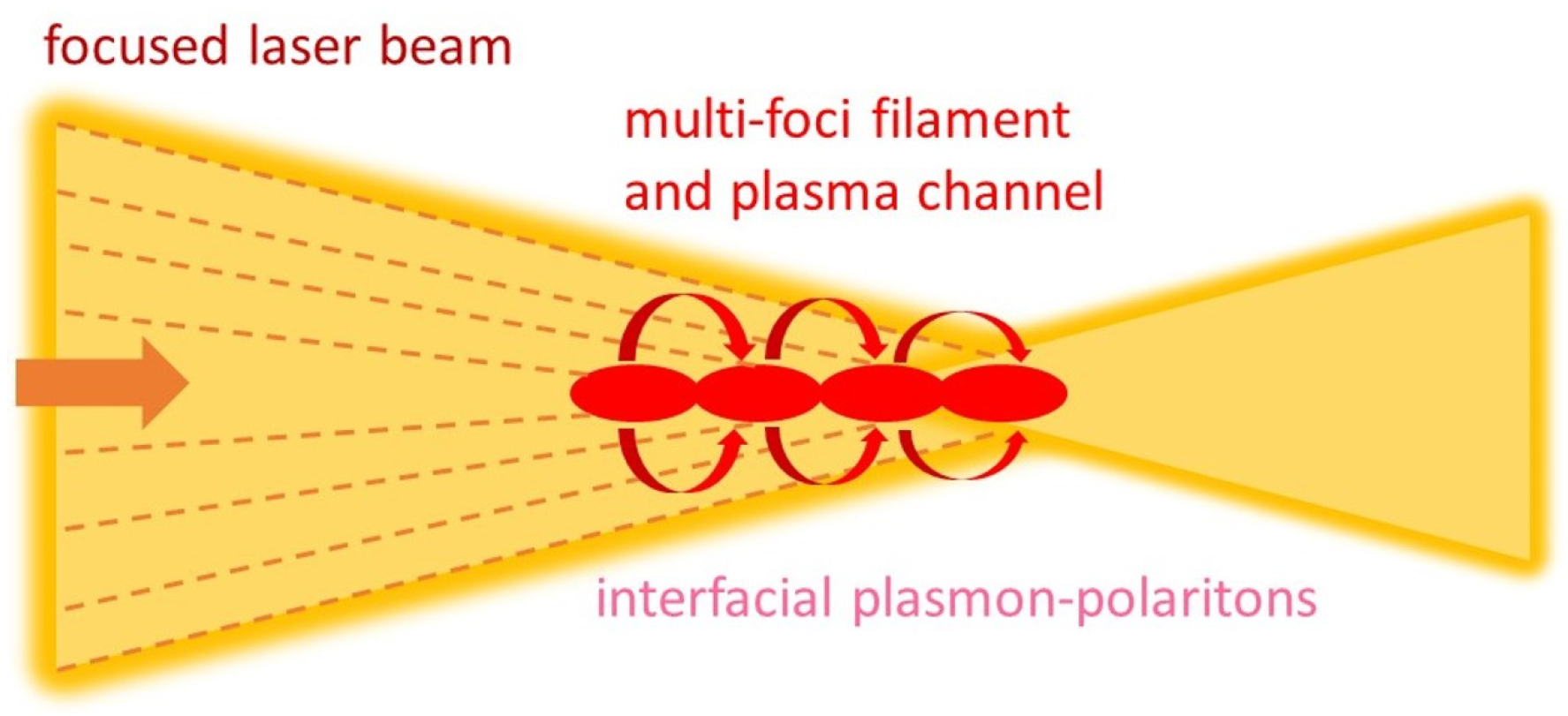
First, we considered the observed filamentation of the ultrashort laser pulses within the “moving-foci” approach [18], as illustrated in Figure 6, rather than their filamentary channeling. However, the plasma channels produced at the high-NA focusing and multi-TW/cm2 laser intensities contained near-critical or even supercritical EHP with the local density Ncr(1030 nm)~1 × 1021 cm−3 at the 1030 nm wavelength and Ncr(515 nm)~4 × 1022 cm−3 at the 515 nm wavelength. This occurs for the refractive index of the plasma in the diamond n*~1 (would be n2 fold higher in an unexcited diamond for its refractive index n(515,1030 nm) ≈ 2.4 [33]), and the dense plasma channels appear during the self-focusing beam collapse as highly reflective micro-optical objects, rather than refractive ones, like in gases [18].
Meanwhile, the supercritical EHP density (Ne > Ncr, Re{ε*} < 0) in the filamentary plasma channels is required for laser excitation of plasmon–polaritons at the plasma channel interface with the surrounding less excited dielectric, according to the threshold condition Re{ε*} ≤ −Re{ε} [34] (Figure 7). Such nanoplasmonic phenomena play important roles in diverse macroscopic electromagnetic phenomena [35,36]. Owing to the uncertainty of the self-focusing collapse arrest by the EHP regarding the final laser incidence angle on the plasma channel and other details, the corresponding plasmon–polaritonic dispersion curves were simulated for the filamentary channel with the supercritical EHP at different tentative self-focusing angles (0, 30, 45 and 600), using the common equations for the plasmon–polariton wavenumber q(ω) [37] under the crude approximation of a flat conductive interface and p-polarized laser radiation [4,34,38]
q P ( ω ) ω c { ε * ε ε * + ε ± sin θ }
where the corresponding expressions for the imaginary magnitudes ε* and ε with Re{ε*,ε} ~ Im{ε*,ε}, representing the lossy EHP in the dielectric, as in [34]. In fact, the exact solutions for excitation of longitudinal plasmon–polaritons in sub-wavelength metallic nanowire-channels (radius aω/c«1) correspond to qualitatively similar dispersion curves h(ω) of surface plasmon–polaritons (specifically, with similar asymptotic dispersion trends at h ⟶ 0 and h ⟶ ∞ [37]), some of which are overviewed in the book [4]. This means non-linear spectral interactions (SC and harmonics generation, etc.) could occur not only in the self-focusing beam, but also in the plasmonic field along the plasma channel, thus becoming considerably enhanced in longer filaments, like in a “channeling” filamentation regime.
Second, prompt interference between the self-focusing laser radiation and interfacial plasmon–polaritons on the plasma channel interface provides their longitudinal interferential electric field patterns along the channels. Their potential periods are near-threshold 1/qP~λ/10 (plasmon resonance in Figure 7) or above-threshold ~λ (plasmon–polaritonic curves near the light-cone line ω = cq/n) magnitudes, similarly to the laser generation of periodical surface structures (LIPSS) on material surfaces [17,38]. Hence, an additional periodical modulation of plasma density (δNe) and refractive index (δn*) emerges in the periodical electric-filed “hot spots” in the filamentary plasma channels, which could work as a longitudinal Bragg grating to filter spectral components at the wavelengths Λfilt [39]
Λ f i l t 2 n e f f q P ,
for the effective refractive index in the filament
n e f f n * ( n * + δ n * ) n * + ( n * + δ n * ) ,
In the plasma channel with the near-critical or supercritical EHP density, n* < n, n* + δn* ≤ n* with 2neff < n; thus, in many dielectrics 2neff~1, though variation of n* near the critical EHP density is sharp and extensive (from n*~n until n* « 1 in the supercritical EHP). Compared to the expected plasmon–polariton wavelength for the flat metal/dielectric interface [17,38] in Equation (2), one could find qP ≈ 1/λ and thus Λfilt ≈ λ. This is consistent with our observations of the strongly modulated SC spectra around the central laser wavelength, which upon normalization to the SC intensity could be represented as the common Bragg grating transmittance spectra (Figure 8). The multiple modulation in this case results not from multiple orders, but from the limited longitudinal number of grating stripes along the plasma channels. Alternatively, in the near-threshold case for n* ≤ n, one has 2neff~n and 1/qP~λ/2n2, thus also resulting in Λfilt ≈ λ/2n; this case is yet to be studied.
Counterintuitively, according to the common diffraction theory, the higher pulse energies in Figure 5 result in the longer filaments and, potentially, the larger longitudinal numbers of regularly spaced grating stripes along the plasma channels and more pronounced central minimum with less distinct side minima. However, in our spectral experiments at the higher pulse energies (pulsewidth—0.6 ps, Figure 8), we observed the increasing number of nearly equal, closely spaced spectral modulation features, thus potentially indicating longitudinal non-uniformity of the Bragg grating periods. This could be a first indirect indication of non-local, e.g., plasmon–polariton propagation, effects along the plasma channels, accumulating plasmon–polariton energy deposition downstream the plasma channels and vice versa, making the filaments/channels longitudinally non-uniform too.
In support of our plasmonic modeling of anticipated Bragg-grating periods of 1/qP ~λ/10 (plasmon resonance in Figure 7) or above-threshold ~λ (plasmon–polaritonic curves near the light-cone line ω = cq/n in Figure 7) and related filtering, our cross-sectional AFM analysis in CLN exactly indicates the expected trends (Figure 9). Just above the nanostructuring threshold ≈ 1–2 μJ, the produced tracks exhibit both sub-wavelength (periods ≈ 200–300 nm) and near-wavelength (periods ~1 μm) structures (Figure 9a), which could be consequently produced during ultrashort laser pulses under resonance and off-resonance excitation of plasmon–polaritons [17,38]. Previously, such sub-wavelength longitudinal gratings were also observed in CLN in a sub-filamentation regime [5], although without the accompanying near-wavelength gratings. Here, by increasing the fs laser pulse energy until 8 μJ, high-aspect longitudinal (length until 20 μm) near-wavelength gratings were produced (Figure 9b), apparently, in the mono-filamentation regime though interfacial plasmon–polaritonic effects on the prompt plasma channel surface. Moreover, because the oblique incidence geometry of geometrical focusing and self-focusing interfacial plasmon–polaritons propagate the filamentary plasma channels/gratings downstream, the resulting Bragg grating periods demonstrate broken longitudinal translational periodicity along the gratings (Figure 9c). An apparent reason for this could be the stronger accumulated plasmon–polaritonic contribution to energy deposition closer the filament end.
These permanent quasi-periodical nano- and microtopographies inscribed by the fs laser pulses in the bulk CLN correlate with the prompt Bragg filtering, appearing in the self-transmission spectra (Figure 10). In comparison to Figure 9, one could see the establishing and developing Bragg filtering at higher fs laser pulse energies (Figure 10b) as the related topographic Bragg gratings in Figure 9 become longer and more structured.

5. Conclusions

Transmittance spectra of focused (NA = 0.25) ultrashort (0.3–12 ps) positively chirped laser pulses at 515 and 1030 nm wavelengths were acquired in the filamentation regime in natural and synthetic diamonds. Besides monotonous pulse-energy-dependent self-phase modulation broadening of laser spectra (supercontinuum generation), becoming pronounced only in the filamentation regime for the laser pulsewidths shorter or equal to 2.4 ps, unusual periodical low-frequency modulation was observed in the spectra for the shorter laser pulsewidths. We suggested the occurrence of supercritical electron-hole plasma in the filaments, inducing plasmon–polariton excitation on their interface with the unexcited dielectric and resulting in nanoplasmonic modulation of plasma density. Then, dynamic filtering of the laser supercontinuum proceeds in the plasma Bragg gratings. Damping of spectral broadening and ultra-modulation for the longer picosecond pulsewidths was related to thermalized electron-hole plasma regime established for the laser pulsewidths longer than 2 ps. The related material Bragg gratings were imprinted in crystalline lithium niobate and visualized by cross-sectional atomic force microscopy as sub-wavelength (periods ≈ 200–300 nm) and near-wavelength (periods ~1 μm) structures.

Author Contributions

Conceptualization, S.K.; project administration, A.A. and S.K.; funding acquisition, S.K. and V.S.; writing—review and editing, S.K. and A.G.; resources, E.G.; visualization, A.T.; methodology, P.D. and V.K.; validation, V.K.; software, V.K.; formal analysis, A.G.; investigation, P.D., M.K. and B.L.; data curation, B.L.; writing—original draft preparation, S.K.; supervision, S.K. and M.K. All authors have read and agreed to the published version of the manuscript.

Funding

The research funding from the Ministry of Science and Higher Education of the Russian Federation (Ural Federal University Program of Development within the Priority-2030 Program) is gratefully acknowledged. The equipment of the Ural Center for Shared Use “Modern nanotechnology” of Ural Federal University (Reg.# 2968), which is supported by the Ministry of Science and Higher Education of Russian Federation (Project #075–15- 2021–677), was used.

Institutional Review Board Statement

Not applicable.

Informed Consent Statement

Not applicable.

Data Availability Statement

Additional data could be provided by the authors upon special request.

Conflicts of Interest

The authors declare no conflict of interest.

References

  1. Sugioka, K.; Cheng, Y. Ultrafast Lasers-Reliable Tools for Advanced Materials Processing. Light Sci. Appl. 2014, 3, 149. [Google Scholar] [CrossRef]
  2. Sotillo, B.; Bharadwaj, V.; Hadden, J.P.; Sakakura, M.; Chiappini, A.; Fernandez, T.T.; Longhi, S.; Jedrkiewicz, O.; Shimotsuma, Y.; Criante, L.; et al. Diamond photonics platform enabled by femtosecond laser writing. Sci. Rep. 2016, 6, 35566. [Google Scholar] [CrossRef] [PubMed]
  3. Couairon, A.; Sudrie, L.; Franco, M.; Prade, B.; Mysyrowicz, A. Filamentation and damage in fused silica induced by tightly focused femtosecond laser pulses. Phys. Rev. B 2005, 71, 125435. [Google Scholar] [CrossRef]
  4. Klimov, V. Nanoplasmonics; CRC Press: Singapore, 2014. [Google Scholar]
  5. Kudryashov, S.; Rupasov, A.; Kosobokov, M.; Akhmatkhanov, A.; Krasin, G.; Danilov, P.; Lisjikh, B.; Abramov, A.; Greshnyakov, E.; Kuzmin, E.; et al. Hierarchical Multi-Scale Coupled Periodical Photonic and Plasmonic Nanopatterns Inscribed by Femtosecond Laser Pulses in Lithium Niobate. Nanomaterials 2022, 12, 4303. [Google Scholar] [CrossRef] [PubMed]
  6. Mouskeftaras, A.; Rode, A.V.; Clady, R.; Sentis, M.; Utéza, O.; Grojo, D. Self-limited underdense microplasmas in bulk silicon induced by ultrashort laser pulses. Appl. Phys. Lett. 2014, 105, 191103. [Google Scholar] [CrossRef]
  7. Papazoglou, D.G.; Abdollahpour, D.; Tzortzakis, S. Ultrafast electron and material dynamics following femtosecond filamentation induced excitation of transparent solids. Appl. Phys. A 2014, 114, 161–168. [Google Scholar] [CrossRef]
  8. Jürgens, P.; Vrakking, M.J.J.; Husakou, A.; Stoian, R.; Mermillod-Blondin, A. Plasma formation and relaxation dynamics in fused silica driven by femtosecond short-wavelength infrared laser pulses. Appl. Phys. Lett. 2019, 115, 191903. [Google Scholar] [CrossRef]
  9. Geints, Y.E.; Zemlyanov, A.A.; Ionin, A.A.; Kudryashov, S.I.; Seleznev, L.V.; Sinitsyn, D.V.; Sunchugasheva, E.S. Peculiarities of filamentation of sharply focused ultrashort laser pulses in air. J. Exp. Theor. Phys. 2010, 111, 724–730. [Google Scholar] [CrossRef]
  10. Song, J.; Wang, X.; Hu, X.; Dai, Y.; Qiu, J.; Cheng, Y.; Xu, Z. Formation mechanism of self-organized voids in dielectrics induced by tightly focused femtosecond laser pulses. Appl. Phys. Lett. 2008, 92, 092904. [Google Scholar] [CrossRef]
  11. Li, X.; Xu, J.; Lin, Z.; Qi, J.; Wang, P.; Chu, W.; Fang, Z.; Wang, Z.; Chai, Z.; Cheng, Y. Polarization-insensitive space-selective etching in fused silica induced by picosecond laser irradiation. Appl. Surf. Sci. 2019, 485, 188–193. [Google Scholar] [CrossRef]
  12. Xu, S.; Fan, H.; Li, Z.-Z.; Hua, J.-G.; Yu, Y.-H.; Wang, L.; Chen, Q.-D.; Sun, H.-B. Ultrafast laser-inscribed nanogratings in sapphire for geometric phase elements. Opt. Lett. 2021, 46, 536–539. [Google Scholar] [CrossRef] [PubMed]
  13. Zhang, B.; Liu, X.; Qiu, J. Single femtosecond laser beam induced nanogratings in transparent media—Mechanisms and applications. J. Mater. 2019, 5, 1–14. [Google Scholar] [CrossRef]
  14. Kudryashov, S.; Rupasov, A.; Zakoldaev, R.; Smaev, M.; Kuchmizhak, A.; Zolot’ko, A.; Kosobokov, M.; Akhmatkhanov, A.; Shur, V. Nanohydrodynamic Local Compaction and Nanoplasmonic Form-Birefringence Inscription by Ultrashort Laser Pulses in Nanoporous Fused Silica. Nanomaterials 2022, 12, 3613. [Google Scholar] [CrossRef] [PubMed]
  15. Shimotsuma, Y.; Kazansky, P.G.; Qiu, J.; Hirao, K. Self-organized nanogratings in glass irradiated by ultrashort light pulses. Phys. Rev. Lett. 2003, 91, 247405. [Google Scholar] [CrossRef] [PubMed]
  16. Taylor, R.; Hnatovsky, C.; Simova, E. Applications of femtosecond laser induced self-organized planar nanocracks inside fused silica glass. Laser Photonics Rev. 2008, 2, 26–46. [Google Scholar] [CrossRef]
  17. Kudryashov, S.I.; Danilov, P.A.; Rupasov, A.E.; Smayev, M.P.; Kirichenko, A.N.; Smirnov, N.A.; Ionin, A.A.; Zolot’ko, A.S.; Zakoldaev, R.A. Birefringent microstructures in bulk fluorite produced by ultrafast pulsewidth-dependent laser inscription. Appl. Surf. Sci. 2021, 568, 150877. [Google Scholar] [CrossRef]
  18. Couairon, A.; Mysyrowicz, A. Femtosecond filamentation in transparent media. Phys. Rep. 2007, 441, 47–189. [Google Scholar] [CrossRef]
  19. Kudryashov, S.; Danilov, P.; Chen, J. Intrapulse correlated dynamics of self-phase modulation and spontaneous Raman scattering in synthetic diamond excited and probed by positively chirped ultrashort laser pulses. Photonics 2023, 10, 626. [Google Scholar] [CrossRef]
  20. Kudryashov, S.; Krasin, G.; Rupasov, A.; Danilov, P.; Kosobokov, M.; Akhmatkhanov, A.; Lisjikh, B.; Turygin, A.; Greshnyakov, E.; Kovalev, M.; et al. Ferroelectric nanodomain engineering in bulk lithium niobate crystals in ultrashort-pulse laser nanopatterning regime. Nanomaterials 2022, 12, 4147. [Google Scholar] [CrossRef]
  21. Krasin, G.K.; Gulina, Y.S.; Kuzmin, E.V.; Martovitskii, V.P.; Kudryashov, S.I. Polarization-Sensitive Nonlinear Optical Interaction of Ultrashort Laser Pulses with HPHT Diamond. Photonics 2023, 10, 106. [Google Scholar] [CrossRef]
  22. Zaitsev, A.M. Optical Properties of Diamond: A Data Handbook; Springer: Berlin/Heidelberg, Germany, 2013. [Google Scholar]
  23. Kudryashov, S.I.; Danilov, P.A.; Kuzmin, E.V.; Gulina, Y.S.; Rupasov, A.E.; Krasin, G.K.; Zubarev, I.G.; Levchenko, A.O.; Kovalev, M.S.; Pakholchuk, P.P.; et al. Pulse-width-dependent critical power for self-focusing of ultrashort laser pulses in bulk dielectrics. Opt. Lett. 2022, 47, 3487–3490. [Google Scholar] [CrossRef] [PubMed]
  24. Ishioka, K.; Hase, M.; Kitajima, M.; Petek, H. Coherent optical phonons in diamond. Appl. Phys. Lett. 2006, 89, 231916. [Google Scholar] [CrossRef]
  25. Zukerstein, M.; Kozák, M.; Trojánek, F.; Malý, P. Experimental observation of anharmonic effects in coherent phonon dynamics in diamond. Diam. Relat. Mater. 2018, 90, 202–206. [Google Scholar] [CrossRef]
  26. Liu, S.; Hammud, A.; Hamada, I.; Wolf, M.; Müller, M.; Kumagai, T. Nanoscale coherent phonon spectroscopy. Sci. Adv. 2022, 8, eabq5682. [Google Scholar] [CrossRef] [PubMed]
  27. Klemens, P.G. Anharmonic decay of optical phonon in diamond. Phys. Rev. B 1975, 11, 3206. [Google Scholar] [CrossRef]
  28. Kudryashov, S.; Danilov, P.; Smirnov, N.; Krasin, G.; Khmelnitskii, R.; Kovalchuk, O.; Kriulina, G.; Martovitskiy, V.; Lednev, V.; Sdvizhenskii, P.; et al. “Stealth Scripts”: Ultrashort Pulse Laser Luminescent Microscale Encoding of Bulk Diamonds via Ultrafast Multi-Scale Atomistic Structural Transformations. Nanomaterials 2023, 13, 192. [Google Scholar] [CrossRef] [PubMed]
  29. Shen, Y.R. The Principles of Nonlinear Optics; Wiley-Interscience: New York, NY, USA, 1984. [Google Scholar]
  30. Grudtsyn, Y.V.; Zubarev, I.G.; Koribut, A.V.; Kuchik, I.E.; Mamaev, S.B.; Mikheev, L.D.; Semjonov, S.L.; Stepanov, S.G.; Trofimov, V.A.; Yalovoi, V.I. Self-Phase modulation in a thin fused silica plate upon interaction with a converging beam of down-chirped femtosecond radiation. Quantum Electron. 2015, 45, 415–420. [Google Scholar] [CrossRef]
  31. Liu, W.; Zhu, L.; Wang, L.; Fang, C. Cascaded four-wave mixing for broadband tunable laser sideband generation. Opt. Lett. 2013, 38, 1772–1774. [Google Scholar] [CrossRef]
  32. Bencivenga, F.; Cucini, R.; Capotondi, F.; Battistoni, A.; Mincigrucci, R.; Giangrisostomi, E.; Gessini, A.; Manfredda, M.; Nikolov, I.P.; Masciovecchio, C.; et al. Four-wave mixing experiments with extreme ultraviolet transient gratings. Nature 2015, 520, 205–208. [Google Scholar] [CrossRef]
  33. Palik, E.D. Handbook of Optical Constants of Solids; Academic Press: Orlando, FL, USA, 1998. [Google Scholar]
  34. Bell, R.J.; Alexander, R.W., Jr.; Parks, W.F.; Kovener, G. Surface excitations in absorbing media. Opt. Commun. 1973, 8, 147–150. [Google Scholar] [CrossRef]
  35. He, L.; Yi, Y.; Zhang, J.; Xu, X.; Tang, B.; Li, G.; Zeng, L.; Chen, J.; Sun, T.; Yi, Z. A four-narrowband terahertz tunable absorber with perfect absorption and high sensitivity. Mater. Res. Bull. 2024, 170, 112572. [Google Scholar] [CrossRef]
  36. Yuan, X.; Xu, Q.; Lang, Y.; Jiang, X.; Xu, Y.; Chen, X.; Han, J.; Zhang, X.; Han, J.; Zhang, W. Tailoring spatiotemporal dynamics of plasmonic vortices. Opto-Electron. Adv. 2023, 6, 220133. [Google Scholar] [CrossRef]
  37. Guo, X.; Ma, Y.; Wang, Y.; Tong, L. Nanowire plasmonic waveguides, circuits and devices. Laser Photonics Rev. 2013, 7, 855–881. [Google Scholar] [CrossRef]
  38. Kudryashov, S.I.; Nastulyavichus, A.A.; Saraeva, I.N.; Rudenko, A.A.; Zayarny, D.A.; Ionin, A.A. Deeply sub-wavelength laser nanopatterning of Si surface in dielectric fluids: Manipulation by surface plasmon resonance. Appl. Surf. Sci. 2020, 519, 146204. [Google Scholar] [CrossRef]
  39. Bak, J.; Suazo Betancourt, J.L.; Rekhy, A.; Abbasszadehrad, A.; Miles, R.B.; Limbach, C.M.; Walker, M.L. High resolution spatially extended 1D laser scattering diagnostics using volume Bragg grating notch filters. Rev. Sci. Instrum. 2023, 94, 023003. [Google Scholar] [CrossRef]
Figure 1. Experimental scheme for spectral acquisition of transmitted laser pulses.
Photonics 10 01316 g001
Figure 2. Comparative transmittance spectra of 1030 nm pulses at the different representative pulse energies (50, 120 and 300 nJ) and pulsewidths (i–vi) for the synthetic (a) and natural (b) diamond samples.
Photonics 10 01316 g002
Figure 3. Threshold (a) pulse energy and (b) peak pulse power for the filamentation onset for 1030 nm pulses in the synthetic (dark curves) and natural (red curves) diamond samples as a function of laser pulsewidth. Relative error bars—10%.
Photonics 10 01316 g003
Figure 4. Transmittance spectra of 515 nm pulses at the different pulse energies and pulsewidths (ad) for the synthetic diamond sample.
Photonics 10 01316 g004
Figure 5. Transmittance spectra of 1030 nm pulses at the different selected pulse energies (color curves, see legend below) and pulsewidths (af) for the synthetic diamond sample. The spectra for different pulse energies are arbitrarily offset along the vertical axis for clarity.
Photonics 10 01316 g005
Figure 6. Schematic view of laser filaments (“moving-foci” regime [18]) with the supercritical EHP and plasmon–polariton excitation on their interface.
Photonics 10 01316 g006
Figure 7. Dispersion curves of interfacial plasmon–polaritons, excited by 1030 nm fs laser pulses in diamond at the incidence angles of 0, 30, 45 and 60° on the interface “diamond-EHP” at supercritical EHP densities Ne ≈ 2 × 1021 cm−3 (a) and Ne ≈ 4 × 1021 cm−3 (b). The bulk electromagnetic wave dispersion line is denoted as “bulk diamond”.
Photonics 10 01316 g007
Figure 8. Transmitted intensity (a,c,e) and transmittance (b,d,f) spectra of 0.6 ps 1030 nm pulses for the synthetic diamond sample: original spectra with the corresponding SC spectra (envelopes in a,c,e) and their ratio (b,d,f) at the different pulse energies (5.0, 2.0 and 0.6 µJ).
Photonics 10 01316 g008
Figure 9. AFM relief maps of longitudinal sub-wavelength (small tilted bright dots) and near-wavelength (large dark dots) Bragg gratings in the cross-sections of CLN inscribed at pulse energies of 5 μJ (a), 7 μJ (b) and 1 μJ (c) and the magnified views of their selected regions highlighted by the yellow frames. The red arrow indicates the laser input position.
Photonics 10 01316 g009
Figure 10. Transmitted intensity (a) and transmittance (b) spectra of 0.2 ps 1050 nm pulses for the crystalline lithium niobate sample: original spectra with the corresponding SC spectra (grey envelope curves in (a)) and their ratio (b) at the different pulse energies: i—9 µJ, ii—4 µJ, iii—1 µJ and iv—0.1 µJ).
Photonics 10 01316 g010
Disclaimer/Publisher’s Note: The statements, opinions and data contained in all publications are solely those of the individual author(s) and contributor(s) and not of MDPI and/or the editor(s). MDPI and/or the editor(s) disclaim responsibility for any injury to people or property resulting from any ideas, methods, instructions or products referred to in the content.