Article

Chemical Dosimetry Using Bisbenzimidazoles: Solvent-Dependent Fluorescence Response of Hoechst 33258 to Radiation Exposure

by
Maria A. Kolyvanova
1,2,
Mikhail A. Klimovich
1,2,
Ekaterina D. Koshevaya
2,
Evgeny A. Nikitin
3,
Nikita S. Lifanovsky
1,
Vladimir Y. Tyurin
3,
Alexandr V. Belousov
2,
Aleksei V. Trofimov
1,
Vladimir A. Kuzmin
1 and
Vladimir N. Morozov
1,*
1
Emanuel Institute of Biochemical Physics, Russian Academy of Sciences, 4 Kosygina, 119334 Moscow, Russia
2
Burnazyan Federal Medical Biophysical Center, Federal Medical Biological Agency of the Russian Federation, 23 Marshala Novikova, 123182 Moscow, Russia
3
Faculty of Chemistry, Lomonosov Moscow State University, 1–3 Leninskie Gory, 119991 Moscow, Russia
*
Author to whom correspondence should be addressed.
Photonics 2023, 10(6), 671; https://doi.org/10.3390/photonics10060671
Submission received: 25 April 2023 / Revised: 2 June 2023 / Accepted: 7 June 2023 / Published: 9 June 2023

Abstract

:
Bisbenzimidazoles have a broad spectrum of potential applications: radioprotectors, drug delivery vectors, antiviral agents, etc. At the same time, they seem to be promising fluorescent probes for radiation measurements. Therefore, in the present work, a fluorescent response to X-ray irradiation of Hoechst 33258, one of the most widely known representatives of the bisbenzimidazole family, was studied for the first time. Irradiation of the dye was performed in aqueous and organic solutions (DMSO and glycerol), as well as in their mixtures. It is shown that the reaction of the dye to radiation exposure is very versatile and may be controlled by the solvent properties, which makes it possible to build relationships between the absorbed dose and a wide variety of parameters of its fluorescence signal. For example, irradiation may induce fluorescence quenching caused by the degradation of the dye, a change in the position of the fluorescence band maximum due to the modification of the dye molecules or to the radiation-induced changes in the properties of the medium, as well as a fluorescence flare-up mediated by the changes in pH.

1. Introduction

Organic dyes are of considerable interest for the development of chemical dosimetry systems with optical response to irradiation for a wide variety of practical tasks from radiation processing and sterilization to nuclear and radiation medicine. However, even though radiation effects on organic dyes have been investigated since the 1940s–1950s [1,2], the studies in the field of their application in radiation measurements are small-scale and non-systematic. According to the literature data, of the variety of organic dyes, triphenylmethane [3,4,5] and azo dyes [6,7] are the most studied in this direction. Moreover, a number of works are known for coumarins [8,9], spiropyrans [10], styryl dyes [11], as well as for some representatives of the other families. Additionally, even though no evident correlation between the magnitude of radiation effect and the dye type has been found yet [12], some general strategies for their use in dosimetry can be identified.
In systems with dyes, irradiation can lead to a variety of optical effects. For example, radiation exposure may cause a change in UV–Vis absorption: initially colorless leuco-forms of dye molecules may acquire color, while bleaching or discoloration may be observed for colored compounds. A number of dosimeters have been developed on the basis of this principle: e.g., Perspex Amber 3042 (Sudan I and Sudan III) [13], Gammachrome YR (dimethyl yellow) [14], and PRESAGE (leuco-malachite green) [15]. Change in the dye fluorescence may be another marker of radiation exposure. In this case, irradiation can lead to both quenching of the signal [16], as well as to its enhancement [9]. However, the response of fluorescent dyes to irradiation is much less studied, even though the fluorescence-based approach of dose recording shows very promising results [17,18,19,20,21,22].
One of the families of fluorescent dyes potentially suitable for radiation measurements due to their physico-chemical properties are bisbenzimidazoles. This family includes original molecules Hoechst 33258, Hoechst 33342, and Hoechst 34580, as well as many of their analogues and derivatives (e.g., see in [23,24,25]). Although Hoechst dyes are known mainly as DNA-specific fluorophores for cell nucleus staining [26], they and their derivates also have a broad spectrum of other potential applications: radioprotection [27], targeted delivery of radionuclides [28], antiviral agents [29], fluorescent sensors [30], etc. Since almost nothing is known about the optical response of Hoechst dyes to irradiation (only the response of the dye–DNA complex to UV exposure was studied [31]), in the present work we have studied for the first time the effect of X-rays on the fluorescence properties of the most famous representative of bisbenzimidazole family—Hoechst 33258. This dye was chosen because of its wide availability and the high demand for it in chemistry and life sciences.

2. Materials and Methods

Experimental samples—solutions of Hoechst 33258 in distilled water, DMSO, and glycerol, as well as in their mixtures (water + DMSO or water + glycerol)—were prepared using commercial dye (Paneko, Moscow region, Russia) and corresponding solvents (Chimmed, Moscow, Russia). In all cases, Hoechst 33258 concentration in the 1.5 mL samples was 3.5 × 10⁻⁶ M. It was determined by the reported values of the dye molar absorption coefficient: ε340 ≈ 42 000 M–1 cm–1 [32]. To provide a conformal dose coverage, the experimental samples were placed in plastic Petri dishes of 35 mm in diameter (GenFollower, Shaoxing city, China), and then irradiated using LNK-268 X-ray machine (80 kVp, molybdenum anode, 8 mA anode current, effective energy of ≈30 keV; Diagnostika-M, Moscow, Russia). The samples were irradiated one at a time. During the exposure, the lid of the dish was left open.
Determination of the dose rate was performed using Fricke dosimeter (air-saturated solution in double distilled water of ammonium iron (II) sulfate (NH4)2Fe(SO4)2 • 6H2O (1 × 10⁻³ M) with the addition of H2SO4 (0.4 M) and NaCl (1 × 10⁻³ M)). Irradiation of the dosimetric solution was performed in the same conditions as experimental samples. The optical density was measured at a wavelength of λ = 304 nm. The absorbed dose rate was found to be 4.17 Gy/s.
Absorption and fluorescence spectra were recorded using UV-3101 PC spectrophotometer and RF5301PC spectrofluorimeter (Shimadzu, Kyoto, Japan). Fluorescence lifetime measurements (λEx = 375 nm) were performed by time-correlated single photon counting (TCSPC) method using a fluorescence steady-state and lifetime spectrometer FluoTime 300 (PicoQuant, Berlin, Germany). These experiments were performed in 1.0 × 0.4 cm quartz cells with 1 cm path length for excitation light. When calculating the lifetime of the excited state, χ2 did not exceed 1.3. The values of pH were registered on PB-11 device (Sartorius, Goettingen, Germany).

3. Results

The structure of Hoechst 33258 (2′-(4-hydroxyphenyl)-5-[5-(4-methylpiperazine-1-yl) benzimidazo-2-yl]-benzimidazole) is shown in Figure 1A. Figure 1B shows normalized absorption and emission spectra of the dye in distilled water (pH = 5.8), DMSO, and glycerol. In all three systems, the absorption maximum of Hoechst 33258 lies in the UV region, while the fluorescence maximum is in the bluish green or blue region of the spectrum (abs./em. maxima: 340/498 nm in water, 352/466 nm in DMSO, and 344/460 nm in glycerol). The lowest Hoechst 33258 fluorescence is found in water, while in DMSO and glycerol it is ≈11.7 and ≈6.8 times higher, respectively. The decay kinetics of Hoechst 33258 fluorescence in these solvents are shown in Figure 1C. The curve for the aqueous solution of the dye is described by single exponential, while those for DMSO and glycerol are described by double exponential. The corresponding lifetimes are τ1 = 3.91 ns (water), τ1 = 4.45 ns and τ2 = 2.79 ns (DMSO), and τ1 = 3.68 ns and τ2 = 2.03 ns (glycerol). Their contributions to the total fluorescence amplitude are shown in the inset to the figure.
Figure 2A shows the fluorescence spectra of the aqueous solutions of Hoechst 33258 irradiated with X-rays. As the dose increases, a drop in the dye fluorescence is observed: its intensity is about 50% of the initial value already at 25 Gy, and at 350 Gy the signal decreases to ≈4% and practically does not change up to the maximum studied dose of 2000 Gy. The position of the band maximum does not change significantly in this case. The radiation-induced quenching of Hoechst 33258 fluorescence is accompanied by a decrease in its absorbance (see inset to Figure 2A). This may indicate that the observed effect is associated with degradation of the dye molecules [16]. At the same time, a low-intensity band with maximum at λ ≈ 390 nm, which is absent in the non-irradiated system, appears in the fluorescence spectrum of Hoechst 33258. We assume that this peak corresponds precisely to the degradation products of the dye molecules. Its amplitude at first increases sharply, reaching a maximum at 200 Gy; then, however, a slightly smoother decrease in the signal is observed. The corresponding dependences of I/I0 (where I and I0 are the peak intensity of the fluorescence signals corresponding to irradiated samples and to non-irradiated control, respectively) at wavelengths of 500 nm and 390 nm on the absorbed dose are shown in Figure 2B. The first one has a clear exponential character, while the second dependence is a right skewed bell curve. The negative logarithm dose dependence pattern of these signals’ ratio (I500/I390) is very close to linear in the region of 0–150 Gy (see inset to Figure 2B).
Figure 2C shows the fluorescence decay kinetics of the irradiated aqueous solutions of Hoechst 33258 recorded at λEm = 500 nm. The corresponding lifetimes of the excited state of the dye and their contributions to the total fluorescence amplitude are presented in Figure 2D. The curve of the control sample is single exponential (τ1 = 3.91 ns). However, an additional short-lived component (τ2 ≈ 0.8 ns) appears in the irradiated solutions. Its contribution to the total fluorescence amplitude increases with the increase in the irradiation dose, and, starting from ≈250 Gy, it begins to exceed the contribution of the first component. It can be assumed that the short-lived component corresponds to the decay of the fluorescence intensity of the Hoechst 33258 products responsible for the appearance of the low-intensity fluorescence band at λ ≈ 390 nm. Indeed, the close-in-magnitude short-lived component dominates in the case of the decay kinetics of the dye fluorescence recorded at λEm = 390 nm (see inset to Figure 2C).
It is well known that the main effect of ionizing radiation in aqueous solutions is caused by the action of radical radiolytic products (i.e., e a q , H, HO, HO2, OH, H3O+, H2, and H2O2 [33]). Therefore, we studied the effect of glycerol and DMSO on the fluorescent response of Hoechst 33258 to radiation exposure. Both compounds are free radical scavengers and can, therefore, have a radioprotective effect [34].
When 6 × 10⁻² M of glycerol (0.4% by volume) is added to the aqueous solution of Hoechst 33258, the sensitivity of the dye to radiation noticeably decreases (see Figure 3A,B). In this case, even at a dose of 2000 Gy, the fluorescence intensity does not fall below 25% of the initial value, and the corresponding dependence of I/I0 at λ = 500 nm on the absorbed dose is linear in the studied range. The peak position remains unchanged. The lifetime of the excited state of the dye also does not change under irradiation (the decay kinetics are shown in the inset to Figure 3A): τ1 = 3.92 ns in the non-irradiated control and τ1 = 3.88 ns at 2000 Gy. As in the case of the aqueous solutions of Hoechst 33258, a low-intensity band at λ ≈ 390 nm also appears in the fluorescence spectra of the samples irradiated in the presence of glycerol. However, its amplitude increases much more slowly—the maximum is reached at a dose of about 1500 Gy (≈7.5-fold higher than in water). The ratiometric fluorescent response to irradiation in semilogarithmic coordinates in this case is linear in the region of 200–1750 Gy.
The results of radiation exposure on the aqueous solutions of Hoechst 33258 in the presence of 6 × 10⁻² M of DMSO (0.5% by volume) are shown in Figure 4. In this case, an effect opposite to radiation-induced quenching is observed: irradiation of the samples causes a sharp and strong increase in the fluorescence of the dye. Thus, the signal amplitude at a dose of 250 Gy increases by more than 3.5-fold. From this point on, the signal reaches a plateau, followed by a linear decline starting from 500 Gy (see inset to Figure 4A). However, even at a dose of 2000 Gy, the fluorescence intensity of Hoechst 33258 in the irradiated sample is ≈1.8-fold higher than in the non-irradiated control. Moreover, in the samples irradiated in the presence of DMSO, no band at λ ≈ 390 nm is observed. There is also no change in the lifetimes (decay curves are shown in the inset to Figure 4A).
Such behavior of Hoechst 33258 fluorescence seems to be partly due to the radical oxidation of DMSO. In aqueous solutions, DMSO preferentially reacts with HO to form methane sulfinic acid (MSIA; H(CH3)SO2) and methyl radical or involves the formation of a hydrogen bond between the hydroxyl radical and the DMSO oxygen, which leads to the proton abstraction [35,36]. The formation of MSIA, in turn, leads to a change in pH of the solution. The corresponding dose dependence of pH mirrors the curve of change in the fluorescence signal in the range of 0–500 Gy (see in Figure 4B). At the same time, it is well known that fluorescence of free Hoechst 33258 flares up with an increase in the acidity of the medium within certain limits [37,38]. However, in our case, the position of the band maximum does not change, in contrast to the work by Barooah et al. (they found a bathochromic shift by ≈22 nm when pH was changed from 7.0 to 4.5) [38], and the type of dependence is somewhat different from that measured by Görner [37]. Nevertheless, the data obtained clearly demonstrate that Hoechst 33258 fluorescence may change not only due to the direct action of ionizing radiation or the interaction of the dye molecules with radiolytic products, but also may be mediated by a radiation induced change in the properties of the solvent.
The results of radiation exposure of the solutions of Hoechst 33258 in DMSO and glycerol are shown in Figure 5 and Figure 6, respectively. The investigated solvents differ from water not only in the type of formed radiolytic products, but also in physico-chemical properties (e.g., dielectric constant and viscosity), which may affect radiation–chemical processes during the exposure [39]. It is clearly seen that the sensitivity of the dye to radiation in these solvents is much lower than in an aqueous solution (differences in the response of the dye to irradiation in different solvents were noted earlier, for example, in the work by Barakat et al. [40]). For example, in DMSO, a decrease in the Hoechst 33258 fluorescence below 40% of the initial value is observed only at a dose of ≈2000 Gy, and the signal drops below 5% only at doses above 8000 Gy (Figure 5B). In this case, a sharp decrease in the signal by more than 2-fold observed at ≈1000 Gy is further replaced by a smoother section from 2000 Gy to 10,000 Gy, where the signal changes from ≈31% to ≈3.4%. In turn, the amplitude of Hoechst 33258 fluorescence in glycerol does not fall by more than 15% of the initial value even at 10,000 Gy (Figure 6B).
In both cases, radiation exposure leads to a shift in the position of the fluorescence maximum, and the corresponding dose dependences are two-component. Such behavior may be associated both with a modification of the chemical structure of the dye molecules and with a change in the properties of the solvent since the main part of the radiation energy is absorbed precisely in the medium. In DMSO, the peak first shifts from 466 nm to 492 nm at 1750–2000 Gy and then drops to 482 nm at 10,000 Gy. The inflection point of this curve corresponds to the inflection point of dose dependence of the relative signal amplitude. In glycerol, a rather sharp shift of the peak position from 460 nm to 474 nm at 1000 Gy is observed at the first stage. Further, it is followed by a much smoother shift up to 480 nm at the maximum studied dose of 10,000 Gy. Both dependences are characterized by pronounced linear sections (see Figure 5C (DMSO) and Figure 6C (glycerol), respectively). Thus, a change in the position of the fluorescence maximum may indicate a change in the absorbed dose even if irradiation does not substantially change the amplitude of the fluorescence signal, as in the case of Hoechst 33258 solution in glycerol.
Previously, in the fluorescence response of organic dyes to ionizing radiation exposure, only a change in the signal amplitude was often observed [9,16,21,22,41,42]. Additionally, although the sensitivity of the systems studied in the present work to radiation is not as high as, for example, that shown in [21], the results obtained seem to be useful. Nowadays, radiation dosimetry is faced with a task of registering doses of 10–6–1010 Gy from sources of various nature and characteristics in omnifarious environmental conditions, and, taking into account the wide availability of Hoechst 33258, it is quite possible to assume its use for the dosimetric support of experiments in the field of radiation chemistry and biology. It also should be noted that there is industrial demand for detecting systems designed to show the fact of irradiation and the achievement of the required dose threshold (first of all, this refers to the tasks of radiation processing and sterilization). In this cases, very simple control methods are needed. Therefore, a change in the emission wavelength noticeable to the naked eye may be fundamentally useful.
We also note that fluorescent dyes may be of interest as auxiliary agents for more complex dosimetric systems. For example, due to their ability to specifically bind to biomacromolecules (e.g., nucleic acids and proteins), they can be used to provide additional functionality to non-fluorescent supramolecular sensors of ionizing radiation, the response of which will not be determined by the first-hand effect of radiation on the dye molecules. Such systems, in particular, include supramolecular ensembles of low molecular weight DNA: in recent works we have demonstrated the ability of such systems to record doses from hundreds of Gy to hundreds of kGy [43,44]. A corresponding study of the possibility of functionalization of these systems with organic dyes for use in dosimetry and the detection of ionizing radiation will be carried out in our future works.

4. Conclusions

In the present work, the fluorescent response of Hoechst 33258, one of the most widely known representatives of the bisbenzimidazole family, to X-ray irradiation was studied for the first time. Solutions of the dye in distilled water and organic solvents—DMSO and glycerol (both are known as free radical scavengers and radioprotectors)—as well as in their mixtures were considered. The reaction of Hoechst 33258 to irradiation, both quantitative and qualitative, strongly depends on the solvent and is very versatile, which makes it possible to build relationships between the absorbed dose and a wide variety of parameters of its fluorescence signal. For example, two fundamentally different mechanisms of changing the intensity of Hoechst 33258 fluorescence are shown: (1) a quenching due to the degradation of the dye molecules caused by the direct action of ionizing radiation and its indirect action through the radical radiolytic products (observed in all cases, except for the aqueous solution in the presence of DMSO) and (2) a flare-up mediated by the radiation-induced changes in solvent properties (observed only in aqueous solution in the presence of DMSO in the dose range up to 350–500 Gy and caused by its radical oxidation and formation of MSIA).
Degradation of the dye is the most efficient in aqueous solution—in this case the fluorescence intensity drops close to zero already at 350 Gy. Since the degradation products of Hoechst 33258 in water seem to have their own fluorescence in the region of λ ≈ 390 nm, it is possible to register the dose change ratiometrically as well as by a change of the contribution of their lifetime to the total fluorescence amplitude. The same decrease in Hoechst 33258 fluorescence is observed in DMSO at the dose of 10,000 Gy, while the dye solution in glycerol is the most resistant to irradiation: even at 10,000 Gy, its fluorescence intensity does not fall by more than 15% of the initial value. Addition of glycerol to the aqueous solution of Hoechst 33258 also significantly protects the dye from irradiation. In this case, a twofold drop in the fluorescence intensity is observed between 1500 and 1750 Gy versus ≈25 Gy in water. Moreover, the absorbed dose may in principle be determined from the change in the position of Hoechst 33258 fluorescence maximum. This effect, well defined in organic solvents, can also work even if irradiation does not substantially change the amplitude of the fluorescence signal, as in the case of the dye solution in glycerol (the peak suffers a bathochromic shift from 460 nm to 480 nm at 10,000 Gy). The reason can be both the modification of the dye molecules and the radiation-induced change in the properties of the medium. Since the exact mechanism of this effect is unclear, it will be studied in detail in our future works. Thus, the example of Hoechst 33258 shows the wide possibilities of using fluorescent bisbenzimidazoles in chemical dosimetry and the detection of ionizing radiation.

Author Contributions

Conceptualization, M.A.K. (Maria A. Kolyvanova) and V.N.M.; methodology, V.N.M.; software, A.V.B.; validation, M.A.K. (Maria A. Kolyvanova), M.A.K. (Mikhail A. Klimovich), A.V.B. and V.N.M.; formal analysis, A.V.B., V.Y.T., A.V.T., V.A.K. and V.N.M.; investigation, M.A.K. (Maria A. Kolyvanova), M.A.K. (Mikhail Klimovich), E.D.K., E.A.N., N.S.L., V.Y.T., A.V.B., A.V.T., V.A.K. and V.N.M.; resources, M.A.K. (Maria A. Kolyvanova), V.Y.T. and V.A.K.; data curation, M.A.K. (Maria A. Kolyvanova), A.V.B., V.Y.T., A.V.T., V.A.K. and V.N.M.; writing—original draft preparation, M.A.K. (Maria A. Kolyvanova) and V.N.M.; writing—review and editing, M.A.K. (Maria A. Kolyvanova), M.A.K. (Mikhail A. Klimovich), E.D.K., E.A.N., A.V.B., V.A.K. and V.N.M.; visualization, M.A.K. (Maria A. Kolyvanova) and V.N.M.; supervision, V.Y.T. and V.A.K.; project administration, M.A.K. (Maria A. Kolyvanova) and V.N.M.; funding acquisition, M.A.K. (Maria A. Kolyvanova), E.A.N. and V.Y.T. All authors have read and agreed to the published version of the manuscript.

Funding

This research was funded by Russian Science Foundation, grant number 23-23-10030.

Institutional Review Board Statement

Not applicable.

Informed Consent Statement

Not applicable.

Data Availability Statement

The data that support the findings of this study are available from the corresponding author upon reasonable request.

Acknowledgments

The authors are grateful to Valery E. Kryuchikhin for the help in samples irradiation and to Alexey A. Kostyukov for discussion of the results.

Conflicts of Interest

The authors declare no conflict of interest.

References

  1. Gallico, E.; Camerino, B. Azione riducente dei raggi X su soluzioni di bleu di metilene. Experientia 1948, 4, 109–110. [Google Scholar] [CrossRef]
  2. Day, M.J.; Stein, G. The action of ionizing radiations on aqueous solutions of methylene blue. Radiat. Res. 1957, 6, 666–679. [Google Scholar] [CrossRef]
  3. El-Assy, N.B.; Chen, Y.D.; Walker, M.L.; Al-Sheikhly, M.; McLaughlin, W.L. Anionic triphenylmethane dye solutions for low-dose food irradiation dosimetry. Radiat. Phys. Chem. 1995, 46, 1189–1197. [Google Scholar] [CrossRef]
  4. Khan, H.M.; Anwer, M.; Chaudhry, Z.S. Dosimetric characterisation of aqueous solution of brilliant green for low-dose food irradiation dosimetry. Radiat. Phys. Chem. 2002, 63, 713–717. [Google Scholar] [CrossRef]
  5. Soliman, Y.S.; Basfar, A.A.; Msalam, R.I. A radiochromic film based on leucomalachite green for high-dose dosimetry applications. Radiat. Meas. 2014, 62, 45–51. [Google Scholar] [CrossRef]
  6. Al Zahrany, A.A.; Rabaeh, K.A.; Basfar, A.A. Radiation-induced color bleaching of methyl red in polyvinyl butyral film dosimeter. Radiat. Phys. Chem. 2011, 80, 1263–1267. [Google Scholar] [CrossRef]
  7. Kayed, S.F. Metal complexes of azo compounds: Synthesis, characterization, molecular modeling and degradation study by gamma radiation. Inorg. Chem. Commun. 2022, 142, 109634. [Google Scholar] [CrossRef]
  8. Ashawa, S.C.; Kini, U.R.; Madhvanath, U. The aqueous coumarin system as a low range chemical dosimeter. Int. J. Appl. Radiat. Isot. 1979, 30, 7–10. [Google Scholar] [CrossRef]
  9. Collins, A.K.; Makrigiorgos, G.M.; Svensson, G.K. Coumarin chemical dosimeter for radiation therapy. Med. Phys. 1994, 21, 1741–1747. [Google Scholar] [CrossRef]
  10. Asai, K.; Koshimizu, M.; Fujimoto, Y.; Asai, K. Isomerization behavior of spiropyran-based compounds upon X-ray irradiation. Radiat. Meas. 2017, 106, 166–169. [Google Scholar] [CrossRef]
  11. Abdel-Mottaleb, M.S.A.; Abdel-Rahim, F.; El-Assy, N.B. Styrylcyanine dye solutions for radiation dosimetry. J. Radioanal. Nucl. Chem. 1984, 81, 67–75. [Google Scholar] [CrossRef]
  12. Rauf, M.A.; Salman Ashraf, S. Radiation induced degradation of dyes—An overview. J. Hazard. Mater. 2009, 16, 6–16. [Google Scholar] [CrossRef] [PubMed]
  13. McLaughlin, W.L. Solid-phase chemical dosimeters. In Technical Developments and Prospects of Sterilization by Ionizing Radiation; Gaughran, E.R.L., Goudie, A.J., Eds.; Multiscience Publications Ltd.: Montréal, CA, USA, 1974; pp. 219–252. [Google Scholar]
  14. Whittaker, B. A new PMMA dosimeter for low doses and low temperatures. Int. J. Radiat. Appl. Instrum. C 1990, 35, 699–702. [Google Scholar] [CrossRef]
  15. Adamovics, J.; Maryanski, M.J. Characterisation of PRESAGE: A new 3-D radiochromic solid polymer dosemeter for ionising radiation. Radiat. Prot. Dosimetry 2006, 120, 107–112. [Google Scholar] [CrossRef] [PubMed]
  16. Beshir, W.B.; Eid, S.; Gafar, S.M.; Ebraheem, S. Application of solutions of Rhodamine B in dosimetry. Appl. Radiat. 2014, 89, 13–17. [Google Scholar] [CrossRef]
  17. Barr, N.F.; Stark, M.B. Chemical dosimetry with fluorescent compounds: The destruction of the fluorescence of quinine by gamma rays. Radiat. Res. 1960, 12, 1–4. [Google Scholar] [CrossRef]
  18. Khan, H.M.; McLaughlin, W.L. Gamma-ray dosimetry by spectrofluorimetry of phenylacetic acid solution. Radiat. Phys. Chem. 1992, 39, 243–249. [Google Scholar] [CrossRef]
  19. Stodilka, R.Z.; Carson, J.J.L.; Yu, K.; Zaman, B.; Li, C.; Wilkinson, D. Optical degradation of CdSe/ZnS quantum dots upon gamma-ray irradiation. J. Phys. Chem. C 2009, 113, 2580–2585. [Google Scholar] [CrossRef]
  20. Liu, Z.; Xue, W.; Cai, Z.; Zhang, G.; Zhang, D. A facile and convenient fluorescence detection of gamma-ray radiation based on the aggregation-induced emission. J. Mater. Chem. 2011, 21, 14487–14491. [Google Scholar] [CrossRef]
  21. Han, J.M.; Xu, M.; Wang, B.; Wu, N.; Yang, X.; Yang, H.; Salter, B.J.; Zang, L. Low dose detection of γ radiation via solvent assisted fluorescence quenching. J. Am. Chem. Soc. 2014, 136, 5090–5096. [Google Scholar] [CrossRef]
  22. Jiang, L.; Li, W.; Nie, J.; Wang, R.; Chen, X.; Fan, W.; Hu, L. Fluorescent nanogel sensors for X-ray dosimetry. ACS Sens. 2021, 6, 1643–1648. [Google Scholar] [CrossRef] [PubMed]
  23. Kelly, D.P.; Bateman, S.A.; Hook, R.J.; Martin, R.F.; Reum, M.E.; Rose, M.; Whittaker, A.R.D. DNA binding compounds. VI. Synthesis and characterization of 2,5’-disubstituted bibenzimidazoles related to the DNA minor groove binder Hoechst-33258. Aust. J. Chem. 1994, 47, 1751–1769. [Google Scholar] [CrossRef]
  24. Sing, A.K.; Lown, J.W. Synthesis of analogs of Hoechst 33258 designed for altered base and sequence recognition. Synth. Commun. 2000, 30, 923–939. [Google Scholar] [CrossRef]
  25. Gromyko, A.V.; Popov, K.V.; Mosoleva, A.P.; Streltsov, S.A.; Grokhovsky, S.L.; Oleinikov, V.A.; Zhuze, A.L. DNA sequence-specific ligands: XII. Synthesis and cytological studies of dimeric Hoechst 33258 molecules. Russ. J. Bioorg. Chem. 2005, 31, 344–351. [Google Scholar] [CrossRef]
  26. Bucevičius, J.; Lukinavičius, G.; Gerasimaitė, R. The use of Hoechst dyes for DNA staining and beyond. Chemosensors 2018, 6, 18. [Google Scholar] [CrossRef] [Green Version]
  27. Nimesh, H.; Tiwari, V.; Yang, C.; Gundala, S.R.; Chuttani, K.; Hazari, P.P.; Mishra, A.K.; Sharma, A.; Lal, J.; Katyal, A.; et al. Preclinical evaluation of DMA, a bisbenzimidazole, as radioprotector: Toxicity, pharmacokinetics, and biodistribution studies in Balb/c mice. Mol. Pharmacol. 2015, 88, 768–778. [Google Scholar] [CrossRef] [Green Version]
  28. Yasui, L.S.; Chen, K.; Wang, K.; Jones, T.P.; Caldwell, J.; Guse, D.; Kassis, A.I. Using Hoechst 33342 to target radioactivity to the cell nucleus. Radiat. Res. 2007, 167, 167–175. [Google Scholar] [CrossRef]
  29. Koval, V.S.; Arutyunyan, A.F.; Salyanov, V.I.; Klimova, R.R.; Kushch, A.A.; Rybalkina, E.Y.; Susova, O.Y.; Zhuze, A.L. DNA sequence-specific ligands. XVII. Synthesis, spectral properties, virological and biochemical studies of fluorescent dimeric bisbenzimidazoles DBA(n). Bioorg. Med. Chem. 2018, 26, 2302–2309. [Google Scholar] [CrossRef]
  30. Hao, X.; Han, S.; Zhu, J.; Hu, Y.; Chang, L.Y.; Pao, C.W.; Chen, J.L.; Chen, J.M.; Hawe, S.C. A bis-benzimidazole PMO ratiometric fluorescence sensor exhibiting AIEE and ESIPT for sensitive detection of Cu2+. RSC Adv. 2019, 9, 13567–13575. [Google Scholar] [CrossRef] [Green Version]
  31. Żurek-Biesiada, D.; Waligórski, P.; Dobrucki, J.W. UV-induced spectral shift and protonation of DNA fluorescent dye Hoechst 33258. J. Fluoresc. 2014, 24, 1791–1801. [Google Scholar] [CrossRef] [Green Version]
  32. Bazhulina, N.P.; Nikitin, A.M.; Rodin, S.A.; Surovaya, A.N.; Kravatsky, Y.V.; Pismensky, V.F.; Archipova, V.S.; Martin, R.; Gursky, G.V. Binding of Hoechst 33258 and its derivatives to DNA. J. Biomol. Struct. Dyn. 2009, 26, 701–718. [Google Scholar] [CrossRef] [PubMed]
  33. Le Caër, S. Water radiolysis: Influence of oxide surfaces on H2 production under ionizing radiation. Water 2011, 3, 235–253. [Google Scholar] [CrossRef] [Green Version]
  34. Haskins, A.H.; Buglewicz, D.J.; Hirakawa, H.; Fujimori, A.; Aizawa, Y.; Kato, T.A. Palmitoyl ascorbic acid 2-glucoside has the potential to protect mammalian cells from high-LET carbon-ion radiation. Sci. Rep. 2018, 8, 13822. [Google Scholar] [CrossRef] [Green Version]
  35. Eberhardt, M.K.; Colina, R. The reaction of OH radicals with dimethyl sulfoxide. A comparative study of Fenton’s reagent and the radiolysis of aqueous dimethyl sulfoxide solutions. J. Org. Chem. 1988, 53, 1071–1074. [Google Scholar] [CrossRef]
  36. Babbs, C.F.; Griffin, D.W. Scatchard analysis of methane sulfinic acid production from dimethyl sulfoxide: A method to quantify hydroxyl radical formation in physiologic systems. Free Radic. Biol. Med. 1989, 6, 493–503. [Google Scholar] [CrossRef] [Green Version]
  37. Görner, H. Direct and sensitized photoprocesses of bis-benzimidazole dyes and the effects of surfactants and DNA. Photochem. Photobiol. 2001, 73, 339–348. [Google Scholar] [CrossRef]
  38. Barooah, N.; Mohanty, J.; Pal, H.; Sarkar, S.K.; Mukherjee, T.; Bhasikuttan, A.C. pH and temperature dependent relaxation dynamics of Hoechst-33258: A time resolved fluorescence study. Photochem. Photobiol. Sci. 2011, 10, 35–41. [Google Scholar] [CrossRef]
  39. Buxton, G.V. An overview of the radiation chemistry of liquids. In Radiation Chemistry: From Basics to Applications in Material and Life Sciences; Spotheim-Maurizot, M., Mostafavi, M., Douki, T., Belloni, J., Eds.; EDP Sciences: Les Ulis, France, 2008; pp. 3–16. [Google Scholar]
  40. Barakat, M.F.; El-Banna, M. Use of some organic dyes in gamma irradiation dose determination. J. Radioanal. Nucl. Chem. 2011, 287, 367–375. [Google Scholar] [CrossRef]
  41. Benevides, C.A.; de Menezes, F.D.; de Araujo, R.E. Evaluation of fluorescent dye degradation indirectly induced by x-ray ionizing radiation. Appl. Opt. 2015, 54, 6935–6939. [Google Scholar] [CrossRef]
  42. Ergun, E. A new fluorometric dosimetry for low-medium gamma radiation doses. J. Fluoresc. 2021, 31, 941–950. [Google Scholar] [CrossRef]
  43. Kolyvanova, M.A.; Klimovich, M.A.; Koshevaya, E.D.; Bushmanov, Y.A.; Belousov, A.V.; Kuzmin, V.A.; Morozov, V.N. Cholesteric liquid-crystalline DNA—A new type of chemical detector of ionizing radiation. Liq. Cryst. 2022, 49, 1359–1366. [Google Scholar] [CrossRef]
  44. Kolyvanova, M.A.; Klimovich, M.A.; Belousov, A.V.; Kuzmin, V.A.; Morozov, V.N. A principal approach to the detection of radiation-induced DNA damage by circular dichroism spectroscopy and its dosimetric application. Photonics 2022, 9, 787. [Google Scholar] [CrossRef]
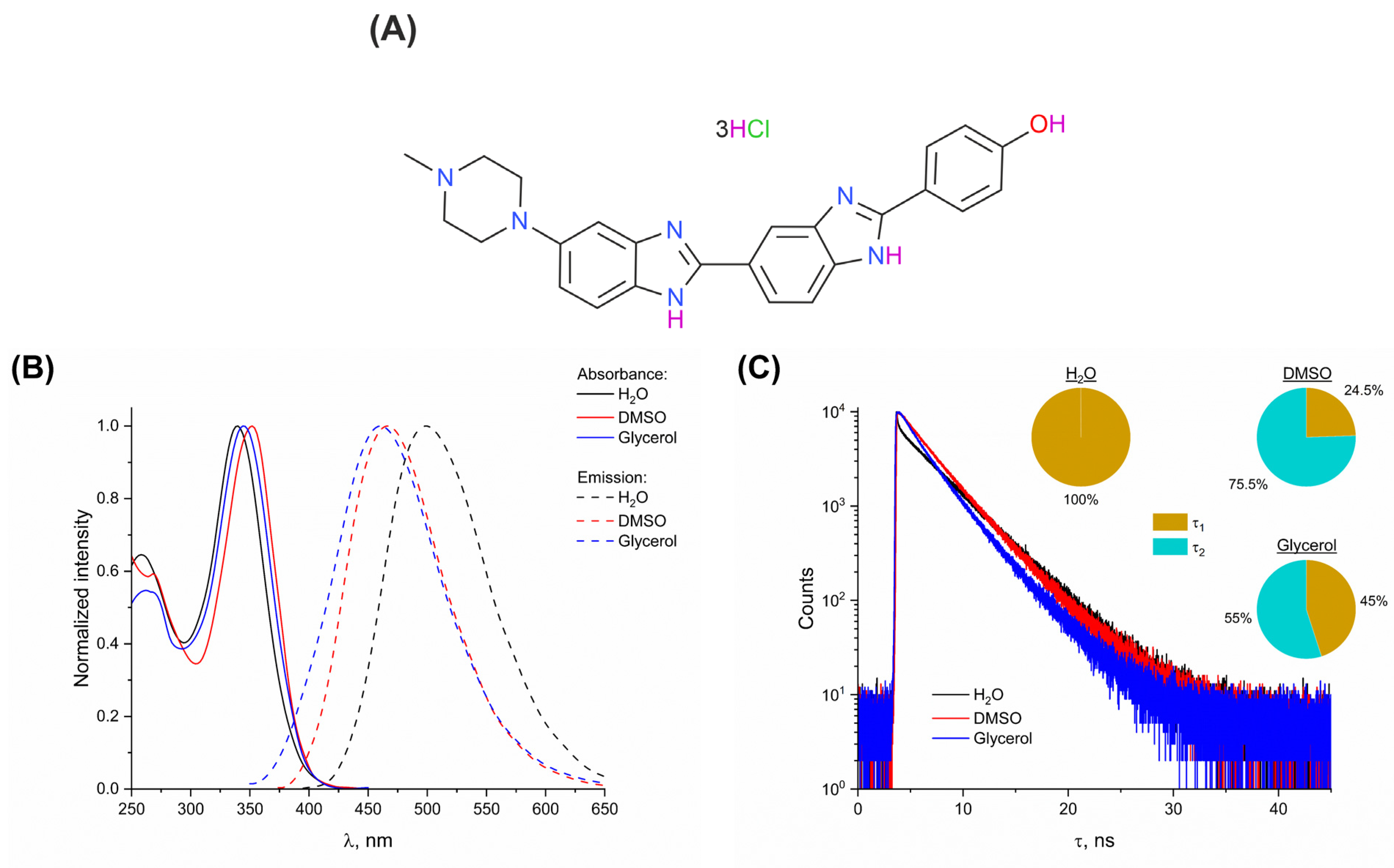
Figure 1. (A) Structure of Hoechst 33258. (B) Normalized absorption (solid lines) and emission (dashed lines) spectra of 5.25 × 10⁻⁶ M Hoechst 33258 in water (black), DMSO (red), and glycerol (blue). (C) Fluorescence decay curves of the dye in water, DMSO, and glycerol (the same color legend is used). (Inset) Contributions of the calculated lifetimes to the total amplitude of Hoechst 33258 fluorescence in the studied media.
Photonics 10 00671 g001
Figure 2. Irradiation of aqueous solutions of the dye: (A) Fluorescence spectra of 3.5 × 10⁻⁶ M Hoechst 33258 at various doses. (Inset) Absorption spectra of non-irradiated dye solution (black) and that irradiated with 2000 Gy (red). (B) Dependence of I/I0 at λ = 500 nm (black) and λ = 390 nm (red) on the absorbed dose. (Inset) Linear section of the ratiometric fluorescence response to irradiation. (C) Fluorescence decay kinetics of Hoechst 33258 (λEm = 500 nm) at various doses. Inset shows the curves recorded at λEm = 390 nm. (D) Dependence of the lifetimes (black) and their contributions to the total fluorescence amplitude (red) on the absorbed dose. In (D), τ1 and A1 values are indicated by filled circles, while τ2 and A2 are indicated by hollow circles, respectively.
Photonics 10 00671 g002
Figure 3. Irradiation of aqueous solutions of the dye in the presence of 6 × 10⁻² M of glycerol: (A) Fluorescence spectra of 3.5 × 10⁻⁶ M Hoechst 33258 at various doses. Inset shows the fluorescence decay kinetics of the dye in non-irradiated control (black) and in the sample irradiated with 2000 Gy (red). (B) Dependence of I/I0 at λ = 500 nm (black) and λ = 390 nm (red) on the absorbed dose.
Photonics 10 00671 g003
Figure 4. Irradiation of aqueous solutions of the dye in the presence of 6 × 10⁻² M of DMSO: (A) Fluorescence spectra of 3.5 × 10⁻⁶ M Hoechst 33258 at various doses. (Inset) Dependence of the fluorescence intensity on the absorbed dose in the region of 500–2000 Gy. (B) Dependence of the relative signal amplitude (I/I0; black) and pH of the solution (red) on the absorbed dose. Inset shows the fluorescence decay kinetics of the dye in non-irradiated control (black) and in the sample irradiated with 500 Gy (red).
Photonics 10 00671 g004
Figure 5. Irradiation of Hoechst 33258 in DMSO: (A) Fluorescence spectra of 3.5 × 10⁻⁶ M of the dye at various doses. Inset shows the normalized spectra. In each case, normalization was conducted using the maximum fluorescence intensity. (B) Dependence of the relative signal amplitude (I/I0; black) and peak position (red) on the absorbed dose. (C) Linear section of the dependence of peak position on the absorbed dose.
Photonics 10 00671 g005
Figure 6. Irradiation of Hoechst 33258 in glycerol: (A) Fluorescence spectra of 3.5 × 10⁻⁶ M of the dye at various doses. Inset shows the normalized spectra. In each case, normalization was conducted using the maximum fluorescence intensity. (B) Dependence of the relative signal amplitude (I/I0; black) and peak position (red) on the absorbed dose. (C) Linear section of the dependence of peak position on the absorbed dose.
Photonics 10 00671 g006
Disclaimer/Publisher’s Note: The statements, opinions and data contained in all publications are solely those of the individual author(s) and contributor(s) and not of MDPI and/or the editor(s). MDPI and/or the editor(s) disclaim responsibility for any injury to people or property resulting from any ideas, methods, instructions or products referred to in the content.