Review

Light in Orthodontics: Applications of High-Intensity Lasers, Photobiomodulation, and Antimicrobial Photodymanic Therapies in Daily Practice

by
Caroline Maria Gomes Dantas
1,*,
Carolina Lapaz Vivan
1,
Gladys Cristina Dominguez
1,
Solange Mongelli de Fantini
1 and
Patricia Moreira de Freitas
2
1
Department of Orthodontics and Pediatric Dentistry, School of Dentistry, University of São Paulo, São Paulo 05508-000, Brazil
2
Department of Restorative Dentistry, School of Dentistry, University of São Paulo, São Paulo 05508-000, Brazil
*
Author to whom correspondence should be addressed.
Photonics 2023, 10(6), 689; https://doi.org/10.3390/photonics10060689
Submission received: 7 May 2023 / Revised: 8 June 2023 / Accepted: 12 June 2023 / Published: 15 June 2023

Abstract

:
Orthodontics is constantly seeking innovation towards mechanical efficiency and better oral-related quality of life during treatment. This narrative review aims to discuss novel scientific reports about light therapies and how they can optimize different stages of orthodontic intervention: before, during, and after treatment. Recurrent conditions that can be treated with laser devices are the removal of carious tissue, dentin hypersensitivity, and temporomandibular disorders. Evidence reveals that laser procedures accelerate health recovery, enabling individuals to initiate orthodontic treatment. Along orthodontic procedure, photobiomodulation therapy, is indicated for analgesia after appliance activations, repair of traumatic ulcers, and acceleration of tooth movement. Moreover, antimicrobial photodynamic therapy is well-indicated for effective decontamination of oral infections such as herpetic lesions and peri-implantitis. Finally, high-intensity lasers are good allies in removing brackets and reconditioning red esthetics. There are many benefits to the use of light sources in the orthodontic routine: simplicity of technique, ease of handling devices, minimal invasiveness, and patient comfort during procedures. It is essential that professionals develop a critical overview of technological advances, offering safe and evidence-based therapies. Recent advances indicate that laser therapies improve patient experiences during orthodontic treatment and minimize the side effects of clinical interventions.

1. Introduction

Orthodontics is a specialty that is constantly in demand of innovations, with academic and market research focused on finding efficient, comfortable, and biologically compatible treatment tools. Efforts have been directed towards improving the patient’s experience and minimizing the side effects from orthodontic interventions [1].
The use of lasers in dentistry started in the 1960s with Goldman et al.’s research on the impact of lasers on dental caries [2]. This technology refers to the use of devices that emit a specific form of light (amplified by stimulated emission of radiation), which can induce different reactions when interacting with biological tissues. In dentistry, class 3B (low-intensity) and class 4 (high-intensity) lasers are commonly used for a variety of procedures, including hard and soft tissue treatments based on photoablation, photothermal, photobiomodulation, and antimicrobial photodynamic phenomena [3]. Depending on the chosen equipment and parameters of irradiation (wavelength, power output, energy density, and mode of emission, among others), it is possible to promote the debridement of soft and hard tissues, disinfection, regulation of inflammation, analgesia, and acceleration of wound healing [4]. Thus, the use of lasers in dental practice has evolved greatly, now attracting special attention from orthodontists. The recent literature shows that light sources can be employed at various stages of orthodontic therapy (OT): before, during, and after treatment.
Initially, patients undergoing OT commonly require prior oral care before tooth movement. Recurrent conditions that can be managed with light include selective removal of demineralized tissue [5], dentin hypersensitivity [6], and temporomandibular disorders (TMD) [7]. Evidence suggests that laser procedures promote accelerated health recovery, enabling the patient to proceed to the next stage. Throughout the treatment, photobiomodulation therapy (PBMT) can be used for analgesia after activations, repair of traumatic ulcers, and acceleration of tooth movement [8]. Meanwhile, antimicrobial photodynamic therapy (aPDT) proves to be an effective decontamination tool for numerous clinical infections caused by viruses, fungi, and bacteria. Among the many applications in the orthodontic routine are herpetic lesions and peri-implantitis [9]. In the final phase of OT, high-intensity lasers may be good allies in bracket removal and in reconditioning “red aesthetics” through gingivectomy [10].
There are many benefits to the use of light sources in the orthodontic routine, particularly in terms of the simplicity of the technique, ease of handling devices, minimally invasive characteristics, and patient comfort during procedures. It is important to emphasize that professionals must develop a critical view of technological advances, practicing evidence-based dentistry. This narrative review aims to discuss, from a broader point of view, the multiple indications of laser light therapies as highly impactful auxiliary tools in orthodontics.

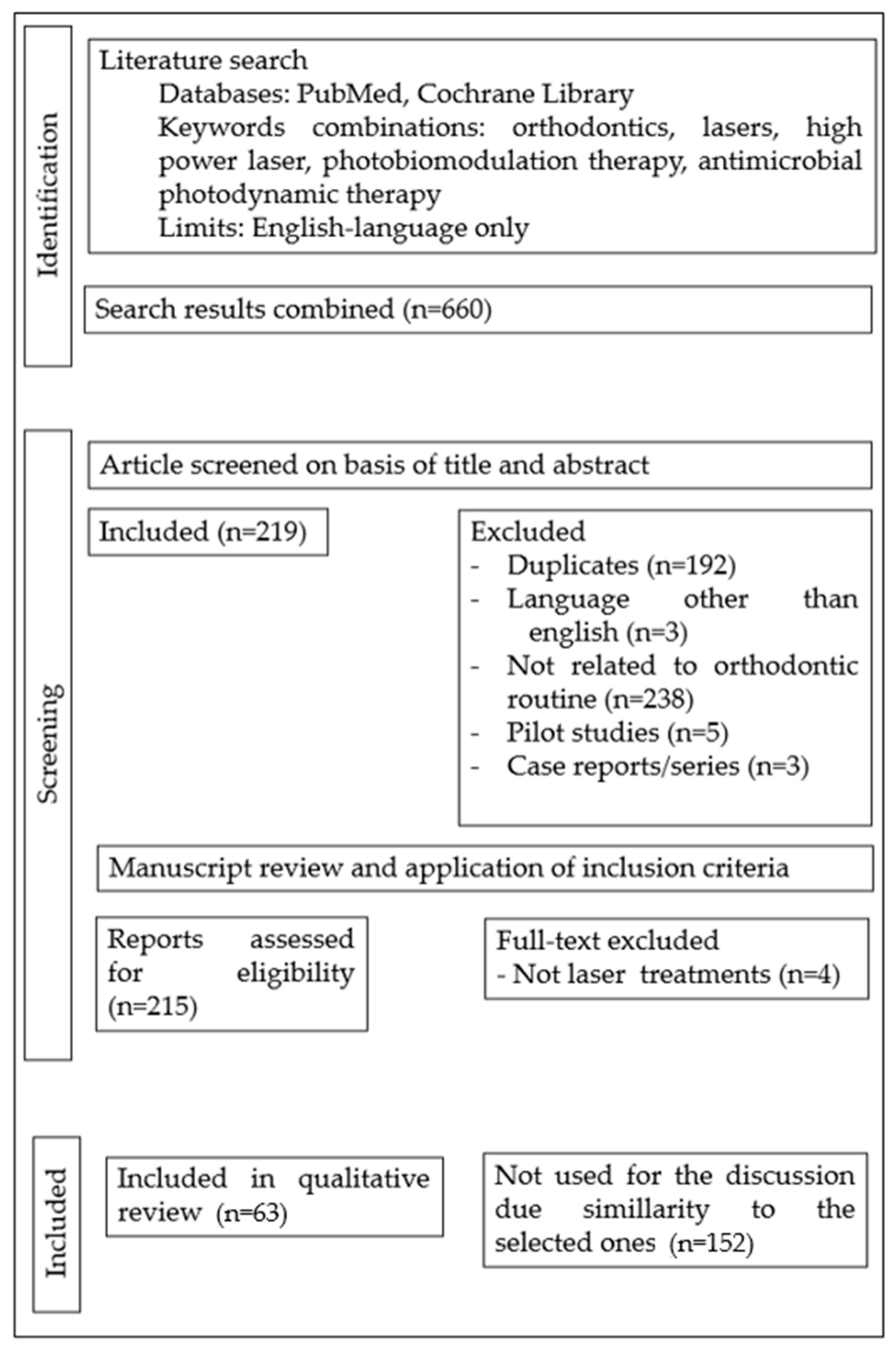
2. Materials and Methods

A comprehensive search of relevant articles about the use of laser devices applied to the orthodontic clinical routine was conducted to compose this narrative review. The selection criteria and literature search strategy are presented below.

2.1. Eligibility Criteria

2.1.1. Inclusion Criteria

  • Scientific articles published up to 30 April 2023.
  • Scientific articles published in the English language.
  • In vitro and in vivo studies that mention tissue reactions to laser light relevant to the orthodontic routine.
  • The literature search was not restricted to any specific age interval, gender, duration of orthodontic treatment, or type of appliance.
  • Only articles related to laser treatments in connection with orthodontic practice were considered relevant to the purpose of this review.

2.1.2. Exclusion Criteria

  • Papers with no clear report or not related to the use of lasers in orthodontic clinical practice.
  • Case reports, case series, pilot studies, study protocols, documents, and book chapters.

2.2. Literature Search Strategy

Data Sources

To obtain an extensive perspective on the subject, a thorough manual search was conducted using the PubMed Central electronic library and Cochrane database. The primary search was conducted up to 30 April 2023, utilizing the keywords “orthodontics”, “laser”, “high power laser”, “photobiomodulation”, and “antimicrobial photodynamic therapy” in various combinations. To quantify the search results, a flow chart based on the PRISMA guidelines [11] is presented (Figure 1).
A total of 660 documents were identified through a comprehensive search of online journals. After removing similar or duplicate publications, 468 distinct papers remained. Upon reviewing the abstracts and titles, an additional 246 articles were disqualified. The remaining studies underwent a manual analysis of their abstracts by two authors (C.M.G.D. and C.L.V.). Titles and abstracts of the retrieved studies were carefully screened, and any studies that met one or more exclusion criteria were excluded. The articles selected for full-text reading were examined by the two authors, and those lacking relevant information for the purpose of this review were excluded (N = 4). Any disagreements were resolved with the assistance of a third reviewer (P.F.). Ultimately, 63 studies were chosen to support the discussion on current applications of light therapies in the clinical practice of orthodontics. Additional literature is presented to further explore and substantiate the line of reasoning, with the aim of elucidating mechanisms of action and/or providing contextual information.

3. Results

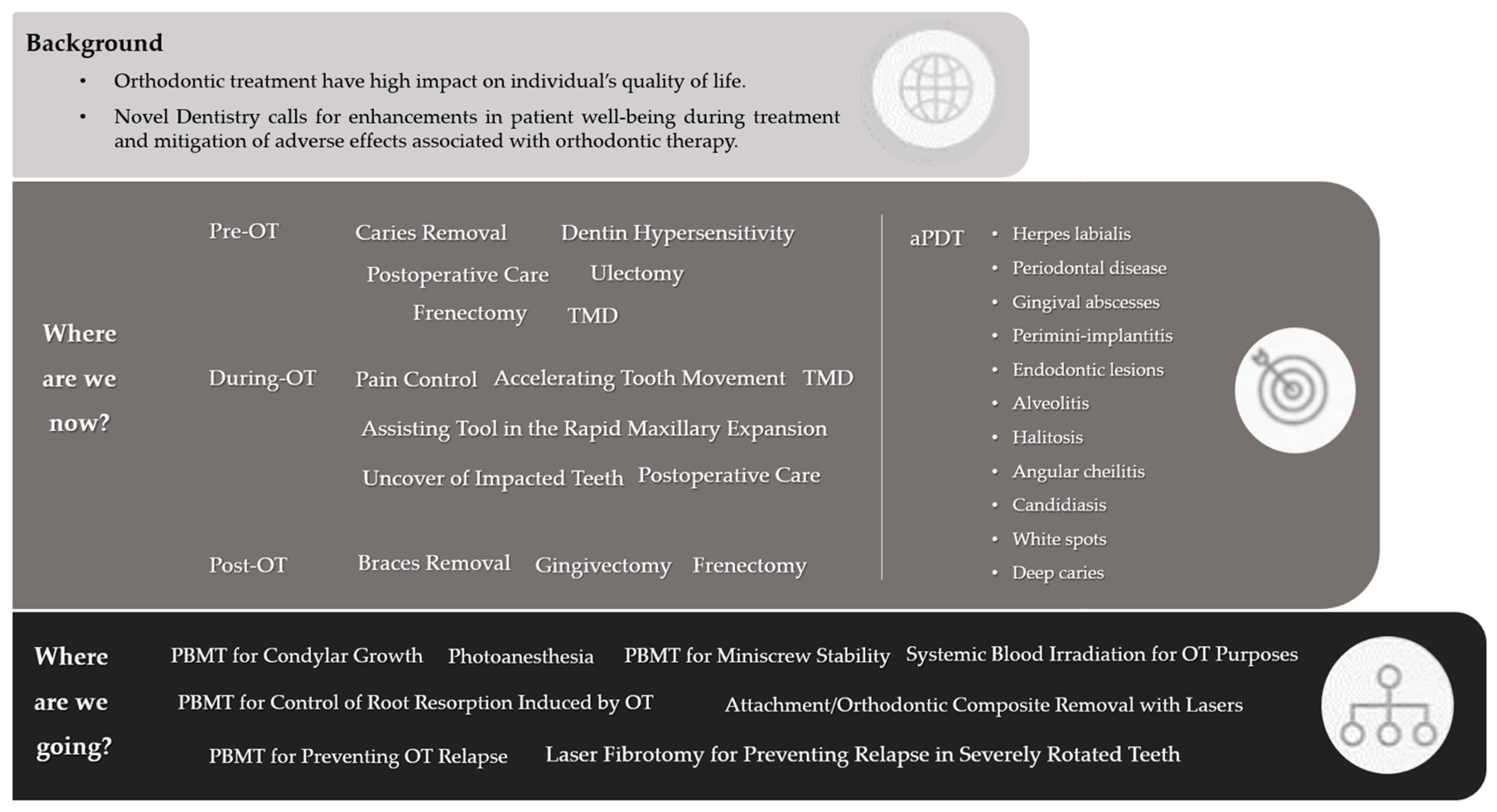
3.1. Where Are We Now?

3.1.1. Pre-Orthodontic Treatment Care

Typically, patients undergoing orthodontic treatment (OT) often present with oral conditions that require prior clinical care, including dental rehabilitation and management of orofacial pathologies. Laser technology can effectively manage recurrent conditions in the clinical routine. Light therapy can selectively remove carious contaminated tissue, offering an ultra-conservative approach [12]. Additionally, dentin hypersensitivity can be alleviated through light surface treatments. High-power lasers such as Nd:YAG or Er:YAG can reorganize the mineral structure, reducing the movement of tubular fluid, while low-power equipment can stimulate—through photobiomodulation—tertiary dentin formation and provide analgesia [13]. Patients undergoing oral surgeries or experiencing postoperative complications can benefit from both high- and low-power laser treatments, as they can help alleviate pain and promote tissue healing [14]. For patients with painful TMD, PBMT can be beneficial in reducing pain, modulating inflammation, and promoting muscular relaxation [15]. It is important to emphasize that these applications represent a few examples of the many ways in which light therapies can enhance treatments in the general practice of dentistry [4].

3.1.2. Pain Control of Orthodontic Activation Responses

Pain response is the most common adverse reaction to OT [16]. It frequently leads to treatment interruption or even reluctance to initiate OT, as patients are concerned about the impact the orthodontic appliance may have on their daily activities [17]. Orthodontic pain is typically associated with tooth discomfort following orthodontic activation, but it can also involve other distressing sensations, such as mucosal ulcers and periodontal lesions caused by the appliances [18].
At the onset of treatment, the presence of orthodontic devices in the oral cavity (such as brackets, aligner attachments, or removable appliances) can cause trauma to the oral mucosa, resulting in ulcerations (Figure 2). An ideal treatment for traumatic ulcers should provide rapid pain relief and optimized tissue repair. Currently, the only therapeutic option that meets these criteria is PBMT. Its mechanism of action involves accelerated extracellular matrix deposition and fibroblast proliferation for tissue repair [19]. Additionally, there is evidence of direct inhibition of pain signaling at the irradiated site, promoting analgesia without any side effects, drug interactions, or age restrictions [20]. Aggarwal et al. [21] demonstrated in a sham-controlled, split-mouth study design that a single session of infrared light PBMT eliminated pain in 93% of patients. Regarding wound healing, a randomized clinical trial (RCT) showed a 70% reduction in the time required for epithelial reconstitution. The current literature supports the superiority of PBMT over topical drug therapy for recurrent aphthous ulcers [22,23,24,25].
Following orthodontic activation (OA), the periodontal ligament immediately responds to stress by initiating a pro-inflammatory cascade, aiming to restore tissue dynamics, vascular changes, and the release of neurogenic and pro-inflammatory mediators, which may also trigger local pain [26]. The gold standard for pain control after orthodontic activation is the use of analgesic or anti-inflammatory drugs. However, not all patients feel comfortable with frequent medication intake, and there is debate regarding the impact of such medication on dental movement [27].
Several studies have demonstrated that PBMT is a non-toxic and effective alternative for managing pain after orthodontic activation [28,29,30,31]. With a single irradiation immediately after OA (Figure 3), PBMT can significantly reduce pain sensitivity by altering the depolarization threshold of nerve endings in the periodontal ligament. It also modulates the production of algogenic mediators and stimulates the peripheral release of endogenous endorphins [20,32,33]. PBMT reduces the number of pain signals reaching the brain, thus preventing the sensitization of central neurons. Brito et al. conducted a double-blind RCT in which patients who received irradiation after the placement of the first alignment arch reported lower levels of pain at 6, 24, 48, and 72 h compared to a control group [28]. This analgesic effect has also been observed for the placement of interdental separators, reducing the need for medication intake [34,35].

3.1.3. Photobiomodulation of Tooth Movement

Orthodontic treatment duration is a significant concern for orthodontic professionals and patients due to the substantial impact of orthodontic appliances on patients’ oral-health-related quality of life [36]. Extensive efforts have been dedicated to the development of therapies that can accelerate tooth movement, including techniques such as corticotomy and microosteoperforation. Studies conducted on animal and human models have already demonstrated that PBMT is a promising adjunctive tool for enhancing tooth movement (Figure 4), exhibiting proven efficacy and safety, and notably, no reported side effects [8,10,37,38,39,40,41,42,43].
Orthodontic mechanics rely on the remodeling of periodontal tissue through the activation of various components, including periodontal fibroblasts, osteoblasts, osteocytes, osteoclasts, and the vascular system. In vitro and in vivo studies have demonstrated that PBMT creates a favorable environment for tooth migration by enhancing cell differentiation, activating osteoclasts and osteoblasts, stimulating collagen synthesis, and promoting neoangiogenesis [30].
In an RCT conducted by Ghaffar et al., the impact of PBMT on the rate of tooth alignment was investigated in patients undergoing OT for Class I malocclusion correction with tooth crowding ranging from 4 to 10 mm. The study revealed that complete alignment was achieved, on average, in 68.2 days for the patients who received irradiation compared to 109.5 days in the control group. Therefore, PBMT shortened the time required for alignment of mild to severe crowding by 37.7% [44]. In a split-mouth RCT by Zheng and Yang, PBMT was found to reduce the time needed for canine retraction by an average of 35% at 4 weeks. The irradiated side exhibited significant changes in bone remodeling markers, including reduced levels of OPG and increased levels of IL-1β and RANKL, indicating that PBMT influenced bone metabolism and resulted in accelerated tooth movement [45]. Systematic reviews support the claim that PBMT can reduce treatment time by 20 to 40%, with no evidence of damage to the periodontium or root resorption [31,37,38,39,40,41,42,43].

3.1.4. Assisting Tool in Rapid Maxillary Expansion

Rapid maxillary expansion (RME) involves not only the bone structure, but also adjacent soft tissues. As it relies on bone remodeling, it is a treatment that induces significant inflammation, often associated with oral pain or discomfort [46]. Low-intensity laser light is recognized for its capability to stimulate the differentiation and activation of osteoclasts, the proliferation of osteoblasts, collagen synthesis, and neoangiogenesis, which, in conjunction with other tissue effects, lead to accelerated bone remodeling, repair, and maturation (Figure 5) [47].
Sasaki et al. [48], in an RCT, demonstrated that the PBMT group experienced a facilitated opening process of the midpalatal suture. This feature is desirable as it may help prevent undesirable dentoalveolar effects. Cepera et al. [49] investigated the influence of PBMT on palatal bone density in patients undergoing RME. The authors observed that patients who received PBMT twice a month after achieving sufficient screw activation showed optimized recovery of bone density during the retention period. PBMT stimulated bone neoformation, which could potentially minimize treatment relapse. Several authors have discussed the potential use of PBMT to reduce the time required for maxillary bone consolidation [50,51,52]. However, further research is needed to support the reduction in the RME-retention phase.

3.1.5. Postoperative Care with Lasers

Light therapy can provide significant benefits in managing the postoperative period following procedures such as tooth extraction, mini-implant installation, orthognathic surgeries, or other events that may lead to complications and impact the oral-health-related quality of life of orthodontic patients.
The immediate postoperative phase is invariably associated with inflammatory symptoms, which can range from swelling and pain to decreased mandibular function. PBMT modulates the inflammatory response, leading to analgesia (by altering the excitation threshold and inducing the production of endogenous endorphins), the resolution of inflammation (by promoting angiogenesis and stimulating lymphatic drainage), and tissue repair (by enhancing enzymatic processes for proper recovery of soft, hard, or nervous tissues) [53,54].
In a split-mouth RCT, Eshghpour et al. [55] demonstrated the effectiveness of PBMT in reducing pain and swelling after the removal of impacted third molars (Figure 6). Domínguez Camacho et al. [56] confirmed that PBMT serves as an important adjunct to oral anti-inflammatory drugs in reducing post-surgical edema in patients undergoing orthognathic surgery. Feslihan and Eroğlu [57] suggested that PBMT can be used as an alternative to corticosteroids after impacted third molar removal, as it exhibits similar clinical efficacy in controlling postoperative pain, edema, and trismus. Considering the toxicity and side effects associated with anti-inflammatory drugs, PBMT is gaining increasing relevance in modern dentistry [58,59,60,61].
Photobiomodulation therapy is highly recommended for addressing one of the most persistent complications following major orofacial surgeries: paresthesia and paralysis. Laser light, when used with the appropriate parameters, aids in the rehabilitation of neurosensory function by optimizing the regeneration of peripheral axons and promoting the sprouting of new adjacent neural terminations [54]. Oliveira et al. [15] draw attention to the fact that the success rate of PBMT for nerve repair depends on the time interval between the nerve damage and the initiation of treatment. Considering that orthodontists schedule the appropriate surgical timing for each case during OT, it is important for professionals to also plan PBMT sessions in the quest of the optimal recovery of their patients.

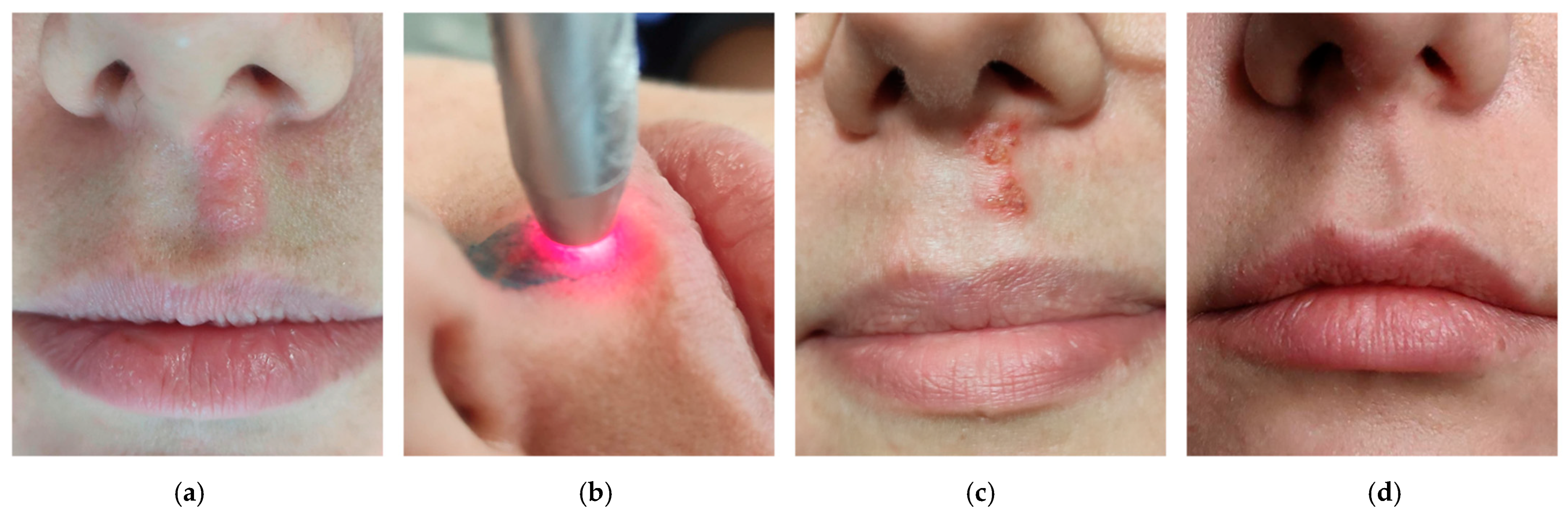
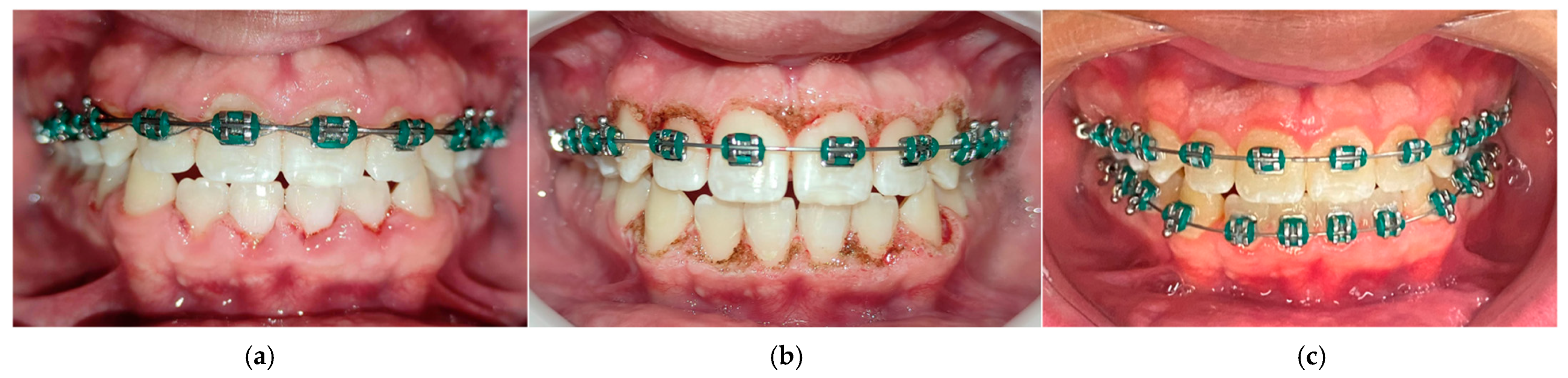
3.1.6. aPDT Easily Solving Infections during Orthodontic Treatment

The occurrence of infectious diseases during OT is relatively common, given the challenges in maintaining proper hygiene due to the presence of orthodontic appliances. It is estimated that approximately 15% of orthodontic patients experience complications that require professional intervention. In the United States alone, this results in an annual cost of over USD 500,000,000 and requires a workload equivalent to that of 1000 full-time dentists [62]. Oral infections during OT can cause significant discomfort and pose risks to the patient’s overall health, as well as impact the scheduled phases of OT.
An interesting approach to manage infectious complications is aPDT due to its simplicity of technique, effectiveness against a wide spectrum of microorganisms (including bacteria, viruses, and fungi), and its non-inductive antimicrobial resistance properties. aPDT involves the application of photosensitizers to the affected area, followed by their activation using a specific wavelength of light. This process triggers a reaction that generates reactive oxygen species at levels sufficient to reduce pathogenic agents without causing any toxicity to the host [63]. Several indications for aPDT are commonly observed in patients undergoing OT, including herpes labialis (Figure 7), periodontal diseases, gingival abscesses, endodontic lesions, alveolitis, perimini-implantitis, halitosis, angular cheilitis, candidiasis, white spots, and deep caries [9].
Baeshen et al. [64] conducted a comprehensive analysis of the impact of aPDT on the management of periodontal diseases in patients undergoing OT with fixed appliances. Clinical parameters, such as gingival inflammation, bacterial population, pro-inflammatory cytokines, and pain perception, showed significant improvement when aPDT was implemented in conjunction with dental scaling. In an RCT, Alshahrani et al. [65] demonstrated that aPDT is also a viable treatment option for adolescents with halitosis during OT. When performed using the tongue scraping technique, aPDT effectively reduced hydrogen sulfide concentration and oral pathogens. Regarding the incidence of herpes labialis, de Paula Eduardo et al. [66] and their study group discussed that aPDT can effectively reduce viral titer during the vesicle phase, allowing for safe continuation of OT after aPDT treatment. They also highlighted the potential implementation of PBMT to reduce the frequency and severity of herpes lesion recurrences [67]. Similar to its uses in various fields of medicine, aPDT proves to be an excellent tool for controlling oral infections, as it not only provides local disinfection, but also promotes pain relief and stimulates tissue repair [9].
The concern for implementing effective measures to control pathogenic complications during OT is of utmost importance, particularly due to the increased risk of tissue lacerations and opportunistic infections during this period. As the literature continues to evolve, aPDT is becoming increasingly recognized as a simple and effective technique for restoring oral health and improving the quality of life for patients undergoing OT [9,68,69,70,71].

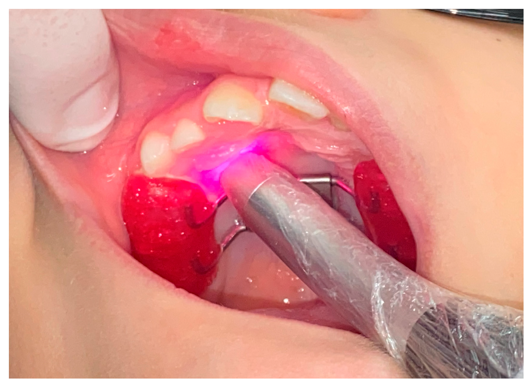
3.1.7. Soft Tissue Management with High-Intensity Lasers Complementing Orthodontic Therapy

High-intensity lasers can be a valuable asset in aiding OT and enhancing treatment outcomes. This technology offers an excellent option for surgery in orthodontic patients as it promotes hemostasis during surgical procedures, significantly reduces the microbial load, and modulates the surrounding tissue to facilitate tissue repair [72].
Currently, two of the most commonly chosen laser devices for surgical purposes in dentistry are high-intensity diode and Er:YAG lasers [72]. High-intensity diode lasers (800–980 nm) interact preferentially with pigmented tissues, primarily absorbed by melanin. When used within the appropriate parameters, they provide good penetration depth in the oral mucosa without causing damage to teeth and bone. High-intensity diode lasers are well-suited for procedures such as gingivectomy, surgical access to impacted teeth, excision of hypertrophic tissue (often induced by orthodontic devices), gingival recontouring to facilitate bracket bonding (Figure 8), frenectomies, and gingival depigmentation [73,74,75,76]. On the other hand, Er:YAG lasers (2.940 nm) are highly absorbed by water and hydroxyapatite. Therefore, in orthodontic patients, they can be used not only for bracket removal, but also for osteotomy in accessing impacted teeth [77]. The clinical application of these lasers relies on the phenomenon of photoablation, where the absorbed light induces rapid vaporization, increasing internal pressure and leading to “micro-explosions” that result in the superficial removal of the irradiated material. Erbium lasers may also be used for soft-tissue incisions; however, special attention must be given to bleeding control [78]. Since these devices act by raising the temperature, they can also be highly valuable in decontaminating infectious processes, such as herpetic lesions [66].
In the context of minimally invasive practices, orthodontists should consider incorporating high-intensity laser technology into their clinical routine. By understanding the characteristics of different light sources and receiving adequate technical training, orthodontists can efficiently manage both soft and hard tissues using lasers, resulting in minimal side effects and a more comfortable postoperative phase for patients.

3.1.8. Braces Removal with the Touch of Light

The removal of orthodontic appliances is traditionally performed using orthodontic pliers, which apply mechanical force to break the adhesion between the appliance and the tooth surface. However, this method can be uncomfortable for the patient and may result in cracks and fractures in the enamel [79]. Therefore, there is a need for alternative methods that allow for the safe removal of appliances without causing damage to the enamel and premature aging of teeth.
High-intensity lasers offer an effective and safe technique for appliance removal. By using the appropriate parameters, the bond strength can be decreased, facilitating the detachment of orthodontic accessories. Various types of lasers, such as diode, Nd:YAG, CO2, Er:YAG, and Er,Cr:YSGG, have been described for bracket debonding [80]. In the thermal softening process, the bonding agent is heated until it softens, allowing the bracket to slide off the tooth surface. Diode lasers are currently more affordable, but caution must be exercised due to temperature increase (which can cause pulp damage), since they do not require water spray [81]. The most commonly used lasers for bracket removal are those that act through photoablation, such as erbium lasers (Figure 9). In photoablation, when the light is absorbed by the orthodontic adhesive, there is a sudden vaporization of the hydroxyl group present in the composite material, resulting in a reduction in shear bonding strength [56]. This process enables the spontaneous detachment of brackets or allows for their removal using college tweezers, eliminating the need for pliers. This procedure is more comfortable for the patient and preserves the enamel structure. Although laser bracket removal is a promising and effective method, it may not be the most practical or cost-effective option for most orthodontists due to limited access to the necessary equipment [80,82,83,84,85].
The technological revolution witnessed in recent decades has led to unprecedented scientific advancements in dentistry. It is crucial to raise awareness among orthodontists regarding the benefits that laser procedures can offer to OT and the oral health of patients. By utilizing efficient, safe, and comfortable therapeutic procedures, it becomes possible to enhance dental aesthetics and functionality, thereby improving the overall health and quality of life of the population.

3.2. The “Ideal Protocol” Conjecture

Defining the ideal parameters for laser therapies in orthodontics can be challenging due to several factors: the heterogeneity of patients/tissue phototypes, the complexity of biological responses, the lack of standardized and well-described protocols, and limitations in scientific research [86]. Despite being under investigation for over 50 years, there is still no consensus on the specific parameters and protocols for each clinical application of laser therapy. The diverse range of parameters involved, including wavelength, energy, fluence, power, irradiance, pulse mode, treatment duration, and repetition, has led researchers to contradictory findings in certain cases.
Orthodontic patients exhibit individual variations in their oral tissues, tooth anatomy, and treatment needs. These variations can influence their response to laser therapy, making it difficult to establish a one-size-fits-all set of parameters. It is known that the biological effects of laser therapy are multifaceted and involve complex interactions with cells, tissues, and biochemical processes [3,4,31]. Determining the optimal parameters requires a comprehensive understanding of the specific mechanisms involved in orthodontic tooth movement and tissue response to laser therapy, which is still an area of ongoing research.
The field of light therapies is still evolving, and there is a need for broad discussion on universally accepted, standardized protocols for orthodontic applications. Different studies may use varying laser devices, beam cross-sectional dynamics, wavelengths, energy densities, treatment durations, and techniques, leading to inconsistencies in results and recommendations [87]. Conducting well-designed, controlled clinical trials specifically focused on laser photobiomodulation in orthodontics can be challenging. Limited sample sizes, variations in study designs, and the lack of long-term follow-up make it difficult to draw definitive conclusions and establish ideal parameters. Taking into account the aforementioned challenges, ongoing investigations and collaboration among researchers, clinicians, and laser manufacturers are necessary to develop evidence-based guidelines and refine the parameters for laser photobiomodulation in orthodontics. It is important to balance the potential benefits of laser therapy with patient safety and the need for reliable and reproducible outcomes.
Considering the great variability of parameters applied in RCTs, it is not possible to suggest a specific protocol for each indication. However, based on the significant results presented in the recent literature, a summary framework incorporating the most frequently reported dosimetric data from systematic reviews published in the last 5 years on common indications of laser therapies in orthodontic practice is presented (Table 1).

3.3. Where Are We Going?

In addition to the above-mentioned evidence-based applications, innovative research is being conducted to uncover more helpful therapeutics for the orthodontist’s clinical routine.
Rationalizing the influence of light therapies on tissue metabolism, Franco et al. [88] hypothesized that PBMT could influence condylar growth during the orthopedic treatment of mandibular advancement in an animal study. The researchers found that PBMT stimulates matrix deposition and cartilage thickening in the condyle towards mandibular propulsion. Additional studies need to be conducted to clarify the possible synergistic correlation between the two therapies.
Aligned with the current demand for less invasive treatments, Sobouti et al. [89] tested the efficacy of photoanesthesia. In an RCT, the researchers compared the analgesic effect of PBMT and topical anesthesia to conventional lidocaine injection on the pain experience during the installation of mini-implants and the postoperative period. It was reported that both techniques performed similarly, indicating that topical anesthesia combined with PBMT provides the necessary anesthetic comfort for the procedure.
Skeletal anchorage has become an important tool in orthodontic mechanics as it is a great ally in solving complex cases. However, the literature shows that 13.5% of installed mini-implants are lost, which may be due, among other factors, to deficient tissue remodeling around the screw and chronic mucosal inflammation. Taking into consideration the well-known mechanism of action of PBMT in hard and soft tissue remodeling, the literature seeks the adequate dosimetry to improve mini-implant stability. Razaghi et al. [90], in a systematic review of human and animal studies, acknowledged that PBMT appears to be beneficial to mini-implant stability due to its anti-inflammatory and bone-stimulating effects. Although the number of trials analyzed to date is still small, the evidence suggests an increase in mini-implant stability over time in laser-treated groups, less mini-implant displacement, and no conclusive data on better pain control. Long-term studies with a larger sample size and clear irradiation parameters are still necessary to further validate PBMT in optimal clinical protocols.
In addition to the above-mentioned evidence-based applications, innovative research is being conducted to further analyze the gray area in the literature regarding PBMT for the control of orthodontically induced root resorption. Nayyer et al. [91], in a meta-analysis of six selected articles from 1509, stated that there is moderate evidence suggesting a beneficial effect of PBMT in root resorption control. Since PBMT is capable of influencing bone and cementum metabolism, inducing neovascularization, and modulating the inflammatory process, PBMT is expected to be a promising therapy for the prevention or reduction in inflammatory processes that lead to root resorption in OT. The selected studies mostly used infrared laser devices, applied buccally and palatal/lingually to the periodontium of the tested elements, with different dosimetry and frequency of irradiation. Meta-analyses resulted in significantly less total root resorption in the PBMT groups, although the duration of the trials and the outcome assessment method were found to be variable. Similar to other developing fields, higher-quality RCTs with proper equivalency in intervention methods and outcome analysis are required.
Aiming for an improved finalization of OT, Jahanbin et al. [92] discussed the efficacy of Er:YAG laser fibrotomy and PBMT in reducing the relapse of incisors with severe rotations compared to conventional surgical techniques. According to the authors, laser fibrotomy offers numerous advantages over conventional surgical techniques, such as minimal or no bleeding, less severe pain and edema, and a lower risk of postoperative infection due to simultaneous decontamination during the procedure. The results seem promising: the control group had a relapse rate of 27.8%, while the treated groups had relapse rates of 9.7% (conventional surgery), 11.7% (PBMT), and 12.7% (Er:YAG). However, the most noteworthy result of this study was that PBMT, administered twice a week for 1 month, was as effective as conventional surgery and laser fibrotomy in controlling relapse. Although innovative, the RCT monitored for only 1 month, without retention after OT. New research in animal and human models is being conducted to determine the optimal PBMT dosimetry to assist in dental stability after OT [93,94].
Preservation of the dental substrate at the end of OT is another major concern of professionals who seek minimally invasive practices. The removal of fixed orthodontic appliances or aligner attachments offers the potential for damage to the tooth structure, especially in enamel topography. After implementing an adequate bracket removal technique, several systems for removing residual orthodontic adhesive are described in the literature [95]. Erbium lasers (Er:YAG and Er,Cr:YSGG) have a high interaction with water and have been widely studied for the removal of resinous materials, including orthodontic adhesive. Their photoablation mechanism is capable of expelling material from the irradiated surface by explosive vaporization [82]. Lizarelli et al. [96] tested different irradiation protocols with an Er:YAG laser, analyzing for which parameters light absorption would occur mainly in the composite. However, the literature still raises questions about the safety of using Er:YAG lasers for this purpose due to the lack of selectivity in relation to dental enamel (since Er:YAG lasers have a high interaction with hydroxyapatite) [97]. Although safe and effective protocols for removing brackets with the Er:YAG laser are available in the literature, systematic reviews indicate that there are still no safe protocols for using the Er:YAG laser to remove orthodontic adhesive from the tooth structure [84, 97]. Scientific research is advancing in this field, but the best evidence for minimal damage to the tooth surface at the end of OT indicates the use of tungsten carbide burs followed by multi-step Sof-Lex discs for polishing the dental surface after bracket removal.
Intravascular irradiation of blood (ILIB), a technique that evokes systemic effects for PBMT, has gained great visibility in daily dentistry, but its mechanisms and dosimetry are still unclear. It states that the low-level laser light directed to blood flow can activate neurohumoral regulation, modulate cell oxidant pattern, and induce analgesic, anti-inflammatory, and sedative effects [98]. However, few studies are available on dentistry matters. Silva and Pinheiro [99], in an RCT, revealed that weekly ILIB sessions were equally effective as aPDT and PBMT in preventing and reducing the severity of mucositis. Rangel and Pinheiro [100] suggested that ILIB and laser acupuncture were helpful in managing dental anxiety in children. Still, no guidelines for the safe use of blood flow irradiation (ILIB, intranasal, or transdermal) for orthodontic purposes are available at the time of this essay. As in any therapeutic modality, it is essential that orthodontists develop a critical overview of the technological developments offered by the industry, acting with caution and via evidence-based approaches. Monitoring scientific developments is mandatory for professionals who are attentive to offering gold-standard clinical care.
Figure 10 presents a map of the applicability and future perspectives for the use of laser procedures in orthodontics.

4. Conclusions

The benefits of the use of light therapies in orthodontic practice are manifold. One of the key highlights of this article is that it is already proven in the literature that the use of lasers can optimize orthodontic mechanics by promoting analgesia, accelerating tooth movement, improving the recovery of surgical cases, and making routine orthodontic procedures more comfortable.
These light therapies offer several advantages, including the technical simplicity of procedures, the ease of handling the technology, minimal invasiveness, and, mostly, the absence of side effects and drug interactions. However, like any treatment technique in health sciences, it requires mastery, skillfulness, and an up-to-date scientific background to achieve the best results with utmost security. Modern dentistry emphasizes the importance of safety procedures that rely on efficiency and minimize patient discomfort. Therefore, it is imperative that oral-health-related quality of life becomes one of the main aspirations in orthodontic treatment, alongside dental alignment or achieving a class I occlusion.

Author Contributions

Conceptualization, C.M.G.D. and C.L.V.; methodology, C.M.G.D., C.L.V. and P.M.d.F.; validation, S.M.d.F. and G.C.D.; investigation, C.M.G.D. and C.L.V.; resources, C.M.G.D.; data curation, C.M.G.D. and C.L.V.; writing—original draft preparation, C.M.G.D. and C.L.V.; writing—review and editing, P.M.d.F., S.M.d.F. and G.C.D.; visualization, C.M.G.D. and C.L.V.; supervision, P.M.d.F., S.M.d.F. and G.C.D.; project administration, C.M.G.D. All authors have read and agreed to the published version of the manuscript.

Funding

This research received no external funding.

Institutional Review Board Statement

Not applicable.

Informed Consent Statement

Not applicable.

Data Availability Statement

Not applicable.

Acknowledgments

The authors kindly thank LELO—Special Laboratory of Lasers in Dentistry of the Faculty of Dentistry of the University of São Paulo for all support with this research.

Conflicts of Interest

The authors declare no conflict of interest.

References

  1. Alam, M.K.; Abutayyem, H.; Kanwal, B.; Shayeb, M.A.L. Future of Orthodontics—A Systematic Review and Meta-Analysis on the Emerging Trends in This Field. J. Clin. Med. 2023, 12, 532. [Google Scholar] [CrossRef]
  2. Goldman, L.; Hornby, P.; Meyer, R.; Goldman, B. Impact of the Laser on Dental Caries. Nature 1964, 203, 417. [Google Scholar] [CrossRef]
  3. Parker, S.; Cronshaw, M.; Anagnostaki, E.; Mylona, V.; Lynch, E.; Grootveld, M. Current Concepts of Laser–Oral Tissue Interaction. Dent. J. 2020, 8, 61. [Google Scholar] [CrossRef] [PubMed]
  4. Luchian, I.; Budală, D.G.; Baciu, E.-R.; Ursu, R.G.; Diaconu-Popa, D.; Butnaru, O.; Tatarciuc, M. The Involvement of Photobiology in Contemporary Dentistry—A Narrative Review. Int. J. Mol. Sci. 2023, 24, 3985. [Google Scholar] [CrossRef] [PubMed]
  5. Douchy, L.; Gauthier, R.; Abouelleil-Sayed, H.; Colon, P.; Grosgogeat, B.; Bosco, J. The effect of therapeutic radiation on dental enamel and dentin: A systematic review. Dent. Mater. 2022, 38, e181–e201. [Google Scholar] [CrossRef] [PubMed]
  6. Pion, L.A.; De Matos, L.L.M.; Gimenez, T.; Palma-Dibb, R.G.; Faraoni, J.J. Treatment outcome for dentin hypersensitivity with laser therapy: Systematic review and meta-analysis. Dent. Med. Probl. 2023, 60, 153–166. [Google Scholar] [CrossRef]
  7. Zhang, Y.; Qian, Y.; Huo, K.; Liu, J.; Huang, X.; Bao, J. Efficacy of laser therapy for temporomandibular disorders: A systematic review and meta-analysis. Complement. Ther. Med. 2023, 74, 102945. [Google Scholar] [CrossRef]
  8. Sipiyaruk, K.; Chintavalakorn, R.; Saengfai, N. The protocol of low-level laser therapy in orthodontic practice: A scoping review of literature. J. Int. Soc. Prev. Community Dent. 2022, 12, 267–286. [Google Scholar] [CrossRef]
  9. Olek, M.; Machorowska-Pieniążek, A.; Stós, W.; Kalukin, J.; Bartusik-Aebisher, D.; Aebisher, D.; Cieślar, G.; Kawczyk-Krupka, A. Photodynamic Therapy in Orthodontics: A Literature Review. Pharmaceutics 2021, 13, 720. [Google Scholar] [CrossRef]
  10. Demirsoy, K.K.; Kurt, G. Use of Laser Systems in Orthodontics. Turk. J. Orthod. 2020, 33, 133–140. [Google Scholar] [CrossRef]
  11. Page, M.J.; McKenzie, J.E.; Bossuyt, P.M.; Boutron, I.; Hoffmann, T.C.; Mulrow, C.D.; Shamseer, L.; Tetzlaff, J.M.; Akl, E.A.; Brennan, S.E.; et al. The PRISMA 2020 Statement: An Updated Guideline for Reporting Systematic Reviews. BMJ 2021, 372, n71. [Google Scholar] [CrossRef]
  12. Li, T.; Zhang, X.; Shi, H.; Ma, Z.; Lv, B.; Xie, M. Er:YAG laser application in caries removal and cavity preparation in children: A meta-analysis. Lasers Med. Sci. 2019, 34, 273–280. [Google Scholar] [CrossRef] [PubMed]
  13. Cattoni, F.; Ferrante, L.; Mandile, S.; Tetè, G.; Polizzi, E.M.; Gastaldi, G. Comparison of Lasers and Desensitizing Agents in Dentinal Hypersensitivity Therapy. Dent. J. 2023, 11, 63. [Google Scholar] [CrossRef] [PubMed]
  14. Dawdy, J.; Halladay, J.; Carrasco-Labra, A.; Araya, I.; Yanine, N.; Brignardello-Petersen, R. Efficacy of adjuvant laser therapy in reducing postsurgical complications after the removal of impacted mandibular third molars: A systematic review update and meta-analysis. J. Am. Dent. Assoc. 2017, 148, 887–902.e4. [Google Scholar] [CrossRef] [PubMed]
  15. De Oliveira, R.F.; Da Silva, A.C.; Simões, A.; Youssef, M.N.; De Freitas, P.M. Laser Therapy in the Treatment of Paresthesia: A Retrospective Study of 125 Clinical Cases. Photomed. Laser Surg. 2015, 33, 415–423. [Google Scholar] [CrossRef] [PubMed]
  16. Kavaliauskiene, A.; Smailiene, D.; Buskiene, I.; Keriene, D. Pain and discomfort perception among patients undergoing orthodontic treatment: Results from one month follow-up study. Stomatologija 2012, 14, 118–125. [Google Scholar]
  17. Erdinç, A.M.E. Perception of pain during orthodontic treatment with fixed appliances. Eur. J. Orthod. 2004, 26, 79–85. [Google Scholar] [CrossRef]
  18. Rakhshan, H.; Rakhshan, V. Pain and discomfort perceived during the initial stage of active fixed orthodontic treatment. Saudi Dent. J. 2015, 27, 81–87. [Google Scholar] [CrossRef] [Green Version]
  19. Suter, V.G.A.; Sjölund, S.; Bornstein, M.M. Effect of laser on pain relief and wound healing of recurrent aphthous stomatitis: A systematic review. Lasers Med. Sci. 2017, 32, 953–963. [Google Scholar] [CrossRef]
  20. Chow, R.T.; Armati, P.J. Photobiomodulation: Implications for Anesthesia and Pain Relief. Photomed. Laser Surg. 2016, 34, 599–609. [Google Scholar] [CrossRef]
  21. Aggarwal, H. Efficacy of Low-Level Laser Therapy in Treatment of Recurrent Aphthous Ulcers—A Sham Controlled, Split Mouth Follow Up Study. J. Clin. Diagn. Res. 2014, 8, 218–221. [Google Scholar] [CrossRef] [PubMed]
  22. Lalabonova, H.; Daskalov, H. Clinical assessment of the therapeutic effect of low-level laser therapy on chronic recurrent aphthous stomatitis. Biotechnol. Biotechnol. Equip. 2014, 28, 929–933. [Google Scholar] [CrossRef] [PubMed]
  23. Parra-Moreno, F.; Egido-Moreno, S.; Schemel-Suárez, M.; González-Navarro, B.; Estrugo-Devesa, A.; López-López, J. Treatment of recurrent aphtous stomatitis: A systematic review. Med. Oral Patol. Oral Cirugía Bucal 2023, 28, e87–e98. [Google Scholar] [CrossRef] [PubMed]
  24. Liu, H.; Tan, L.; Fu, G.; Chen, L.; Tan, H. Efficacy of Topical Intervention for Recurrent Aphthous Stomatitis: A Network Meta-Analysis. Medicina 2022, 58, 771. [Google Scholar] [CrossRef] [PubMed]
  25. Ahmed, M.K.; Jafer, M.; Nayeem, M.; Moafa, I.H.; Quadri, M.F.A.; Gopalaiah, H. Low-Level Laser Therapy and Topical Medications for Treating Aphthous Ulcers: A Systematic Review. J. Multidiscip. Healthc. 2020, 13, 1595–1605. [Google Scholar] [CrossRef]
  26. Ngan, P.; Kess, B.; Wilson, S. Perception of discomfort by patients undergoing orthodontic treatment. Am. J. Orthod. Dentofac. Orthop. 1989, 96, 47–53. [Google Scholar] [CrossRef]
  27. Bertolini, A.; Ferrari, A.; Ottani, A.; Guerzoni, S.; Tacchi, R.; Leone, S. Paracetamol: New Vistas of an Old Drug. CNS Drug Rev. 2006, 12, 250–275. [Google Scholar] [CrossRef]
  28. Brito, M.H.; Nogueira, C.Q.; Cotrin, P.; Fialho, T.; Oliveira, R.C.; Oliveira, R.G.; Salmeron, S.; Valarelli, F.P.; Freitas, K.M.S.; Cançado, R.H. Efficacy of Low-Level Laser Therapy in Reducing Pain in the Initial Stages of Orthodontic Treatment. Int. J. Dent. 2022, 2022, 3934900. [Google Scholar] [CrossRef]
  29. Zhi, C.; Guo, Z.; Wang, T.; Liu, D.; Duan, X.; Yu, X.; Zhang, C. Viability of Photobiomodulaton Therapy in Decreasing Orthodontic-Related Pain: A Systematic Review and Meta-Analysis. Photobiomodul. Photomed. Laser Surg. 2021, 39, 504–517. [Google Scholar] [CrossRef]
  30. Camacho, A.D.; Reyes, M.B.; Cujar, S.A.V. A systematic review of the effective laser wavelength range in delivering photobiomodulation for pain relief in active orthodontic treatment. Int. Orthod. 2020, 18, 684–695. [Google Scholar] [CrossRef]
  31. Cronshaw, M.; Parker, S.; Anagnostaki, E.; Lynch, E. Systematic Review of Orthodontic Treatment Management with Photobiomodulation Therapy. Photobiomodul. Photomed. Laser Surg. 2019, 37, 862–868. [Google Scholar] [CrossRef] [Green Version]
  32. Cambier, D.; Blom, K.; Witvrouw, E.; Ollevier, G.; De Muynck, M.; Vanderstraeten, G. The Influence of Low Intensity Infrared Laser Irradiation on Conduction Characteristics of Peripheral Nerve: A Randomised, Controlled, Double Blind Study on the Sural Nerve. Lasers Med. Sci. 2000, 15, 195–200. [Google Scholar] [CrossRef]
  33. Hagiwara, S.; Iwasaka, H.; Okuda, K.; Noguchi, T. GaAlAs (830 nm) low-level laser enhances peripheral endogenous opioid an-algesia in rats. Lasers Surg. Med. 2007, 39, 797–802. [Google Scholar] [CrossRef]
  34. Farzan, A.; Khaleghi, K. The Effectiveness of Low-Level Laser Therapy in Pain Induced by Orthodontic Separator Placement: A Systematic Review. J. Lasers Med. Sci. 2021, 12, e29. [Google Scholar] [CrossRef] [PubMed]
  35. Al-Jewair, T.; Farsaii, A. Low-Level Laser Therapy May Reduce Pain Associated with Orthodontic Separator Placement. J. Evid. Based Dent. Pract. 2022, 22, 101752. [Google Scholar] [CrossRef]
  36. Kaklamanos, E.G.; Makrygiannakis, M.A.; Athanasiou, A.E. Oral Health-Related Quality of Life throughout Treatment with Clear Aligners in Comparison to Conventional Metal Fixed Orthodontic Appliances: A Systematic Review. Int. J. Environ. Res. Public Health 2023, 20, 3537. [Google Scholar] [CrossRef] [PubMed]
  37. AlShahrani, I.; Togoo, R.A.; Hosmani, J.; Alhaizaey, A. Photobiomodulation in acceleration of orthodontic tooth movement: A systematic review and meta analysis. Complement. Ther. Med. 2019, 47, 102220. [Google Scholar] [CrossRef]
  38. Huang, T.; Wang, Z.; Li, J. Efficiency of photobiomodulation on accelerating the tooth movement in the alignment phase of orthodontic treatment—A systematic review and meta-analysis. Heliyon 2023, 9, e13220. [Google Scholar] [CrossRef]
  39. Olmedo-Hernández, O.L.; Mota-Rodríguez, A.N.; Torres-Rosas, R.; Argueta-Figueroa, L. Effect of the photobiomodulation for acceleration of the orthodontic tooth movement: A systematic review and meta-analysis. Lasers Med. Sci. 2022, 37, 2323–2341. [Google Scholar] [CrossRef]
  40. Yavagal, C.M.; Matondkar, S.P.; Yavagal, P.C. Efficacy of Laser Photobiomodulation in Accelerating Orthodontic Tooth Movement in Children: A Systematic Review with Meta-analysis. Int. J. Clin. Pediatr. Dent. 2021, 14 (Suppl. S1), S94–S100. [Google Scholar] [CrossRef]
  41. Li, J.; Ge, X.; Guan, H.; Jia, L.; Chang, W.; Ma, W. The Effectiveness of Photobiomodulation on Accelerating Tooth Movement in Orthodontics: A Systematic Review and Meta-Analysis. Photobiomodul. Photomed. Laser Surg. 2021, 39, 232–244. [Google Scholar] [CrossRef] [PubMed]
  42. Jedliński, M.; Romeo, U.; del Vecchio, A.; Palaia, G.; Galluccio, G. Comparison of the Effects of Photobiomodulation with Different Lasers on Orthodontic Movement and Reduction of the Treatment Time with Fixed Appliances in Novel Scientific Reports: A Systematic Review with Meta-Analysis. Photobiomodul. Photomed. Laser Surg. 2020, 38, 455–465. [Google Scholar] [CrossRef] [PubMed]
  43. Bakdach, W.M.; Hadad, R. Effectiveness of low-level laser therapy in accelerating the orthodontic tooth movement: A systematic review and meta-analysis. Dent. Med. Probl. 2020, 57, 73–94. [Google Scholar] [CrossRef] [PubMed]
  44. Ghaffar, Y.K.A.; El Sharaby, F.A.; Negm, I.M. Effect of low-level laser therapy on the time needed for leveling and alignment of mandibular anterior crowding. Angle Orthod. 2022, 92, 478–486. [Google Scholar] [CrossRef]
  45. Zheng, J.; Yang, K. Clinical research: Low-level laser therapy in accelerating orthodontic tooth movement. BMC Oral Health 2021, 21, 324. [Google Scholar] [CrossRef]
  46. Bastos, R.T.D.R.M.; Blagitz, M.N.; Aragón, M.L.S.D.C.; Maia, L.C.; Normando, D. Periodontal side effects of rapid and slow maxillary expansion: A systematic review. Angle Orthod. 2019, 89, 651–660. [Google Scholar] [CrossRef] [Green Version]
  47. Saito, S.; Shimizu, N. Stimulatory effects of low-power laser irradiation on bone regeneration in midpalatal suture during ex-pansion in the rat. Am. J. Orthod. Dentofac. Orthop. 1997, 111, 525–532. [Google Scholar] [CrossRef] [PubMed]
  48. Sasaki, A.; Touma, Y.; Ishino, Y.; Tanaka, E.; Aoyama, J.; Hanaoka, K.; Watanabe, M.; Tanne, K. Linear polarized near-infrared irradiation stimulates mechanical expansion of the rat sagittal suture. Luminescence 2003, 18, 58–60. [Google Scholar] [CrossRef]
  49. Cepera, F.; Torres, F.C.; Scanavini, M.A.; Paranhos, L.R.; Filho, L.C.; Cardoso, M.A.; Siqueira, D.C.; Siqueira, D.F. Effect of a low-level laser on bone regeneration after rapid maxillary expansion. Am. J. Orthod. Dentofac. Orthop. 2012, 141, 444–450. [Google Scholar] [CrossRef]
  50. Farzan, A.; Khaleghi, K.; Pirayesh, Z. Effect of Low-Level Laser Therapy on Bone Formation in Rapid Palatal Expansion: A Systematic Review. J. Lasers Med. Sci. 2022, 13, e13. [Google Scholar] [CrossRef]
  51. Lai, P.-S.; Fierro, C.; Bravo, L.; Perez-Flores, A. Benefits of Using Low-level Laser Therapy in the Rapid Maxillary Expansion: A Systematic Review. Int. J. Clin. Pediatr. Dent. 2021, 14 (Suppl. S1), S101–S106. [Google Scholar] [CrossRef]
  52. Davoudi, A.; Amrolahi, M.; Khaki, H. Effects of laser therapy on patients who underwent rapid maxillary expansion; a systematic review. Lasers Med. Sci. 2018, 33, 1387–1395. [Google Scholar] [CrossRef] [PubMed]
  53. Albertini, R.; Villaverde, A.; Aimbire, F.; Salgado, M.; Bjordal, J.; Alves, L.; Munin, E.; Costa, M. Anti-inflammatory effects of low-level laser therapy (LLLT) with two different red wavelengths (660 nm and 684 nm) in carrageenan-induced rat paw edema. J. Photochem. Photobiol. B Biol. 2007, 89, 50–55. [Google Scholar] [CrossRef]
  54. Muniz, X.C.; de Assis, A.C.C.; de Oliveira, B.S.A.; Ferreira, L.F.R.; Bilal, M.; Iqbal, H.M.N.; Soriano, R.N. Efficacy of low-level laser therapy in nerve injury repair—A new era in therapeutic agents and regenerative treatments. Neurol. Sci. 2021, 42, 4029–4043. [Google Scholar] [CrossRef] [PubMed]
  55. Eshghpour, M.; Ahrari, F.; Takallu, M. Is Low-Level Laser Therapy Effective in the Management of Pain and Swelling After Mandibular Third Molar Surgery? J. Oral Maxillofac. Surg. 2016, 74, 1322.e1–1322.e8. [Google Scholar] [CrossRef] [PubMed]
  56. Camacho, A.D.; Velásquez, S.A.; Marulanda, N.J.B.; Moreno, M. Photobiomodulation as oedema adjuvant in post-orthognathic surgery patients: A randomized clinical trial. Int. Orthod. 2020, 18, 69–78. [Google Scholar] [CrossRef] [PubMed]
  57. Feslihan, E.; Eroğlu, C.N. Can Photobiomodulation Therapy Be an Alternative to Methylprednisolone in Reducing Pain, Swelling, and Trismus After Removal of Impacted Third Molars? Photobiomodul. Photomed. Laser Surg. 2019, 37, 700–705. [Google Scholar] [CrossRef]
  58. de Barros, D.D.; Catão, J.S.d.S.B.; Ferreira, A.C.D.; Simões, T.M.S.; Almeida, R.D.A.C.; Catão, M.H.C.D.V. Low-level laser therapy is effective in controlling postoperative pain in lower third molar extractions: A systematic review and meta-analysis. Lasers Med. Sci. 2022, 37, 2363–2377, Erratum in Lasers Med. Sci. 2022. [Google Scholar] [CrossRef]
  59. de Oliveira, F.J.D.; Brasil, G.M.L.C.; Soares, G.P.A.; Paiva, D.F.F.; Júnior, F.D.A.D.S. Use of low-level laser therapy to reduce postoperative pain, edema, and trismus following third molar surgery: A systematic review and meta-analysis. J. Cranio-Maxillofac. Surg. 2021, 49, 1088–1096. [Google Scholar] [CrossRef]
  60. Hakimiha, N.; Bassir, S.H.; Romanos, G.E.; Shamshiri, A.R.; Moslemi, N. Efficacy of photobiomodulation therapy on neurosensory recovery in patients with inferior alveolar nerve injury following oral surgical procedures: A systematic review. Quintessence Int. 2021, 52, 140–153. [Google Scholar] [CrossRef]
  61. Hosseinpour, S.; Tunér, J.; Fekrazad, R. Photobiomodulation in Oral Surgery: A Review. Photobiomodul. Photomed. Laser Surg. 2019, 37, 814–825. [Google Scholar] [CrossRef] [Green Version]
  62. Ren, Y.; Jongsma, M.A.; Mei, L.; Van Der Mei, H.C.; Busscher, H.J. Orthodontic treatment with fixed appliances and biofilm formation—A potential public health threat? Clin. Oral Investig. 2014, 18, 1711–1718. [Google Scholar] [CrossRef]
  63. Ozog, D.M.; Rkein, A.M.; Fabi, S.G.; Gold, M.H.; Goldman, M.P.; Lowe, N.J.; Martin, G.M.; Munavalli, G.S. Photodynamic Therapy: A Clinical Consensus Guide. Dermatol. Surg. 2016, 42, 804–827, Erratum in Dermatol. Surg. 2017, 43, 319. [Google Scholar] [CrossRef]
  64. Baeshen, H.A.; Alshahrani, A.; Kamran, M.A.; Alnazeh, A.A.; Alhaizaey, A.; Alshahrani, I. Effectiveness of antimicrobial photodynamic therapy in restoring clinical, microbial, proinflammatory cytokines and pain scores in adolescent patients having generalized gingivitis and undergoing fixed orthodontic treatment. Photodiagn. Photodyn. Ther. 2020, 32, 101998. [Google Scholar] [CrossRef]
  65. Alshahrani, A.A.; Alhaizaey, A.; Kamran, M.A.; Alshahrani, I. Efficacy of antimicrobial photodynamic therapy against halitosis in adolescent patients undergoing orthodontic treatment. Photodiagn. Photodyn. Ther. 2020, 32, 102019. [Google Scholar] [CrossRef] [PubMed]
  66. Eduardo, C.D.P.; Aranha, A.C.C.; Simões, A.; Bello-Silva, M.S.; Ramalho, K.M.; Esteves-Oliveira, M.; de Freitas, P.M.; Marotti, J.; Tunér, J. Laser treatment of recurrent herpes labialis: A literature review. Lasers Med. Sci. 2014, 29, 1517–1529. [Google Scholar] [CrossRef]
  67. Zanella, P.A.; Onuchic, L.F.; Watanabe, E.H.; Azevedo, L.H.; Aranha, A.C.C.; Ramalho, K.M.; Eduardo, C.D.P. Photobiomodulation for Preventive Therapy of Recurrent Herpes Labialis: A 2-Year In Vivo Randomized Controlled Study. Photobiomodul. Photomed. Laser Surg. 2022, 40, 682–690. [Google Scholar] [CrossRef] [PubMed]
  68. Woźniak, A.; Matys, J.; Grzech-Leśniak, K. Effectiveness of lasers and aPDT in elimination of intraoral halitosis: A systematic review based on clinical trials. Lasers Med. Sci. 2022, 37, 3403–3411. [Google Scholar] [CrossRef]
  69. Ferrisse, T.M.; Dias, L.M.; de Oliveira, A.B.; Jordão, C.C.; Mima, E.G.D.O.; Pavarina, A.C. Efficacy of curcumin-mediated antibacterial photodynamic therapy for oral antisepsis: A systematic review and network meta-analysis of randomized clinical trials. Photodiagn. Photodyn. Ther. 2022, 39, 102876. [Google Scholar] [CrossRef] [PubMed]
  70. Sales, L.S.; Miranda, M.L.; de Oliveira, A.B.; Ferrisse, T.M.; Fontana, C.R.; Milward, M.; Brighenti, F.L. Effect of the technique of photodynamic therapy against the main microorganisms responsible for periodontitis: A systematic review of in-vitro studies. Arch. Oral Biol. 2022, 138, 105425. [Google Scholar] [CrossRef]
  71. Al-Shammery, D.; Michelogiannakis, D.; Ahmed, Z.U.; Ahmed, H.B.; Rossouw, P.E.; Romanos, G.E.; Javed, F. Scope of antimicrobial photodynamic therapy in Orthodontics and related research: A review. Photodiagn. Photodyn. Ther. 2019, 25, 456–459. [Google Scholar] [CrossRef]
  72. Ahmed, A.; Fida, M.; Javed, F.; Maaz, M.; Ali, U.S. Soft tissue lasers: An innovative tool enhancing treatment outcomes in orthodontics—A narrative review. J. Pak. Med. Assoc. 2023, 73, 346–351. [Google Scholar] [CrossRef] [PubMed]
  73. Sarver, D.M. Use of the 810 nm diode laser: Soft tissue management and orthodontic applications of innovative technology. Pract. Proced. Aesthetic Dent. PPAD 2006, 18, suppl 7–13. [Google Scholar]
  74. Inchingolo, A.M.; Malcangi, G.; Ferrara, I.; Viapiano, F.; Netti, A.; Buongiorno, S.; Latini, G.; Azzollini, D.; De Leonardis, N.; de Ruvo, E.; et al. Laser Surgical Approach of Upper Labial Frenulum: A Systematic Review. Int. J. Environ. Res. Public Health 2023, 20, 1302. [Google Scholar] [CrossRef] [PubMed]
  75. Sadiq, M.S.K.; Maqsood, A.; Akhter, F.; Alam, M.K.; Abbasi, M.S.; Minallah, S.; Vohra, F.; Alswairki, H.J.; Abutayyem, H.; Mussallam, S.; et al. The Effectiveness of Lasers in Treatment of Oral Mucocele in Pediatric Patients: A Systematic Review. Materials 2022, 15, 2452. [Google Scholar] [CrossRef]
  76. Khosraviani, F.; Ehsani, S.; Fathi, M.; Saberi-Demneh, A. Therapeutic effect of laser on pediatric oral soft tissue problems: A systematic literature review. Lasers Med. Sci. 2019, 34, 1735–1746. [Google Scholar] [CrossRef]
  77. Bornstein, E.S.; Lomke, M.A. The safety and effectiveness of dental Er:YAG lasers. A literature review with specific reference to bone. Dent. Today 2003, 22, 129–133. [Google Scholar] [PubMed]
  78. Ahn, J.H.; Power, S.; Thickett, E. Application of the diode laser for soft-tissue surgery in orthodontics: Case series. J. Orthod. 2021, 48, 82–87. [Google Scholar] [CrossRef] [PubMed]
  79. Dumbryte, I.; Vebriene, J.; Linkeviciene, L.; Malinauskas, M. Enamel microcracks in the form of tooth damage during orthodontic debonding: A systematic review and meta-analysis of in vitro studies. Eur. J. Orthod. 2018, 40, 636–648. [Google Scholar] [CrossRef]
  80. Ghazanfari, R.; Nokhbatolfoghahaei, H.; Alikhasi, M. Laser-Aided Ceramic Bracket Debonding: A Comprehensive Review. J. Lasers Med. Sci. 2016, 7, 2–11. [Google Scholar] [CrossRef]
  81. Stein, S.; Wenzler, J.; Hellak, A.; Schauseil, M.; Korbmacher-Steiner, H.; Braun, A. Intrapulpal Temperature Increases Caused by 445-nm Diode Laser-Assisted Debonding of Self-Ligating Ceramic Brackets During Simulated Pulpal Fluid Circulation. Photomed. Laser Surg. 2018, 36, 185–190. [Google Scholar] [CrossRef] [PubMed]
  82. Hoteit, M.; Nammour, S.; Zeinoun, T. Evaluation of Enamel Topography after Debonding Orthodontic Ceramic Brackets by Different Er,Cr:YSGG and Er:YAG Lasers Settings. Dent. J. 2020, 8, 6. [Google Scholar] [CrossRef] [Green Version]
  83. Mesaroș, A.; Mesaroș, M.; Buduru, S. Orthodontic Bracket Removal Using LASER-Technology—A Short Systematic Literature Review of the Past 30 Years. Materials 2022, 15, 548. [Google Scholar] [CrossRef]
  84. Ajwa, N.; Alfayez, H.; Al-Oqab, H.; Melibary, R.; Alzamil, Y. The Effect of Erbium-Doped Yttrium Aluminum Garnet Laser in Debonding of Orthodontic Brackets: A Systematic Review of the Literature. Photobiomodul. Photomed. Laser Surg. 2021, 39, 725–733. [Google Scholar] [CrossRef]
  85. Francisco, I.; Travassos, R.; Nunes, C.; Ribeiro, M.; Marques, F.; Pereira, F.; Marto, C.M.; Carrilho, E.; Oliveiros, B.; Paula, A.B.; et al. What Is the Most Effective Technique for Bonding Brackets on Ceramic—A Systematic Review and Meta-Analysis. Bioengineering 2022, 9, 14. [Google Scholar] [CrossRef]
  86. Zein, R.; Selting, W.; Hamblin, M.R. Review of light parameters and photobiomodulation efficacy: Dive into complexity. J. Biomed. Opt. 2018, 23, 120901. [Google Scholar] [CrossRef] [Green Version]
  87. Parker, S.; Cronshaw, M.; Anagnostaki, E.; Bordin-Aykroyd, S.R.; Lynch, E. Systematic Review of Delivery Parameters Used in Dental Photobiomodulation Therapy. Photobiomodul. Photomed. Laser Surg. 2019, 37, 784–797. [Google Scholar] [CrossRef]
  88. Franco, W.F.; Galdino, M.V.B.; Capeletti, L.R.; Sberowsky, B.H.; Vieira, R.A.; Figueiredo, A.C.; Ramalho, K.M.; dos Santos, F.C.A.; Biancardi, M.F.; de Marco, J.P.; et al. Photobiomodulation and Mandibular Advancement Modulates Cartilage Thickness and Matrix Deposition in the Mandibular Condyle. Photobiomodul. Photomed. Laser Surg. 2020, 38, 3–10. [Google Scholar] [CrossRef] [Green Version]
  89. Sobouti, F.; Chiniforush, N.; Saravani, H.J.; Noroozian, M.; Cronshaw, M.; Navaei, R.A.; Rakhshan, V.; Dadgar, S. Efficacy of compound topical anesthesia combined with photobiomodulation therapy in pain control for placement of orthodontic miniscrew: A double-blind, randomized clinical trial. Lasers Med. Sci. 2022, 37, 589–594. [Google Scholar] [CrossRef] [PubMed]
  90. Razaghi, P.; Haghgou, J.M.; Khazaei, S.; Farhadian, N.; Fekrazad, R.; Gholami, L. The Effect of Photobiomodulation Therapy on the Stability of Orthodontic Mini-implants in Human and Animal Studies: A Systematic Review and Meta-analysis. J. Lasers Med. Sci. 2022, 13, e27. [Google Scholar] [CrossRef] [PubMed]
  91. Nayyer, N.; Tripathi, T.; Ganesh, G.; Rai, P. Impact of photobiomodulation on external root resorption during orthodontic tooth movement in humans—A systematic review and meta-analysis. J. Oral Biol. Craniofacial Res. 2022, 12, 469–480. [Google Scholar] [CrossRef] [PubMed]
  92. Jahanbin, A.; Ramazanzadeh, B.; Ahrari, F.; Forouzanfar, A.; Beidokhti, M. Effectiveness of Er:YAG laser-aided fiberotomy and low-level laser therapy in alleviating relapse of rotated incisors. Am. J. Orthod. Dentofac. Orthop. 2014, 146, 565–572. [Google Scholar] [CrossRef] [PubMed]
  93. Miresmæili, A.F.; Mollabashi, V.; Gholami, L.; Farhadian, M.; Rezaei-Soufi, L.; Javanshir, B.; Malekshoar, M. Comparison of conventional and laser-aided fiberotomy in relapse tendency of rotated tooth: A randomized controlled clinical trial. Int. Orthod. 2019, 17, 103–113. [Google Scholar] [CrossRef] [PubMed]
  94. Salehi, P.; Heidari, S.; Tanideh, N.; Torkan, S. Effect of low-level laser irradiation on the rate and short-term stability of rotational tooth movement in dogs. Am. J. Orthod. Dentofac. Orthop. 2015, 147, 578–586. [Google Scholar] [CrossRef]
  95. Amasyalİ, M.; Sabuncuoglu, F.A.; Ersahan, S.; Oktay, E.A. Comparison of the Effects of Various Methods Used to Remove Adhesive from Tooth Surfaces on Surface Roughness and Temperature Changes in the Pulp Chamber. Turk. J. Orthod. 2019, 32, 132–138. [Google Scholar] [CrossRef]
  96. Lizarelli, R.d.F.; Moriyama, L.T.; Bagnato, V.S. Ablation of composite resins using Er:YAG laser—Comparison with enamel and dentin. Lasers Surg. Med. 2003, 33, 132–139. [Google Scholar] [CrossRef]
  97. Grocholewicz, K.; Janiszewska-Olszowska, J.; Szatkiewicz, T.; Tomkowski, R.; Tandecka, K. Effect of Orthodontic Debonding and Adhesive Removal on the Enamel—Current Knowledge and Future Perspectives—A Systematic Review. Med. Sci. Monit. 2014, 20, 1991–2001. [Google Scholar] [CrossRef] [Green Version]
  98. Weber, M.H.; Fußgänger-May, T.; Wolf, T. The intravenous laser blood irradiation. Introduction of a new therapy. Ger. J. Acupunct. Relat. Tech. 2007, 50, 12–23. [Google Scholar]
  99. da Silva, L.A.; Pinheiro, S.L. Clinical Evaluation of Intravascular Blood Irradiation with Laser, Photobiomodulation, and Photodynamic Therapy in Cancer Patients with Mucositis. Photobiomodul. Photomed. Laser Surg. 2021, 39, 687–695. [Google Scholar] [CrossRef]
  100. Rangel, C.R.G.; Pinheiro, S.L. Laser acupuncture and intravascular laser irradiation of blood for management of pediatric dental anxiety. J. Oral Sci. 2021, 63, 355–357. [Google Scholar] [CrossRef]
Figure 1. PRISMA-based flow chart of the methodology for the conducted study.
Photonics 10 00689 g001
Figure 2. Photobiomodulation therapy for traumatic ulcers. (a) Traumatic injury on buccal mucosa associated with the orthodontic appliance. (b) PBMT for analgesia and tissue repair (Laser Duo; MMO, São Carlos, Brazil).
Photonics 10 00689 g002
Figure 3. Photobiomodulation therapy for orthodontic activation pain control.
Photonics 10 00689 g003
Figure 4. Photobiomodulation therapy for orthodontic tooth movement.
Photonics 10 00689 g004
Figure 5. Photobiomodulation therapy during rapid maxillary expansion.
Photonics 10 00689 g005
Figure 6. Photobiomodulation therapy for postoperative care. (a) One day after impacted 38 extraction, first session of PBMT. (b) Three days after surgery, second session of PBMT. (c) Seven days after surgery, zero discomfort reported.
Photonics 10 00689 g006
Figure 7. Photodynamic therapy for herpes labialis. (a) Second day of manifestation, scab formation. (b) aPDT.with methylene blue and low-level red laser irradiation. (c) One day after aPDT, crusting phase. (d) Five days after aPDT, completely healed.
Photonics 10 00689 g007
Figure 8. Gingival recontouring to facilitate appliance bonding. (a) Pre-gingivectomy. (b) Immediate post-op with high-intensity diode laser. (c) One month after surgery, fixed orthodontic device fully installed.
Photonics 10 00689 g008
Figure 9. Ceramic bracket removal with an erbium laser (LiteTouch; Light Instruments, Yokneam, Israel).
Photonics 10 00689 g009
Figure 10. Indications and future perspectives of the applicability of lasers in orthodontics.
Photonics 10 00689 g010
Table 1. Parameters of laser treatments most frequently discussed in the novel literature.
INDICATIONS vs. PARAMETERSSource of LightWavelenght (nm)Power (mW)Energy (J/point)Energy Density (J/cm2)Exposure Durantion (s/point)Points of IrradiationFrequency of IrradiationMain OutcomeNovel Systematic Reviews
Traumatic UlcersDiode658–98027–20000.2–13–110.675–180Over and around the ulcersSingle or daily until complete healingPain relief, faster healing23–25
Post-Activation PainDiode780–98020–3000.2–82.25–1895–60Along the periodontium, buccallySinglePain relief29–31, 34, 35
Orthodontic Tooth MovementDiode810–98020–2000.2–2.35–21620–40Along the periodontium, buccal and liguallyDay 0, 3, 7, 14, every 15 daysAccelerated tooth movement, reduced treatment time31, 37–43
Rapid Maxillary ExpansionDiode780–83010–1000.1–210–14010–844–10 distributed to the palatumOnce a week to daily during activation; weekly during retentionAccelerated bone repair50–52
Post-Operative CareDiode660–980100–300*****Pain relief, edema reduction, faster healing58–61
Infeccted LesionsDiode; CO2; Nd:YAG; Er:YAG; Er,Cr:YSGG*****Over the lesionSingle or multiple (if necessary)(aPDT/HILT) Desinfection, faster healing 9, 68–76
Soft Tissue SurgeriesDiode; CO2; Nd:YAG; Er:YAG; Er,Cr:YSGG******SingleBetter homeostasis, reduction of operation time and post-operative complications, faster healing72–76
Bracket RemovalDiode; CO2; Nd:YAG; Er,Cr:YSGG445–10,600****Around metalic brackets; sweeping ceramic bracket surfaceSingleBracket debonding82–85
Er:YAG29402.5–50.125–0.6 4–8
nm—Nanometers; mW—Miliwatts; J/point—Joules per point; J/cm2—Joules per square centimiter; s/point—secounds per point; CO2—Carbon dioxide; Nd:YAG—Neodymium-doped yttrium aluminum garnet; Er:YAG—Erbium-doped yttrium aluminum garnet; Er,Cr:YAG—Erbium, chromium: yttrium scandium gallium garnet. * varies with the condition.
Disclaimer/Publisher’s Note: The statements, opinions and data contained in all publications are solely those of the individual author(s) and contributor(s) and not of MDPI and/or the editor(s). MDPI and/or the editor(s) disclaim responsibility for any injury to people or property resulting from any ideas, methods, instructions or products referred to in the content.

