Protocol

Transcranial Photobiomodulation for Executive Function in Bipolar Disorder (TPEB): Study Protocol

by
David Richer Araujo Coelho
1,2,†,
Aura Maria Hurtado Puerto
1,2,3,*,†,
Willians Fernando Vieira
1,2,4,
Carlos Alberto Lohmann
1,2,
Muhammad Hamza Shahab
1,2,
Maia Beth Gersten
1,
Farzan Vahedifard
5,
Kayla Marie McEachern
1,
Julie A. Clancy
1 and
Paolo Cassano
1,2
1
Division of Neuropsychiatry and Neuromodulation, Massachusetts General Hospital, 149 13th Street (2612), Boston, MA 02129, USA
2
Department of Psychiatry, Harvard Medical School, Boston, MA 02215, USA
3
Department of Biomedical Science, Universidad del Valle, Cali 760042, Colombia
4
Department of Anatomy, Institute of Biomedical Sciences, University of São Paulo, São Paulo 05508-000, SP, Brazil
5
Department of Psychiatry, University of Pittsburgh, Pittsburgh, PA 15213, USA
*
Author to whom correspondence should be addressed.
These authors contributed equally to this work.
Photonics 2024, 11(8), 761; https://doi.org/10.3390/photonics11080761
Submission received: 1 June 2024 / Revised: 9 July 2024 / Accepted: 10 July 2024 / Published: 15 August 2024
(This article belongs to the Section Biophotonics and Biomedical Optics)

Abstract

:
Bipolar disorder (BD) is a debilitating psychiatric disorder characterized by mood disturbances and executive function deficits. Impairments in executive function, including impulsivity, significantly impact the daily lives of individuals with BD. Transcranial photobiomodulation (t-PBM) with near-infrared light offers a promising noninvasive neurostimulation approach to improve cognitive function. The Transcranial Photobiomodulation for Executive Function in Bipolar Disorder (TPEB) study aims to explore the potential of t-PBM in individuals with BD and executive function impairments. This study will include 20 adults with BD who will each receive one sham and one t-PBM session on the first day of stimulation (treatment day 1), followed by one daily t-PBM stimulation session for four days (treatment days 2 to 5). Cerebral blood flow changes will be evaluated using functional magnetic resonance imaging. Impulsivity, decision-making, and reward responsiveness will be assessed using the Barratt Impulsiveness Scale, the Iowa Gambling Task, and a gambling task that evaluates reward. The outcomes involve examining changes in cerebral blood flow, improvements in decision-making, and reductions in impulsivity and manic symptoms. The TPEB study aims to provide valuable insights into the potential of t-PBM as a therapeutic intervention to enhance executive function in BD.

1. Introduction

1.1. Bipolar Disorder

Bipolar disorder (BD) is a debilitating psychiatric condition characterized by depressive and manic or hypomanic episodes, affecting an estimated 45 million people worldwide [1]. The lifetime prevalence of BD is estimated at 2.4%, with specific rates of 0.6% for BD type I (characterized by the presence of manic episodes), 0.4% for BD type II (characterized by hypomanic episodes), and 1.4% for subthreshold BD [2]. BD exhibits two distinct peaks in the age of onset, occurring between 15 and 24 years and between 45 and 54 years [3]. It is noteworthy that over 70% of individuals with the condition experience clinical manifestations before age 25 [4]. Its prevalence is widely considered to have a relatively equal distribution across sex, ethnicity, and urban versus rural areas [5,6].
BD is among the most impairing psychiatric disorders, typically causing a pronounced deterioration in quality of life [7]. Furthermore, BD is associated with premature mortality due to a suicide rate 10 to 30 times higher than the general population [8]. According to an analysis performed using the World Mental Health surveys, BD was identified as the second most impactful illness in terms of the number of days individuals are unable to fulfill their roles and responsibilities [7]. Clinical manifestations of a manic episode in BD are characterized by impulsive behavior, which increases with the severity of other manic symptoms [9]. However, even in a euthymic state, people with BD retain high impulsivity [10]. These behaviors can lead to harmful psychological, social, and financial outcomes [11].
The first line of treatment for symptom management typically involves pharmacological interventions, with more intensive approaches like electroconvulsive therapy (ECT) considered at later stages of the treatment algorithm [12]. Medications for manic symptoms, such as mood stabilizers and antipsychotics, have significant drawbacks, including potential metabolic syndrome, cognitive dulling, allergic reactions, and sexual dysfunction [13,14,15,16,17]. Due to these side effects, nonadherence to medications is common in BD, with a reported rate between 30 and 50% [18,19,20]. Nonadherence can lead to the recurrence of depression, mania, psychosis, hospitalization, and suicide attempts, especially after abrupt discontinuation of medications [21,22,23]. Overall, nonadherence decreases the likelihood of achieving remission and recovery [24,25]. While medications have been shown to reduce impulsivity—with a combination of antipsychotics and mood stabilizers having a greater effect than mood stabilizers alone—overall, these impulsivity scores continue to be higher than in healthy controls [26].
Device-based interventions show promise in controlling manic symptoms [27,28]. Before the advent of mood stabilizers and antipsychotic medications, ECT was the only effective intervention for mania, mixed states, and delirious mania [12]. Nowadays, the use of ECT for the treatment of manic symptoms is limited by poor tolerability, stigma, access, lack of specialists, cost, healthcare disparities, and national laws [29,30,31]. Modern noninvasive brain stimulation (NIBS) techniques could potentially achieve the same efficacy as ECT in controlling manic symptoms, however, with better tolerability, less stigma, and easier access [32,33].
Impulsivity, a prominent symptom of BD characterized by poor decision-making, is often associated with severe functional impairment [34,35,36]. This trait tends to persist in individuals with BD even outside of manic episodes, suggesting it may be a core characteristic of BD [37]. Previous imaging studies have shown ventral striatum and ventromedial prefrontal cortex (PFC) hyperactivation with reward anticipation in euthymic patients [38]. In subjects with subsyndromal hypomania, when presented with cues related to rewards, the striatal activation related to reward value was stronger than controls [39]. Similarly, when the same participants experienced outcomes related to rewards, their striatal activation related to prediction error was also stronger than controls [39]. Accordingly, most treatment studies show reduced impulsivity through neuromodulation of the PFC. Specifically, excitatory high-frequency repetitive transcranial magnetic stimulation (rTMS) on the right dorsolateral PFC (R-dlPFC) decreases hasty, impulsive choices driven by immediate reward [40,41]. Similarly, right anodal (excitatory) and left cathodal (inhibitory) transcranial current direct stimulation (tDCS) on the dlPFC reduces impulsivity [42,43]. Therefore, it is evident that the dlPFC plays an essential role in some of the cognitive processes of impulsivity and that excitation of the R-dlPFC by NIBS techniques is a potential mechanism for treating impulsivity in BD.
However, electrical-based NIBS devices, such as rTMS and tDCS, have restricted use in patients for whom these techniques are contraindicated (e.g., aneurysm clips, deep brain stimulation, and cochlear implants, metal in the head, epilepsy) [33,44]. Furthermore, the administration of TMS requires frequent in-office sessions with specialized staff [45,46]. These limitations hamper the scalability of these techniques to meet the needs of our society. Alternatively, the use of at-home, less restricted, self-administered NIBS techniques could provide widespread access to non-stigmatizing, well-tolerated interventions for managing manic symptoms. One such emerging approach is transcranial photobiomodulation (t-PBM) with near-infrared (NIR) light applied over the forehead [47]. t-PBM is a novel form of NIBS technique that offers the advantages of being cost-effective, user-friendly, and safe for home-based administration without the need for supervision [48,49,50].

1.2. Transcranial Photobiomodulation (t-PBM)

A substantial body of literature suggests that t-PBM with NIR light penetrates deeply into the cerebral cortex, modulates cortical excitability [51,52], and improves cerebral perfusion [53,54,55] and oxygenation [56]. Its safety has been demonstrated in feasibility studies with 1410 acute stroke patients [57,58] and a systematic review of clinical and pre-clinical trials [59]. t-PBM improves inhibition, attention, memory, working memory, and learning, with evidence for cognitive benefits, and it has been used for the treatment of several neuropsychiatric conditions [60,61,62,63,64,65,66,67,68,69]. Some minor side effects reported include transient headaches, insomnia, irritable mood, strange taste in the mouth, abdominal bloating, and illusions such as “seeing vivid colors” [49,70,71]. t-PBM is ideally suited to excite the R-dlPFC and to improve inhibition—similarly to rTMS and tDCS—with the substantial advantage that t-PBM is also suitable for in-home self-administration [49,72,73]. Recent research has shown that transcranial infrared laser stimulation over the prefrontal cortex can reduce impulsivity and improve cognition in euthymic older adults with BD [74]. In this proposed study, we will test t-PBM on the R-dlPFC to control impulsivity (improve decision-making) in patients with a diagnosis of BD who are currently on the spectrum between euthymic and hypomanic affective states. We will also use cerebral blood flow as a reliable biomarker of target engagement of the R-dlPFC.
Mechanistically, t-PBM with NIR light (wavelength 800–1100 nm) is absorbed by the cytochrome C oxidase in neuronal mitochondria, leading to an increased synthesis of adenosine triphosphate, along with the photodissociation of nitric oxide and cerebral vasodilation and a subsequent increase in cerebral blood flow (CBF) [75]. t-PBM increases cerebral blood flow (CBF) based on single-photon emission computerized tomography (SPECT) [53,76], doppler ultrasound [55], and functional near-infrared spectroscopy (fNIRS) [56,77,78] reports. This proposal will use the functional magnetic resonance imaging (fMRI) blood-oxygen-level-dependent (BOLD) signal as the biomarker of change in CBF induced by t-PBM. The BOLD signal was previously validated as a marker of target engagement and as a predictor of treatment outcomes in other NIBS techniques [79,80]. Additionally, an increased BOLD signal following t-PBM over the R-dlPFC has been associated with cognitive improvement in adults with cognitive deficits [81]. We will test the potential of t-PBM to modulate the R-dlPFC in patients with impulsivity due to BD via modulating the CBF.

1.3. Preliminary Data

1.3.1. Within Our Institution

At Massachusetts General Hospital (MGH), we are conducting a single-blind, sham-controlled (cross-over design) study to test the effect of bilateral t-PBM on fMRI BOLD signal within the dlPFC in adults with major depressive disorder (MDD). We have demonstrated the feasibility of delivering multiple t-PBM sessions per week and t-PBM within a magnetic resonance imaging (MRI) scanner while recording a BOLD signal (NCT04366258). Siemens MAGNETOM Prisma 3T MRI scanner (Erlangen, Germany), with an XR200 gradient system.

1.3.2. Within Our Collaborators in the Region

In a previous fMRI study conducted by Dr. Jacek Dmochowski at the City University of New York (CUNY) [82]—a collaborator of MGH—the effects of t-PBM treatment were investigated in a group of 20 healthy participants. Dmochowski et al. (2018) [82] aimed to differentiate the effects on CBF from those on blood oxygen by employing a multi-echo magnetic resonance sequence. During a 30 min BOLD signal acquisition, t-PBM was administered for 10 min starting at the 11th minute. The treatment used an 808 nm laser on a 1 cm2 area of the right forehead. To investigate changes in brain activity during and after t-PBM, the group at CUNY analyzed the group-averaged BOLD signal at different echo times. Their analysis indicated a significant BOLD signal increase of 31% during the first echo time in the course of t-PBM stimulation compared to the first echo time during the period before stimulation. This shift suggests that t-PBM increases CBF, as shown by a significant BOLD signal rise at an initial echo time [83,84]. This shift was observed in a cluster of 214 voxels located on the right mid-frontal gyrus (MFG), 24 mm from the irradiation site. Notably, the effects of t-PBM on the early-echo BOLD signal persisted beyond the stimulation period. These findings provide support for using quantitative changes in fMRI BOLD as a biomarker to assess target engagement during t-PBM in our study design.

1.3.3. Within Our International Collaborators

A collaboration between MGH and colleagues in Italy piloted bilateral t-PBM in adults (n = 4) with BD [85]. t-PBM was applied for 20 min over the electroencephalogram (EEG) locations for F3 and F4 (dlPFC) twice a week for four weeks. The parameters consisted of t-PBM in continuous wave, a wavelength of 830 nm, an irradiance of 33.2 mW/cm2, an average fluence of 40 J/cm2, an irradiation area of 28.7 cm2 × 2, and a total energy of 2.3 kJ per session, totaling 18.4 kJ per four-week cycle [Omnilux New U (28 LED) handheld probe; Photomedex, Inc., Horsham, PA, USA]. All four patients in the study reported improvements in residual symptoms of BD, including impulsivity and irritability.

1.4. Innovation

Available medications for BD primarily target mood symptoms and psychosis, with a limited focus on executive function impairment [86]. This highlights the need for innovative approaches that specifically address deficits in executive function. Furthermore, traditional NIBS devices, such as rTMS and tDCS, are still understudied for manic symptoms, and safety concerns and implementation challenges limit their widespread use in home settings [87,88]. Our study aims to address this gap by utilizing t-PBM to target dysregulated brain circuitry associated with impulsivity, executive function, and mood, including the Executive Control Network (R-dlPFC) and, indirectly, the Salience Network and the Default Mode Network (Figure 1).
To evaluate the impact of t-PBM on impulsivity, decision-making, and reward responsiveness, we will incorporate the Barratt Impulsiveness Scale (BIS), the Iowa Gambling Task (IGT), and a card-guessing task that assesses reward processing (reward gambling task, or RGT) [89,90,91]. Additionally, t-PBM stimulation will be administered in-scanner, allowing us to measure changes in BOLD signals.
By integrating computerized neuropsychological tasks, such as the IGT and the RGT, with in-scanner photobiomodulation and recording the BOLD signal, our study employs an innovative approach that may clarify the immediate cognitive effects of t-PBM and its underlying neurophysiological mechanisms. This comprehensive assessment strategy will enable us to understand the impacts and benefits of t-PBM in individuals with BD, with a focus on decision-making abilities, impulsivity, and overall manic symptomatology.
Through these innovations, our study aims to explore the potential of t-PBM as a treatment option for individuals with BD, specifically intervening in subthreshold symptomatology characterized by ongoing, trait-like manifestations of impulsivity. Because of the versatility of PBM, we expect this study will help to set the foundation for the development of at-home NIBS treatment options for BD. This unique focus on the inter-episodic phase of BD has the potential to significantly enhance the quality of life and prevent the development of major episodes of illness. By addressing this gap in the treatment options for BD, our study offers a promising avenue for intervention.

2. Aims

Aim 1: To test the effect of a single t-PBM session on cerebral blood flow (CBF) while irradiating R-dlPFC in subjects with impulsivity due to BD. Hypothesis: A single t-PBM session on the R-dlPFC will significantly increase the BOLD signal in the R-dlPFC, compared to sham.
Aim 2: To test the effect of repeated t-PBM sessions on CBF after irradiating R-dlPFC in subjects with impulsivity due to BD. Hypothesis: Repeated t-PBM sessions on the R-dlPFC will significantly increase the BOLD signal in the R-dlPFC immediately after the course of the intervention (treatment 5) relative to baseline.
Aim 3: To test the effect of repeated t-PBM sessions on reward processing and impaired decision-making in subjects with impulsivity due to BD. Hypothesis: Repeated t-PBM sessions over the R-dlPFC will significantly improve decision-making as measured by an increase in net gain in the IGT, an increase in reaction time during the rewarding presentations of the RGT, a decrease in the BIS score, a decrease in the Behavior Rating Inventory of Executive Function-Adults (BRIEF-A) score, and a decrease in Impulsiveness and Venturesomeness Questionnaire (I-7) score, immediately after the course of the intervention (treatment 5) and during the follow-up.
Aim 4: To test the effect of repeated t-PBM sessions on manic symptoms in subjects with BD. Hypothesis: Repeated t-PBM sessions over the R-dlPFC will significantly decrease the total Mood Spectrum-Self Report (MOODS-SR) score immediately after the course of the intervention (treatment 5) and at follow-up. Dimensional improvements in insight, content, and speech (single-item scores) will be examined with a secondary analysis.

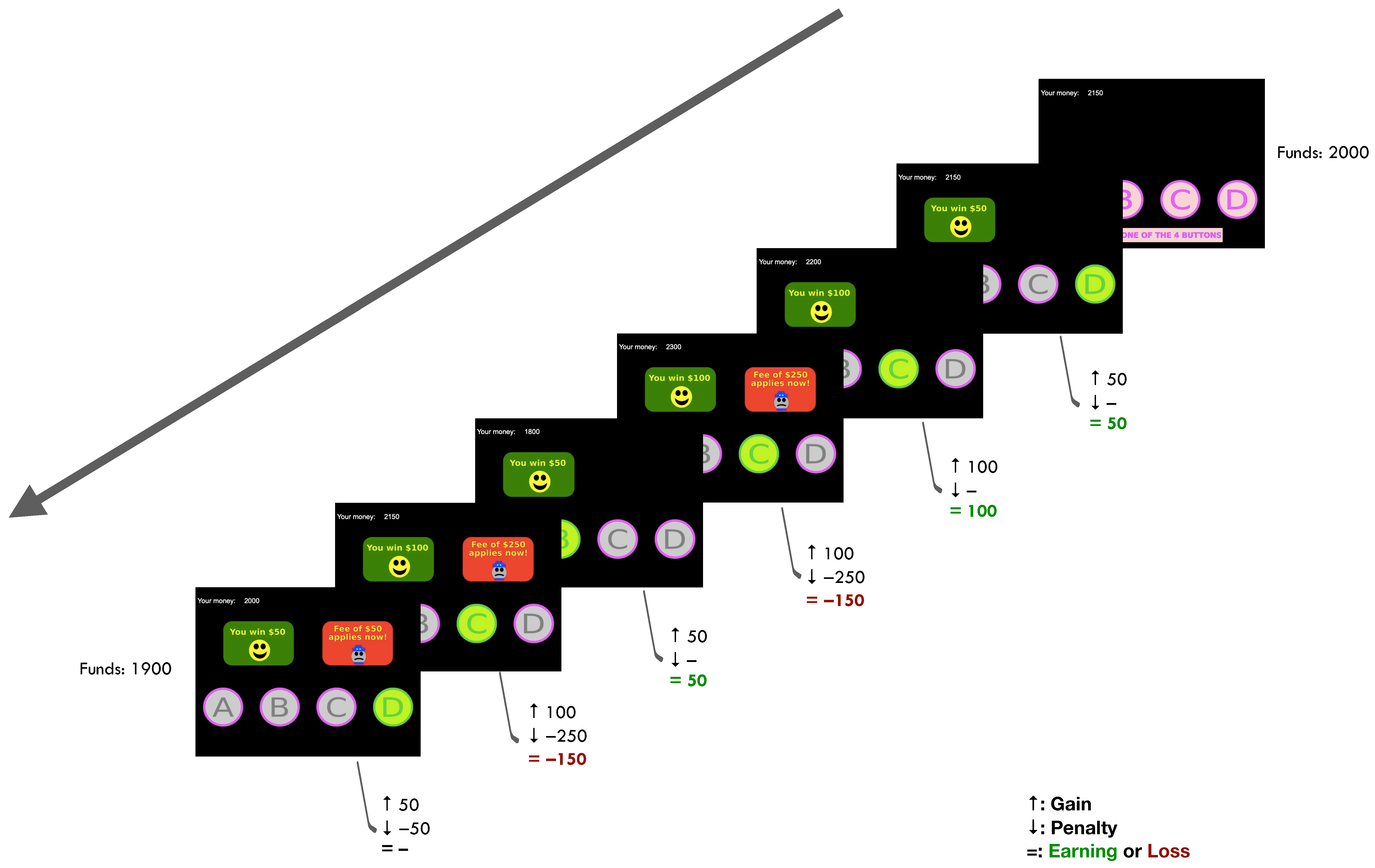
3. Gambling Tasks

3.1. Iowa Gambling Task

In the TPEB study, we will incorporate the IGT, a computerized task widely used in research to assess decision-making and impulsivity [89]. Participants partake in a simulated game of chance, choosing from four decks of cards labeled A, B, C, and D. Each deck has different probabilities of winning or losing fake money. The goal of the participants is to maximize their winnings over multiple trials by selecting cards from the decks [92].
Two decks (e.g., A and B) provide lower immediate rewards but long-term gains, while the other two decks (e.g., C and D) offer high immediate rewards but long-term losses. The participants are then told that there are some decks that are worse than others and that they can win the game if they stay away from the bad decks, regardless of the accrued losses at any given point. They are also informed that the system is not adaptive, meaning that the order of presentations is the same regardless of their responses. After choosing a card, participants receive feedback indicating whether they have won or lost fake money. With repeated trials, participants should learn to favor the advantageous decks (e.g., A and B) and avoid the disadvantageous decks (e.g., C and D) to accumulate the most winnings [89].
The IGT serves as a measure of impulsivity as it requires individuals to balance immediate gratification with long-term consequences [93]. Individuals exhibiting impulsivity tend to prioritize immediate rewards and initially show a preference for the disadvantageous decks (e.g., C and D) [94]. They struggle to adjust their strategy over time and persist in choosing from these disadvantageous decks. In contrast, individuals with better decision-making abilities and lower impulsivity learn to favor the advantageous decks (e.g., A and B) and avoid risky choices [95].
Analyzing participants’ choices and learning patterns in the IGT provides valuable insights into decision-making processes, impulsivity, and risk-taking behavior [96]. The task is widely utilized to investigate decision-making impairments in clinical populations and explore the neural mechanisms underlying impulsive behaviors [96,97,98,99]. The task is depicted in Figure 2.

3.2. Reward Gambling Task

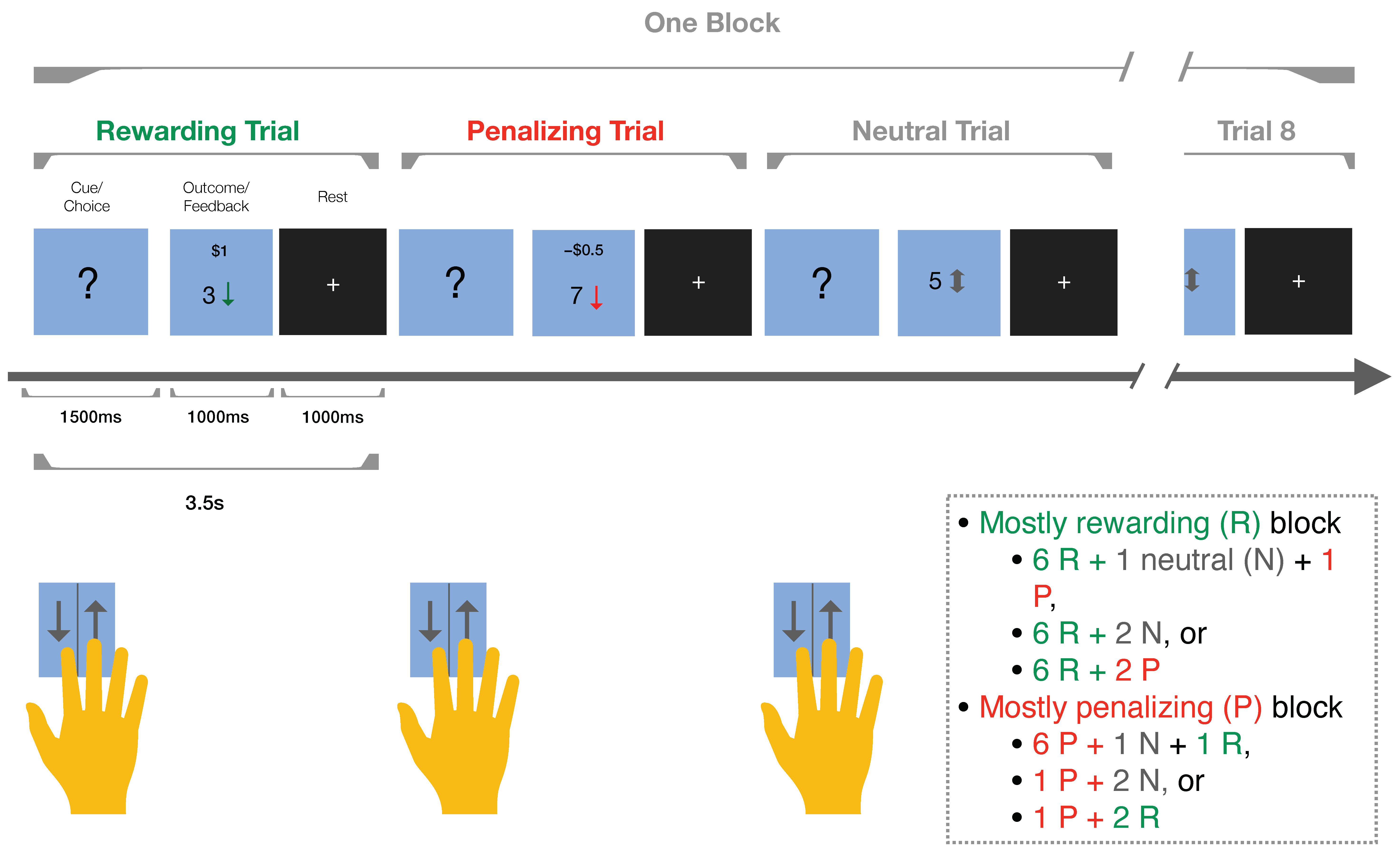
The RGT is specifically designed to probe decision-making and risk-taking propensities in individuals with BD and will also be used in the TPEB study [90]. Like the IGT, the RGT operates through a computerized interface widely adopted in psychological research. In the RGT, participants engage in a cognitive exercise in which they predict the value of a concealed card, represented by a “?”. The goal is to ascertain whether the card’s value is above or below five, either gaining rewards or facing penalties based on their decision. This process is important for understanding decision-making and risk assessment, especially in behavioral disorders such as BD.
Each participant’s decision triggers a feedback mechanism, which is programmed to reflect the nature of the trial, as follows: reward-based, penalizing, or neutral. During the task, participants receive visual feedback through contrasting symbols; rewards are represented by a green upward arrow with “$1”, penalties by a red downward arrow with “−$0.50”, and neutral outcomes by a gray bidirectional arrow with the numeral 5. This process is fundamental for the learning curve [90].
The RGT consists of trial blocks, with each block consisting of either reward or penalty outcomes. This design tests participants’ decision-making strategies and exposes them to varying risk-reward situations. The task consists of two types of blocks—reward-focused and penalty-focused—interspersed with fixation blocks to allow for mental recalibration [90].
The RGT holds significance in cognitive psychology and decision-making research. By systematically presenting stimuli, it measures risk-taking behaviors, impulse control, and adaptive decision-making, offering valuable insights into these cognitive processes. The task’s design and execution provide an understanding of the complexities of cognitive processing in risk and reward situations, particularly in behavioral disorders like BD [90]. The task is represented in Figure 3.

4. Study Design, Schedule, and Assessments

4.1. Stimulation Sessions

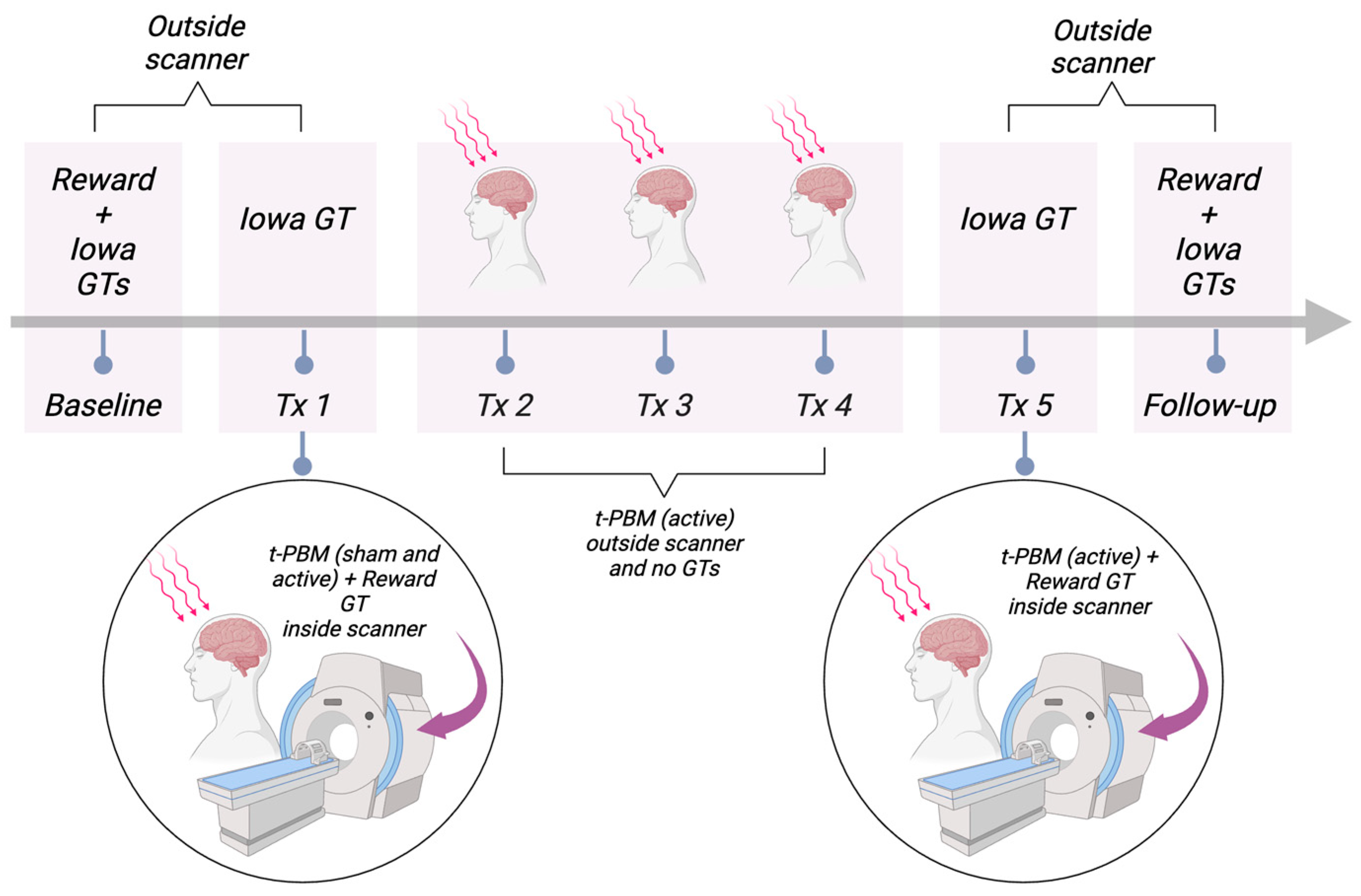
This will be a single-blind study to test the neuropsychological and clinical effects of NIR t-PBM on BD. This study will be conducted at MGH. For our primary aim of examining the effect of t-PBM on CBF, we will incorporate a sham-controlled study design. Twenty patients (n = 20) with a diagnosis of BD, who are not currently in a depressive, manic, or mixed episode, will be included. Each participant will go in the MRI scanner and first undergo one sham stimulation session and then one t-PBM stimulation session. This will occur during the first day of treatment (treatment day 1). Participants will be told that, on this day, they will receive both active and sham stimulation. However, they will be blinded to the order (i.e., immediately before each type of stimulation is delivered, they will be told that the type may be active or sham t-PBM). During each of the four following days, participants will receive one active t-PBM session daily (treatment days 2 to 5). On treatment day 5, participants will receive the t-PBM session in the MRI scanner (Figure 4).

4.2. Outcomes

The fMRI scan will measure BOLD changes during the sham t-PBM session (on treatment day 1 immediately before active stimulation), during the first active t-PBM session (treatment day 1), and during the last active t-PBM session (treatment day 5) on the R-dlPFC. Impulsivity will be assessed using the BIS during screening (for inclusion criteria), baseline, immediately after treatment 5, and during the follow-up visit and using the gambling tasks during baseline, immediately after the first t-PBM session (treatment 1), immediately after the fifth t-PBM session (treatment 5), and during the follow-up visit. Other aspects of impulsivity and executive functioning will be assessed using the I-7 and the BRIEF during baseline, immediately after treatment 5, and during the follow-up visit. The MOODS-SR Last Week will be administered during the baseline, immediately after treatment 5, and during the follow-up visit to assess changes in mood, particularly hypomanic symptoms. During the screening, subjects will complete the MOODS-SR Lifetime. A check-in event will take place via phone or video call on the weekday after the last stimulation visit. This encounter will evaluate suicidality, side effects, and depression symptoms, among others. A follow-up visit will be scheduled within the week after the final treatment (Table 1).

5. Subjects

We intend on enrolling 30 subjects diagnosed with BD, with the aim of randomizing 20 subjects. Inclusion criteria for this study include (1) adults between the ages of 18 and 65; (2) a diagnosis of BD assessed with a Mini International Neuropsychiatric Interview (MINI); (3) currently experiencing symptoms of impulsivity, as measured using a BIS total cut-off score equal to or greater than 70 [100]; and (4) if of child-bearing potential, they must agree to use adequate contraception (e.g., oral contraceptives, intrauterine device, double barrier methods, or total abstinence from intercourse).
Exclusion criteria for this study include (1) currently in a depressive, manic, or mixed episode; (2) currently experiencing psychotic symptoms; (3) judged to be at serious and imminent suicidal risk, as measured by a Columbia Suicide Severity Rating Scale (C-SSRS) score of ≥4; (4) currently meeting the criteria for alcohol or substance use disorder (or within the past three months, as assessed using the MINI diagnostic interview); (5) having unstable medical conditions; (6) being unable to consent or to complete study procedures; (7) failing to meet standard MRI safety requirements (e.g., claustrophobia, non-removable piercings, implanted medical devices, other non-removable metals); (8) having changed medications or having used augmentative devices and other interventions within two weeks prior to this study; (9) currently participating in other clinical research trials that may influence the primary outcomes or adherence to this study; and (10) being currently pregnant.
Recruitment sources will include outpatient clinics at the MGH and McLean Hospital, referrals from physicians, flyers, online advertisements, and platforms, and patient registries from the MGH network. One of these is a digital platform that provides a secure and central location where patients and hospital members coordinate and manage patients’ healthcare. Another is a secure and centralized clinical data registry that collects data from various hospital systems in the MGH network and centralizes them, making clinical information readily accessible to researchers. A third is a free recruitment platform offering a public webpage that allows community members to express their interest in participating in a study, secure tools for managing communications with potential volunteers, and inclusion in recruitment emails that are sent to MGH network employees and subscribed users from the broader public. Lastly, we will utilize an online recruitment platform funded by the National Institutes of Health (NIH) that connects researchers with potential volunteers interested in research study opportunities. We will only enroll English-speaking participants in this study since the measures used are developed and validated in the English language and because non-English-speaking subjects may not be able to understand study procedures, which could undermine the validity of the results.
Interested individuals will be contacted by trained study staff for pre-screening. The trained study staff will review study details and procedures, ask a series of questions, and administer two questionnaires—the Altman Self-Rating Mania Scale (ASRM) and the Patient Health Questionnaire (PHQ-9)—to assess eligibility for this study. Individuals will be asked to verbally authorize the recording of their responses to any data collection. They will have the option to refrain from answering questions if they choose not to do so. Additionally, individuals will be given the opportunity to provide verbal consent for unencrypted email communication. Those meeting the eligibility criteria will be invited to attend a screening visit. In cases in which individuals are ineligible or decide not to enroll, any information collected during the initial phone call will be destroyed to protect privacy. Conversely, if an individual is eligible and decides to enroll in this study, the information collected during the pre-screening phase will be incorporated into their research file.
Initial contact with potential research subjects will collect minimal protected health information (name and email). Participants will be asked to consent to ongoing communication via either unencrypted emails or a secure method. Those who prefer secure communication can notify staff accordingly. If individuals initiate contact via email, it will be understood that they accept email communication. Staff will confirm participants’ comfort with unencrypted emails and obtain consent for further communication.
t-PBM is not considered a standard of care and is, therefore, an alternative treatment for BD. The standard of care for BD includes, but is not limited to, psychotherapy, medications (such as mood stabilizers, antidepressants, or antipsychotics) or device-based treatments (such as ECT) [101]. Information regarding the standard of care for BD will be provided to all subjects. Subjects may continue with their current treatment regimen throughout this study.

6. Transcranial Photobiomodulation Administration

The t-PBM device used in this study is the Litecure LightForce® EXPi Deep Tissue Laser TherapyTM System (Carlsbad, CA, USA) Transcranial PhotoBioModulation-1000 (t-PBM-2.0). The t-PBM-2.0 is an investigational device based on LiteCure’s LightForce® EXPi Deep Tissue Laser TherapyTM System. For the investigational study, the EXPi System’s beam delivery—Empower TM is modified to non-invasively deliver NIR to subjects diagnosed with BD. The modified system is configured to provide sham (placebo) treatment, as well. The device is manufactured and supplied by LiteCure LLC, 101 Lukens Dr., Suite A, New Castle, DE 18720, USA. The treatment parameters outlined in Table 2 will be programmed into the device. The console displays the time and energy delivered and automatically shuts off once the treatment is completed.
t-PBM-2.0 is considered a Class II medical device according to 21 CFR 890.5500 and 878.4810 and is manufactured according to 21 CFR 820. It utilizes a laser diode source with a maximum continuous (CW) output of ≤30 Watts at a wavelength of 808 nm and a nominal beam diameter of 40 mm at the outside aperture.
t-PBM-2.0 consists of a therapeutic laser console, which emits laser energy in the NIR spectrum, and an optical delivery system. This system consists of a flexible, double-sheathed optical fiber connected to a custom helmet (cap). The cap is designed to deliver NIR light to the EEG site F4 (or in close proximity, if obstructed by hair), covering a total treatment area of approximately 24 cm2 [12 cm2 × 2]. It also includes laser safety eyewear with an optical density rating >5.0 at 808 nm.
The therapeutic laser console is the only component of the device that is not MRI-compatible. It will be stored in an equipment room located in Zone 2 (an area with no detectable magnetic field but to which access is restricted due to scanner proximity). An MRI-compatible optical fiber will connect the laser console to the MRI-compatible cap. The safety eyewear is also MRI-compatible. Aside from the cap, the t-PBM-2.0 is the same device as LiteCure’s EXPi System, Model LTS-2500, which is marketed under the FDA’s 510(K) # K107637.
The cap is a custom 3D-printed headgear made of plastic and serves to hold the laser probe in place between the MRI coil and the subject’s head. The cap is provided by the device manufacturer (LiteCure) and is very similar to those used in our previous t-PBM studies. The laser probe, attached to the cap, will be connected to the laser console via a multimode MR-compatible optical fiber. Ceramic ferrules at the distal end of the fiber will be securely attached to the cap using clamps. This cap serves as the sole modification to the device delivery system, required for conducting t-PBM inside the MRI scanner. It is a component of the t-PBM-2.0 device adapted to apply NIR to custom EEG sites.
The t-PBM-2.0 device has been used by our team in two other studies at MGH, as follows: Transcranial Photobiomodulation for Alzheimer’s Disease (NCT04784416) and Transcranial Near Infrared Radiation and Cerebral Blood Flow in Depression (NCT05573074).
The two laser outlets will be placed on the scalp over the right dlPFC. The location will be determined using the scalp heuristics method [102]. We found that on the heads of three volunteers, the right frontopolar region (Fp2) of the 10–20 system was located anteriorly contiguous to the marker of the right anterior dlPFC, as identified with this method.

7. MRI Scanning and Imagining Processing

The MRI images will be collected using a 3T MAGNETOM Prisma® scanner (Malvern, PA, USA) and a Head/Neck 20-channel coil by Siemens Medical Solutions. The imaging protocols include Echo Planar Imaging sequences based on those developed by the Human Connectome Project [103], which have been validated at the Martinos Center for Biomedical Imaging [104,105].
Participants will undergo a series of MRI scans, including conventional high-resolution 3D T1-weighted MPRAGE scans, resting state functional connectivity MRI sequences, and task-based functional connectivity MRI sequences.
We will examine areas associated with impulsivity, mood, and executive function, including, but not limited to, the salience network, executive control network, default mode network, limbic areas, and the dlPFC.
This data will be used to test the effect of a single t-PBM session over the right dlPFC (SA1) on cerebral blood flow (CBF) of subjects with impulsivity due to BD and to test the effect of repeated t-PBM sessions on CBF (SA2).
We will preprocess MRI and fMRI data using the FreeSurfer software package (https://surfer.nmr.mgh.harvard.edu/), scripted in Bash. This will involve slice-time correction, motion correction, and the co-registration of a functional BOLD scan with the participants’ anatomical images. BOLD signals will then be smoothed to a full-width half maximum (FWHM) of 3 mm and z-scored at every voxel.
The t-PBM-related increase in BOLD percent signal change (PSC) averaged across all ROI voxels will be assessed during the first scanning session, in which the participant receives both sham and NIR over the R-dlPFC. The PSC of t-PBM-related increase in BOLD both during and after stimulation will be the dependent variable, with the independent variable being the condition of t-PBM during the first session (sham vs. NIR).
We will assess the BOLD signal during the fifth NIR session as a PSC across all ROI voxels, comparing it with the BOLD PSC from the first NIR session. This will allow us to observe any changes in BOLD after five NIR treatment sessions. The dependent variable will be the BOLD signal, and the independent variable will be the time point, as follows: treatment 1 vs. treatment 5.

8. Statistical Analysis

We determined the required sample size for our proposed TPEB study by referencing two previous studies. The first study, conducted by Mannu et al. (2019) [85], enrolled patients with BD and showed that t-PBM reduced lithium doses from a baseline mean of 938 mg (SD 124) to 763 mg (SD 108) after t-PBM. Given an 80% power and an alpha of 0.05 in our study, a sample size of seven patients (n = 7) would be necessary. The second study, conducted by our group at MGH (NCT04366258), involved patients with MDD receiving various dosages of t-PBM (high, medium, low, and sham). After the medium dose, the mean percentage change in the raw BOLD signal was 100.36 (SD 70.72), while after the sham dose, the mean percentage change in the raw BOLD signal was 1.99 (SD 30.65). Considering 80% power and an alpha of 0.05 in our study, we would also need a sample size of seven patients (n = 7). Our proposed TPEB study aims to randomize 20 patients with BD. With this sample size, we anticipate having sufficient power to detect statistically significant differences.
Aim 1: To test the effect of a single t-PBM session on CBF while irradiating R-dlPFC in subjects with impulsivity due to BD. The dependent variable will be the t-PBM-induced increase in BOLD signal during and after stimulation, measured as PSC at the R-dlPFC. The independent variable will be the t-PBM condition (sham vs. NIR) within the first session. A statistical analysis will be performed using a Wilcoxon signed-rank test (one-tailed, alpha = 0.05).
Aim 2: To test the effect of repeated t-PBM sessions on CBF after irradiating R-dlPFC in subjects with impulsivity due to BD. The dependent variable will be the BOLD signal, measured as PSC at the R-dlPFC. The independent variable will be the time point (baseline vs. treatment 5) after four t-PBM sessions. A statistical analysis will be conducted using a Wilcoxon signed-rank test (one-tailed, alpha = 0.05).
Aim 3: To test the effect of repeated t-PBM sessions on reward processing and impaired decision-making in subjects with impulsivity due to BD. The dependent variables will include the net gain score at IGT, reaction times at RGT, BIS, and Behavior Rating BRIEF-A and I-7 scores. The independent variable will be the time point (baseline, treatment 5, and follow-up). The statistical analysis will involve general mixed model regression, treating time as a fixed effect and subject as a random effect.
Aim 4: To test the effect of repeated t-PBM sessions on manic symptoms in subjects with BD. The dependent variable will be the MOODS-SR score. The independent variable will be the time point (baseline, treatment 5, and follow-up). The statistical analysis will employ general mixed model regression to assess the change in MOODS-SR scores. Dimensional improvements, such as insight, content, and speech (single-item scores), will be analyzed as secondary measures.
By using appropriate statistical tests for each aim, this study aims to provide reliable and valid results regarding the effects of t-PBM on CBF, BOLD signal, decision-making, and manic symptoms among individuals with hypomania. The consideration of effect sizes and statistical power will ensure the ability to detect meaningful differences and contribute to the understanding of t-PBM as a potential therapeutic intervention for bipolar disorder.

9. Demographic Data on Existing Participants

As of 2 March 2024, we have successfully enrolled 11 participants, with four completing this study. Of the subjects enrolled, the median age was 30 years (range: 19–59). Sex at birth was predominantly female (73%), and 27% was male. We had one participant who identified as a transgender man. Regarding race/ethnicity, most participants self-identified as White (73%), with 9% identifying as Black or African American, 9% as Asian, and 9% preferring not to answer. Educational levels were uniformly high, with all participants having completed at least twelve years of education, averaging 15 years (SD: 2). Marital status varied, with a majority never being married (55%), three participants who were married (27%), and two who were living with a partner (18%). For employment status, 46% were employed full-time, 45% were employed part-time, and one participant (9%) was unemployed. In terms of handedness, a significant majority of the subjects were right-handed (82%), and the remainder were left-handed (18%).

10. Discussion

The TPEB study stands at the forefront of exploring novel therapeutic avenues for BD, particularly focusing on deficits in executive function. This exploration is significant given the complex interplay between cognitive functions and mood regulation in BD. By targeting the R-dlPFC, this study is anchored on a key area implicated in executive function and mood regulation. This targeted approach could address the gaps left by conventional treatments, which primarily focus on mood stabilization.
The incorporation of t-PBM using NIR light to stimulate the mitochondrial respiratory chain represents a cutting-edge approach to neuromodulation. This technique’s potential to increase CBF is particularly promising, as it suggests a direct physiological mechanism for enhancing cognitive function. This study’s design, employing both single-session and repeated-session t-PBM interventions, is strategically poised to assess both the immediate and longer-term effects on the R-dlPFC in individuals with BD. In addition, fMRI plays an important role in this process, enabling the visualization and measurement of changes in BOLD signals. This methodology not only allows for real-time monitoring of the effects of t-PBM but also contributes to a deeper understanding of the neural underpinnings of BD.
The TPEB study employs comprehensive assessments of impulsivity, decision-making, and manic symptoms, which will be important in evaluating the multifaceted impact of t-PBM. These assessments will provide a holistic view of how t-PBM influences various cognitive and mood aspects of BD. t-PBM has the potential to improve decision-making, reduce impulsivity, and alleviate manic symptoms. If successful, t-PBM could represent a paradigm shift in the management of BD, offering a noninvasive, well-tolerated, and potentially more effective treatment option for addressing the cognitive symptoms of this disorder.
We believe this study has the potential to deliver meaningful findings. Nevertheless, we identify some limitations. First, the sample size of this study is relatively small, which may hinder its external validity. The sample size also limits our ability to randomize the order of the type of stimulation that participants will receive on their first treatment session, which may introduce a systematic error. We attempt to mitigate this by blinding participants to the type of stimulation during the first session. We will also discuss the impact of this measure after analyzing the study results. Additionally, our inclusion criteria allow for enrolling patients with some comorbidities up to three months before their enrollment, such as substance use. We made this decision based on the high prevalence of this disorder in the BD population, which could severely limit our ability to recruit a realistic sample. While this may hamper a mechanistic analysis and might affect treatment responses, it more accurately reflects the general population with this disorder. Future studies should address these limitations by using larger sample sizes that allow for stricter inclusion criteria and a randomized order of stimulation.
Despite these limitations, the findings from the TPEB study have promising far-reaching implications beyond BD. By elucidating the neural and cognitive mechanisms of t-PBM, this research could pave the way for novel neuromodulation strategies applicable to a range of psychiatric and neurological conditions characterized by executive function impairments. This opens avenues for several research opportunities, potentially revolutionizing our approach to treating various mental health disorders.

11. Conclusions

The TPEB study shows a promising approach to addressing executive function impairments in individuals with BD through t-PBM. By targeting the R-dlPFC with NIR light, this study aims to improve decision-making, reduce impulsivity, and enhance reward responsiveness. If successful, this noninvasive intervention could offer a well-tolerated, at-home treatment option, potentially transforming the management of cognitive symptoms in BD.

Author Contributions

Conceptualization, D.R.A.C., A.M.H.P. and P.C.; methodology, A.M.H.P., M.B.G., K.M.M. and J.A.C.; investigation, D.R.A.C., A.M.H.P., W.F.V., C.A.L., M.H.S., M.B.G., F.V., K.M.M., J.A.C. and P.C.; resources, P.C.; writing—original draft preparation, D.R.A.C., A.M.H.P., M.B.G., F.V., K.M.M., J.A.C. and P.C.; writing—review and editing, D.R.A.C., A.M.H.P., W.F.V., C.A.L., M.H.S., M.B.G., F.V., K.M.M., J.A.C. and P.C.; supervision, P.C.; project administration, P.C.; funding acquisition, P.C. All authors have read and agreed to the published version of the manuscript.

Funding

This study is financed through the Milken Institute; Bessemer Giving Fund/Baszucki Brain Research Fund (grant 2021A009969).

Institutional Review Board Statement

The study protocol was approved by the MGH Institutional Review Board (IRB protocol No. 2022P000289).

Informed Consent Statement

This report outlines a forthcoming research study. All participants involved will furnish IRB-approved, informed consent.

Data Availability Statement

The data generated in this study will be uploaded to the NIH data archive and will be available for sharing with interested investigators, in agreement with NIH policy.

Conflicts of Interest

Cassano consulted for Janssen Research and Development and Niraxx Light Therapeutics Inc.; was funded by PhotoThera Inc., LiteCure LLC, and Cerebral Sciences Inc. to conduct studies on transcranial photobiomodulation; is a shareholder of Niraxx Inc.; and has filed several patents related to the use of NIR light in psychiatry. The other authors have nothing to disclose.

References

  1. GBD 2017 Disease and Injury Incidence and Prevalence Collaborators. Global, regional, and national incidence, prevalence, and years lived with disability for 354 diseases and injuries for 195 countries and territories, 1990–2017: A systematic analysis for the Global Burden of Disease Study 2017. Lancet 2018, 392, 1789–1858. [Google Scholar] [CrossRef]
  2. Merikangas, K.R.; Jin, R.; He, J.-P.; Kessler, R.C.; Lee, S.; Sampson, N.A.; Viana, M.C.; Andrade, L.H.; Hu, C.; Karam, E.G.; et al. Prevalence and correlates of bipolar spectrum disorder in the world mental health survey initiative. Arch. Gen. Psychiatry 2011, 68, 241–251. [Google Scholar] [CrossRef] [PubMed]
  3. Nowrouzi, B.; McIntyre, R.S.; MacQueen, G.; Kennedy, S.H.; Kennedy, J.L.; Ravindran, A.; Yatham, L.; De Luca, V. Admixture analysis of age at onset in first episode bipolar disorder. J. Affect. Disord. 2016, 201, 88–94. [Google Scholar] [CrossRef] [PubMed]
  4. Moreno, C.; Laje, G.; Blanco, C.; Jiang, H.; Schmidt, A.B.; Olfson, M. National trends in the outpatient diagnosis and treatment of bipolar disorder in youth. Arch. Gen. Psychiatry 2007, 64, 1032–1039. [Google Scholar] [CrossRef] [PubMed]
  5. Rowland, T.A.; Marwaha, S. Epidemiology and risk factors for bipolar disorder. Ther. Adv. Psychopharmacol. 2018, 8, 251. [Google Scholar] [CrossRef] [PubMed]
  6. Kroon, J.S.; Wohlfarth, T.D.; Dieleman, J.; Sutterland, A.L.; Storosum, J.G.; Denys, D.; de Haan, L.; Sturkenboom, M.C. Incidence rates and risk factors of bipolar disorder in the general population: A population-based cohort study. Bipolar Disord. 2013, 15, 306–313. [Google Scholar] [CrossRef] [PubMed]
  7. Alonso, J.; Petukhova, M.; Vilagut, G.; Chatterji, S.; Heeringa, S.; Üstün, T.B.; Alhamzawi, A.O.; Viana, M.C.; Angermeyer, M.; Bromet, E.; et al. Days out of role due to common physical and mental conditions: Results from the WHO World Mental Health surveys. Mol. Psychiatry 2011, 16, 1234–1246. [Google Scholar] [CrossRef] [PubMed]
  8. Dome, P.; Rihmer, Z.; Gonda, X. Suicide Risk in Bipolar Disorder: A Brief Review. Medicina 2019, 55, 403. [Google Scholar] [CrossRef]
  9. Strakowski, S.M.; Fleck, D.E.; DelBello, M.P.; Adler, C.M.; Shear, P.K.; Kotwal, R.; Arndt, S. Impulsivity across the course of bipolar disorder. Bipolar Disord. 2010, 12, 285–297. [Google Scholar] [CrossRef]
  10. Newman, A.L.; Meyer, T.D. Impulsivity: Present during euthymia in bipolar disorder?—A systematic review. Int. J. Bipolar Disord. 2014, 2, 2. [Google Scholar] [CrossRef] [PubMed]
  11. Sylvia, L.G.; Montana, R.E.; Deckersbach, T.; Thase, M.E.; Tohen, M.; Reilly-Harrington, N.; McInnis, M.G.; Kocsis, J.H.; Bowden, C.; Calabrese, J.; et al. Poor quality of life and functioning in bipolar disorder. Int. J. Bipolar Disord. 2017, 5, 10. [Google Scholar] [CrossRef] [PubMed]
  12. Perugi, G.; Medda, P.; Toni, C.; Mariani, M.G.; Socci, C.; Mauri, M. The Role of Electroconvulsive Therapy (ECT) in Bipolar Disorder: Effectiveness in 522 Patients with Bipolar Depression, Mixed-state, Mania and Catatonic Features. Curr. Neuropharmacol. 2017, 15, 359–371. [Google Scholar] [CrossRef] [PubMed]
  13. Barton, B.B.; Segger, F.; Fischer, K.; Obermeier, M.; Musil, R. Update on weight-gain caused by antipsychotics: A systematic review and meta-analysis. Expert. Opin. Drug Saf. 2020, 19, 295–314. [Google Scholar] [CrossRef] [PubMed]
  14. Yee, C.S.; Hawken, E.R.; Baldessarini, R.J.; Vázquez, G.H. Maintenance Pharmacological Treatment of Juvenile Bipolar Disorder: Review and Meta-Analyses. Int. J. Neuropsychopharmacol. 2019, 22, 531–540. [Google Scholar] [CrossRef] [PubMed]
  15. García-Blanco, A.; García-Portilla, M.P.; de la Fuente-Tomás, L.; Batalla, M.; Sánchez-Autet, M.; Arranz, B.; Safont, G.; Arqués, S.; Livianos, L.; Sierra, P. Sexual Dysfunction and Mood Stabilizers in Long-Term Stable Patients with Bipolar Disorder. J. Sex. Med. 2020, 17, 930–940. [Google Scholar] [CrossRef] [PubMed]
  16. Bahji, A.; Ermacora, D.; Stephenson, C.; Hawken, E.R.; Vazquez, G. Comparative efficacy and tolerability of pharmacological treatments for the treatment of acute bipolar depression: A systematic review and network meta-analysis. J. Affect. Disord. 2020, 269, 154–184. [Google Scholar] [CrossRef]
  17. Parker, G.; Ricciardi, T.; Tavella, G.; Hadzi-Pavlovic, D. A Single-Blind Randomized Comparison of Lithium and Lamotrigine as Maintenance Treatments for Managing Bipolar II Disorder. J. Clin. Psychopharmacol. 2021, 41, 381–388. [Google Scholar] [CrossRef]
  18. Sajatovic, M.; Valenstein, M.; Blow, F.; Ganoczy, D.; Ignacio, R. Treatment adherence with lithium and anticonvulsant medications among patients with bipolar disorder. Psychiatr. Serv. 2007, 58, 855–863. [Google Scholar] [CrossRef] [PubMed]
  19. Jawad, I.; Watson, S.; Haddad, P.M.; Talbot, P.S.; McAllister-Williams, R.H. Medication nonadherence in bipolar disorder: A narrative review. Ther. Adv. Psychopharmacol. 2018, 8, 349–363. [Google Scholar] [CrossRef] [PubMed]
  20. Semahegn, A.; Torpey, K.; Manu, A.; Assefa, N.; Tesfaye, G.; Ankomah, A. Psychotropic medication non-adherence and its associated factors among patients with major psychiatric disorders: A systematic review and meta-analysis. Syst. Rev. 2020, 9, 17. [Google Scholar] [CrossRef] [PubMed]
  21. Lintunen, J.; Lähteenvuo, M.; Tanskanen, A.; Tiihonen, J.; Taipale, H. Non-adherence to mood stabilizers and antipsychotics among persons with bipolar disorder—A nationwide cohort study. J. Affect. Disord. 2023, 333, 403–408. [Google Scholar] [CrossRef]
  22. Abdullah-Koolmees, H.; Nawzad, S.; Egberts, T.C.G.; Vuyk, J.; Gardarsdottir, H.; Heerdink, E.R. The effect of non-adherence to antipsychotic treatment on rehospitalization in patients with psychotic disorders. Ther. Adv. Psychopharmacol. 2021, 11, 20451253211027449. [Google Scholar] [CrossRef]
  23. Prajapati, A.R.; Dima, A.L.; Clark, A.B.; Gant, C.; Gibbons, C.; Gorrod, R.; Mosa, G.; Scott, S.; Song, F.; Teague, B.; et al. Mapping of modifiable barriers and facilitators of medication adherence in bipolar disorder to the Theoretical Domains Framework: A systematic review protocol. BMJ Open 2019, 9, e026980. [Google Scholar] [CrossRef] [PubMed]
  24. Yerevanian, B.I.; Koek, R.J.; Mintz, J. Bipolar pharmacotherapy and suicidal behavior. Part I: Lithium, divalproex and carbamazepine. J. Affect. Disord. 2007, 103, 5–11. [Google Scholar] [CrossRef]
  25. Lew, K.H.; Chang, E.Y.; Rajagopalan, K.; Knoth, R.L. The effect of medication adherence on health care utilization in bipolar disorder. Manag. Care Interface 2006, 19, 41–46. [Google Scholar] [PubMed]
  26. Powers, R.L.; Russo, M.; Mahon, K.; Brand, J.; Braga, R.J.; Malhotra, A.K.; Burdick, K.E. Impulsivity in bipolar disorder: Relationships with neurocognitive dysfunction and substance use history. Bipolar Disord. 2013, 15, 876–884. [Google Scholar] [CrossRef]
  27. Madero, S.; Anmella, G.; Sagué-Vilavella, M.; Pons, M.T.; Giménez, A.; Murru, A.; Gómez-Ramiro, M.; Gil-Badenes, J.; Rios, J.; Bioque, M.; et al. Evaluating maintenance electroconvulsive therapy in Bipolar Disorders: 3-year mirror-image study. J. Affect. Disord. 2022, 298, 58–64. [Google Scholar] [CrossRef]
  28. Mutz, J. Brain stimulation treatment for bipolar disorder. Bipolar Disord. 2023, 25, 9–24. [Google Scholar] [CrossRef] [PubMed]
  29. Agbese, E.; Leslie, D.L.; Ba, D.M.; Rosenheck, R. Does Electroconvulsive Therapy for Patients with Mood Disorders Extend Hospital Length of Stays and Increase Inpatient Costs? Adm. Policy Ment. Health 2022, 49, 71–78. [Google Scholar] [CrossRef]
  30. Paris, J. Why electroconvulsive therapy still carries a stigma. Br. J. Psychiatry 2022, 220, 113–114. [Google Scholar] [CrossRef]
  31. Bahji, A. The Rise, Fall, and Resurgence of Electroconvulsive Therapy. J. Psychiatr. Pract. 2022, 28, 440–444. [Google Scholar] [CrossRef] [PubMed]
  32. Mutz, J.; Edgcumbe, D.R.; Brunoni, A.R.; Fu, C.H.Y. Efficacy and acceptability of non-invasive brain stimulation for the treatment of adult unipolar and bipolar depression: A systematic review and meta-analysis of randomised sham-controlled trials. Neurosci. Biobehav. Rev. 2018, 92, 291–303. [Google Scholar] [CrossRef] [PubMed]
  33. Piccoli, E.; Cerioli, M.; Castiglioni, M.; Larini, L.; Scarpa, C.; Dell’Osso, B. Recent innovations in non-invasive brain stimulation (NIBS) for the treatment of unipolar and bipolar depression: A narrative review. Int. Rev. Psychiatry 2022, 34, 715–726. [Google Scholar] [CrossRef] [PubMed]
  34. Johnson, S.L.; Carver, C.S.; Mulé, S.; Joormann, J. Impulsivity and risk for mania: Towards greater specificity. Psychol. Psychother. 2013, 86, 401–412. [Google Scholar] [CrossRef] [PubMed]
  35. Burdick, K.E.; Millett, C.E.; Yocum, A.K.; Altimus, C.M.; Andreassen, O.A.; Aubin, V.; Belzeaux, R.; Berk, M.; Biernacka, J.M.; Blumberg, H.P.; et al. Predictors of functional impairment in bipolar disorder: Results from 13 cohorts from seven countries by the global bipolar cohort collaborative. Bipolar Disord. 2022, 24, 709–719. [Google Scholar] [CrossRef] [PubMed]
  36. Chan, C.C.; Alter, S.; Hazlett, E.A.; Shafritz, K.M.; Yehuda, R.; Goodman, M.; Haznedar, M.M.; Szeszko, P.R. Neural correlates of impulsivity in bipolar disorder: A systematic review and clinical implications. Neurosci. Biobehav. Rev. 2023, 147, 105109. [Google Scholar] [CrossRef] [PubMed]
  37. Santana, R.P.; Kerr-Gaffney, J.; Ancane, A.; Young, A.H. Impulsivity in Bipolar Disorder: State or Trait? Brain Sci. 2022, 12, 1351. [Google Scholar] [CrossRef] [PubMed]
  38. Nusslock, R.; Almeida, J.R.; Forbes, E.E.; Versace, A.; Frank, E.; LaBarbara, E.J.; Klein, C.R.; Phillips, M.L. Waiting to win: Elevated striatal and orbitofrontal cortical activity during reward anticipation in euthymic bipolar disorder adults. Bipolar Disord. 2012, 14, 249–260. [Google Scholar] [CrossRef] [PubMed]
  39. O’Sullivan, N.; Szczepanowski, R.; El-Deredy, W.; Mason, L.; Bentall, R.P. fMRI evidence of a relationship between hypomania and both increased goal-sensitivity and positive outcome-expectancy bias. Neuropsychologia 2011, 49, 2825–2835. [Google Scholar] [CrossRef] [PubMed]
  40. Cho, S.S.; Koshimori, Y.; Aminian, K.; Obeso, I.; Rusjan, P.; Lang, A.E.; Daskalakis, Z.J.; Houle, S.; Strafella, A.P. Investing in the Future: Stimulation of the Medial Prefrontal Cortex Reduces Discounting of Delayed Rewards. Neuropsychopharmacology 2015, 40, 546–553. [Google Scholar] [CrossRef] [PubMed]
  41. Brevet-Aeby, C.; Brunelin, J.; Iceta, S.; Padovan, C.; Poulet, E. Prefrontal cortex and impulsivity: Interest of noninvasive brain stimulation. Neurosci. Biobehav. Rev. 2016, 71, 112–134. [Google Scholar] [CrossRef]
  42. Hsu, T.-Y.; Tseng, L.-Y.; Yu, J.-X.; Kuo, W.-J.; Hung, D.L.; Tzeng, O.J.; Walsh, V.; Muggleton, N.G.; Juan, C.-H. Modulating inhibitory control with direct current stimulation of the superior medial frontal cortex. Neuroimage 2011, 56, 2249–2257. [Google Scholar] [CrossRef] [PubMed]
  43. Meinzer, M.; Lindenberg, R.; Antonenko, D.; Flaisch, T.; Flöel, A. Anodal transcranial direct current stimulation temporarily reverses age-associated cognitive decline and functional brain activity changes. J. Neurosci. 2013, 33, 12470–12478. [Google Scholar] [CrossRef] [PubMed]
  44. Rossi, S.; Hallett, M.; Rossini, P.M.; Pascual-Leone, A.; Safety of TMS Consensus Group. Safety, ethical considerations, and application guidelines for the use of transcranial magnetic stimulation in clinical practice and research. Clin. Neurophysiol. 2009, 120, 2008–2039. [Google Scholar] [CrossRef]
  45. Pohar, R.; Farrah, K. Repetitive Transcranial Magnetic Stimulation for Patients with Depression: A Review of Clinical Effectiveness, Cost-Effectiveness and Guidelines—An Update; CADTH Rapid Response Reports; Canadian Agency for Drugs and Technologies in Health: Ottawa, ON, Canada, 2019. Available online: http://www.ncbi.nlm.nih.gov/books/NBK545105/ (accessed on 28 January 2024).
  46. Sauvaget, A.; Tostivint, A.; Etcheverrigaray, F.; Pichot, A.; Dert, C.; Schirr-Bonnais, S.; Clouet, J.; Sellal, O.; Mauduit, N.; Leux, C.; et al. Hospital production cost of transcranial direct current stimulation (tDCS) in the treatment of depression. Neurophysiol. Clin. 2019, 49, 11–18. [Google Scholar] [CrossRef]
  47. Hamblin, M.R. Shining light on the head: Photobiomodulation for brain disorders. BBA Clin. 2016, 6, 113–124. [Google Scholar] [CrossRef]
  48. Gavish, L.; Houreld, N.N. Therapeutic Efficacy of Home-Use Photobiomodulation Devices: A Systematic Literature Review. Photobiomodulation. Photomed. Laser Surg. 2019, 37, 4–16. [Google Scholar] [CrossRef]
  49. Cassano, P.; Caldieraro, M.A.; Norton, R.; Mischoulon, D.; Trinh, N.H.; Nyer, M.; Dording, C.; Hamblin, M.R.; Campbell, B.; Iosifescu, D.V. Reported Side Effects, Weight and Blood Pressure, After Repeated Sessions of Transcranial Photobiomodulation. Photobiomodul. Photomed. Laser Surg. 2019, 37, 651–656. [Google Scholar] [CrossRef]
  50. Cassano, P.; Norton, R.; Caldieraro, M.A.; Vahedifard, F.; Vizcaino, F.; McEachern, K.M.; Iosifescu, D. Tolerability and Safety of Transcranial Photobiomodulation for Mood and Anxiety Disorders. Photonics 2022, 9, 507. [Google Scholar] [CrossRef]
  51. Konstantinović, L.M.; Jelić, M.B.; Jeremić, A.; Stevanović, V.B.; Milanović, S.D.; Filipović, S.R. Transcranial application of near-infrared low-level laser can modulate cortical excitability. Lasers Surg. Med. 2013, 45, 648–653. [Google Scholar] [CrossRef] [PubMed]
  52. Chaieb, L.; Antal, A.; Masurat, F.; Paulus, W. Neuroplastic effects of transcranial near-infrared stimulation (tNIRS) on the motor cortex. Front. Behav. Neurosci. 2015, 9, 147. [Google Scholar] [CrossRef] [PubMed]
  53. Henderson, T.A.; Morries, L.D. SPECT Perfusion Imaging Demonstrates Improvement of Traumatic Brain Injury with Transcranial Near-infrared Laser Phototherapy. Adv. Mind Body Med. 2015, 29, 27–33. [Google Scholar] [PubMed]
  54. Nawashiro, H.; Wada, K.; Nakai, K.; Sato, S. Focal increase in cerebral blood flow after treatment with near-infrared light to the forehead in a patient in a persistent vegetative state. Photomed. Laser Surg. 2012, 30, 231–233. [Google Scholar] [CrossRef] [PubMed]
  55. Salgado, A.S.I.; Zângaro, R.A.; Parreira, R.B.; Kerppers, I.I. The effects of transcranial LED therapy (TCLT) on cerebral blood flow in the elderly women. Lasers Med. Sci. 2015, 30, 339–346. [Google Scholar] [CrossRef] [PubMed]
  56. Tian, F.; Hase, S.N.; Gonzalez-Lima, F.; Liu, H. Transcranial laser stimulation improves human cerebral oxygenation. Lasers Surg. Med. 2016, 48, 343–349. [Google Scholar] [CrossRef] [PubMed]
  57. Hacke, W.; Schellinger, P.D.; Albers, G.W.; Bornstein, N.M.; Dahlof, B.L.; Fulton, R.; Kasner, S.E.; Shuaib, A.; Richieri, S.P.; Dilly, S.G.; et al. Transcranial laser therapy in acute stroke treatment: Results of neurothera effectiveness and safety trial 3, a phase III clinical end point device trial. Stroke 2014, 45, 3187–3193. [Google Scholar] [CrossRef] [PubMed]
  58. Lampl, Y.; Zivin, J.A.; Fisher, M.; Lew, R.; Welin, L.; Dahlof, B.; Borenstein, P.; Andersson, B.; Perez, J.; Caparo, C.; et al. Infrared laser therapy for ischemic stroke: A new treatment strategy: Results of the NeuroThera Effectiveness and Safety Trial-1 (NEST-1). Stroke 2007, 38, 1843–1849. [Google Scholar] [CrossRef] [PubMed]
  59. Montazeri, K.; Farhadi, M.; Fekrazad, R.; Akbarnejad, Z.; Chaibakhsh, S.; Mahmoudian, S. Transcranial photobiomodulation in the management of brain disorders. J. Photochem. Photobiol. B Biol. 2021, 221, 112207. [Google Scholar] [CrossRef] [PubMed]
  60. Barrett, D.W.; Gonzalez-Lima, F. Transcranial infrared laser stimulation produces beneficial cognitive and emotional effects in humans. Neuroscience 2013, 230, 13–23. [Google Scholar] [CrossRef] [PubMed]
  61. Blanco, N.J.; Saucedo, C.L.; Gonzalez-Lima, F. Transcranial infrared laser stimulation improves rule-based, but not information-integration, category learning in humans. Neurobiol. Learn. Mem. 2017, 139, 69–75. [Google Scholar] [CrossRef] [PubMed]
  62. Hwang, J.; Castelli, D.M.; Gonzalez-Lima, F. Cognitive enhancement by transcranial laser stimulation and acute aerobic exercise. Lasers Med. Sci. 2016, 31, 1151–1160. [Google Scholar] [CrossRef] [PubMed]
  63. Morries, L.D.; Cassano, P.; Henderson, T.A. Treatments for traumatic brain injury with emphasis on transcranial near-infrared laser phototherapy. Neuropsychiatr. Dis. Treat. 2015, 11, 2159–2175. [Google Scholar] [PubMed]
  64. Naeser, M.A.; Saltmarche, A.; Krengel, M.H.; Hamblin, M.R.; Knight, J.A. Improved cognitive function after transcranial, light-emitting diode treatments in chronic, traumatic brain injury: Two case reports. Photomed. Laser Surg. 2011, 29, 351–358. [Google Scholar] [CrossRef]
  65. Naeser, M.A.; Zafonte, R.; Krengel, M.H.; Martin, P.I.; Frazier, J.; Hamblin, M.R.; Knight, J.A.; Meehan, W.P.; Baker, E.H. Significant improvements in cognitive performance post-transcranial, red/near-infrared light-emitting diode treatments in chronic, mild traumatic brain injury: Open-protocol study. J. Neurotrauma 2014, 31, 1008–1017. [Google Scholar] [CrossRef] [PubMed]
  66. Saltmarche, A.E.; Naeser, M.A.; Ho, K.F.; Hamblin, M.R.; Lim, L. Significant Improvement in Cognition in Mild to Moderately Severe Dementia Cases Treated with Transcranial Plus Intranasal Photobiomodulation: Case Series Report. Photomed. Laser Surg. 2017, 35, 432–441. [Google Scholar] [CrossRef] [PubMed]
  67. Disner, S.G.; Beevers, C.G.; Gonzalez-Lima, F. Transcranial Laser Stimulation as Neuroenhancement for Attention Bias Modification in Adults with Elevated Depression Symptoms. Brain Stimul. 2016, 9, 780–787. [Google Scholar] [CrossRef] [PubMed]
  68. Coelho, D.R.A.; Gersten, M.; Jimenez, A.S.; Fregni, F.; Cassano, P.; Vieira, W.F. Treating neuropathic pain and comorbid affective disorders: Preclinical and clinical evidence. Pain. Pract. 2024, 00, 1–19. [Google Scholar] [CrossRef] [PubMed]
  69. Coelho, D.R.A.; Salvi, J.D.; Vieira, W.F.; Cassano, P. Inflammation in obsessive-compulsive disorder: A literature review and hypothesis-based potential of transcranial photobiomodulation. J. Neurosci. Res. 2024, 102, e25317. [Google Scholar] [CrossRef] [PubMed]
  70. Cassano, P.; Petrie, S.R.; Mischoulon, D.; Cusin, C.; Katnani, H.; Yeung, A.; De Taboada, L.; Archibald, A.; Bui, E.; Baer, L.; et al. Transcranial Photobiomodulation for the Treatment of Major Depressive Disorder. The ELATED-2 Pilot Trial. Photomed. Laser Surg. 2018, 36, 634–646. [Google Scholar] [CrossRef] [PubMed]
  71. Fradkin, Y.; De Taboada, L.; Naeser, M.; Saltmarche, A.; Snyder, W.; Steingold, E. Transcranial photobiomodulation in children aged 2–6 years: A randomized sham-controlled clinical trial assessing safety, efficacy, and impact on autism spectrum disorder symptoms and brain electrophysiology. Front. Neurol. 2024, 15, 1221193. [Google Scholar] [CrossRef] [PubMed]
  72. Schiffer, F.; Khan, A.; Bolger, E.; Flynn, E.; Seltzer, W.P.; Teicher, M.H. An Effective and Safe Novel Treatment of Opioid Use Disorder: Unilateral Transcranial Photobiomodulation. Front. Psychiatry 2021, 12, 713686. [Google Scholar] [CrossRef] [PubMed]
  73. Qu, X.; Li, L.; Zhou, X.; Dong, Q.; Liu, H.; Liu, H.; Yang, Q.; Han, Y.; Niu, H. Repeated transcranial photobiomodulation improves working memory of healthy older adults: Behavioral outcomes of poststimulation including a three-week follow-up. Neurophotonics 2022, 9, 035005. [Google Scholar] [CrossRef] [PubMed]
  74. O’Donnell, C.M.; Barrett, D.W.; Fink, L.H.; Garcia-Pittman, E.C.; Gonzalez-Lima, F. Transcranial Infrared Laser Stimulation Improves Cognition in Older Bipolar Patients: Proof of Concept Study. J. Geriatr. Psychiatry Neurol. 2022, 35, 321–332. [Google Scholar] [CrossRef] [PubMed]
  75. Uozumi, Y.; Nawashiro, H.; Sato, S.; Kawauchi, S.; Shima, K.; Kikuchi, M. Targeted increase in cerebral blood flow by transcranial near-infrared laser irradiation. Lasers Surg. Med. 2010, 42, 566–576. [Google Scholar] [CrossRef] [PubMed]
  76. Hipskind, S.G.; Grover, F.L., Jr.; Fort, T.R.; Helffenstein, D.; Burke, T.J.; Quint, S.A.; Bussiere, G.; Stone, M.; Hurtado, T. Pulsed Transcranial Red/Near-Infrared Light Therapy Using Light-Emitting Diodes Improves Cerebral Blood Flow and Cognitive Function in Veterans with Chronic Traumatic Brain Injury: A Case Series. Photomed. Laser Surg. 2018, 37, 77–84. [Google Scholar] [CrossRef] [PubMed]
  77. Urquhart, E.L.; Wanniarachchi, H.; Wang, X.; Gonzalez-Lima, F.; Alexandrakis, G.; Liu, H. Transcranial photobiomodulation-induced changes in human brain functional connectivity and network metrics mapped by whole-head functional near-infrared spectroscopy in vivo. Biomed. Opt. Express 2020, 11, 5783–5799. [Google Scholar] [CrossRef] [PubMed]
  78. Truong, N.C.D.; Wang, X.; Wanniarachchi, H.; Liu, H. Enhancement of Frequency-Specific Hemodynamic Power and Functional Connectivity by Transcranial Photobiomodulation in Healthy Humans. Front. Neurosci. 2022, 16, 896502. [Google Scholar] [CrossRef] [PubMed]
  79. Jog, M.; Jann, K.; Yan, L.; Huang, Y.; Parra, L.; Narr, K.; Bikson, M.; Wang, D.J.J. Concurrent Imaging of Markers of Current Flow and Neurophysiological Changes During tDCS. Front. Neurosci. 2020, 14, 374. [Google Scholar] [CrossRef] [PubMed]
  80. Jog, M.S.; Kim, E.; Anderson, C.; Kubicki, A.; Kayathi, R.; Jann, K.; Yan, L.; Leaver, A.; Hellemann, G.; Iacoboni, M.; et al. In-vivo imaging of targeting and modulation of depression-relevant circuitry by transcranial direct current stimulation: A randomized clinical trial. Transl. Psychiatry 2021, 11, 138. [Google Scholar] [CrossRef] [PubMed]
  81. Vargas, E.; Barrett, D.W.; Saucedo, C.L.; Huang, L.-D.; Abraham, J.A.; Tanaka, H.; Haley, A.P.; Gonzalez-Lima, F. Beneficial neurocognitive effects of transcranial laser in older adults. Lasers Med. Sci. 2017, 32, 1153–1162. [Google Scholar] [CrossRef] [PubMed]
  82. Dmochowski, G.M.; Dmochowski, J.P. Increased Blood Flow and Oxidative Metabolism in the Human Brain by Transcranial Laser Stimulation. bioRxiv 2018. Available online: https://www.biorxiv.org/content/10.1101/459883v1 (accessed on 11 March 2024). [CrossRef]
  83. Lebon, V.; Carlier, P.G.; Brillault-Salvat, C.; Leroy-Willig, A. Simultaneous measurement of perfusion and oxygenation changes using a multiple gradient-echo sequence: Application to human muscle study. Magn. Reson. Imaging 1998, 16, 721–729. [Google Scholar] [CrossRef] [PubMed]
  84. Posse, S.; Wiese, S.; Gembris, D.; Mathiak, K.; Kessler, C.; Grosse-Ruyken, M.-L.; Elghahwagi, B.; Richards, T.; Dager, S.R.; Kiselev, V.G. Enhancement of BOLD-contrast sensitivity by single-shot multi-echo functional MR imaging. Magn. Reson. Med. 1999, 42, 87–97. [Google Scholar] [CrossRef]
  85. Mannu, P.; Saccaro, L.F.; Spera, V.; Cassano, P. Transcranial Photobiomodulation to Augment Lithium in Bipolar-I Disorder. Photobiomodul Photomed. Laser Surg. 2019, 37, 577–578. [Google Scholar] [CrossRef] [PubMed]
  86. Sanches, M.; Bauer, I.E.; Galvez, J.F.; Zunta-Soares, G.B.; Soares, J.C. The management of cognitive impairment in bipolar disorder: Current status and perspectives. Am. J. Ther. 2015, 22, 477–486. [Google Scholar] [CrossRef] [PubMed]
  87. Konstantinou, G.; Hui, J.; Ortiz, A.; Kaster, T.S.; Downar, J.; Blumberger, D.M.; Daskalakis, Z.J. Repetitive transcranial magnetic stimulation (rTMS) in bipolar disorder: A systematic review. Bipolar Disord. 2022, 24, 10–26. [Google Scholar] [CrossRef] [PubMed]
  88. Dondé, C.; Neufeld, N.H.; Geoffroy, P.A. The Impact of Transcranial Direct Current Stimulation (tDCS) on Bipolar Depression, Mania, and Euthymia: A Systematic Review of Preliminary Data. Psychiatr. Q. 2018, 89, 855–867. [Google Scholar] [CrossRef] [PubMed]
  89. Bechara, A. Iowa Gambling Task. Professional Manual; Psychological Assessment Resources, Inc.: Lake Magdalene, FL, USA, 2007. [Google Scholar]
  90. Delgado, M.R.; Nystrom, L.E.; Fissell, C.; Noll, D.C.; Fiez, J.A. Tracking the hemodynamic responses to reward and punishment in the striatum. J. Neurophysiol. 2000, 84, 3072–3077. [Google Scholar] [CrossRef] [PubMed]
  91. Patton, J.H.; Stanford, M.S.; Barratt, E.S. Factor structure of the Barratt impulsiveness scale. J. Clin. Psychol. 1995, 51, 768–774. [Google Scholar] [CrossRef] [PubMed]
  92. Li, X.; Lu, Z.L.; D’Argembeau, A.; Ng, M.; Bechara, A. The Iowa Gambling Task in fMRI images. Hum. Brain Mapp. 2010, 31, 410–423. [Google Scholar] [CrossRef] [PubMed]
  93. Bull, P.N.; Tippett, L.J.; Addis, D.R. Decision making in healthy participants on the Iowa Gambling Task: New insights from an operant approach. Front. Psychol. 2015, 6, 391. [Google Scholar] [CrossRef]
  94. Brevers, D.; Bechara, A.; Cleeremans, A.; Noël, X. Iowa Gambling Task (IGT): Twenty years after—Gambling disorder and IGT. Front. Psychol. 2013, 4, 665. [Google Scholar] [CrossRef] [PubMed]
  95. Kumar, R.; Janakiprasad Kumar, K.; Benegal, V. Underlying decision-making processes on Iowa Gambling Task. Asian J. Psychiatry 2019, 39, 63–69. [Google Scholar] [CrossRef] [PubMed]
  96. Edge, M.D.; Johnson, S.L.; Ng, T.; Carver, C.S. Iowa Gambling Task performance in euthymic bipolar I disorder: A meta-analysis and empirical study. J. Affect. Disord. 2013, 150, 115–122. [Google Scholar] [CrossRef] [PubMed]
  97. Gorlyn, M.; Keilp, J.G.; Oquendo, M.A.; Burke, A.K.; John Mann, J. Iowa gambling task performance in currently depressed suicide attempters. Psychiatry Res. 2013, 207, 150–157. [Google Scholar] [CrossRef] [PubMed]
  98. Nejtek, V.A.; Kaiser, K.A.; Zhang, B.; Djokovic, M. Iowa Gambling Task scores predict future drug use in bipolar disorder outpatients with stimulant dependence. Psychiatry Res. 2013, 210, 871–879. [Google Scholar] [CrossRef] [PubMed]
  99. Ono, Y.; Kikuchi, M.; Hirosawa, T.; Hino, S.; Nagasawa, T.; Hashimoto, T.; Munesue, T.; Minabe, Y. Reduced prefrontal activation during performance of the Iowa Gambling Task in patients with bipolar disorder. Psychiatry Res. 2015, 233, 1–8. [Google Scholar] [CrossRef] [PubMed]
  100. Stanford, M.S.; Mathias, C.W.; Dougherty, D.M.; Lake, S.L.; Anderson, N.E.; Patton, J.H. Fifty years of the Barratt Impulsiveness Scale: An update and review. Personal. Individ. Differ. 2009, 47, 385–395. [Google Scholar] [CrossRef]
  101. McIntyre, R.S.; Berk, M.; Brietzke, E.; Goldstein, B.I.; López-Jaramillo, C.; Kessing, L.V.; Malhi, G.S.; Nierenberg, A.A.; Rosenblat, J.D.; Majeed, A.; et al. Bipolar disorders. Lancet 2020, 396, 1841–1856. [Google Scholar] [CrossRef] [PubMed]
  102. Mir-Moghtadaei, A.; Siddiqi, S.H.; Mir-Moghtadaei, K.; Blumberger, D.M.; Vila-Rodriguez, F.; Daskalakis, Z.J.; Fox, M.D.; Downar, J. Updated scalp heuristics for localizing the dorsolateral prefrontal cortex based on convergent evidence of lesion and brain stimulation studies in depression. Brain Stimul. 2022, 15, 291–295. [Google Scholar] [CrossRef] [PubMed]
  103. Moeller, S.; Yacoub, E.; Olman, C.A.; Auerbach, E.; Strupp, J.; Harel, N.; Uğurbil, K. Multiband multislice GE-EPI at 7 tesla, with 16-fold acceleration using partial parallel imaging with application to high spatial and temporal whole-brain fMRI. Magn. Reson. Med. 2010, 63, 1144–1153. [Google Scholar] [CrossRef] [PubMed]
  104. Glasser, M.F.; Smith, S.M.; Marcus, D.S.; Andersson, J.L.R.; Auerbach, E.J.; Behrens, T.E.J.; Coalson, T.S.; Harms, M.P.; Jenkinson, M.; Moeller, S.; et al. The Human Connectome Project’s Neuroimaging Approach. Nat. Neurosci. 2016, 19, 1175–1187. [Google Scholar] [CrossRef]
  105. Elam, J.S.; Glasser, M.F.; Harms, M.P.; Sotiropoulos, S.N.; Andersson, J.L.; Burgess, G.C.; Curtiss, S.W.; Oostenveld, R.; Larson-Prior, L.J.; Schoffelen, J.-M.; et al. The Human Connectome Project: A retrospective. Neuroimage 2021, 244, 118543. [Google Scholar] [CrossRef] [PubMed]
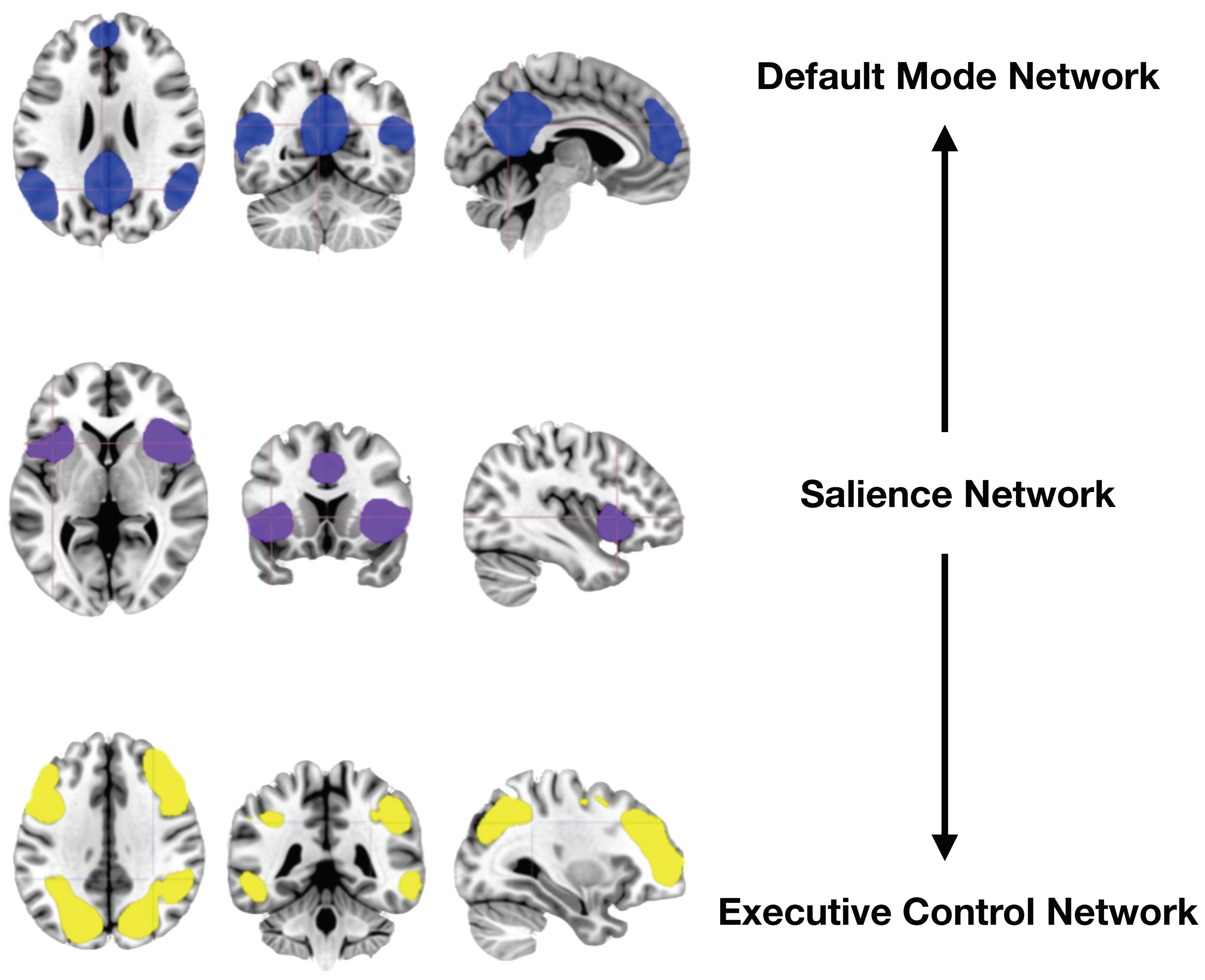
Figure 1. Three-network paradigm with the Central Executive Network (CEN), the Salience Network (SN), and the Default Mode Network (DMN). At the helm of decision-making, the SN in purple allocates cognitive resources to respond to endogenous and exogenous stimuli by switching cognitive assignments between the CEN and the DMN. The DMN, in exchange, funnels these stimuli toward self-references, self-awareness, social cognition, and autobiographical memory. Key nodes of the CEN in yellow include the dorsolateral prefrontal cortex (dlPFC), inferior frontal gyrus (iFG), frontal eye fields (FEF, not noted in the figure), anterior cingulate cortex (ACC, not noted in the figure), inferior parietal lobule (IPL), and posterior parietal cortex (PCC). Nodes of the SN in purple include dorsal anterior cingulate cortex (dACC), supplementary motor area (SMA, not noted in the figure), anterior insula (aI), and limbic structures (not noted in the figure) like the amygdala (Amy), thalamus, hypothalamus, and ventral striatum (VS). Classic nodes of the DMN in blue include the dACC, the dorsomedial prefrontal cortex (dmPFC), the posterior cingulate cortex (PCC), and the precuneus.
Photonics 11 00761 g001
Figure 2. Iowa Gambling Task. In the Iowa Gambling Task (IGT), during each presentation, participants must choose one of four decks of cards (i.e., A, B, C, or D). Each card selection results in gaining or losing a certain amount of play money. The participants do not earn or lose the exact amount of money for the task, but they are led to believe that their performance will affect the amount of monetary compensation for this study. Two decks (in this figure, A and B) have lower immediate rewards but also lower long-term losses, while the other two decks (in this figure, C and D) have higher immediate rewards but also higher long-term losses. Participants are instructed to select cards from any deck of their choice and accumulate as much play money as possible, and they are not informed about the decks’ different gain and loss structures. The task consists of multiple blocks, totaling 200 trials, with each block subdivided into smaller sets of trials. During each trial, participants select a card by pressing the corresponding key on a 4-key keyboard. The selected card is then revealed, and the outcome (gain or loss) and the updated total play money are displayed. Each presentation is shown for a maximum of 3.5 s, after which the program makes a random choice, marks it on the screen for the participant to see, and shows the immediate and accumulated net gain for the choice made. The software used is PsyToolkit 3.4.2. The software ensures precise timing and the accurate recording of participant responses. Before the task, participants receive instructions and practice trials to ensure they understand the hardware’s operation and the task’s objectives. Four versions of the task were built with random permutations on the assignments of advantageous and disadvantageous decks to reduce the learning effect of repeated applications of the task.
Photonics 11 00761 g002
Figure 3. Reward Gambling Task. In the Reward Gambling Task, participants are required to predict the number on a concealed card, represented by a “?”, with the objective of either receiving rewards or penalties. Participants are informed that the card numbers would range from 1 to 9. Their task is to assess whether the concealed card number is above or below 5, making their choice known by pressing one of two buttons on a response box. The feedback mechanism involves revealing the card’s number, which is determined by a program based on whether the trial was reward-oriented, penalizing, or neutral. This feedback is visually presented through a green upward arrow with “$1” for reward trials, a red downward arrow with “−$0.50” for penalizing trials, or the digit 5 accompanied by a gray bidirectional arrow in neutral trials. The mystery card “?” is displayed for up to 1500 ms. If participants respond earlier, a fixation cross appears for the remaining time. This is followed by a feedback duration of 1000 ms and an intertrial interval (ITI) of the same length, during which a “+” sign is shown. The task encompasses blocks of 8 trials, categorized as reward- or penalty-oriented. Reward-oriented blocks include six reward trials interspersed with a mix of neutral and penalizing trials, whereas penalty-focused blocks comprise six penalizing trials combined with either reward or neutral trials. Each of the two runs in the task features two blocks, each of reward-focused and penalty-focused trials, interspersed with four 15-s fixation blocks.
Photonics 11 00761 g003
Figure 4. Timeline of this study. t-PBM: Transcranial photobiomodulation. GT: gambling task. Tx: treatment visit.
Photonics 11 00761 g004
Table 1. Schedule of events.
MeasurePre-ScreenScreenBaselineTx 1Tx 2Tx 3Tx 4Tx 5Check-InFollow-Up
Pre-Screen FormX
ASRMX X
PHQ-9X X
Consent Form X
Demographic Form X
Concomitant Treatments Log XXXXXXXXX
AE XXXXXXXX
Prior Treatment Log X
Medical History X
Drug Screen X
Pregnancy Test X
MINI X
C-SSRS XX XX
MOODS-SR Lifetime X
YMRS X
Screening Note X
NIS-SCS X
MOODS-SR Last Week X X X
BISX X X X
I-7 X X X
BRIEF X X X
t-PBM X *XXXX *
Reward Gambling Task XX * X * X
lowa Gambling Task XX X X
Vitals X X
MRI Safety Form X X X
SAFTEE X X X
CGI-S X XX
CGI-I XX
PBQ X
TSRQ XXXXX
CAST-IRR XXXXXX
Intervention Tracking XXXXX
Adverse Event Log XXXXXXXX
Legend: * Indicates administered inside scanner; Tx: treatment visit; ASRM: Altman Self-Rating Mania Scale; PHQ-9: Patient Health Questionnaire-9; AE: adverse events; MINI: Mini International Neuropsychiatric Interview; C-SSRS: Columbia Suicide Severity Rating Scale (C-SSRS); MOODS-SR: Mood Spectrum-Self Report; YMRS: Young Mania Rating Scale; NIS-SCS: New-Imigrant Survey-Skin Color Scale; BIS: Barrat Impulsiveness Scale; I-7: Impulsiveness and Venturesomeness Questionnaire; BRIEF: Behavior Rating Inventory of Executive Function; t-PBM: transcranial photobiomodulation; MRI: magnetic resonance imaging; SAFTEE: Systematic Assessment for Treatment Emergent Events; CGI-S: Clinical Global Impressions—Severity; CGI-I: Clinical Global Impressions—Improvement; PBQ: Perceived Blinding Questionnaire; TSRQ: t-PBM Self-Report Questionnaire; CAST-IRR: Concise Associated Symptom Tracking Irritability Scale.
Table 2. Transcranial photobiomodulation parameters.
Measure (Unit)Value
Wavelength (nm)808
Exposure time (s)333
Area of exposure (cm2)24
Irradiance, Power Density (mW/cm2)300
Fluence (J/cm2)100
Total Energy (kJ)2.4
NIR SourceLaser
Laser Output (W)3.5 per fiber (7 total)
Wave ModeContinuous
Anatomical TargetsF4 & Fp2
DeviceLitecure’s LightForce® EXPi Deep Tissue Laser Therapy TM System, Transcranial PhotoBioModulation-1000 (t-PBM-2.0)
Disclaimer/Publisher’s Note: The statements, opinions and data contained in all publications are solely those of the individual author(s) and contributor(s) and not of MDPI and/or the editor(s). MDPI and/or the editor(s) disclaim responsibility for any injury to people or property resulting from any ideas, methods, instructions or products referred to in the content.