Article

Thermoacoustic Imaging Using Single-Channel Data Acquisition System for Non-Invasive Assessment of Liver Microwave Ablation: A Feasibility Study

1
Department of Ultrasound, West China Hospital, Sichuan University, Chengdu 610041, China
2
School of Information Engineering, Southwest University of Science and Technology, Mianyang 621010, China
3
School of Electronic Science and Engineering, University of Electronic Science and Technology of China, Chengdu 611731, China
*
Authors to whom correspondence should be addressed.
Photonics 2024, 11(9), 807; https://doi.org/10.3390/photonics11090807
Submission received: 29 July 2024 / Revised: 21 August 2024 / Accepted: 28 August 2024 / Published: 29 August 2024
(This article belongs to the Special Issue Technologies and Applications of Biophotonics)

Abstract

:
Microwave ablation (MWA) plays a crucial role in non-surgical liver cancer treatment, but the existing efficacy evaluation tools lack the characteristics of being real-time, non-invasive, and efficient. As an emerging imaging technology, thermoacoustic imaging (TAI) has attracted extensive clinical attention for its excellent merits, which combine the advantages of high contrast in microwave imaging and high resolution in ultrasound imaging. Particularly, the application of a circular scanned single-channel data acquisition system maximizes the capture of thermoacoustic signals, thereby providing more comprehensive image information and rendering reconstructed images closer to reality. This study aimed to verify the feasibility of TAI in non-invasive evaluation of the efficacy of MWA on ex vivo porcine liver and in vivo rabbit liver. During the experiments, ultrasound is used to cross-verify the results of TAI to ensure the accuracy and reliability of the method. Additionally, by altering the thickness of porcine liver tissue to increase the distance (from 0 mm to 80 mm) between the horn antenna and the target (soy sauce tube), TAI is used to observe the change of the image signal-to-noise ratio to preliminarily explore the imaging depth of TAI. The results of ex and in vivo experiments can not only promote the clinical application of TAI, but also be expected to provide a more accurate and reliable efficacy assessment method for MWA in liver cancer treatment.

1. Introduction

Liver cancer is one of the leading causes of death worldwide. With the global population increasing, the World Health Organization (WHO) estimates that approximately 1.4 million people could be diagnosed with liver cancer by 2040, and 1.3 million are projected to succumb to the disease, a number that represents a 56.4% increase from 2020 [1]. Although surgery, including hepatic resection and liver transplantation, is considered the preferred treatment for liver cancer, only 20–30% of patients can tolerate it [2]. Microwave ablation (MWA), as a mature thermal ablation treatment, plays a significant role in the minimally invasive treatment of small liver cancer (less than 3 cm in diameter) and the palliative treatment of advanced liver cancer [3]. MWA induces coagulative necrosis in tissues by causing protein denaturation in cancer cells through microwave thermal effects. MWA is frequently recommended in clinical guidelines, due to its minimal invasiveness, simplicity of operation, and significant efficacy [4].
As with other local thermal ablation treatments, damage to healthy tissue should be minimized in MWA while ensuring treatment efficacy. This is crucial for determining whether the patient will experience recurrence postoperatively [5]. However, due to the lack of real-time imaging techniques for evaluating the efficacy of MWA in clinical practice, MWA is often treated incompletely or overtreated [6]. Contrast-enhanced ultrasound (CEUS) can accurately assess the efficacy of MWA, but it is usually recommended to evaluate at least 2 h after MWA [7]. Contrast-enhanced CT, MRI, and FDG-PET/CT can accurately evaluate the extent of MWA, but do not provide the results of real-time assessment [8]. Therefore, an imaging technique capable of timely and accurate intraoperative assessment of MWA is essential to improve the efficacy of minimally invasive treatment for HCC.
Thermoacoustic imaging (TAI) is a novel non-invasive imaging technique that has emerged in recent years. Its principle is based on the transient thermoelastic expansion effect induced by pulsed microwave, which can generate thermoacoustic signals (TASs). The TASs collected by the probe can then be further reconstructed into TAI [9]. The strength of TASs is primarily influenced by the dielectric properties (DPs) of biological tissues. It is well known that DPs are an intrinsic property of biological tissues which are related to the tissue’s water content, ionic concentration, and temperature [10]. When MWA is performed, the temperature within the biological tissues will rise sharply, and the distribution of water within the tissues will become uneven. Consequently, there will be a noticeable difference in the DPs between the ablated tissue and the normal tissue. This difference in DPs leads to variations in microwave absorption between normal and ablated tissues which can be measured using TAI [11]. This is the basis for TAI to identify the ablation range of MWA, and this technology offers higher contrast than ultrasound imaging. Thus far, TAI has been investigated for the detection of breast [12,13,14], kidney [15], and prostate cancers [16] and joint [17,18] and brain imaging [19].
This study provides a detailed description of the circular scanned single-channel data acquisition TAI system and its application in the evaluation of the ablation effect on ex vivo porcine liver and in vivo rabbit liver. Simultaneously, the imaging depth of TAI was also preliminarily investigated, demonstrating that TAI has the potential for real-time efficacy evaluation after MWA in the future.

2. Materials and Methods

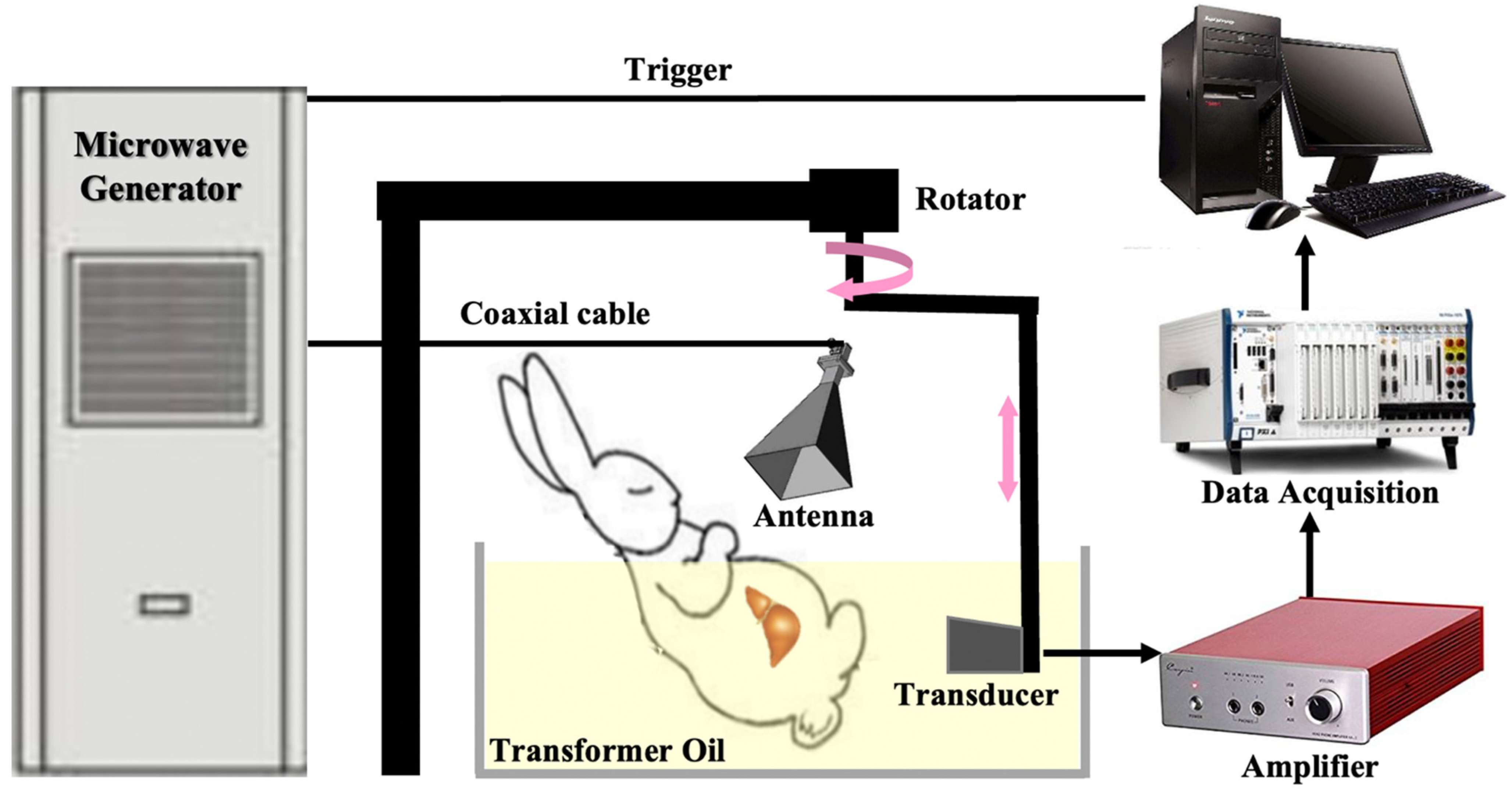
2.1. The Circular Scanned Single-Channel Data Acquisition TAI System

The schematic diagram of the single-channel TAI system is shown in Figure 1. The microwave excitation source used in this study was a custom-designed microwave generator (center frequency: 3.0 GHz, bandwidth: 50 MHz, peak power: 70 kW, pulse duration: 0.75 μs). The pulsed microwave was coupled into a pyramid-shaped horn antenna (114 × 144 mm2) via a semi-rigid coaxial cable (1.5 m long with 1.2 dB insertion loss). This is consistent with our previous study [20,21,22]. The aperture size of the pyramid horn antenna ensured coverage of the liver tissue area. It is noteworthy that the average microwave power density on the surface of object was about 15 mW/cm2 when a 50 Hz repetition frequency was used, which is below the IEEE safety standard (20 mW/cm2 at 3.0 GHz) [23]. To ensure effective coupling of microwave and ultrasound signals, both the object and transducer were immersed in a transformer oil-filled tank to avoid interference from the air medium. An immersion transducer with a central frequency of 2.25 MHz (V323-SU, Japan Probe Co., Ltd., Yokohama, Japan), which was mounted on a rotary stage (RSA 100, Beijing Zolix Instruments Co., Ltd., Beijing, China), was circularly scanned over 360° and 220° for the ex and in vivo experiments, respectively. A step size of 2° was used to detect the generated TASs. The TAS received by the transducer were amplified using a homemade amplifier (bandwidth: 200 kHz–2 MHz, gain: 55 dB) [22]. Subsequently, the amplified TASs were converted into digital signals by a data acquisition card (NI5752B, National Instruments, Inc., Austin, TX, USA) with a sampling frequency of 50 MHz and transmitted to a computer. In the computer, the acquired data were averaged 50 times to improve the signal-to-noise ratio and finally formed a frame TAI image [21]. The total experimental duration for the entire process was approximately 4 min, which includes all steps from signal acquisition, amplification, digitization, and image reconstruction.
The image reconstruction in this study utilized the delay-and-sum (DAS) algorithm, a classic method in TAI research and the fundamental beamforming technique in current medical imaging applications [24]. The basic principle involves adjusting time delays to achieve signal focusing and summation. Specifically, the algorithm reconstructs the image of the target object by calculating the time delay of the signals received at different detection points propagating at an assumed speed of sound, and then aligning and superimposing these signals in the appropriate time domain. The advantages of this algorithm lie in its simplicity and computational efficiency, enabling rapid large-scale data processing and image reconstruction. This is one of the reasons DAS is widely used in many imaging techniques, including TAI. By employing the DAS algorithm, we can effectively reconstruct microwave thermoacoustic images with high contrast and resolution, thereby providing reliable data support for pathological detection of biological tissues [25]. The DAS algorithm can be expressed as the following Equation (1):
p r = i = 1 n S i ( r r i c ) ,
In the equation, p ( r ) represents the initial acoustic pressure value at r , S i denotes the acquired signal from the i t h channel, r i represents the position of the i t h channel, r r i stands for the distance between the specimen under test and the i t h channel, and c denotes the speed of sound.

2.2. TAI of Porcine Liver Ex Vivo

We obtained fresh porcine livers from a local slaughterhouse for ex vivo experiments. The livers were sliced into rectangular slices. We simulated the MWA process on the porcine liver tissue by using a soldering iron (7 °C, 60 s). A damaged area of approximately 8 × 10 × 5 mm3 was generated in the porcine liver. The damaged area is located in the lower right corner of Figure 2a, and the red arrows are used to indicate the boundaries of normal tissue and the yellow arrows are used to indicate the boundaries of damaged tissue. Subsequently, the treated liver tissue was immersed in transformer oil and fixed. The microwave generator was activated, directing microwave energy onto the porcine liver tissue through a horn antenna. So that the microwave energy can be completely irradiated on the pig liver, the rotating stage was turned on and the probe began rotating to collect TASs. The TASs were amplified and then transmitted to a computer for image reconstruction.
Figure 2b shows a TAI image of an ex vivo porcine liver. Normal tissue and damaged tissue are indicated in the same way as in Figure 2a. In the image, it is evident that the TASs from the damaged area are significantly reduced compared to normal tissue, resulting in a distinct “blank” area at the right corner of the liver, and the contours of the damaged tissue can still be observed (the yellow arrows). This result indicates that TAI images can accurately reflect the differences in the thermoacoustic properties of different tissues, and it is roughly consistent with the physical images. It is noteworthy that the boundary between normal tissue and damaged tissue presents as a strong signal, which may be related to the higher moisture content in this area. During the burning process, the moisture in the tissue undergoes evaporation and migration.

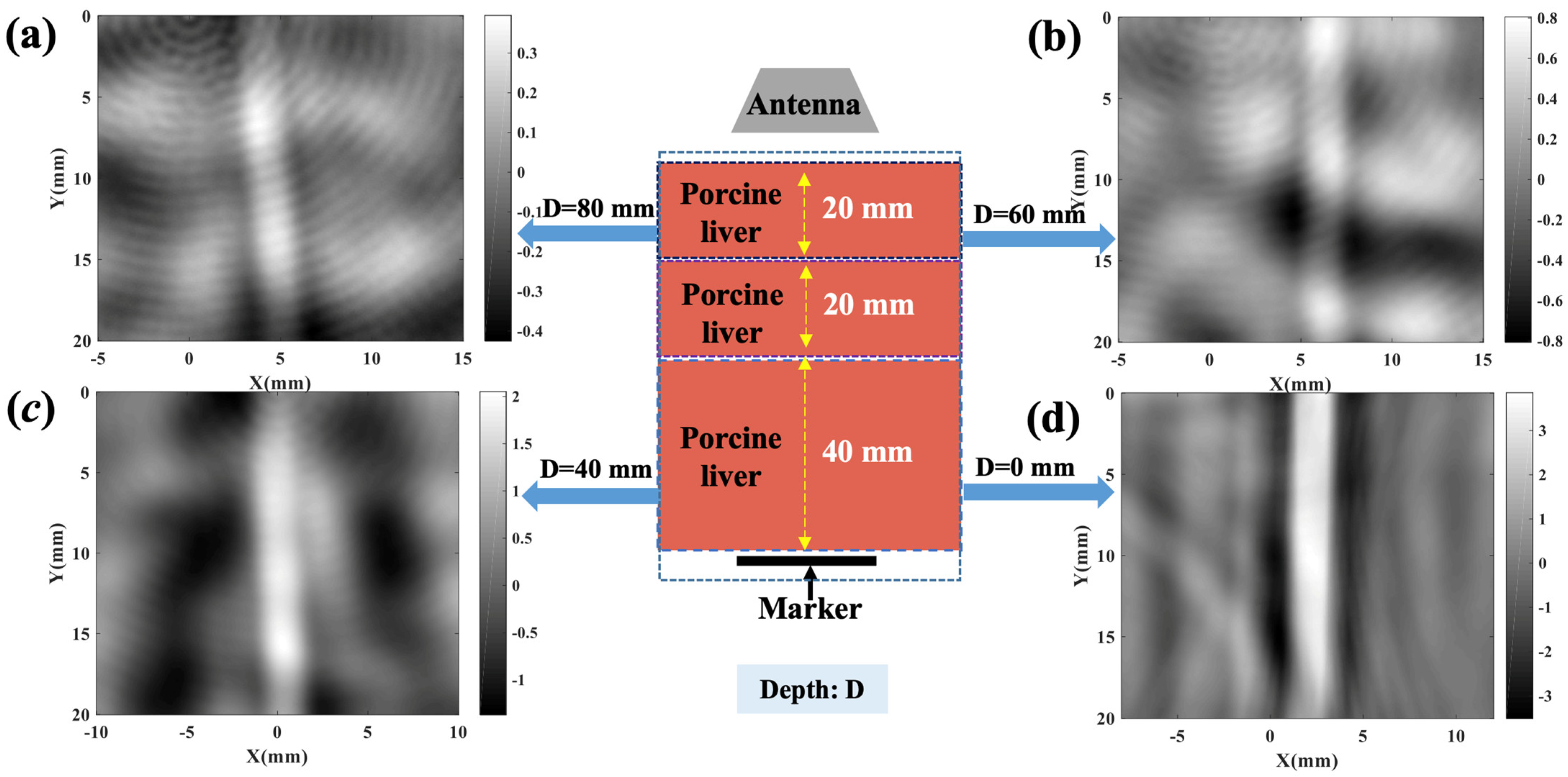
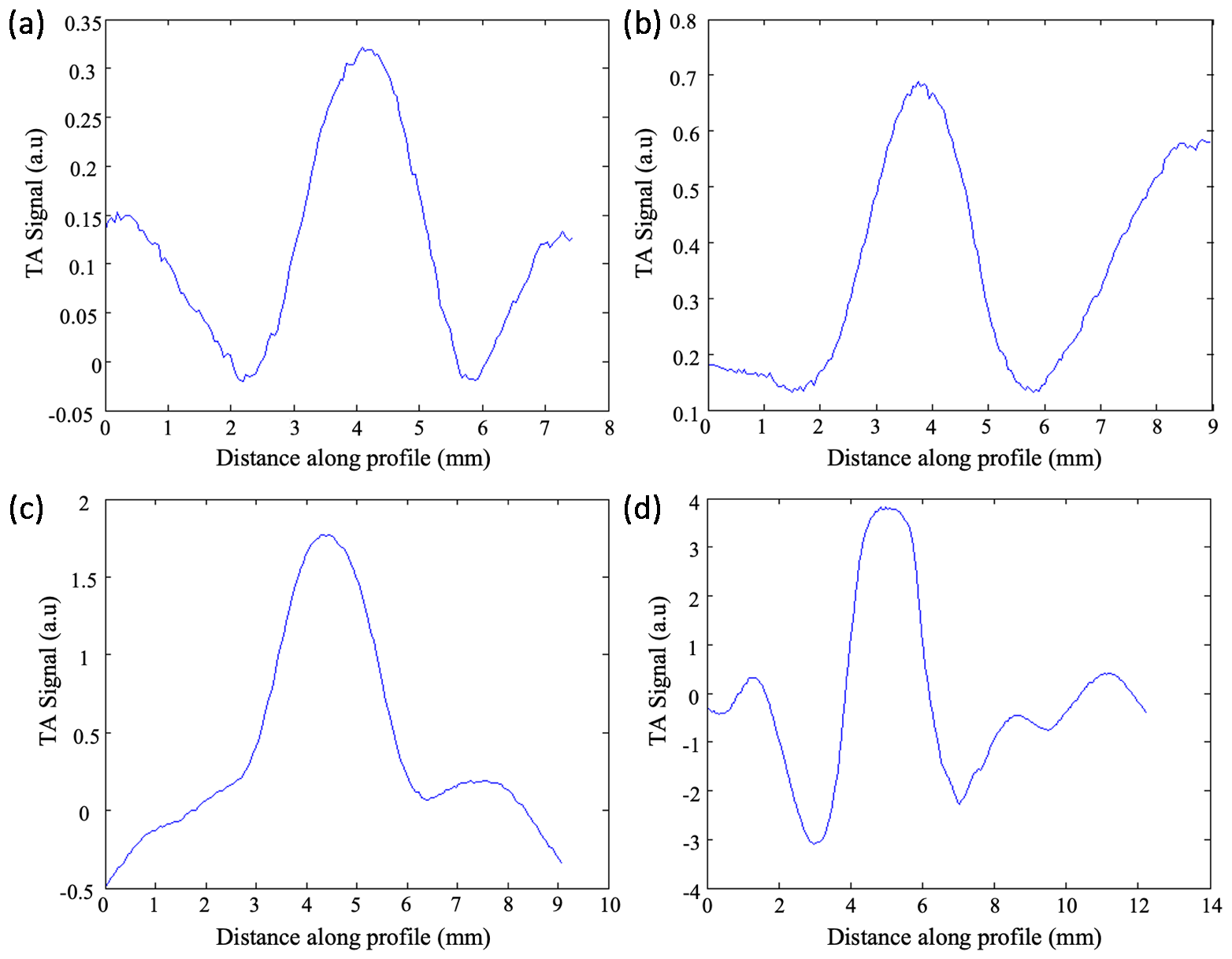
2.3. TAI Imaging Depth Investigation

To assess the imaging depth capability of TAI, a soy sauce tube with a diameter of 3 mm was placed at the bottom of fresh porcine livers of various thicknesses. The probe for collecting TASs was positioned on the opposite side of the soy sauce tube. The imaging distance between the probe and the soy sauce tube was adjusted by gradually increasing the thickness of the porcine liver tissue. The soy sauce tubes were imaged at different depths (80 mm, 60 mm, 40 mm, and 0 mm), as shown in Figure 3a–d.
From Figure 3d, it can be observed that when no porcine liver is placed above the soy sauce tube (0 mm), the outline of the soy sauce tube is the most distinct. The sidewall of the soy sauce tube is a straight line with minimal shape distortion. At this moment, the signal-to-noise ratio (SNR) is 36 dB. The experimental results showed that when the thickness of the porcine liver was 80 mm, the outline of the soy sauce tube could still be clearly observed (Figure 3a). However, during this process we can observe that the wall of the soy sauce tube gradually becomes blurred. At the same time, a small number of artifacts began to appear in other areas. With increasing imaging depth, the SNR significantly decreased, from 36 dB to 21 dB (Figure 3c and Table 1). The curve of TA signal changing with depth is shown in Figure 4. The relative microwave absorption amplitude of the soy sauce tube showed a slight downward trend, but the contrast seemed to change little. These results indicate that although TAI system still has imaging capability at greater depths, the increase of imaging depth has a negative effect on the relative microwave absorption amplitude and SNR.

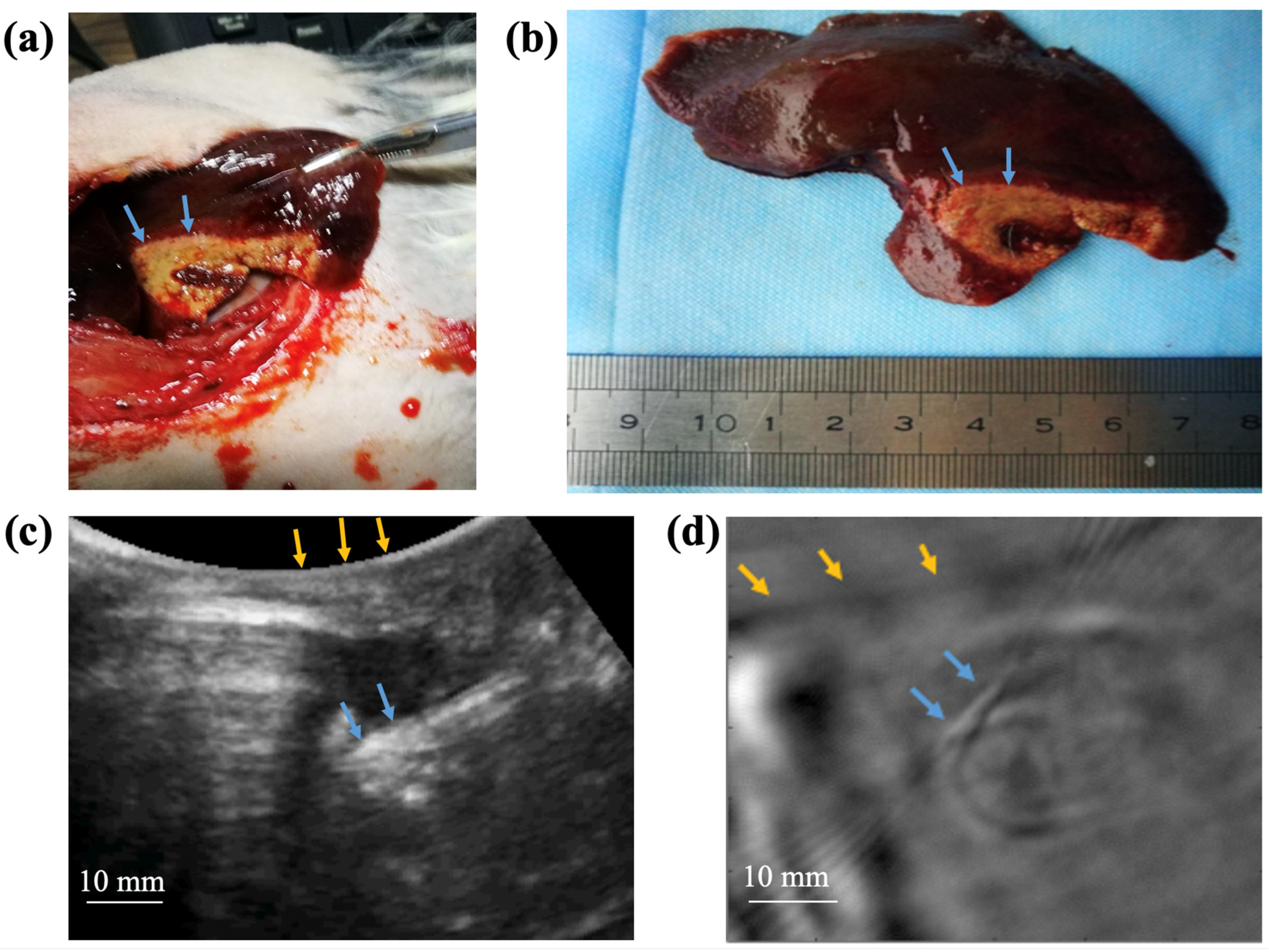
2.4. TAI of Rabbit Liver In Vivo

To verify the efficacy of TAI in a more complex in vivo environment, MWA experiments were conducted in live normal rabbit livers. An adult male New Zealand rabbit weighing approximately 2.5 kg was used for the in vivo experiments (Chengdu Dossy Experimental Animal Co., Ltd., Chengdu, China). The protocols and procedures used for the animal experiments was approved by the Animal Ethics Committee of West China Hospital, Sichuan University (Approval No.2018031A). Prior to the experiment, the hair on the abdomen of the rabbit was removed to minimize interference with TASs. Anesthesia was induced by intravenous injection of 30 mg/kg sodium pentobarbital via the ear vein. The anesthetized rabbit was restrained in a metal frame to maintain a supine position and immersed in oil. Under ultrasound guidance using CX50 ultrasound system (Philips, Amsterdam, The Netherlands) with a C5-1 convex probe, a clinician with over 5 years of clinical MWA therapy experience placed a MWA needle into the rabbit liver percutaneous. MWA was performed using a 2450 MHz MWA device (KY2200, Nanjing Canyou Medical Technology Co., Ltd., Nanjing, China). Microwave energy was delivered to the rabbit liver via the KY2450A antenna (ablation needle). MWA was performed for 3 min at 30 W power. After MWA, ultrasonic and TAI images were collected separately. The rabbits were executed using overdose anesthesia at the end of the experiment. The livers were carefully removed from the abdomen, and photographs of the liver were retained. The ablated liver tissue was outlined with red lines and marked with blue arrows in both ultrasound and TAI images.
In the post-dissection liver photographs, normal liver tissue appears deep red, while the ablated tissue is yellow (blue arrows, Figure 5a,b). The ablated tissue appears as a strongly echogenic circular area in the ultrasound images (blue arrow, Figure 5c). This is because during MWA in live liver tissue, a large number of bubbles are generated, along with coagulative necrosis in the normal liver tissue. Compared with liver photographs and ultrasound images, TAI can clearly demonstrate MWA lesions in terms of shape and size (Figure 5d). We observed from TAI image that the ablated lesion was 10 mm in diameter (blue arrows), and located 22 mm beneath the rabbit’s skin (yellow arrows), which corresponds well with the ultrasound images. Additionally, TAI images showed an improved contrast-to-noise ratio and clearer margin of lesion compared to ultrasound images.

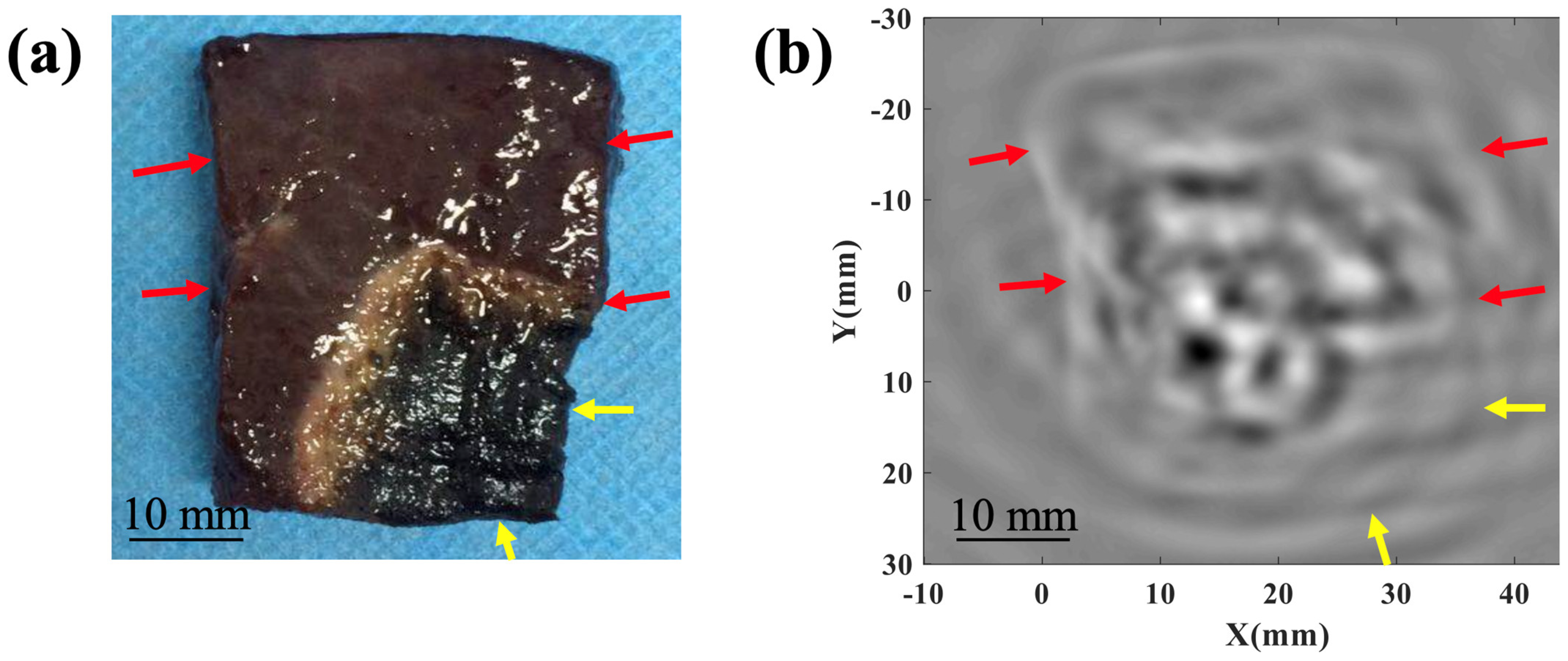
3. Result and Discussion

This study proposes a non-invasive method for evaluating the effect of liver ablation using the circular scanned single-channel data acquisition TAI system. Through a series of ex vivo and in vivo experiments, we demonstrated that TAI has the potential to assess the extent of ablation necrosis.
Based on the alteration of tissue DPs induced by thermal ablation, TAI has a higher contrast in distinguishing necrotic tissues from normal tissues. In order to validate this, and to realize the advantages of TAI’s high contrast in clinical work, further research has been conducted. Jin et al. [26] induced damage using high-intensity focused ultrasound in fat samples implanted in porcine muscle under pulsed microwaves at 3 GHz, and imaged them via the circular scanned single-channel data acquisition TAI system. The general damage area can be observed in the final TAI image result. This is consistent with the experimental findings of ex vivo porcine livers in our study. However, due to the limitation of the bandwidth of the transducer, there is a large error in predicting the boundaries of the injuries. Cavagnaro et al. [27] developed a numerical model of MWA which provides a useful tool for optimizing TAI simulation data. Bucci et al. [28] used this model to generate microwave tomography for the initial observation of MWA. However, this can only provide qualitative results and cannot prove a necessary link with the actual situation. In addition, Evans et al. [29] had even designed antennas that can simultaneously produce TASs and melt tissue. Their study further elucidated the effects of temperature and tissue coagulation on TAS arrival time and energy. Unfortunately, the study did not show the image effects of TAI. These results provide preliminary experimental evidence for the application of TAI in local thermal ablation imaging. Additionally, Srishti et al. presented preliminary phantom data to establish a TAI-guided platform for focused microwave treatment of solid breast tumors, which marks new advancements and perspectives in the clinical application of TAI [13]. Nevertheless, there is always some difference between an imitation and the actual situation. In order to simulate the effect of TAI in a more realistic environment, this study conducted experiments in ex vivo porcine livers and in vivo rabbit livers. This not only enhances the credibility of the experimental results, but also helps to identify directions that need to be optimized.
In our study, TAI achieved a visualization depth of up to 80 mm, which meets the imaging requirements for most human organs. Compared to photoacoustic tomography (maximum imaging depth of 5 cm), TAI exhibits significant advantages in imaging depth [30]. However, to investigate the pattern of microwave energy attenuation with increasing liver tissue thickness, we used a soy sauce pipe as a reference marker. This experimental method will require further improvement in our future experiments. For instance, we could consider using an insertable positioning needle. The imaging depth of TAI still needs further experimental research.
In our previous studies, it has been demonstrated that TAI can visualize the entire liver in live rabbits through the abdominal wall, and preliminary image calibration was performed using a localization needle [21]. On this basis, we conducted real-time MWA experiments in live rabbit livers. It is worth mentioning that our in vivo experiments involved the percutaneous placement of the ablation needle, which closely simulates the human MWA treatment scenario. We chose to validate the imaging effects of TAI in the most complex experimental conditions, which is highly significant, and the results show that the ablated necrotic zone exhibits significant imaging effects in TAI and could be clearly outlined. This result demonstrates the application prospect of TAI technology in the real-time and accurate assessment of ablation extents.
In summary, this study represents the first application of microwave TAI for non-invasive evaluation of liver MWA. Through ex and in vivo experiments, we validated the feasibility of TAI in evaluating the efficacy of MWA. This research lays a foundation for future clinical applications, but it still needs to be further optimized and improved. First, to improve the temporal resolution, it is necessary to adopt array transducers and a multi-channel data acquisition system to achieve more efficient data collection and processing. Secondly, reconstruction algorithms based on the finite element method offer significant advantages in recovering tissue parameters such as electrical conductivity, speed of sound, and temperature, which will provide additional value for TAI in assessing MWA. With these improvements, TAI is expected to become a standardized, real-time, non-invasive ablation evaluation tool to provide more accurate and reliable imaging support for the treatment of liver tumors.

Author Contributions

Conceptualization, Y.L. and L.H.; methodology, Q.L.; software, Z.Y.; validation, L.F., Z.Y., and W.P.; formal analysis, L.S.; investigation, W.P. and L.S.; resources, L.S.; data curation, L.H.; writing—original draft preparation, L.S.; writing—review and editing, L.S., Y.L. and L.H; visualization, L.S.; supervision, Y.L.; project administration, L.H. All authors have read and agreed to the published version of the manuscript.

Funding

This work was supported in part by the National Natural Science Foundation of China under Grant Nos. 82071940 and 82371977, in part by the Key Research and Development Program of Science and Technology Department of Sichuan Province under Grant No. 2023YFG0322, and in part by the Clinical and Translational Medicine Research Special Project of Chinese Academy of Medical Sciences (2022-I2M-C&T-B-105).

Institutional Review Board Statement

The protocols and procedures used for the animal experiments were approved by the Animal Ethics Committee of West China Hospital, Sichuan University (Approval No.: 2018031A).

Informed Consent Statement

Not applicable.

Data Availability Statement

The data presented in this study are available from the authors upon reasonable request.

Acknowledgments

We thank Boyang Yu from West China Hospital for his guidance on microwave ablation surgery.

Conflicts of Interest

The authors declare no conflicts of interest.

References

  1. Rumgay, H.; Arnold, M.; Ferlay, J.; Lesi, O.; Cabasag, C.J.; Vignat, J.; Laversanne, M.; McGlynn, K.A.; Soerjomataram, I. Global Burden of Primary Liver Cancer in 2020 and Predictions to 2040. J. Hepatol. 2022, 77, 1598–1606. [Google Scholar] [CrossRef] [PubMed]
  2. Yarchoan, M.; Agarwal, P.; Villanueva, A.; Rao, S.; Dawson, L.A.; Karasic, T.; Llovet, J.M.; Finn, R.S.; Groopman, J.D.; El-Serag, H.B.; et al. Recent Developments and Therapeutic Strategies against Hepatocellular Carcinoma. Cancer Res. 2019, 79, 4326–4330. [Google Scholar] [CrossRef]
  3. Gordan, J.D.; Kennedy, E.B.; Abou-Alfa, G.K.; Beg, M.S.; Brower, S.T.; Gade, T.P.; Goff, L.; Gupta, S.; Guy, J.; Harris, W.P.; et al. Systemic Therapy for Advanced Hepatocellular Carcinoma: ASCO Guideline. J. Clin. Oncol. 2020, 38, 4317–4345. [Google Scholar] [CrossRef]
  4. Chakraborty, E.; Sarkar, D. Emerging Therapies for Hepatocellular Carcinoma (HCC). Cancers 2022, 14, 2798. [Google Scholar] [CrossRef]
  5. Xie, D.-Y.; Ren, Z.-G.; Zhou, J.; Fan, J.; Gao, Q. 2019 Chinese Clinical Guidelines for the Management of Hepatocellular Carcinoma: Updates and Insights. Hepatobiliary Surg. Nutr. 2020, 9, 452–463. [Google Scholar] [CrossRef] [PubMed]
  6. Kok, H.P.; Cressman, E.N.K.; Ceelen, W.; Brace, C.L.; Ivkov, R.; Grüll, H.; Ter Haar, G.; Wust, P.; Crezee, J. Heating Technology for Malignant Tumors: A Review. Int. J. Hyperth. 2020, 37, 711–741. [Google Scholar] [CrossRef]
  7. Wu, H.; Wilkins, L.R.; Ziats, N.P.; Haaga, J.R.; Exner, A.A. Real-Time Monitoring of Radiofrequency Ablation and Postablation Assessment: Accuracy of Contrast-Enhanced US in Experimental Rat Liver Model. Radiology 2014, 270, 107–116. [Google Scholar] [CrossRef] [PubMed]
  8. Puijk, R.S.; Ruarus, A.H.; Scheffer, H.J.; Vroomen, L.G.P.H.; Van Tilborg, A.A.J.M.; De Vries, J.J.J.; Berger, F.H.; Van Den Tol, P.M.P.; Meijerink, M.R. Percutaneous Liver Tumour Ablation: Image Guidance, Endpoint Assessment, and Quality Control. Can. Assoc. Radiol. J. 2018, 69, 51–62. [Google Scholar] [CrossRef] [PubMed]
  9. Chen, H.; Tang, X.; Nie, G.; Wang, Z.; Hu, J.; Hu, J.; Qin, H. Quantitative Tracing of Bioprobes by Simultaneously Monitoring Radiative and Nonradiative Relaxations. J. Innov. Opt. Health Sci. 2023, 16, 2243002. [Google Scholar] [CrossRef]
  10. Etoz, S.; Brace, C.L. Computed Tomography-Based Modeling of Water Vapor-Induced Changes in Permittivity During Microwave Ablation. IEEE Trans. Biomed. Eng. 2020, 67, 2427–2433. [Google Scholar] [CrossRef]
  11. Lopresto, V.; Pinto, R.; Lovisolo, G.A.; Cavagnaro, M. Changes in the Dielectric Properties of Ex Vivo Bovine Liver during Microwave Thermal Ablation at 2.45 GHz. Phys. Med. Biol. 2012, 57, 2309–2327. [Google Scholar] [CrossRef] [PubMed]
  12. Zhao, Y.; Ji, Z.; Qin, B.; Xing, D. A Thermoacoustic Imaging System with Variable Curvature and Multi-Dimensional Detection Adapted to Breast Tumor Screening. J. Appl. Phys. 2018, 124, 144902. [Google Scholar] [CrossRef]
  13. Saraswat, S.; Tak, J.; Liang, M.; Lyu, C.; Witte, R.S.; Xin, H. Towards Study on Thermoacoustic Imaging Guided Focused Microwave Therapy for Breast Cancer Treatment. In Proceedings of the 2018 IEEE/MTT-S International Microwave Symposium—IMS, Philadelphia, PA, USA, 10–15 June 2018; IEEE: Piscataway, NJ, USA, 2018; pp. 953–956. [Google Scholar]
  14. Huang, G.; Li, Y.; Ren, M.; Zhang, H.; Qin, H. Real-Time Thermoacoustic Imaging for Breast Tumor Biomarker Biopsy Navigation Basing on a Semi-Ring Ultrasonic Transducer. Appl. Phys. Lett. 2023, 123, 183702. [Google Scholar] [CrossRef]
  15. Cao, C.; Nie, L.; Lou, C.; Xing, D. The Feasibility of Using Microwave-Induced Thermoacoustic Tomography for Detection and Evaluation of Renal Calculi. Phys. Med. Biol. 2010, 55, 5203–5212. [Google Scholar] [CrossRef]
  16. Patch, S.K.; Hanson, E.; Thomas, M.; Kelly, H.; Jacobsohn, K.; See, W.A. Thermoacoustic Imaging of Fresh Prostates up to 6-cm Diameter; Oraevsky, A.A., Wang, L.V., Eds.; SPIE: San Francisco, CA, USA, 2013; p. 85812K. [Google Scholar]
  17. Chi, Z.; Huang, L.; Ge, S.; Jiang, H. Technical Note: Anti-phase Microwave Illumination-based Thermoacoustic Tomography of in Vivo Human Finger Joints. Med. Phys. 2019, 46, 2363–2369. [Google Scholar] [CrossRef]
  18. Chi, Z.; Zhao, Y.; Huang, L.; Zheng, Z.; Jiang, H. Thermoacoustic Imaging of Rabbit Knee Joints. Med. Phys. 2016, 43, 6226. [Google Scholar] [CrossRef]
  19. Zhao, Y.; Shan, T.; Chi, Z.; Jiang, H. Thermoacoustic Tomography of Germinal Matrix Hemorrhage in Neonatal Mouse Cerebrum. J. X-ray Sci. Technol. 2020, 28, 83–93. [Google Scholar] [CrossRef]
  20. Wang, F.; Yang, Z.; Peng, W.; Song, L.; Luo, Y.; Zhao, Z.; Huang, L. RPCA-Based Thermoacoustic Imaging for Microwave Ablation Monitoring. Photoacoustics 2024, 38, 100622. [Google Scholar] [CrossRef] [PubMed]
  21. Xiang, H.; Zheng, Z.; Huang, L.; Qiu, T.; Luo, Y.; Jiang, H. In Vivo Liver Thermoacoustic Imaging and Demonstration Based on Localization Wire. Med. Phys. 2021, 48, 1608–1615. [Google Scholar] [CrossRef]
  22. Huang, L.; Li, T.; Jiang, H. Technical Note: Thermoacoustic Imaging of Hemorrhagic Stroke: A Feasibility Study with a Human Skull. Med. Phys. 2017, 44, 1494–1499. [Google Scholar] [CrossRef]
  23. Huang, L.; Yao, L.; Liu, L.; Rong, J.; Jiang, H. Quantitative Thermoacoustic Tomography: Recovery of Conductivity Maps of Heterogeneous Media. Appl. Phys. Lett. 2012, 101, 244106. [Google Scholar] [CrossRef]
  24. Hoelen, C.G.A.; De Mul, F.F.M. Image Reconstruction for Photoacoustic Scanning of Tissue Structures. Appl. Opt. 2000, 39, 5872. [Google Scholar] [CrossRef] [PubMed]
  25. McKeighen, R.E.; Buchin, M.P. New Techniques for Dynamically Variable Electronic Delays for Real Time Ultrasonic Imaging. In Proceedings of the 1977 Ultrasonics Symposium, Phoenix, AZ, USA, 26–28 October 1977; IEEE: Piscataway, NJ, USA, 1977; pp. 250–254. [Google Scholar]
  26. Jin, X.; Xu, Y.; Wang, L.V.; Fang, Y.R.; Zanelli, C.I.; Howard, S.M. Imaging of High-Intensity Focused Ultrasound-Induced Lesions in Soft Biological Tissue Using Thermoacoustic Tomography: Thermoacoustic Tomography of HIFU-Induced Lesions. Med. Phys. 2004, 32, 5–11. [Google Scholar] [CrossRef]
  27. Cavagnaro, M.; Pinto, R.; Lopresto, V. Numerical Models to Evaluate the Temperature Increase Induced by Ex Vivo Microwave Thermal Ablation. Phys. Med. Biol. 2015, 60, 3287–3311. [Google Scholar] [CrossRef] [PubMed]
  28. Bucci, O.M.; Cavagnaro, M.; Crocco, L.; Lopresto, V.; Scapaticci, R. Microwave Ablation Monitoring via Microwave Tomography: A Numerical Feasibility Assessment. In Proceedings of the 2016 10th European Conference on Antennas and Propagation (EuCAP), Davos, Switzerland, 10–15 April 2016; IEEE: Piscataway, NJ, USA, 2016; pp. 1–5. [Google Scholar]
  29. Evans, A.L.; Liu, R.L.; Ma, C.; Hagness, S.C. The Evolution of Microwave-Induced Thermoacoustic Signals Generated During Pulsed Microwave Ablation in Bovine Liver. IEEE J. Electromagn. RF Microw. Med. Biol. 2023, 7, 273–280. [Google Scholar] [CrossRef]
  30. Pramanik, M.; Wang, L.V. Thermoacoustic and Photoacoustic Sensing of Temperature. J. Biomed. Opt. 2009, 14, 054024. [Google Scholar] [CrossRef]
Figure 1. Schematic of our TAI experimental setup.
Photonics 11 00807 g001
Figure 2. Photography (a) and TAI image (b) of a piece of ex vivo porcine liver having a soldering iron-induced lesion in the right corner. The red arrows indicate the boundary of normal liver tissue. The yellow arrows indicate the boundary of the damaged tissue.
Photonics 11 00807 g002
Figure 3. (ad) TAI recovered images by adding 80, 60, 40, and 0 mm porcine liver to the top of a 3 mm-diameter tube containing soybean sauce.
Photonics 11 00807 g003
Figure 4. The relative microwave absorption amplitude of the soy sauce tube. (a) The imaging depth is 80 mm. (b) The imaging depth is 60 mm. (c) The imaging depth is 40 mm. (d) The imaging depth is 0 mm. a.u: arbitrary unit.
Photonics 11 00807 g004
Figure 5. Photography (a,b), and in vivo ultrasound (c) and TAI (d) images of the rabbit liver. The blue and yellow arrows indicate the ablation tissue and the skin, respectively. Yellow arrow: the skin; blue arrow: the ablation tissue.
Photonics 11 00807 g005
Table 1. SNR at different imaging depths.
Center Frequency (GHz)Imaging Depth (mm)SNR (dB)
3.0036
4033
6023
8021
Disclaimer/Publisher’s Note: The statements, opinions and data contained in all publications are solely those of the individual author(s) and contributor(s) and not of MDPI and/or the editor(s). MDPI and/or the editor(s) disclaim responsibility for any injury to people or property resulting from any ideas, methods, instructions or products referred to in the content.

