Article

Optical Fibers Use in On-Chip Fabry–Pérot Refractometry to Achieve High Q-Factor: Modeling and Experimental Assessment

by
Mohamed Abdelsalam Mansour
1,
Alaa M. Ali
1,2,
Frédéric Marty
3,
Tarik Bourouina
3 and
Noha Gaber
1,*
1
Center for Nanotechnology and Program of Nanotechnology and Nanoelectronics Engineering, Zewail City of Science and Technology, Giza 12578, Egypt
2
FEMTO-ST Institute, University of Franche-Comté, 25000 Besançon, France
3
ESIEE/Esycom-Lab, Cité Descartes 2 Bd Blaise Pascal, 93162 Noisy-le-Grand, France
*
Author to whom correspondence should be addressed.
Photonics 2024, 11(9), 852; https://doi.org/10.3390/photonics11090852
Submission received: 31 July 2024 / Revised: 7 September 2024 / Accepted: 9 September 2024 / Published: 10 September 2024
(This article belongs to the Special Issue Advances in Optical Fiber Sensing Technology)

Abstract

:
This paper investigates the integration of optical fibers into an on-chip Fabry–Pérot (FP) resonator to achieve high-quality (Q) factors, which is favorable in sensing applications. Initially designed for high-speed data transmission, optical fibers are now utilized in sensing applications because of their flexibility and sensitivity to optical phenomena. This article focuses on the role of single-mode fibers (SMF) and the geometry of different structures in enhancing light confinement within FP resonators. Two distinct on-chip designs utilizing SMFs are demonstrated, modeled, and experimentally evaluated. One achieves a Q-factor higher than 5200, demonstrating significant improvement in light confinement, while the other maximizes the spectral range between the resonant modes’ peaks, maximizing the sensing range through the wavelength shift. This is supported by visualized simulation and coupling efficiencies calculations for fundamental and higher-order modes for comprehensive analysis. Comparison with existing literature is also made, underscoring the advancements achieved by the presented approaches. The findings contribute to the development of microscale refractive index sensing applications, highlighting the vital role of optical fiber integration for high-performance sensing.

1. Introduction

Optical fibers originally were developed for high-speed, high-performance data transmission in communications, effectively transmitting light over long distances with minimal losses [1]. However, they can also be used in sensing, offering numerous benefits over traditional sensors, including compactness, minimal weight, flexibility, robustness against harsh environments, and immunity to electromagnetic interference (EMI) [2,3]. The sensing process in optical fiber is categorized based on the sensor place according to [3,4]; if the sensing part is directly incorporated within the optical fiber, then it is categorized as an intrinsic sensor where both modulation and signal transport occur along the fiber. On the other hand, if the sensing process is carried out by an external transducer, it is categorized as an extrinsic sensor, where the optical fiber only guides the light from the source to the transducer, where the modulation happens, and from the transducer to the detector. Optical fiber sensors are also categorized according to [4] into three types: Grating-Based [5], Distributed [6], and Interferometric sensors. Interferometric optical sensors use the idea of interference of light to check the phase difference according to the sensed material [4]. Fabry–Pérot (FP)-based devices are examples of extrinsic interferometric sensors, where they measure the shift in phase resulting from the interaction between transmitted and reflected light signals through a sample. They are featured by an FP cavity at the end of an input fiber providing the light, and an output fiber to transport the modulated signal straight to the optical detector. It allows users to perform sensing of numerous parameters, such as pressure, temperature, and refractive index (RI) [4].
FP resonators offer benefits such as simplicity in fabrication, acceptable sensitivity, and ease of handling with respect to other techniques such as Mach–Zehnder [7], surface plasmon resonators [8], and photonic crystals [9]. These techniques require either a large footprint or a high-index medium where only the evanescent wave portion of the light interacts with the analyte, limiting their application to surface refractometry. While being highly sensitive due to their potentially high quality (Q) factor, they are less effective for contaminated surfaces or applications requiring volumetric sample analysis [10]. In volume refractometry, unlike surface methods, the entirety of the light wave interacts deeply with the sample, allowing for more comprehensive analysis. Sensing by FP resonators is based on the variations in the optical path length caused by light traversing different media, using shifts in resonance peaks to deduce the sample’s RI. It can be designed to give both compact device and competitive performance.
The light beam injected by optical fiber into free space is usually modeled as a Gaussian Beam (GB). Optical coupling elements are designed to counteract the divergence of this GB propagation inside the resonator and to facilitate phase front matching with the FP mirror. Silicon micro-mirrors have been fabricated on chip in various shapes, including flat [11], cylindrical [12], and spherical designs [13]. High coupling efficiency in sub-millimeter optical path setups can be achieved by utilizing lensed fibers, beam-matching structures, or a combination of both [14,15]. Previously, there were multiple attempts on working on different on-chip FP structures to achieve high performance and good coupling. Waveguide-coupled resonators (WCR) can be used as FP resonators, achieving a considerably high Q. A high Q factor of 106 could be attained [16], however, a cavity length of more than 8 mm was needed to achieve Q around 2 × 10⁶. In general, designing a WCR involves either macroscopic structures or design complexity. Moreover, WCR primarily relies on evanescent field coupling with the resonating structure, which restricts the range of usable wavelengths. To address any fluctuations and maintain operational stability, heaters are frequently incorporated, adding complexity to the device design [17,18]. There is also another approach that uses cascaded FP interferometers in an attempt to enhance performance [19,20]. One design used fluidic microlenses before and after the cavity to confine the light. The design has flexibility in tuning the lens based on the inner fluid to give a certain focal length [21]. Other structures worked on the confinement idea by engineering special structures and modifying the optical fibers used [12,14,22].
The presented research in this paper focuses on analyzing the role of optical fibers in enhancing light confinement for on-chip FP resonators for refractometry applications. We conducted experimental investigations on two distinct chip designs utilizing SMF for both input and output signal propagation. The coupling efficiency has been analytically determined for the fundamental and several higher-order modes. Additionally, COMSOL simulations are performed on the design that achieves the better performance to illustrate the mechanism of light confinement by the structure’s shape. Finally, a comparison against existing literature has been performed, contributing to the ongoing efforts to integrate optical fibers into microscale RI sensing applications.

2. Materials and Methods

2.1. Design and Theory

2.1.1. Optical FP Resonators’ Geometry and Performance Enhancement

The performance of optical FP resonators is critically dependent on their geometry, the optical fibers used, and the characteristics of the material under test, including its RI. In order to optimally reduce power loss and diffraction, the cavity mirror needs to be a 3-D mirror for fully beam-profile matching with the input GB; however, this is not efficiently available by current fabrication processes. Instead, there are other ways to break down this issue to fit with the planar techniques by designing curvature structures in the two dimensions. The light from optical fiber could be confined in both longitudinal and transverse directions before it enters the cavity to compensate for the 3-D matching problem [23]. Figure 1 demonstrates various FP cavity designs previously presented that improve coupling and confinement, using cylindrical or planar mirrors combined with different fiber configurations.
The beam profile from the fiber could greatly affect the light coupling into the cavity according to the percentile matching between the input beam profile and the resonant mode shapes. So, modifying the fiber tip into certain lens shapes (e.g., spherical, tapered, parabolic) could facilitate the compatibility with the modal shapes of different FP structures. They act as focusing lenses to adjust spot sizes and control beam divergence, which helps in high power delivery and conserved beam shape, as in Figure 1a,b. Despite their advantages, these lensed fibers are susceptible to surface scratches that can impair wave propagation. Additionally, the interface between the fiber lens and the optical fiber often experiences a gradient in both dimensions of core and cladding and the material distribution percentage, potentially causing signal perturbations. Moreover, their specialized nature limits their use to specific applications and environments where the lens is vulnerable to fracture. Finally, some shapes require complicated fabrication steps, which may not always be available [24,25]. In response to these challenges, implementing curvature-matched structures can help in confinement and minimize the beam’s spot size and divergence in an attempt to match with the beam shape from cleaved ends of normal commercial fibers to enhance the resonator’s overall performance.
To be able to work in different circumstances and still give the desired results of lensed fibers, a different track could be followed in the design of the system’s geometry and fibers used. First, for a certain output beam shape, some structures can be engineered to give good matching with the light beam profile in the normal direction to assure better coupling efficiency and less divergence. Second, cleaving optical fibers aims to produce fiber ends with clean, straight tips. These surfaces must be as flat as possible across the fiber core area to ensure optimal light transmission. It is a critical technique within the field of fiber optics, particularly when fibers are utilized in their bare form, such as in applications involving light delivery or collection from a chip device.
In Figure 1a, a cavity with cylindrical micro-mirrors is demonstrated, and lensed fibers are used to minimize the spot size and enhance the shape of the light beam for better coupling efficiency [14]. Another on-chip design employs cylindrical Bragg mirrors while the analyte is contained in a micro-tube between the mirrors in an attempt to confine the beam in both directions, as shown in Figure 1b. The design also uses lensed fibers for input and output coupling, with the chip achieving the goal of good confinement with the help of the curvature in the Bragg mirrors and the micro-tube [12]. The same team continued with another setup to make the performance better. Instead of inserting the sample in a tube between the cavities, it is better to fill the cavity with analyte to increase sensitivity. So, they went with the same cylindrical mirrors but put FRL before and after the cavity to make the role of good coupling, and at the same time, the volume within the cavity can be filled with the test sample. Alternatively, cleaved fibers were used for injecting and collecting the light into and from the chip instead of lensed fiber, as depicted in Figure 1c [22].
A new on-chip FP structure has been designed to obtain confined light and a high Q-factor without the need of lensed fibers, which is shown in Figure 1d. The structure aims to completely confine the input light before entering the FP, so the cavity is made of parallel straight reflectors and then there is no need for curved mirrors. The diverged input is confined at the cavity entrance by micro-lenses to reduce beam expansion and better match the wavefront of GB with the straight mirror. The designed lens system consists of a micro up-right lens that couples first with the beam working on transverse (in plan) wave-front matching. Then, an FRL functions as a cylindrical lens to limit divergence in the vertical (out-of-plan) direction. These lenses help in designing longer cavities to fit large test samples without sacrificing the Q-factor from diversion loss. The lense system is repeated after the cavity to collect the output signal to be received by the output fiber carrying it to the spectrum analyzer [15,23]. Since the confinement is carried out by the structure, it provides an option to use SMF with a cleaved tip instead of lensed fiber.

2.1.2. Analytical Model and Coupling Efficiency

Most light sources do not emit perfect plane waves; instead, the intensity and wavefront have certain variations along propagation, and one of the most common approximations used is the Gaussian shape. Assuming paraxial approximation, the transverse electric field component in GB form of light propagating in z-direction is expressed as
E x , y = A 0   e [ j k x 2 + y 2 2   q ( z ) ] ,
q z = 1 1 R ( z ) j λ π   W ( z ) 2 ,
W z = W 0 1 + ( z z 0 ) 2 ,
R z = z 1 + z 0 z 2 ,
z 0 = π   W 0 2 λ ,
where A 0 is the amplitude evaluated at z = 0, q ( z ) is the complex phase radius of curvature, z 0 is the Rayleigh range at which the intensity drops to its half on the beam axis, W 0 is the GB waist radius, k is the wavenumber at wavelength of operation, W ( z ) is the beam width, and R ( z ) is the radius of curvature of the beam wavefront at certain z from propagation axis [26]. The input fiber is assumed to inject a GB profile with W 0 = 5.2 µm (mode field diameter = 10.4 µm).
The modes generated inside a spherical or cylindrical mirror cavity are approximated by Hermite-Gaussian transverse modes ψ l . m :
ψ l . m x , y = A 0 W 0 W z H l ( u )   H m ( v )   e [ j k x 2 + y 2 2   q ( z ) ] ,
H l u = 2 u H l u 2 l   H l 1 u ,  
H m v = 2 v H m v 2 l   H m 1 v ,
u = 2 x W z ,   v = 2 y W z
where H l   and H m are the Hermite polynomial functions of order l and m respectively. The indices ( l , m ) denote various transverse modes, indicating spatial dependencies on the transverse coordinates x and y.
The input beam to cavity power coupling efficiency η can be evaluated by normalizing the inner product of the input GB and the transverse modes inside the cavity [14,27].
η = E · ψ l . m d x d y 2 E 2 d x d y · ψ l . m 2 d x d y
The integral has been performed analytically along the mirror’s area for designs in Figure 1a,b. The parameter has been evaluated for both horizontal and side views. To calculate this integral, E x , y needs to be evaluated right before the FP mirror. The optical system considered consists of various components, including air gaps, lenses, and refraction through mediums, each described by its own ABCD matrix. The equivalent ABCD matrix M e q is calculated by cascading these matrices from the fiber’s position to the z -position of the FP mirror for both structures as follows:
M e q = A i B i C i D i   A i + 1 B i + 1 C i + 1 D i + 1 A n B n C n D n = A e q B e q C e q D e q ,
where i is the index of the matrix, n is the number of matrices, and ( A e q ,   B e q ,   C e q ,   D e q ) are the parameters of the equivalent matrix. q 2 can then be expressed in terms of q 0 as [26],
q 2 = A e q   q 0 + B e q C e q   q 0 + D e q ,

2.2. Simulation

In order to visualize the confinement achieved by the structure in Figure 1d, the FP resonator is modeled by the COMSOL Multiphysics program. A simulation of a GB with a mode field diameter of 10 µm (equivalent to the SMF core diameter) is injected into the system, passing through the upright lens and FRL for confinement before the cavity. Then, it resonates inside the cavity and exits again towards another lens and FRL similar to the input ones. The upright lens and the FP cavity mirrors are made of silicon, whose RI is 3.48 at the employed wavelength range. The FRL is a striped optical fiber from silicon dioxide possessing an RI of 1.467. The upright lenses feature a radius of curvature set at 125 µm at each interface. The FRL has a radius of 62.5 µm, and the mirrors’ width is 3.8 µm. The simulation is performed by scanning a wavelength range from 1531.6 to 1537.6 nm. A perfectly matched layer has been assumed around the whole structure, and electromagnetic waves and frequency domain physics have been used. Due to limitations in the computational domain, the 3D simulation of such a large system could not be carried out. Alternatively, the analysis is decoupled into two plans depicting the top view and side view, and 2D simulations are accomplished. Figure 2 shows the results of such a 2D simulation showing the electric field distribution and the influence of the lenses and FRL on its confinement.
The modeling results in Figure 2a,b show that the GB is confined horizontally and vertically before entering the FP. After transmitting out of the cavity, the GB is confined again to have a high electric field entering the output fiber. The numerical verification has been carried out by calculating power distribution at the output fiber region and the whole line above and beneath at a wavelength of 1537 nm (Figure 3). It is observed that this is a (0,2) mode because in the side view the light is collected in one spot at the output fiber, while it is propagating as 3 spots in the top view [15,23].
To simulate the refractometry-sensing capabilities of the device, the medium inside the cavity is changed, and the output spectrum for different RI is obtained. The sensitivity is simulated using blood plasma samples containing different concentrations of estrogen hormone: 0.7 nmol/L (n3 = 1.3351), 1.3 nmol/L (n1 = 1.3355), and 3.1 nmol/L (n2 = 1.3359). Figure 4 shows the output optical power of the three concentrations at a cavity space of 182.7 µm, and it can be observed that the FP cavity is sensitive enough to distinguish between hormonal concentrations with even such a small difference in RI value [28,29]. By analyzing the first and third bundles of peaks in the three tested concentrations, the calculated sensitivity values for the first bundle of peaks are found to be δ λ δ n 2 1 = 222.75 nm/RIU and δ λ δ n 2 3 = 278.875 nm/RIU with an average of 250.8125 nm/RIU. Following the same manner in the third bundle of peaks, the sensitivity is calculated and found to be δ λ δ n 2 1 = 424.75 nm/RIU and δ λ δ n 2 3 = 375.75 nm/RIU with an average of 400.25 nm/RIU. Moreover, it is worth mentioning that the second bundle of peaks is not accurate enough as there is an undesired crosstalk between two peaks transmitted from the FP. It is concluded from the sensitivity calculations that the sensitivity of the FP structure increases as the wavelength of the light passing through the cavity material increases at a constant FP cavity [15]. The performed simulation range was only 6 nm due to the limitations in simulation resources; however, it can operate experimentally on wider ranges, as will be discussed in the following section.

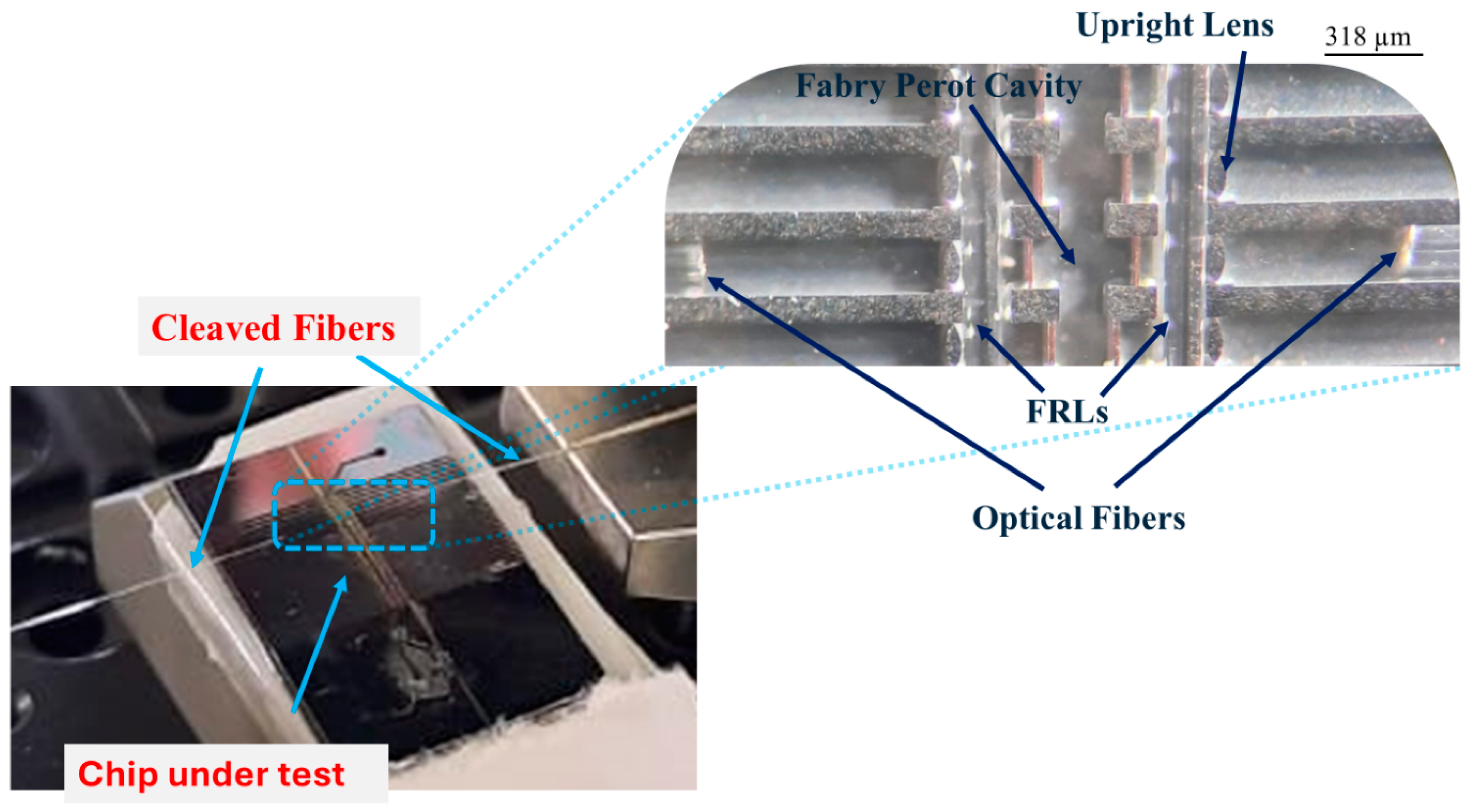
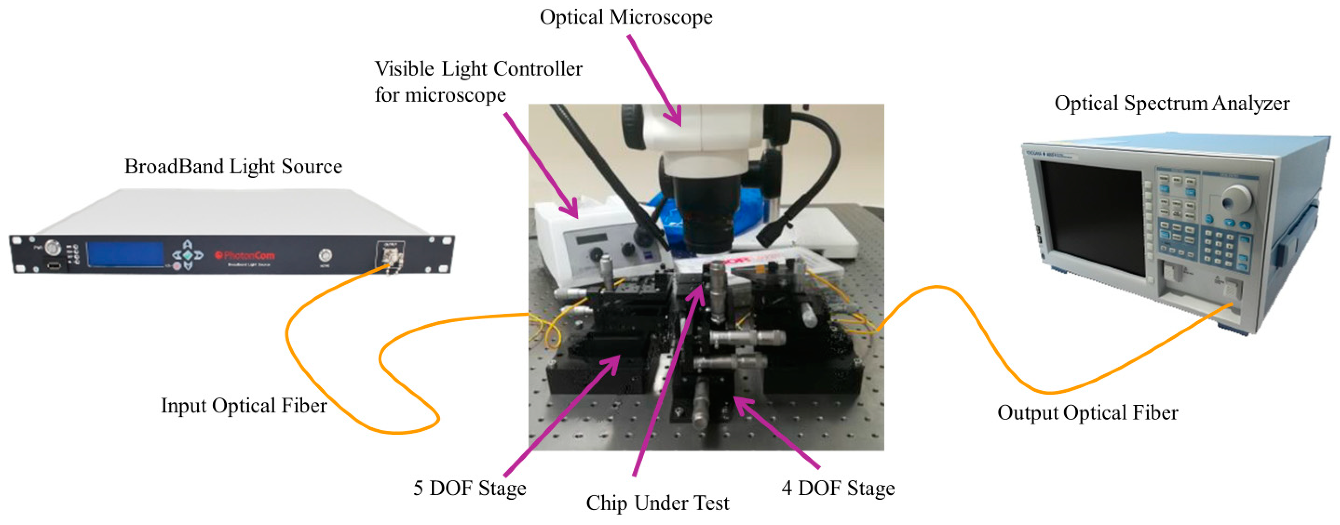
2.3. Experimental Methodology

The characteristics of a fiber, such as its RI, core diameter, and the wavelength at which it operates, directly influence the types and quantities of modes that can propagate through it. In a SMF, the fiber is specifically designed to support only the fundamental mode of light propagation. This design ensures that most of the light’s energy is confined within the core of the fiber, with only a minimal amount extending into the cladding. In our experiment, the single mode fiber (SMF-28-J9 from Thorlabs) has been used in the insertion and receiving of light processes in the operating wavelength. From its datasheet, its mode field diameter is equal to 10.4 µm.
In the experimental setup, shown in Figure 5, one of such fiber cables is used to inject light into the on-chip device, and the transmitted light is collected using another fiber cable. Inserting the fibers exactly at their places is ensured by the etched grooves of widths about 150 µm in the fabricated silicon chip covered by polydimethylsiloxane (PDMS) capping and etched depth over 130 µm to accommodate the bare fibers of diameters about 125 µm. A photo of the fibers inserted into the chip is shown in Figure 5, where the inset shows the microscopic view of a portion of the fabricated chip after insertion of fibers. The fabrication steps can be found in [23].
Figure 6 shows the entire experimental setup for characterizing the on-chip FP micro-device. The alignment and positioning of fibers and chips is handled using alignment stages. A stage of 5 degrees of freedom (DOF) is used for aligning each fiber to maximize power coupling to the chip. Also, a 4 DOF stage is used for positioning the chip under test. For the light source, a broadband light from ‘PhotonCom’ is used, covering the band from 1528 to 1608 nm. The first end of the input fiber is connected to the light source, and the other cleaved end is integrated into the chip under test. The output light from the microcavity is coupled to the cleaved end of the output fiber to carry the signal to the Optical Spectrum Analyzer (OSA), model AQ6374 OSA from YOKOGAWA.
With the experimental setup in place, it is important to consider the operating wavelength limits of the system, particularly in relation to the performance of the FP mirror and the FRL. The bandwidth Δ λ of the FP mirror can be approximated by the following relation:
Δ λ λ o 4 π   sin 1 n 1 n 2 n 1 + n 2 ,
where n 1 is the refractive of the silicon, n 2 is the refractive of air, and λ o is the central wavelength, set as 1560 nm. Hence, the bandwidth can be evaluated as:
Δ λ = 1165   n m
According to the datasheet specifications of the fibers, the operating wavelength is 1260–1625 nm. The FRL is a piece of the same fiber, but it is used as a lens and the light enters it laterally, so the stated bandwidth within which the light is guided as a single mode is not of concern here. After investigating the FRL ability to focus the light as a lens, we found that the focal length becomes larger at smaller wavelengths according to the relation [26]:
W 0 = λ π W 0   f ,
where W 0 is the waist radius at the focal length. The focal length does not show divergence with the wavelength in both side and top views.
All of these bandwidths are much wider than the light source range (from 1528 to 1608 nm). So, they are not expected to limit the performance in our system.

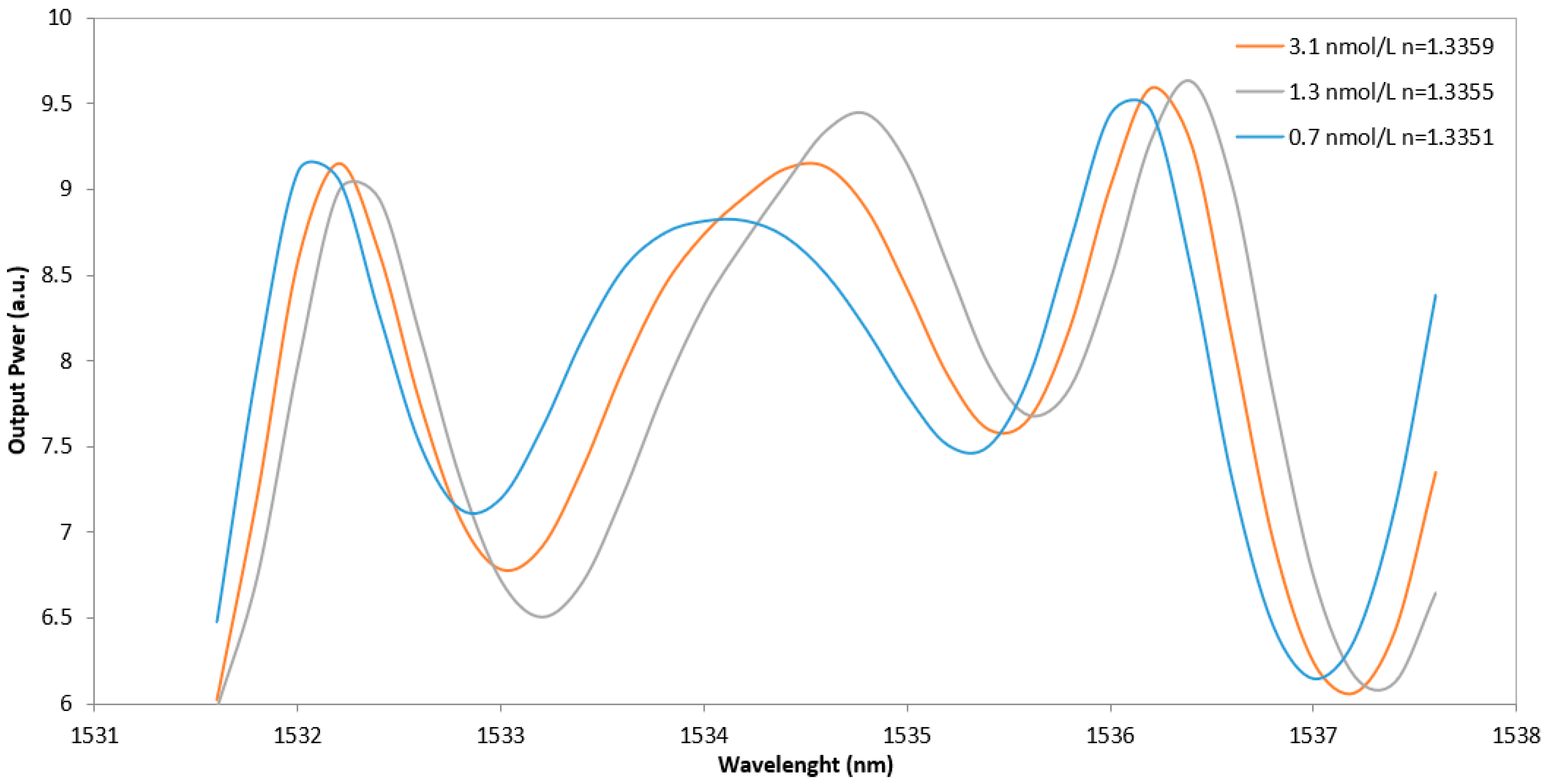
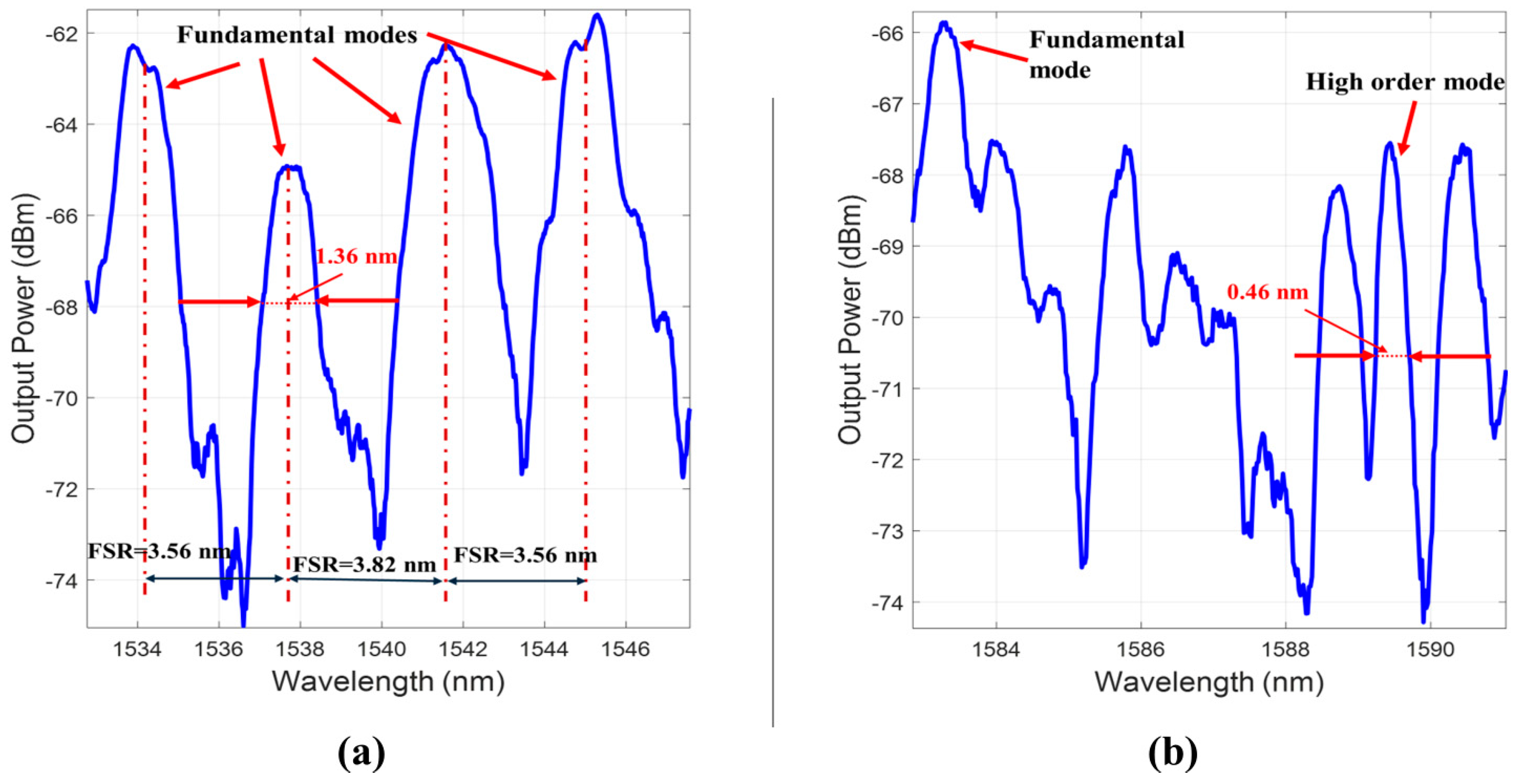
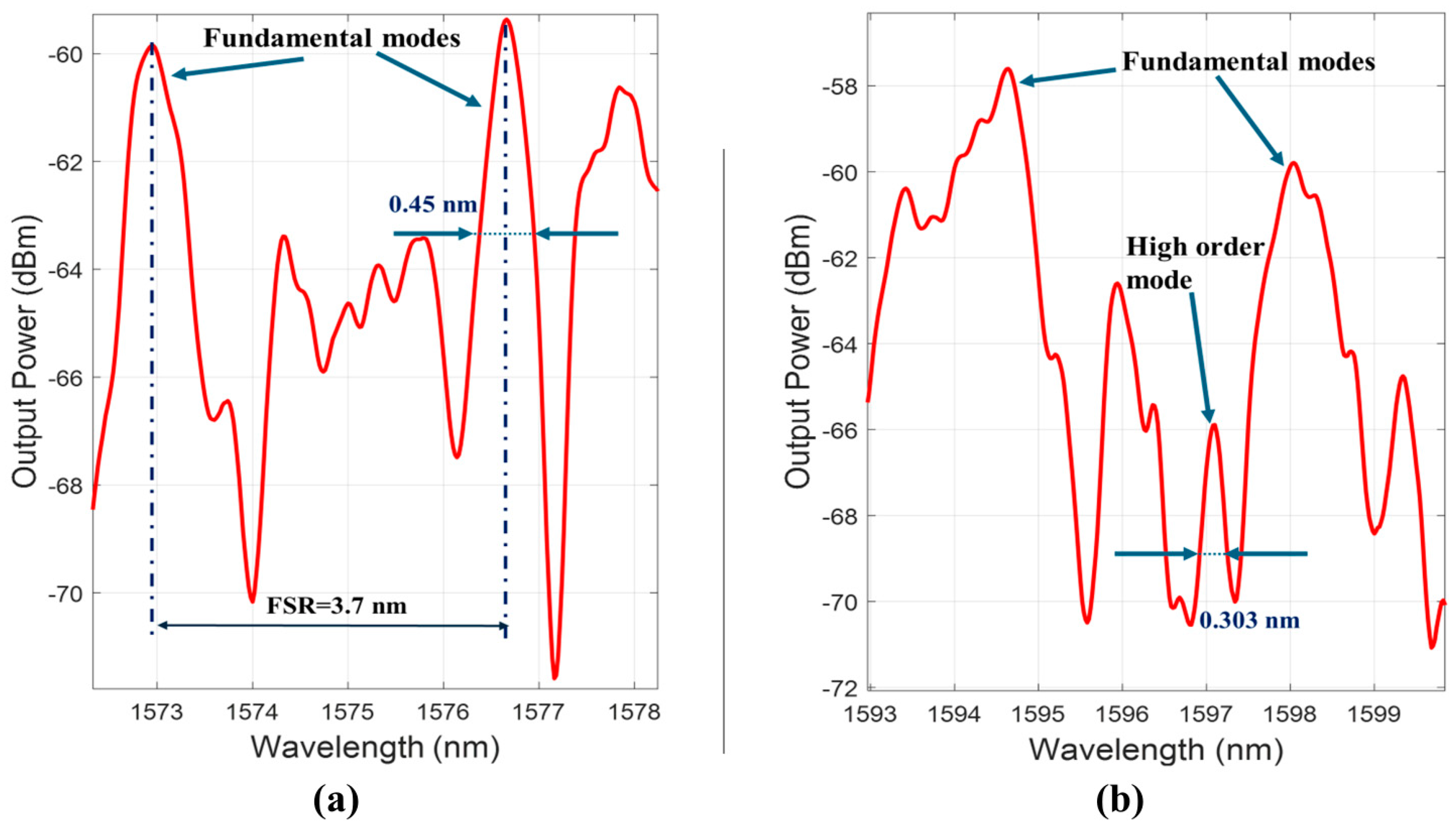
3. Results and Discussion

The experimental investigation is carried out for the chip in Figure 1c with 2 silicon layers (Bragg mirrors) to increase reflectivity, curved mirrors having a radius of curvature of 318 µm and gap of 318 µm filled with air. The transmitted output power is expressed in dBm and calculated by subtracting the output power when the chip is tested from the source power when there is no chip. The cavity achieved a Q-factor of fundamental modes as high as 1100 for a bandwidth of 1.36 nm (Figure 7a) and 3455 for a higher-order mode peak of a bandwidth of 0.46 nm (Figure 7b). There are other peaks that also correspond to the fundamental modes that appear in the spectrum at different wavelengths. Some of these peaks have slightly less Q-factors, which have values of 993, 1131, and 1037, whose average is 1054. However, there are not many side peaks corresponding to higher-order modes. The distance between two consecutive fundamental modes, known as Free Spectral Range (FSR), is found to be around the average of 3.65 nm, which fits well with the analytically determined value of 3.73 nm obtained by the relation:
F S R = λ 2 2   L ,
where λ is the central wavelength of the spectrum, and L is the cavity space.
The experimental investigation is performed also on the design shown in Figure 1d with two-layer Bragg mirrors and a cavity gap of 318 µm. As shown in Figure 8a, the Q factor can reach as high as 3500 for fundamental modes of 0.45 nm linewidth and 5270 for higher-order modes of 0.303 linewidth (Figure 8b). Again, as expected, there are other peaks that also correspond to the fundamental modes that appear in the spectrum. Some of these peaks have still high Q-factor values, such as 3503, 3268, and 3480, whose average is 3417. As for other side peaks corresponding to higher-order modes, some of them have Q-factor values of 5271, 5037, and 5113, whose average is 5140. This value is very competitive for an experimentally tested on-chip extrinsic FP micro-resonator. Again, the FSR calculated is mapping the cavity gap value with an error of only 5%.
To highlight the role of the input beam shape and the focusing lens structures, the coupling efficiency η is calculated from Equation (3) by evaluating the coupling between the fiber GB profile E x , y at the entrance of the cavity after being shaped by the lenses and the Hermite–Gaussian transverse modes inside the cavity ψ l . m x , y . The values are calculated for both structures in Figure 1c,d. Table 1 shows the values of η for both side and top views. Higher-order modes are excited in both designs. This matches with the experimental spectra shown in Figure 7 and Figure 8. Also, confirmed with the simulation results previously discussed, where the side view has one peak corresponding to the fundamental while three peak in the top view, which concludes to the exciting of (0,2) mode. It is worth noting that while certain lens parameters may theoretically improve coupling efficiency for both side and top views, achieving optimal performance in both orientations simultaneously is challenging. This difficulty arises because the lenses used are not perfectly spherical, meaning that enhancing efficiency in one view often comes at the expense of the other. Additionally, practical limitations in lens fabrication and the inherent properties of commercially available fiber components that function as the FRL further constrain the possibility of achieving ideal coupling conditions. These factors collectively make it unrealistic to expect perfect coupling efficiency across all viewing angles.
Table 2 shows a comparison with the experimentally tested FP cavities found in the literature with different specifications and mirrors’ structure regarding the Q-factor. For high Q-factor, as in [30], a cavity space exceeding 100 mm is required. In analogy, the device in [31] has a footprint of 0.205 mm2. In [32], they used polymeric mirrors alongside a chip size of several millimeters. Either a long cavity or different mirror materials are not preferable in the miniaturization trend and broader commercial application. Notably, the reported Q-factor in [22] was achieved using ethanol as the analyte and mirrors of five bilayers; however, even with air as the medium and mirrors with two bilayers only, this work achieved a Q-factor of 5270, which is believed to be enhanced by higher RI analytes.
Further research could explore the integration of these high-performance chips into real-world devices, examining aspects such as durability, cost-efficiency, and integration with other components. Additionally, extending the study to include more diverse analyte materials could provide better performance.

4. Conclusions

In this work, the effectiveness of using cleaved optical fibers to couple light into FP resonators is shown by achieving high-quality factors, which is useful in refractometry applications. Two different designs have been investigated by injecting GB from input SMF into the system. To confine the light beam, on-chip micro-lenses integrated with the system are employed before and after the FP cavity. The calculated coupling efficiency has shown that most of the power is coupled to the first fundamental mode, and it is better for the second design. This design has been simulated to illustrate the confinement and beam matching achieved by the geometry. The experimental work demonstrated high Q-factors for both designs compared with other studies taking into account the analyte and the geometry of the structure used. A Q-factor of up to 5270 has been reached, surpassing many previously reported values. Future work will focus on integrating these high-performance chips into real-world devices, examining durability, cost-efficiency, and compatibility with diverse analyte materials to further enhance performance.

Author Contributions

M.A.M. was responsible for primary manuscript preparation, experimental work, and analytical modeling and calculations. A.M.A. and M.A.M. worked on the simulation. F.M. fabricated the chips and T.B. facilitated the process. N.G. provided the structure idea and chip design, supervised the work, and revised the manuscript. All authors have read and agreed to the published version of the manuscript.

Funding

This research received no external funding.

Informed Consent Statement

Not applicable.

Data Availability Statement

The raw data supporting the conclusions of this article will be made available by the authors on request.

Acknowledgments

Authors are thankful to Mohamed Hatem for his help and support in the analysis of the simulation.

Conflicts of Interest

The authors declare no conflicts of interest.

References

  1. Jali, M.H.; Rahim, H.R.A.; Johari, A.M.; Baharom, M.F.; Ahmad, A.; Yusof, H.H.M.; Harun, S.W. Optical Microfiber Sensor: A Review. J. Physics Conf. Ser. 2021, 2075, 012021. [Google Scholar] [CrossRef]
  2. Floris, I.; Adam, J.M.; Calderón, P.A.; Sales, S. Fiber Optic Shape Sensors: A comprehensive review. Opt. Lasers Eng. 2021, 139, 106508. [Google Scholar] [CrossRef]
  3. Krohn, D.A.; MacDougall, T.; Mendez, A. Fiber Optic Sensor Fundamentals and Applications, 4th ed.; Society of Photo-Optical Instrumentation Engineers (SPIE): Washington, DC, USA, 2015. [Google Scholar] [CrossRef]
  4. Pendão, C.; Silva, I. Optical Fiber Sensors and Sensing Networks: Overview of the Main Principles and Applications. Sensors 2022, 22, 7554. [Google Scholar] [CrossRef]
  5. Bonopera, M. Fiber-Bragg-Grating-Based Displacement Sensors: Review of Recent Advances. Materials 2022, 15, 5561. [Google Scholar] [CrossRef]
  6. Barrias, A.; Casas, J.R.; Villalba, S. A review of distributed optical fiber sensors for civil engineering applications. Sensors 2016, 16, 748. [Google Scholar] [CrossRef] [PubMed]
  7. Mu, X.; Yang, Y.; Wang, J.; Bi, L. High sensitive Mach–Zehnder interferometer for seawater refractive index measurement. Ferroelectrics 2022, 595, 27–34. [Google Scholar] [CrossRef]
  8. Uniyal, A.; Srivastava, G.; Pal, A.; Taya, S.; Muduli, A. Recent Advances in Optical Biosensors for Sensing Applications: A Review. Plasmonics 2023, 18, 735–750. [Google Scholar] [CrossRef]
  9. Senthil, R.; Anand, U.; Krishnan, P. Hollow-core high-sensitive photonic crystal fiber for liquid-/gas-sensing applications. Appl. Phys. A 2021, 127, 282. [Google Scholar] [CrossRef]
  10. Bitarafan, M.H.; DeCorby, R.G. On-Chip High-Finesse Fabry-Perot Microcavities for Optical Sensing and Quantum Information. Sensors 2017, 17, 1748. [Google Scholar] [CrossRef]
  11. Syms, R.; Lohmann, A. MOEMS tuning element for a Littrow external cavity laser. J. Microelectromechan. Syst. 2003, 12, 921–928. [Google Scholar] [CrossRef]
  12. Gaber, N.; Sabry, Y.M.; Marty, F.; Bourouina, T. Optofluidic Fabry-Pérot Micro-Cavities Comprising Curved Surfaces for Homogeneous Liquid Refractometry—Design, Simulation, and Experimental Performance Assessment. Micromachines 2016, 7, 62. [Google Scholar] [CrossRef] [PubMed]
  13. Sabry, Y.; Bourouina, T.; Saadany, B.; Khalil, D. In-plane diffraction loss free optical cavity using coated optical fiber and silicon micromachined spherical mirror. In Proceedings of the SPIE—The International Society for Optical Engineering, San Diego, CA, USA, 25 August 2013. [Google Scholar] [CrossRef]
  14. Malak, M.; Gaber, N.; Marty, F.; Pavy, N.; Richalot, E.; Bourouina, T. Analysis of Fabry-Perot optical micro-cavities based on coating-free all-silicon cylindrical Bragg reflectors. Opt. Express 2013, 21, 2378–2392. [Google Scholar] [CrossRef] [PubMed]
  15. Ali, A.M.; Mansour, M.A.; Ahmed, M.H.S.; Gaber, N.; Marty, F.; Bourouina, T. On-chip Optofluidic Structure for Hormonal Detection Using Stable Fabry-Pérot Resonator. In Proceedings of the 2022 International Conference on Electrical, Computer and Energy Technologies (ICECET), Prague, Czech Republic, 20–22 July 2022. [Google Scholar] [CrossRef]
  16. Xu, H.; Qin, Y.; Hu, G.; Tsang, H.K. Million-Q integrated Fabry-Perot cavity using ultralow-loss multimode retroreflectors. Photon. Res. 2022, 10, 2549–2559. [Google Scholar] [CrossRef]
  17. Wang, Y.; Gao, S.; Wang, K.; Li, H.; Skafidas, E. Ultra-broadband, compact, and high-reflectivity circular Bragg grating mirror based on 220 nm silicon-on-insulator platform. Opt. Express 2017, 25, 6653–6663. [Google Scholar] [CrossRef]
  18. Huang, Q.; Liu, Q.; Xia, J. Traveling wave-like Fabry–Perot resonator-based add-drop filters. Opt. Lett. 2017, 42, 5158–5161. [Google Scholar] [CrossRef]
  19. ElZeiny, W.E.; Sabry, Y.M.; Khalil, D.A. Complex Kernel-based spectrum reconstruction algorithm for cascaded Fabry–Perot interferometric spectrometer. Appl. Opt. 2021, 60, 8999–9006. [Google Scholar] [CrossRef] [PubMed]
  20. El-Sayed, I.S.; Sabry, Y.M.; ElZeiny, W.E.; Badra, N.; Khalil, D.A. Transformation algorithm and analysis of the Fourier transform spectrometer based on cascaded Fabry-Perot interferometers. Appl. Opt. 2018, 57, 7225–7231. [Google Scholar] [CrossRef]
  21. Chin, L.K.; Liu, A.Q.; Lim, C.S.; Lin, C.L.; Ayi, T.C.; Yap, P.H. An optofluidic volume refractometer using Fabry–Pérot resonator with tunable liquid microlenses. Biomicrofluidics 2010, 4, 024107. [Google Scholar] [CrossRef]
  22. Gaber, N.; Sabry, Y.M.; Erfan, M.; Marty, F.; Bourouina, T. High-Q Fabry–Pérot Micro-Cavities for High-Sensitivity Volume Refractometry. Micromachines 2018, 9, 54. [Google Scholar] [CrossRef]
  23. Toraya, A.; Aboulela, N.G.A.; Marty, F.; Bourouina, T. On-chip structure for optofluidic sensing using stable Fabry-Pérot resonator. In MOEMS and Miniaturized Systems XX; SPIE: San Francisco, CA, USA, 2021. [Google Scholar] [CrossRef]
  24. Pal, B.P. Optical Fiber Sensors: A Versatile Technology Platform for Sensing. J. Indian Inst. Sci. 2014, 94, 283–310. [Google Scholar]
  25. Zheng, W. Optic lenses manufactured on fiber ends. In Proceedings of the 2015 Optoelectronics Global Conference (OGC), Shenzhen, China, 29 August 2015; pp. 1–7. [Google Scholar] [CrossRef]
  26. Saleh, B.E.A.; Teich, M.C. Fundamentals of Photonics; John Wiley & Sons: Hoboken, NJ, USA, 1991; ISBN 0-471-83965-5. [Google Scholar]
  27. Yariv, A. Quantum Electronics; Wiley: New York, NY, USA, 1989. [Google Scholar]
  28. Liu, P.Y.; Chin, L.K.; Ser, W.; Chen, H.F.; Hsieh, C.-M.; Lee, C.-H.; Sung, K.-B.; Ayi, T.C.; Yap, P.H.; Liedberg, B.; et al. Cell refractive index for cell biology and disease diagnosis: Past, present and future. Lab Chip 2016, 16, 634–644. [Google Scholar] [CrossRef] [PubMed]
  29. Aly, A.H.; Awasthi, S.K.; Mohamed, A.M.; Matar, Z.S.; Mohaseb, M.A.; Al-Dossari, M.; Tammam, M.T.; Zaky, Z.A.; Amin, A.F.; Sabra, W. Detection of Reproductive Hormones in Females by Using 1D Photonic Crystal-Based Simple Reconfigurable Biosensing Design. Crystals 2021, 11, 1533. [Google Scholar] [CrossRef]
  30. Krupka, J.; Cwikla, A.; Mrozowski, M.; Clarke, R.N.; Tobar, M. High Q-factor microwave Fabry-Perot resonator with distributed Bragg reflectors. IEEE Trans. Ultrason. Ferroelectr. Freq. Control 2005, 52, 1443–1451. [Google Scholar] [CrossRef] [PubMed]
  31. Zhang, W.; Ehteshami, N.; Liu, W.; Yao, J. Silicon-based on-chip electrically tunable sidewall Bragg grating Fabry–Perot filter. Opt. Lett. 2015, 40, 3153–3156. [Google Scholar] [CrossRef]
  32. Testa, G.; Persichetti, G.; Bernini, R. All-polymeric high-Q optofluidic Fabry–Perot resonator. Opt. Lett. 2021, 46, 352–355. [Google Scholar] [CrossRef]
  33. Guertin, R.; Lacasse, T.; Lemieux-Leduc, C.; Bianki, M.-A.; Peter, Y.-A. High-Performance, Low-Cost On-Chip Fabry-Perot Interferometer With Microfluidic Channel Using Single Anisotropic Wet Etching. J. Microelectromechan. Syst. 2023, 32, 347–351. [Google Scholar] [CrossRef]
  34. Leça, J.M.; Magalhães, Y.; Antunes, P.; Pereira, V.; Ferreira, M.S. Real-Time Measurement of Refractive Index Using 3D-Printed Optofluidic Fiber Sensor. Sensors 2022, 22, 9377. [Google Scholar] [CrossRef]
  35. You, K.E.; Uddin, N.; Kim, T.H.; Fan, Q.H.; Yoon, H.J. Highly sensitive detection of biological substances using microfluidic enhanced Fabry-Perot etalon-based optical biosensors. Sensors Actuators B Chem. 2018, 277, 62–68. [Google Scholar] [CrossRef]
  36. Tu, L.; Huang, L.; Wang, W. A novel micromachined Fabry-Perot interferometer integrating nano-holes and dielectrophoresis for enhanced biochemical sensing. Biosens. Bioelectron. 2019, 127, 19–24. [Google Scholar] [CrossRef]
  37. Jian, A.; Wu, Y.; Ji, J.; Li, D.; Zhang, Y.; Xue, X.; Sang, S. Low cell concentration detection by Fabry-Pérot resonator with sensitivity enhancement by dielectrophoresis. Sensors Actuators A Phys. 2021, 331, 112977. [Google Scholar] [CrossRef]
  38. Zhu, J.M.; Shi, Y.; Zhu, X.Q.; Yang, Y.; Jiang, F.H.; Sun, C.J.; Zhao, W.H.; Han, X.T. Optofluidic marine phosphate detection with enhanced absorption using a Fabry–Pérot resonator. Lab Chip 2017, 17, 4025–4030. [Google Scholar] [CrossRef] [PubMed]
  39. Guertin, R.; Bianki, M.-A.; Peter, Y.-A. Low-cost fabrication of an on-chip Fabry-Perot interferometer for dry environmental monitoring. In Proceedings of the 2023 International Conference on Optical MEMS and Nanophotonics (OMN) and SBFoton International Optics and Photonics Conference (SBFoton IOPC), Campinas, Brazil, 30 July–3 August 2023. [Google Scholar] [CrossRef]
Figure 1. Schematics of optical cavity designs using cylindrical and planar mirrors for improved coupling and confinement: (a) Cylindrical mirrors with lensed fibers [14]. (b) Combination of cylindrical mirrors, microtubes, and lensed fibers [12]. (c) Fiber Rod Lens (FRL) with cylindrical mirrors and cleaved SMF [22]. (d) Combination setup integrating planar mirrors, upright lenses, FRL, and cleaved SMF [23].
Photonics 11 00852 g001
Figure 2. Two-dimensional simulation of the optical setup described in Figure 1d showing the electric field distribution and the confinement effects of lenses and FRL on the GB in an FP resonator for (a) the side view and (b) the top view.
Photonics 11 00852 g002
Figure 3. The power distribution at the output fiber region and the whole line above and beneath for the side and top views.
Photonics 11 00852 g003
Figure 4. The output power from the FP cavity for different concentrations of estrogen hormone showing the sensitivity to small changes in RI.
Photonics 11 00852 g004
Figure 5. Experimental setup for testing the optical chip. The inset is a microscope view of the fabricated chip detailing the FP cavity and fiber alignment after insertion.
Photonics 11 00852 g005
Figure 6. Characterization system for the on-chip FP resonators featuring the equipment used: a broadband light source, an input optical fiber connected to the light source, two 5 DOF stages aligning the fiber, a 4 DOF stage holding the chip under test, and an output optical fiber routing light to Optical Spectrum Analyzer. The optical microscope above the setup is used for precise positioning and observation.
Photonics 11 00852 g006
Figure 7. Transmitted power from cylindrical FP cavity described in Figure 1c. (a) Fundamental modes with 1100 Q−factor; (b) high-order mode of 3455 Q−factor.
Photonics 11 00852 g007
Figure 8. Transmission analysis of FP cavity with straight mirrors and external lens described in Figure 1d: (a) Fundamental modes with FSR of 3.7 nm and 3500 Q−factor, (b) High-order mode of 5270 Q−factor.
Photonics 11 00852 g008
Table 1. Coupling efficiency (η) values for the coupling between the shaped fiber GB profile and Hermite-Gaussian transverse modes for side and top views of the structures shown in Figure 1c,d.
Straight Mirrors (Figure 1d)Cylindrical Mirrors (Figure 1c)
Transverse mode order ( l , m ) η Side view Top viewSide view Top view
(0,0) 99.55%48.19%91.99%73.2%
(0,2) 99.55%18.50%91.99%16.99%
(2,0)0.45%48.19%7.07%73.2%
(2,2) 0.45%18.50%7.07%16.99%
Table 2. Comparison of Q-factors achieved by experimentally tested FP cavities from literature.
Ref. Mirror ShapeQ-FactorAnalyte
[30]Straight>106Air
[31]Straight 3.35 × 10⁴N/A
[32]Straight3.03 × 104N/A
[33]Straight2000N/A
[34]Straight106Air
[35]Straight260Air
[36]Straight128.4Deionized Water
[37]Straight 3500Yeast Solution (3.65 × 10⁵ cells/mL)
[38]Straight 270Deionized Water
[39]Straight1150Air
[21]Straight 407n = 1.493
[14]Cylindrical 1059Air
[12]Cylindrical2896Toluene
[22]Cylindrical9896Ethanol
Experimentally tested in this paperCylindrical3455Air
Experimentally tested in this paperStraight5270Air
Disclaimer/Publisher’s Note: The statements, opinions and data contained in all publications are solely those of the individual author(s) and contributor(s) and not of MDPI and/or the editor(s). MDPI and/or the editor(s) disclaim responsibility for any injury to people or property resulting from any ideas, methods, instructions or products referred to in the content.

