Review

Advances in Photoacoustic Endoscopic Imaging Technology for Prostate Cancer Detection

1
School of Medical Informatics and Engineering, Hunan University of Medicine, Huaihua 418000, China
2
School of Basic Medical Sciences, Hunan University of Medicine, Huaihua 418000, China
3
Department of Biomedical Engineering, School of Basic Medical Science, Central South University, Changsha 410083, China
*
Author to whom correspondence should be addressed.
Photonics 2024, 11(9), 872; https://doi.org/10.3390/photonics11090872
Submission received: 21 August 2024 / Revised: 6 September 2024 / Accepted: 11 September 2024 / Published: 18 September 2024
(This article belongs to the Special Issue New Perspectives in Biomedical Optics and Optical Imaging)

Abstract

:
The rapid progress in biomedical imaging technology has generated considerable interest in new non-invasive photoacoustic endoscopy imaging techniques. This emerging technology offers significant benefits, including high spectral specificity, strong tissue penetration, and real-time multidimensional high-resolution imaging capabilities, which enhance clinical diagnosis and treatment of prostate cancer. This paper delivers a thorough review of current prostate cancer screening techniques, the core principles of photoacoustic endoscopy imaging, and the latest research on its use in detecting prostate cancer. Additionally, the limitations of this technology in prostate cancer detection are discussed, and future development trends are anticipated.

1. Introduction

Prostate cancer is the second most common lethal malignancy among men, with incidence rates steadily increasing and a noticeable trend towards younger demographics [1,2,3,4]. The early clinical signs of prostate cancer are frequently subtle, resulting in diagnoses at more advanced stages. Therefore, early detection and intervention are crucial for significantly reducing the mortality rate associated with prostate cancer and improving patient outcomes [5].
The primary diagnostic and screening techniques for prostate cancer in clinical practice include digital rectal examination (DRE), transrectal ultrasound-guided biopsy (TRUS), prostate-specific antigen blood test (PSA), and magnetic resonance imaging (MRI). As prostate cancer lesions are often located in the peripheral zone of the prostate, DRE detects these lesions through palpation of the prostate surface. This method is non-invasive and relatively simple to perform. DRE is commonly utilized for early prostate cancer diagnosis and holds significant value in both diagnosis and staging [6,7,8]. However, DRE has low sensitivity and can be affected by external factors. Current research indicates that the positive predictive value of DRE for diagnosing prostate cancer is only 10%. The traditional screening method, PSA testing, has been extensively debated due to its relatively low sensitivity and specificity [9,10,11,12]. While serum PSA testing can enhance the detection rate of prostate cancer, PSA levels are influenced by various factors, including age, prostate volume, DRE, and inflammation. Acute prostatitis and prostate enlargement can elevate serum PSA levels, reducing specificity and leading to excessive treatment. Moreover, the accuracy of prostate cancer diagnosis improves slightly when additional methods such as PSA density and PSA velocity are employed. Nevertheless, these indicators are similar to PSA and do not fundamentally resolve its limitations [13,14]. TRUS, which uses transrectal ultrasound imaging-guided biopsy after identifying suspicious lesions during a DRE, has relatively poor sensitivity. This is due to most prostate cancer lesions being isoechoic on ultrasound and some benign prostate conditions showing similar features [15,16,17,18]. MRI demonstrates superior sensitivity for prostate cancer detection compared to TRUS. By combining magnetic resonance diffusion–weighted imaging and dynamic contrast-enhanced imaging, multiparametric MRI provides improved accuracy in diagnosing prostate cancer, enhancing both specificity and sensitivity. However, its high cost and long duration limit its use to targeted imaging-guided prostate biopsy, restricting its broader application in prostate cancer diagnosis [19,20,21,22]. The comparison of these technologies is shown in Table 1.

2. Photoacoustic Endoscope Imaging Technique

Photoacoustic endoscopic imaging (PAE) represents an advanced embodiment of non-invasive photoacoustic imaging (PAI) technology. In PAI, when a pulsed laser irradiates biological tissues, it causes local heating and rapid expansion due to transient optical absorption. This process generates ultrasound pulses, which are then detected by an ultrasonic transducer to form an image. The fundamental principle of this hybrid technology is illustrated in Figure 1. This technique uniquely combines the high contrast and specificity of optical imaging with the superior spatial resolution and penetration capabilities of acoustic imaging, making it a promising tool for disease diagnosis in deep soft tissues. PAI provides a strong signal for hemoglobin absorption, with cancerous lesions often exhibiting significant abnormal angiogenesis. Thus, the signal intensity at cancer sites is markedly higher than in normal tissues, resulting in enhanced detection specificity and sensitivity for malignant tissues [23,24,25,26,27,28]. PAE technology extends the imaging principle of PAI by using ultrasound detection signals, effectively addressing the limitations of low penetration depth and spatial resolution associated with traditional optical imaging. Moreover, photoacoustic endoscopy and ultrasound imaging not only share intrinsic compatibility but also have the potential to overcome challenges faced by ultrasound endoscopy in clinical medical testing. PAE offers advantages such as nondestructive detection and reduced radiation exposure to patients. It also mitigates the limitations of light diffusion in tissues, proving valuable in biological sciences. Particularly for tumor detection, this technology allows for precise localization and microdissection of tumor lesions, thereby improving the sensitivity and specificity of tumor detection. These attributes highlight the potential of PAE for early tumor detection and localization, making it suitable for imaging smaller tissue or organ sizes [29,30,31,32,33,34,35]. Despite its promising potential for deep imaging of blood vessels and tissue structures, further research and development are required to enhance its accuracy and reliability in clinical applications, enabling it to play a more significant role in future clinical practice.
In recent years, efforts have been made to enhance the depth of field in photoacoustic images by employing ring array detectors for faster signal reception. However, the high cost of equipment required for processing signals from multiple channels of the ring array, along with the complex manufacturing process of ring array ultrasound transducers, has constrained their use in photoacoustic endoscopy imaging [36,37,38,39,40,41]. Additionally, some researchers have developed a liquid-based acoustic divergent lens to adjust the probe’s focal length and increase its acceptance angle. Nonetheless, this device has limitations due to its slow focal length adjustment rate, leading to fixed focal lengths and unstable performance [42,43]. To improve the imaging resolution of PAE technology, optimization of its system and algorithms is crucial. To tackle the issue of low-resolution imaging, the Xing Da research group introduced an intravascular confocal photoacoustic endoscope with a dual-element ultrasound probe. This novel device captures focused photoacoustic signals, enhancing both resolution and signal-to-noise ratio in intravascular PAI. The team used this endoscope for intravascular photoacoustic tomography of normal and atherosclerotic blood vessels, demonstrating the system’s imaging capabilities. This probe provides valuable technical guidance for intravascular plaque imaging and subsequent clinical diagnosis [29,44]. Jiang Huabei’s research team utilized the PAE system to image early-stage colorectal cancer tissues and normal tissues separately from the human body. They performed statistical analysis of the light absorption intensity distribution at different array element positions, demonstrating the resolution capability of photoacoustic endoscopic imaging for these tissues. This technology shows potential for improving the accuracy of early colorectal cancer diagnosis [30,45]. Additionally, Wang Lihong’s research group developed a non-invasive transvaginal PAI system for human ovaries. Features extracted from the images were analyzed using logical classifiers and support vector machine classifiers to determine whether the ovaries were benign or malignant. This system has been shown to effectively enhance the accuracy of diagnosing early-stage ovarian cancer and distinguishing between benign and malignant ovarian tumors [31,46,47].

3. Research on Photoacoustic Endoscopic Imaging Technology for Prostate Cancer Detection

The image quality of photoacoustic endoscopy is primarily dependent on the stability of the system and the effectiveness of the reconstruction algorithms. This technology is mainly applied in in vivo animal studies and ex vivo human examinations for the diagnosis and treatment of prostate cancer. System-level advancements involve integrating photoacoustic imaging with other medical imaging techniques, such as magnetic resonance imaging and ultrasound imaging, to provide more comprehensive early detection of prostate cancer [48,49]. At the reconstruction algorithm level, various image reconstruction algorithms have been developed, including delay and sum (DAS) [50], backprojection (BP) [51], model-based (MB) [52,53], and synthetic aperture (SAFT) [54] algorithms. These algorithms are designed to accommodate the unique structure of prostate tissue and the characteristics of photoacoustic signals, with the goal of enhancing the endoscopic image quality of prostate cancer tissues.

3.1. Prostate Detection System Utilizing Photoacoustic Endoscopic Imaging Technology

Given that the prostate in a typical male is located at a depth exceeding the penetration capability of the skin surface and considering the inherent optical scattering properties of this tissue in photoacoustic imaging, it is important to note that the typical penetration depth for human skin in photoacoustic imaging is about 3–5 cm [55,56]. Therefore, external light sources are unsuitable for photoacoustic imaging of the normal human prostate. Instead, only built-in light sources can be utilized for simulations and animal experiments. The feasibility of this approach has been validated through various research findings [57,58,59,60,61]. To effectively illuminate the entire prostate while minimizing tissue invasion, the primary challenge in prostate PAI is the method of light source transmission. Researchers have explored two minimally invasive optical transmission methods: transrectal light delivery and transurethral light delivery. Both methods are performed under endoscopic guidance, allowing prostate PAI to provide a comprehensive in vivo image of the prostate. The technology involved in this process, known as transurethral photoacoustic excitation rectal ultrasound detection and prostate photoacoustic scanning, is depicted in Figure 2.
In recent years, several PAI systems have been developed to enhance the early detection of malignant prostate cancer tissues by providing photoacoustic contrast images. Bauer et al. [62] introduced a high-resolution pulse-echo and photoacoustic co-registration ultrasound system designed for tumor microenvironment imaging. This technology enables real-time monitoring of tumor growth in mice implanted with PC-3 prostate tumor cells and allows for functional imaging of intratumoral blood vessels, offering complementary contrast and depth information compared to traditional optical systems. Wang et al. [58] conducted photoacoustic tomography experiments on blood-rich lesion tissues within in vivo and ex vivo prostate tumors of a simulated canine model using a commercial medical ultrasound system. This study explored the potential of this imaging technology for non-invasive prostate cancer imaging and demonstrated that photoacoustic tomography provides superior sensitivity and contrast for deep lesion visualization. The findings suggest that PAI can achieve detailed imaging of prostate tumors, thereby improving tumor localization, disease staging, and recurrence detection. Dogra et al. [63] used multispectral PAI technology to image frozen sections of prostate cancer tissues, effectively distinguishing between malignant prostate tissue, benign prostatic hyperplasia, and normal human prostate tissue. This study highlighted the limitations of current screening methods and demonstrated the specificity and sensitivity of PAI as a valuable tool for the early diagnosis of prostate cancer.

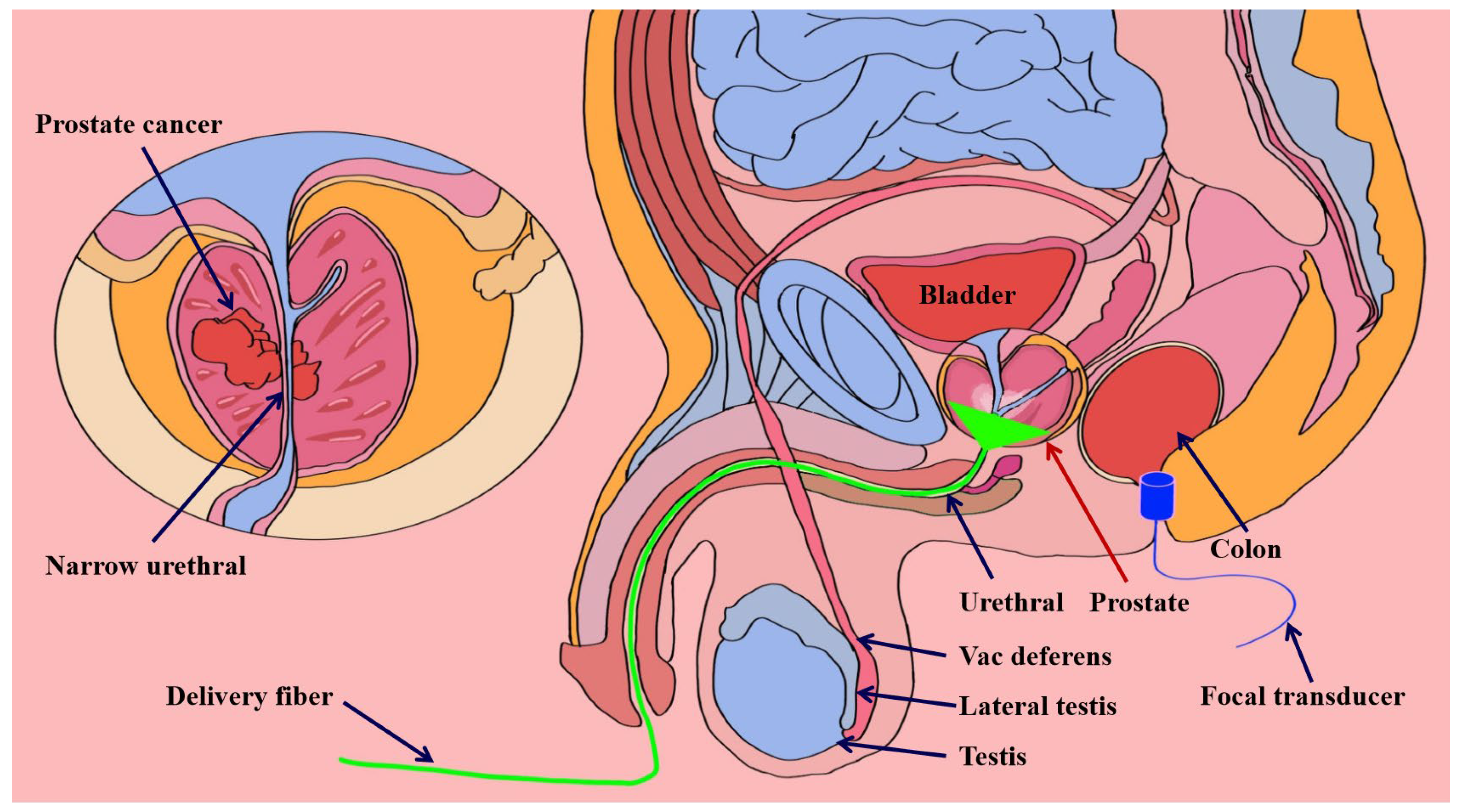
3.1.1. Transrectal Photodelivery

The prevailing method for detecting and diagnosing prostate cancer in clinical practice involves the combination of transrectal ultrasound probes and transrectal optical fiber delivery. This approach is not only widely used but also simple and less likely to cause infection. However, it suffers from significant energy loss due to intense light scattering caused by the rectal wall, making it less effective for imaging the entire living prostate. To address the need for larger imaging sizes and fields of view, as well as to enhance the sensitivity of prostate cancer imaging, Jang et al. [64] developed a novel transrectal ultrasound and photoacoustic imaging probe. This probe features a 20 mm diameter, a 134.5° field of view, a 128-element array, a central frequency of 7 MHz, a bifurcated optical fiber bundle, and two optical lenses. To demonstrate the impact of the new photoacoustic probe on both the field of view and PA signal intensity, the system was used to image tungsten wires at various depths and angles. Figure 3A shows that the photoacoustic image of the tungsten wire signal is significantly weaker without an optical lens (Figure 3(Aa)). In contrast, the application of an optical lens results in a considerably higher-resolution photoacoustic image (Figure 3(Ab)). The normalized envelope profiles of the transverse resolution of the tungsten wire at a depth of 28 mm along the axial direction are shown in Figure 3(Ac,Ad). Based on these findings, photoacoustic imaging was performed on chicken breast samples wrapped in pig intestines, which were then placed over dorsal tumor samples in mice. Additionally, photoacoustic imaging was conducted on all covered pig intestines. The results, depicted in Figure 3B, indicate that it is feasible to obtain combined ultrasound and photoacoustic images of the prostate via the human rectal intestinal tract. This probe facilitates clear visualization of the distribution and morphological changes in prostate microvessels, thereby aiding in the accurate diagnosis of prostate cancer.
Liu et al. [65] developed a highly sensitive, compact transrectal array photoacoustic probe for in vivo imaging of the canine prostate, designed to efficiently transmit laser energy into tissues without loss. The effectiveness of this design was validated through simulation experiments, which improved both laser utilization and the photoacoustic probe’s imaging sensitivity. The probe’s feasibility for prostate cancer detection was confirmed using a canine prostate phantom and in vivo imaging. Figure 4A illustrates the results of Monte Carlo simulations regarding the laser energy distribution within a simulated biological tissue scattering medium, comparing the designed and conventional probes. The image shows the distribution of laser energy density at various imaging depths, with each white box representing the detection area of the ultrasound transducer at its respective depth (Figure 4(Aa)). A comparative analysis of the average laser energy density detected by the designed and conventional probes is shown in Figure 4(Ab). Figure 4(Ac) provides an enlarged view of the imaging depth from the purple box in Figure 4(Ab). These results indicate that the newly designed probe can deliver sufficient laser energy for all imaging depths, demonstrating superior imaging sensitivity compared to traditional probes. The potential of transrectal photoacoustic probes for in vivo imaging applications is thus validated. Additionally, an image of a prostate in a healthy dog model with implanted sutures was captured. Figure 4(Ba,Bb) show the actual images of the prostate before and after suture implantation, respectively. Figure 4(Bc,Be) present combined photoacoustic/ultrasound images of the prostate in the same healthy dog two days post-operation. Horiguchi et al. [66] developed a PAI system integrating traditional linear array ultrasound handheld probes with optical illumination. In vivo model experiments were conducted, gathering prostate peripheral tissue signals from excised specimens using a linear array photoacoustic probe. This probe could display blood vessels with a diameter of 300 µm within a depth of 10 mm, allowing observation of microvascular distribution and nerve fiber consistency. Ishihara et al. [67] utilized a transrectal PAI probe with an array of 128 convex transducers for raw data collection. This probe had an effective scanning range of up to 170°. Compared to pure ultrasound imaging, in vivo PAI demonstrated superior capability in visualizing blood-rich tumors. However, the limitations of the transrectal light delivery method were confirmed in phantom experiments [55,59,60]. During transrectal light transmission, the light must penetrate the rectal wall to reach the prostate. Notably, at a laser wavelength of 1000 nm, the absorption and scattering coefficients of the rectum are two and five times greater than those of the prostate, respectively [55,68,69]. Thus, this mode of transrectal illumination can lead to significant energy loss due to intense light scattering within the rectal wall, making it suboptimal for whole-prostate imaging in vivo. Furthermore, Peng et al. [59,60] have shown that the prostate exhibits asymmetry under transrectal light irradiation, which is advantageous for detecting tumors near the rectum. Therefore, to achieve comprehensive prostate imaging, more uniformly distributed laser illumination is required.

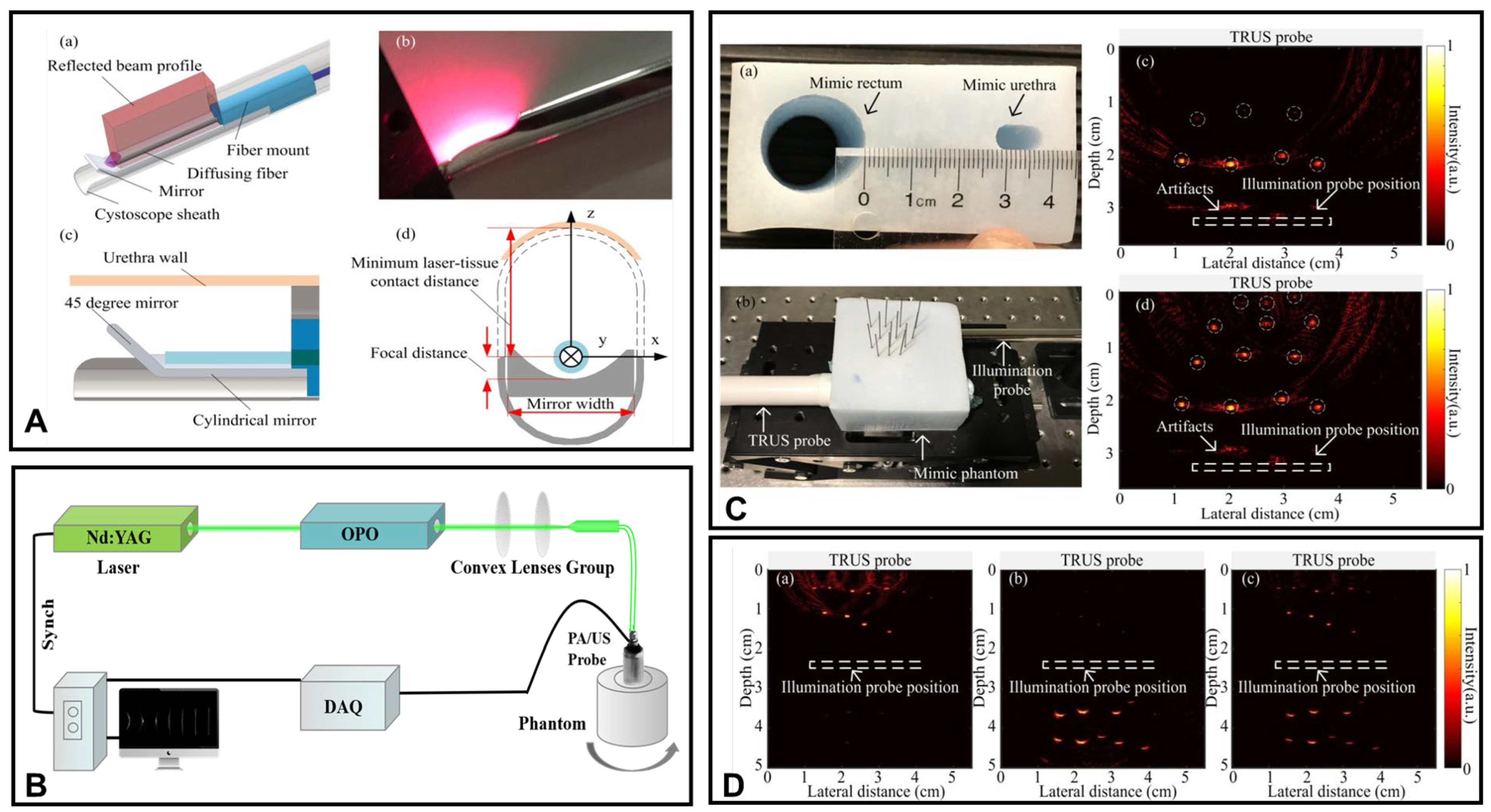
3.1.2. Transurethral Light Delivery

Compared to the rectal wall, transurethral light delivery technology not only results in partial light energy loss due to the significantly lower absorption and scattering coefficients of the urethral wall but also allows for uniform illumination of the prostate as the urethra passes directly through its center. Thus, this technology facilitates the early detection, diagnosis, and treatment of prostate cancer [58]. In 2019, Ai et al. [70] developed a probe for transurethral prostate illumination using high-energy pulses produced by multimode optical fibers. As shown in Figure 5A, the probe incorporates a cylindrical reflector to direct light, achieving parallel side illumination and increasing luminous flux. To overcome the issue of a limited field of view in prostate imaging, the research team introduced an image reconstruction enhancement method based on the variance stochastic gradient descent algorithm. This algorithm’s efficacy in reducing noise and artifacts from linear array sensors was confirmed through numerical simulations and two-dimensional photoacoustic imaging experiments. The imaging system is illustrated in Figure 5B, with photoacoustic tomography of the simulated prostate phantom presented in Figure 5C and PAT images featuring hair embedded within the prostate phantom shown in Figure 5D. The study validated that transurethral illumination photoacoustic imaging technology is practical for prostate detection and diagnosis. Zhang et al. [71] used an ex vivo human prostate model illuminated through the urethral wall to capture photoacoustic and ultrasound images showing its tissue structure alongside an indocyanine green solution. The study revealed that different concentrations of the indocyanine green solution could be effectively visualized within a 2 cm radius of the urethral wall. Therefore, combining transurethral light delivery with transrectal ultrasound detection offers a non-invasive method for photoacoustic molecular imaging of the entire prostate. This method improves the accuracy and diagnostic effectiveness of prostate cancer biopsies, highlighting the potential of whole prostate organ molecular imaging via transurethral photoacoustic tomography. Tang et al. [60] enhanced the maximum penetration depth of photoacoustic imaging technology for prostate cancer detection and treatment monitoring by optimizing the configuration of photoacoustic endoscopy. Their research employed a body model with varying concentrations of photosensitizer and prostate tumors for simulation experiments. The results indicated that the improved PAE configuration for urinary light transmission not only increased imaging penetration depth but also enhanced image quality. Yaseen et al. [57] addressed light attenuation by positioning the ultrasound transducer in the rectum through transurethral optical transmission. However, in this method, where the ultrasound transducer and light source are on the same side, compensating for light attenuation proves challenging. This conclusion was supported by Monte Carlo simulation experiments, which further confirmed that combining transurethral optical transmission with transrectal ultrasound imaging represents the optimal treatment strategy for prostate cancer. Lediju Bell et al. [72,73] investigated the use of photoacoustic imaging for intraoperative updates of brachytherapy plans. The sound waves produced were detected via a transrectal ultrasound probe, and seed positions were verified by integrating postoperative computed tomography images with ex vivo photoacoustic images. This approach provides essential technical support for the diagnosis and treatment of prostate cancer. Subsequently, a side-fiber prototype for transurethral prostate photoacoustic imaging was developed using a dual-array rectal ultrasound probe for experimental application. Imaging of canine prostates with brachytherapy seeds via this transrectal ultrasound probe PAI enhances image contrast and signal-to-noise ratio. Consequently, this optical transmission and beamforming method can improve key detection and treatment strategies for prostate cancer. Brachytherapy seeds are small radioactive capsules that can be permanently implanted in the prostate to treat tumors.
The process of inserting transurethral optical fibers, which facilitates transurethral light transmission, does not add complexity to imaging procedures. Prostate arterial imaging (PAI) provides real-time imaging capabilities and is relatively cost-effective, making it a commonly used clinical detection technology. Consequently, PAI has substantial potential for assisting in the implantation of brachytherapy seeds and monitoring intraprostatic seeds. It also allows for rapid targeted prostate biopsy. Additionally, PAI can distinguish between malignant tumors and benign prostatic hyperplasia, thus preventing unnecessary biopsies in patients with benign prostatic hyperplasia. To confirm the feasibility of PAI for the clinical detection of prostate cancer, additional in vivo experimental studies on transurethral optical fiber insertion are required.

3.2. Research Progress on Photoacoustic Endoscopic Imaging Reconstruction Algorithms

The image reconstruction algorithm, a crucial technology in photoacoustic endoscopy imaging, utilizes the principle of photoacoustic imaging to reverse reconstruct the initial sound field. This process produces an electromagnetic absorption distribution image of the tissue. Currently, most PAE imaging algorithms use methods similar to those in ultrasound imaging. These methods assume straight-line propagation of ultrasound and directly organize ultrasound signals obtained at various angles for reverse projection, thus reconstructing images akin to B-mode sector scanning. However, in practical experiments, due to the use of focused ultrasound transducers for data collection, ultrasound propagation is only about linear near the focal area. As a result, this algorithm achieves optimal tangential resolution only near the focal area. When the target is located further from the focal area, the tangential resolution decreases rapidly, which significantly limits the depth of field of the image. To improve the object’s depth of focus, collecting data with a long focal length ultrasonic transducer is a frequently used method. However, using this kind of detector may compromise the lateral resolution at the focus and affect the quality of the imaging. Therefore, choosing an appropriate PAE reconstruction algorithm is crucial for maintaining the image quality.

3.2.1. General PAE Algorithm

Currently, photoacoustic endoscopic imaging algorithms that rely on stringent models often treat ultrasonic transducers as point detectors. This approach frequently leads to a mismatch between the model’s assumptions and practical application [52,53]. Additionally, due to the inherent limitations of these models and the computational demands they impose, such algorithms significantly increase the complexity of experimental procedures. Consequently, their use in PAE system imaging is relatively restricted. To achieve enhanced lateral resolution for targets, some researchers have proposed techniques that combine synthetic aperture focusing with coherent weighting. This method generates large apertures by summing delayed signals received at various positions. By using signal coherence as a metric for image quality, the focusing quality of the resulting images can be improved further [74,75,76,77,78,79,80,81,82,83]. In this context, Cai et al. [54] developed a side-viewing PAE probe based on SAFT. By optimizing the probe position and rod lens diameter through simulation and employing both the SAFT and coherent factor (CF), they achieved improved lateral resolution over a broad depth of focus in the radial direction, thus enhancing the focusing quality of SAFT images. Empirical data show that this probe provides a lateral resolution of 115–190 µm across a substantial depth of focus of 3.5 mm in the radial direction. Figure 6 demonstrates this, with Figure 6a,b showing cross-sectional images reconstructed using the original algorithm and the SAFT+CF algorithm, respectively. Figure 6c,d present signal images before and after the application of the SAFT+CF algorithm. The use of SAFT+CF facilitates a clearer identification of the positions and orientations of four rendered hairs in 3D, while also reducing background noise and thereby enhancing target resolution. Wang et al. [84] utilized compressed sensing theory and L-2 norm optimization techniques to incorporate sparse prior information into the photoacoustic image reconstruction process, which significantly reduced reconstruction artifacts. Ma et al. [50] proposed a beamforming algorithm employing multiple delays and summations to mitigate potential high sidelobes and substantial artifacts associated with delay and summation techniques. This algorithm not only determines the initial values of the beamforming signal but also computes the entire photoacoustic signal for each pixel, thus eliminating artifacts. The algorithm reorders computation sequences and uses GPU acceleration for parallel computing to meet the requirements of real-time clinical applications. To minimize noise impact on photoacoustic signals, several researchers have proposed effective regularization algorithms for PAI reconstruction [85,86]. Sheu et al. [51] developed a method for intravascular photoacoustic imaging reconstruction and explored the viability of traditional analytical formulas in this context. Unlike conventional photoacoustic tomography, this technique encapsulates the scanning aperture within the imaging target, resulting in limited data collection and pronounced artifacts in the visualization of intravascular structures. To enhance the quality of reconstructed images, iterative expectation maximization and compensation least squares techniques were employed to reduce discrepancies between measured and reconstructed image signals. These results highlight that both iterative expectation maximization and compensation least squares can significantly improve image quality. Analyzing the suitability and limitations of these reconstruction methods is essential for advancing intravascular photoacoustic imaging in clinical settings.

3.2.2. Improved PAE Imaging Algorithm

Wang et al. [87,88] proposed an advanced dynamic focusing back-projection reconstruction algorithm to address the challenge of achieving high resolution with a large depth of field using a fixed-focus transducer in photoacoustic endoscopy. This method, validated through both two-dimensional and three-dimensional experiments, collects data from multiple transducer positions while also accounting for the geometric shape of the transducer detection surface. This approach significantly improves lateral resolution across the entire imaging depth. Extensive numerical simulations and model experiments, as shown in Figure 7, demonstrate that this algorithm enhances the lateral resolution of targets in defocused areas by 30% to 40%. The algorithm has been incorporated into most current acoustic-resolution-based PAE (ARPAE) systems, producing high-quality two-dimensional and three-dimensional images in both focused and defocused areas of the probe. This development may spur further advancements for clinical applications. In 2022, Wang et al. [89] introduced a novel algorithm based on the approximate Gaussian sound field for photoacoustic/ultrasound endoscopic imaging. This algorithm significantly improves resolution and signal-to-noise ratio in out-of-focus regions and includes a model-based dynamic focusing mechanism to enhance its robustness. To evaluate the algorithm’s effectiveness in optimizing the transverse resolution of target points in defocused zones, extensive numerical simulations, chicken breast experiments, and rabbit rectum endoscopic tests were conducted. The results are shown in Figure 8. The algorithm reduced the transverse resolution of the indocyanine green tube in the photoacoustic image from 3.975 mm to 1.857 mm, representing a 52.3% improvement. This advancement enables rapid acoustic resolution photoacoustic/ultrasound dynamic focusing, significantly enhancing the system’s imaging quality and providing valuable insights for designing acoustic resolution photoacoustic/ultrasound endoscopic systems. Image reconstruction algorithms have substantial potential for improving image contrast, resolution, and full-field parameters. Therefore, rapid and precise image reconstruction techniques are essential for ensuring both imaging accuracy and speed [90,91,92,93]. Currently, various accurate and fast reconstruction algorithms are utilized in PAE both nationally and internationally [94,95,96,97,98,99].

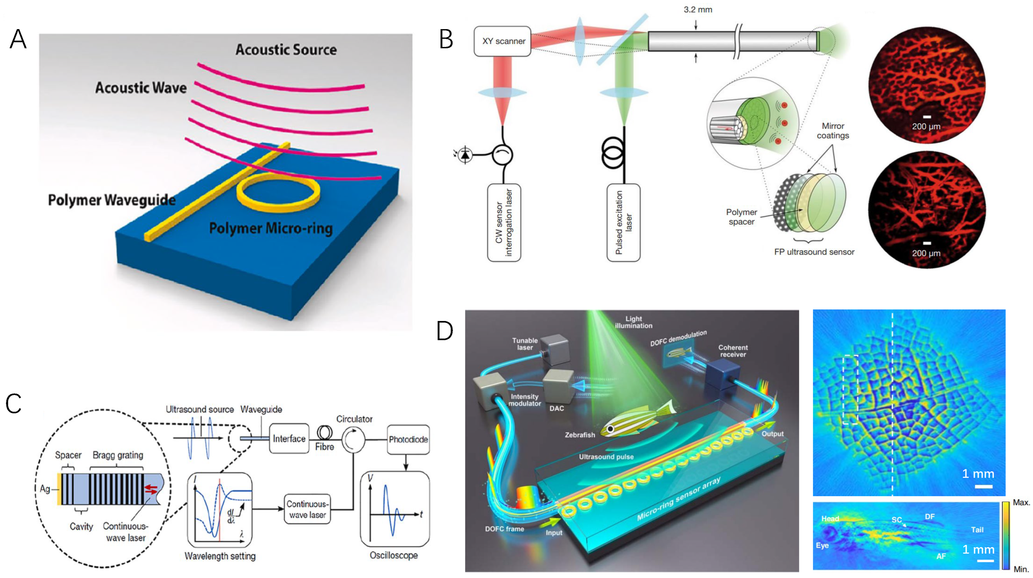
3.3. Optical Ultrasound Sensing Technologies and Their Applications in PAE

Photoacoustic endoscopic imaging (PAE) is a sophisticated technique that not only involves the excitation of light and the detection of ultrasound but also must consider the size and shape of internal human organs. The spatial constraints of these organs present a significant challenge in the design of photoacoustic endoscopic probes compared to traditional optical and ultrasound imaging techniques. Traditional piezoelectric ultrasound sensors lose sensitivity quadratically with size reduction, which hinders the miniaturization necessary for endoscopic probes. Moreover, their susceptibility to electromagnetic interference limits their performance in various applications. Recently, optical ultrasound sensors based on cavity optomechanical sensing have been developed to address these limitations [100,101]. These sensors, characterized by their small size, high sensitivity, large bandwidth, and optical transparency, offer a superior alternative for the design of photoacoustic endoscopic probes. The technology of optical ultrasound sensors can be broadly categorized into optical microring resonators (Figure 9A) [100,102], Fabry–Pérot etalons (Figure 9B) [101], and Bragg grating-based devices (Figure 9C) [100,103].
Fabry–Pérot etalons, for instance, can be implemented using a single optical fiber for photoacoustic endoscopic imaging [54] or with fiber bundles for forward-looking photoacoustic endoscopic imaging [101,104]. Both optical microring resonators [105] and Bragg grating-based silicon-on-insulators [106] have been utilized in PAE, providing high-definition imaging of vascular or rectal vascular networks through lateral scanning. Notably, the silicon-on-insulator-based optical resonator design achieves a per-area sensitivity that is 1000 times higher than that of microring resonators and 100,000,000 times better than that of piezoelectric detectors [103]. Additionally, these probes often feature a transparent structure that allows excitation light to pass through while detecting ultrasound, significantly simplifying the design of photoacoustic endoscopic probes. Arrays of microring resonators have also been developed for photoacoustic tomography (Figure 9D) [107], promising to revolutionize photoacoustic endoscopic imaging by shifting from point-scanning to array-based imaging, thereby greatly enhancing the scanning speed. This advancement in optical ultrasound sensor technology holds great potential to transform the field of photoacoustic endoscopic imaging, offering not only improved diagnostic capabilities but also paving the way for more efficient and non-invasive medical imaging techniques.
Figure 9. Optical ultrasound sensing technologies with different principles. (A) Optical microring resonators, (B) optical sensor based on fiber bundle Fabry–Pérot etalons, (C) Bragg grating-based optical sensor, and (D) optical microring-based ultrasound detector array. (A,C) are adapted with permission from Li et al. [100], (B) is adapted with permission from Fu et al. [101], and (D) is adapted with permission from Pan et al. [107].
Photonics 11 00872 g009

4. Discussion and Conclusions

In recent years, PAE technology has encountered both challenges and opportunities in prostate cancer detection. There is an urgent need to develop innovative imaging technologies and equipment to address growing clinical demands and support real-time monitoring. Simultaneously, existing imaging algorithms must be refined to improve their speed, imaging depth, and resolution. Integrating PAE with magnetic resonance imaging or PET-CT could provide clinicians with a more comprehensive dataset. As artificial intelligence and deep learning technologies progress, their incorporation into PAE can lead to more intelligent and efficient imaging, enhancing the precision and timeliness of diagnoses. In summary, ongoing advancements in PAE systems and algorithms suggest significant potential for growth and extensive application in prostate cancer detection.

Author Contributions

Investigation and research were conducted by N.W., B.L. and H.C.; resources were collected by H.C. and X.D.; data were compiled by N.W. and B.W. All authors have read and agreed to the published version of the manuscript.

Funding

This research was funded by the Natural Science Foundation of Hunan Province, China (Project No. 2021JJ40385, 2022JJ40292), the Scientific Research Project of the Hunan Provincial Department of Education (Project No. 22C1182), and the Innovation and Entrepreneurship Training Program for College Students in Hunan Province (Project No. S202412214036).

Institutional Review Board Statement

Not applicable.

Informed Consent Statement

Not applicable.

Data Availability Statement

Not applicable.

Conflicts of Interest

The authors declare no conflicts of interest.

References

  1. Rawla, P. Epidemiology of prostate cancer. World J. Oncol. 2019, 10, 63–89. [Google Scholar] [CrossRef] [PubMed]
  2. Borno, H.; George, D.J.; Schnipper, L.E.; Cavalli, F.; Cerny, T.; Gillessen, S. All men are created equal: Addressing disparities in prostate cancercare. Am. Soc. Clin. Oncol. Educ. Book 2019, 39, 302–308. [Google Scholar] [CrossRef] [PubMed]
  3. Niu, T.L.; Zhang, Q.C. Research progress of Chinese traditional medicine on the treatment of prostate cancer. Glob. Tradit. Chin. Med. CNKI China 2015, 8, 253–256. [Google Scholar]
  4. Miao, Y.S.U. New progress of traditional Chinese medicine and pharmacy in the treatment of prostate cancer. Glob. Tradit. Chin. Med. CNKI China 2012, 5, 152–156. [Google Scholar]
  5. Descotes, J.L. Diagnosis of prostate cancer. Asian J. Urol. 2019, 6, 129–136. [Google Scholar] [CrossRef]
  6. Andersson, J.; Palsdottir, T.; Lantz, A.; Aly, M.; Grönberg, H.; Egevad, L.; Eklund, M.; Nordström, T. Digital Rectal Examination in Stockholm3 Biomarker-based Prostate Cancer Screening. Eur. Urol. Open Sci. 2022, 44, 69–75. [Google Scholar] [CrossRef]
  7. Roobol, M.J.; Vugt, H.A.V.; Loeb, S.; Zhu, X.; Bul, M.; Bangma, C.H.; VanLeenders, A.G.L.J.H.; Steyerberg, E.W.; Schröder, F.H. Prediction of prostate cancer risk: The role of prostate volume and digital rectal examination in the ERSPC risk calculators. Eur. Urol. 2012, 61, 577–583. [Google Scholar] [CrossRef]
  8. Ankerst, D.P.; Straubinger, J.; Selig, K.; Guerrios, L.; De Hoedt, A.; Hernandez, J.; Liss, M.A.; Leach, R.J.; Freedland, S.J.; Kattan, M.W.; et al. A contemporary prostate biopsy risk calculator based on multiple heterogeneous cohorts. Eur. Urol. 2018, 74, 197–203. [Google Scholar] [CrossRef]
  9. Ferraro, S.; Bussetti, M.; Panteghini, M. Serum prostate-specific antigen testing for early detection of prostate cancer: Managing the gap between clinical and laboratory practice. Clin. Chem. 2021, 67, 602–609. [Google Scholar] [CrossRef]
  10. Halpern, J.A.; Oromendia, C.; Shoag, J.E.; Mittal, S.; Cosiano, M.F.; Ballman, K.V.; Vickers, A.J.; Hu, J.C. Use of digital rectal examination as an adjunct to prostate specific antigen in the detection of clinically significant prostate cancer. J. Urol. 2018, 199, 947–953. [Google Scholar] [CrossRef]
  11. Beemsterboer, P.M.M.; Kranse, R.; Koning, H.J.D.; Habbema, J.D.F.; Schröder, F.H. Changing role of 3 screening modalities in the European randomized study of screening for prostate cancer (Rotterdam). Int. J. Cancer 1999, 84, 437–441. [Google Scholar] [CrossRef]
  12. Schröder, F.H.; Roobol-Bouts, M.; Vis, A.N.; Kwast, T.V.D.; Kranse, R. Prostate-specific antigen-based early detection of prostate cancer—Validation of screening without rectal examination. Urology 2001, 57, 83–90. [Google Scholar] [CrossRef] [PubMed]
  13. Merriel, S.W.D.; Pocock, L.; Gilbert, E.; Creavin, S.; Walter, F.M.; Spencer, A.; Hamilton, W. Systematic review and meta-analysis of the diagnostic accuracy of prostate-specific antigen (PSA) for the detection of prostate cancer in symptomatic patients. BMC Med. 2022, 20, 1–11. [Google Scholar] [CrossRef] [PubMed]
  14. Simmons, M.N.; Berglund, R.K.; Jones, J.S. A practical guide to prostate cancer diagnosis and management. Cleve. Clin. J. Med. 2011, 78, 321–331. [Google Scholar] [CrossRef]
  15. Klotz, L.; Chin, J.; Black, P.C.; Finelli, A.; Anidjar, M.; Bladou, F.; Machado, A.; Levental, M.; Ghai, S.; Chang, S. Comparison of multiparametric magnetic resonance imaging–targeted biopsy with systematic transrectal ultrasonography biopsy for biopsy-naive men at risk for prostate cancer: A phase 3 randomized clinical trial. JAMA Oncol. 2021, 7, 534–542. [Google Scholar] [CrossRef]
  16. Bass, E.J.; Pantovic, A.; Connor, M.J.; Loeb, S.; Rastinehad, A.R.; Winkler, M.; Gabe, R.; Ahmed, H.U. Diagnostic accuracy of magnetic resonance imaging targeted biopsy techniques compared to transrectal ultrasound guided biopsy of the prostate: A systematic review and meta-analysis. Prostate Cancer Prostatic Dis. 2022, 25, 174–179. [Google Scholar] [CrossRef]
  17. Loeb, S.; Gonzalez, C.M.; Roehl, K.A.; Han, M.; Antenor, J.A.V.; Yap, R.L.; Catalona, W.J. Pathological characteristics of prostate cancer detected through prostate specific antigen based screening. Urology 2006, 175, 902–906. [Google Scholar] [CrossRef]
  18. Mottet, N.; Bergh, R.C.N.V.D.; Briers, E.; Broeck, T.V.D.; Catto, J. EAU-EANM-ESTRO-ESUR-SIOG guidelines on prostate cancer—2020 update. Part 1: Screening, diagnosis, and local treatment with curative intent. Eur. Urol. 2021, 79, 243–262. [Google Scholar] [CrossRef]
  19. Ahdoot, M.; Wilbur, A.R.; Reese, S.E.; Lebastchi, A.H.; Mehralivand, S.; Gomella, P.T.; Jonathan Bloom, M.D.; Gurram, S.; Siddiqui, M.; Pinsky, P.; et al. MRI-Targeted, Systematic, and Combined Biopsy for Prostate Cancer Diagnosis. N. Engl. J. Med. 2020, 382, 917–928. [Google Scholar] [CrossRef]
  20. de Rooij, M.; Hamoen, E.H.J.; Fütterer, J.J.; Barentsz, J.O.; Rovers, M.M. Accuracy of multiparametric MRI for prostate cancer detection: A meta-analysis. Ajr Am. J. Roentgenol. 2014, 202, 343–351. [Google Scholar] [CrossRef]
  21. Liu, L.; Tian, Z.; Zhang, Z.; Fei, B. Computer-aided detection of prostate cancer with MRI: Technology and applications. Acad. Radiol. 2016, 23, 1024–1046. [Google Scholar] [CrossRef] [PubMed]
  22. Fusco, R.; Sansone, M.; Granata, V.; Setola, S.V.; Petrillo, A. A systematic review on multiparametric MR imaging in prostate cancer detection. Infect. Agents Cancer 2017, 12, 1–14. [Google Scholar] [CrossRef] [PubMed]
  23. Wang, L.V.; Yao, J. A practical guide to photoacoustic tomography in the life sciences. Nat. Methods 2016, 13, 627–638. [Google Scholar] [CrossRef] [PubMed]
  24. Zhou, Y.; Yao, J.; Wang, L.V. Tutorial on photoacoustic tomography. Biomed. Opt. 2016, 21, 061007. [Google Scholar] [CrossRef]
  25. Maslov, K.I.; Wang, L.V. Photoacoustic imaging of biological tissue with intensity-modulated continuous-wave laser. J. Biomed. Opt. 2008, 13, 024006–024011. [Google Scholar] [CrossRef]
  26. Tang, K.; Zhang, S.; Liang, Z.; Wang, Y.; Ge, J.; Chen, W.; Qi, L. Advanced image post-processing methods for photoacoustic tomography: A review. Photonics 2023, 10, 707–730. [Google Scholar] [CrossRef]
  27. Le, T.D.; Kwon, S.Y.; Lee, C. Segmentation and quantitative analysis of photoacoustic imaging: A review. Photonics 2022, 9, 176. [Google Scholar] [CrossRef]
  28. Guo, H.; Li, Y.; Qi, W.; Xi, L. Photoacoustic endoscopy: A progress review. Biophotonics 2020, 13, e202000217. [Google Scholar] [CrossRef]
  29. Ji, X.; Xiong, K.; Yang, S.; Xing, D. Intravascular confocal photoacoustic endoscope with dual-element ultrasonic transducer. Opt. Express 2015, 23, 9130–9136. [Google Scholar] [CrossRef]
  30. Chen, B.Z.; Hang, Y.; Yang, J.G.; Chi, Z.H.; Jian, R.; Bing, H.; Jiang, H.B. The human colorectal cancer tissue in vitro experimental study based on photoacoustic endoscopic system. Acta Phys. Sin. 2014, 63, 84204. [Google Scholar] [CrossRef]
  31. Aguirre, A.; Ardeshirpour, Y.; Sanders, M.M.; Brewer, M.; Zhu, Q. Potential role of coregistered photoacoustic and ultrasound imaging in ovarian cancer detection and characterization. Transl. Oncol. 2011, 4, 29–37. [Google Scholar] [CrossRef] [PubMed]
  32. Tang, Y.; Wu, H.; Klippel, P.; Zhang, B.; Huang, H.Y.S.; Jing, Y.; Jiang, X.; Yao, J. Deep thrombosis characterization using photoacoustic imaging with intravascular light delivery. Biomed. Eng. Lett. 2022, 12, 135–145. [Google Scholar] [CrossRef]
  33. Yang, J.M.; Favazza, C.; Yao, J.; Chen, R.; Zhou, Q.; Shung, K.K.; Wang, L.V. Three-dimensional photoacoustic endoscopic imaging of the rabbit esophagus. PLoS ONE 2015, 10, e0120269. [Google Scholar] [CrossRef] [PubMed]
  34. Li, Y.; Lu, G.; Zhou, Q.; Chen, Z. Advances in endoscopic photoacoustic imaging. Photonics 2021, 8, 281. [Google Scholar] [CrossRef]
  35. Gao, W.; Mo, H.; Wu, G.; Yang, D.; Yin, L. Compressive endoscopic imaging with complementary light modulation. Appl. Opt. 2021, 60, 8221–8225. [Google Scholar] [CrossRef]
  36. Yang, D.W.; Zeng, L.M.; Ji, X.R.; Huang, Z.; Chen, X.H.; Tan, Z. Fast photoacoustic imaging of blood vessels based on an annular transducer array. Chin. Phys. Lett. 2012, 29, 1–5. [Google Scholar] [CrossRef]
  37. Yang, D.W.; Zhou, Z.B.; Zeng, L.M.; Xin, Z.; Chen, X.H. Photoacoustic Imaging of Animals with an Annular Transducer Array. Chin. Phys. Lett. 2014, 31, 1–4. [Google Scholar] [CrossRef]
  38. Bauer-Marschallinger, J.; Höllinger, A.; Jakoby, B.; Burgholzer, P.; Berer, T. Fiber-optic annular detector array for large depth of field photoacoustic macroscopy. Photoacoustics 2017, 5, 1–9. [Google Scholar] [CrossRef]
  39. Xiao, J.; Wang, T.; Peng, K.; Zhang, X.; Wang, B. Lithography of Aluminum Coated PVDF Annular Array for Photoacoustic Endoscopy. Commun. Comput. Phys. 2018, 23, 561–571. [Google Scholar] [CrossRef]
  40. Passler, K.; Nuster, R.; Gratt, S.; Burgholzer, P.; Paltauf, G. Piezoelectric annular array for large depth of field photoacoustic imaging. Biomed. Opt. Express 2011, 2, 2655–2664. [Google Scholar] [CrossRef]
  41. Wang, H.; Xing, D.; Xiang, L. Photoacoustic imaging using an ultrasonic Fresnel zone plate transducer. J. Phys. D Appl. Phys. 2009, 41, 95111–95118. [Google Scholar] [CrossRef]
  42. Song, C.; Xi, L.; Jiang, H. Acoustic lens with variable focal length for photoacoustic microscopy. Appl. Phys. 2013, 114, 194703–194708. [Google Scholar] [CrossRef]
  43. Song, C.; Xi, L.; Jiang, H. Liquid acoustic lens for photoacoustic tomography. Opt. Lett. 2013, 38, 2930–2933. [Google Scholar] [CrossRef] [PubMed]
  44. Zhang, J.; Yang, S.; Ji, X.; Zhou, Q.; Xing, D. Characterization of lipid-rich aortic plaques by intravascular photoacoustic tomography: Ex vivo and in vivo validation in a rabbit atherosclerosis model with histologic correlation. Am. Coll. Cardiol. 2014, 64, 385–390. [Google Scholar] [CrossRef]
  45. Dai, X.; Yang, H.; Shan, T.; Xie, H.K.; Berceli, S.A.; Jiang, H. Miniature endoscope for multimodal imaging. ACS Photonics 2017, 4, 174–180. [Google Scholar] [CrossRef]
  46. Zhu, Q.C. A review of co-registered transvaginal Photoacoustic and ultrasound Imaging for Ovarian Cancer Diagnosis. Curr. Opin. Biomed. Eng. 2022, 22, 1–9. [Google Scholar] [CrossRef]
  47. Wang, L.; Lei, P.; Wen, X.; Zhang, P.; Yang, S. Tapered fiber-based intravascular photoacoustic endoscopy for high-resolution and deep-penetration imaging of lipid-rich plaque. Opt. Express 2019, 27, 12832–12840. [Google Scholar] [CrossRef]
  48. Kothapalli, S.R.; Sonn, G.A.; Choe, J.W.; Nikoozadeh, A.; Bhuyan, A.; Park, K.K.; Cristman, P.; Fan, R.; Moini, A.; Chullee, B.; et al. Simultaneous transrectal ultrasound and photoacoustic human prostate imaging. Sci. Transl. Med. 2019, 11, eaav2169. [Google Scholar] [CrossRef]
  49. Agrawal, S.; Johnstonbaugh, K.; Clark, J.Y.; Raman, J.D.; Wang, X.; Kothapalli, S.R. Design, development, and multi-characterization of an integrated clinical transrectal ultrasound and photoacoustic device for human prostate imaging. Diagnostics 2020, 10, 566–582. [Google Scholar] [CrossRef]
  50. Ma, X.; Peng, C.; Yuan, J.; Cheng, Q.; Xu, G.; Wang, X.; Carson, P.L. Multiple delay and sum with enveloping beamforming algorithm for photoacoustic imaging. IEEE Trans. Med. Imaging 2019, 39, 1812–1821. [Google Scholar] [CrossRef]
  51. Sheu, Y.L.; Chou, C.Y.; Hsieh, B.Y.; Li, P.C. Image reconstruction in intravascular photoacoustic imaging. IEEE Trans. Ultrason. Ferroelectr. Freq. Control 2011, 58, 2067–2077. [Google Scholar] [CrossRef] [PubMed]
  52. Gamelin, J.; Maurudis, A.; Aguirre, A.; Huang, F.; Guo, P.; Wang, L.V.; Zhu, Q. A real-time photoacoustic tomography system for small animals. Opt. Express 2009, 17, 10489–10498. [Google Scholar] [CrossRef] [PubMed]
  53. Xia, J.; Chatni, M.R.; Maslov, K.; Guo, Z.; Wang, K.; Anastasio, M.; Wang, L.V. Whole-body ring-shaped confocal photoacoustic computed tomography of small animals in vivo. J. Biomed. Opt. 2012, 17, 50506–50509. [Google Scholar] [CrossRef]
  54. Cai, D.; Li, G.; Xia, D.; Li, Z.; Guo, Z.; Chen, S.L. Synthetic aperture focusing technique for photoacoustic endoscopy. Opt. Express 2017, 25, 20162–20171. [Google Scholar] [CrossRef]
  55. El-Gohary, S.H.; Metwally, M.K.; Eom, S.; Jeon, S.H.; Byun, K.M.; Kim, T.S. Design study on photoacoustic probe to detect prostate cancer using 3D Monte Carlo simulation and finite element method. Biomed. Eng. Lett. 2014, 4, 250–257. [Google Scholar] [CrossRef]
  56. Lee, C.H.; Akin-Olugbade, O.; Kirschenbaum, A. Overview of prostate anatomy, histology, and pathology. Endocrinol. Metab. Clin. 2011, 40, 565–575. [Google Scholar] [CrossRef]
  57. Yaseen, M.A.; Ermilov, S.A.; Brecht, H.P.; Su, R.; Oraevsky, A.A. Optoacoustic imaging of the prostate: Development toward image-guided biopsy. J. Biomed. Opt. 2010, 15, 021310. [Google Scholar] [CrossRef] [PubMed]
  58. Wang, X.; Roberts, W.W.; Carson, P.L.; Wood, D.P.; Fowlkes, J.B. Photoacoustic tomography: A potential new tool for prostate cancer. Biomed. Opt. Express 2010, 1, 1117–1126. [Google Scholar] [CrossRef]
  59. Peng, D.Q.; Peng, Y.Y.; Guo, J.; Li, H. Laser illumination modality of photoacoustic imaging technique for prostate cancer. J. Phys. Conf. Ser. 2016, 679, 012026. [Google Scholar] [CrossRef]
  60. Tang, S.; Chen, J.; Samant, P.; Stratton, K.; Xiang, L. Transurethral photoacoustic endoscopy for prostate cancer: A simulation study. IEEE Trans. Med. Imaging 2016, 35, 1780–1787. [Google Scholar] [CrossRef]
  61. Singh, N.; Cherin, E.; Soenjaya, Y.; Matharoo, M.; Brisbane, B.; Papernick, S.; Roa, C.F.; Zheng, G.; Wodlinger, B.; Foster, F.S.; et al. A new photoacoustic imaging platform for potential applications in prostate cancer. In Proceedings of the 2020 IEEE International Ultrasonics Symposium (IUS), Las Vegas, NV, USA, 17 November 2020; pp. 1–4. [Google Scholar]
  62. Bauer, D.R.; Olafsson, R.; Montilla, L.G.; Witte, R.S. 3-D photoacoustic and pulse echo imaging of prostate tumor progression in the mouse window chamber. Biomed. Opt. 2011, 16, 026012. [Google Scholar] [CrossRef] [PubMed]
  63. Dogra, V.S.; Chinni, B.K.; Valluru, K.S.; Joseph, J.V.; Ghazi, A.; Yao, J.L.; Evans, K.; Messing, E.M.; Rao, N.A. Multispectral photoacoustic imaging of prostate cancer: Preliminary ex-vivo results. Clin. Imaging Sci. 2013, 3, 1–8. [Google Scholar] [CrossRef] [PubMed]
  64. Jang, J.; Kim, J.; Lee, H.J.; Chang, J.H. Transrectal ultrasound and photoacoustic imaging probe for diagnosis of prostate cancer. Sensors 2021, 21, 1217. [Google Scholar] [CrossRef]
  65. Liu, C.; Xing, M.; Cong, B.; Qiu, C.; He, D.; Wang, C.; Xiao, Y.; Yin, T.H.; Shao, M.; Qiu, W.B.; et al. In vivo transrectal imaging of canine prostate with a sensitive and compact handheld transrectal array photoacoustic probe for early diagnosis of prostate cancer. Biomed. Opt. Express 2019, 10, 1707–1717. [Google Scholar] [CrossRef]
  66. Horiguchi, A.; Tsujita, K.; Irisawa, K.; Kasamatsu, T.; Hirota, K.; Kawaguchi, M.; Shinchi, M.; Ito, K.; Asano, T.; Shinmoto, H.; et al. A pilot study of photoacoustic imaging system for improved real-time visualization of neurovascular bundle during radical prostatectomy. Prostate 2016, 76, 307–315. [Google Scholar] [CrossRef]
  67. IIshihara, M.; Shinchi, M.; Horiguchi, A.; Shinmoto, H.; Tsuda, H.; Irisawa, K.; Wada, T.; Asano, T. Possibility of transrectal photoacoustic imaging-guided biopsy for detection of prostate cancer. In Proceedings of the Photons Plus Ultrasound Imaging Sensing. SPIE 2017, BiOS, Medicine, Engineerin, San Francisco, CA, USA, 3 March 2017; Volume 10064, pp. 388–392. [Google Scholar]
  68. Agrba, P.D.; Kirillin, M.Y.; Abelevich, A.I.; Kamensky, V.A. Mechanical compression for biotissue image enhancement in optical coherence tomography. In Proceedings of the Saratov Fall Meeting 2009: International School for Junior Scientists and Students on Optics, Laser Physics, and Biophotonics. SPIE, Saratov Fall Meeting 2009, Saratov, Russia, 11 February 2010; Volume 7547, pp. 21–27. [Google Scholar]
  69. Mobley, J.; Vo-Dinh, T.; Tuchin, V.V. Optical properties of tissue. Biomed. Photonics Handb. 2003, 2, 1–2. [Google Scholar]
  70. Ai, M.; Youn, J.I.; Salcudean, S.E.; Rohling, R.; Abolmaesumi, P.; Tang, S. Photoacoustic tomography for imaging the prostate: A transurethral illumination probe design and application. Biomed. Opt. Express 2019, 10, 2588–2605. [Google Scholar] [CrossRef]
  71. Zhang, H.; Huang, S.; Chen, Y.; Xie, W.; Zhang, M.; Pan, J.; Sato, N.; Wang, X.D.; Wu, D.L.; Cheng, Q. Examining the technical feasibility of prostate cancer molecular imaging by transrectal photoacoustic tomography with transurethral illumination. Exp. Biol. Med. 2020, 245, 313–320. [Google Scholar] [CrossRef] [PubMed]
  72. Lediju Bell, M.A.; Kuo, N.P.; Song, D.Y.; Kang, J.U.; Boctor, E.M. In vivo visualization of prostate brachytherapy seeds with photoacoustic imaging. Biomed. Opt. 2014, 19, 126011. [Google Scholar] [CrossRef]
  73. Lediju Bell, M.A.; Guo, X.; Song, D.Y.; Boctor, E.M. Transurethral light delivery for prostate photoacoustic imaging. Biomed. Opt. 2015, 20, 036002. [Google Scholar] [CrossRef]
  74. Liao, C.K.; Li, M.L.; Li, P.C. Optoacoustic imaging with synthetic aperture focusing and coherence weighting. Opt. Lett. 2004, 29, 2506–2508. [Google Scholar] [CrossRef] [PubMed]
  75. Deng, Z.; Yang, X.; Gong, H.; Luo, Q. Two-dimensional synthetic-aperture focusing technique in photoacoustic microscopy. J. Appl. Phys. 2011, 109, 104701. [Google Scholar] [CrossRef]
  76. Turner, J.; Estrada, H.; Kneipp, M.; Razansky, D. Improved optoacoustic microscopy through three-dimensional spatial impulse response synthetic aperture focusing technique. Opt. Lett. 2014, 39, 3390–3393. [Google Scholar] [CrossRef] [PubMed]
  77. Park, J.; Jeon, S.; Meng, J.; Song, L.; Lee, J.S.; Kim, C. Delay-multiply-and-sum-based synthetic aperture focusing in photoacoustic microscopy. Biomed. Opt. 2016, 21, 036010. [Google Scholar] [CrossRef] [PubMed]
  78. Jeon, S.; Park, J.; Managuli, R.; Kim, C. A novel 2-D synthetic aperture focusing technique for acoustic-resolution photoacoustic microscopy. IEEE Trans. Med. Imaging 2018, 38, 250–260. [Google Scholar] [CrossRef]
  79. Cai, D.; Li, Z.; Li, Y.; Guo, Z.; Chen, S.L. Photoacoustic microscopy in vivo using synthetic-aperture focusing technique combined with three-dimensional deconvolution. Opt. Express 2017, 25, 1421–1434. [Google Scholar] [CrossRef]
  80. Cai, D.; Li, Z.; Chen, S.L. Photoacoustic microscopy by scanning mirror-based synthetic aperture focusing technique. Chin. Opt. Lett. 2015, 13, 101101. [Google Scholar] [CrossRef]
  81. Lin, Y.; Zheng, R.; Zhang, X.; Li, Z.; Li, H. Image enhancement of photoacoustic imaging for early endometrial cancer detection by employing a filtered delay multiply and sum beamforming algorithm. AIP Adv. 2019, 9, 125303. [Google Scholar] [CrossRef]
  82. Paul, S.; Mulani, S.; Daimary, N.; Singh, M.S. Simplified-delay-multiply-and-sum-based promising beamformer for real-time photoacoustic imaging. IEEE Trans. Instrum. Meas. 2022, 71, 1–9. [Google Scholar] [CrossRef]
  83. Prakash, R.; Manwar, R.; Avanaki, K. Evaluation of 10 current image reconstruction algorithms for linear array photoacoustic imaging. Biophotonics 2024, 17, e202300117. [Google Scholar] [CrossRef]
  84. Wang, Z.X.; Wang, H.Q.; Ren, S.L. Research on ADMM Reconstruction algorithm of Photoacoustic tomography with limited sampling data. IEEE Access 2021, 9, 113631–113641. [Google Scholar] [CrossRef]
  85. Kong, Q.; Gong, R.; Liu, J.T.; Shao, X. Investigation on reconstruction for frequency domain photoacoustic imaging via TVAL3 regularization algorithm. IEEE Photonics J. 2018, 10, 1–16. [Google Scholar] [CrossRef]
  86. Liu, X.; Zhang, L.; Zhang, Y.; Qiao, L. A photoacoustic imaging algorithm based on regularized smoothed L0 norm minimization. Mol. Imaging 2021, 2021, 6689194. [Google Scholar] [CrossRef] [PubMed]
  87. Wang, B.; Wei, N.N.; Peng, K.; Xiao, J.Y. Modified back-projection method in acoustic resolution based photoacoustic endoscopy for improved lateral resolution. Med. Phys 2018, 45, 4430–4438. [Google Scholar] [CrossRef] [PubMed]
  88. Wang, B.; Wang, C.; Zhong, F.; Pang, W.; Guo, L.; Peng, K.; Xiao, J. 3D acoustic resolution-based photoacoustic endoscopy with dynamic focusing. Quant. Imaging Med. Surg. 2021, 11, 685–696. [Google Scholar] [CrossRef]
  89. Wang, Y.; Yuan, C.; Jiang, J.; Peng, K.; Wang, B. Photoacoustic/Ultrasound Endoscopic Imaging Reconstruction Algorithm Based on the Approximate Gaussian Acoustic Field. Biosensors 2022, 12, 463–477. [Google Scholar] [CrossRef]
  90. Wang, Y.; Xing, D.; Zeng, Y.; Chen, Q. Photoacoustic imaging with deconvolution algorithm. Phys. Med. Biol. 2004, 49, 3117. [Google Scholar] [CrossRef]
  91. Ding, T.; Ren, K.; Vallélian, S. A one-step reconstruction algorithm for quantitative photoacoustic imaging. Inverse Probl. 2015, 31, 095005. [Google Scholar] [CrossRef]
  92. Mozaffarzadeh, M.; Mahloojifar, A.; Orooji, M.; Kratkiewicz, K.; Adabi, S.; Nasiriavanaki, M. Linear-array photoacoustic imaging using minimum variance-based delay multiply and sum adaptive beamforming algorithm. Biomed. Opt. 2018, 23, 026002. [Google Scholar] [CrossRef]
  93. Hakakzadeh, S.; Amjadian, M.; Zhang, Y.; Mostafavi, S.M.; Kavehvash, Z.; Wang, L. Signal restoration algorithm for photoacoustic imaging systems. Biomed. Opt. Express 2023, 14, 651–666. [Google Scholar] [CrossRef]
  94. Wang, H.; Ma, Y.; Yang, H.; Jiang, H.; Ding, Y.; Xie, H. MEMS ultrasound transducers for endoscopic photoacoustic imaging applications. Micromachines 2020, 11, 928. [Google Scholar] [CrossRef] [PubMed]
  95. Zheng, S.; Duoduo, H.; Yuan, Y. 2-D image reconstruction of photoacoustic endoscopic imaging based on time-reversal. Comput. Biol. Med. 2016, 76, 60–68. [Google Scholar] [CrossRef]
  96. Liu, X.; Peng, D.; Guo, W.; Ma, X.; Yang, X.; Tian, J. Compressed sensing photoacoustic imaging based on fast alternating direction algorithm. Int. J. Biomed. Imaging 2012, 2012, 206214. [Google Scholar] [CrossRef]
  97. Gulenko, O.; Yang, H.; Kim, K.; Youm, J.Y.; Kim, M.; Kim, Y.; Yang, J.M. Deep-learning-based algorithm for the removal of electromagnetic interference noise in photoacoustic endoscopic image processing. Sensors 2022, 22, 3961. [Google Scholar] [CrossRef]
  98. Gröhl, J.; Schellenberg, M.; Dreher, K.; Maier-Hein, L. Deep learning for biomedical photoacoustic imaging: A review. Photoacoustics 2021, 22, 100241. [Google Scholar] [CrossRef] [PubMed]
  99. Pang, W.; Wang, Y.; Guo, L.; Wang, B.; Lai, P.; Xiao, J. Two-dimensional photoacoustic/ultrasonic endoscopic imaging based on a line-focused transducer. Front. Bioeng. Biotechnol. 2022, 9, 807633. [Google Scholar] [CrossRef]
  100. Li, B.B.; Ou, L.; Lei, Y.; Liu, Y.C. Cavity optomechanical sensing. Nanophotonics 2021, 10, 2799–2832. [Google Scholar] [CrossRef]
  101. Fu, B.; Cheng, Y.; Shang, C.; Li, J.; Wang, G.; Zhang, C.; Sun, J.; Ma, J.; Ji, X.; He, B. Optical ultrasound sensors for photoacoustic imaging: A narrative review. Quant. Imaging Med. Surg. 2022, 12, 1608. [Google Scholar] [CrossRef]
  102. Westerveld, W.J.; Mahmud-Ul-Hasan, M.; Shnaiderman, R.; Ntziachristos, V.; Rottenberg, X.; Severi, S.; Rochus, V. Sensitive, small, broadband and scalable optomechanical ultrasound sensor in silicon photonics. Nat. Photonics 2021, 15, 341–345. [Google Scholar] [CrossRef]
  103. Shnaiderman, R.; Wissmeyer, G.; Ülgen, O.; Mustafa, Q.; Chmyrov, A.; Ntziachristos, V. A submicrometre silicon-on-insulator resonator for ultrasound detection. Nature 2020, 585, 372–378. [Google Scholar] [CrossRef]
  104. Ansari, R.; Zhang, E.Z.; Desjardins, A.E.; Beard, P.C. All-optical forward-viewing photoacoustic probe for high-resolution 3D endoscopy. Light Sci. Appl. 2018, 7, 75. [Google Scholar] [CrossRef] [PubMed]
  105. Dong, B.; Chen, S.; Zhang, Z.; Sun, C.; Zhang, H.F. Photoacoustic probe using a microring resonator ultrasonic sensor for endoscopic applications. Opt. Lett. 2014, 39, 4372–4375. [Google Scholar] [CrossRef] [PubMed]
  106. Liang, Y.; Fu, W.; Li, Q.; Chen, X.; Sun, H.; Wang, L.; Jin, L.; Huang, W.; Guan, B.O. Optical-resolution functional gastrointestinal photoacoustic endoscopy based on optical heterodyne detection of ultrasound. Nat. Commun. 2022, 13, 7604. [Google Scholar] [CrossRef] [PubMed]
  107. Pan, J.; Li, Q.; Feng, Y.; Zhong, R.; Fu, Z.; Yang, S.; Sun, W.; Zhang, B.; Sui, Q.; Chen, J.; et al. Parallel interrogation of the chalcogenide-based micro-ring sensor array for photoacoustic tomography. Nat. Commun. 2023, 14, 3250. [Google Scholar] [CrossRef]
Figure 1. Principle diagram of photoacoustic imaging.
Photonics 11 00872 g001
Figure 2. Transurethral photoacoustic excitation for ultrasound detection of prostate photoacoustic scanning technology diagram.
Photonics 11 00872 g002
Figure 3. (A) Photoacoustic (PA) image of a tungsten wire target, with (a) depicting the target without and (b) with a developed optical lens. The white arrows in (a) highlight the wire targets that are not discernible in the PA image in (a) but become visible in (b). (c,d) Normalized envelope line profiles of the wire target images, with (c) representing the axial center position and (d) the transverse direction at a depth of 28 mm. (B) The results of the feasibility study on combining ultrasound with photoacoustic imaging in pig intestine are as follows: (a) A photo of the imaging target used for the ex vivo experiment, (b) An integrated ultrasound and photoacoustic image of five graphite rods embedded in chicken breast tissue overlying pig intestine, with the ultrasound and photoacoustic images being log-compressed using dynamic ranges of 55 dB and 25 dB, respectively. (c) A photo of a BALB/c (albino, immunodeficient inbred) mouse overlaid on pig intestine for in vivo experiments, (d) An integrated ultrasound and photoacoustic image of the tumor site in the mouse, with the images being log-compressed using dynamic ranges of 45 dB and 25 dB, respectively. Reproduced with permission from Jang et al. [64].
Photonics 11 00872 g003
Figure 4. (A) The results of Monte Carlo simulations for both the designed and conventional probes in a tissue comparing the laser energy density distribution. (a) This figure displays the distribution of laser energy density across different transverse sections or imaging depths perpendicular to the probe axis. The white rectangles within each image indicate the detection area of the ultrasound transducer at the corresponding depth. (b) A quantitative comparison is provided between the average laser energy density within the ultrasound detection region at various depths for both the designed and conventional probes. (c) An enlarged view of the section of the purple rectangle in (b) is presented. (B) Imaging results from a healthy canine subject, with sutures implanted in the prostate to simulate tumor angiogenesis. (a) Images are shown prior to and (b) following the implantation of the sutures. Ultrasound images (c), photoacoustic images (d), and their integrated image (e) of the canine prostate region are displayed. Reproduced with permission from Liu et al. [65].
Photonics 11 00872 g004
Figure 5. Design of the transurethral illumination probe (A). (a) Illumination of the probe by the urethra. (b) Beam profile of the probe illustrated by 660 nm light. (c) Longitudinal cross-section of the probe. (d) Axial section view of the probe. Fiber-optic transmission high-energy pulsed photoacoustic tomography experimental setup (B). Photoacoustic tomographic imaging of a simulated prostate phantom (C). (a) Phantom photograph (two tubular chambers indicating the positions of the rectum and urethra). (b) Experimental setup. (c,d) PAT images with and without attenuation compensation, respectively, embedded in the phantom with a pencil point. Hair PAT image embedded in a simulated prostate phantom (D). (a) Front illumination of the TRUS probe. (b) Back illumination. (c) Results of combining (a,b). Reproduced with permission from Ai et al. [70].
Photonics 11 00872 g005
Figure 6. Cross-sectional B-mode images at z = 2 mm for the (a) original and (b) SAFT+CF images are provided. Volumetric rendering images of the human hair phantom for the (c) original and (d) SAFT+CF images are also shown. Reproduced with permission from Cai et al. [54].
Photonics 11 00872 g006
Figure 7. Experimental results of the imaging phantom. (a,b) Reconstruction results obtained using the traditional method and the improved method, respectively. (c) A Photograph of the phantom. (d) Received signal for target 1. (e) Amplitude−frequency curve of this signal. (fh) Transverse contours of targets 1, 2, and 3, reconstructed using the traditional method (blue line) and the improved method (red line). Reproduced with permission from Wang et al. [87].
Photonics 11 00872 g007
Figure 8. (a) Experimental images of a rabbit rectal endoscope, (b) rabbit rectal photoacoustic endoscopic images processed by the conventional and improved PAE algorithms, and (c) rabbit rectal ultrasound images processed by the conventional and improved PAE algorithms. Reproduced with permission from Wang et al. [89].
Photonics 11 00872 g008
Table 1. Comparison of the characteristics of several main methods for the diagnosis and screening of early prostate cancer.
MethodSpecificitySensitivityPriceDamage
DRElowerlowergenerallyno
TRUSlowerlowergenerallyno
PSAlowerlowergenerallyyes
MRIhigherhigherhigherno
PAIhigherhighergenerallyno
Disclaimer/Publisher’s Note: The statements, opinions and data contained in all publications are solely those of the individual author(s) and contributor(s) and not of MDPI and/or the editor(s). MDPI and/or the editor(s) disclaim responsibility for any injury to people or property resulting from any ideas, methods, instructions or products referred to in the content.

