Article

Exciton-Resonance-Enhanced Two-Photon Absorption in Three-Dimensional Hybrid Organic–Inorganic Perovskites

by
Xing Ran
,
Xin Xiang
,
Feng Zhou
and
Shunbin Lu
*
International Collaborative Laboratory of 2D Materials for Optoelectronic Science & Technology of Ministry of Education, Institute of Microscale Optoelectronics (IMO), Shenzhen University, Shenzhen 518060, China
*
Author to whom correspondence should be addressed.
Photonics 2025, 12(3), 261; https://doi.org/10.3390/photonics12030261
Submission received: 19 February 2025 / Revised: 7 March 2025 / Accepted: 10 March 2025 / Published: 13 March 2025
(This article belongs to the Special Issue Novel Two-Dimensional Materials Based on Nonlinear Photonics)

Abstract

:
Three-dimensional (3D) hybrid organic–inorganic perovskites (HOIPs) have attracted tremendous interest due to strong excitonic effects and large optical nonlinearities. Taking the advantages, 3D HOIPs show great potential for applications in excitonic and nonlinear devices. However, understanding the relevant mechanisms of exciton-associated nonlinear optical phenomena in 3D perovskites is still challenging. Here, we apply the quantum perturbation theory to calculate the exciton-associated degenerate 2PA spectra of 3D HOIPs. The calculated 2PA spectra of twelve 3D HOIPs are predicted to exhibit resonance peaks at both the sub-band and band edges. The exciton-resonance-associated 2PA coefficients are at least one order of magnitude larger than those of band-to-band transitions and are comparable to those of low-dimensional perovskites. To validate our model, we carried out measurements of the static light-intensity-dependent transmission on MAPbBr3 single crystals. Enhancements of 2PA coefficients are predicted theoretically and observed experimentally with a resonant peak at 1100 nm, indicating intrinsic two-photon transitions to excitonic states in MAPbBr3 single crystals.

1. Introduction

An exciton is an electrically neutral quasi-particle composed of a Coulombically bound electron–hole pair. The Coulomb interaction between an electron and a hole can be either strong or weak, which refers to two types of excitons: Frenkel and Wannier–Mott excitons [1]. In general, Frenkel excitons are generated in organic semiconductors due to the associated low dielectric constant and have large binding energy (0.1~1 eV), while Wannier–Mott excitons favor inorganic semiconductors with relatively small binding energy (~0.01 eV) [2]. The exciton nature in semiconductors provides them with excellent optical and electrical properties, such as clearly resolved spectrum excitonic absorption [3], high photoluminescence (PL) efficiency [4] and long charge carrier lifetime [5]. Furthermore, excitonic effects have been demonstrated to enhance materials’ optical nonlinearities in a resonance manner and make them great potential for various applications in frequency up-conversion [6], biological imaging [7], microfabrication [8], sub-band photodetection [9], optical data storage [10] and multiphoton-pumped laser [11].
Recently, a renewed interest in hybrid organic–inorganic perovskites (HOIPs) has attracted tremendous research attention due to their excitonic nature at room temperature [12]. The HOIPs are a class of semiconducting materials with the chemical formula of ABX3, where A interprets a monovalent organic cation (such as CH3NH3+ = MA+ and CH(NH2)2 + = FA+), B interprets a metal cation (typically Pb2+, Sn2+), and X is the anion referring to halide ions (Cl, Br, I). The B-site cation is six-coordinated by the X-site anion to form [BX6]4− octahedrons, which are corner-sharing to constitute three-dimensional frameworks [13,14]. It is commonly demonstrated that the excitons generated in the bulk perovskites are Wannier–Mott excitons arising from the pronounced dielectric constants in the inorganic parts [15]. The motion of organic parts in HOIPs plays a significant role in the exciton–phonon coupling, broadening the PL spectra at room temperature [16]. The Wannier–Mott excitons in HOIPs are hydrogen-like species and the corresponding exciton binding energy generally ranges from tens to hundreds of meV [17,18]. Astonishingly, the exciton binding energies are sensitive to the dielectric environment and can be finely adjusted via tuning chemical components and multiple structures [19]. For example, MAPbCl3, MAPbBr3 and MAPbI3 single crystals have exciton binding energies around 8~69, 15~150 and 2~45 meV, respectively [20,21]. HOIPs in 2D structures, such as layered (C4H9NH3)2(CH3NH3)n−1PbnI3n+1, (C9H19NH3)2(CH3NH3)n−1PbnI3n+1 (n = 1~4), and (C6H13NH3)2(CH3NH3)n−1PbnI3n+1, are demonstrated to exhibit huge exciton binding energies of several hundred meV due to both quantum and dielectric confinements [22,23,24]. Previous work has shown that the exciton nature in 2D HOIPs enhances the third harmonic generation, as well as multiphoton absorption efficiency by several orders of magnitude [25,26]. A theoretical model based on the quantum perturbation theory has also been proposed for 2PA of (C4H9NH3)n(CH3NH3)n−1PbnI3n+1 (n = 1~4), where excitonic resonances essentially dominate the nonlinear optical process [27]. Despite the rapid progress in such a research field that has been achieved [28,29,30,31], a systematic theoretical understanding of the exciton-associated 2PA is still lacking in 3D HOIPs, which have been demonstrated to exhibit better thermal stability and higher optical damage threshold for applications in NLO devices [32].
Here, we present a theoretical calculation on degenerate 2PA coefficients of 3D HOIPs by applying a quantum perturbation theory. The calculated 2PA spectra of 3D HOIPs are predicted to be enhanced by at least one order of magnitude over the band edge where the incoming photon is on resonance to the energy of excitonic states. An experimental case study of MAPbBr3 single crystals to verify our model was conducted. The static light-intensity-dependent transmission measurement is used to measure the 2PA coefficients of MAPbBr3 single crystals in the near-infrared and visible regions. Excellent agreements between our theory and the experimental findings explicitly confirm a pivotal role of exciton in the 2PA of 3D HOIPs. The excitons in 3D HOIPs provide researchers with an ideal strategy to greatly enhance 2PA for further improving the performance of next-generation photonics and nano-optical devices.

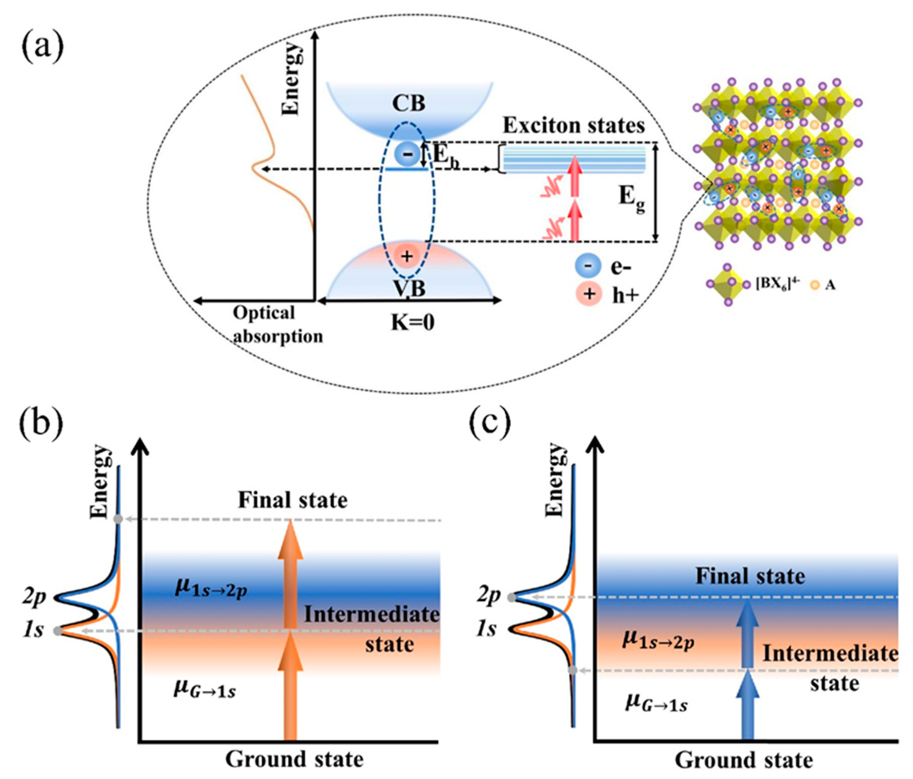
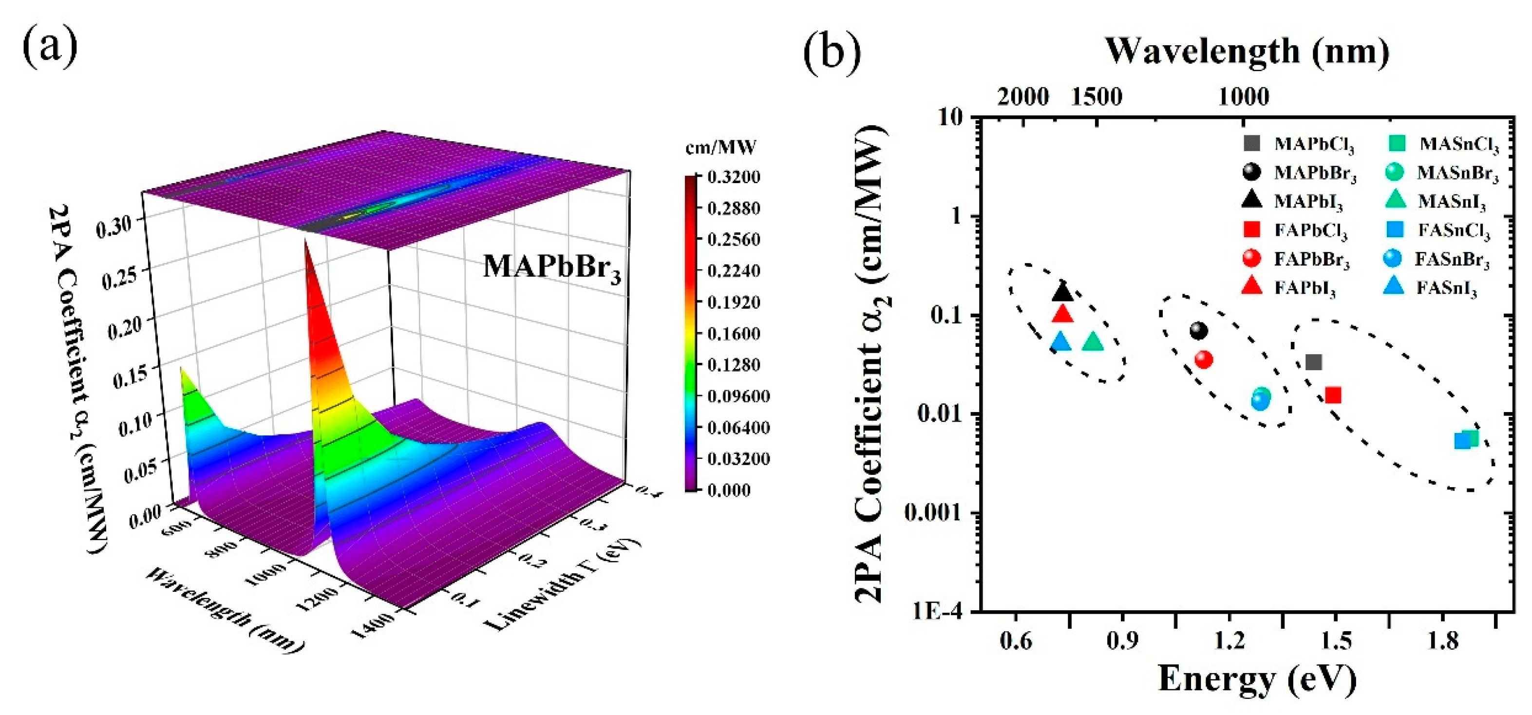
2. Principles and Simulations

Excitons in semiconducting materials are generally characterized via a resonance peak in the optical absorption spectrum, as illustrated in Figure 1a. With a large binding energy, eigenstates of excitons corresponding to the optical resonance are naturally present near the band edge with a Rydberg series. For a simple case with the many-body effects excluded [33], the wavefunction of excitons in bulk crystals is approximated by a 3D hydrogen model as follows [34]:
ψ n , l , m r = 2 n a B 3 D 3 n l 1 ! 2 n n + l ! 3 2 r n a B 3 D l e r n a B 3 D L n l 1 2 l + 1 2 r n a B 3 D Y l m θ , φ
where n, l and m are the principal quantum number, the angular momentum quantum number, and the magnetic quantum number, respectively; a B 3 D corresponds to the effective Bohr radius that is much larger than the lattice space. Such excitons are usually called Wannier (Wannier–Mott) excitons, with the motion extending in three dimensions along their crystal structure. L(x) is the generalized Laguerre polynomial and Y(x) is the spherical harmonics. For 2PA, these excitons act as both the intermediate and the final states for electronic transitions when interacting with incoming photons simultaneously.
In the electronic transitions, the 1s exciton acts as a real intermediate state leading to the primary transition from the ground state. The transition dipole moment is given by the following:
μ s g = ψ s ( r e ) e r e ψ g ( r e )
where ψ s ( r e ) and ψ g ( r e ) are wavefunctions of the 1s-exciton state and the ground state, respectively. Here, ψ g ( r e ) corresponds to the ground state describing the wavefunction of orbital electrons. ψ s ( r e ) is expressed in the formation as ψ s r e = ψ c r e × ψ n = 1 , l = 0 , m = 0 r e r h d r h , with ψ c ( r e ) being the Bloch wave functions for the conduction band and ψ n = 1 , l = 0 , m = 0 ( r e r h ) being the hydrogen wave functions of the 1s exciton.
Following the first electronic transition, the 1s exciton makes an intra-excitonic transition to a p exciton as the final state in the 2PA process, the corresponding transition dipole moment is given by the following:
μ p s = ψ p r e r h e r e r h ψ s r e r h
here, ψ s ( r e r h ) = ψ n = 1 , l = 0 , m = 0 ( r e r h ) is calculated based on the 3D hydrogen model for the 1s exciton. ψ p ( r e r h ) for the p exciton is derived as follows:
ψ p r e r h = ψ n p , l = 1 , m = ± 1 r = r e r h = 2 n a B 3 D 3 n 2 ! 2 n n + 1 ! 3 2 r n a B 3 D e r n a B 3 D L n 2 3 2 r n a B 3 D Y 1 ± 1 θ , φ
With transition dipole moments calculated from Equations (2) and (3), the coefficients of degenerate two-photon resonances with excitons in 3D semiconducting materials can be derived as follows:
α 2 h ν = C N h ν n 0 2 + 2 4 μ s g 2 E s h ν 2 + ( Γ s / 2 ) 2 p μ p s 2 Γ p / 2 π E p 2 h ν 2 + ( Γ p / 2 ) 2
where denotes the energy of an incident photon; N is the density of unit cells; ∑ refers to the summation of all final states; μsg interprets the transition dipole moment from the ground state to a 1s exciton; μps interprets the transition dipole moment from the 1s exciton to an np exciton; Es or Ep refers to a certain excitonic state energy level; Γs or Γp is the linewidth of the corresponding exciton state and is assumed to have the Lorentzian function of density distribution; n0 is the refractive index; and C is a constant with a value of 4.28 × 1043 in units such that α2 is in cm/MW, where N is in units of cm−3, Es, Ep, Γs, and Γp are in units of eV, and μsg and μps are in units of esu.
Here, we assume that only 2p exciton is taken into consideration. As derived from Equation (5), there are resonant peaks at both the sub-band and band edges, indicating intrinsic two-photon transitions to excitonic states. At the sub-band edges, individual excitation photon energy is onresonance to the real intermediate states of E1s and the transition process, as illustrated by Figure 1b. Interestingly, another resonant peak of the α 2 -spectra is predicted near E2p/2 at the band edge, where the resonance with the 2p-exciton state is the final state for 2PA transitions; see Figure 1c. And the peak values of the α 2 -spectra at the band edge are larger than that at the sub-band edge; this is attributed to the higher density of np-exciton states at the band edge for the two-photon resonance. In our model, only one resonance with the discrete excitonic state is achieved to fulfill the energy conservation requirement, and the calculated α 2 -values are proportional to the density of excitonic states on resonance.
It is noted that the linewidth Γ in our model is a variable parameter that is influenced by acoustic- and longitudinal-optical phonon scatterings. Figure 2a plots the calculated α2 values of MAPbBr3 as functions of the excitation wavelength and the linewidth. It is noted that α2 values exhibit two peaks that correspond to the resonance with the discrete excitonic states as mentioned above. The maximal α2 value is calculated to be 0.061 cm/MW at 1112 nm. As Γ decreases from 0.4 to 0.05 eV, peak values of 2PA coefficients exhibit a blue shift and increase by at least one order of magnitude. The empirical fitting to the numerical modeling reveals that α 2 Γ 0.92 .
More importantly, α2 values at the exciton-resonance peak are calculated to be inversely proportional to the excitonic energy level, giving rise to variations in 3D HOIPs with different halide anions (X = Cl, Br, I); see Figure 2b. Furthermore, α2 values of APbX3 are twice that of ASnX3 at the resonance peak, which may be attributed to larger light–matter interaction.

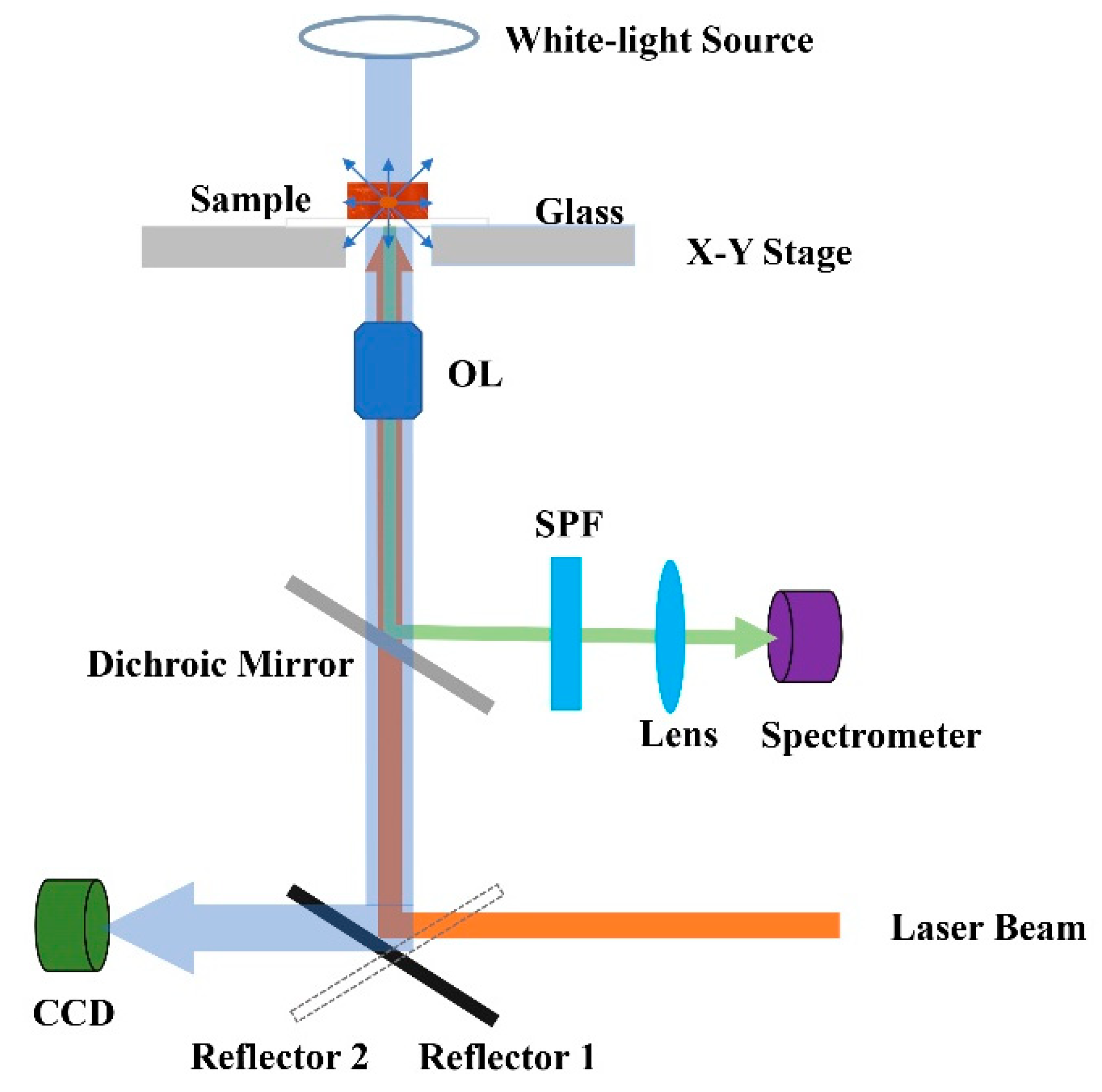
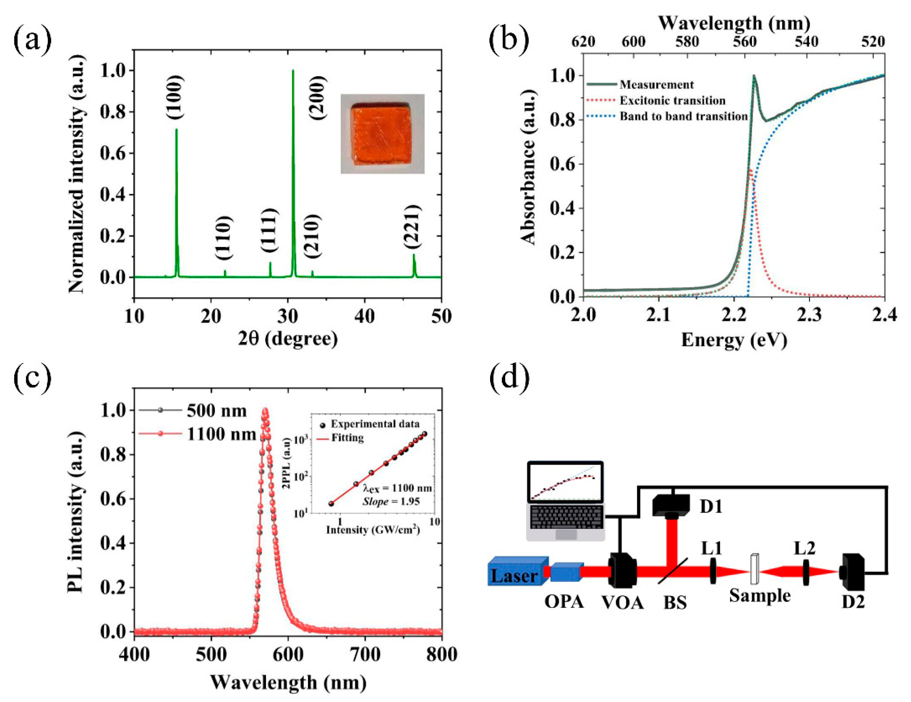
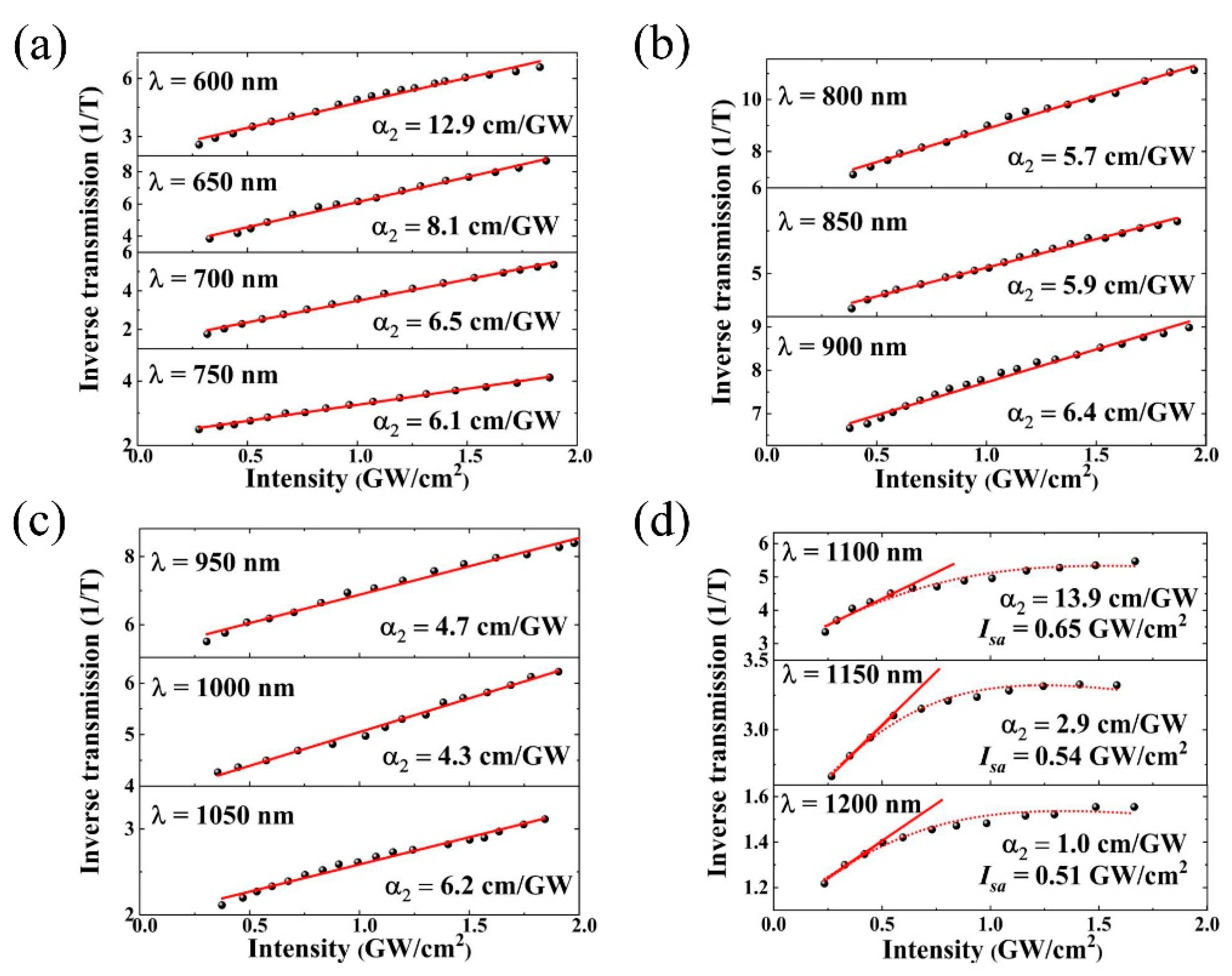
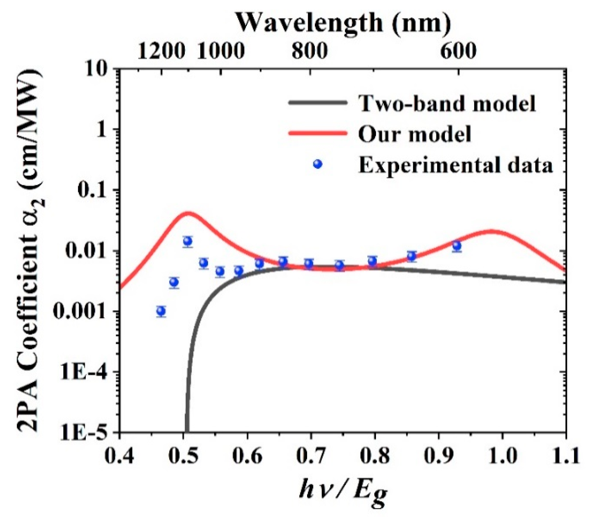
3. Experimental Results and Discussions

In order to demonstrate the enhancement of 2PA in 3D HOIPs, we conducted the 2PA measurements on MAPbBr3 single crystals. Single crystals of MAPbBr3 were synthesized via an antisolvent vapor-assisted growth method (See Appendix A.1) and then prepared in the size of 4 × 4 × 1 mm3; see an image in the inset of Figure 3a. The grew high-quality, millimeter-sized MAPbBr3 single crystals whose shape conformed to the underlying symmetry of the crystal lattice. The phase purity of the as-grown crystals was confirmed by X-ray diffraction (XRD) from a large batch of crystals (See Appendix A.2). The diffraction peaks of the perovskite are located at 15.45°, 31.06°, and 47.52°, which correspond to the lattice planes (100), (200), and (221), which revealed the characteristics of the perovskite square phase. The XRD pattern in Figure 3a illustrates the well-preferred orientation of MAPbBr3 single crystals, in good agreement with the previously reported results [35]. This indicates our MAPbBr3 samples are of high crystalline quality. To characterize the optical properties of these crystals, both linear absorption (See Appendix A.3) and photoluminescence (PL) measurements were carried out. As shown in Figure 3b, a resonance peak near the bandgap rather than a clear band edge cut off in the linear absorption spectrum clearly demonstrates the excitonic effects in MAPbBr3 single crystals. By fitting the measured linear absorption spectrum with a step function and a Lorentzian function, the optical bandgap of MAPbBr3 single crystals (Eg) is estimated to be 2.22 eV and the exciton binding energy (Eb) is 40 meV. Under an excitation of 500 nm laser pulses, the one-photon photoluminescence (1PPL) spectrum was measured and it exhibits a peak located at the wavelength of 570 nm and with a full-width half-maximum (FWHM) of 22 nm, indicating homogeneity of excitonic state; see the black dotted line in Figure 3c. It is noted that the same PL peak occurs under the excitation of a pulsed laser at 1100 nm, implying that 2PA takes place where individual photon energy is near the sub-bandgap. The inset shows two-photon-excited photoluminescence (2PPL) versus laser intensity on the log–log scale for the crystals at 1100 nm, and a nearly square dependence of 2PPL on the laser intensity.
A set of static intensity-dependent transmission measurements was conducted [36]. A schematic diagram of the experimental setup is shown in Figure 3d, where a laser system (Carbide-CB5, Light Conversion) is used with a femtosecond laser source, producing 0.1 mJ, 216 fs (τ), 1030 nm pulses at a repetition rate (T) of 60 kHz. The laser source was utilized to pump an optical parametric generator/amplifier (OPA) (Orpheus, Light Conversion, Lithuania) for the generation of laser pulses with the wavelength tuning from 500 nm to 1200 nm by a step of 50 nm. The output power of the laser was continuously tuned via a variable optical attenuator (VOA) (VA-CB-4-CONEX, Newport, America). The detector D1 (919P, Newport, America) after the beam splitter (BS) (BSW29R, Thorlabs, America) was used as a reference channel to measure the incident laser power (P0), while the detector D2 recorded the laser power transmitted through the sample (Pt). The beam waist (w0) of the focused laser was measured to be ~3 mm at the focal point. The incident light intensity was limited by 2.0 GW/cm2 to avoid thermal effects and laser-induced damage to the sample. To further eliminate the influence of nonlinear refraction, a lens (L2) with a focused length of 10 cm was used to collect signals from the sample. The intensity-dependent transmission of MAPbBr3 single crystals was recorded as T = Pt/P0.
Inverse transmissions (1/T) versus light intensity are plotted in Figure 4. At low-intensity light (I < 0.5 GW/cm2), the measured inverse transmissions exhibit linear increase, which manifests a typical characteristic for 2PA, and it is in accordance with the non-saturation model. To extract the 2PA coefficients α2 of MAPbBr3 single crystals, we fit the intensity-dependent transmission with the following expression:
1 T ( I ) = ( 1 R ) α 2 I L e f f + 1 ( 1 R ) 2 e α 0 L
where R is the reduction factor caused by the reflection and scattering. α0 is the linear absorption coefficient and Leff = [1 − exp(−α0L)]/α0 is the effective length of the sample, with L = 1 mm being thickness of the sample. Using Equation (7), 2PA coefficients of MAPbBr3 single crystals are fitted to be in the range of 4.3~12.9 cm/GW, which are on the same order of magnitude as those reported previously at the same wavelength [37,38]. On the other hand, the measured transmission tends to be stable at high light intensity and with excitation wavelengths of 1100, 1150, and 1200 nm; see Figure 4d. This indicates a saturation of 2PA associated with excitonic states that exist below the band edge. These dark excitonic states (or np excitons) provide MAPbBr3 single crystals with 2PA where individual photon energy is below the sub-bandgap. The saturation process of the MAPbBr3 single crystals can be well fitted using homogeneously broadened systems:
α 2 - sa ( I ) = α 2 1 + ( I / I s a ) 2
here, α 2 - s a is the saturated 2PA coefficient, and Isa is the saturable intensity of 2PA from which the density of excitonic states can be estimated as N = I s a τ 2 h ν π ω 2 . With the best fit, the density of excitonic states involved in the 2PA is estimated to be 1.09 × 1014 cm−3, 8.91 × 1013 cm−3, and 7.27 × 1013 cm−3 for 1100, 1150, and 1200 nm, respectively. It is consistent with the Lorentz distribution of excitonic states in our model.
The experimental measured 2PA coefficients of MAPbBr3 single crystals are compared to calculated results from our theoretical model and the two-band model [39]. The 2PA coefficients are plotted as a function of hν/Eg, as shown in Figure 5. It is noted that the experimentally extracted α2 values exhibit a well-defined peak over the sub-band edge (0.45 < hv/Eg < 0.6), featuring 2PA associated with exciton effects in MAPbBr3 single crystals. The maximal α2 value is measured to be 13.9 cm/GW at the excitation wavelength of 1100 nm, corresponding to the resonance with the 2p-excitonic state or the final state in 2PA transitions (2hv = E2p). As the laser wavelength is tuned from 750 to 600 nm, the experimentally extracted α2 values show a rising trend, implying another resonance peak near the bandgap (hv~E1s). The fitting results derived from our model shown by the red solid curve are in good agreement with the experimental data in this wavelength range. However, there is still a discrepancy within one order of magnitude between results from our theoretical model and the experimental data at wavelengths near 1050 nm. It is explained by thermal and scattering effects during measurement, which hinders the intrinsic NLO properties of our samples. More importantly, both experimental and theoretical calculated α2 values are enhanced by at least one order of magnitude as compared to the results from the two-band model; see the black solid curve in Figure 5. This demonstrates that exciton effects play a significant role in the 2PA in MAPbBr3 single crystals.

4. Conclusions

In summary, 2PA associated with excitonic effects are theoretically studied in 3D HOIPs based on the quantum perturbation theory. They are at least one order of magnitude larger than the values predicted by the band-to-band transitions and are comparable to those of low-dimensional perovskites. To demonstrate our theory, 2PA spectra of MAPbBr3 single crystals are calculated in the near-infrared and visible regions. The 2PA coefficients of MAPbBr3 single crystals were determined via the static intensity-dependent transmission measurement and confirmed the exciton-associated 2PA enhancements. The excitons in 3D HOIPs provide researchers with an ideal strategy to enhance 2PA to further improve the performance of next-generation photonics and nano-optical devices.

Author Contributions

X.R. and X.X. contributed equally to this work. Conceptualization, S.L.; methodology, F.Z. and S.L.; validation, S.L.; formal analysis, X.R.; investigation, X.R. and X.X.; resources, S.L.; data curation, X.R. and X.X.; writing—original draft preparation, X.R. and X.X.; writing—review and editing, F.Z. and S.L.; visualization, X.R.; supervision, S.L.; project administration, S.L.; funding acquisition, S.L. All authors have read and agreed to the published version of the manuscript.

Funding

This research was funded by the Shenzhen Science and Technology Program (Grant Number JCYJ20220531103016036) and the Shenzhen Key Laboratory of 2D Meta-materials for Information Technology (Grant Number ZDSYS201707271014468).

Institutional Review Board Statement

Not applicable.

Informed Consent Statement

Not applicable.

Data Availability Statement

Data are contained within the article.

Conflicts of Interest

The authors declare no conflicts of interest.

Appendix A. Experimental Section

Appendix A.1. Synthesis of MAPbBr3 Single Crystals

We employed a vapor-assisted single-crystal growth technique to obtain samples. The methylammonium halide precursors CH3NH3Br (CH3NH3 = MA) were synthesized through the reaction of hydrohalide acid HBr with methylamine followed by recrystallization from ethanol. An equimolar amount of HBr acid solution in water was added dropwise into the methylamine (45% in methanol) at 0 °C under stirring. Then, the mixture was stirred for 2 h at 0 °C. Removal of the solvent was followed by recrystallization from ethanol to yield snow-white MABr crystals. PbBr2 and MABr were dissolved in N, N-dimethylformamide (DMF). MAPbBr3 single crystals were grown, along with the slow diffusion of the vapor of the anti-solvent dichloromethane (DCM) into the solution. The resulting crystals are orange in color and millimeter-sized with regular rectangular facets. The synthesized crystals were of sufficient quality and macroscopic dimensions, which enabled accurate measurements of two-photon absorption for the longer propagation distance.

Appendix A.2. X-Ray Diffraction Characterization

Single-crystal X-ray diffraction to verify the crystal structure was performed on ULTIMA IV (Rigaku, Japan). For a highly sensitive analysis of a small (~mm diameter) section of material, the diffraction system was equipped with Cross Beam Optics (CBO), a 3 kW sealed tube X-ray source and a D/teX high-speed two-dimensional X-ray detector. A seamlessly integrated X-Y-Z positioning stage and magnified CCD camera system allow X-ray diffraction data to be easily collected from different points on a sample surface. In our measurements, the X-ray generator output is 30 kV, 50 mA, and 1.5 kW. Cu-Kα radiation (λ = 1.54056 Å) and X-ray radiation were used with the measuring range (2θ) from 10° to 50°, and scanning was performed at 2°/min. Data reduction and numerical absorption corrections were performed using the PDXL suite.

Appendix A.3. The Linear Absorption

The high transparency of the MAPbBr3 single crystal enabled us to record its UV-Vis absorbance in transmission mode. The linear absorption spectrum of the MAPbBr3 single crystal was characterized using a UV/vis/NIR spectrophotometer equipped with deuterium and tungsten halogen lamps and an integrating sphere (UH4150, Hitachi, Japan). It contains an integral sphere detection system; a direct light detection system detector; a photomultiplier tube (UV-VIS) and a cooled PbS detector (NIR); high-sensitivity full integrating sphere (2 port); and small signal level differences at detector switching afford highly accurate measurements even when the wavelength of the detector is being switched. Multiple detectors are installed in the integrating sphere to perform measurements over a wide range of wavelengths, from ultraviolet to visible to near-infrared regions. For our measurements, intervals were 1 nm at a scanning speed of 1200 nm/min. The incident wavelength ranges from 240 to 2600 nm.

Appendix A.4. 1PPL and 2PPL

A confocal microscope (Nikon Ti-2) was utilized. Its setup is schematically shown in Figure A1. The sample is placed on the stage of the microscope. Firstly, we set the bottom reflector to “reflector 2” mode, and, under the illumination of the white light source, the morphology of the sample can be observed by the CCD. The area to be measured is selected at this time. Then, the reflector is switched to the “Reflector 1” mode, and the laser beam is introduced. It passes through the Dichroic Mirror and the OL to focus on the selected area of the sample. The sample is stimulated by the laser and emits fluorescence. Part of the fluorescence is collected by the OL and reflected to the spectrometer by the Dichroic Mirror. In front of the spectrometer, a short-pass filter (SPF) is used to filter out the excitation beam, and a lens is used to improve the collection efficiency.
For the one-photon photoluminescence (1PPL), the excitation source was a mode-locked Ti: sapphire laser (Carbide-CB5, Spectra Physics, Milpitas, CA, USA) with a pulse width of around 216 fs and a repetition rate of 60 kHz. An excitation wavelength of 500 nm was illuminated on the perovskite sample and its transmitted light was channeled to a spectrometer (QEpro, Ocean Optics, Largo, FL, USA). Integration time: 8 ms to 60 min; dynamic range: 85,000:1; signal-to-noise ratio: 1000:1; optical resolution: 0.14~7.7 nm. The same laser system was used as an excitation source to measure two-photon photoluminescence (2PPL). The laser (excitation wavelength: 1100 nm) was focused by an objective lens through the glass substrate onto the selected area (≈3 µm in diameter) of the perovskite sample and 2PPL was collected by the objective lens and channeled to the spectrometer.
Figure A1. Schematic of the PL system for optical measurement.
Photonics 12 00261 g0a1

References

  1. Boyd, R.W. Nonlinear Optics; Academic Press: Burlington, ON, Canada, 2008. [Google Scholar]
  2. Baranowski, M.; Plochocka, P. Excitons in Metal-Halide Perovskites. Adv. Energy Mater. 2020, 10, 1903659. [Google Scholar] [CrossRef]
  3. Green, M.A.; Ho-Baillie, A.; Snaith, H.J. The emergence of perovskite solar cells. Nat. Photonics 2014, 8, 506–514. [Google Scholar] [CrossRef]
  4. Xiao, Z.; Kerner, R.A.; Zhao, L.; Tran, N.L.; Lee, K.M.; Koh, T.W.; Scholes, G.D.; Rand, B.P. Efficient perovskite light-emitting diodes featuring nanometre-sized crystallites. Nat. Photonics 2017, 11, 108–115. [Google Scholar] [CrossRef]
  5. Bi, Y.; Hutter, E.M.; Fang, Y.; Dong, Q.; Huang, J.; Savenije, T.J. Charge Carrier Lifetimes Exceeding 15 μs in Methylammonium Lead Iodide Single Crystals. J. Phys. Chem. Lett. 2016, 7, 923–928. [Google Scholar] [CrossRef]
  6. Cui, Y.; Fang, Q.; Huang, Z.; Xue, G.; Xu, G.; Yu, W. Frequency up-conversion of s-triazine derivatives via two photon absorption and second-harmonic generation. J. Mater. Chem. 2004, 15, 2443–2449. [Google Scholar] [CrossRef]
  7. Bharali, D.J.; Lucey, D.W.; Jayakumar, H.; Pudavar, H.E.; Prasad, P.N. Folate-Receptor-Mediated Delivery of InP Quantum Dots for Bioimaging Using Confocal and Two-Photon Microscopy. J. Am. Chem. Soc. 2005, 127, 11364–11371. [Google Scholar] [CrossRef] [PubMed]
  8. Kawata, S.; Sun, H.; Tanaka, T.; Takada, K. Finer features for functional microdevices. Nature 2001, 412, 697–698. [Google Scholar] [CrossRef]
  9. Zhou, F.; Ji, W. Two-photon absorption and subband photodetection in monolayer MoS2. Opt. Lett. 2017, 42, 3113–3116. [Google Scholar] [CrossRef]
  10. Lott, J.; Ryan, C.; Valle, B.; Johnson, J.R.; Schiraldi, D.A.; Shan, J.; Singer, K.D.; Weder, C. Two-Photon 3D Optical Data Storage via Aggregate Switching of Excimer-Forming Dyes. Adv. Mater. 2011, 23, 2425–2429. [Google Scholar] [CrossRef]
  11. Xu, Y.; Chen, Q.; Zhang, C.; Wang, R.; Wu, H.; Zhang, X.; Xing, G.; Yu, W.W.; Wang, X.; Zhang, Y.; et al. Two-Photon-Pumped Perovskite Semiconductor Nanocrystal Lasers. J. Am. Chem. Soc. 2016, 138, 3761–3768. [Google Scholar] [CrossRef]
  12. Chouhan, L.; Ghimire, S.; Subrahmanyam, C.; Miyasaka, T.; Biju, V. Synthesis, optoelectronic properties and applications of halide perovskites. Chem. Soc. Rev. 2020, 49, 2869–2885. [Google Scholar] [CrossRef] [PubMed]
  13. Gratzel, M. The light and shade of perovskite solar cells. Nat. Mater. 2014, 13, 838–841. [Google Scholar] [CrossRef]
  14. Manser, J.S.; Christians, J.A.; Kamat, P.V. Intriguing Optoelectronic Properties of Metal Halide Perovskites. Chem. Rev. 2016, 116, 12956–13008. [Google Scholar] [CrossRef]
  15. Fakharuddin, A.; Shabbir, U.; Qiu, W.; Iqbal, T.; Sultan, M.; Heremans, P.; Schmidt-Mende, L. Inorganic and Layered Perovskites for Optoelectronic Devices. Adv. Mat. 2019, 31, 1807095. [Google Scholar] [CrossRef]
  16. Srimath Kandada, A.R.; Silva, C. Exciton Polarons in Two-Dimensional Hybrid Metal-Halide Perovskites. J. Phys. Chem. Lett. 2020, 11, 3173–3184. [Google Scholar] [CrossRef] [PubMed]
  17. Mauck, C.M.; Tisdale, W.A. Excitons in 2D Organic–Inorganic Halide Perovskites. Trends Chem. 2019, 1, 380–392. [Google Scholar] [CrossRef]
  18. Marongiu, D.; Saba, M.; Quochi, F.; Mura, A.; Bongiovanni, G. The role of excitons in 3D and 2D lead halide perovskites. J. Mater. Chem. C 2019, 7, 12006–12018. [Google Scholar] [CrossRef]
  19. Ghimire, S.; Klinke, C. Two-dimensional halide perovskites: Synthesis, optoelectronic properties, stability, and applications. Nanoscale 2021, 13, 12394–12422. [Google Scholar] [CrossRef] [PubMed]
  20. Sercel, P.C.; Lyons, J.L.; Bernstein, N.; Efros, A.L. Quasicubic model for metal halide perovskite nanocrystals. J. Chem. Phys. 2019, 151, 234106. [Google Scholar] [CrossRef]
  21. Liu, Y.; Lorenz, M.; Ievlev, A.V.; Ovchinnikova, O.S. Secondary Ion Mass Spectrometry (SIMS) for Chemical Characterization of Metal Halide Perovskites. Adv. Funct. Mater. 2020, 30, 2002201. [Google Scholar] [CrossRef]
  22. Zhou, F.; Ran, X.; Fan, D.; Lu, S.; Ji, W. Perovskites: Multiphoton Absorption and Applications Adv. Opt. Mater. 2021, 9, 202100292. [Google Scholar] [CrossRef]
  23. Lu, W.G.; Chen, C.; Han, D.; Yao, L.; Han, J.; Zhong, H.; Wang, Y. Nonlinear Optical Properties of Colloidal CH3NH3PbBr3 and CsPbBr3 Quantum Dots: A Comparison Study Using Z-Scan Technique. Adv. Opt. Mater. 2016, 4, 1732–1737. [Google Scholar] [CrossRef]
  24. Wei, Q.; Du, B.; Wu, B.; Guo, J.; Li, M.J.; Fu, J.; Zhang, Z.; Yu, J.; Hou, T.; Xing, G.; et al. Two-Photon Optical Properties in Individual Organic Inorganic Perovskite Microplates. Adv. Opt. Mater. 2017, 5, 1700809. [Google Scholar] [CrossRef]
  25. Abdelwahab, I.; Grinblat, G.; Leng, K.; Li, Y.; Chi, X.; Rusydi, A.; Maier, S.A.; Loh, K.P. Highly Enhanced Third-Harmonic Generation in 2D Perovskites at Excitonic Resonances. ACS Nano 2018, 12, 644–650. [Google Scholar] [CrossRef] [PubMed]
  26. Lu, S.; Zhou, F.; Zhang, Q.; Eda, G.; Ji, W. Layered Hybrid Perovskites for Highly Efficient Three-Photon Absorbers: Theory and Experimental Observation. Adv. Sci. 2019, 6, 1801626. [Google Scholar] [CrossRef]
  27. Zhou, F.; Abdelwahab, I.; Leng, K.; Loh, K.P.; Ji, W. 2D Perovskites with Giant Excitonic Optical Nonlinearities for High-Performance Sub-Bandgap Photodetection. Adv. Mater. 2019, 31, 1904155. [Google Scholar] [CrossRef]
  28. Wang, W.; Liu, R.; Zhu, S.; Zhou, M.; Jin, B.; Lu, S.; Liu, M.; Su, X.; Ji, W. Enhanced three-photon absorption excited at near-infrared laser pulses and stability of MAPbBr3 quantum dots encapsulated in mesoporous single MOF-5 crystals. J. Mater. Chem. C. 2025. [Google Scholar] [CrossRef]
  29. Zhou, F.; Nieva, C.J.; Fan, D.; Lu, S.; Ji, W. Superior optics Kerr effects induced by two-dimensional excitons. Photonics Res. 2022, 10, 834–842. [Google Scholar] [CrossRef]
  30. Lin, J.H.; Hsu, J.F.; Yang, Y.C.; Lin, C.; Huang, C.C.; Ge, Y. Nonlinear Absorption in 2D Ruddlesden–Popper Perovskites: Pathways to Ultrafast Optical Applications. J. Phys. Chem. Lett. 2024, 15, 9644–9651. [Google Scholar] [CrossRef]
  31. Yu, W.; Lee, K.J.; Li, Y.; Huang, Z.; Zhou, R.; Chen, A.; Guo, C. Advancements in halide perovskite photonics. Adv. Opt. Photonics 2024, 16, 868–957. [Google Scholar] [CrossRef]
  32. Shen, W.; Chen, J.; Wu, J.; Li, X.; Zeng, H. Nonlinear Optics in Lead Halide Perovskites: Mechanisms and Applications. ACS Photonics 2021, 8, 113–124. [Google Scholar] [CrossRef]
  33. Qiu, D.Y.; Da Jornada, F.H.; Louie, S.G. Optical Spectrum of MoS2: Many-Body Effects and Diversity of Exciton States. Phys. Rev. Lett. 2013, 111, 216805. [Google Scholar] [CrossRef] [PubMed]
  34. Griffiths, D.J. Introduction to Quantum Mechanics; Cambridge University Press: Cambridge, UK, 2016. [Google Scholar]
  35. Saouma, F.O.; Park, D.Y.; Kim, S.H.; Jeong, M.S.; Jang, J.I. Multiphoton Absorption Coefficients of Organic−Inorganic Lead Halide Perovskites CH3NH3PbX3 (X = Cl, Br, I) Single Crystals. Chem. Mater. 2017, 29, 6876–6882. [Google Scholar] [CrossRef]
  36. Walters, G.; Sutherland, B.R.; Hoogland, S.; Shi, D.; Comin, R.; Sellan, D.P.; Bakr, O.M.; Sargent, E.H. Two-Photon Absorption in Organometallic Bromide Perovskites. ACS Nano 2015, 9, 9340–9346. [Google Scholar] [CrossRef]
  37. Faghihnasiri, M.; Izadifard, M.; Ghazi, M.E. Study of strain effects on electronic and optical properties of CH3NH3PbX3 (X¼Cl, Br, I) perovskites. Phys. B 2020, 582, 412024. [Google Scholar] [CrossRef]
  38. Wei, T.C.; Mokkapati, S.; Li, T.Y.; Lin, C.H.; Lin, G.R.; Jagadish, C.; He, J.H. Nonlinear Absorption Applications of CH3NH3PbBr3 Perovskite Crystals. Adv. Funct. Mater. 2018, 28, 1707175. [Google Scholar] [CrossRef]
  39. Van Stryland, E.W.; Woodall, M.A.; Vanherzeele, H.; Soileau, M.J. Energy band-gap dependence of two-photon absorption. Opt. Lett. 1985, 10, 490–492. [Google Scholar] [CrossRef]
Figure 1. (a) Schematic of 2PA associated with exciton of 3D HOIPs. The orange curve represents the linear absorption spectrum, a resonance peak in the linear absorption spectrum clearly demonstrates the presence of exciton with virtually zero momentum. The blue dashed line is a schematic diagram of excitons formed by electron–hole pairs in the momentum space near K point, blue and pink circles depict electrons and holes in sequence, the downward curve in pink is the valence band (VB), and the upward curve in blue is the conduction band (CB). Eb is exciton binding energy. Eg is bandgap. The blue rectangle regions represent the energy levels of excitons. The degenerate two-photon absorption (2PA) process associated with the exciton is shown by the red arrows. (b,c) represent the resonance with the real intermediate states of E1s or the final state of E2p for 2PA transitions, respectively.
Photonics 12 00261 g001
Figure 2. Numerical modeling of (a) 2PA coefficient spectra of MAPbBr3 single crystals depended on wavelength and linewidth. (b) The comparison of the maximal 2PA values of HOIPs at the band edge at room temperature.
Photonics 12 00261 g002
Figure 3. Characterization of MAPbBr3 single crystals and experimental setup. (a) X-ray diffraction (XRD) pattern of a MAPbBr3 single crystal, the inset is a photo of such a crystal. (b) Absorption spectrum of the crystal (black solid line) is fitted by the green dotted line, which can be described by the sum of a Lorentzian function for the excitonic resonance (red dotted line) and a function for the band-to-band transition (blue dotted line). (c) Normalized one-photon-excited photoluminescence (1PPL) and two-photon-excited photoluminescence (2PPL) spectra measured for a MAPbBr3 single crystal at the laser excitation wavelengths of 500 and 1100 nm, respectively, the inset is 2PPL versus laser intensity on the log–log scale for the crystal. (d) Diagrammatic sketch of the measuring experimental setup.
Photonics 12 00261 g003
Figure 4. Inverse transmission (1/T) as a function of peak intensity for the MAPbBr3 single crystals at the spectral region of (a) 600~750 nm, (b) 800~900, (c) 950~1050 nm and (d) 1100~1200 nm. Solid dots are the experimental data. Solid lines and dashed lines represent the results fitted by 2PA non-saturation model and 2PA saturation model, respectively.
Photonics 12 00261 g004
Figure 5. Experimentally measured and theoretically calculated 2PA spectra of MAPbBr3 single crystals. The blue solid dots are experimental data with an error bar of ~20%. The red and black solid lines are calculated by our model and the two-band model, respectively.
Photonics 12 00261 g005
Disclaimer/Publisher’s Note: The statements, opinions and data contained in all publications are solely those of the individual author(s) and contributor(s) and not of MDPI and/or the editor(s). MDPI and/or the editor(s) disclaim responsibility for any injury to people or property resulting from any ideas, methods, instructions or products referred to in the content.

