Review

Photoacoustic Imaging in Visualization of Acupuncture Mechanisms

by
Yun Wu
1,
Dan Wu
1,
Yanting Wen
2,
Ying Yang
2,
Jing Zhang
2,
Zihui Chi
1 and
Huabei Jiang
3,*
1
School of Computer Science and Technology/School of Artificial Intelligence, Chongqing University of Posts and Telecommunications, Chongqing 400065, China
2
Chengdu Fifth People’s Hospital/The Affiliated Fifth People’s Hospital of Chengdu University of Traditional Chinese Medicine, Chengdu 610000, China
3
Department of Medical Engineering, University of South Florida, Tampa, FL 33620, USA
*
Author to whom correspondence should be addressed.
Photonics 2025, 12(4), 365; https://doi.org/10.3390/photonics12040365
Submission received: 19 March 2025 / Revised: 8 April 2025 / Accepted: 10 April 2025 / Published: 11 April 2025
(This article belongs to the Special Issue New Perspectives in Biomedical Optics and Optical Imaging)

Abstract

:
Photoacoustic imaging (PAI) has emerged as a transformative modality for bridging traditional Chinese medicine (TCM) theory and contemporary biomedical research in acupuncture mechanism studies. This review assesses PAI’s capacity to decode acupuncture-induced neuromodulatory and hemodynamic effects, with dual focus on the central nervous system (CNS) responses and acupoint-specific microcirculatory dynamics. Leveraging the photoacoustic effect coupled with ultrasonic detection, PAI enables non-invasive, high-resolution mapping of cerebral hemodynamic parameters, including blood flow, oxygen saturation and hemoglobin concentrations, in real time. Experimental evidence from murine models of cerebral hypoperfusion and ischemic stroke demonstrates acupoint-specific spatiotemporal activation patterns, particularly at Yongquan (KI1) and Yanglingquan (GB34), revealing cortical hemodynamic reorganization and angiogenesis. At the microcirculatory level, PAI identifies functional transitions from quiescent to activated vascular states during disease progression, characterized by altered perfusion dynamics and vascular permeability. While structural metrics (e.g., microvascular density and curvature) show no significant differences in knee osteoarthritis models, functional parameters such as hemoglobin flux and oxygen metabolism emerge as critical biomarkers of acupoint specificity. PAI further enhances treatment precision through standardized acupoint localization, as evidenced by electrostimulation studies at Hegu (LI4) and Zhongwan (CV12). This synthesis highlights PAI’s dual contributions: (1) validating CNS-mediated systemic regulation via acupoint-brain functional correlations, and (2) providing multimodal quantification of microcirculatory dynamics. Future directions emphasize integration of molecular probes for neuroendocrine pathway visualization and multimodal imaging to address unresolved thermal/optical interactions. By synergizing TCM principles with advanced biophotonics, PAI establishes a paradigm for mechanistic acupuncture research and clinical translation.

1. Introduction

Traditional Chinese medicine (TCM) conceptualizes the human body as an organic whole interconnected via acupoints, meridians, and organ systems. Acupoints are strategically distributed along meridians—pathways governing the circulation of Qi (vital energy) and blood [1,2,3,4]. Meridian theory, the cornerstone of acupuncture and moxibustion, provides the foundational framework for TCM therapeutics [1,2]. As an exogenous intervention, acupuncture exerts simultaneous regulation across multiple organ systems. When targeting specific organs, it induces both system-specific modulations and cross-system synergistic effects, achieving integrated physiological regulation [1,2,3,4,5,6]. These regulatory effects initiate locally at acupoints, propagate along meridians, and ultimately manifest as therapeutic outcomes in disease prevention and treatment [4,5,6,7]. With over three millennia of clinical application in China [1,2,6], acupuncture was formally recognized by the World Health Organization (WHO) in 1996 as an evidence-based therapy for 64 conditions including stroke, chronic pain, and facial paralysis [1,2,3,4,7,8,9,10,11].
Modern mechanistic interpretations attribute acupuncture’s efficacy to Besedovsky’s neuroendocrine-immune (NEI) network [7]. This integrative system coordinates neurological, endocrine, and immune functions through shared mediators (e.g., neurotransmitters and cytokines), facilitating adaptive responses to physiological challenges under the central nervous system (CNS) regulation [7,8]. Converging evidence identifies neurocentral regulation as acupuncture’s primary therapeutic pathway. Acupuncture stimuli are processed through cortico-limbic-brainstem neural circuits, subsequently modulating autonomic nervous system activity—a critical homeostatic regulator—via neuroendocrine-immune crosstalk [11,12,13,14,15]. Efferent signaling from these integrated networks induces bidirectional adjustments of visceral and somatic functions, establishing the neurobiological basis for acupuncture’s systemic therapeutic effects [11,12,13,14,15]. Recent neuroimaging studies further suggest functional-linking specific acupoints/meridians to discrete brain regions, reinforcing neuroendocrine-mediated mechanisms [8,9,10,11]. However, global adoption of acupuncture remains hindered by the absence of a unified theoretical model substantiated through modern technological methodologies [6,7,8,9,10].
Medical imaging advancements have revolutionized mechanistic investigations of acupuncture. While techniques like functional magnetic resonance imaging (fMRI), positron emission tomography (PET), and calcium imaging provide unique insights, their utility is constrained by inherent limitations (Table 1). fMRI suffers from prohibitive costs and limited spatiotemporal resolution [15], whereas PET requires radioactive tracers and offers suboptimal spatial resolution [15]. These technical barriers highlight the urgent need for cost-effective, non-invasive modalities capable of real-time monitoring of acupuncture-induced neural dynamics.
Photoacoustic imaging (PAI) has recently emerged as a transformative biomedical modality, combining non-invasiveness with high acoustic resolution and superior optical contrast [13,14,15,31,32,33]. This review evaluates PAI’s strengths and limitations in acupuncture research through two complementary lenses: (1) the effects of acupuncture on the central nervous system and (2) local effects of acupoints–microcirculation sensitization. By bridging local physiological responses to systemic regulatory effects, we aim to advance mechanistic understanding of acupuncture and propose novel investigative frameworks.

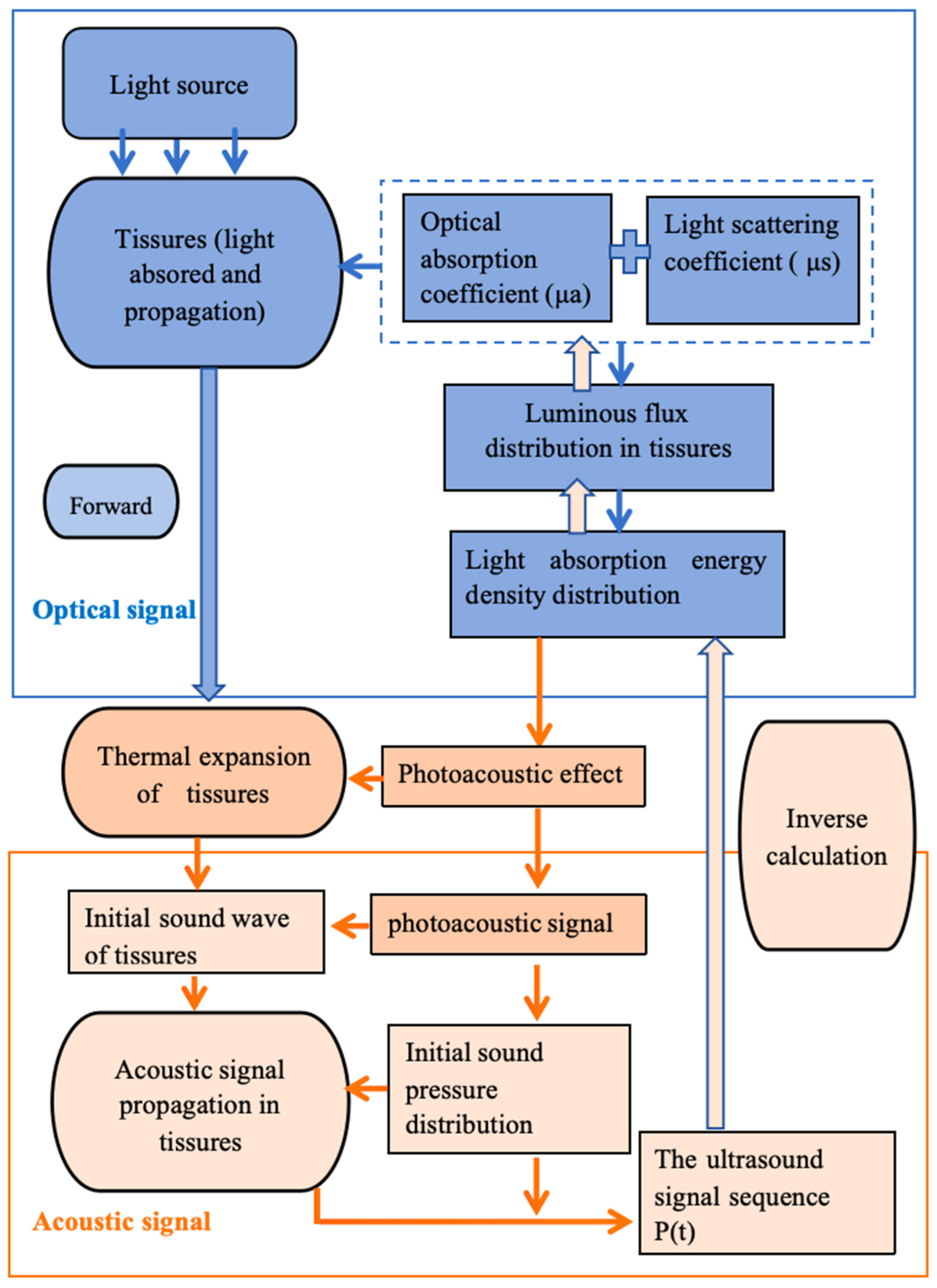
2. Photoacoustic Imaging (PAI)

Photoacoustic imaging (PAI) is a hybrid biomedical modality that synergizes optical excitation with ultrasonic detection. A nanosecond pulsed laser irradiates target tissues, generating ultrasonic waves via the photoacoustic effect. Ultrasonic transducers arrayed around the specimen acquire these signals, which are subsequently reconstructed through advanced algorithms to generate high-resolution images [15,31,32,33]. Photon absorption by endogenous chromophores (e.g., hemoglobin) during irradiation induces transient thermoelastic expansion. This phenomenon produces broadband ultrasonic emissions (0.1–50 MHz) that propagate through biological media, ultimately forming images through time-resolved signal detection (Figure 1) [34,35,36,37].
The calculation of initial photoacoustic pressure necessitates adherence to two governing constraints: thermal confinement and stress confinement.
  • Stress confinement condition: This condition mandates that the laser pulse duration (τp) be shorter than the acoustic transit time across the irradiated volume:
    τp < d/Vs
    where d denotes the characteristic dimension of the irradiated region (m), and Vs represents the speed of sound in the medium (m·s−1). Stress confinement ensures localized energy deposition by restricting acoustic wave propagation beyond the thermal source region, thereby enabling efficient thermoelastic pressure generation.
  • Thermal confinement condition: This requires the laser pulse duration to be shorter than the thermal relaxation time (τth):
    τth < dth2/αth
    where αth is the thermal diffusivity (m2·s−1), and corresponds to the thermal diffusion length, empirically approximated as dth ≈ 1 μm in biological tissues. Compliance with this criterion minimizes thermal conduction losses (<5% energy dissipation) during irradiation, preserving spatial resolution.
The photoacoustic signal, exhibiting linear proportionality to the optical absorption coefficient (μa), is captured via ultrasound transducers and digitized through data acquisition (DAQ) systems. Time-resolved signals undergo computational processing using advanced reconstruction algorithms to generate maps of optical absorption distribution. This approach merges the high optical contrast of pure optical modalities with the superior penetration depth and spatial resolution of ultrasound imaging [38,39,40]. The reconstruction techniques encompass conventional image reconstruction methods and deep learning (DL)-based image reconstruction methods. Conventional image reconstruction methods do not involve deep learning and mainly include five classes of algorithms: FBP, delay and sum (DAS), series expansion (SE), time reversal (TR), and iterative reconstruction (IR) [38,39,40].
PAI encompasses three primary modalities: photoacoustic tomography (PAT), photoacoustic microscopy (PAM), and photoacoustic endoscopy (PAE) [41]. PAT employs ultrasound transducer arrays for macroscopic and microscopic imaging, whereas PAM and PAE utilize focused transducers to achieve micrometer-scale resolution at millimeter depths [42,43]. Applications span from cellular to organ-level imaging, enabling anatomical, functional, and molecular visualization. A few examples include tracking erythrocytes [44], mapping vasculature [45,46], whole brain structural imaging [47,48], monitoring tumor dynamics [49,50], hemodynamics [51,52], and neuronal activity [53,54].
PAT image reconstruction involves solving the inverse problem of photoacoustic wave propagation. Surface-detected acoustic signals are processed via inversion algorithms (e.g., time reversal and back-projection) to reconstruct initial pressure distributions that correlate with tissue optical absorption coefficients (Figure 1). As optical absorption varies with molecular composition, PAT enables quantitative measurement of physiological parameters including oxygenated/deoxygenated hemoglobin concentrations, blood flow velocity, and oxygen metabolism rates [15,37,38,39,40]. The accurate evaluation of chromophore concentrations obtained by such imaging requires accurate estimation of the absorption coefficient (µa) of target tissues, which is challenging because it depends on photon fluence (Φ) that attenuates as the light propagates deeper into the tissue. Quantitative optoacoustic tomography (QOAT) aims to improve the reconstruction accuracy of chromophore concentrations by starting from conventional images of the initial pressure distribution and treating the pressure as the product of the absorption coefficient (μa) and the photon fluence. However, this approach requires solving a non-linear, ill-posed optical inverse problem, which presents significant challenges. Three main strategies have been proposed to address this issue. First, homogeneous or empirical optical properties, in which the μa is estimated by assuming homogeneous or empirical optical properties. However, this assumption may not hold true, leading to substantial reconstruction errors. Second, multimodal imaging, which combines optoacoustic tomography (OAT) with other imaging modalities, such as diffuse optical tomography (DOT) or acoustic–optic tomography, to calculate the fluence distribution. While effective, this method requires more complex systems and computational resources. Third, iterative reconstruction, which employs iterative reconstruction to minimize errors by incorporating an appropriate optimization strategy and a forward model of photon transport. This method, however, relies heavily on accurate calibration between the acoustic reconstruction and the optical model [38,55]. Compared to fMRI and PET, PAT provides non-invasive cerebrovascular imaging with high spatiotemporal resolution, rendering it uniquely suited for mapping acupuncture-induced hemodynamic alterations. By leveraging endogenous chromophores (e.g., hemoglobin and neurotransmitters) and tissue-specific optoacoustic properties, PAI serves as a powerful modality for visualizing acupuncture’s dual-regulatory mechanisms. This review specifically examines PAI’s capacity to modulate the central nervous system via acupuncture and resolve localized microcirculatory responses at stimulated acupoints. Through this dual-lens approach, PAI bridges the gap between holistic neuromodulation and site-specific physiological effects in acupuncture research.

3. PAI in Acupuncture

3.1. Central Nervous System Modulation via Acupuncture

Research teams, including Prof. Huabei Jiang’s group at the University of South Florida, have employed photoacoustic imaging (PAI) to investigate acupoint–brain functional correlations, elucidating the neurocentral regulatory relationships between acupoints and target organs [15,23,24,56,57,58,59,60]. This work critically addresses the technological validation of acupoint specificity essential for mechanistic acupuncture research.
Li Tingting et al. [56] employed photoacoustic tomography (PAT) to examine cerebral hemodynamic alterations in mice, following stimulation of the Yongquan (KI1) acupoint. The study conducted pre- and post-stimulation PAT imaging of cerebral vasculature, revealing through quantitative analysis of hemoglobin (Hb) optical absorption, that unilateral stimulation at either the left or right Yongquan acupoint significantly increased regional cerebral blood flow (rCBF) in specific brain regions. Notably, bilateral simultaneous stimulation of both Yongquan acupoints did not produce additive effects on regional blood flow, but rather induced more complex, distributed hemodynamic responses across multiple brain areas [56]. Chen et al. [23] extended this research by applying PAT to investigate acupuncture effects on total hemoglobin (HbT) concentration in a murine model of cerebral hypoperfusion following unilateral carotid artery ligation. Their findings demonstrated that stimulation of the Yanglingquan (GB34) acupoint significantly increased HbT levels in hypoperfused regions, with similar effects observed following either the left or right acupoint stimulation. Importantly, the observed HbT elevation not only correlated with cerebral blood flow (CBF) changes but also exhibited sustained duration. The study further identified vascular remodeling in hypoperfused regions, characterized by both vasodilation (increased vessel diameter) and angiogenesis (new vessel formation), suggesting potential therapeutic mechanisms for vascular occlusion and cerebral perfusion deficits [23]. These collective findings substantiate the utility of PAT for non-invasive detection of acupuncture-induced cerebral blood flow modifications. The technique’s capacity to quantify both immediate hemodynamic changes and longer-term vascular adaptations provides valuable insights into the neurovascular mechanisms underlying acupuncture therapy.
Jinge Yang et al. [24] utilized photoacoustic microscopy (PAM) to investigate cerebral hemodynamic responses to electroacupuncture (EA) stimulation at the Zusanli acupoint (ST36). By comparing real acupoint stimulation with sham point interventions, distinct hemodynamic patterns were observed between the two groups, with significant differences in temporal and spatial dynamics. PAM imaging revealed a pronounced temporal delay and cumulative effects of acupuncture. Specifically, significant increases in cerebral blood volume (CBV) were detected in the sensorimotor cortex and retrosplenial agranular cortex during stimulation. These CNS-mediated responses may represent a key pathway for acupuncture’s neuromodulatory effects. The observed divergence between EA and sham groups further corroborated acupuncture-specific activation patterns within the integrated nervous system. EA-specific activation of pain-related CNS pathways and motor-analgesic circuits highlights its therapeutic potential in stroke rehabilitation [24]. Jiang Shuhua et al. [57] employed PAT to longitudinally assess cerebral hemodynamic alterations in mice following acupuncture stimulation at three acupoints: Yongquan (KI1), Yanglingquan (GB34), and Taichong (LR3). Semi-quantitative PAI analysis showed Yanglingquan induced the most pronounced CBF increase, followed by Yongquan, while Taichong elicited minimal changes. This acupoint-specific hemodynamic differentiation validates the capacity of PAT to resolve spatiotemporal neurovascular dynamics, providing mechanistic insights into acupuncture’s site-selective therapeutic effects [57].
To evaluate factors influencing acupuncture efficacy, comparative studies were conducted to assess cerebral hemodynamic responses to the different acupuncture modalities (e.g., manual acupuncture [MA], electroacupuncture [EA], and moxibustion) and anesthetic regimens. Cui Ruihuan et al. [58] investigated Yongquan (KI1) acupoint stimulation effects using these three modalities, revealing distinct cerebral hemodynamic modulation patterns. MA predominantly enhanced perfusion in most brain regions except the right auditory cortex, which showed inhibition. Conversely, EA induced inhibitory effects in the right visual/auditory cortices, left posterior parietal association area, somatosensory cortex, and left motor cortex, with facilitatory effects elsewhere. Moxibustion demonstrated intermediate region-specific activation. Furthermore, sodium pentobarbital anesthesia minimally interfered with needling effects compared to chloral hydrate and urethane, establishing an optimized protocol for photoacoustic imaging (PAI) standardization in acupuncture research [59].
The following research provides a more comprehensive interpretation of the mechanisms of acupuncture by PAI. Wu Dan et al. [15] employed PAT to quantify real-time cerebral hemodynamic parameters, including oxygen saturation, hemoglobin (Hb), and deoxyhemoglobin (dHb) concentrations, in response to stimulation of 17 distinct acupoints in murine models. Through functional photoacoustic neuroimaging experiments involving over 200 subjects, the study established a non-invasive dynamic visualization platform for neuroacupuncture research. This systematic mapping of acupuncture-induced hemodynamic responses demonstrated cortical-level spatiotemporal specificity across acupoints. In a dual-modality imaging paradigm integrating PAT with laser speckle contrast imaging (LSCI), the team investigated ischemic stroke diagnosis and acupuncture intervention effects. This approach simultaneously monitored Hb concentration and blood flow velocity during Yanglingquan (GB34) acupoint stimulation in cerebral ischemia models. Results demonstrated acupuncture-induced dilation of ischemic vasculature and enhanced reperfusion, underscoring its therapeutic potential. Crucially, PAT exhibited differential diagnostic capacity between ischemic and hemorrhagic stroke models through distinct hemodynamic signatures. These findings validate PAT’s utility for evaluating acupuncture efficacy and mechanistic studies in stroke management [15]. Shang Qiquan et al. [60] further demonstrated acupoint-specific neuromodulation by stimulating Shenyu (BL23), Zhongwan (KN12), Huantiao (GB30), and Sanyinjiao (Sp6) in mice. Immediate and delayed blood flow changes were observed in distinct brain regions, suggesting that acupoint stimulation selectively activates or inhibits disease-associated neural circuits. These findings highlight PAI’s capacity to map spatiotemporal hemodynamic patterns, bridging acupoint specificity with therapeutic outcomes.
As demonstrated by prior studies (Table 2), photoacoustic imaging (PAI) integrates the high optical contrast of laser excitation with the deep penetration of ultrasound, enabling high-resolution visualization of cortical vascular responses to acupuncture in small animal models. Comparative analyses of hemodynamic patterns, between stimulated acupoints and non-acupoints, as well as across distinct acupoints, revealed statistically significant spatiotemporal differences. Specifically, brain regions temporally linked to acupoint stimulation exhibited marked hemodynamic divergence. Photoacoustic tomography (PAT) further quantifies real-time changes in cerebral blood parameters, including oxygen saturation, hemoglobin (Hb), and deoxyhemoglobin(dHB) concentrations, within acupoint-associated functional zones [41]. PAI’s versatility extends to neuroimaging applications such as mapping cerebral oxygenation, metabolic activity, and functional connectivity, as well as diagnosing neurological disorders (e.g., epilepsy, ischemic/hemorrhagic stroke, and traumatic injury) [41,61]. Its compatibility with molecular probes and multimodal systems enhances its utility in mechanistic studies. Collectively, these findings validate the specificity of acupoint–brain interactions and highlight PAI’s potential to elucidate acupuncture’s neurophysiological mechanisms, bridging gaps between localized effects and systemic regulation.
To further elucidate the therapeutic mechanisms of acupuncture, our investigation extended beyond the central nervous system responses to acupoints, employing photoacoustic imaging (PAI) to monitor real-time microvascular dynamics at stimulated acupoints [61]. This multimodal approach demonstrated the mechanistic link between local acupoint sensitization processes (e.g., perfusion modulation and vascular permeability changes) and systemic therapeutic outcomes.

3.2. Local Effects of Acupoints—Microcirculation Sensitization

Acupoint sensitization refers to the transition from a “silent” (healthy) to an “activated” (disease-associated) state, characterized by altered responses to external stimuli, including localized thermal, optical, and microcirculatory changes. Microcirculation is critical for metabolic exchange, tissue perfusion, and homeostasis [62,63]. Prior studies suggest that sensitization correlates with enhanced local microcirculatory perfusion [62,63].
Tao et al. [64] employed laser scattering imaging to assess microvascular dynamics at the Hegu acupoint in primary dysmenorrhea patients. The results showed that acupuncture induced significant capillary dilation, underscoring disease-specific microcirculatory alterations. These findings demonstrate that pathological states induce acupoint-specific microcirculatory sensitization—a dynamic process reflecting disease-related bioinformation. Clinically, such sensitized acupoints manifest as body surface biomarkers with diagnostic and therapeutic relevance. Integrating photoacoustic imaging (PAI) could further advance studies on acupoint-specific microcirculatory dynamics.
Contrastingly, Liu Xiaoxia [65] investigated microvascular morphology at Zusanli (ST36), Yanglingquan (GB34), and non-acupoint sites in knee osteoarthritis (KOA) mice using photoacoustic imaging. Quantitative analysis revealed no significant differences in vascular curvature, diameter distribution, or microvascular density between these sites across control, KOA model, and sham model groups (p > 0.05). Similarly, Ding et al. [66] employed optical-resolution photoacoustic microscopy to dynamically monitor microcirculatory structures during acupoint sensitization in KOA models, corroborating the absence of significant morphological differences in vascular density, diameter, or tortuosity at ST36, GB34, and non-acupoint regions. These consistent findings suggest that acupoint sensitization may not involve microcirculatory morphological remodeling, emphasizing the need to investigate functional hemodynamic alterations in sensitized acupoints.
Liu Hao [67] examined microcirculatory alterations at myocardial ischemia-related acupoints (Feiyu BL13, Jueyinshu BL14, Quze PC3, Chize LU5) in mice stratified into normal, sham-operated, and ischemia groups. Photoacoustic imaging revealed acupoint-specific variations in microvascular curvature count metrics (ICM), suggesting potential correlations with sensitization phenomena. Shang Qiquan et al. [60] employed high-speed photoacoustic imaging to monitor stimulation-induced microcirculatory changes. Electro-stimulation of the Hegu acupoint (LI4) in humans elicited localized blood flow increases in distal extremities (fingers/wrists/arms), with LI4 stimulation producing significantly greater hemodynamic responses than non-acupoint regions (p < 0.05). Jing Jiang et al. [68] conducted in vivo comparisons of acupoint microcirculation between normal and Alzheimer’s disease (AD) mice using acoustic-resolution photoacoustic microscopy (AR-PAM). AD mice exhibited significant microvascular attenuation at Zhongwan (CV12) and cephalic regions (p < 0.01). Acupuncture intervention partially ameliorated these pathological alterations, providing visual evidence for mechanistic studies of acupuncture efficacy [46,53,54].
While the central nervous system (CNS) centrally regulates acupoint responsiveness, acupoints themselves constitute critical somatic–visceral interfaces in systemic pathophysiology. Functioning as both therapeutic targets and biological sensors, these specialized loci exhibit pathological sensitization characterized by quantifiable microcirculatory alterations, including modulated perfusion rates and enhanced vascular permeability during disease progression. Structural microvascular remodeling (e.g., density and tortuosity) shows no significant correlation with acupoint sensitization (p > 0.05); whereas, functional hemodynamic parameters, particularly perfusion dynamics and quantitative metrics such as hemoglobin flux and oxygen saturation, emerge as critical biomarkers of acupoint activation [56,69]. These functional adaptations likely underlie the clinical relevance of sensitized acupoints as somatic manifestations of visceral pathology. Further investigation into real-time hemodynamic monitoring and neurovascular coupling mechanisms (e.g., glutamate-mediated vasodilation) is essential to decode the therapeutic foundations of acupuncture.

4. Discussion and Conclusions

Photoacoustic imaging (PAI) provides a multimodal platform for investigating acupuncture by integrating structural and functional perspectives across spatiotemporal scales. This technology enables visualization of vascular architecture and hemodynamics, revealing acupoint-specific modulation of cerebral metabolic activity and microcirculatory flux [15,70,71,72,73]. By correlating local acupoint stimulation with distal organ responses, PAI bridges traditional Chinese medicine principles with modern neurophysiological frameworks.
PAI facilitates both qualitative and quantitative assessment of acupuncture effects through high-resolution mapping of cortical hemodynamics in preclinical models. Studies demonstrate its capacity to differentiate acupoint-specific brain activation patterns and track neurovascular plasticity during treatment [70,71,72,73]. The technique’s anatomical precision further supports investigations of neuromodulatory pathways, particularly in decoding acupuncture’s therapeutic efficacy for stroke rehabilitation and chronic pain management [15].
Advancing PAI applications require integration with molecular probes targeting neurotransmitters (e.g., dopamine and serotonin) and inflammatory markers to resolve neuroendocrine interactions. Multimodal approaches combining hyperspectral PAI with thermal imaging could address unresolved mechanisms of acupoint sensitization, potentially yielding quantitative biomarkers (e.g., thermal–optical coupling coefficients). Such innovations may facilitate the development of standardized protocols for clinical translation while deepening mechanistic understanding of acupuncture’s holistic effects.

Author Contributions

Y.W. (Yun Wu): Conceptualization (lead); Data curation (lead); Formal analysis (lead); Methodology (lead); Writing—original draft (lead). H.J.: Validation (lead); Visualization (lead); Writing—review and editing (equal). D.W., Y.W. (Yanting Wen), Y.Y., J.Z. and Z.C.: Conceptualization (supporting); Project administration (supporting); Resources (supporting); Supervision (supporting). All authors have read and agreed to the published version of the manuscript.

Funding

Chengdu Health Commission—Chengdu University of Traditional Chinese Medicine 2024 Joint Innovation Fund Project, Title: Clinical Application of Musculoskeletal Ultrasound Combined with Photoacoustic Imaging for Therapeutic Assessment of Acupuncture in Cervical Spondylosis Management. No.: WXLH202403258.

Conflicts of Interest

The authors have no conflicts of interest to disclose.

Abbreviations

PAIPhotoacoustic imaging
TCMTraditional Chinese medicine
NEINeuro-endocrine-immunity
CNSCentral nervous system
NMRNuclear magnetic resonance
PETPositron emission tomography
PAPhotoacoustic effect
HBHemoglobin
PATPhotoacoustic tomography
PAMPhotoacoustic microscopy
PAEPhotoacoustic endoscopy
EAElectronic acupuncture
ST36Zusanli
CBVCerebral blood volume
BL23Shenyu
KN12Zhongwan
GB30Huantiao
Sp6Sanyinjiao
HbTHemoglobin
CBFCerebral blood flow
KOAKnee osteoarthritis
GB34Yanglingquan
CCMCurvature count metric
ADAlzheimer’s disease
CV12Zhongwan

References

  1. Ondrejkovicova, A.; Petrovics, G.; Svitkova, K.; Bajtekova, B.; Bangha, O. Why acupuncture in pain treatment? Neuro Endocrinol. Lett. 2016, 37, 163–168. [Google Scholar] [PubMed]
  2. Zhang, S. Acupuncture theory in Huangdi Neijing and launching the natural history of the body. Zhongguo Zhen Jiu 2024, 44, 593–598. [Google Scholar] [PubMed]
  3. Li, L.; Wang, X.; Guo, J.; Chen, Y.; Wang, Z. The effect of acupuncture in the acute phase of intracerebral hemorrhage on prognosis and serum BDNF: A randomized controlled trial. Front. Neurosci. 2023, 17, 1167620. [Google Scholar] [CrossRef] [PubMed]
  4. Itoh, K.; Minakawa, Y.; Kitakoji, H. The effect of acupuncture depth on muscle pain. Chin. Med. 2011, 6, 24. [Google Scholar] [CrossRef]
  5. Li, S.; Ye, Y.; Chen, Z.; Zhao, M.; Jiang, Y.; Wang, Z.; Jiang, Y. Efficacy and safety evaluation of acupuncture therapy for patients with salpingitis undergoing IVF-ET: A protocol for systematic review and meta-analysis. Medicine 2021, 100, e24015. [Google Scholar] [CrossRef]
  6. Xi, Z.; Wei, X.; Ye, Z.; Wang, K.; Zhou, J. Acupuncture for adult lung cancer: A systematic review and meta-analysis of patient-reported outcomes. Front. Oncol. 2022, 12, 921151. [Google Scholar] [CrossRef]
  7. Li, T. Application of Photoacoustic Imaging Technology in Acupuncture Therapy. Master’s Thesis, University of Electronic Science and Technology, Chengdu, China, 2014. [Google Scholar]
  8. Yang, L.; Zheng, Y. Progress in the application of medical imaging in the study of acupuncture mechanisms. Mod. Med. Health 2016, 32, 2359–2361. [Google Scholar]
  9. Xu, Y.; Nie, L. Progress in the application of optical imaging technology in the study of acupuncture in Chinese medicine. China Laser 2023, 50, 71–83. [Google Scholar]
  10. Qiu, K.; Jing, M.; Sun, R.; Yang, J.; Liu, X.; He, Z.; Yin, S.; Lan, Y.; Cheng, S.; Gao, F.; et al. The status of quality control in acupuncture-neuroimaging studies. Evid.-Based Complement. Altern. Med. 2016, 2016, 3685785. [Google Scholar] [CrossRef]
  11. Chen, L.; Li, M.; Fan, L.; Zhu, X.; Liu, J.; Li, H.; Xu, Z.; Chen, J.; Liang, Z.; Liu, Z.; et al. Optimized acupuncture treatment (acupuncture and intradermal needling) for cervical spondylosis-related neck pain: A multicenter randomized controlled trial. Pain 2021, 162, 728–739. [Google Scholar] [CrossRef]
  12. Koes, B.W.; van Tulder, M.W.; Thomas, S. Diagnosis and treatment of low back pain. BMJ 2006, 332, 1430–1434. [Google Scholar] [CrossRef] [PubMed]
  13. Qin, Z.S.; Zang, Z.W.; Wu, J.N.; Zhou, J.; Liu, Z. Efficacy of acupuncture for chronic prostatitis/chronic pelvic pain syndromes: Study protocol for a randomized. 2016, sham acupuncture-controlled trial. BMC Complement. Altern. Med. 2016, 16, 440. [Google Scholar] [CrossRef] [PubMed]
  14. Zhang, Y.H.; Wu, T.; Shen, W.; Huang, F.Y.; Liu, C.R. Effect of Fu’s subcutaneous needling on thickness and elasticity of affected muscles in shoulder neck pain based on ultrasonic elastography. Chin. Acupunct. Moxibustion 2020, 40, 939–941. [Google Scholar]
  15. Wu, D. Optoacoustic Imaging of Brain Function and Acupuncture Brain Mapping. Doctoral Dissertation, University of Electronic Science and Technology, Chengdu, China, 2019. [Google Scholar]
  16. Gao, X.Y.; Han, S.; Huang, Q.; He, S.Q.; Ford, N.C.; Zheng, Q.; Chen, Z.; Yu, S.; Dong, X.; Guan, Y. Calcium imaging in population of dorsal root ganglion neurons unravels novel mechanisms of visceral pain sensitization and referred somatic hypersensitivity. Pain 2021, 162, 1068–1081. [Google Scholar] [CrossRef]
  17. Gong, P.; Zhang, S.S.; Ren, L.; Zhang, J.; Zhao, Y.; Mao, X.; Gan, L.; Wang, H.; Ma, C.; Lin, Y.; et al. Electroacupuncture of the trigeminal nerve causes N-methyl-D-aspartate receptors to mediate blood-brain barrier opening and induces neuronal excitatory changes. Front. Cell. Neurosci. 2022, 16, 1020644. [Google Scholar] [CrossRef]
  18. Li, S.; Liu, Y.; Zhang, N.; Li, W.; Xu, W.J.; Xu, Y.Q.; Chen, Y.Y.; Cui, X.; Zhu, B.; Gao, X.Y. Perspective of calcium imaging technology applied to acupuncture research. Chin. J. Integr. Med. 2023, 30, 3–9. [Google Scholar] [CrossRef]
  19. Cao, J.; Tang, Y.S.; Li, Y.J.; Gao, K.; Shi, X.; Li, Z. Behavioral changes, and hippocampus glucose metabolism in APP/PS1 transgenic mice via electro-acupuncture at governor vessel acupoints. Front. Aging Neurosci. 2017, 9, 5. [Google Scholar] [CrossRef]
  20. Cai, M.D.; Lee, J.H.; Yang, E.J. Electroacupuncture attenuates cognition impairment via anti-neuroinflammation in an Alzheimer’s disease animal model. J. Neuroinflamm. 2019, 16, 264. [Google Scholar] [CrossRef]
  21. Jiang, Y.M.; Lin, Y.S.; Tan, Y.H.; Shen, X.; Liao, M.; Wang, H.; Lu, N.; Han, F.; Xu, N.; Tang, C.; et al. Electroacupuncture ameliorates cerebrovascular impairment in Alzheimer’s disease mice via melatonin signaling. CNS Neurosci. Ther. 2023, 29, 917–931. [Google Scholar] [CrossRef]
  22. Cui, S.; Yao, S.Q.; Wu, C.X.; Yao, L.; Huang, P.; Chen, Y.; Tang, C.; Xu, N. Electroacupuncture involved in motor cortex and hypoglossal neural control to improve voluntary swallowing of poststroke dysphagia mice. Neural Plast. 2020, 2020, 8857543. [Google Scholar] [CrossRef]
  23. Chen, B.Z.; Yang, J.G.; Wu, D.; Zeng, D.W.; Yi, Y.; Yang, N.; Jiang, H.B. Photoacoustic imaging of cerebral hypoperfusion during acupuncture. Biomed. Opt. Express 2015, 6, 3225–3234. [Google Scholar] [CrossRef] [PubMed]
  24. Yang, J.G.; Wu, D.; Tang, Y.; Jiang, H. Photoacoustic microscopy of electronic acupuncture (EA) effect in small animals. J. Biophotonics 2017, 10, 217–223. [Google Scholar] [CrossRef] [PubMed]
  25. Ye, Y.; Wei, Y.; Jia, J.; Yan, X. Efficacy of needling Baihui (GV20), Neiguan (PC6), Shenmen (HT7) and Taichong (LR3) on cerebral cortical blood oxygen level in rats with insomnia. J. Tradit. Chin. Med. 2023, 43, 523–532. [Google Scholar]
  26. Chen, J.; Li, H.; Zeng, C.; Li, J.; Zhao, B. Evaluation of the recovery outcome of poststroke cognitive impairment after cluster needling of scalp acupuncture therapy based on functional near-infrared spectroscopy. Brain Behav. 2020, 10, e01731. [Google Scholar] [CrossRef]
  27. Liang, S.X.; Lin, Y.J.; Lin, B.B.; Li, J.; Liu, W.; Chen, L.; Zhao, S.; Tao, J. Resting-state functional magnetic resonance imaging analysis of brain functional activity in rats with ischemic stroke treated by electro-acupuncture. J. Stroke Cerebrovasc. Dis. 2017, 26, 1953–1959. [Google Scholar] [CrossRef]
  28. Wang, Z.X.; Fan, X.W.; Chen, K.Y.; Yu, X.; Gao, J. Effects of three kinds of head acupuncture therapies on regulation of brain microenvironment and rehabilitation of nerve function in rats with cerebral palsy. J. Tradit. Chin. Med. 2021, 41, 276–283. [Google Scholar]
  29. Chang, X.Y.; Chen, K.; Cheng, T.; Lai, P.T.; Zhang, L.; So, K.F.; Yang, E.S. In vivo neuronal and astrocytic activation in somatosensory cortex by acupuncture stimuli. Neural Regen. Res. 2022, 17, 2526–2529. [Google Scholar]
  30. Leow, M.Q.; Lee, S.H.; Mohamed-Shah, M.T.; Cao, T.; Cui, S.L.; Tay, S.C.; Ooi, C.C. Exploring the physiological and physical effects of acupuncture using ultrasound. Acupunct. Med. 2016, 35, 231–235. [Google Scholar] [CrossRef]
  31. Zhao, C.Y.; Wang, Q.; Tao, X.X.; Wang, M.; Yu, C.; Liu, S.R.; Li, M.; Tian, X.; Qi, Z.; Li, J.; et al. Multimodal photoacoustic/ultrasonic imaging system: A promising imaging method for the evaluation of disease activity in rheumatoid arthritis. Eur. Radiol. 2021, 31, 3542–3552. [Google Scholar] [CrossRef]
  32. Steinberg, I.; Huland, D.M.; Vermesh, O.; Frostig, H.E.; Tummers, W.S.; Gambhir, S.S. Photoacoustic clinical imaging. Photoacoustics 2019, 14, 77–98. [Google Scholar] [CrossRef]
  33. Chen, L.; Ma, H.H.; Liu, H.; Shou, K.Q.; Zheng, X.; Fan, Q.L.; Yu, A.; Hu, X. Quantitative photoacoustic imaging for early detection of muscle ischemia injury. Am. J. Transl. Res. 2017, 9, 2255–2265. [Google Scholar] [PubMed]
  34. Yang, J.G.; Zhang, G.; Chang, W.; Chi, Z.H.; Shang, Q.Q.; Wu, M.; Pan, T.; Huang, L.; Jiang, H. Photoacoustic imaging of hemodynamic changes in forearm skeletal muscle during cuff occlusion. Biomed. Opt. Express 2020, 11, 4560–4570. [Google Scholar] [PubMed]
  35. Liba, O.; de la Zerda, A. Photoacoustic tomography: Breathtaking whole-body imaging. Nat. Biomed. Eng. 2017, 1, 0075. [Google Scholar]
  36. Ranjbaran, S.M.; Aghamiry, H.S.; Gholami, A.; Operto, S.; Avanaki, K. Quantitative photoacoustic tomography using iteratively refined wavefield reconstruction inversion: A simulation study. IEEE Trans. Med. Imaging 2024, 43, 874–885. [Google Scholar]
  37. Tarvainen, T.; Cox, B. Quantitative photoacoustic tomography: Modeling and inverse problems. J. Biomed. Opt. 2024, 29 (Suppl. 1), S11509. [Google Scholar]
  38. Pattyn, A.; Mumm, Z.; Alijabbari, N.; Duric, N.; Anastasio, M.A.; Mehrmohammadi, M. Model-based optical and acoustical compensation for photoacoustic tomography of heterogeneous media. Photoacoustics 2021, 23, 100275. [Google Scholar]
  39. Ahn, J.; Kim, J.Y.; Choi, W.; Kim, C. High-resolution functional photoacoustic monitoring of vascular dynamics in human fingers. Photoacoustics 2021, 23, 100282. [Google Scholar]
  40. Fonseca, M.; An, L.; Beard, P.; Cox, B. Sulfates as chromophores for multiwavelength photoacoustic imaging phantoms. J. Biomed. Opt. 2017, 22, 125007. [Google Scholar]
  41. Sun, M.; Wu, D.; Jiang, H. Photoacoustic imaging and Chinese medicine research. World Tradit. Chin. Med. 2020, 15, 1527–1534. [Google Scholar]
  42. Wang, L.V.; Hu, S. Photoacoustic tomography: In vivo imaging from organelles to organs. Science 2012, 335, 1458–1462. [Google Scholar]
  43. Kneipp, M.; Turner, J.; Hambauer, S.; Krieg, S.M.; Lehmberg, J.; Lindauer, U.; Razansky, D. Functional real-time optoacoustic imaging of middle cerebral artery occlusion in mice. PLoS ONE 2014, 9, e96118. [Google Scholar] [CrossRef] [PubMed]
  44. Dean-Ben, X.L.; Sela, G.; Lauri, A.; Kneipp, M.; Ntziachristos, V.; Westmeyer, G.G.; Shoham, S.; Razansky, D. Functional optoacoustic neuro-tomography for scalable whole-brain monitoring of calcium indicators. Light Sci. Appl. 2016, 5, 16201. [Google Scholar] [CrossRef] [PubMed]
  45. Estrada, H.; Huang, X.; Rebling, J.; Zwack, M.; Gottschalk, S.; Razansky, D. Virtual craniotomy for high-resolution optoacoustic brain microscopy. Sci. Rep. 2018, 8, 1459. [Google Scholar] [CrossRef] [PubMed]
  46. Heywang-Kobrunner, S.H.; Hacker, A. Advantages and disadvantages of mammography screening. Breast Care 2011, 6, 199–207. [Google Scholar] [CrossRef]
  47. Lin, L.; Hu, P.; Shi, J.; Appleton, C.M.; Maslov, K.; Li, L.; Zhang, R.; Wang, L.V. Single-breath-hold photoacoustic computed tomography of the breast. Nat. Commun. 2018, 9, 2352. [Google Scholar] [CrossRef]
  48. Heijblom, M.; Piras, D.; van den Engh, F.M.; van der Schaaf, M.; Klaase, J.M.; Steenbergen, W.; Manohar, S. The state of the art in breast imaging using the Twente Photoacoustic Mammoscope: Results from 31 measurements on malignancies. Eur. Radiol. 2016, 26, 3874–3887. [Google Scholar] [CrossRef]
  49. Toi, M.; Asao, Y.; Matsumoto, Y.; Sekiguchi, H.; Yoshikawa, A.; Takada, M.; Kataoka, M.; Endo, T.; Kawaguchi-Sakita, N.; Kawashima, M.; et al. Visualization of tumor-related blood vessels in the human breast using a photoacoustic imaging system with a hemispherical detector array. Sci. Rep. 2017, 7, 41970. [Google Scholar] [CrossRef]
  50. Attia, A.B.E.; Balasundaram, G.; Moothanchery, M.; Dinish, U.S.; Bi, R.; Ntziachristos, V.; Olivo, M. A review of clinical photoacoustic imaging: Current and future trends. Photoacoustics 2019, 16, 100144. [Google Scholar] [CrossRef]
  51. Xi, L.; Grobmyer, S.R.; Zhou, G.; Qian, W.; Yang, L.; Jiang, H. Molecular photoacoustic tomography of breast cancer using receptor-targeted magnetic iron oxide nanoparticles as contrast agents. J. Biophotonics 2014, 7, 401–409. [Google Scholar] [CrossRef]
  52. Xi, L.; Li, X.; Yao, L.; Grobmyer, S.; Jiang, H. Design and evaluation of a hybrid photoacoustic tomography and diffuse optical tomography system for breast cancer detection. Med. Phys. 2012, 39, 2584–2594. [Google Scholar] [CrossRef]
  53. Li, X.; Heldermon, C.D.; Yao, L.; Xi, L.; Jiang, H. High-resolution functional photoacoustic tomography of breast cancer. Med. Phys. 2015, 42, 5321–5328. [Google Scholar] [CrossRef] [PubMed]
  54. Jo, J.; Tian, C.; Xu, G.; Sarazin, J.; Schiopu, E.; Gandikota, G.; Wang, X. Photoacoustic tomography for human musculoskeletal imaging and inflammatory arthritis detection. Photoacoustics 2018, 12, 82–89. [Google Scholar] [CrossRef] [PubMed]
  55. Li, J.; Wang, C.; Chen, T.; Lu, T.; Li, S.; Sun, B.; Gao, F.; Ntziachristos, V. Deep learning-based quantitative optoacoustic tomography of deeptissues in the absence of labeled experimental data. Optica 2022, 9, 32–41. [Google Scholar] [CrossRef]
  56. Li, T.; Xu, X.; Chen, B.; Rong, J.; Jiang, H. Photoacoustic imaging of acupuncture effects in small animals. J. Biophotonics 2015, 6, 433–442. [Google Scholar] [CrossRef]
  57. Jiang, S. Experimental Study of Acupuncture at Different Acupoints Based on a Photoacoustic Tomography Imaging System. Doctoral Dissertation, University of Electronic Science and Technology, Chengdu, China, 2016. [Google Scholar]
  58. Cui, R. PAT In Vivo Study of Monitoring the Response of Different Acupuncture Methods on the Cerebral Cortex. Doctoral Dissertation, University of Electronic Science and Technology, Chengdu, China, 2019. [Google Scholar]
  59. Guo, X. Monitoring of Cerebral Cortex Response to Acupuncture Under Different Anaesthetic States. Doctoral Dissertation, University of Electronic Science and Technology, Chengdu, China, 2019. [Google Scholar]
  60. Shang, Q. Photoacoustic Imaging Study of Organism Response to Needling. Doctoral Dissertation, University of Electronic Science and Technology, Chengdu, China, 2020. [Google Scholar]
  61. Ding, N.; Jiang, J.; Qin, P.; Hu, J.; Wang, Q.; Xu, Y.; Liu, X.; Li, Z. Prospects for the application of photoacoustic imaging in microcirculation sensitization imaging study of acupoints. Acupunct. Res. 2017, 42, 454–458. [Google Scholar]
  62. Mirg, S.; Turner, K.L.; Chen, H.; Drew, P.J.; Kothapalli, S.R. Photoacoustic imaging for microcirculation. Microcirculation 2022, 29, e12776. [Google Scholar] [CrossRef]
  63. Hsu, H.-C.; Wang, L.; Wang, L.V. In vivo photoacoustic microscopy of human cuticle microvasculature with single-cell resolution. J. Biomed. Opt. 2016, 21, 056004. [Google Scholar] [CrossRef]
  64. Tao, H.; Yang, L.; Jia, S.; Mu, X.; Wu, M.; Ye, H.; Liu, W.; Cheng, X. Capillary blood flow in patients with dysmenorrhea treated with acupuncture. J. Tradit. Chin. Med. 2013, 33, 757–760. [Google Scholar]
  65. Liu, X. Photoacoustic Imaging Study of Acupoint Sensitization in a Mouse Model of Knee Osteoarthritis. Doctoral Dissertation, Beijing University of Traditional Chinese Medicine, Beijing, China, 2019. [Google Scholar]
  66. Ding, N.; Liu, X.; Chen, N.; Jiang, J.; Zhao, H.; Li, Z.; Zhang, J.; Liu, C. Lack of association between acupoint sensitization and microcirculatory structural changes in a mouse model of knee osteoarthritis: A pilot study. J. Biophotonics 2019, 12, e201800458. [Google Scholar] [CrossRef]
  67. Liu, H.; Tian, H.; Wang, Z.; Jiang, J.; Pan, Y.H.; Liu, C.B.; Li, Z.G. Photoacoustic imaging study of microvascular spatial structure of relevant acupoints in mice with acute myocardial ischemia. Acupunct. Res. 2021, 46, 497–504. [Google Scholar]
  68. Jiang, J.; Wang, Z.; Yu, R.; Yang, J.; Wang, Q.; Wu, G.; Tao, Y.; Zhao, X.; Wang, Y.; Li, Z.; et al. Visualization of microvascular dynamics in response to acupuncture intervention in a mouse model. Biomed. Opt. Express 2021, 12, 2202–2215. [Google Scholar]
  69. Wu, D.; Wen, Y.; Xie, Y.; Liu, L.; Fu, Y.; Shen, Y.; Chi, Z.; Sun, M.; Tang, Y.; Jiang, H.B. Photoacoustic image guidance for acupuncture. In Proceedings of the 2022 IEEE MTT-S International Wireless Symposium (IWS), Harbin, China, 12–15 August 2022; Volume 1, pp. 1–3. [Google Scholar]
  70. Cao, R.; Li, J.; Ning, B.; Sun, N.; Wang, T.; Zuo, Z.; Hu, S. Functional and oxygen-metabolic photoacoustic microscopy of the awake mouse brain. NeuroImage 2017, 150, 77–87. [Google Scholar] [CrossRef] [PubMed]
  71. Mirg, S.; Chen, H.; Turner, K.L.; Gheres, K.W.; Liu, J.; Gluckman, B.J.; Drew, P.J.; Kothapalli, S.R. Awake mouse brain photoacoustic and optical imaging through a transparent ultrasound cranial window. Opt. Lett. 2022, 47, 1121–1124. [Google Scholar] [CrossRef] [PubMed]
  72. Tang, Y.; Wu, H.; Klippel, P.; Zhang, B.; Huang, H.S.; Jing, Y.; Jiang, X.; Yao, J. Deep thrombosis characterization using photoacoustic imaging with intravascular light delivery. Biomed. Eng. Lett. 2022, 12, 135–145. [Google Scholar] [CrossRef]
  73. Li, L.; Yao, J.; Wang, L.V. Photoacoustic tomography of neural systems. In Neural Engineering; He, B., Ed.; Springer International Publishing: Berlin/Heidelberg, Germany, 2020; pp. 349–378. [Google Scholar]
Figure 1. Photoacoustic effects include optical and acoustic section.
Photonics 12 00365 g001
Table 1. Comparing the medical imaging.
Imaging MethodSpatial ResolutionTemporal ResolutionExpensivePortableAdvantagesDisadvantagesApplicable AreasApplication in Acupuncture Research
Calcium Imaging<1 μm10–100 msNoNoHigh resolution, large field of view, long-term monitoringShallow observation depthObserving activity of neurons and glial cellsAcupoint–brain function relationship, acupuncture–neural circuit research [16,17,18]
PET4 mm30–60 sYesNoHigh sensitivityLow spatial resolutionDynamic observation of physiological, chemical reactions, and metabolic processesAcupoint specificity, acupoint–brain system research [19,20]
Laser Speckle2~3 μm10–100 msNoYesHigh image resolution, large monitoring area, non-invasiveShallow imaging depthContrast imaging (LSCI)real-time monitoring of local tissue blood perfusionAcupuncture–microcirculation research [21,22]
Photoacoustic Imaging50–500 μm1–100 msNoYesHigh resolution, high contrast, strong penetrationRequires coupling mediumMonitoring cerebral hemodynamics and blood oxygen levelsAcupuncture–brain effects research [23,24]
Near Infrared Spectroscopy (f-NIRS)10–30 mm 0.1–10 HzNoYesReal-time measurement of hemodynamics, non-invasiveAccuracy slightly reduced when penetrating skullProvides metabolic, physiological, molecular, and anatomical informationAcupoint–brain function relationship research [25,26]
fMRI2–3 mm1–3 sYesNoThree-dimensional data acquisition, non-invasiveLong imaging time, slightly lower sensitivityCellular, tissue, and in vivo imagingAcupoint–brain function relationship, acupoint–organ connection research [27,28]
Two-Photon In Vivo Imaging<1 μm1–100 msNoYesHigh sensitivity, low light toxicityOnly fluorescent imaging, invasiveUsed for vascular and neural diseasesAcupoint–brain microcirculation research [29]
US100–500 μm10–100 HzNoYesNon-invasive, high contrast, strong penetration.Limited spatial resolutionUsed for vascular and structuresMonitoring the effects of acupuncture on blood flow and soft tissue [30]
Table 2. PAI of the acupuncture effect on the brain based on above references.
ReferencesTimeImaging
System
MethodAcupuncture Effect on the BrainAcupoints
Li Tingting et al. [56]2015PATComparison of before acupuncture vs. after acupunctureThe blood flow in the mouse brain increased after acupunctureYongquan
Chen et al. [23]2015PATComparison of before acupuncture vs. after in a mouse model of cerebral hypoperfusionpromoting the generation of new blood vessels and increasing the diameter of the blood vessels in the hypoperfusion area by acupunctureYanglingquan
Jinge Yang et al. [24]2016PAMComparison of real acupuncture vs. sham acupunctureRemarkable cerebral blood volume (CBV) and hemoglobin concentration changes in sensorimotor and retro splenial agranular cortex by delayed effect and accumulated effect of acupuncture.Zusanli (ST36)
Jiang Shuhua et al. [57]2016PATComparison of acupuncture effects among different acupoints in different periodsSemi-quantitative analysis
of acupoint-specific hemodynamic differentiation among different acupoints
Yongquan, Yanglingquan and Taichong
Cui Ruihuan et al. [58]2019PATComparison of different acupuncture methodsDifferent changes in cerebral hemodynamics for different methodYongquan
Guo Xiuyun [59]2019PATComparison of different anesthetic statesCerebral cortical response to acupuncture under different anesthetic statesZusanli (ST36)
Wu, Dan [15]2019PATComparison of multiple acupoints(1) Built comprehensive mapping of acupuncture-induced cerebral hemodynamic responses (e.g., oxygen saturation, hemoglobin and deoxyhemoglobin concentration); (2) Monitoring and evaluating the efficacy and mechanisms of acupuncture in stroke management17 acupoints
Shang Qiquan [60]2020PATComparison of immediate and aftereffects of acupunctureEliciting a positive response from specific regions for different acupointsShenyu(BL23), Zhongjiao (KN12), Huanjiao (GB30), and Sanyinjiao (Sp6)
Disclaimer/Publisher’s Note: The statements, opinions and data contained in all publications are solely those of the individual author(s) and contributor(s) and not of MDPI and/or the editor(s). MDPI and/or the editor(s) disclaim responsibility for any injury to people or property resulting from any ideas, methods, instructions or products referred to in the content.

