Communication

Efficacy of Photodynamic Inactivation against the Major Human Antibiotic-Resistant Uropathogens

1
Department of Epidemiology, Microbiology and Evidence-Based Medicine, Privolzhsky Research Medical University, 10/1 Minin and Pozharsky Sq., 603005 Nizhny Novgorod, Russia
2
Medical and Preventative Care Department, Privolzhsky Research Medical University, 10/1 Minin and Pozharsky Sq., 603005 Nizhny Novgorod, Russia
3
Department of Urology Named after E.V. Shakhov, Privolzhsky Research Medical University, 10/1 Minin and Pozharsky Sq., 603005 Nizhny Novgorod, Russia
4
Institute of Biology and Biomedicine, National Research Lobachevsky State University of Nizhny Novgorod, 23 Prospekt Gagarina (Gagarin Avenue), 603022 Nizhny Novgorod, Russia
5
Institute of Experimental Oncology and Biomedical Technologies, Privolzhsky Research Medical University, 10/1 Minin and Pozharsky Sq., 603005 Nizhny Novgorod, Russia
6
Federal Research Center Institute of Applied Physics of the Russian Academy of Sciences, 46 Ul’yanov Street, 603950 Nizhny Novgorod, Russia
*
Author to whom correspondence should be addressed.
Photonics 2021, 8(11), 495; https://doi.org/10.3390/photonics8110495
Submission received: 29 September 2021 / Revised: 29 October 2021 / Accepted: 3 November 2021 / Published: 5 November 2021
(This article belongs to the Special Issue Topical Problems of Biophotonics)

Abstract

:
Photodynamic inactivation (PDI) is considered to be an effective method of prevention of postoperative complications of urolithiasis. The present study shows a complex approach to assess the efficacy of PDI of drug resistant bacteria associated with renal calculi. Bacterial strains associated with renal calculi were isolated and identified using standard methods of bacteriological analysis and tested for drug resistance to 10 antibiotics by the disco-diffusion method. Uropathogenic bacterial strains present in 78.7 ± 5.2% of the infected samples from the total number of analyzed calculi. The most frequent representatives belonged to the genera Staphylococcus, Escherichia, and Enterococcus. All tested strains showed high antibiotic resistance. Representatives of the most common bacterial genera in the calculi were used as models for the selection of PD exposure modes. It was found that the maximum time of photosensitizer accumulation depends on the structure of the bacterial cell wall: 30 min for Gram-negative strains and 60 min for Gram-positive ones. Optimal modes of PD exposure to antibiotic-resistant uropathogenic microorganisms were selected: 50 µg/mL Fotoditazin and 150 mW laser power. The maximal bactericidal activity of PDI against uropathogenic microorganisms was shown for Enterococcus faecalis, and Staphylococcus aureus. The bacteriostatic effect was found against Escherichia coli and Proteus mirabilis.

1. Introduction

The frequent, prolonged, and uncontrolled use of antibiotics in the treatment of infections has resulted in an increasing number of bacterial strains resistant to a wide range of antibiotics [1]. The antibiotic resistant bacteria such as Enterococcus faecium, Staphylococcus aureus, Klebsiella pneumoniae, Acinetobacter baumannii, Pseudomonas aeruginosa, and Enterobacter species cause the majority of hospital infections with high mortality of patients [2]. Antibiotic resistant pathogens are of great significance in the case of infected urinary stones. Urolithiasis is well known to be a widespread disease of the urinary system in middle-age people. According to the annual costs of diagnosis, treatment and hospitalization, it ranks second among all urinary tract infections [3]. Accordingly, in countries with a high standard of life, renal stone rate is notably high (>10%). The increase in urolithiasis rate of more than 37% was reported over the last 20 years for some areas [4]. Urologists are especially challenged by stones with infections, which comprise ~15% of kidney stones [5]. The history describes infective stone or struvite as the most common type of urinary stones containing magnesium ammonium phosphate. It is known, that urea-splitting bacteria such as Proteus spp., Staphylococcus aureus, Klebsiella spp., Providencia spp., and Ureaplasma urealyticum are commonly responsible for struvite stones [6]. The study of bacterial isolates from patients with calculi revealed the following genus Proteus, Staphylococcus, Pseudomonas, Providencia, Klebsiella, and Escherichia [7], that may also be drug resistant.
Present day methods of endoscopic and extracorporeal lithotripsy are based on the fine-size stone fragmentation. In case of infected calculi, pathogens diffused in the organ cavity may induce new types of postoperative complications, such as pyelonephritis, systemic inflammatory reaction syndrome, and urosepsis [8]. Traditional treatment of a urinary infection by peroral or intravenous administration of an antibiotic is limited by antibiotic penetration into the urinary tract, as well as by the drug resistance of pathogens. Local irrigation of either the renal or the bladder cavity with an antibiotic after lithotripsy is also limited by drug resistance and irrigation time. Moreover, in both approaches, antibiotics do not achieve a concentration required for the bactericidal effect. On the other hand, antibiotic therapy is accompanied by killing of the gut microbiota and damage of the natural colonization resistance. The prevention and treatment of postoperative complications with antibiotics often leads to the emergence of new variants of the disease associated with antibiotic therapy, such as pseudomembranous colitis, gut dysbacteriosis, and others [9,10]. However, using antibiotics is the main way to treat and prevent postoperative infectious and inflammatory complications. These facts have inspired a search for new antibiotics able to inactivate resistant microorganisms to the existent antibiotics.
Nowadays, photodynamic inactivation is considered to be an alternative to the antibiotic treatment of localized infectious processes [11,12]. The photodynamic effect is based on the interaction of 3 ingredients: light, photosensitizer, and oxygen. After exposure of the photosensitizer to light, reactive oxygen species, especially singlet oxygen, are generated. The reactive oxygen species affect various intracellular components, including proteins and DNA, resulting in cell death [13]. Thereby the photodynamic inactivation has several targets within the bacterial cell. In addition, the evidence of the promotion resistance/tolerance of microorganisms to photoinactivation have not yet been established [14]. It may be caused by the limited number of microbial antioxidant defense enzymes [15]. PDI has already been used in stomatology for treating infected root canals, which have polymicrobial infections [16]. There are some reports about an effective application of PDI to some Gram-positive [17] and Gram-negative standard and antibiotic-resistant bacteria strains [18].
Thus, the application of photodynamic inactivation to uropathogenic bacteria would make it possible to involve this approach into clinical practice for the prevention of infections caused by antibiotic-resistant microorganisms.
The purpose of the study was complex analysis of bacteria associated with renal calculi as a model for photodynamic inactivation.

2. Materials and Methods

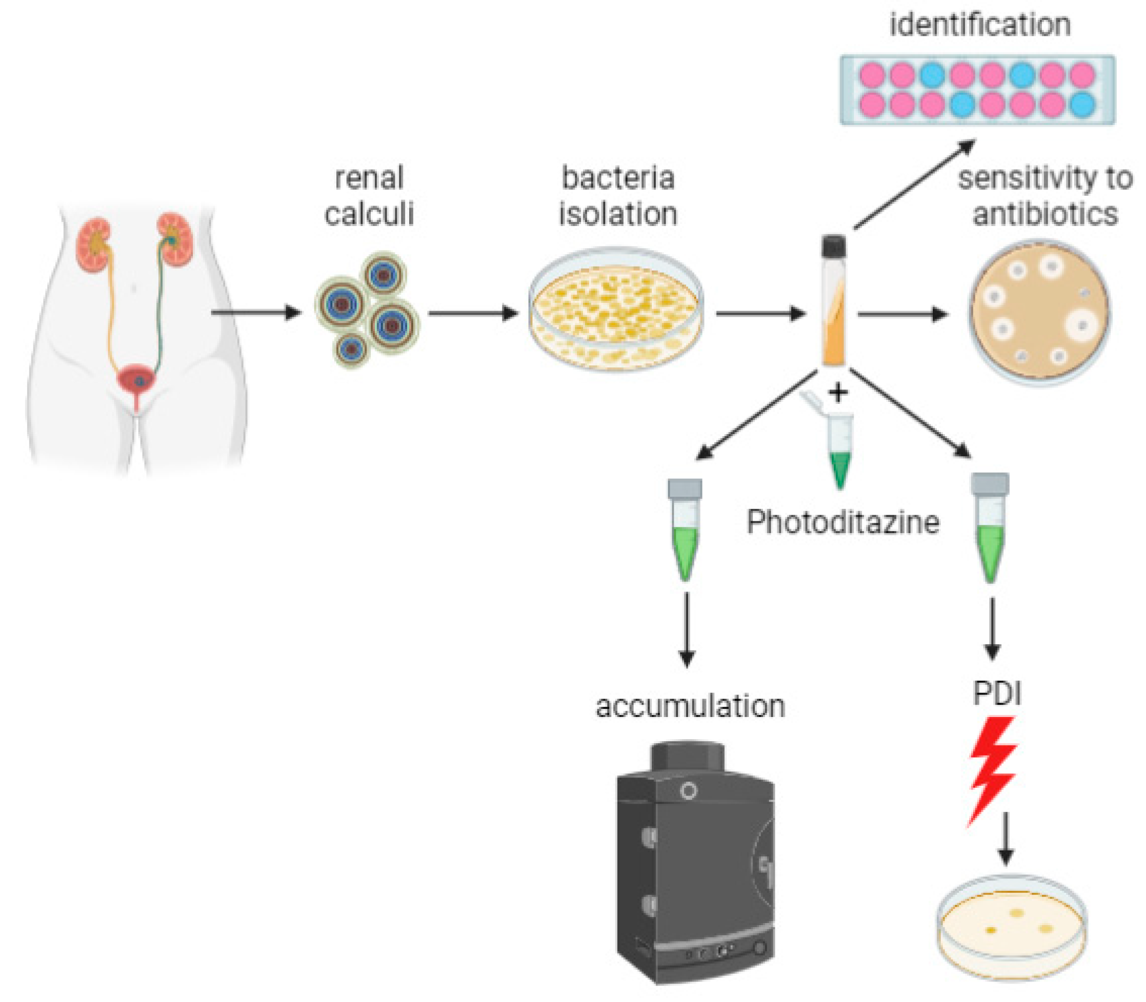
2.1. Isolation and Identification of Pathogenic Microorganisms

Uropathogenic microorganisms were sampled from renal calculi preliminary selected based on their X-ray density. The common type of urinary stones contains magnesium ammonium phosphate. Urinary stones extracted during a laparoscopic surgery were placed in a sterile PBS solution and were crushed after 2 h incubation. The obtained suspension was used for inoculation of Yolk-salt agar, enterococcus agar, MacConkey agar, and nutrient agar. Samples were cultivated overnight at 37 °C. For primary identification, the specimens were stained by Gram. The differentiation of Enterobacteriaceae and Staphylococcaceae representatives was carried out by bacterial biochemical identification kits (RPC Diagnostic Systems, Nizhny Novgorod, Russia). The scheme of the experiments is shown on Figure 1.

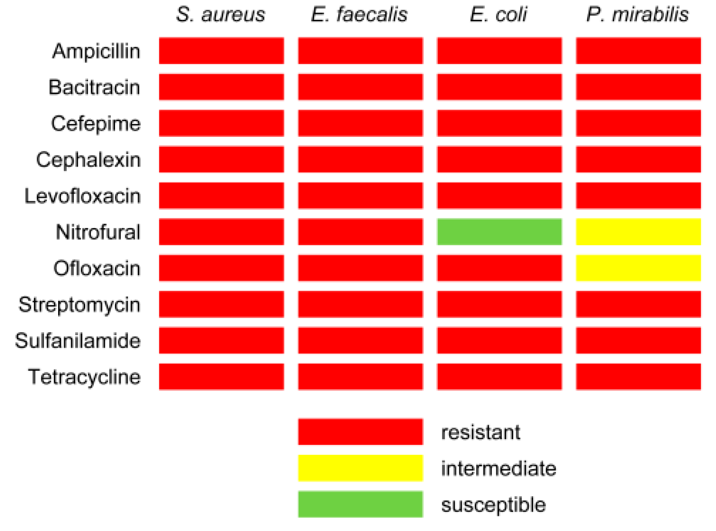
2.2. Antibiotic Susceptibility Testing

The disk diffusion susceptibility method was used according to the approved standard [19]. The strain’s sensitivity to 10 antibiotics (ampicillin, bacitracin, cefepime, cephalexin, levofloxacin, nitrofural, ofloxacin, streptomycin, sulfanilamide, and tetracycline) was evaluated. The test was performed by applying a bacterial inoculum of approximately 1–2 × 10⁸ CFU/mL to the surface of a nutrient agar plate with a diameter of 60 mm. A commercially prepared, fixed-concentration paper antibiotic disk was placed on the inoculated agar surface. The results were assessed after 18–24 h incubation of the plates at 37 °C. The zones of growth inhibition surrounding the antibiotic disk were measured to the nearest millimeter. The zone diameters of each drug were interpreted using the criteria published by the manufacturer’s instruction. (NICF, St. Petersburg, Russia).

2.3. Multiple Antibiotic Resistance (MAR) Index

MAR index is a number of antibiotics to which the test isolate displayed resistance divided by the total number of anbiotics to which the test organism has been evaluated for sensitivity. So, the MAR index for each isolate was calculated [1].

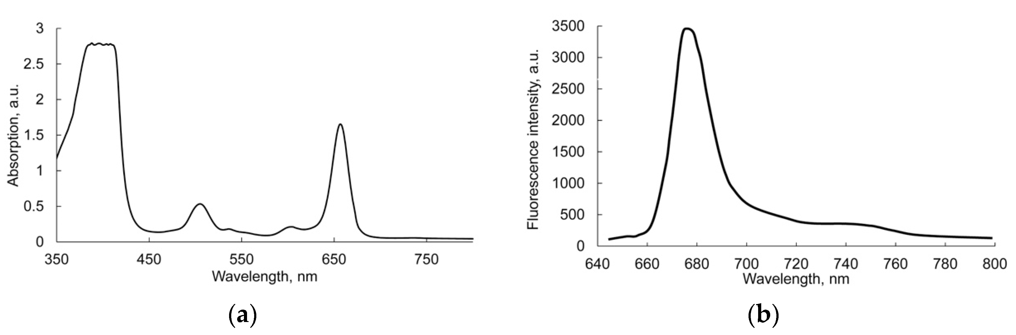
2.4. Accumulation of Photosensitizer by Microorganisms

Fotoditazin® (Veta-Grand LLC, Russia), the chlorin e6 dimeglumine-based photosensitizer (PS) of the second generation, was used in this study. The absorption and fluorescence spectra of PS are depicted in Figure 2. The accumulation of photosensitizer by Gram-positive (Staphylococcus aureus and Enterococcus faecalis) and Gram-negative (Escherichia coli and Proteus mirabilis) species was studied. The photosensitizer at final concentrations of 5 µg/mL and 50 µg/mL was added to the bacterial suspension containing 2.5 × 10⁸ CFU/mL. In 15, 30, and 60 min of incubation in the dark, the microorganisms were twice washed by PBS solution from the unbound photosensitizer and were resuspended in 500 µL of PBS solution for fluorescence assessment. The photosensitizer accumulation was analyzed by the increase in the intensity of the fluorescence using the IVIS Spectrum (Caliper Life Sciences, Waltham, MA, USA) with an excitation filter 640/35 nm and an emission filter 680/20 nm. The average intensity of fluorescence was calculated for the same size areas of each sample using Living Image software (Caliper Life Sciences, Waltham, MA, USA).

2.5. Preparation of Competent Bacterial Cells

A slightly modified rapid protocol for the preparation of electrocompetent bacterial cells [20] with slight modification was used. The bacterial suspension containing 2.5 × 10⁸ CFU/mL in ice-cold PBS was centrifuged for 7 min at 7000 rpm using a microcentrifuge. After the supernatant was discarded, the bacterial pellet was resuspended in the same volume of ice-cold deionized water. This step was repeated twice for a total of three washes. At the next step, the bacterial pellet was resuspended in the ice cold 10% glycerol on deionized water and kept at +4 °C for 20 min. Then, the bacterial cells were centrifuged and resuspended in PBS.

2.6. PDI Assay In Vitro

The microorganisms were diluted from a nutrient broth to the work concentration of 2.5 × 10⁸ CFU/mL in a phosphate buffer saline. The photosensitizer at final concentrations of 5 µg/mL and 50 µg/mL was added to the bacteria with subsequent incubation for 15 min in the dark at room temperature. For laser treatment 100 µL of the samples were transferred into a 96-well plate. The fiber-coupled diode laser (Atcus LLC, Russia) with a wavelength of 659 nm was used to illuminate samples. Laser irradiation was performed for 9 min with an output power (total light dose) of 50 mW (90 J/cm2), 100 mW (180 J/cm2), and 150 mW (270 J/cm2). The laser beam was focused in a circle of 5 mm in diameter. After that, the samples were diluted at a ratio 1:1000 and inoculated into plates with nutrient agar. In 18–24 h the plates were photographed and the colonies were counted using ImageJ software (NIH, Bethesda, MD, USA). Bacteria without any treatment were served as a control.

2.7. Statistical Analysis

The amount of sterile and contaminated calculi by mono and mixed pathogens as well as various strains was expressed as percentages ± standard error of the proportion. The fluorescence intensity was presented as mean ± standard deviation. The reduction of colony forming units of each strain was calculated in percentages of the control and shown as a mean ± standard deviation. To calculate the statistical significance of the differences, the ANOVA with Bonferroni post-hoc test was used. Statistical analysis was performed with Statistica 10 (StatSoft. Inc., Tusla, OK, USA). p-values ≤ 0.05 were considered statistically significant.

3. Results

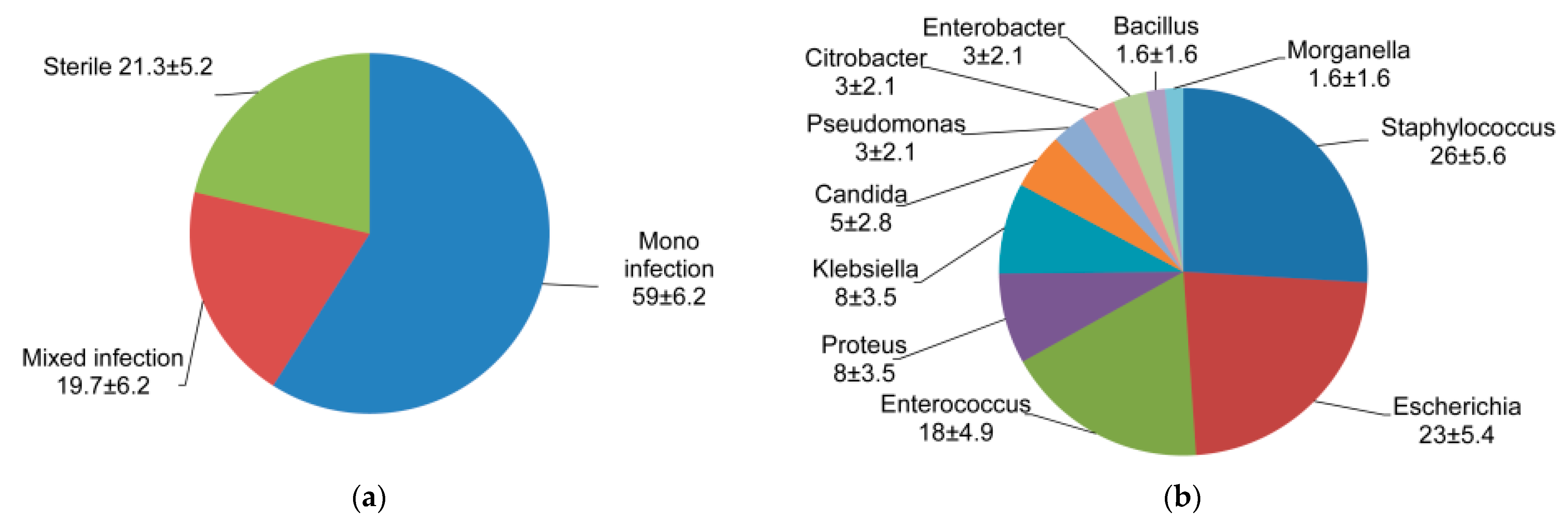
3.1. Spectrum of Uropathogenic Bacteria Associated with Renal Calculi

It was found that renal calculi obtained during laparoscopic surgeries were contaminated in 78.7 ± 5.2% of cases (59 ± 6.2% by one strain and 19.7 ± 6.2% by 2 or more various strains) (Figure 3a). Thus, 66 species of microorganisms referred to various genera were isolated. The species of Staphylococcus, Escherichia, Enterococcus and Proteus were found to be abundant in contaminated renal calculi and accounted for 26%, 23%, 18% and 8% of isolates, respectively (Figure 3b). In addition, opportunistic species of the next genera were isolated, such as Klebsiella (8%), Candida (5%), Pseudomonas (3%), Citrobacter (3%), Enterobacter (3%), Bacillus (1.6%), and Morganella (1.6%). Representatives of four abundant bacterial genera were used for further studies.

3.2. Antibiotic Susceptibility Testing

Testing the sensitivity of the isolated strains to 10 antibiotics of different mechanisms of action, the most widely used in urological practice, showed that the studied strains have a high antibiotic resistance (Figure 4).
Staphylococcus aureus and Enterococcus faecalis strains were shown to be resistant to all the tested drugs (MAR = 1). It was found that Escherichia coli was susceptible to nitrofural (MAR = 0.9) and Proteus mirabilis had intermediate sensitivity to ofloxacin and nitrofural (MAR = 0.8). High sensitivity was not found to any of the drugs.

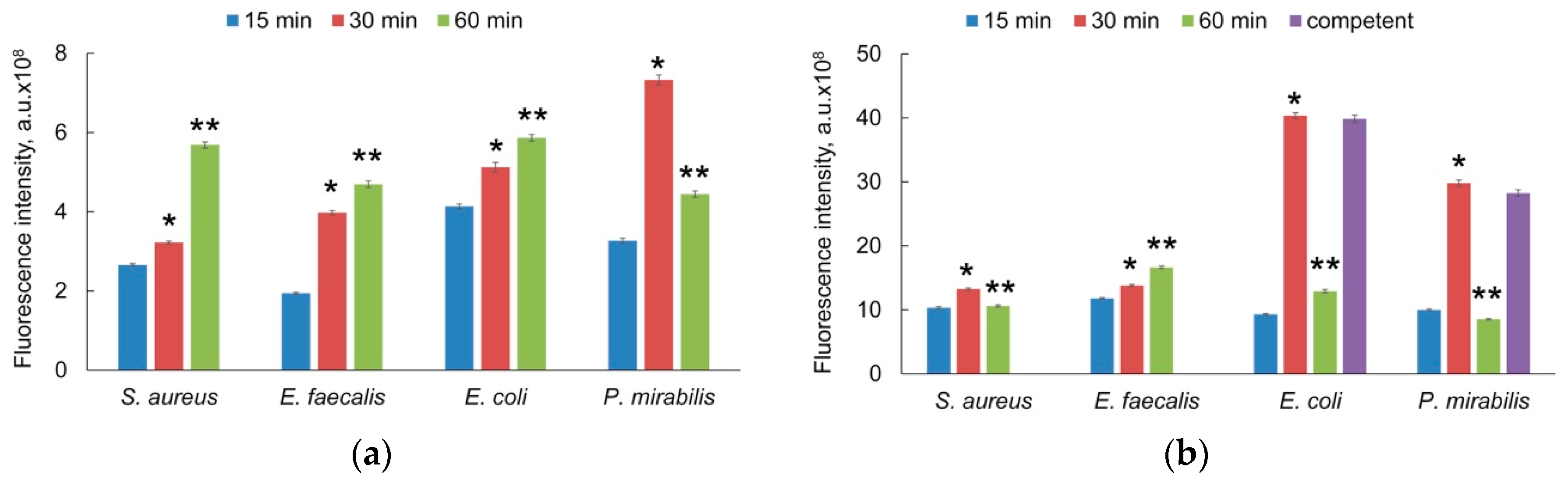
3.3. Photosensitizer Accumulation

The interaction between photosensitizer and urapothogenic microorganisms was analyzed on Gram-negative (E. coli and P. mirabilis) and Gram-positive (E. faecalis and S. aureus) species. Since the samples were washed from free molecules of the photosensitizer, the fluorescence was only detected from the photosensitizer that had penetrated into cells and/or was bound with the cell wall. The photosensitizer accumulation was estimated to be dependent on both incubation time and concentration. The fluorescence intensity was found to be higher for Gram-negative strains than for the Gram-positive ones regardless of the photosensitizer concentration. The strains of E. faecalis and S. aureus demonstrated the enhancement of the fluorescence intensity in a time-dependent manner with the maximal value at 60 min. E. coli and P. mirabilis had the maximal value of fluorescence intensity after 30 min and that significantly decreased by 60 min (Figure 5).
In addition, the assessment of photosensitizer accumulation by competent cells of E. coli and P. mirabilis was performed. After 30 min of the incubation, bacteria with Fotoditazin at a final concentration of 50 µg/mL fluorescence intensity had the same values as the corresponding non-competent strains. The optimal incubation time was found to be 30 min. However, we are planning to use this technique during laser lithotripsy for sanitation, where the time is a limiting factor; therefore, we have chosen incubation for 15 min. The working concentration of the photosensitizer was selected to be 50 µg/mL.

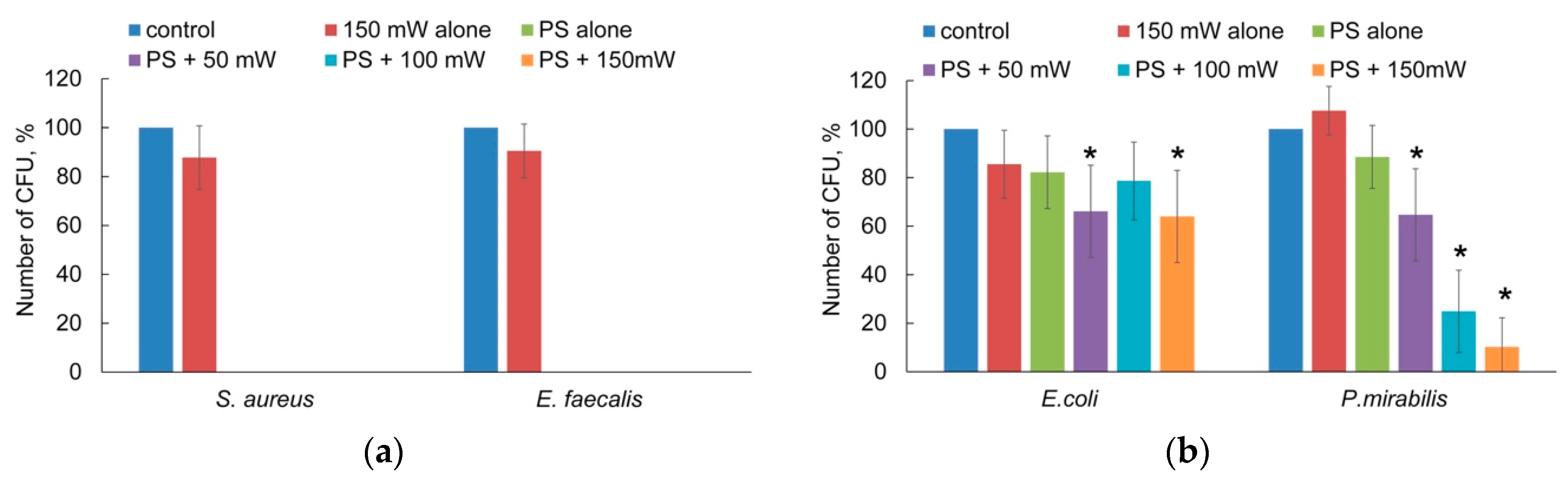
3.4. Photodynamic Inactivation

The output laser power in the study was chosen so as to achieve the bactericidal effect. The fraction of viable bacteria for the photodynamic inactivation experiments was expressed as the percent of CFUs out of the strains treated with both light and photosensitizer. As a control, CFUs measured for strains were treated with neither photosensitizer nor light. Therefore, the viable fraction corresponding to PS is a measure of the toxicity of the photosensitizer and ambient light to the bacteria. The bar graphs of bacteria viability are shown in Figure 6. The toxicity of Fotoditazin alone had a strong effect on Gram-positive strains (Figure 6a). After 15 min of incubation in the dark followed by 15 min of manipulation (dilution, inoculation) at ambient light, no colony was detected on the plates. There was no difference in killing between S. aureus and E. faecalis. In contrast to the photosensitizer, the treatment of either Gram-positive or Gram-negative bacteria by laser light only did not induce significant reduction of CFUs. The survival rate of P. mirabilis for photodynamic inactivation was power-dependent. The number of viable bacteria decreased from 65% to 10% with an increase in power from 50 mW to 150 mW. The maximal bactericidal effect was reached at 150 mW (Figure 6b).
Isolates of E. coli had a weaker response to photodynamic inactivation. The number of viable bacteria was counted to be only 64% after photodynamic inactivation at 150 mW.

4. Discussion

Photodynamic inactivation is considered to be an effective method to control localized bacterial infection. The present study shows a complex approach to assess the efficacy of photodynamic inactivation of drug resistant bacteria associated with renal calculi. Bacteria isolated from urinary stones were tested to be drug resistant for 10 antibiotics. The same isolates were used to analyze the photosensitizer accumulation and to select the photodynamic inactivation parameters.
The results have shown that bacteria are quite often present in the concretions. Uric acid urolithiasis is usually associated with persistently low urine pH. All patients with uric acid calculi demonstrate constantly low urinary pH, while the majority excrete normal amounts of urates [21]. Decreasing pH of urine may be a result of bacterial contamination. It was shown that patients with inflammatory bowel disease, ileostomy, or multiple bowel resections, especially involving the terminal ileum are predisposed to uric acid nephrolithiasis [21]. In our study about 78.7 ± 5.2% of all analyzed calculi contained microorganisms in both mono and mixed cultures. In agreement with the reports from other researchers [6], Escherichia coli was the most often presented species in calculi. E. coli is an intestinal commensal that may migrate to the urinary tract due to poor personal hygiene, decreasing of colonization resistance, urinary retention, and other predisposing factors [22]. Moreover, bacteria associated with calculi may be not found in urine [6].
For this study, abundant species Escherichia coli, Enterococcus faecalis and Staphylococcus aureus were selected. The next most common strains are Proteus spp., Klebsiella spp., and Pseudomonas spp. [23]. Complicated infections prolong treatment and may cause catheter biofilm or stone formation, particularly with Proteus mirabilis [24]. We choose P. mirabilis because this pathogen causes monomicrobial and polymicrobial catheter-associated urinary tract infection primarily through indwelling catheters [25]. The urinary tract usually cleans out the microbe before its exponential growth, but the catheter accumulates pathogens on the surface and prevents this cleaning. P. mirabilis can then adhere to the catheter and form biofilms. Once established, bacteria can spread through the urethra via capacity to motion and may reach the bladder. There P. mirabilis binds to the bladder epithelial cells and can also lead to a urease-mediated mechanism of the formation of kidney and bladder stones. Proteus mirabilis urease hydrolyzes urine urea to ammonia 6 to 10 times faster than urease of other species [26].
Antibiotic susceptibility testing revealed the strong resistance of all isolates to the studied antibiotics. According to the present-day definition, a microorganism is multidrug resistant if it acquired non-susceptibility to at least one agent in three or more antimicrobial categories [27]. In this study, all isolates are multidrug resistant as they have resistance to antibiotics in at least six categories. Therefore, the use of common drugs to treat such infections will be inefficient. Moreover, the antimicrobial agents cannot invade, where these bacteria are present within the interspace of the stones. Thus, the outcome is progressive expansion of stones because of a persistent infection over a period of weeks or months [6]. Some scientific groups are developing a photodynamic inactivation methodology as an alternative to antibiotic treatment [28].
The accumulation of a photosensitizer is one of the major factors to establish an efficient photodynamic treatment for bacterial eradication. Accumulation in cells is generally described by three main mechanisms: external action, intracellular action (including self-promoted uptake), and active transport [29]. All of them have provided efficient photokilling of both Gram-positive and Gram-negative bacteria. So, it is well known that the efficiency of a photodynamic inactivation depends on photosensitizer and molecular oxygen concentrations, incubation time and light dose. We analyzed the accumulation of Fotoditazin by Gram-positive and Gram-negative bacteria isolated from renal calculi by the fluorescence imaging technique. According to this technique, the value of fluorescence intensity of a sample is directly related to photosensitizer concentration. It was found that the fluorescence intensity of photosensitizer in bacterial suspension increases with increasing the working concentration of photosensitizer or incubation time, which is logical and correlates with the other paper [29]. However, it was revealed that fluorescence intensity of Gram-negative bacteria was higher than Gram-positive. Differences in the outer membrane structure of Gram-negative bacteria may affect the photosensitizer penetration and accumulation. This membrane is an asymmetric bilayer of lipopolysaccharides and phospholipids containing specific uptake channels and nonspecific pores [30]. The bilayers containing lipopolysaccharides are more rigid than normal bilayers, slowing passive diffusion of hydrophobic compounds, whereas pores limit by size the penetration of hydrophilic drugs [31]. Moreover, photosensitizers with a negative charge, such as Fotoditazin, may interact with negatively charged lipopolysaccharides and do not penetrate through the inner membrane. In addition, the differences in the intensity of fluorescence between gram-positive and gram-negative bacteria may be associated with the assessment approach used in our work. The measurement of fluorescence was carried out on a suspension of non-lysed bacteria. Thus, the fluorescence of the photosensitizer was located both inside the bacterial cell and in the cell wall. The paper, which shows a greater fluorescence level for gram-negative bacteria, analyzes living bacteria [32]. In the case of assessment of lysed cells, the fluorescence for gram-negative and gram-positive bacteria does not differ [33,34]. The rapid protocol for the preparation of electrocompetent bacterial cells was used as a control for photosensitizer accumulation by Gram-negative bacteria [20]. During this procedure all dissolved ions were removed from the sample solution and bacterial wall. In addition, it is well known that glycerol is capable to form hydrogen bonds with water molecules and cell wall components [35]. Glycerol interacts with the polar heads of the lipid bilayer, which leads to a decrease in the lateral mobility of molecules and to increase in the local membrane stiffness [36]. Thus, the fluorescence intensity should have decreased due to an increase in the total negative charge of the cell wall and a decrease in the photosensitizer accumulation. However, the fluorescence intensity value did not vary. At the same time, the amount of the photosensitizer extracted by sodium dodecyl sulfate from the competent E. coli wall was higher by 54% compared to non-competent cells (data not shown). An increase in the amount of the extracted photosensitizer may be due to the following reasons: On the one hand, efflux pumps may be inactivated due to changes in the stiffness of cell membrane. On the other hand, the electrostatic interaction between the photosensitizer molecules and the cell wall components may be reduced that result in weak sticking of the photosensitizer with following washing out by SDS solution. The dramatic decrease of fluorescence after 1 h incubation is probably due to the activity of efflux pumps [37], which lead to a decrease in the concentration of the intracellular photosensitizer. In addition, cell division can lead to a decrease in the amount of photosensitizer sticking to the cell wall. Cell division is accompanied by hydrolysis of cross-links between existing glycan strands and cleavage of the peptide stem from the glycan backbone of peptidoglycan to produce stemless (“denuded”) glycans [38], which can lead to the release of the photosensitizer. Probably, the low efficiency of photodynamic inactivation of Gram-negative bacteria is caused by this interaction. The singlet oxygen producing in a photodynamic reaction has a lifetime of 3 µs in karyoplasm of eukaryotic cells. It travels a distance of about 134 nm during this time [39]. However, a karyoplasm has a low concentration of quenchers. The periplasm of Gram-negative bacteria is densely packed with proteins [40] that may act as quenchers; in addition, it is more viscous than the cytoplasm that may limit diffuse distance of singlet oxygen. Probably, singlet oxygen does not reach targets, such as DNA or lipids. On the contrary, Gram-positive bacteria have a good response to photosensitizer and photodynamic inactivation. The primary human uropathogens demonstrate the accumulation of photosensitizer and response to photodynamic inactivation in the manner inherent in strains isolated from other areas. These results correlate with the previous papers [41,42]. Thus, the methodology of photodynamic inactivation of Gram-negative bacteria should be adapted.

5. Conclusions

The results of this study indicate that the photosensitizer accumulation by four abundant primary human uropathogens depends on concentration and time. Importantly, this relation is observed for Gram-positive as well as for Gram-negative strains. The photodynamic inactivation has an obvious bactericidal effect against S. aureus and E. faecalis. Only the bacteriostatic effect is achieved at a high light dose for Gram-negative strains. Our data provide the foundation for further studies aimed at the development of the antimicrobial photodynamic inactivation technique. This therapeutic strategy can be adapted for use against uropathogens during laser lithotripsy.

Author Contributions

Conceptualization, N.I. and V.E.; methodology, O.S., V.K. and A.A.; investigation, T.I., I.B. and V.E.; writing—original draft preparation, N.I. and V.E.; writing—review and editing, V.K.; project administration, O.S.; funding acquisition, V.K. All authors have read and agreed to the published version of the manuscript.

Funding

This research was funded by the Russian Science Foundation under grant No. 21-15-00371.

Institutional Review Board Statement

The study was conducted according to the guidelines of the Declaration of Helsinki, and approved by the Institutional Ethics Committee Privolzhsky Research Medical University (protocol No 13, 7 July 2021).

Informed Consent Statement

Informed consent was obtained from all subjects involved in the study.

Data Availability Statement

All relevant data are within the paper.

Acknowledgments

The authors are thankful to Vasily Vlasov (N.A. Semashko Nizhny Novgorod regional clinical hospital, Russia) for provision of renal calculi.

Conflicts of Interest

The authors declare no conflict of interest.

References

  1. Mahato, S.; Mandal, P.; Mahato, A. Biofilm producon by uropathogens like Klebsiella spp. and Pseudomonas spp. and their antibiotic susceptibility. BJHS 2020, 5, 902–906. [Google Scholar]
  2. Rice, L.B. Federal funding for the study of antimicrobial resistance in nosocomial pathogens: No ESKAPE. J. Infect. Dis. 2008, 15, 1079–1081. [Google Scholar] [CrossRef] [PubMed]
  3. Türk, C.; Knoll, T.; Petrik, A.; Sarica, K.; Skolarikos, A.; Straub, M.; Seitz, C. Guidelines on Urolithiasis. Eur. Urol. 2014, 20, 308–341. [Google Scholar]
  4. Sánchez-Martín, F.M.; Rodríguez, M.F.; Fernández, E.S.; Tomás, S.J.; Rousaud Barón, F.; Martínez-Rodríguez, R.; Villavicencio Mavrich, H. Incidencia y prevalencia de la urolitiasis en España: Revisión de los datos originales disponibles hasta la actualidad (Incidence and prevalence of published studies about urolithiasis in Spain. A review). Actas Urológicas Españolas 2007, 31, 511–520. [Google Scholar] [CrossRef] [Green Version]
  5. Bazin, D.; André, G.; Weil, R.; Matzen, G.; Emmanuel, V.; Carpentier, X.; Daudon, M. Absence of bacterial imprints on struvite-containing kidney stones: A structural investigation at the mesoscopic and atomic scale. Urology 2012, 79, 786–790. [Google Scholar] [CrossRef]
  6. Shah, P.; Baral, R.; Agrawal, C.S.; Lamsal, M.; Baral, D.; Khanal, B. Urinary Calculi: A Microbiological and Biochemical Analysis at a Tertiary Care Hospital in Eastern Nepal. Int. J. Microbiol. 2020, 2020, 8880403. [Google Scholar] [CrossRef] [PubMed]
  7. Flannigan, R.; Choy, W.H.; Chew, B.; Lange, D. Renal struvite stones—Pathogenesis, microbiology, and management strategies. Nat. Rev. Urol. 2014, 11, 333–341. [Google Scholar] [CrossRef]
  8. Strohmaier, W. Epidemiologie und Pathogenese der Urolithiasis. In Die Urologie; Part of the series Springer Reference Medizin; Springer: Berlin/Heidelberg, Germany, 2016; pp. 473–478. [Google Scholar] [CrossRef]
  9. Surawicz, C.M.; McFarland, L.V. Pseudomembranous colitis: Causes and cures. Digestion 1999, 60, 91–100. [Google Scholar] [CrossRef]
  10. Cohen, S.H.; Gerding, D.N.; Johnson, S.; Kelly, C.P.; Loo, V.G.; McDonald, L.C.; Pepin, J.; Wilcox, M.H.; Society for Healthcare Epidemiology of America; Infectious Diseases Society of America. Clinical practice guidelines for Clostridium difficile infection in adults: 2010 update by the society for healthcare epidemiology of America (SHEA) and the infectious diseases society of America (IDSA). Infect. Control Hosp. Epidemiol. 2010, 31, 431–455. [Google Scholar] [CrossRef]
  11. Sperandio, F.F.; Huang, Y.Y.; Hamblin, M.R. Antimicrobial photodynamic therapy to kill gram-negative bacteria. Recent Pat. Anti-Infect. Drug Discov. 2013, 8, 108–120. [Google Scholar] [CrossRef] [Green Version]
  12. Cieplik, F.; Deng, D.; Crielaard, W.; Buchalla, W.; Hellwig, E.; Al-Ahmad, A.; Maisch, T. Antimicrobial photodynamic therapy—What we know and what we don’t. Crit. Rev. Microbiol. 2018, 44, 571–589. [Google Scholar] [CrossRef] [PubMed] [Green Version]
  13. Rkein, A.M.; Ozog, D.M. Photodynamic therapy. Dermatol. Clin. 2014, 32, 415–425. [Google Scholar] [CrossRef] [PubMed]
  14. Kashef, N.; Hamblin, M.R. Can microbial cells develop resistance to oxidative stress in antimicrobial photodynamic inactivation? Drug Resist. Updates 2017, 31, 31–42. [Google Scholar] [CrossRef]
  15. Staerck, C.; Gastebois, A.; Vandeputte, P.; Calenda, A.; Larcher, G.; Gillmann, L.; Papon, N.; Bouchara, J.P.; Fleury, M.J.J. Microbial antioxidant defense enzymes. Microb. Pathog. 2017, 110, 56–65. [Google Scholar] [CrossRef] [PubMed]
  16. Sarda, R.A.; Shetty, R.M.; Tamrakar, A.; Shetty, S.Y. Antimicrobial efficacy of photodynamic therapy, diode laser, and sodium hypochlorite and their combinations on endodontic pathogens. Photodiagnosis Photodyn. Ther. 2019, 28, 265–272. [Google Scholar] [CrossRef]
  17. Schreiner, M.; Baumler, W.; Eckl, D.B.; Spath, A.; Konig, B.; Eichner, A. Photodynamic inactivation of bacteria to decolonize meticillin-resistant Staphylococcus aureus from human skin. Brit. J. Dermatol. 2018, 179, 1358–1367. [Google Scholar] [CrossRef]
  18. Otieno, W.; Liu, C.; Deng, H.; Li, J.; Zeng, X.; Ji, Y. Hypocrellin B-Mediated photodynamic inactivation of gram-positive antibiotic-resistant bacteria: An in vitro study. Photobiomodul. Photomed. Laser Surg. 2019, 38, 36–42. [Google Scholar] [CrossRef]
  19. Wayne, P.A. Performance Standards for Antimicrobial Disk Susceptibility Tests, 12th ed.; CLSI document M02-A12; Clinical and Laboratory Standards Institute: Wayne, PA, USA, 2015. [Google Scholar]
  20. Gonzales, M.F.; Brooks, T.; Pukatzki, S.U.; Provenzano, D. Rapid protocol for preparation of electrocompetent Escherichia coli and Vibrio cholerae. J. Vis. Exp. 2013, 8, 50684. [Google Scholar] [CrossRef] [Green Version]
  21. Abou-Elela, A. Epidemiology, pathophysiology, and management of uric acid urolithiasis: A narrative review. J. Adv. Res. 2017, 8, 513–527. [Google Scholar] [CrossRef]
  22. Thass, N.; Kumar, M.; Kaur, R. Phenotypic methods to detect virulence factors of uropathogenic e.coli isolates and their antimicrobial susceptibility pattern. Int. J. Cur. Adv. Res. 2020, 9, 22345–22348. [Google Scholar]
  23. Flores-Mireles, A.L.; Walker, J.N.; Caparon, M.; Hultgren, S.J. Urinary tract infections: Epidemiology, mechanisms of infection and treatment options. Nat. Rev. Microbiol. 2015, 13, 269–284. [Google Scholar] [CrossRef] [PubMed]
  24. Cortes-Penfield, N.W.; Trautner, B.W.; Jump, R.L.P. Urinary tract infection and asymptomac bacteriuria in older adults. Infect. Dis. Clin. N. Am. 2017, 31, 673–688. [Google Scholar] [CrossRef] [Green Version]
  25. Smith, S.N.; Armbruster, C.E. Indwelling urinary catheter model of Proteus mirabilis infection. Methods Mol. Biol. 2019, 2021, 187–200. [Google Scholar] [CrossRef]
  26. Morris, N.S.; Stickler, D.J.; McLean, R.J. The development of bacterial biofilms on indwelling urethral catheters. World J. Urol. 1999, 17, 345–350. [Google Scholar] [CrossRef] [PubMed]
  27. Magiorakos, A.P.; Srinivasan, A.; Carey, R.B.; Carmeli, Y.; Falagas, M.E.; Giske, C.G.; Harbarth, S.; Hindler, J.F.; Kahlmeter, G.; Olsson-Liljequist, B.; et al. Multidrug-resistant, extensively drug-resistant and pandrug-resistant bacteria: An international expert proposal for interim standard definitions for acquired resistance. Clin. Microbiol. Infect. 2012, 18, 268–281. [Google Scholar] [CrossRef] [Green Version]
  28. Liu, Y.; Qin, R.; Zaat, S.A.J.; Breukink, E.; Heger, M. Antibacterial photodynamic therapy: Overview of a promising approach to fight antibiotic-resistant bacterial infections. J. Clin. Transl. Res. 2015, 1, 140–167. [Google Scholar] [PubMed]
  29. Rapacka-Zdończyk, A.; Woźniak, A.; Michalska, K.; Pierański, M.; Ogonowska, P.; Grinholc, M.; Nakonieczna, J. Factors determining the susceptibility of bacteria to antibacterial photodynamic inactivation. Front. Med. (Lausanne) 2021, 8, 642609. [Google Scholar] [CrossRef]
  30. Delcour, A.H. Outer membrane permeability and antibiotic resistance. Biochim. Et Biophys. Acta 2009, 1794, 808–816. [Google Scholar] [CrossRef] [Green Version]
  31. Buzalewicz, I.; Ulatowska-Jarża, A.; Kaczorowska, A.; Gąsior-Głogowska, M.; Podbielska, H.; Karwańska, M.; Wieliczko, A.; Matczuk, A.K.; Kowal, K.; Kopaczyńska, M. Bacteria single-cell and photosensitizer interaction revealed by quantitative phase imaging. Int. J. Mol. Sci. 2021, 22, 5068. [Google Scholar] [CrossRef]
  32. George, S.; Hamblin, M.R.; Kishen, A. Uptake pathways of anionic and cationic photosensitizers into bacteria. Photochem. Photobiol. Sci. 2009, 8, 788–795. [Google Scholar] [CrossRef] [Green Version]
  33. Sułek, A.; Pucelik, B.; Kobielusz, M.; Barzowska, A.; Dąbrowski, J.M. Photodynamic inactivation of bacteria with porphyrin derivatives: Effect of charge, lipophilicity, ROS generation, and cellular uptake on their biological activity in vitro. Int. J. Mol. Sci. 2020, 21, 8716. [Google Scholar] [CrossRef] [PubMed]
  34. Schrader, A.M.; Cheng, C.-Y.; Israelachvili, J.N.; Han, S. Communication: Contrasting effects of glycerol and DMSO on lipid membrane surface hydration dynamics and forces. J. Chem. Phys. 2016, 145, 041101. [Google Scholar] [CrossRef] [PubMed] [Green Version]
  35. Pocivavsek, L.; Gavrilov, K.; Cao, K.D.; Chi, E.Y.; Li, D.; Lin, B.; Meron, M.; Majewski, J.; Lee, K.Y. Glycerol-induced membrane stiffening: The role of viscous fluid adlayers. Biophys. J. 2011, 101, 118–127. [Google Scholar] [CrossRef] [PubMed] [Green Version]
  36. Kourtesi, C.; Ball, A.R.; Huang, Y.Y.; Jachak, S.M.; Vera, D.M.; Khondkar, P.; Gibbons, S.; Hamblin, M.R.; Tegos, G.P. Microbial efflux systems and inhibitors: Approaches to drug discovery and the challenge of clinical implementation. Open Microbiol. J. 2013, 7, 34–52. [Google Scholar] [CrossRef] [Green Version]
  37. Mueller, E.A.; Levin, P.A. Bacterial cell wall quality control during environmental stress. mBio 2020, 11, e02456-20. [Google Scholar] [CrossRef] [PubMed]
  38. Zgurskaya, H.I.; Löpez, C.A.; Gnanakaran, S. Permeability barrier of gram-negative cell envelopes and approaches to bypass it. ACS Infect. Dis. 2015, 1, 512–522. [Google Scholar] [CrossRef] [Green Version]
  39. Skovsen, E.; Snyder, J.W.; Lambert, J.D.; Ogilby, P.R. Lifetime and diffusion of singlet oxygen in a cell. J. Phys. Chem. 2005, 12, 8570–8573. [Google Scholar] [CrossRef]
  40. Mullineaux, C.W.; Nenninger, A.; Ray, N.; Robinson, C. Diffusion of green fluorescent protein in three cell environments in Escherichia coli. J. Bacteriol. 2006, 188, 3442–3448. [Google Scholar] [CrossRef] [Green Version]
  41. Fekrazad, R.; Zare, H.; Mohammadi, S.S.; Morsali, P. The effect of antimicrobial photodynamic therapy with radachlorin® on Staphylococcus aureus and Escherichia coli: An in vitro study. J. Lasers Med. Sci. 2014, 5, 82–85. [Google Scholar]
  42. Luke-Marshall, N.R.; Hansen, L.A.; Shafirstein, G.; Campagnari, A.A. Antimicrobial photodynamic therapy with chlorin e6 is bactericidal against biofilms of the primary human otopathogens. mSphere 2020, 5, e00492-20. [Google Scholar] [CrossRef] [PubMed]
Figure 1. The study design.
Photonics 08 00495 g001
Figure 2. Absorption (a) and fluorescence (b) spectra of Fotoditazin®.
Photonics 08 00495 g002
Figure 3. Microorganisms isolated from renal calculi. Frequency of contaminated calculi (a) and the species accumulation chart of microorganisms (b).
Photonics 08 00495 g003
Figure 4. Results of antibiotic susceptibility testing.
Photonics 08 00495 g004
Figure 5. Accumulation of Fotoditazin at 5 µg/mL (a) and 50 µg/mL (b) of final concentration by Gram-positive and Gram-negative bacterial strains for 15 min, 30 min, and 60 min. Statistically significant differences between 15 min (*) and 30 min (**) are marked.
Photonics 08 00495 g005
Figure 6. Results of photodynamic inactivation of Gram-positive (a) and Gram-negative (b) bacterial strains. Statistically significant differences between the corresponding control group (*) are marked.
Photonics 08 00495 g006
Publisher’s Note: MDPI stays neutral with regard to jurisdictional claims in published maps and institutional affiliations.

