Autofluorescence Imaging of Living Yeast Cells with Deep-Ultraviolet Surface Plasmon Resonance

Abstract
1. Introduction
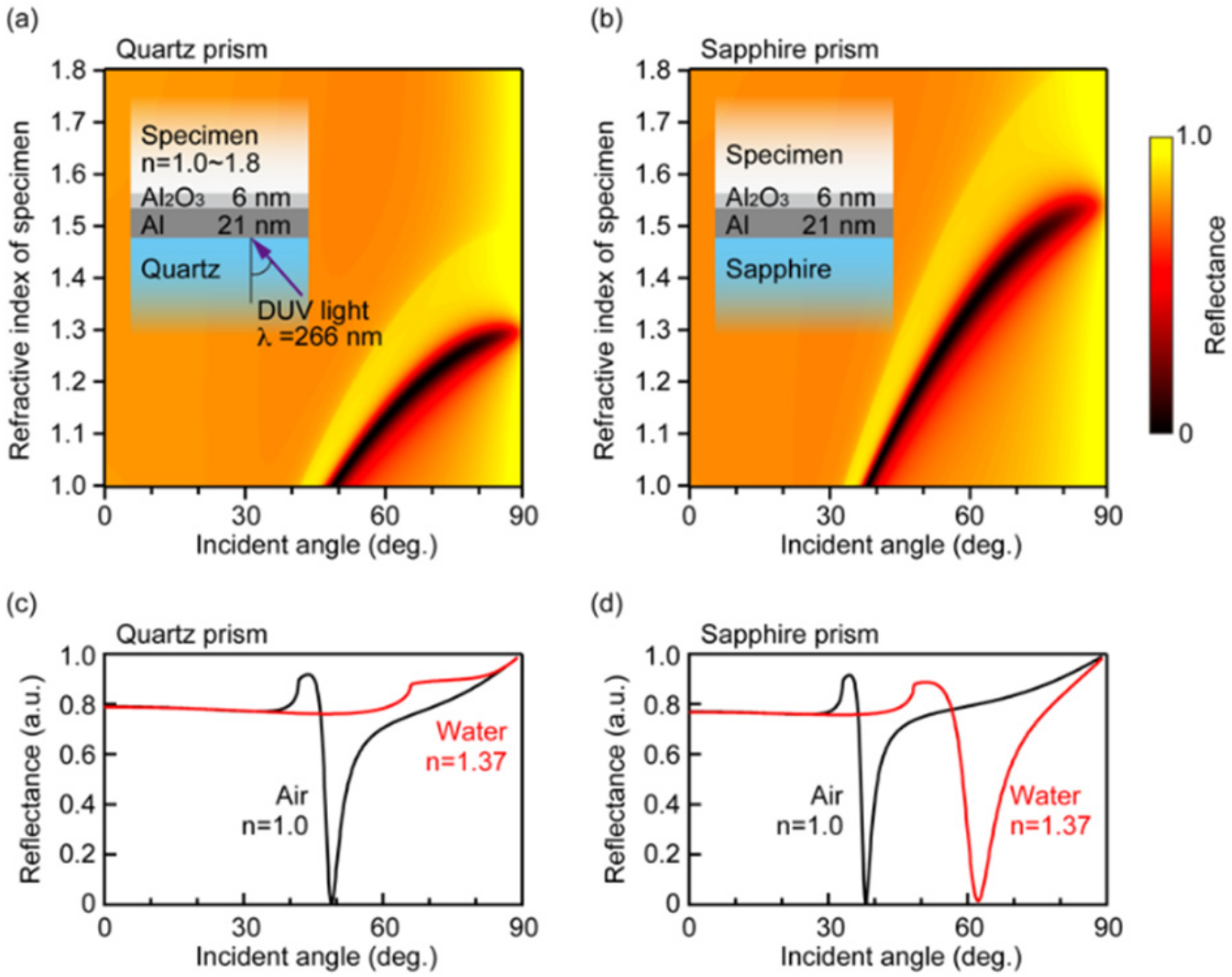
2. Theoretical Analysis
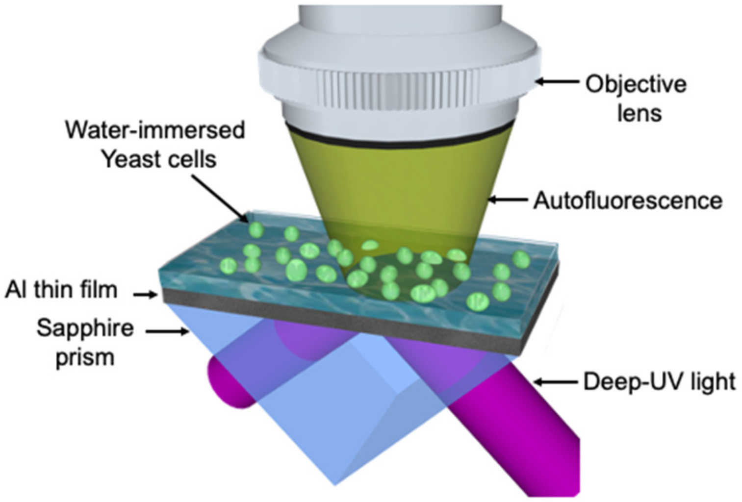
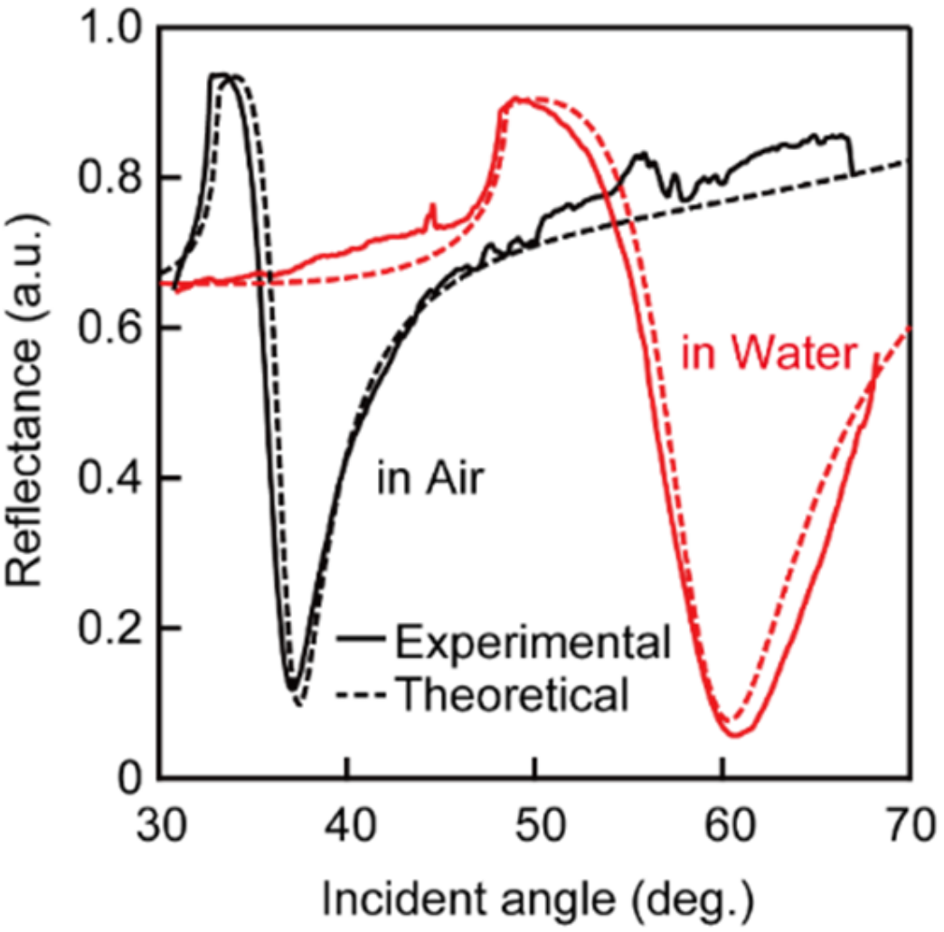
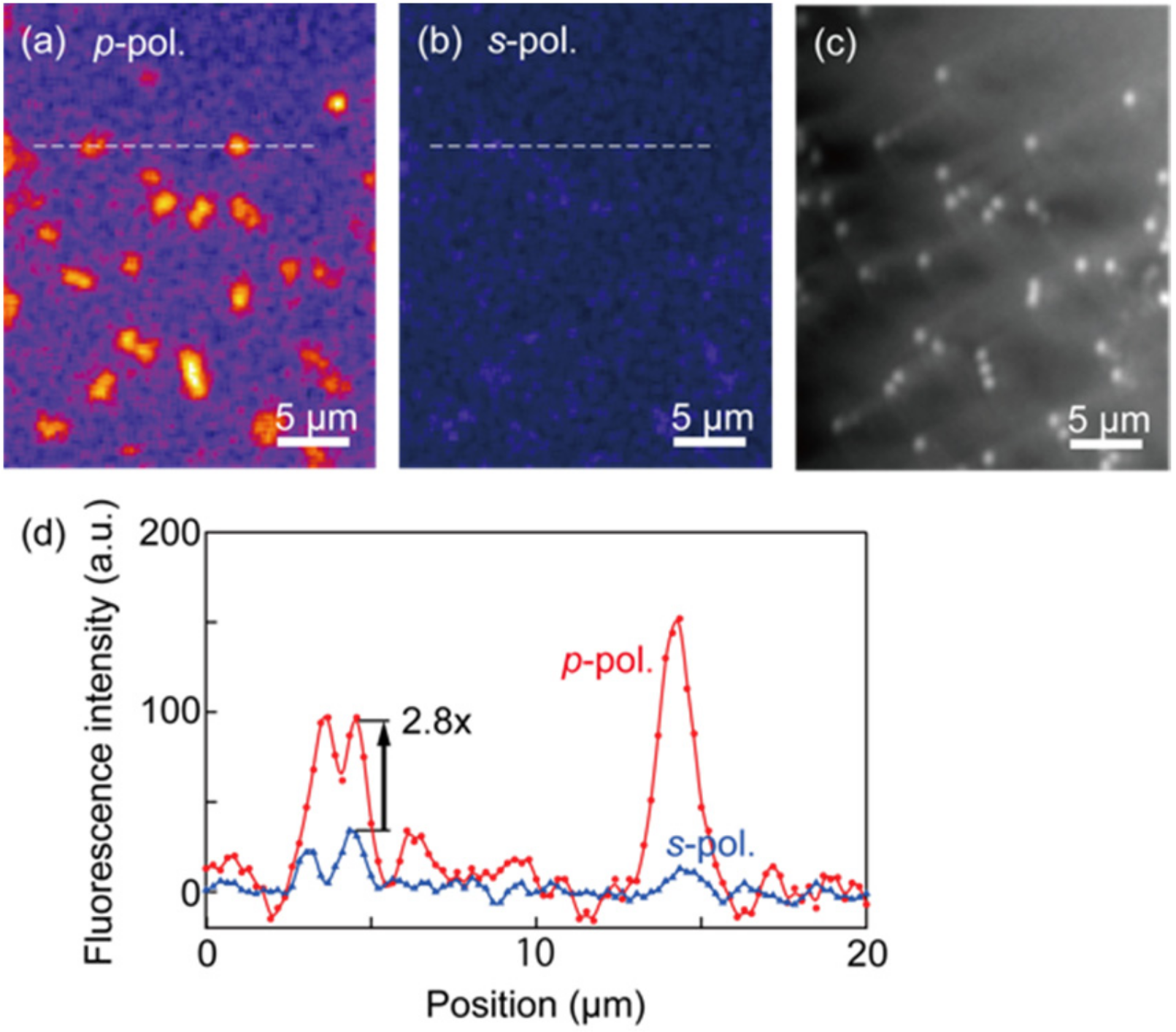
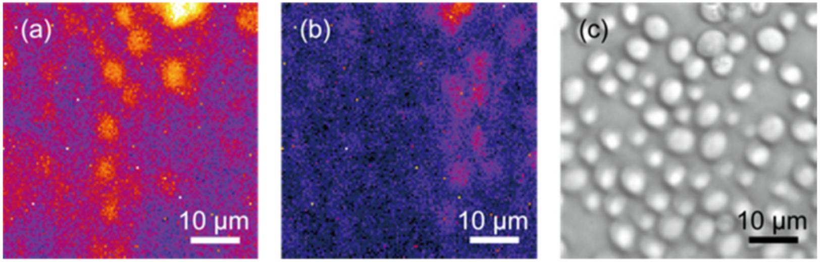
3. Experimental Methods and Results
4. Discussion
5. Conclusions
Author Contributions
Funding
Institutional Review Board Statement
Informed Consent Statement
Data Availability Statement
Conflicts of Interest
References
- Hu, S.; Ren, Y.; Wang, Y.; Li, J.; Qu, J.; Liu, L.; Ma, H.; Tang, Y. Surface plasmon resonance enhancement of photoluminescence intensity and bioimaging application of gold nanorod@CdSe/ZnS quantum dots. Beilstein J. Nanotechnol. 2019, 10, 22–31. [Google Scholar] [CrossRef] [PubMed]
- Uludag, Y.; Tothill, I.E. Cancer Biomarker Detection in Serum Samples Using Surface Plasmon Resonance and Quartz Crystal Microbalance Sensors with Nanoparticle Signal Amplification. Anal. Chem. 2012, 84, 5898–5904. [Google Scholar] [CrossRef] [PubMed]
- Maltais, J.-S.; Denault, J.-B.; Gendron, L.; Grandbois, M. Label-free monitoring of apoptosis by surface plasmon resonance detection of morphological changes. Apoptosis 2012, 17, 916–925. [Google Scholar] [CrossRef] [PubMed]
- Li, D.; Li, L.; Li, P.; Li, Y.; Chen, X. Apoptosis of HeLa cells induced by a new targeting photosensitizer-based PDT via a mitochondrial pathway and ER stress. OncoTargets Ther. 2015, 8, 703–711. [Google Scholar] [CrossRef]
- Zhang, R.; Niu, G.; Liu, Z.; Chau, J.H.; Su, H.; Lee, M.S.; Gu, Y.; Kwok, R.T.; Lam, J.W.; Tang, B.Z. Single AIEgen for multiple tasks: Imaging of dual organelles and evaluation of cell viability. Biomaterials 2020, 242, 119924. [Google Scholar] [CrossRef]
- Hanulia, T.; Inami, W.; Ono, A.; Kawata, Y. Fluorescence lifetime measurement excited with ultraviolet surface plasmon resonance. Opt. Commun. 2018, 427, 266–270. [Google Scholar] [CrossRef]
- Jamme, F.; Kascakova, S.; Villette, S.; Allouche, F.; Pallu, S.; Rouam, V.; Réfrégiers, M. Deep UV autofluorescence microscopy for cell biology and tissue histology. Biol. Cell 2013, 105, 277–288. [Google Scholar] [CrossRef]
- Monici, M. Cell and tissue autofluorescence research and diagnostic applications. Biotechnol. Annu. Rev. 2005, 11, 227–256. [Google Scholar] [CrossRef]
- Cideciyan, A.V.; Swider, M.; Jacobson, S.G. Autofluorescence imaging with near-infrared excitation: Normal-ization by reflectance to reduce signal from choroidal fluorophores. Investig. Ophthalmol. Vis. Sci. 2015, 56, 3393–3406. [Google Scholar] [CrossRef]
- Miljanić, S.; Frkanec, L.; Biljan, T.; Žini, Z.M.M. Recent Advances in linear and non-linear Raman spectroscopy I. J. Raman Spectrosc. 2007, 38, 1538–1553. [Google Scholar]
- Jha, S.K.; Ahmed, Z.; Agio, M.; Ekinci, Y.; Löffler, J.F. Deep-UV Surface-Enhanced Resonance Raman Scattering of Adenine on Aluminum Nanoparticle Arrays. J. Am. Chem. Soc. 2012, 134, 1966–1969. [Google Scholar] [CrossRef]
- Lenzi, E.; De Aberasturi, D.J.; Liz-Marzán, L.M. Surface-Enhanced Raman Scattering Tags for Three-Dimensional Bioimaging and Biomarker Detection. ACS Sens. 2019, 4, 1126–1137. [Google Scholar] [CrossRef]
- Shi, K.; Edwards, P.S.; Hu, J.; Xu, Q.; Wang, Y.; Psaltis, D.; Liu, Z. Holographic coherent anti-Stokes Raman scattering bio-imaging. Biomed. Opt. Express 2012, 3, 1744–1749. [Google Scholar] [CrossRef][Green Version]
- Day, J.P.R.; Domke, K.F.; Rago, G.; Kano, H.; Hamaguchi, H.O.; Vartiainen, E.M.; Bonn, M. Quantitative coherent anti-stokes raman scattering (CARS) microscopy. J. Phys. Chem. B 2011, 115, 7713–7725. [Google Scholar] [CrossRef]
- Steuwe, C.; Kaminski, C.F.; Baumberg, J.J.; Mahajan, S. Surface Enhanced Coherent Anti-Stokes Raman Scattering on Nanostructured Gold Surfaces. Nano Lett. 2011, 11, 5339–5343. [Google Scholar] [CrossRef]
- Kikawada, M.; Ono, A.; Inami, W.; Kawata, Y. Plasmon-Enhanced Autofluorescence Imaging of Organelles in Label-Free Cells by Deep-Ultraviolet Excitation. Anal. Chem. 2016, 88, 1407–1411. [Google Scholar] [CrossRef]
- Ono, A.; Kikawada, M.; Akimoto, R.; Inami, W.; Kawata, Y. Fluorescence enhancement with deep-ultraviolet surface plasmon excitation. Opt. Express 2013, 21, 17447–17453. [Google Scholar] [CrossRef]
- Ray, K.; Chowdhury, M.H.; Lakowicz, J.R. Aluminum nanostructured films as substrates for enhanced fluo-rescence in the ultraviolet-blue spectral region. Anal. Chem. 2007, 79, 6480–6487. [Google Scholar] [CrossRef]
- Szmacinski, H.; Ray, K.; Lakowicz, J.R. Metal-enhanced fluorescence of tryptophan residues in proteins: Application toward label-free bioassays. Anal. Biochem. 2009, 385, 358–364. [Google Scholar] [CrossRef]
- Gryczynski, I.; Malicka, J.; Gryczynski, Z.; Nowaczyk, A.K.; Lakowicz, J.R. Ultraviolet Surface Plasmon-Coupled Emission Using Thin Aluminum Films. Anal. Chem. 2004, 76, 4076–4081. [Google Scholar] [CrossRef]
- Ono, A.; Shiroshita, N.; Kikawada, M.; Inami, W.; Kawata, Y. Enhanced photoelectron emission from aluminum thin film by surface plasmon resonance under deep-ultraviolet excitation. J. Phys. D Appl. Phys. 2014, 48, 184005. [Google Scholar] [CrossRef]
- Watanabe, Y.; Inami, W.; Kawata, Y. Deep-ultraviolet light excites surface plasmon for the enhancement of photoelectron emission. J. Appl. Phys. 2011, 109, 023112. [Google Scholar] [CrossRef]
- Block, I.D.; Mathias, P.C.; Ganesh, N.; Jones, S.I.; Dorvel, B.R.; Chaudhery, V.; Vodkin, L.O.; Bashir, R.; Cunningham, B.T. A detection instrument for enhanced-fluorescence and label-free imaging on photonic crystal surfaces. Opt. Express 2009, 17, 13222–13235. [Google Scholar] [CrossRef]
- Kikawada, M.; Ono, A.; Inami, W.; Kawata, Y. Enhanced multicolor fluorescence in bioimaging using deep-ultraviolet surface plasmon resonance. Appl. Phys. Lett. 2014, 104, 223703. [Google Scholar] [CrossRef]
- Kikawada, M.; Ono, A.; Inami, W.; Kawata, Y. Surface plasmon-enhanced fluorescence cell imaging in deep-UV region. Appl. Phys. Express 2015, 8, 072401. [Google Scholar] [CrossRef]
- Taguchi, A.; Hayazawa, N.; Furusawa, K.; Ishitobi, H.; Kawata, S. Deep-UV tip-enhanced Raman scattering. J. Raman Spectrosc. 2009, 40, 1324–1330. [Google Scholar] [CrossRef]
- Kumamoto, Y.; Taguchi, A.; Honda, M.; Watanabe, K.; Saito, Y.; Kawata, S. Indium for Deep-Ultraviolet Surface-Enhanced Resonance Raman Scattering. ACS Photonics 2014, 1, 598–603. [Google Scholar] [CrossRef]
- Born, M.; Wolf, E. Principle of Optics; Cambridge University Press: Cambridge, UK, 1997; Volume 7, pp. 360–409. [Google Scholar]
- Daimon, M.; Masumura, A. Measurement of the refractive index of distilled water from the near-infrared region to the ultraviolet region. Appl. Opt. 2007, 46, 3811–3820. [Google Scholar] [CrossRef]


| Material | Refractive Index@266nm |
|---|---|
| Aluminum | 0.21 + i3.14 |
| Alumina | 1.83 |
| Quartz prism | 1.52 |
| Sapphire prism | 1.83 |
| Water | 1.37 |
Publisher’s Note: MDPI stays neutral with regard to jurisdictional claims in published maps and institutional affiliations. |
© 2022 by the authors. Licensee MDPI, Basel, Switzerland. This article is an open access article distributed under the terms and conditions of the Creative Commons Attribution (CC BY) license (https://creativecommons.org/licenses/by/4.0/).
Share and Cite
Che Lah, C.N.H.; Morisawa, H.; Kobayashi, K.; Ono, A.; Inami, W.; Kawata, Y. Autofluorescence Imaging of Living Yeast Cells with Deep-Ultraviolet Surface Plasmon Resonance. Photonics 2022, 9, 424. https://doi.org/10.3390/photonics9060424
Che Lah CNH, Morisawa H, Kobayashi K, Ono A, Inami W, Kawata Y. Autofluorescence Imaging of Living Yeast Cells with Deep-Ultraviolet Surface Plasmon Resonance. Photonics. 2022; 9(6):424. https://doi.org/10.3390/photonics9060424
Chicago/Turabian StyleChe Lah, Che Nur Hamizah, Hirofumi Morisawa, Keita Kobayashi, Atsushi Ono, Wataru Inami, and Yoshimasa Kawata. 2022. "Autofluorescence Imaging of Living Yeast Cells with Deep-Ultraviolet Surface Plasmon Resonance" Photonics 9, no. 6: 424. https://doi.org/10.3390/photonics9060424
APA StyleChe Lah, C. N. H., Morisawa, H., Kobayashi, K., Ono, A., Inami, W., & Kawata, Y. (2022). Autofluorescence Imaging of Living Yeast Cells with Deep-Ultraviolet Surface Plasmon Resonance. Photonics, 9(6), 424. https://doi.org/10.3390/photonics9060424