Review

Unique Pulmonary Hypertensive Vascular Diseases Associated with Heart and Lung Developmental Defects

1
Department of Pediatrics, Osaka University Graduate School of Medicine, 2-2 Yamadaoka, Suita 565-0871, Osaka, Japan
2
Department of Cardiology, Tokyo Metropolitan Children’s Medical Center, 2-8-29 Musashidai, Fuchu 183-8561, Tokyo, Japan
3
Department of Pediatrics, Keio University of Medicine, 35 Shinanomachi, Shinjuku-ku 160-8582, Tokyo, Japan
4
Keio University Health Center, 4-1-1 Hiyoshi, Kohoku-ku, Yokohama 223-8521, Kanagawa, Japan
*
Author to whom correspondence should be addressed.
J. Cardiovasc. Dev. Dis. 2023, 10(8), 333; https://doi.org/10.3390/jcdd10080333
Submission received: 28 May 2023 / Revised: 10 July 2023 / Accepted: 1 August 2023 / Published: 3 August 2023

Abstract

:
Although pediatric pulmonary hypertension (PH) shares features and mechanisms with adult PH, there are also some significant differences between the two conditions. Segmental PH is a unique pediatric subtype of PH with unclear and/or multifactorial pathophysiological mechanisms, and is often associated with complex congenital heart disease (CHD), pulmonary atresia with ventricular septal defect, and aortopulmonary collateral arteries. Some cases of complex CHD, associated with a single ventricle after Fontan operation, show pathological changes in the small peripheral pulmonary arteries and pulmonary vascular resistance similar to those observed in pulmonary arterial hypertension (PAH). This condition is termed as the pediatric pulmonary hypertensive vascular disease (PPHVD). Recent advances in genetics have identified the genes responsible for PAH associated with developmental defects of the heart and lungs, such as TBX4 and SOX17. Targeted therapies for PAH have been developed; however, their effects on PH associated with developmental heart and lung defects remain to be established. Real-world data analyses on the anatomy, pathophysiology, genetics, and molecular biology of unique PPHVD cases associated with developmental defects of the heart and lungs, using nationwide and/or international registries, should be conducted in order to improve the treatments and prognosis of patients with these types of pediatric PH.

1. Introduction

Pulmonary arterial hypertension (PAH) is a progressive and lethal disorder. Its estimated incidence is 2.5–7.5 cases per million per year and its prevalence is 15–50 cases per million individuals, according to French [1] and Scottish registries [2]. Also, its estimated prevalence is 4.8–8.1 cases per million for pediatric-onset [3] and 5.6–25 cases per million for adult-onset disease [4]. PAH is defined as a mean pulmonary arterial pressure of >20 mmHg and pulmonary vascular resistance of ≥3 Wood Units (WU) [5]. Childhood PAH is frequently associated with congenital heart disease (CHD) resulting from developmental defects of the cardiovascular system, and belongs to group 1.4.4 of the sixth World Symposium of Pulmonary Hypertension (WSPH) classification (Table 1) [6]. Pulmonary hypertension (PH) has a negative impact on the natural course of CHD and worsens the clinical status and overall outcome. Patients with PAH or CHD are a heterogeneous population. Other than group 1.4.4 of PAH associated with CHD, group 5.4 in the 6th WSPH includes several anomalies involving differential pulmonary blood flow under the category of “segmental PH”, indicating the distinct nature of these entities as compared with other forms of PH [7]. Some types of complex CHDs are associated with congenital abnormalities in the pulmonary vascular structure, leading to segmental PH [6]. Other complex CHDs are associated with a single ventricle after Fontan operation [7]. Although patients with Fontan circulation do not have PH, those with poor pulmonary circulation show similar pathological changes in the small peripheral pulmonary arteries, as well as pulmonary vascular resistance (PVR). This condition is termed pediatric pulmonary hypertensive vascular disease (PPHVD). Finally, recent advances in genetic analyses have identified genes responsible for PAH, which are implicated not only in pulmonary endothelial function, but also in the development of the heart and lungs. For instance, pathogenic variants of TBX4 are strongly associated with developmental disorders of the lungs [7], while variants of SOX17 are strongly associated with simple CHD.
In this review, we focus on unique pulmonary hypertensive vascular diseases associated with developmental defects of the heart and lungs, including segmental PH and PPHVD in the Fontan circulation, and the developmental genes TBX4 and SOX17. Recent advances highlighting the pathophysiological mechanisms, clinical management, and genetic origins of these conditions are discussed.

2. Segmental PH Associated with Complex CHD

2.1. Overview of Segmental PH

Segmental PH is a unique subtype of pediatric PH with unclear and multifactorial underlying pathophysiological mechanisms [8]. The concept of segmental PH was introduced in the 2015 European Society of Cardiology (ESC)/European Respiratory Society (ERS) Guidelines for the Diagnosis and Treatment of PH, reflecting the 2013 discussions in the 5th WSPH [8]. In 2018, the 6th WSPH revised their recommendations based on updated research [6], with the Pediatric Task Force of the WSPH refining the definition, classification, epidemiology, diagnostics, and treatment of pediatric PH [7]. Although the latest ESC/ERC PH guidelines have revised the major clinical classification of segmental PH [6], it is still emphasized in the pediatric field and is classified in group 5.4 in the 6th WSPH as pediatric PH with complex CHD. These complex CHD types commonly include tetralogy of Fallot (TOF) with aortopulmonary collateral arteries (MAPCAs), and rarely include isolated pulmonary artery of ductal origin, absent pulmonary artery, and hemitruncus with unbalanced pulmonary circulation, etc. [7]. Segmental PH indicates PH affecting one or several lobes of the lungs to different extents, with the remaining lobes being normotensive [9].

2.2. Segmental PH Associated with MAPCAs

MAPCAs are developmentally aberrant vessels derived from the embryonic intersegmental artery. During the early fetal period, the vascular plexus in the lung buds is connected to the intersegmental arteries arising from the dorsal aorta (Figure 1A) [10,11]. This remains until fetal day 50, when the segmental arteries regress and the sixth pharyngeal arch gives rise to a central pulmonary artery that is connected to the right ventricle. Developmental defects in the sixth pharyngeal arch artery result in a diminutive central pulmonary artery and failure to develop the right ventricular outflow tract. Under these circumstances, the intersegmental arteries maintain the aorto-pulmonary connection and ensure pulmonary blood flow from the aorta after birth. The more severe the developmental defects of the sixth pharyngeal arch artery are, the more diminutive the central pulmonary artery becomes, with the development of MAPCAs (Figure 1B–E).
MAPCAs are commonly associated with TOF, particularly when atresia or severe stenosis of the pulmonary outflow tract exists. Clinically significant MAPCAs generally exceed 3 mm in diameter. As mentioned previously, MAPCAs are believed to originate from the intersegmental arteries connected to the splanchnic vascular plexus. After birth, MAPCAs mostly branch from the descending aorta but can also branch from the ascending aorta, subclavian artery, brachiocephalic artery, and—rarely—from the coronary artery [10,12], connecting with the vascular beds of the lungs. Morphologically, MAPCAs resemble dilated bronchial arteries and may be considered as a substitute for the physiological role of the pulmonary artery in terms of growth and vasoreactivity [12,13]. A histological study demonstrated the same histological findings as for the systemic arteries, with muscular media and distal merging into efficient pulmonary vascular beds [14]. However, the origins and characteristics of MAPCAs remain unclear [12,13,15].
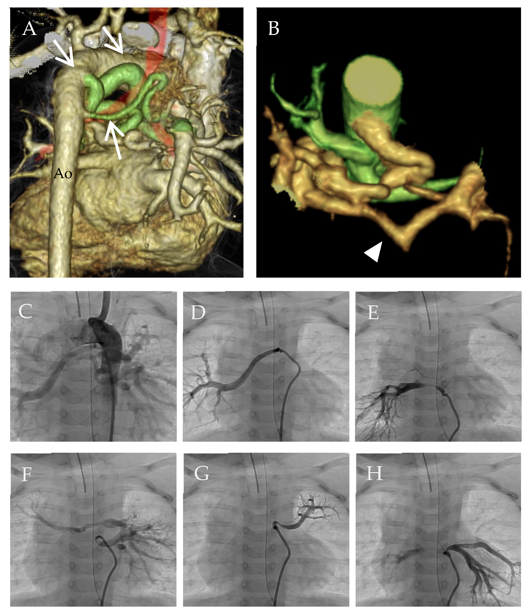
In cases with MAPCAs, pulmonary overcirculation and undercirculation may coexist in different lung segments of the same patient [10]. When MAPCAs are non-stenotic, pulmonary vascular beds are prone to high shear stress under systemic arterial pressure. Excessive pulmonary blood flow may cause PH. On the other hand, peripheral stenosis in MAPCAs may protect some, but not all, pulmonary vascular beds against systemic blood pressure. Moreover, peripheral stenosis of MAPCAs may lead to the development of proximal PH, if severe, and may decrease pulmonary blood flow segmentally. This undercirculation may lead to hypoplasia of the pulmonary vascular beds in some lung segments, eventually increasing the vascular resistance and resulting in PH in corresponding segments. These changes can occur heterogeneously in the lungs of individuals with MAPCAs (Figure 2).

2.3. Surgical Treatment for MAPCAs to Avoid Segmental PH

Surgical intervention is an effective treatment option for creating an integrated unified pulmonary blood supply. Traditionally, multistage unifocalization of the MAPCAs or pulmonary rehabilitation has been performed. Unifocalization includes anastomosing the ipsilateral MAPCAs to unify them and then incorporating them into the central pulmonary artery, if this is present. Systemic pulmonary shunts are also used to maintain the pulmonary blood flow. Pulmonary rehabilitation involves promoting the growth of the hypoplastic central pulmonary artery by using a central aortopulmonary shunt or a right ventricle-to-pulmonary artery connection. Single-stage unifocalization has become the preferred surgical option as it can achieve a lower right ventricular pressure, a lower re-intervention rate, and is a simpler surgical procedure than multi-stage unifocalization and pulmonary rehabilitation [13,15]. After these palliative procedures, if the pulmonary vascular beds are well developed, segmental PH may not occur.
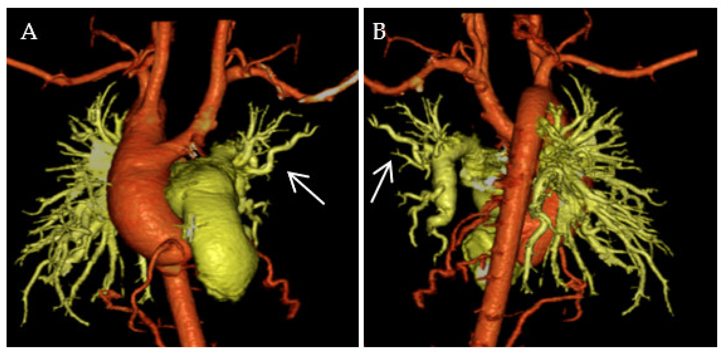
Despite completing the surgical repair of TOF and MAPCAs, segmental PH may persist or may even develop [9]. It can be caused by hypoplasia, obstruction, or hypoxic spasm of the pulmonary vessels, in addition to original unrepaired conditions. Figure 3 shows a three-dimensional multidetector computed tomography imaging of an infant with repaired TOF, MAPCAs, and residual PH. The left pulmonary arteries were more hypoplastic and tortuous than the right pulmonary arteries, suggesting abnormal arborization. In such cases, surgical or catheter interventions, including angioplasty and stenting, may be indicated and medical treatments should be considered.

2.4. Pulmonary Vasodilators for Segmental PH

Pulmonary vasodilators are a medical option for targeting segmental PH. Although the exact origin of MAPCAs is not well understood, distal lesions of MAPCAs appear to be similar to those of pulmonary vascular architecture, as previously described, which supports the efficacy of pulmonary vasodilators [14]. Yasuhara and Yamagishi detailed the case of a 5-year-old girl with repaired TOF and MAPCAs who had segmental PH. They applied combination therapy using both percutaneous pulmonary angioplasty and pulmonary vasodilators, including a phosphodiesterase type 5 (PDE5) inhibitor, sildenafil, and the endothelin-receptor antagonist (ERA), bosentan, which successfully improved segmental PH [11]. Yamamura et al. reported the efficacy of bosentan for treating segmental PH in two pediatric patients with repaired TOF and MAPCAs [16]. Moreover, a few small-scale studies have described the effects of bosentan and/or sildenafil in patients with repaired TOF and MAPCAs with PH (Table 2). All study patients experienced remission of clinical symptoms [17,18,19], but their PH did not always improve, which is consistent with previous findings [9]. Some patients experienced adverse effects from pulmonary vasodilators, such as headache, nausea, lethargy, and dyspnea, including a patient requiring cessation of sildenafil treatment [17,18]. Further pulmonary vasodilators may cause pulmonary edema in sections of the lung where arterial pressures are normal, particularly in patients with diastolic dysfunction. Careful monitoring of patients after administration of these agents is needed.
Because the development and distribution of MAPCA are significantly heterogeneous and individually different, a patient-based approach is necessary to evaluate segmental PH to plan a treatment strategy. Pulmonary vasodilators appear to be effective in only a small number of patients with segmental PH; however, evidence is insufficient. The International Registry of PH associated with CHD is currently enrolling more than 700 patients [20] and is expected to provide a comprehensive overview of their management. In Japan, a nationwide registry of the Japanese Association of CHD-PH Registry (JACPHR) is also ongoing, and several patients with TOF and MAPCAs with segmental PH have been enrolled [21]. Accumulating clinical data on segmental PH may help delineate the most appropriate treatment strategy in future.

3. Pulmonary Hypertensive Vascular Disease in the Fontan Circulation

3.1. Overview of Fontan Circulation

In complex CHD with single-ventricle physiology, the Fontan procedure is a well-established palliative procedure. The original Fontan procedure was reported more than 50 years ago [22] and has been a landmark contribution for improving mortality and morbidity in patients with a single ventricle [23,24,25]. In Fontan circulation, the superior and inferior vena cava are directly connected to the pulmonary arteries without the subpulmonary ventricle. Pulmonary circulation after the Fontan procedure is unique, differing from that associated with biventricular physiology. The diastolic kinetics of the ventricle and central venous pressure (CVP) drive the pulmonary blood flow. Therefore, well-developed pulmonary vascular beds and low PVR are mandatory for establishing Fontan circulation [26,27]. Additionally, ventricular diastolic function and atrioventricular valve regurgitation, which contribute to high pulmonary wedge pressure, are additional factors for optimal Fontan circulation [28,29]. Most of the serious complications of Fontan patients are attributed to the high PVR and CVP [27], and in some cases of so-called “failing Fontan,” the pulmonary circulation is not fully functional. Chronic liver congestion with a high CVP is thought to induce liver fibrosis and inflammation, leading to cirrhosis and eventually to hepatic carcinoma, which is called Fontan-associated liver disease [30,31]. Chronic congestion of the intestines, kidneys, and lymphoid system can cause protein-losing enteropathy (PLE), plastic bronchitis, and renal failure [32,33]. These complications can increase overall mortality, and deteriorate exercise capacity and quality of life in Fontan patients [23,34]. Therefore, understanding the pulmonary vasculature biology and the effects of pulmonary vasodilators is important for managing Fontan patients.

3.2. Pulmonary Vascular Remodeling in Fontan Circulation

Several hypotheses have been proposed regarding the progression of pulmonary vascular remodeling in patients with Fontan disease. The most common pathogenic hypothesis is the lack of pulsatile blood flow in the pulmonary arteries of the Fontan circulation. Several previous studies, including animal models of nonpulsatile cavopulmonary anastomosis, have demonstrated that vascular endothelial function is significantly impaired under conditions of nonpulsatile pulmonary blood flow [35,36,37,38]. Pulsatile blood flow evokes optimal shear stress in endothelial cells and induces the expression of endothelial nitric oxide synthetase (eNOS), which induces NO production for vasodilatation. Impairment of eNOS production leads to vasoconstriction of the pulmonary arteries.
Another hypothesis involves endothelin (ET)-1, a potent vasoconstrictive protein. ET-1 has mitogenic activity in vascular smooth muscle cells and promotes vascular remodeling. The pathogenicity of ET-1 has been previously demonstrated for various types of PAH [39]. Previous clinical studies have demonstrated that plasma ET-1 levels are significantly elevated in Fontan patients and correlate positively with CVP [40,41]. Immunohistological and quantitative real-time polymerase chain reaction analyses demonstrated that the expression of not only ET-1 but also ET receptor types A and B was significantly elevated in the pulmonary arteries of failing Fontan patients as compared to those of healthy controls and non-failing Fontan patients [42]. In patients with failed Fontan circulation, the medial walls of the pulmonary arteries were thickened, and the intima was significantly hypertrophied. Histological findings of pulmonary arterial remodeling were similar to those of patients with idiopathic PAH (IPAH) [43]. Notably, patients with non-failing Fontan circulation who suddenly died of ventricular arrhythmia or infection showed relatively milder remodeling of the pulmonary arteries and lower levels of ET-1 and its receptors than in failing Fontan circulation patients with PLE or plastic bronchitis [42,44]. Although these studies did not analyze the expression levels of eNOS in the pulmonary arteries of failed Fontan patients, they indicated that ET-1 and its signaling pathway may play important roles in the pathogenic mechanisms of pulmonary arterial remodeling in the Fontan circulation and provide a rationale for the use of ERA to treat failing Fontan patients.
Recently, hypoxia-inducible factor (HIF)-1 has been considered as a major player in the pathogenesis of pulmonary vascular remodeling in patients with PAH [45]. The pulmonary vasculature is hypoxic in Fontan patients. Low cardiac output and insufficient vascularization and oxygenation can induce tissue hypoxia, even without cyanosis, after the Fontan operation [46]. Patients with complex CHD often experience hypoxic tissue conditions before and after the Fontan procedure. HIF-1 induces the expression of several downstream factors, including ET-1 and vascular endothelial growth factor (VEGF). VEGF, a growth factor that induces angiogenesis, is elevated in patients with Fontan disease [47]. VEGF plays an important role in the pathogenesis of PAH and in the remodeling of the pulmonary arterial vasculature. Taken together, multiple pathogenic signaling cascades may be involved in the remodeling seen in Fontan patients. Pulmonary vasculature remodeling progresses more readily, particularly in failing Fontan patients, rather than in biventricular repaired patients.

3.3. Pulmonary Vasodilators for Failing Fontan

Numerous pulmonary vasodilative drugs have beneficial effects on exercise capacity, morbidity, and mortality in patients with IPAH and hereditary PAH (HPAH). Considering the pathophysiology of the pulmonary vasculature in failing Fontan patients, pulmonary vasodilators are theoretically likely to be beneficial. Since it was reported that inhaled NO elicits acute vasoreactivity late after the Fontan operation, the potential therapeutic effects of pulmonary vasodilators have been extensively investigated [48]. A randomized controlled trial (RCT) was the first to report that treatment with sildenafil was effective for increasing cardiac output and exercise capacity in Fontan patients [49]. However, a later study found no beneficial effects of sildenafil on Fontan circulation, including the peak VO2 [50]. More recently, in a retrospective cohort study (RC), it was reported that the routine use of sildenafil soon after a Fontan operation did not improve postoperative morbidity or the length of hospital stay [51]. Udenafil, a recently developed PDE5 inhibitor, was administered to Fontan patients in the FUEL trial and was found to improve multiple measures of exercise performance, but not peak VO2 [52]. Bosentan, a potent ERA, was also provided to Fontan patients in an RCT and demonstrated several beneficial effects. Although Schuuring et al. showed that 6 months of treatment with bosentan did not provide benefits for Fontan patients, Shang et al. demonstrated that bosentan reduced the incidence of pulmonary arteriovenous fistulae and protein-losing enteropathy while improving heart function [53,54]. Another small size RCT demonstrated that ambrisentan, a more specific antagonist of the ET receptor type A, could improve the exercise capacity of adult Fontan patients [55]. Illoprost, a synthetic analog of prostacyclin, was also investigated in a small RCT, which found it capable of improving peak oxygen pulse and peak VO2 in Fontan patients, appearing to be particularly beneficial in patients with impaired exercise function [56].
Recently, meta-analyses of the efficacy and safety of pulmonary vasodilators in Fontan patients were published by Wang et al. [57] and Li et al. [58]. Peak VO2 was significantly improved with pulmonary vasodilator treatment, whereas no significant changes were noted in the subgroup analysis of ERA and PDE5 inhibitors. However, quality of life, assessed using the SF-36 score, did not significantly change with treatment. Wang et al. reported that the New York Heart Association (NYHA) functional class and mean pulmonary arterial pressure were significantly improved in the pulmonary vasodilator group [57]. However, Li et al. concluded that these parameters did not differ significantly [58]. Both meta-analyses demonstrated that the overall mortality was not significantly different between the drug and control groups. Pulmonary vasodilators were deemed to be safe for administration to Fontan patients (Table 3).
Taken together, the beneficial effects of pulmonary vasodilators in patients with Fontan disease have not been universally established. However, in some Fontan patients, pulmonary vasodilators can improve exercise capacity and NYHA functional class, regardless of improvement in pulmonary hemodynamics. The positive effects of pulmonary vasodilators in Fontan patients may be attributed to a complex phenomenon rather than to a simple improvement in pulmonary hypertension. The discrepancy in the effect of pulmonary vasodilators in patients with Fontan circulation may be mainly due to the marked variation in the etiology and pathology of failing Fontan circulation among patients; additionally, the cause of Fontan failure is not only dependent on PVR. Ventricular systolic and diastolic functions, atrioventricular valve regurgitation, systemic arterial-pulmonary shunts, and arrhythmias affect the exercise capacity and mortality in Fontan patients. Large-scale RCTs are required to determine which subpopulations of Fontan patients are more likely to respond to pulmonary vasodilators. However, a registry of PAH associated with CHD will provide promising real-world data for further analysis of pulmonary vasodilators in Fontan patients. The analysis of the COMPERA registry, published in 2021, included only nine patients with pulmonary vascular disease and Fontan circulation; however, the data suggested that monotherapy with PDE5 inhibitors may be preferable in Fontan patients [20]. Large-scale comprehensive registry analyses, such as our JACPHR study, would uncover the role of pulmonary vasodilators in Fontan circulation.

4. Genetic Origins of PAH Associated with Developmental Defects of the Heart and Lungs

4.1. Overview of Genetics in PAH

In approximately 70–87% of HPAH and 12–20% of IPAH patients, a genetic cause was identified in PAH-associated genes [6,59]. Early genetic analyses indicated an autosomal dominant mode of inheritance. Recent genetic analyses of large cohorts using high-throughput next-generation sequencing have defined the frequency of individuals with deleterious variants in known PAH-associated genes and identified novel risk genes [60,61,62] of which 17 were listed as pathogenesis-associated genes in the 6th WSPH reported in 2018 [59]. However, known susceptibility variants are not completely penetrant. Many individuals carrying monogenic risk variants do not develop PAH, while a subset of patients have deleterious variants in more than one PAH-associated gene.
HPAH is most commonly caused by heterozygous pathogenic variants in the gene encoding a member of the bone morphogenetic protein (BMP) receptor family of transmembrane serine/threonine kinases (BMPR2) [63,64,65]. To date, more than 800 independent pathogenic BMPR2 variants have been identified [59]. The inheritance of PAH is autosomal dominant, with incomplete penetrance. For BMPR2 variants, penetrance has been estimated to be approximately 30% [59,66]. BMPR2 mutation carriers have a younger mean age of onset of PAH and are less responsive to PAH targeted therapy than noncarriers [60,67,68]. Genes responsible for hereditary hemorrhagic telangiectasia (HHT; Osler–Weber–Rendu disease) are also identified as PAH-predisposing genes: activin A receptor type II-like kinase 1 (ACVRL1), endoglin (ENG), mothers against decapentaplegic, drosophila, homolog of 4 (SMAD4), and those belonging to the BMP/transforming growth factor-beta (TGF-β) super family [64]. ACVRL1 and ENG variants contribute to approximately 0.8% of the PAH cases [60]. Patients with PAH harboring an ACVRL1 mutation are characterized by a young median age of onset of 20 years and subsequently develop HHT [69]. The frequency of variants in the growth differentiation factor 2 gene (GDF2), which encodes the ligand of BMPR2/ACVRL1 (BMP9), was reported to be approximately 1% in the PAH cases in European cohorts, but was higher in Chinese patients (approximately 6.7%) [60,62,70]. The prevalence of variants in SMAD genes, which encode the downstream mediators of BMP signaling, was much lower.
Consequently, loss of or dysfunction in the finely tuned balance between BMP signaling and TGF-β signaling is considered to be the major molecular defect involved in the predisposition to and disease progression of PAH. Recently, some newly identified deleterious genetic variants in additional genes have been implicated in childhood-onset PAH, such as genes encoding the transcription factors TBX4 and SOX17. These findings have revised our understanding of the mechanism underlying PAH associated with developmental defects of the heart and lungs (Table 4).

4.2. TBX4 in Pediatric PAH

TBX4, which encodes T-box transcription factors, together with TBX5, is crucial for embryonic development of the limbs and lungs [73,74]. Heterozygous TBX4 variants cause the small patella syndrome (also known as the ischiocoxopodopatellar syndrome [ICPPS; OMIM #147891]). TBX4 variants have been confirmed to be a substantial cause of PAH, accounting for up to 8% of HPAH and IPAH cases, with or without ICPPS [75,76]. Enrichment of pathogenic TBX4 variants has been observed in pediatric patients with PAH [77]. Zhu et al. reported that patients with HPAH harboring a TBX4 variant had a 20-year younger age of onset than that of BMPR2 mutation carriers [76]. TBX4 pathogenic variants, or copy number variant deletions, have been identified in patients with developmental disorders of the lungs, including acinar dysplasia, congenital alveolar dysplasia, and alveolar growth abnormalities [78,79,80,81]. It has been reported that patients with TBX4-related PAH have a low diffusion capacity in the lungs and maldevelopment of the lung buds [82]. While early reports indicated that some PAH patients with TBX4 variants showed a milder presentation, it has recently been recognized that the clinical phenotypes associated with TBX4 disruption represent a broad spectrum, ranging from transient neonatal PH to severe progressive or biphasic PH, with or without developmental disorders [77].
TBX4 is expressed throughout the lung mesenchyme during the early stages of embryogenesis [83,84]. In animal studies, a reduction in TBX4 and TBX5 levels in mice led to a severe decrease in branching [84]. We recently identified three novel TBX4 variants associated with PAH. In vitro functional analyses have revealed that TBX4 directly regulates the transcriptional activity of the fibroblast growth factor 10 gene (FGF10), whereas the identified TBX4 variant proteins failed to activate FGF10 transcription because of disruption of the nuclear localization signal or poor DNA-binding affinity. FGF10 is directly regulated by TBX4, expressed in the lung mesenchyme, and is involved in the initial lung bud formation stages from the ventral foregut to the outgrowth of the main bronchi; thus, the TBX4-FGF10 pathway plays a key role in subsequent branching morphogenesis [84,85,86]. Ex vivo inhibition of TBX4 resulted in insufficient lung morphogenesis and specific downregulation of TIE2 and Kruppel-like factor 4 gene (KLF4) expression. Our study suggested that variants in TBX4 may lead to PAH through insufficient lung morphogenesis by disrupting the TBX4-mediated direct regulation of FGF10 signaling and pulmonary vascular endothelial dysfunction involving PAH-related molecules [71]. In contrast, Cai et al. have reported that TBX4 positively regulates SMAD1/5 crosstalk via BMP signaling [87]. Taken together, the impaired function of TBX4 may result in PH through two underlying mechanisms: (1) congenital lung hypoplasia could be the cause of group 3.5 PH with the developmental lung disorders in group 3 PH due to lung disease and/or hypoxia, and (2) dysfunction of pulmonary vascular endothelial cells could be the usual cause of group 1.2 PH: HPAH in group 1.

4.3. SOX17 in PAH Associated with CHD

SOX17 encodes a member of the conserved SRY-related high-mobility group (HMG) box gene family of transcription factors that is widely expressed during development. SOX17 and SOX18, which belong to the subgroup of SOXF genes, are crucial during embryonic development, and play a role in processes such as vasculogenesis, angiogenesis, arterial specification, and vascular remodeling [88,89,90]. During embryogenesis, SOX17 is selectively expressed in arterial endothelial cells [91]. Endothelium-specific inactivation of Sox17 in murine embryos or postnatal retinas leads to impaired arterial specification, embryonic lethality, or arteriovenous malformations, respectively [92]. It has also been reported that SOX17 is associated with intracranial aneurysms in genome-wide association studies, while the endothelial-specific knockout of Sox17 induced intracranial aneurysms [93]. Furthermore, conditional knockout of Sox17 in mesenchymal progenitor cells demonstrated that SOX17 is required for the normal formation of lung microvessels in utero [94].
Childhood forms of PAH are frequently associated with CHD, forming group 1.4.4 of the 6th WSPH [5]. In 2018, SOX17 was identified as a PAH-associated gene via gene burden testing in an IPAH cohort [95]. This study showed that one nonsense mutation detected in a family may underlie early-onset PAH associated with atrial septal defects (ASD). Whole-exome sequencing in a cohort of 256 cases with CHD confirmed SOX17 as a major cause of PAH associated with CHD. In addition, the striking identification of rare deleterious missense variants in 9 of the 13 pediatric cases with simple CHD (i.e., ASD, patent ductus arteriosus, or ventricular septal defect) was carried out. The precise pattern of SOX17 expression is controlled by master transcription factors for cardiogenesis, such as GATA4, MEF2C, TBX5, and NKX2-5 [96]. Moreover, SOX17 inhibits WNT/ß-catenin signaling through direct protein interaction, while NOTCH1 is a direct transcription target of SOX17 during embryonic arterial development [97]. These molecular mechanisms may be implicated in the impairment of SOX17 in PAH and in the development of heart defects. Other reports of childhood-onset PAH included four likely pathogenic variants in a cohort of 2572 PAH cases from a PAH biobank, a report of 12 families [60], and 128 IPAH or HPAH index cases from Japan, in which SOX17 variants were identified in four patients, among whom three patients had ASD or patent foramen ovale [98]. Most patients had severe clinical PAH with systemic or super-systemic resting pulmonary arterial pressure and intravenous vasodilator treatment. Furthermore, severe PAH was observed in all patients carrying variants in the conserved HMG-box domain, which is essential for DNA binding [95].

5. Conclusions

In this review, we summarized the recent advances in the field of unique PPHVD associated with developmental defects of the heart and lungs, focusing on segmental PH associated with complex CHD, Fontan circulation with single-ventricle CHD, and mutated genes associated with PAH and cardiopulmonary development, namely TBX4 and SOX17. Notably, these entities are not classified in major PAH group 1, but in group 5, indicating unclear and/or multifactorial mechanisms or overlap between groups 1 and 3 in the 6th WSPH, suggesting complex multifactorial mechanisms [5]; however, they are very important in the clinical practice of pediatric cardiology. Therefore, relatively little is known about the clinical picture of and therapeutic strategies for these entities, although recent advances in pulmonary vasodilators have improved the prognoses of patients with typical IPAH and HPAH. Further investigation of real-world data on the anatomy, pathophysiology, genetics, and molecular biology of these PPHVDs associated with developmental defects of the heart and lungs based on nationwide and/or international registries would provide new insights into the disease and facilitate an understanding of the prognosis and therapies for these unique types of PAH.

Author Contributions

Writing—original draft preparation, H.I., J.M., K.U. and H.Y.; writing—review and editing, K.U. and H.Y.; supervision, H.Y.; funding acquisition, H.Y. All authors have read and agreed to the published version of the manuscript.

Funding

This work was supported by the Japan Agency for Medical Research and Development (AMED) under grant number JP23ek0109546. K.U., J.M., and H.Y. were supported by JSPS KAKENHI [grant numbers JP17K10153 and JP20K08267 to K.U.; JP19K08352 to J.M.; and JP16H05359 to H.Y.].

Institutional Review Board Statement

Not applicable.

Informed Consent Statement

Not applicable.

Data Availability Statement

Not applicable.

Acknowledgments

We thank the patients, their family members, and the referring physicians who agreed to enroll in the Japanese Association of CHD-PH Registry, JACPHR, and thank Shouzaburo Doi, Taku Ishii, and Susumu Hosokawa for providing us the opportunity to collaborate with the JACPHR study.

Conflicts of Interest

H.Y. received research grants from Jansen Pharmaceuticals Japan, Ltd. The sponsors had no role in the design, execution, interpretation, or writing of the study. The authors declare that the research was conducted in the absence of commercial or financial relationships that could be construed as potential conflicts of interest.

References

  1. Humbert, M.; Sitbon, O.; Chaouat, A.; Bertocchi, M.; Habib, G.; Gressin, V.; Yaici, A.; Weitzenblum, E.; Cordier, J.-F.; Chabot, F.; et al. Pulmonary Arterial Hypertension in France: Results from a National. Am. J. Respir. Crit. Care Med. 2006, 173, 1023–1030. [Google Scholar] [CrossRef] [Green Version]
  2. Hurdman, J.; Condliffe, R.; Elliot, C.A.; Davies, C.; Hill, C.; Wild, J.M.; Capener, D.; Sephton, P.; Hamilton, N.; Armstrong, I.J.; et al. ASPIRE Registry: Assessing the Spectrum of Pulmonary Hypertension Identified at a REferral Centre. Eur. Respir. J. 2012, 39, 945–955. [Google Scholar] [CrossRef] [Green Version]
  3. Li, L.; Jick, S.; Breitenstein, S.; Hernandez, G.; Michel, A.; Vizcaya, D. Pulmonary Arterial Hypertension in the USA: An Epidemiological Study in a Large Insured Pediatric Population. Pulm. Circ. 2017, 7, 126–136. [Google Scholar] [CrossRef] [Green Version]
  4. Swinnen, K.; Quarck, R.; Godinas, L.; Belge, C.; Delcroix, M. Learning from Registries in Pulmonary Arterial Hypertension: Pitfalls and Recommendations. Eur. Respir. Rev. 2019, 28, 190050. [Google Scholar] [CrossRef]
  5. Simonneau, G.; Montani, D.; Celermajer, D.S.; Denton, C.P.; Gatzoulis, M.A.; Krowka, M.; Williams, P.G.; Souza, R. Haemodynamic Definitions and Updated Clinical Classification of Pulmonary Hypertension. Eur. Respir. J. 2019, 53, 1801913. [Google Scholar] [CrossRef]
  6. Humbert, M.; Kovacs, G.; Hoeper, M.M.; Badagliacca, R.; Berger, R.M.F.; Brida, M.; Carlsen, J.; Coats, A.J.S.; Escribano-Subias, P.; Ferrari, P.; et al. 2022 ESC/ERS Guidelines for the Diagnosis and Treatment of Pulmonary Hypertension. Eur. Heart J. 2022, 43, 3618–3731. [Google Scholar] [CrossRef]
  7. Rosenzweig, E.B.; Abman, S.H.; Adatia, I.; Beghetti, M.; Bonnet, D.; Haworth, S.; Ivy, D.D.; Berger, R.M.F. Paediatric Pulmonary Arterial Hypertension: Updates on Definition, Classification, Diagnostics and Management. Eur. Respir. J. 2019, 53, 1801916. [Google Scholar] [CrossRef]
  8. Galiè, N.; Humbert, M.; Vachiery, J.-L.; Gibbs, S.; Lang, I.; Torbicki, A.; Simonneau, G.; Peacock, A.; Vonk Noordegraaf, A.; Beghetti, M.; et al. 2015 ESC/ERS Guidelines for the Diagnosis and Treatment of Pulmonary Hypertension. Eur. Respir. J. 2015, 46, 903–975. [Google Scholar] [CrossRef]
  9. Dimopoulos, K.; Diller, G.; Opotowsky, A.R.; D’Alto, M.; Gu, H.; Giannakoulas, G.; Budts, W.; Broberg, C.S.; Veldtman, G.; Swan, L.; et al. Definition and Management of Segmental Pulmonary Hypertension. J. Am. Heart Assoc. 2018, 7, e008587. [Google Scholar] [CrossRef] [Green Version]
  10. Alex, A.; Ayyappan, A.; Valakkada, J.; Kramadhari, H.; Sasikumar, D.; Menon, S. Major Aortopulmonary Collateral Arteries. Radiol. Cardiothorac. Imaging 2022, 4, e210157. [Google Scholar] [CrossRef]
  11. Yasuhara, J.; Yamagishi, H. Pulmonary Arterial Hypertension Associated with Tetralogy of Fallot. Int. Heart J. 2015, 56, S17–S21. [Google Scholar] [CrossRef] [Green Version]
  12. Nørgaard, M.A.; Alphonso, N.; Cochrane, A.D.; Menahem, S.; Brizard, C.P.; d’Udekem, Y. Major Aorto-Pulmonary Collateral Arteries of Patients with Pulmonary Atresia and Ventricular Septal Defect Are Dilated Bronchial Arteries. Eur. J. Cardio-Thorac. Surg. 2006, 29, 653–658. [Google Scholar] [CrossRef] [Green Version]
  13. Ikai, A. Surgical Strategies for Pulmonary Atresia with Ventricular Septal Defect Associated with Major Aortopulmonary Collateral Arteries. Gen. Thorac. Cardiovasc. Surg. 2018, 66, 390–397. [Google Scholar] [CrossRef] [PubMed]
  14. Thiene, G.; Frescura, C.; Bini, R.M.; Valente, M.; Gallucci, V. Histology of Pulmonary Arterial Supply in Pulmonary Atresia with Ventricular Septal Defect. Circulation 1979, 60, 1066–1074. [Google Scholar] [CrossRef] [Green Version]
  15. Soquet, J.; Barron, D.J.; d’Udekem, Y. A Review of the Management of Pulmonary Atresia, Ventricular Septal Defect, and Major Aortopulmonary Collateral Arteries. Ann. Thorac. Surg. 2019, 108, 601–612. [Google Scholar] [CrossRef]
  16. Yamamura, K.; Nagata, H.; Ikeda, K.; Ihara, K.; Hara, T. Efficacy of Bosentan Therapy for Segmental Pulmonary Artery Hypertension Due to Major Aortopulmonary Collateral Arteries in Children. Int. J. Cardiol. 2012, 161, e1–e3. [Google Scholar] [CrossRef] [PubMed]
  17. Lim, Z.S.; Vettukattill, J.J.; Salmon, A.P.; Veldtman, G.R. Sildenafil Therapy in Complex Pulmonary Atresia with Pulmonary Arterial Hypertension. Int. J. Cardiol. 2008, 129, 339–343. [Google Scholar] [CrossRef]
  18. Schuuring, M.J.; Bouma, B.J.; Cordina, R.; Gatzoulis, M.A.; Budts, W.; Mullen, M.P.; Vis, J.C.; Celermajer, D.; Mulder, B.J.M. Treatment of Segmental Pulmonary Artery Hypertension in Adults with Congenital Heart Disease. Int. J. Cardiol. 2013, 164, 106–110. [Google Scholar] [CrossRef]
  19. Apostolopoulou, S.C.; Vagenakis, G.; Rammos, S. Pulmonary Vasodilator Therapy in Tetralogy of Fallot with Pulmonary Atresia and Major Aortopulmonary Collaterals: Case Series and Review of Literature. Cardiol. Young 2017, 27, 1861–1864. [Google Scholar] [CrossRef]
  20. Kaemmerer, A.-S.; Gorenflo, M.; Huscher, D.; Pittrow, D.; Ewert, P.; Pausch, C.; Delcroix, M.; Ghofrani, H.A.; Hoeper, M.M.; Kozlik-Feldmann, R.; et al. Medical Treatment of Pulmonary Hypertension in Adults with Congenital Heart Disease: Updated and Extended Results from the International COMPERA-CHD Registry. Cardiovasc. Diagn. Ther. 2021, 11, 1255–1268. [Google Scholar] [CrossRef]
  21. Japanese Association of CHD-PH Registry. Available online: https://jacphr.jp/ (accessed on 15 May 2023).
  22. Fontan, F.; Baudet, E. Surgical Repair of Tricuspid Atresia. Thorax 1971, 26, 240–248. [Google Scholar] [CrossRef] [Green Version]
  23. Rychik, J.; Atz, A.M.; Celermajer, D.S.; Deal, B.J.; Gatzoulis, M.A.; Gewillig, M.H.; Hsia, T.-Y.; Hsu, D.T.; Kovacs, A.H.; McCrindle, B.W.; et al. Evaluation and Management of the Child and Adult with Fontan Circulation: A Scientific Statement From the American Heart Association. Circulation 2019, 140, e234–e284. [Google Scholar] [CrossRef] [PubMed]
  24. Khairy, P.; Fernandes, S.M.; Mayer, J.E.; Triedman, J.K.; Walsh, E.P.; Lock, J.E.; Landzberg, M.J. Long-Term Survival, Modes of Death, and Predictors of Mortality in Patients with Fontan Surgery. Circulation 2008, 117, 85–92. [Google Scholar] [CrossRef] [Green Version]
  25. Ohuchi, H.; Inai, K.; Nakamura, M.; Park, I.-S.; Watanabe, M.; Hiroshi, O.; Kim, K.-S.; Sakazaki, H.; Waki, K.; Yamagishi, H.; et al. Mode of Death and Predictors of Mortality in Adult Fontan Survivors: A Japanese Multicenter Observational Study. Int. J. Cardiol. 2019, 276, 74–80. [Google Scholar] [CrossRef]
  26. Gewillig, M.; Brown, S.C. The Fontan Circulation after 45 Years: Update in Physiology. Heart 2016, 102, 1081–1086. [Google Scholar] [CrossRef] [PubMed]
  27. Egbe, A.C.; Miranda, W.R.; Anderson, J.H.; Borlaug, B.A. Hemodynamic and Clinical Implications of Impaired Pulmonary Vascular Reserve in the Fontan Circulation. J. Am. Coll. Cardiol. 2020, 76, 2755–2763. [Google Scholar] [CrossRef]
  28. Krimly, A.; Jain, C.C.; Egbe, A.; Alzahrani, A.; Al Najashi, K.; Albert-Brotons, D.; Veldtman, G.R. The Pulmonary Vascular Bed in Patients with Functionally Univentricular Physiology and a Fontan Circulation. Cardiol. Young 2021, 31, 1241–1250. [Google Scholar] [CrossRef]
  29. Goldstein, B.H.; Connor, C.E.; Gooding, L.; Rocchini, A.P. Relation of Systemic Venous Return, Pulmonary Vascular Resistance, and Diastolic Dysfunction to Exercise Capacity in Patients with Single Ventricle Receiving Fontan Palliation. Am. J. Cardiol. 2010, 105, 1169–1175. [Google Scholar] [CrossRef]
  30. Ohuchi, H.; Hayama, Y.; Nakajima, K.; Kurosaki, K.; Shiraishi, I.; Nakai, M. Incidence, Predictors, and Mortality in Patients with Liver Cancer After Fontan Operation. J. Am. Heart Assoc. 2021, 10, e016617. [Google Scholar] [CrossRef]
  31. De Lange, C.; Möller, T.; Hebelka, H. Fontan-Associated Liver Disease: Diagnosis, Surveillance, and Management. Front. Pediatr. 2023, 11, 1100514. [Google Scholar] [CrossRef]
  32. Dori, Y.; Smith, C.L. Lymphatic Disorders in Patients with Single Ventricle Heart Disease. Front. Pediatr. 2022, 10, 828107. [Google Scholar] [CrossRef]
  33. Sharma, S.; Ruebner, R.L.; Furth, S.L.; Dodds, K.M.; Rychik, J.; Goldberg, D.J. Assessment of Kidney Function in Survivors Following Fontan Palliation. Congenit. Heart Dis. 2016, 11, 630–636. [Google Scholar] [CrossRef]
  34. Diller, G.-P.; Giardini, A.; Dimopoulos, K.; Gargiulo, G.; Muller, J.; Derrick, G.; Giannakoulas, G.; Khambadkone, S.; Lammers, A.E.; Picchio, F.M.; et al. Predictors of Morbidity and Mortality in Contemporary Fontan Patients: Results from a Multicenter Study Including Cardiopulmonary Exercise Testing in 321 Patients. Eur. Heart J. 2010, 31, 3073–3083. [Google Scholar] [CrossRef]
  35. Kurotobi, S.; Sano, T.; Kogaki, S.; Matsushita, T.; Miwatani, T.; Takeuchi, M.; Matsuda, H.; Okada, S. Bidirectional Cavopulmonary Shunt with Right Ventricular Outflow Patency: The Impact of Pulsatility on Pulmonary Endothelial Function. J. Thorac. Cardiovasc. Surg. 2001, 121, 1161–1168. [Google Scholar] [CrossRef] [PubMed] [Green Version]
  36. Zongtao, Y.; Huishan, W.; Zengwei, W.; Hongyu, Z.; Minhua, F.; Xinmin, L.; Nanbin, Z.; Hongguang, H. Experimental Study of Nonpulsatile Flow Perfusion and Structural Remodeling of Pulmonary Microcirculation Vessels. Thorac. Cardiovasc. Surg. 2010, 58, 468–472. [Google Scholar] [CrossRef] [PubMed]
  37. Henaine, R.; Vergnat, M.; Bacha, E.A.; Baudet, B.; Lambert, V.; Belli, E.; Serraf, A. Effects of Lack of Pulsatility on Pulmonary Endothelial Function in the Fontan Circulation. J. Thorac. Cardiovasc. Surg. 2013, 146, 522–529. [Google Scholar] [CrossRef] [Green Version]
  38. Kalia, K.; Walker-Smith, P.; Ordoñez, M.V.; Barlatay, F.G.; Chen, Q.; Weaver, H.; Caputo, M.; Stoica, S.; Parry, A.; Tulloh, R.M.R. Does Maintenance of Pulmonary Blood Flow Pulsatility at the Time of the Fontan Operation Improve Hemodynamic Outcome in Functionally Univentricular Hearts? Pediatr. Cardiol. 2021, 42, 1180–1189. [Google Scholar] [CrossRef]
  39. Barton, M.; Yanagisawa, M. Endothelin: 30 Years from Discovery to Therapy. Hypertension 2019, 74, 1232–1265. [Google Scholar] [CrossRef] [PubMed]
  40. Hiramatsu, T.; Imai, Y.; Takanashi, Y.; Seo, K.; Terada, M.; Aoki, M.; Nakazawa, M. Time Course of Endothelin-1 and Adrenomedullin after the Fontan Procedure. Ann. Thorac. Surg. 1999, 68, 169–172. [Google Scholar] [CrossRef] [PubMed]
  41. Yamagishi, M.; Kurosawa, H.; Hashimoto, K.; Nomura, K.; Kitamura, N. The Role of Plasma Endothelin in the Fontan Circulation. J. Cardiovasc. Surg. 2002, 43, 793–797. [Google Scholar]
  42. Ishida, H.; Kogaki, S.; Ichimori, H.; Narita, J.; Nawa, N.; Ueno, T.; Takahashi, K.; Kayatani, F.; Kishimoto, H.; Nakayama, M.; et al. Overexpression of Endothelin-1 and Endothelin Receptors in the Pulmonary Arteries of Failed Fontan Patients. Int. J. Cardiol. 2012, 159, 34–39. [Google Scholar] [CrossRef] [PubMed]
  43. Ridderbos, F.-J.S.; Wolff, D.; Timmer, A.; van Melle, J.P.; Ebels, T.; Dickinson, M.G.; Timens, W.; Berger, R.M.F. Adverse Pulmonary Vascular Remodeling in the Fontan Circulation. J. Heart Lung Transplant. 2015, 34, 404–413. [Google Scholar] [CrossRef] [PubMed]
  44. Ishida, H.; Kogaki, S.; Takahashi, K.; Ozono, K. Attenuation of Bone Morphogenetic Protein Receptor Type 2 Expression in the Pulmonary Arteries of Patients with Failed Fontan Circulation. J. Thorac. Cardiovasc. Surg. 2012, 143, e24–e26. [Google Scholar] [CrossRef]
  45. Pullamsetti, S.S.; Mamazhakypov, A.; Weissmann, N.; Seeger, W.; Savai, R. Hypoxia-Inducible Factor Signaling in Pulmonary Hypertension. J. Clin. Investig. 2020, 130, 5638–5651. [Google Scholar] [CrossRef] [PubMed]
  46. Becker, K.; Uebing, A.; Hansen, J.H. Pulmonary Vascular Disease in Fontan Circulation—Is There a Rationale for Pulmonary Vasodilator Therapies? Cardiovasc. Diagn. Ther. 2021, 11, 1111–1121. [Google Scholar] [CrossRef]
  47. Suda, K.; Matsumura, M.; Miyanish, S.; Uehara, K.; Sugita, T.; Matsumoto, M. Increased Vascular Endothelial Growth Factor in Patients with Cyanotic Congenital Heart Diseases May Not Be Normalized after a Fontan Type Operation. Ann. Thorac. Surg. 2004, 78, 942–946. [Google Scholar] [CrossRef]
  48. Khambadkone, S.; Li, J.; de Leval, M.R.; Cullen, S.; Deanfield, J.E.; Redington, A.N. Basal Pulmonary Vascular Resistance and Nitric Oxide Responsiveness Late After Fontan-Type Operation. Circulation 2003, 107, 3204–3208. [Google Scholar] [CrossRef] [Green Version]
  49. Giardini, A.; Balducci, A.; Specchia, S.; Gargiulo, G.; Bonvicini, M.; Picchio, F.M. Effect of Sildenafil on Haemodynamic Response to Exercise and Exercise Capacity in Fontan Patients. Eur. Heart J. 2008, 29, 1681–1687. [Google Scholar] [CrossRef]
  50. Goldberg, D.J.; French, B.; McBride, M.G.; Marino, B.S.; Mirarchi, N.; Hanna, B.D.; Wernovsky, G.; Paridon, S.M.; Rychik, J. Impact of Oral Sildenafil on Exercise Performance in Children and Young Adults After the Fontan Operation. Circulation 2011, 123, 1185–1193. [Google Scholar] [CrossRef]
  51. Collins, J.L.G.; Law, M.A.; Borasino, S.; Erwin, W.C.; Cleveland, D.C.; Alten, J.A. Routine Sildenafil Does Not Improve Clinical Outcomes After Fontan Operation. Pediatr. Cardiol. 2017, 38, 1703–1708. [Google Scholar] [CrossRef]
  52. Goldberg, D.J.; Zak, V.; Goldstein, B.H.; Schumacher, K.R.; Rhodes, J.; Penny, D.J.; Petit, C.J.; Ginde, S.; Menon, S.C.; Kim, S.-H.; et al. Results of the FUEL Trial. Circulation 2020, 141, 641–651. [Google Scholar] [CrossRef] [PubMed]
  53. Schuuring, M.J.; Vis, J.C.; van Dijk, A.P.J.; van Melle, J.P.; Vliegen, H.W.; Pieper, P.G.; Sieswerda, G.T.; de Bruin-Bon, R.H.A.C.M.; Mulder, B.J.M.; Bouma, B.J. Impact of Bosentan on Exercise Capacity in Adults after the Fontan Procedure: A Randomized Controlled Trial. Eur. J. Heart Fail. 2013, 15, 690–698. [Google Scholar] [CrossRef] [PubMed]
  54. Shang, X.; Li, Y.; Liu, M.; Zhou, H.; Peng, T.; Deng, X.; Tao, L.; Zhang, G. Efficacy of Endothelin Receptor Antagonist Bosentan on the Long-Term Prognosis in Patients after Fontan Operation. Zhonghua Xin Xue Guan Bing Za Zhi 2013, 41, 1025–1028. [Google Scholar]
  55. Cedars, A.M.; Saef, J.; Peterson, L.R.; Coggan, A.R.; Novak, E.L.; Kemp, D.; Ludbrook, P.A. Effect of Ambrisentan on Exercise Capacity in Adult Patients After the Fontan Procedure. Am. J. Cardiol. 2016, 117, 1524–1532. [Google Scholar] [CrossRef] [PubMed]
  56. Rhodes, J.; Ubeda-Tikkanen, A.; Clair, M.; Fernandes, S.M.; Graham, D.A.; Milliren, C.E.; Daly, K.P.; Mullen, M.P.; Landzberg, M.J. Effect of Inhaled Iloprost on the Exercise Function of Fontan Patients: A Demonstration of Concept. Int. J. Cardiol. 2013, 168, 2435–2440. [Google Scholar] [CrossRef] [Green Version]
  57. Wang, W.; Hu, X.; Liao, W.; Rutahoile, W.H.; Malenka, D.J.; Zeng, X.; Yang, Y.; Feng, P.; Wen, L.; Huang, W. The Efficacy and Safety of Pulmonary Vasodilators in Patients with Fontan Circulation: A Meta-analysis of Randomized Controlled Trials. Pulm. Circ. 2019, 9, 1–9. [Google Scholar] [CrossRef] [PubMed] [Green Version]
  58. Li, D.; Zhou, X.; An, Q.; Feng, Y. Pulmonary Vasodilator Therapy after the Fontan Procedure: A Meta-Analysis. Heart Fail. Rev. 2021, 26, 91–100. [Google Scholar] [CrossRef]
  59. Morrell, N.W.; Aldred, M.A.; Chung, W.K.; Elliott, C.G.; Nichols, W.C.; Soubrier, F.; Trembath, R.C.; Loyd, J.E. Genetics and Genomics of Pulmonary Arterial Hypertension. Eur. Respir. J. 2019, 53, 1801899. [Google Scholar] [CrossRef] [Green Version]
  60. Zhu, N.; Pauciulo, M.W.; Welch, C.L.; Lutz, K.A.; Coleman, A.W.; Gonzaga-Jauregui, C.; Wang, J.; Grimes, J.M.; Martin, L.J.; He, H.; et al. Novel Risk Genes and Mechanisms Implicated by Exome Sequencing of 2572 Individuals with Pulmonary Arterial Hypertension. Genome Med. 2019, 11, 69. [Google Scholar] [CrossRef] [Green Version]
  61. Eichstaedt, C.A.; Saßmannshausen, Z.; Shaukat, M.; Cao, D.; Xanthouli, P.; Gall, H.; Sommer, N.; Ghofrani, H.-A.; Seyfarth, H.-J.; Lerche, M.; et al. Gene Panel Diagnostics Reveals New Pathogenic Variants in Pulmonary Arterial Hypertension. Respir. Res. 2022, 23, 74. [Google Scholar] [CrossRef]
  62. Gräf, S.; Haimel, M.; Bleda, M.; Hadinnapola, C.; Southgate, L.; Li, W.; Hodgson, J.; Liu, B.; Salmon, R.M.; Southwood, M.; et al. Identification of Rare Sequence Variation Underlying Heritable Pulmonary Arterial Hypertension. Nat. Commun. 2018, 9, 1416. [Google Scholar] [CrossRef] [Green Version]
  63. Machado, R.D.; Southgate, L.; Eichstaedt, C.A.; Aldred, M.A.; Austin, E.D.; Best, D.H.; Chung, W.K.; Benjamin, N.; Elliott, C.G.; Eyries, M.; et al. Pulmonary Arterial Hypertension: A Current Perspective on Established and Emerging Molecular Genetic Defects. Hum. Mutat. 2015, 36, 1113–1127. [Google Scholar] [CrossRef] [PubMed] [Green Version]
  64. Southgate, L.; Machado, R.D.; Gräf, S.; Morrell, N.W. Molecular Genetic Framework Underlying Pulmonary Arterial Hypertension. Nat. Rev. Cardiol. 2020, 17, 85–95. [Google Scholar] [CrossRef]
  65. Lane, K.B.; Machado, R.D.; Pauciulo, M.W.; Thomson, J.R.; Phillips, J.A.; Loyd, J.E.; Nichols, W.C.; Trembath, R.C. Heterozygous Germline Mutations in BMPR2, Encoding a TGF-β Receptor, Cause Familial Primary Pulmonary Hypertension. Nat. Genet. 2000, 26, 81–84. [Google Scholar] [CrossRef]
  66. Larkin, E.K.; Newman, J.H.; Austin, E.D.; Hemnes, A.R.; Wheeler, L.; Robbins, I.M.; West, J.D.; Phillips, J.A.; Hamid, R.; Loyd, J.E. Longitudinal Analysis Casts Doubt on the Presence of Genetic Anticipation in Heritable Pulmonary Arterial Hypertension. Am. J. Respir. Crit. Care Med. 2012, 186, 892–896. [Google Scholar] [CrossRef] [PubMed] [Green Version]
  67. Evans, J.D.W.; Girerd, B.; Montani, D.; Wang, X.-J.; Galiè, N.; Austin, E.D.; Elliott, G.; Asano, K.; Grünig, E.; Yan, Y.; et al. BMPR2 Mutations and Survival in Pulmonary Arterial Hypertension: An Individual Participant Data Meta-Analysis. Lancet Respir. Med. 2016, 4, 129–137. [Google Scholar] [CrossRef] [PubMed] [Green Version]
  68. Yang, H.; Zeng, Q.; Ma, Y.; Liu, B.; Chen, Q.; Li, W.; Xiong, C.; Zhou, Z. Genetic Analyses in a Cohort of 191 Pulmonary Arterial Hypertension Patients. Respir. Res. 2018, 19, 87. [Google Scholar] [CrossRef]
  69. Girerd, B.; Montani, D.; Coulet, F.; Sztrymf, B.; Yaici, A.; Jaïs, X.; Tregouet, D.; Reis, A.; Drouin-Garraud, V.; Fraisse, A.; et al. Clinical Outcomes of Pulmonary Arterial Hypertension in Patients Carrying an ACVRL1 (ALK1) Mutation. Am. J. Respir. Crit. Care Med. 2010, 181, 851–861. [Google Scholar] [CrossRef] [Green Version]
  70. Wang, X.-J.; Lian, T.-Y.; Jiang, X.; Liu, S.-F.; Li, S.-Q.; Jiang, R.; Wu, W.-H.; Ye, J.; Cheng, C.-Y.; Du, Y.; et al. Germline BMP9 Mutation Causes Idiopathic Pulmonary Arterial Hypertension. Eur. Respir. J. 2019, 53, 1801609. [Google Scholar] [CrossRef]
  71. Yoshida, Y.; Uchida, K.; Kodo, K.; Shibata, H.; Furutani, Y.; Nakayama, T.; Sakai, S.; Nakanishi, T.; Takahashi, T.; Yamagishi, H. Genetic and Functional Analyses of TBX4 Reveal Novel Mechanisms Underlying Pulmonary Arterial Hypertension. J. Mol. Cell Cardiol. 2022, 171, 105–116. [Google Scholar] [CrossRef]
  72. Kerstjens-Frederikse, W.S.; Bongers, E.M.H.F.; Roofthooft, M.T.R.; Leter, E.M.; Douwes, J.M.; Van Dijk, A.; Vonk-Noordegraaf, A.; Dijk-Bos, K.K.; Hoefsloot, L.H.; Hoendermis, E.S.; et al. TBX4 Mutations (Small Patella Syndrome) Are Associated with Childhood-Onset Pulmonary Arterial Hypertension. J. Med. Genet. 2013, 50, 500–506. [Google Scholar] [CrossRef] [Green Version]
  73. Uchida, K.; Yoshida, Y.; Kodo, K.; Yamagishi, H. Roles of Tbx4 in the lung mesenchyme for airway and vascular development. In Molecular Mechanism of Congenital Heart Disease and Pulmonary Hypertension; Nakanishi, T., Baldwin, H.S., Fineman, J.R., Yamagishi, H., Eds.; Springer: Tokyo, Japan, 2020; pp. 79–81. [Google Scholar]
  74. Sheeba, C.J.; Logan, M.P.O. The roles of T-box genes in vertebrate limb development. Curr. Top. Dev. Biol. 2017, 122, 355–381. [Google Scholar]
  75. Levy, M.; Eyries, M.; Szezepanski, I.; Ladouceur, M.; Nadaud, S.; Bonnet, D.; Soubrier, F. Genetic Analyses in a Cohort of Children with Pulmonary Hypertension. Eur. Respir. J. 2016, 48, 1118–1126. [Google Scholar] [CrossRef] [PubMed] [Green Version]
  76. Zhu, N.; Gonzaga-Jauregui, C.; Welch, C.L.; Ma, L.; Qi, H.; King, A.K.; Krishnan, U.; Rosenzweig, E.B.; Ivy, D.D.; Austin, E.D.; et al. Exome Sequencing in Children with Pulmonary Arterial Hypertension Demonstrates Differences Compared with Adults. Circ. Genom. Precis. Med. 2018, 11, e001887. [Google Scholar] [CrossRef] [PubMed] [Green Version]
  77. Galambos, C.; Mullen, M.P.; Shieh, J.T.; Schwerk, N.; Kielt, M.J.; Ullmann, N.; Boldrini, R.; Stucin-Gantar, I.; Haass, C.; Bansal, M.; et al. Phenotype Characterisation of TBX4 Mutation and Deletion Carriers with Neonatal and Paediatric Pulmonary Hypertension. Eur. Respir. J. 2019, 54, 1801965. [Google Scholar] [CrossRef]
  78. Szafranski, P.; Coban-Akdemir, Z.H.; Rupps, R.; Grazioli, S.; Wensley, D.; Jhangiani, S.N.; Popek, E.; Lee, A.F.; Lupski, J.R.; Boerkoel, C.F.; et al. Phenotypic Expansion of TBX4 Mutations to Include Acinar Dysplasia of the Lungs. Am. J. Med. Genet. A 2016, 170, 2440–2444. [Google Scholar] [CrossRef]
  79. German, K.; Deutsch, G.H.; Freed, A.S.; Dipple, K.M.; Chabra, S.; Bennett, J.T. Identification of a Deletion Containing TBX4 in a Neonate with Acinar Dysplasia by Rapid Exome Sequencing. Am. J. Med. Genet. A 2019, 179, 842–845. [Google Scholar] [CrossRef]
  80. Karolak, J.A.; Vincent, M.; Deutsch, G.; Gambin, T.; Cogné, B.; Pichon, O.; Vetrini, F.; Mefford, H.C.; Dines, J.N.; Golden-Grant, K.; et al. Complex Compound Inheritance of Lethal Lung Developmental Disorders Due to Disruption of the TBX-FGF Pathway. Am. J. Human. Genet. 2019, 104, 213–228. [Google Scholar] [CrossRef] [Green Version]
  81. Suhrie, K.; Pajor, N.M.; Ahlfeld, S.K.; Dawson, D.B.; Dufendach, K.R.; Kitzmiller, J.A.; Leino, D.; Lombardo, R.C.; Smolarek, T.A.; Rathbun, P.A.; et al. Neonatal Lung Disease Associated with TBX4 Mutations. J. Pediatr. 2019, 206, 286–292.e1. [Google Scholar] [CrossRef] [PubMed]
  82. Thoré, P.; Girerd, B.; Jaïs, X.; Savale, L.; Ghigna, M.-R.; Eyries, M.; Levy, M.; Ovaert, C.; Servettaz, A.; Guillaumot, A.; et al. Phenotype and Outcome of Pulmonary Arterial Hypertension Patients Carrying a TBX4 Mutation. Eur. Respir. J. 2020, 55, 1902340. [Google Scholar] [CrossRef] [PubMed]
  83. Naiche, L.A.; Arora, R.; Kania, A.; Lewandoski, M.; Papaioannou, V.E. Identity and Fate of Tbx4-Expressing Cells Reveal Developmental Cell Fate Decisions in the Allantois, Limb, and External Genitalia. Dev. Dyn. 2011, 240, 2290–2300. [Google Scholar] [CrossRef] [PubMed] [Green Version]
  84. Arora, R.; Metzger, R.J.; Papaioannou, V.E. Multiple Roles and Interactions of Tbx4 and Tbx5 in Development of the Respiratory System. PLoS Genet. 2012, 8, e1002866. [Google Scholar] [CrossRef] [Green Version]
  85. Sekine, K.; Ohuchi, H.; Fujiwara, M.; Yamasaki, M.; Yoshizawa, T.; Sato, T.; Yagishita, N.; Matsui, D.; Koga, Y.; Itoh, N.; et al. Fgf10 Is Essential for Limb and Lung Formation. Nat. Genet. 1999, 21, 138–141. [Google Scholar] [CrossRef] [PubMed]
  86. Itoh, N. FGF10: A Multifunctional Mesenchymal–Epithelial Signaling Growth Factor in Development, Health, and Disease. Cytokine Growth Factor. Rev. 2016, 28, 63–69. [Google Scholar] [CrossRef] [PubMed]
  87. Cai, Y.; Yan, L.; Kielt, M.J.; Cogan, J.D.; Hedges, L.K.; Nunley, B.; West, J.; Austin, E.D.; Hamid, R. TBX4 Transcription Factor Is a Positive Feedback Regulator of Itself and Phospho-SMAD1/5. Am. J. Respir. Cell Mol. Biol. 2021, 64, 140–143. [Google Scholar] [CrossRef]
  88. Francois, M.; Koopman, P.; Beltrame, M. SoxF Genes: Key Players in the Development of the Cardio-Vascular System. Int. J. Biochem. Cell Biol. 2010, 42, 445–448. [Google Scholar] [CrossRef]
  89. Matsui, T.; Kanai-Azuma, M.; Hara, K.; Matoba, S.; Hiramatsu, R.; Kawakami, H.; Kurohmaru, M.; Koopman, P.; Kanai, Y. Redundant Roles of Sox17 and Sox18 in Postnatal Angiogenesis in Mice. J. Cell Sci. 2006, 119, 3513–3526. [Google Scholar] [CrossRef] [Green Version]
  90. Sakamoto, Y.; Hara, K.; Kanai-Azuma, M.; Matsui, T.; Miura, Y.; Tsunekawa, N.; Kurohmaru, M.; Saijoh, Y.; Koopman, P.; Kanai, Y. Redundant Roles of Sox17 and Sox18 in Early Cardiovascular Development of Mouse Embryos. Biochem. Biophys. Res. Commun. 2007, 360, 539–544. [Google Scholar] [CrossRef] [PubMed]
  91. Lilly, A.J.; Lacaud, G.; Kouskoff, V. SOXF Transcription Factors in Cardiovascular Development. Semin. Cell Dev. Biol. 2017, 63, 50–57. [Google Scholar] [CrossRef] [PubMed]
  92. Corada, M.; Orsenigo, F.; Morini, M.F.; Pitulescu, M.E.; Bhat, G.; Nyqvist, D.; Breviario, F.; Conti, V.; Briot, A.; Iruela-Arispe, M.L.; et al. Sox17 Is Indispensable for Acquisition and Maintenance of Arterial Identity. Nat. Commun. 2013, 4, 2609. [Google Scholar] [CrossRef] [PubMed] [Green Version]
  93. Lee, S.; Kim, I.-K.; Ahn, J.S.; Woo, D.-C.; Kim, S.-T.; Song, S.; Koh, G.Y.; Kim, H.-S.; Jeon, B.H.; Kim, I. Deficiency of Endothelium-Specific Transcription Factor Sox17 Induces Intracranial Aneurysm. Circulation 2015, 131, 995–1005. [Google Scholar] [CrossRef] [PubMed] [Green Version]
  94. Lange, A.W.; Haitchi, H.M.; LeCras, T.D.; Sridharan, A.; Xu, Y.; Wert, S.E.; James, J.; Udell, N.; Thurner, P.J.; Whitsett, J.A. Sox17 Is Required for Normal Pulmonary Vascular Morphogenesis. Dev. Biol. 2014, 387, 109–120. [Google Scholar] [CrossRef] [Green Version]
  95. Zhu, N.; Welch, C.L.; Wang, J.; Allen, P.M.; Gonzaga-Jauregui, C.; Ma, L.; King, A.K.; Krishnan, U.; Rosenzweig, E.B.; Ivy, D.D.; et al. Rare Variants in SOX17 Are Associated with Pulmonary Arterial Hypertension with Congenital Heart Disease. Genome Med. 2018, 10, 56. [Google Scholar] [CrossRef] [Green Version]
  96. McCulley, D.J.; Black, B.L. Transcription Factor Pathways and Congenital Heart Disease. Curr. Top. Dev. Biol. 2012, 100, 253–277. [Google Scholar] [PubMed] [Green Version]
  97. Zorn, A.M.; Barish, G.D.; Williams, B.O.; Lavender, P.; Klymkowsky, M.W.; Varmus, H.E. Regulation of Wnt Signaling by Sox Proteins. Mol. Cell 1999, 4, 487–498. [Google Scholar] [CrossRef] [PubMed]
  98. Hiraide, T.; Kataoka, M.; Suzuki, H.; Aimi, Y.; Chiba, T.; Kanekura, K.; Satoh, T.; Fukuda, K.; Gamou, S.; Kosaki, K. SOX17 Mutations in Japanese Patients with Pulmonary Arterial Hypertension. Am. J. Respir. Crit. Care Med. 2018, 198, 1231–1233. [Google Scholar] [CrossRef] [PubMed]
Figure 1. Development of the normal pulmonary artery and types of MAPCA [10]. (A) The developing pulmonary vascular system in the early embryonic stage. (BE) Development of the pulmonary artery and MAPCA. The development of the sixth pharyngeal arch artery, pulmonary blood supply, and frequency of each type are shown in the table below the figure. Ao, aorta; ISA, intersegmental artery; MAPCA, major aorto-pulmonary collateral arteries; PAA, pharyngeal arch artery; PPA, peripheral pulmonary artery; PT, pulmonary trunk.
Jcdd 10 00333 g001
Figure 2. MDCT imaging and aortogram of MAPCA. (A) MAPCA (arrows) viewed from the back. (B) The central PA (arrowhead) and MAPCA. (C) Aortogram showing the total image of MAPCA. (DH) Selective angiograms of MAPCA. Ao, descending aorta; MAPCA, major aorto-pulmonary collateral arteries; MDCT, multidetector computed tomography; PA, pulmonary artery.
Jcdd 10 00333 g002
Figure 3. MDCT imaging of repaired TOF and MAPCA in frontal (A) and back (B) views. The left pulmonary arteries are hypoplastic and tortuous (arrows). MAPCA, major aorto-pulmonary collateral arteries; MDCT, multidetector computed tomography; TOF, tetralogy of Fallot.
Jcdd 10 00333 g003
Table 1. Clinical classification of PH 1.
Group 1PAH
1.1IPAH
1.2HPAH
1.3Drug- and toxin-induced PAH
1.4APAH
1.4.1PAH associated with connective tissue disease (APAH-CTD)
1.4.2PAH associated with human immunodeficiency virus infection
1.4.3PAH associated with portal hypertension
1.4.4APAH-CHD
1.4.5PAH associated with schistosomiasis
1.5PAH long-term responders to calcium channel blockers
1.6PAH with overt features of venous/capillaries (PVOD/PCH) involvement
1.7PPHN syndrome
Group 2PH due to left heart disease
2.1PH due to heart failure with preserved left ventricular ejection fraction
2.2PH due to heart failure with reduced left ventricular ejection fraction
2.3Valvular heart disease
2.4Congenital/acquired cardiovascular conditions leading to post-capillary PH
Group 3PH due to lung diseases and/or hypoxia
3.1Obstructive lung disease
3.2Restrictive lung disease
3.3Other lung disease with mixed restrictive/obstructive pattern
3.4Hypoxia without lung disease
3.5Developmental lung disorders
Group 4PH due to pulmonary artery obstructions
4.1Chronic thromboembolic pulmonary hypertension
4.2Other pulmonary artery obstructions
Group 5PH with unclear and/or multifactorial mechanisms
5.1Hematological disorders
5.2Systemic and metabolic disorders
5.3Others
5.4Complex CHD
1 Updated clinical classification according to the sixth World Symposium on Pulmonary Hypertension. Group 1 comprises subgroups of PAH, with IPAH (1.1), HPAH (1.2), and APAH-CHD (1.4.4) being most common in children. PH, pulmonary hypertension; PAH, pulmonary arterial hypertension; IPAH, idiopathic pulmonary arterial hypertension; HPAH, heritable pulmonary arterial hypertension; APAH, associated pulmonary arterial hypertension; CHD, congenital heart disease; PPHN, persistent pulmonary hypertension of the newborn.
Table 2. The effect of pulmonary vasodilators in cases with repaired pulmonary atresia with ventricular septal defect and major aortopulmonary collateral arteries with pulmonary hypertension.
StudynAge (Year)
Median (Range)
Duration (Year)
Median (Range)
MedicationOutcome
Lim et al.
[17]
519 (17–47)(0.5–2)S: 5Symptoms were improved in 5 cases.
6 MWT was increased in 2 cases.
Schuuring et al.
[18]
732 (23–42)1B: 7Symptoms were improved in 7 cases.
6 MWT was increased in 6 cases.
Apostolopoulou et al.
[19]
326 (21–31)10 (5–14)B 3Symptoms were improved in 3 cases.
B, bosentan; PVR, pulmonary vascular resistance; RVP, right ventricular pressure; S, sildenafil; 6 MWT, six-minute walking test.
Table 3. The effect of pulmonary vasodilators in pulmonary hypertensive vascular disease related to Fontan circulation.
StudyYearCountryDesignAgeNo. of PatientsDrugsOutcomesConclusions
Giardini et al. [49]2008ItalyRCT22.8 ± 4.927Sildenafil
0.7 mg/kg
peak VO2Sildenafil improved exercise capacity, pulmonary blood flow, and cardiac index
Goldberg et al. [50]2011USARCT14.9 ± 5.155Sildenafil
60 mg/day
peak VO2Sildenafil did not improve VO2 max. However, increased ventilatory efficiency and improved oxygen consumption occurred in two subgroups
Collins et al. [51]2017USARC4.0 ± 0.3103Sildenafil
1.05–3.0 mg/kg/day
Chest tube output
Length of hospital stay
Duration of mechanical ventilation
Routine early administration of sildenafil after Fontan operation did not improve postoperative chest tube output, length of stay, or duration of mechanical ventilation.
Goldberg et al. [52]2020USARCT15.5 ± 2.0400Udenafil
175 mg/day
peak VO2
Anaerobic threshold
Myocardial performance index
BNP
Udenafil could not improve peak VO2, but improved multiple measures of exercise performance at the anaerobic threshold.
Schuuring et al. [53]2013NetherlandsRCT28 (18–55)42Bosentan
250 mg/day
Exercise capacity
NT-proBNP
SF-36 quality of life
NYHA class
Six months treatment of bosentan was not beneficial.
Shang et al. [54]2013ChinaRCT2.5–1839Bosentan,
31.25–250 mg/day according to body weight
Mortality
PLE
PAF
6MWD
NYHA class
Bosentan therapy in post-Fontan patients could reduce the incidence of PAF and PLE and improve heart function.
Cedars et al. [55]2016USARCT24.9 ± 5.247Ambrisentan
10 mg/day
peak VO2
SF-36 quality of life
Ambrisentan improved exercise capacity in adult Fontan patients.
Rhodes et al. [56]2013USARCTmedian 16.718Illoprost
5.0 μg
peak VO2Iloprost improved the peak oxygen pulse and peak VO2 of Fontan patients and appeared to be particularly beneficial among patients with impaired exercise function.
Table 4. Genetic variants in pulmonary arterial hypertension (PAH) associated with developmental defects of the heart and lungs.
GenePulmonary Hypertension Phenotypic AssociationPutative Molecular MechanismInheritance PatternAssociated Clinical FeaturesPopulations
TBX4 [62,71,72]Heritable and idiopathic PAH
ischiocoxopodopatellar syndrome
Parenchymal lung disease
Bronchopulmonary dysplasia
Persistent pulmonary hypertension of the neonate
Loss of functionAutosomal dominantPatellar aplasia
Skeletal abnormalities, particularly pelvis, knees, and feet
Pediatric and (less commonly) adult
SOX17 [62]Heritable and idiopathic PAH
Congenital heart disease
UnknownAutosomal dominantAtrial septal defect, patent ductus arteriosus, and ventricular septal defectPediatric and adult
Disclaimer/Publisher’s Note: The statements, opinions and data contained in all publications are solely those of the individual author(s) and contributor(s) and not of MDPI and/or the editor(s). MDPI and/or the editor(s) disclaim responsibility for any injury to people or property resulting from any ideas, methods, instructions or products referred to in the content.