Myocardial Contraction during the Diastolic Isovolumetric Period: Analysis of Longitudinal Strain by Means of Speckle Tracking Echocardiography

 , ,
, ,
Abstract
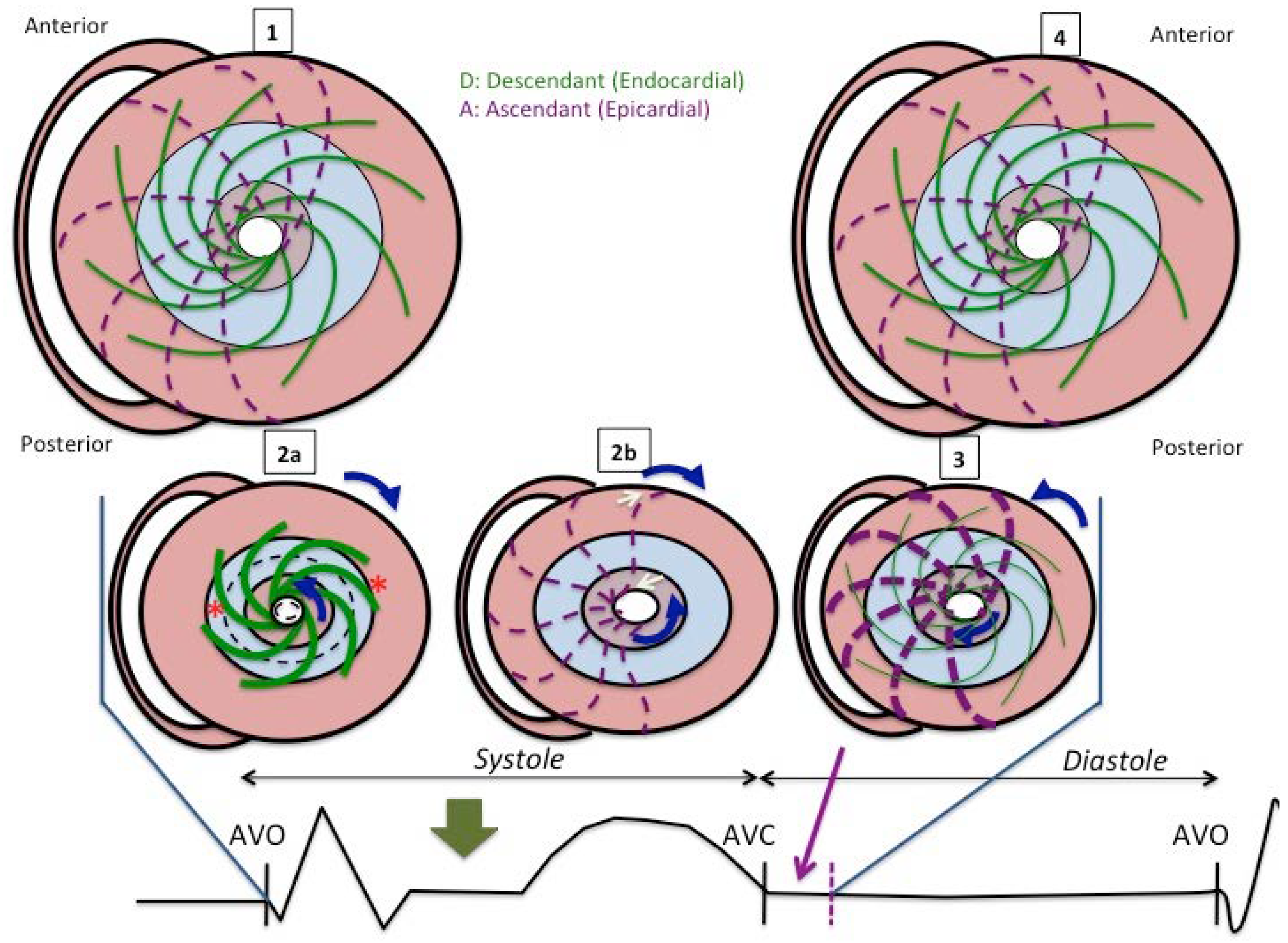
:1. Introduction
2. Materials and Methods
2.1. Study Population
2.2. Echocardiography
2.3. Statistical Analysis
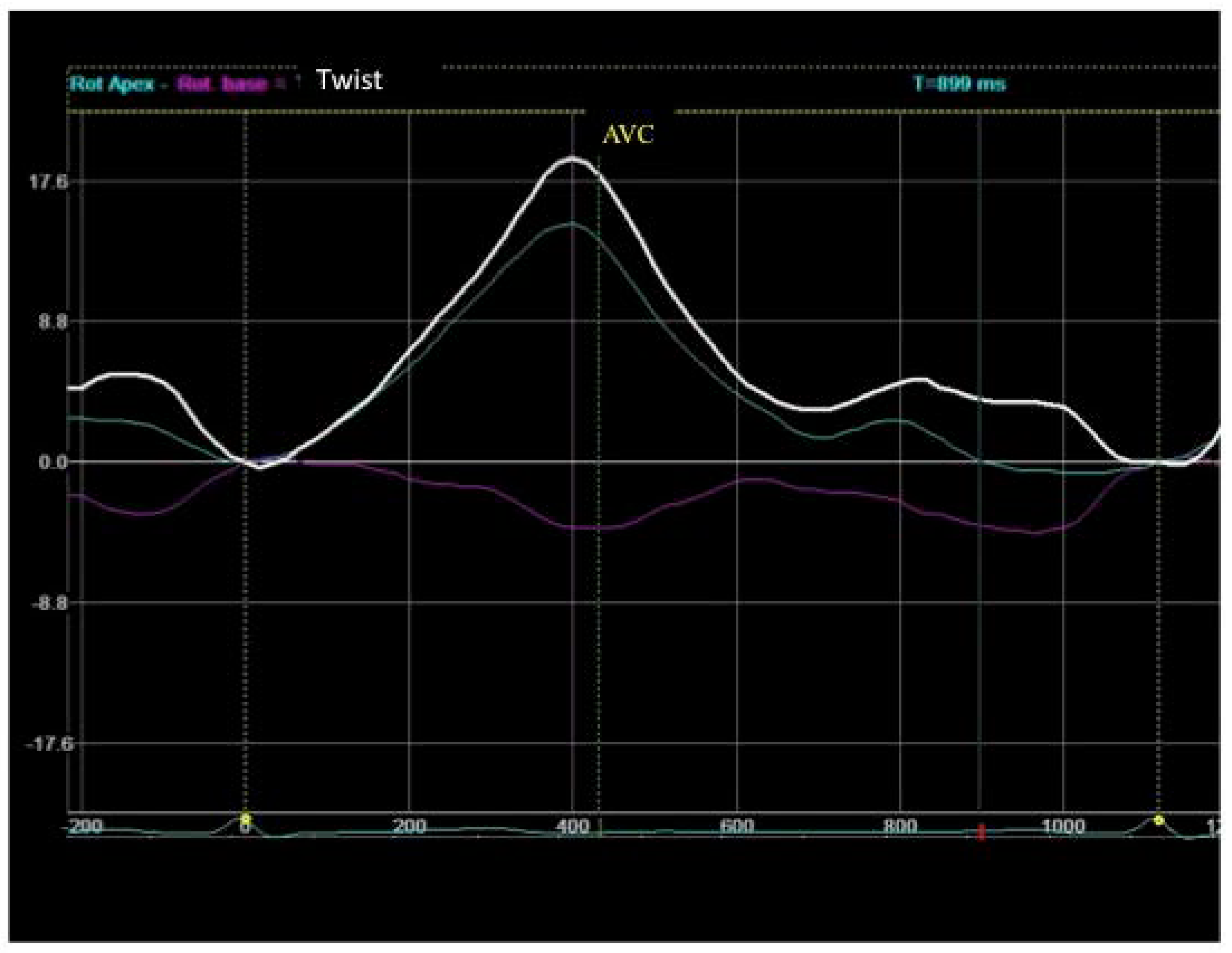
3. Results
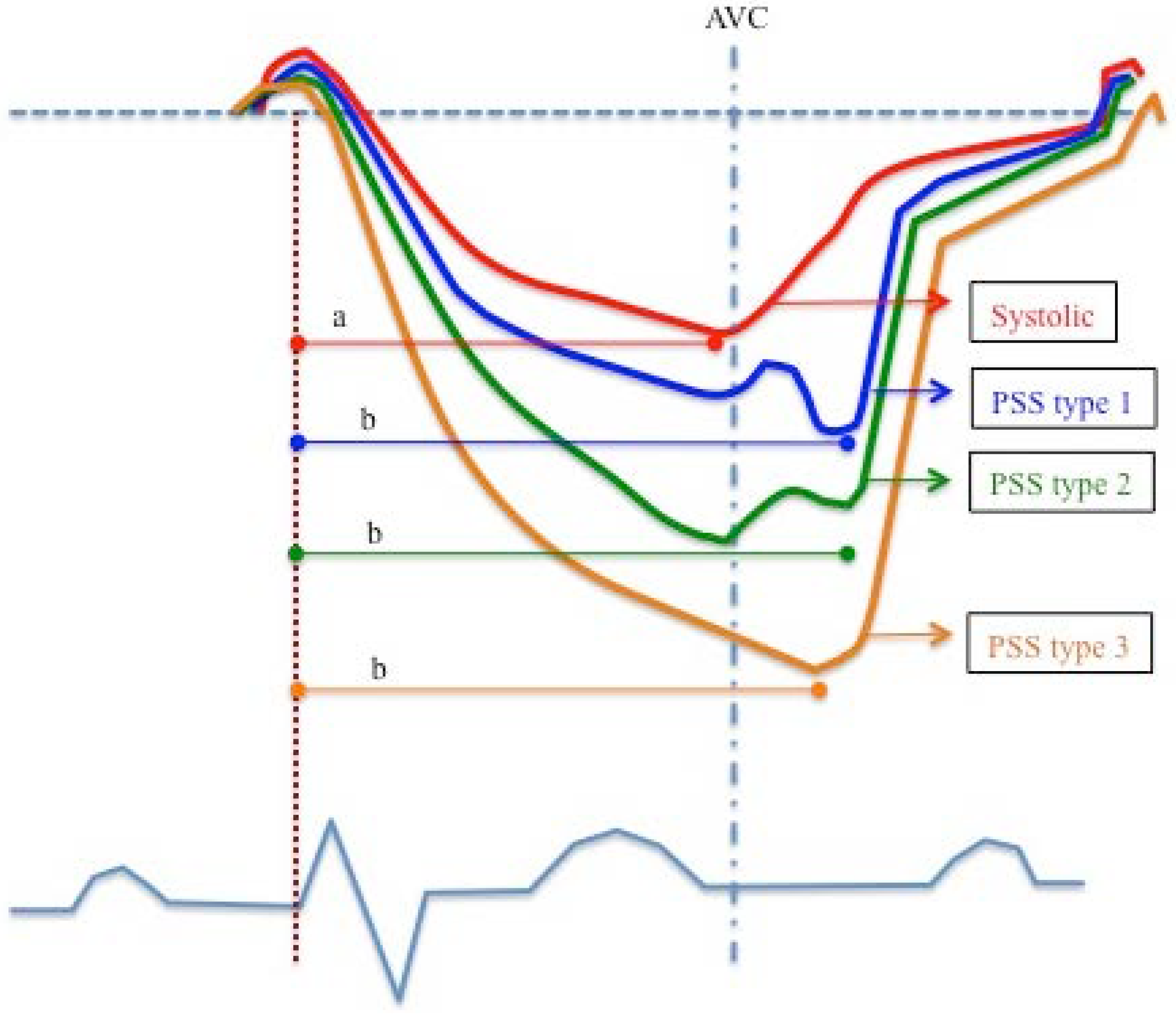
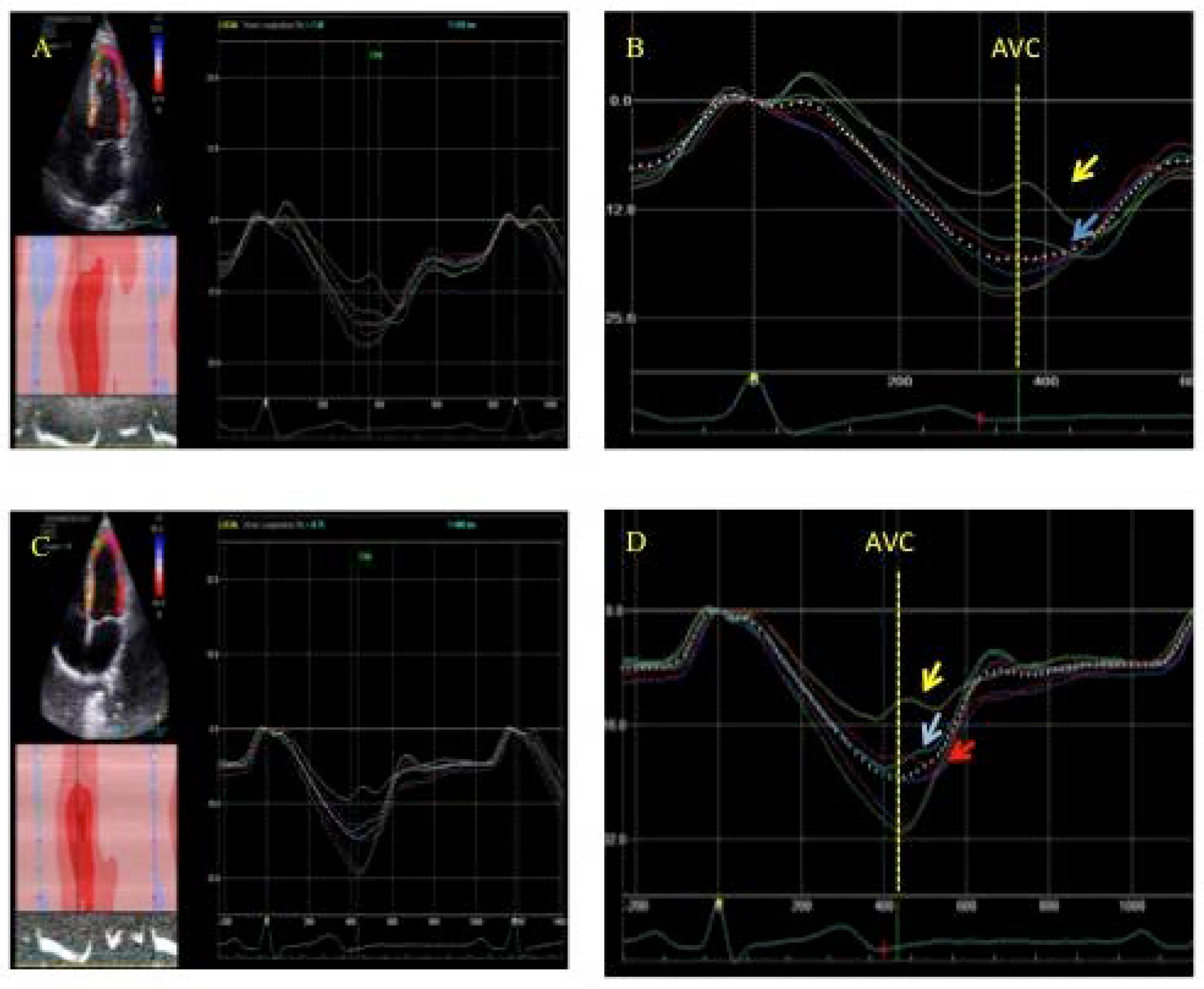
3.1. Postsystolic Shortening
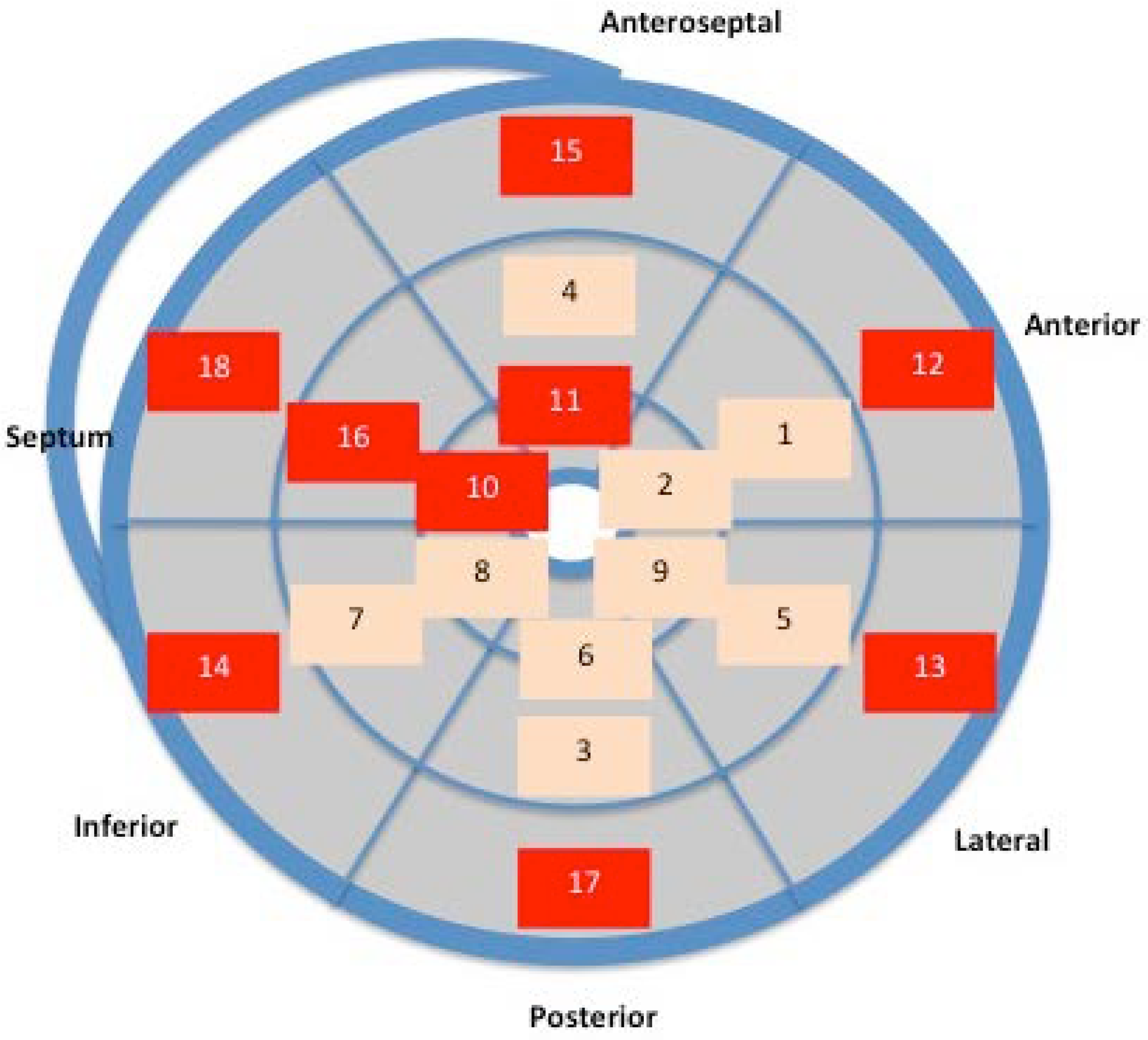
3.2. Anatomical Distribution
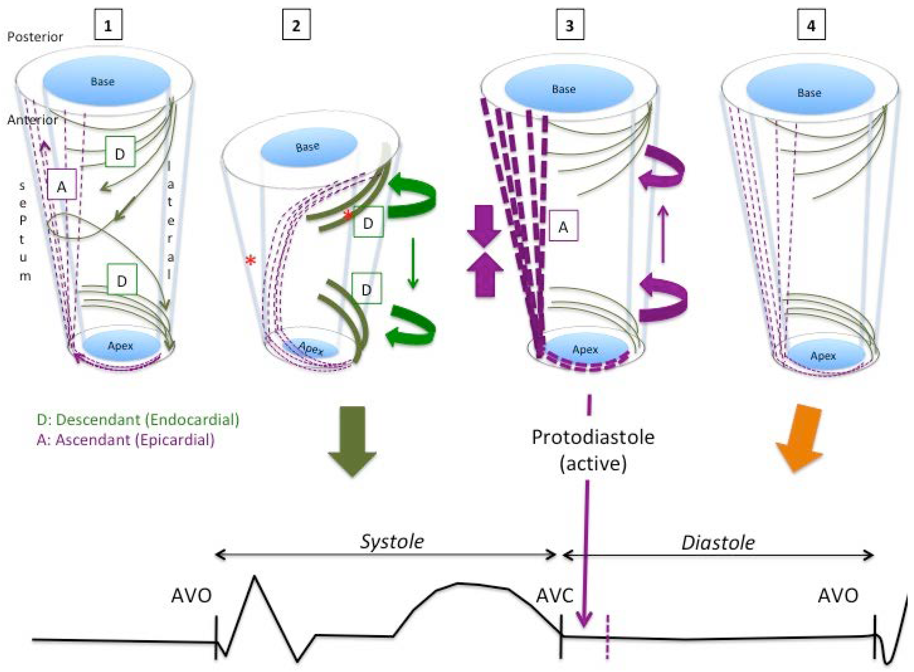
4. Discussion
5. Conclusions
Author Contributions
Funding
Conflicts of Interest
References
- Zaca, V.; Ballo, P.; Galderisi, M.; Mondillo, S. Echocardiography in the assessment of left ventricular longitudinal systolic function: Current methodology and clinical applications. Heart Fail. Rev. 2010, 15, 23–37. [Google Scholar] [CrossRef] [PubMed]
- Kocabay, G.; Muraru, D.; Peluso, D.; Cucchini, U.; Mihaila, S.; Padayattil-Jose, S.; Gentian, D.; Iliceto, S.; Vinereanu, D.; Badanoa, L.P. Mecánica ventricular izquierda normal mediante ecocardiografía speckle tracking bidimensional. Valores de referencia para adultos sanos. Revista Española de Cardiología 2014, 67, 651–658. [Google Scholar] [CrossRef] [PubMed]
- Torrent-Guasp, F. Estructura y función del corazón. Revista Española de Cardiología 1998, 51, 91–102. [Google Scholar] [CrossRef]
- Torrent-Guasp, F.; Ballester, M.; Buckberg, G.; Carreras, F.; Flotats, A.; Carrio, I.; Ferreira, A.; Samuels, L.E.; Narula, J. Spatial orientation of the ventricular muscle band: Physiologic contribution and surgical implications. J. Thorac. Cardiovasc. Surg. 2001, 122, 389–392. [Google Scholar] [CrossRef] [PubMed] [Green Version]
- Cosín, J.A.; Hernándiz, A.; Tuzón, M.T.; Agüero, J.; Torrent-Guasp, F. Estudio experimental de la llamada fase de relajación isovolumétrica del ventrículo izquierdo. Revista Española de Cardiología 2009, 62, 392–399. [Google Scholar] [CrossRef]
- Torrent-Guasp, F. El Ciclo Cardiaco; Espasa-Calpe: Madrid, Spain, 1954. [Google Scholar]
- Streeter, D.D. The cardiovascular system I. In American Physiological Society. Handbook of Physiology; Williams & Wilkins: Baltimore, MD, USA, 1979; pp. 61–112. [Google Scholar]
- Torrent Guasp, F. La estructuración macroscópica del miocardio ventricular. Revista Española de Cardiología 1980, 33, 265–287. [Google Scholar] [PubMed]
- Torrent-Guasp, F.; Bukberg, G.D.; Clemente, C.; Cox, J.L.; Coghlan, H.C.; Gharib, M. The structure and function of the helical Heart and its buttress wrapping. I. The normal macroscopic structure of the heart. Semin. Thorac. Cardiovasc. Surg. 2001, 13, 301–319. [Google Scholar] [CrossRef] [PubMed]
- Buckberg, G.D. Basic science review: The helix and the heart. J. Thorac. Cardiovasc. Surg. 2002, 124, 863–883. [Google Scholar] [CrossRef] [PubMed]
- Trainini, J.C.; Elencwajg, B.; López-Cabanillas, N.; Herreros, J.; Lago, N.E.; Lowenstein, J.A.; Trainini, A. Propagación de los estímulos, torsión muscular y efecto de succión cardiaca a través de la investigación electrofisiológica. In Fundamentos de La Nueva Mecánica Cardiac. La Bomba de Succión, 1st ed.; En Lumen, S.R.L., Ed.; Lumen: Ciudad Autónoma de Buenos Aires, Argentina, 2015; pp. 41–67. [Google Scholar]
- Sengupta, P.P.; Khandheria, B.K.; Korinek, J.; Wang, J.; Jahangir, A.; Seward, J.B.; Belohlavek, M. Apex-to-base dispersión in regional timing of left ventricular shortening and lengthening. Am. Coll. Cardiol. 2006, 47, 163–172. [Google Scholar] [CrossRef] [PubMed]
- Ashikaga, H.; Coppola, B.A.; Hopenfeld, B.; Leifer, E.S.; McVeigh, E.R.; Omens, J.H. Transmural Dispersion of Myofiber Mechanics: Implications for Electrical Heterogeneity In Vivo. J. Am. Coll. Cardiol. 2007, 49, 909–916. [Google Scholar] [CrossRef] [PubMed]
- Coghlan, H.C.; Coghlan, A.R.; Buckberg, G.D.; Gharib, M.; Cox, J.L. The estructure and function of the helical heart and its buttress wrapping. III. The electric spiral of the heart: The hypothesis of the anisotropic conducting matrix. Semin.Thorac. Cardiovasc. Surg. 2001, 13, 333–341. [Google Scholar] [CrossRef] [PubMed]
- Ballester-Rodés, M.; Carreras, F.; Narula, J.; Oschman, J.L. A New Look at the T Wave. J. Fluid Mech. Open Access 2016, 3, 128. [Google Scholar]
- Buckberg, G.D.; Castellá, M.; Gharib, M.; Saleh, S. Active myocyte shortening during the ‘isovolumetric relaxation’ phase of diastole is responsible for ventricular suction; ‘systolic ventricular filling’. Eur. J. Cardiothorac. Surg. 2006, 29 (Suppl. 1), 98–106. [Google Scholar] [CrossRef] [PubMed]
- Buckberg, G.D.; Nanda, N.C.; Nguyen Ch Kocica, M.J. What is the heart? Anatomy, function, pathophysiology, and misconceptions. J. Cardiovasc. Dev. Dis. 2018, 5, 33. [Google Scholar] [CrossRef] [PubMed]
- Kukulski, T.; Jamal, F.; D’Hooge, J.; Bijnens, B.; De Scheerder, I.; Sutherland, G.R. Acute changes in systolic and diastolic events during clinical coronary angioplasty: A comparison of regional velocity, strain rate and strain measurement. J. Am. Soc. Echocardiogr. 2002, 15, 1–12. [Google Scholar] [CrossRef] [PubMed]
- Voigt, J.U.; Regenfus, M.; Lindenmeier, G.; Schlundt, C.; Werner, D.; Nixdorff, U.; UFlachskampf, F.A.; Daniel, W.G. The relation between post systolic shortening and residual viable myocardium after acute myocardial infarction: A comparison of strain rate imaging with magnetic resonance tomography and scintigraphy. J. Am. Coll. Cardiol. 2001, 37, 450A. [Google Scholar]
- Voigt, J.U.; Lindenmeier, G.; Exner, B.; Regenfus, M.; Werner, D.; Reulbach, U.; Nixdorff, U.; Flachskampf, F.A.; Daniel, W.G. Incidence and characteristics of segmental postsystolic longitudinal shortening in normal, acutely ischemic, and scarred myocardium. J. Am. Soc. Echocardiogr. 2003, 16, 415–423. [Google Scholar] [CrossRef]
- Poveda, F.; Martí, E.; Gil, D.; Carreras, F.; Ballester, M. Helical structure of ventricular anatomy by diffusion tensor cardiac MR tractography. J. Am. Coll. Cardiol. Cardiovasc. Imaging 2012, 5, 754–755. [Google Scholar] [CrossRef] [PubMed]
- Poveda, F.; Gil, D.; Martí, E.; Andaluz, A.; Ballester, M.; Carreras, F. Estudio tractográfico de la anatomía helicoidal del miocardio ventricular mediante resonancia magnética por tensor de difusión. Revista Española de Cardiología 2014, 66, 782–790. [Google Scholar] [CrossRef]
- Martín, G.; Gimeno, J.V.; Cosín, J.; Guillem, M.I. Time constant of isovolumic pressure fall, new numerical approaches and significance. Am. J. Physiol. 1984, 247, H283–H294. [Google Scholar] [CrossRef] [PubMed]
- Ashikaga, H.; Criscione, J.C.; Omens, J.H.; Cowell, J.W.; Ingels, N.B. Transmural left ventricular mechanisms underlying torsional recoil during relaxation. Am. J. Physiol. Heart Circ. Physiol. 2004, 286, H640–H647. [Google Scholar] [CrossRef] [PubMed]
- Lorenz, C.H.; Pastorek, J.S.; Bundy, J.M. Delineation of normal human left ventricular twist throughout systole by tagget cine magnetic resonance imaging. J. Cardiovasc. Magn. Reson. 2000, 2, 97–108. [Google Scholar] [CrossRef] [PubMed]
- Sengupta, P.P.; Tondato, F.; Khandheria, B.K.; Belohlavek, M.; Jahangir, A. Electromechanical activation sequence in normal heart. Heart Fail. Clin. 2008, 4, 303–314. [Google Scholar] [CrossRef] [PubMed]
- Buckberg, G.; Hoffman, J.I.E.; Mahajan, A.; Saleh, S.; Coghlan, C. Cardiac mechanics revisited. The relationship of cardiac architecture to ventricular function. Circulation 2008, 118, 2571–2587. [Google Scholar] [CrossRef] [PubMed]
- Fonofagou, E.E.; Provost, J. Electromechanical wave imaging for noninvasive mapping of the 3D electrical activation sequence in canines and humans in vivo. J. Biomech. 2012, 45, 856–864. [Google Scholar] [CrossRef] [PubMed] [Green Version]
- Gurev, V.; Constantino, J.; Rice, J.J.; Trayanova, N.A. Distribution of electromechanical delay in the heart: Insights from a three-dimensional electromechanical model. Biophys. J. 2010, 99, 745–754. [Google Scholar] [CrossRef] [PubMed]
- Waldman, L.K.; Nosan, D.; Villarreal, F.; Covell, J.W. Relation between transmural deformation and local myofiber direction in canine left ventricle. Circ. Res. 1988, 63, 550–562. [Google Scholar] [CrossRef] [PubMed]
- Opdahl, A.; Remme, E.W.; Helle-Valle, T.; Edvardsen, T.; Smiseth, O.A. Myocardial relaxation, restoring forces, and early-diastolic load are independent determinants of left ventricular untwisting rate. Circulation 2012, 126, 1441–1451. [Google Scholar] [CrossRef] [PubMed]


| Total (n = 90) | Men (n = 52) | Women (n = 38) | p | |
|---|---|---|---|---|
| Age | 50.3 ± 11.1 | 50.4 ± 11.1 | 50.1 ± 11.1 | 0.88 |
| Body surface | 1.8 ± 0.2 | 1.9 ± 0.1 | 1.6 ± 0.1 | 0.001 |
| Heart rate | 65 ± 10 | 65 ± 11 | 66 ± 9 | 0.65 |
| Systolic blood pressure (mmHg) | 119 ± 16 | 121 ± 16 | 115 ± 15 | 0.07 |
| Diastolic left ventricle (mm) | 45.5 ± 4.5 | 47.4 ± 4.2 | 43.0 ± 3.4 | 0.001 |
| Left ventricular mass | 151.6 ± 51.0 | 170.7 ± 53.6 | 125.4 ± 32.8 | 0.001 |
| LVED (mL) | 89.2 ± 28.4 | 101.1 ± 27.9 | 73.0 ± 20.1 | 0.001 |
| LVES (mL) | 29.8 ± 11.1 | 34.1 ± 11.5 | 24.1 ± 7.5 | 0.001 |
| EFLV (%) | 66.6 ± 5.5 | 66.4 ± 5.4 | 66.9 ± 5.7 | 0.68 |
| Global Longitudinal Strain | −21.1 ± 2.1 | −20.7 ± 2.0 | −21.7 ± 2.1 | 0.02 |
| Segments | PSS Total | Type 1 | Type 2 | Type 3 |
|---|---|---|---|---|
| Basal (n = 540) | 321 (59.4%) | 84 (15.5%) | 139 (25.7%) | 91 (16.8%) |
| Medial (n = 540) | 218 (40.3%) | 43 (7.9%) | 102 (18.9%) | 73 (13.5%) |
| Apical (n = 540) | 245 (45.4%) | 19 (3.5%) | 76 (14.1%) | 150 (27.8%) |
| Segment | PSS (%) | Type 1 (%) | Type 2 (%) | Type 3 (%) |
|---|---|---|---|---|
| 1 Basal anteroseptal | 51 (56.6) | 13 (14.4) | 28 (31.2) | 9 (10.0) |
| 2 Basal septal | 60 (66.6) | 19 (21.2) | 25(27.7) | 16 (17.7) |
| 3 Basal inferior | 57 (63.3) | 19 (21.2) | 24 (26.6) | 14 (15.5) |
| 4 Basal posterior | 60 (66.6) | 9 (10.0) | 29 (32.2) | 22 (24.4) |
| 5 Basal lateral | 46 (51.1) | 11 (12.3) | 21 (23.3) | 14 (15.5) |
| 6 Basal anterior | 47 (52.2) | 19 (21.2) | 12 (13.3) | 16 (17.7) |
| 7 Medial anteroseptal | 37 (41.1) | 6 (6.7) | 17 (18.8) | 14 (15.5) |
| 8 Medial septal | 46 (51.1) | 7 (7.7) | 28 (31.2) | 11 (12.2) |
| 9 Medial inferior | 44 (48.8) | 13 (14.4) | 18 (20.0) | 13 (14.4) |
| 10 Medial posterior | 33 (36.6) | 6 (6.6) | 16 (17.7) | 11 (12.3) |
| 11 Medial lateral | 36 (40.0) | 8 (8.9) | 17 (18.8) | 11 (12.2) |
| 12 Medial anterior | 22 (24.4) | 3 (3.4) | 6 (6.6) | 13 (14.4) |
| 13 Apical anteroseptal | 47 (52.2) | 2 (2.2) | 18 (20.0) | 27 (30.0) |
| 14 Apical septal | 43 (47.7) | 2 (2.2) | 16 (17.8) | 25 (27.7) |
| 15 Apical inferior | 44 (48.8) | 7 (7.7) | 9 (10.0) | 28 (31.1) |
| 16 Apical posterior | 34 (37.7) | 2 (2.2) | 16 (17.8) | 16 (17.7) |
| 17 Apical lateral | 40 (44.4) | 3 (3.3) | 12 (13.3) | 25 (27.8) |
| 18 Apical anterior | 37 (41.1) | 3 (3.3) | 5 (5.6) | 29 (32.2) |
| Segment | Mean (Milliseconds) | ±SD |
|---|---|---|
| 12 Medial anterior | 372 | 43 |
| 18 Apical anterior | 379 | 48 |
| 10 Medial posterior | 382 | 47 |
| 7 Medial anteroseptal | 385 | 51 |
| 11 Medial lateral | 388 | 53 |
| 16 Apical posterior | 389 | 57 |
| 9 Medial inferior | 390 | 53 |
| 15 Basal lateral | 391 | 51 |
| 17 Apical lateral | 392 | 57 |
| 14 Apical inferior | 394 | 58 |
| 13 Apical anteroseptal | 395 | 55 |
| 6 Basal anterior | 395 | 54 |
| 5 Apical septum | 397 | 56 |
| 3 Basal inferior | 405 | 59 |
| 1 Basal anteroseptal | 405 | 60 |
| 8 Medial septum | 407 | 74 |
| 4 Basal posterior | 408 | 54 |
| 2 Basal septum | 416 | 60 |
© 2018 by the authors. Licensee MDPI, Basel, Switzerland. This article is an open access article distributed under the terms and conditions of the Creative Commons Attribution (CC BY) license (http://creativecommons.org/licenses/by/4.0/).
Share and Cite
Mora, V.; Roldán, I.; Romero, E.; Saurí, A.; Romero, D.; Pérez-Gozalbo, J.; Ugalde, N.; Bertolín, J.; Rodriguez-Israel, M.; Delgado, C.P.-O.; et al. Myocardial Contraction during the Diastolic Isovolumetric Period: Analysis of Longitudinal Strain by Means of Speckle Tracking Echocardiography. J. Cardiovasc. Dev. Dis. 2018, 5, 41. https://doi.org/10.3390/jcdd5030041
Mora V, Roldán I, Romero E, Saurí A, Romero D, Pérez-Gozalbo J, Ugalde N, Bertolín J, Rodriguez-Israel M, Delgado CP-O, et al. Myocardial Contraction during the Diastolic Isovolumetric Period: Analysis of Longitudinal Strain by Means of Speckle Tracking Echocardiography. Journal of Cardiovascular Development and Disease. 2018; 5(3):41. https://doi.org/10.3390/jcdd5030041
Chicago/Turabian StyleMora, Vicente, Ildefonso Roldán, Elena Romero, Assumpció Saurí, Diana Romero, Jana Pérez-Gozalbo, Natalia Ugalde, Javier Bertolín, Melisa Rodriguez-Israel, Carmen Pérez-Olivares Delgado, and et al. 2018. "Myocardial Contraction during the Diastolic Isovolumetric Period: Analysis of Longitudinal Strain by Means of Speckle Tracking Echocardiography" Journal of Cardiovascular Development and Disease 5, no. 3: 41. https://doi.org/10.3390/jcdd5030041
APA StyleMora, V., Roldán, I., Romero, E., Saurí, A., Romero, D., Pérez-Gozalbo, J., Ugalde, N., Bertolín, J., Rodriguez-Israel, M., Delgado, C. P.-O., & Lowenstein, J. A. (2018). Myocardial Contraction during the Diastolic Isovolumetric Period: Analysis of Longitudinal Strain by Means of Speckle Tracking Echocardiography. Journal of Cardiovascular Development and Disease, 5(3), 41. https://doi.org/10.3390/jcdd5030041