
Inherent and Composite Hydrogels as Promising Materials to Limit Antimicrobial Resistance


Abstract
1. Introduction
2. Classification of Hydrogels
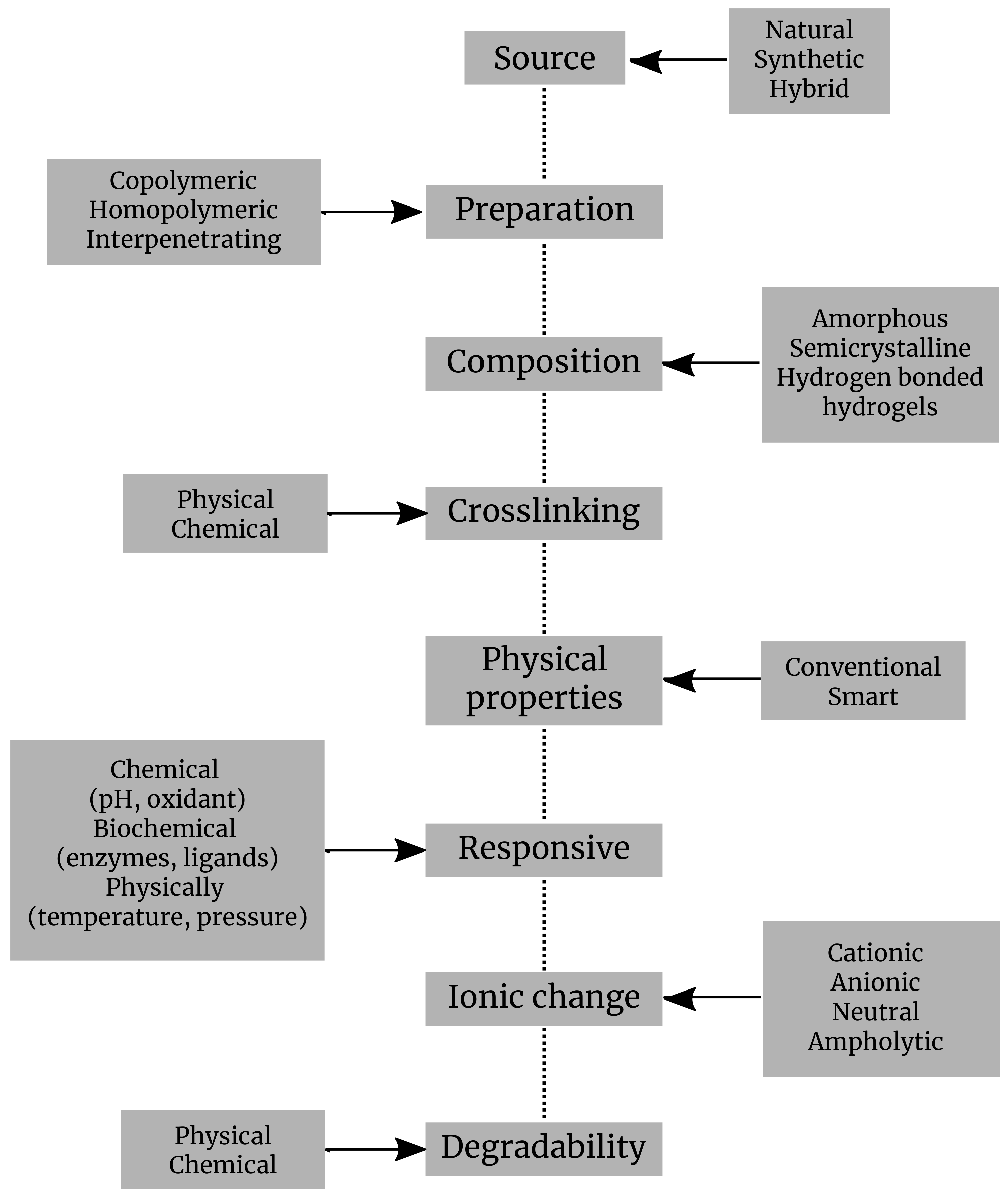
- Depending upon their origin, hydrogels can be split into natural, synthetic, or hybrid hydrogels. Generally, hydrogels with natural origin exhibit a superior biocompatibility and favor biological processes, while the synthetic hydrogels exhibit more consistent mechanical and biochemical attributes. The hydrogels of natural origin are obtained based on precursors belonging to different structural categories of biopolymers, chains representing polysaccharides, or peptides/proteins [25];
- Depending upon their preparation, hydrogels were defined in various ways. The most popular definition which describes a hydrogel as a cross-linked polymeric network which is water-swollen, derived from the basic reaction of one or more units of monomer/polymer/cross-linker. A different description presents it as a polymeric material capable of swelling and retaining a large amount of water in its three-dimensional matrix, but which does not dissolve in water [26]. They are also illustrated as polymeric systems that present the capability to swell in water and retain a significant proportion of water inside their three-dimensional net, without dissolving in water. Food and biomaterial researchers are using two similar terms, gels and hydrogels, to describe polymeric cross-linked net structures [27]. Homopolymers are the polymers which have only one type of monomer in their assemblage. They may have a cross-linked structure, due to the nature of the monomer and the technique of polymerization. Copolymeric hydrogels are the ones that are made of two types of monomers, at least one of them being hydrophilic [28];
- Hydrogels can be also classified according to their structure, which may be amorphous, semicrystalline, crystalline, or hydrocolloid [28];
- As the hydrogels are basically built by cross-linking networking, therefore based on cross-linking, they are classified regarding this feature into two categories: (a) physically cross-linked or self-assembled hydrogels are formed through reversible bonds based on ionic interactions, crystallization, formation of stereocomplex, hydrophobization of polysaccharides, interaction of proteins or hydrogen bonds; (b) a chemically cross-linked hydrogel, linked by permanent covalent bonds which can be polymerized by chain growth, addition, and condensation [27]. Several types of physical and chemical hydrogels were prepared from natural or synthetic polymers in order to be used in miscellaneous applications (Table 1).
- As regards the administration to patients, hydrogels are either implanted or injected. Injectable hydrogels are preformed before injection or are formed in situ [41];
- Depending upon their response, the hydrogels are broken down into physically, chemically, and biochemically responsive hydrogels. They can further be designed to be responsive to environmental variables, such as temperature, light, pH, antigens, or even enzymes. Hence, hydrogels can be divided into physical, chemical, or biochemical classifications. Physical hydrogels can pass from liquid to gel in response to a specific change in environmental parameters, such as temperature, pH, concentration of ions, or changes in the state of two such components. Chemical gels use covalent bonding that provides mechanical integrity and degradation resistance in comparison with other weak materials. In biochemical hydrogels, the gelation process is performed with the involvement of biological agents, as enzymes or amino acids [14];
- According to their ionic charge, hydrogels can be designated as cationic, anionic, neutral, and ampholytic. For instance, poly(norbornene) is a cationic polymer, and it was thoroughly scrutinized for its antimicrobial properties [42];
- Depending upon their physical properties, there are two types of hydrogels: conventional and smart hydrogels. The first are the ones already known, previously established in the past. Smart hydrogel systems include elements capable of chemically or structurally displaying responses to a range of external stimuli comprising light, temperature, concentration of ions, pH, chemicals, and even magnetic or electric fields. This change in structure and volume as a response to the stimuli as the ones above opens a huge research potential and a large array of applications [43];
- Depending upon their degradability process, the hydrogels are split into two types: biodegradable and non-biodegradable. The biodegradability and biocompatibility make them a strong candidate for biological and environmental applications, as implants or materials for pollutants removal. They can even bring biodegradability to electronics, meaning that hydrogels represent a new option for the designing and creation of supercapacitors. Natural hydrogels are not only biodegradable and biocompatible. For instance, chitosan has become the preferred hydrogel for developing antimicrobial hydrogels of natural origins, as its properties include fast cross-linking [14]. Hydrogels can be engineered to fit a number of large range application due to their pliability, the possibility to be modulated according to needs [14].
3. Processing Procedure
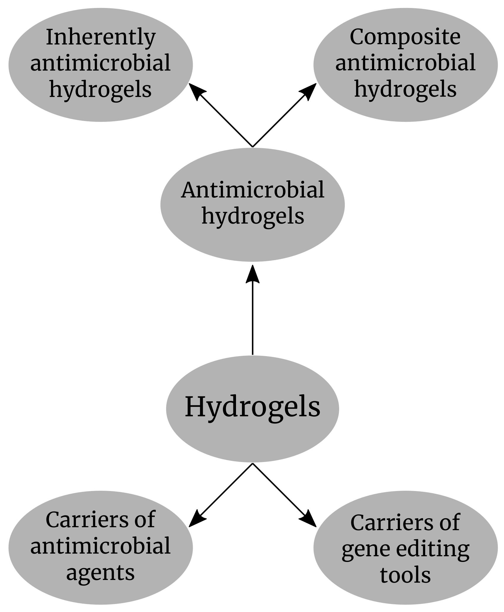
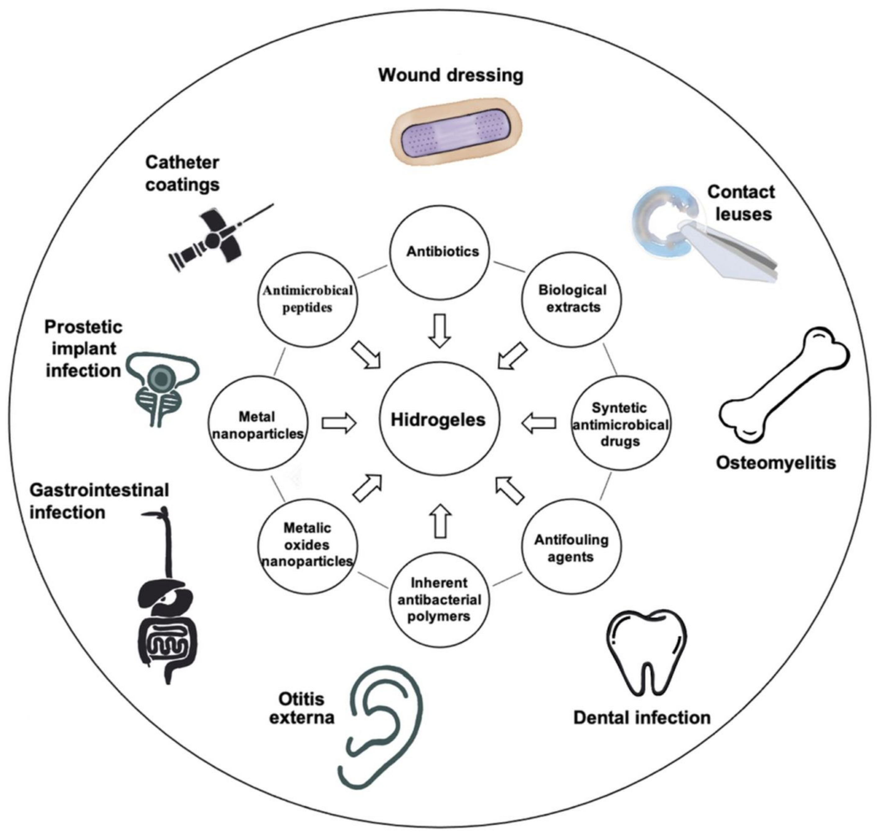
4. Inherently Antimicrobial Hydrogels
4.1. Natural Hydrogels
4.1.1. Microbial Sources
- Gellan gum is an anionic product extracellularly secreted by Sphingomonas elodea (ATCC 31461) following a microbial fermentation process [61]. It has a structure of linear polysaccharide, formed by a repeating tetrasaccharide unit made of two D-glucose, one L-rhamnose and one D-glucuronic acid (Table 2). This gum has two commercialization forms: high acyl (acetylated) gellan gum and low acyl (deacetylated) gellan gum. Both of them are capable of gelation. The difference is that the acetylated one makes elastic and translucent gels, while the deacetylated form produces gels which are rigid and, thus, more suitable for tissue engineering and regenerative medicine applications. The gelation process is conducted by a two-step mechanism [62]. The first step is a thermic process. The aqueous solution of gellan gum is heated above 80 °C for about 25 min and then cooled, driving the formation of highly ordered double helices from the linear polymers of gellan gum with randomly coiled chains. Afterwards, the cations are added and the helices are cross-linked to complete a stable hydrogel. There are several favorable characteristics of gellan gum hydrogels, including biocompatibility, similarity in structure with the inner glycosaminoglycans of the body, and mild conditions of gelation, that facilitate the incorporation of cells, making gellan gum-based hydrogels appropriate for various tissue engineering and regenerative medicine applications [51,63].
- Xanthan gum is extracellularly secreted by bacteria of the genus Xanthomonas, resulting from polysaccharide fermentation [64]. It is not toxic. Xanthan gum is a polysaccharide with a branched structure and is made of a repeating unit of D-glucose, D-mannose, and D-glucuronic acid, having the molar ratio of 2:2:1 (Table 2) [65]. Its harmless nature and shear characteristics make it promising for attaining an injectable scaffold for cartilage tissue repairing and for biocompatibility [66]. Xanthan gum is produced by a single-step thermic gelation process. A colloidal heterogeneous suspension, made of pockets of molecular assemblies, is constituted when, at room temperature, xanthan gum polymers are added in water. If this heterogeneous suspension is brought above 40 °C, for 3 h, annealing takes place and thus the suspension becomes homogenous. After cooling the hydrogels become robust [67].
- Dextran was the first microbial polysaccharide commercially available. It is secreted by two species of bacteria, Leuconostoc mesenteroides and Streptococcus mutans. Linear alpha-1,6 and branch alpha-1,3 glycosidic linkages between glucose monomers are at the base of its edification (Table 2). Dextran is very important in medicine, being used extensively as a volume expander and antithrombotic. The downside is that dextran cannot form hydrogels in its native state. However, composite hydrogels based on dextran were developed in order to be used in tissue regeneration [68]. Dextran is also exhibiting antimicrobial features if long alkyl tail is attached at the reducing end. More explicitly, a mixture of DMSO-MeF and NaCNBH3 with excess of dodecyl or octadecyl is mediating the reductive alkylation [69,70].
4.1.2. Algal Sources
- Alginate may be present in the salts located in the cell wall of brown algae or in acid form. The composition of alginate consists of 1,4-linked alternate alpha-L-guluronic acid and beta-D-mannuronic acid residues. The chemical composition of alginates slightly differs from one algae species to another. Hydrogel preparation is mainly used in the biomedical field, in drug release or tissue regeneration. For a hydrogel to be formed, divalent cations are needed. Calcium chloride is such a cation which provides the cross-linkage. The salts of alginate also exhibit antimicrobial effects. Percival et al. (2011) [71] reported effects, including the growth inhibition of infectious agents as Streptococcus viridans and Candida albicans. Such properties can be boosted by adding alkyl groups to alginate [52].
- Carrageenans. There are only three forms of carrageenans found in nature, represented by kappa, iota, and lambda. The k-carrageenan is obtained from the alga Kappaphycus alvarezii, while i-carrageenan is extracted from Euchema denticulatum. Carrageenans vary in about 15 different structural ways. They are generally made of differently linked D-galactopyranose units. Carrageenans also include sulfate groups in their structure. Several hydrogels were developed from carrageenans. For bone tissue regeneration, a sensitive medical issue, a composite hydrogel from k-carrageenan/collagen-hydroxyapatite was developed [72]. Injectable hydrogels based on the same carrageenans are produced to be involved in tissue engineering [72]. Azizi et al. (2017) [55] fabricated a bio-nanocomposite hydrogel by incorporating biosynthesized silver nanoparticles with kappa- carrageenan. Diverse plant extracts were used for the synthesis of Ag nanoparticles. It demonstrates an excellent antimicrobial effect against S. aureus, methicillin-resistant S. aureus E. coli, and Pseudomonas aeruginosa.
4.1.3. Animal Sources
- Polysaccharides from animal sources are also widely used in order to obtain hydrogels. From them, chitin is the one most common. The animal polysaccharides are chemically modified before being used to obtain hydrogels, the native form lacking the needed characteristics. For instance, chitin needs to be transformed to chitosan. Chitin is structured by 1–4 glycosidic bonds linking N acetyl glucosamine. The highly acetylated residues present in chitin make it rigid, and therefore not suitable. Chitin is found in the exoskeleton of insects, but it is mostly obtained from crab shells, which contain a large amount of calcium, and so need to be subjected to a demineralization process. Chitosan was accidentally obtained by Rouget in 1859 [72]. Its structure consists of two units of 2-acetamido-2-deoxy-beta-D-glucan and 2-amino-2-deoxy-beta-D-glucan. The extent of the deacetylation by which chitosan is obtained determines the hydrophilicity of the final product. Native chitosan needs to be made less hydrophilic in order to be used for drug delivery systems. Suitable chitosan hydrogels can only be obtained from modified chitosan [73]. Chitosan not only has antimicrobial properties, it is also able to involve neutrophils and macrophages in the healing of wounds, thus improving its benefits. Allan and Hadwiger (1979) [74] were the first research group, who claimed that chitosan demonstrates antagonistic behavior towards fungi. Following the report by Allan and Hadwiger (1979) [74], many studies were published which discussed fungicidal and antimicrobial characteristics [73,75,76,77]. However, the exact mechanism of antimicrobial activity remains obscure. Their antimicrobial properties can even be increased by augmenting the cationic charges along the polymer backbone. Thus, it was observed that the hydrogels of quaternized chitosan, which contain tertiary amino groups, provide a reduced risk of infection and sustain tissue repair at the same time [27]. The chitosan gels are easy to prepare. Chitosan is dissolved in acetic acid, and then a sodium hydroxide solution is added until the solution reaches 9 (pH). Then, the raw hydrogel is decanted, washed, and dialyzed [78].
- Chondroitin sulfate is another source of hydrogels belonging to the glycosaminoglycans; with the compounds exhibiting linear heteropolysaccharide chains formed of repeating units of disaccharides [79]. Chondroitin sulfate can be found widely, in many different tissues (hyaline cartilage, skin, blood vessels, etc.). Barkat et al (2019) [80] used chondroitin sulfate hydrogels packed with oxaliplatin against colorectal cancer [72].
- Hyaluronic acid is a mucopolysaccharide, also formed in living organisms, present in the synovial fluid, which functions as a lubricant. Hyaluronic acid is a linear polysaccharide made of nonsulfated glycosaminoglycan units. Hydrogels based on hyaluronic acid are obtained by cross-linking. An injectable hydrogel made of hyaluronic acid is used for drug delivery in cancer therapy [57].
4.1.4. Plant Sources
- Cellulose hydrogels can be obtained by cross-linking of cellulose in the solution [72]. As cellulose has a variety of the hydroxyl group, it can easily form networks by linking through H2 bonding. Huang et al. prepared a nanofiber hydrogel with healing capacity with dialdehyde cellulose nanocrystals and carboxymethyl chitosan [82]. Double network hydrogel was achieved by diffusion of isopropylacrylamide in cellulose hydrogels cross-linked to epichlorohydrin. Double network hydrogels were analogically obtained by changing the ratio between isopropylacrylamide to acrylamide [83]. Cellulose hydrogels exhibiting remarkable stretchability can be manufactured using the sequential cross-linking and dual network techniques [84]. Fabrication in the NaOH/urea system requires two steps. The first step includes cross-linking of cellulose by epichlorohydrin. By electron microscopy techniques, it was observed the morphology of the first network is changed, resulting in improved mechanical properties. First, the precursors are diffusing within the first network, then the polymerization is UV-light initiated and, thus, dual network hydrogels are emerging.
- Locust (Carob) bean gum represents a natural nonstarch galactomannan, it is not ionically branched and can be used in various fields based on its inner flexibility. The locust bean gum and its hydrogel-derived preparations are very popular, being widely used in food, pharmaceutical, biomedical, or cosmetic fields. Locust bean gum is also used as a carrier for drug delivery applications. Alongside the use of this popular material, novel versions were obtained by different modifications processes. Locust bean gum can be used for specific functions through its combination with several other polymers. It even responds to various stimuli, enhancing the applicability in various therapies [72].
4.2. Synthetic Hydrogels
5. Composite Antimicrobial Hydrogels
5.1. Chitosan Grafted Hydrogels
5.2. Hydrogels Containing Immobilized Antimicrobial Compounds
5.2.1. Antibiotic-Loaded Hydrogels
5.2.2. Biological Extract-Loaded Hydrogels
5.2.3. Synthetic Antibacterial Drug-Loaded Hydrogels
5.2.4. Peptide Hybridized Hydrogels
5.2.5. Immobilized Metal, Metal Oxide Nanoparticles
5.2.6. Carbon Material-Loaded Hydrogels
6. Hydrogels as Carriers of Antimicrobial Agents
6.1. Physical Incorporation of NPs in Hydrogels
6.2. Integration of Enzyme Cleavage Sites into Hydrogels
6.3. Optimization of Hydrogel Properties
6.4. Development of Bacteria Responsive Hydrogels
7. Delivery Systems for Gene Editing Tools for Curing the Bacterial Resistance
8. Challenges of Development and Uses of Antibacterial Hydrogels
9. Conclusions and Perspectives
Author Contributions
Funding
Institutional Review Board Statement
Informed Consent Statement
Data Availability Statement
Conflicts of Interest
References
- Omulo, S.; Thumbi, S.; Njenga, M.; Call, D. A review of 40 years of enteric antimicrobial resistance research in Eastern Africa: What can be done better? Antimicrob. Resist. Infect. Control. 2015, 4, 1. [Google Scholar] [CrossRef]
- Bassetti, M.; Merelli, M.; Temperoni, C.; Astilean, A. New antibiotics for bad bugs: Where are we? Ann. Clin. Microbiol. Antimicrob. 2013, 12, 22. [Google Scholar] [CrossRef]
- Lim, W.; Wu, P.; Bond, H.; Wong, J.; Ni, K.; Seto, W.; Jit, M.; Cowling, B. Determinants of methicillin-resistant Staphylococcus aureus (MRSA) prevalence in the Asia-Pacific region: A systematic review and meta-analysis. J. Glob. Antimicrob. Resist. 2019, 16, 17–27. [Google Scholar] [CrossRef] [PubMed]
- Hall, J.; Ingram, P.; O’Reilly, L.; Inglis, T. Temporal flux in β-lactam resistance among Klebsiella pneumoniae in Western Australia. J. Med. Microbiol. 2016, 65, 429–437. [Google Scholar] [CrossRef][Green Version]
- Wyres, K.; Hawkey, J.; Hetland, M.; Fostervold, A.; Wick, R.; Judd, L.; Hamidian, M.; Howden, B.; Löhr, I.; Holt, K. Emergence and rapid global dissemination of CTX-M-15-associated Klebsiella pneumoniae strain ST307. J. Antimicrob. Chemother. 2018, 74, 577–581. [Google Scholar] [CrossRef] [PubMed]
- Xie, R.; Zhang, X.; Zhao, Q.; Peng, B.; Zheng, J. Analysis of global prevalence of antibiotic resistance in Acinetobacter baumannii infections disclosed a faster increase in OECD countries. Emerg. Microbes Infect. 2018, 7, 1–10. [Google Scholar] [CrossRef]
- Fan, X.; Wu, Y.; Xiao, M.; Xu, Z.P.; Kudinha, T.; Bazaj, A.; Kong, F.; Xu, Y.C. Diverse Genetic Background of Multidrug-Resistant Pseudomonas aeruginosa from Mainland China and Emergence of an Extensively Drug-Resistant ST292 Clone in Kunming. Sci. Rep. 2016, 6, 26522. [Google Scholar] [CrossRef]
- Magill, S.; Edwards, J.; Bamberg, W.; Beldavs, Z.; Dumyati, G.; Kainer, M.; Lynfield, R.; Maloney, M.; McAllister-Hollod, L.; Nadle, J.; et al. Multistate Point-Prevalence Survey of Health Care–Associated Infections. N. Engl. J. Med. 2014, 370, 1198–1208. [Google Scholar] [CrossRef]
- Ramirez, M.; Traglia, G.; Lin, D.; Tran, T.; Tolmasky, M. Plasmid-Mediated Antibiotic Resistance and Virulence in Gram-Negatives: The Klebsiella pneumoniae Paradigm. Microbiol. Spectr. 2014, 2, 5. [Google Scholar] [CrossRef] [PubMed]
- Oliveira, D.M.P.D.; Forde, B.M.; Kidd, T.J.; Harris, P.N.A.; Schembri, M.A.; Beatson, S.A.; Paterson, D.L.; Walker, M.J. Antimicrobial Resistance in ESKAPE Pathogens. Clin. Microbiol. Rev. 2020, 33, e00181-19. [Google Scholar] [CrossRef]
- Hall, C.W.; Mah, T.F. Molecular mechanisms of biofilm-based antibiotic resistance and tolerance in pathogenic bacteria. FEMS Microbiol. Rev. 2017, 41, 276–301. [Google Scholar] [CrossRef]
- Partridge, S.; Kwong, S.; Firth, N.; Jensen, S. Mobile Genetic Elements Associated with Antimicrobial Resistance. Clin. Microbiol. Rev. 2018, 31. [Google Scholar] [CrossRef]
- Trastoy, R.; Lucia, B.; German, B.; Maria, T. Fighting antimicrobial resistance in ESKAPE pathogens. Fight Antimicrob. Resist. 2018, 2018, 1–18. [Google Scholar] [CrossRef]
- Joshi Navare, K.; Eggermont, L.; Rogers, Z.; Mohammed, H.; Colombani, T.; Bencherif, S. Antimicrobial Hydrogels: Key Considerations and Engineering Strategies for Biomedical Applications; Springer: Berlin/Heidelberg, Germany, 2020; pp. 511–542. [Google Scholar] [CrossRef]
- Casolaro, M.; Casolaro, I.; Akimoto, J.; Ueda, M.; Ueki, M.; Ito, Y. Antibacterial Properties of Silver Nanoparticles Embedded on Polyelectrolyte Hydrogels Based on α-Amino Acid Residues. Gels 2018, 4, 42. [Google Scholar] [CrossRef] [PubMed]
- Censi, R.; Martino, P.D.; Vermonden, T.; Hennink, W.E. Hydrogels for protein delivery in tissue engineering. J. Control. Release 2012, 161, 680–692. [Google Scholar] [CrossRef]
- Hixon, K.R.; Bogner, S.J.; Ronning-Arnesen, G.; Janowiak, B.E.; Sell, S.A. Investigating Manuka Honey Antibacterial Properties When Incorporated into Cryogel, Hydrogel, and Electrospun Tissue Engineering Scaffolds. Gels 2019, 5, 21. [Google Scholar] [CrossRef]
- Drago, L.; Boot, W.; Dimas, K.; Malizos, K.; Hänsch, G.M.; Stuyck, J.; Gawlitta, D.; Romanò, C.L. Does Implant Coating With Antibacterial-Loaded Hydrogel Reduce Bacterial Colonization and Biofilm Formation in Vitro? Clin. Orthop. Relat. Res. 2014, 472, 3311–3323. [Google Scholar] [CrossRef]
- Meo, D.D.; Ceccarelli, G.; Iaiani, G.; Torto, F.L.; Ribuffo, D.; Persiani, P.; Villani, C. Clinical Application of Antibacterial Hydrogel and Coating in Orthopaedic and Traumatology Surgery. Gels 2021, 7, 126. [Google Scholar] [CrossRef] [PubMed]
- Eoh, J.; Gu, L. Biomaterials as vectors for the delivery of CRISPR–Cas9. Biomater. Sci. 2019, 7, 1240–1261. [Google Scholar] [CrossRef]
- Wan, F.; Draz, M.; Gu, M.; Yu, W.; Ruan, Z.; Luo, Q. Novel Strategy to Combat Antibiotic Resistance: A Sight into the Combination of CRISPR/Cas9 and Nanoparticles. Pharmaceutics 2021, 13, 352. [Google Scholar] [CrossRef] [PubMed]
- Høiby, N.; Bjarnsholt, T.; Givskov, M.; Molin, S.; Ciofu, O. Antibiotic resistance of bacterial biofilms. Int. J. Antimicrob. Agents 2010, 35, 322–332. [Google Scholar] [CrossRef]
- Pérez-Cobas, A.E.; Artacho, A.; Knecht, H.; Ferrús, M.L.; Friedrichs, A.; Ott, S.J.; Moya, A.; Latorre, A.; Gosalbes, M.J. Differential Effects of Antibiotic Therapy on the Structure and Function of Human Gut Microbiota. PLoS ONE 2013, 8, e80201. [Google Scholar] [CrossRef]
- Sahiner, N. Soft and flexible hydrogel templates of different sizes and various functionalities for metal nanoparticle preparation and their use in catalysis. Prog. Polym. Sci. 2013, 38, 1329–1356. [Google Scholar] [CrossRef]
- González-Henríquez, C.; Sarabia-Vallejos, M.; Rodriguez-Hernandez, J. Advances in the Fabrication of Antimicrobial Hydrogels for Biomedical Applications. Materials 2017, 10, 232. [Google Scholar] [CrossRef]
- Ahmed, E.M. Hydrogel: Preparation, characterization, and applications: A review. J. Adv. Res. 2015, 6, 105–121. [Google Scholar] [CrossRef] [PubMed]
- Varaprasad, K.; Raghavendra, G.M.; Jayaramudu, T.; Yallapu, M.M.; Sadiku, R. A mini review on hydrogels classification and recent developments in miscellaneous applications. Mater. Sci. Eng. C 2017, 79, 958–971. [Google Scholar] [CrossRef]
- Bahram, M.; Mohseni, N.; Moghtader, M. An Introduction to Hydrogels and Some Recent Applications. In Emerging Concepts in Analysis and Applications of Hydrogels; InTech: London, UK, 2016. [Google Scholar] [CrossRef]
- Schulze, J.; Hendrikx, S.; Schulz-Siegmund, M.; Aigner, A. Microparticulate poly(vinyl alcohol) hydrogel formulations for embedding and controlled release of polyethylenimine (PEI)-based nanoparticles. Acta Biomater. 2016, 45, 210–222. [Google Scholar] [CrossRef]
- Ye, X.; Li, X.; Shen, Y.; Chang, G.; Yang, J.; Gu, Z. Self-healing pH-sensitive cytosine- and guanosine-modified hyaluronic acid hydrogels via hydrogen bonding. Polymer 2017, 108, 348–360. [Google Scholar] [CrossRef]
- Chellat, F.; Tabrizian, M.; Dumitriu, S.; Chornet, E.; Magny, P.; Rivard, C.H.; Yahia, L. In vitro andin vivo biocompatibility of chitosan-xanthan polyionic complex. J. Biomed. Mater. Res. 2000, 51, 107–116. [Google Scholar] [CrossRef]
- Yu, Y.; Moncal, K.K.; Li, J.; Peng, W.; Rivero, I.; Martin, J.A.; Ozbolat, I.T. Three-dimensional bioprinting using self-assembling scalable scaffold-free “tissue strands” as a new bioink. Sci. Rep. 2016, 6, 28714. [Google Scholar] [CrossRef]
- El-Meliegy, E.; Mabrouk, M.; Kamal, G.M.; Awad, S.M.; El-Tohamy, A.M.; Gohary, M.I.E. Anticancer drug carriers using dicalcium phosphate/dextran/CMCnanocomposite scaffolds. J. Drug Deliv. Sci. Technol. 2018, 45, 315–322. [Google Scholar] [CrossRef]
- Gentile, P.; Chiono, V.; Carmagnola, I.; Hatton, P. An Overview of Poly(lactic-co-glycolic) Acid (PLGA)-Based Biomaterials for Bone Tissue Engineering. Int. J. Mol. Sci. 2014, 15, 3640–3659. [Google Scholar] [CrossRef]
- Zhu, J. Bioactive modification of poly(ethylene glycol) hydrogels for tissue engineering. Biomaterials 2010, 31, 4639–4656. [Google Scholar] [CrossRef]
- Wang, J.; Wei, J. Hydrogel brushes grafted from stainless steel via surface-initiated atom transfer radical polymerization for marine antifouling. Appl. Surf. Sci. 2016, 382, 202–216. [Google Scholar] [CrossRef]
- Jayakumar, R.; Prabaharan, M.; Kumar, P.S.; Nair, S.; Tamura, H. Biomaterials based on chitin and chitosan in wound dressing applications. Biotechnol. Adv. 2011, 29, 322–337. [Google Scholar] [CrossRef] [PubMed]
- Loh, X.J.; Goh, S.H.; Li, J. Hydrolytic degradation and protein release studies of thermogelling polyurethane copolymers consisting of poly[(R)-3-hydroxybutyrate], poly(ethylene glycol), and poly(propylene glycol). Biomaterials 2007, 28, 4113–4123. [Google Scholar] [CrossRef]
- Vashisth, P.; Bellare, J.R. Development of hybrid scaffold with biomimetic 3D architecture for bone regeneration. Nanomed. Nanotechnol. Biol. Med. 2018, 14, 1325–1336. [Google Scholar] [CrossRef] [PubMed]
- Pereira, D.R.; Silva-Correia, J.; Oliveira, J.M.; Reis, R.L.; Pandit, A.; Biggs, M.J. Nanocellulose reinforced gellan-gum hydrogels as potential biological substitutes for annulus fibrosus tissue regeneration. Nanomed. Nanotechnol. Biol. Med. 2018, 14, 897–908. [Google Scholar] [CrossRef]
- Blache, U.; Ehrbar, M. Inspired by Nature: Hydrogels as Versatile Tools for Vascular Engineering. Adv. Wound Care 2018, 7, 232–246. [Google Scholar] [CrossRef]
- Konai, M.M.; Bhattacharjee, B.; Ghosh, S.; Haldar, J. Recent Progress in Polymer Research to Tackle Infections and Antimicrobial Resistance. Biomacromolecules 2018, 19, 1888–1917. [Google Scholar] [CrossRef]
- Yahia, L. History and Applications of Hydrogels. J. Biomed. Sci. 2015, 4, 2. [Google Scholar] [CrossRef]
- Das, D.; Das, R.; Ghosh, P.; Dhara, S.; Panda, A.B.; Pal, S. Dextrin cross linked with poly(HEMA): A novel hydrogel for colon specific delivery of ornidazole. RSC Adv. 2013, 3, 25340. [Google Scholar] [CrossRef]
- Salomé Veiga, A.; Schneider, J. Antimicrobial hydrogels for the treatment of infection. Biopolymers 2013, 100, 637–644. [Google Scholar] [CrossRef] [PubMed]
- Wang, T.; Zhu, X.; Xue, X.; Wu, D. Hydrogel sheets of chitosan, honey and gelatin as burn wound dressings. Carbohydr. Polym. 2012, 88, 75–83. [Google Scholar] [CrossRef]
- Ng, V.; Chan, J.; Sardon, H.; Ono, R.; García, J.; Yang, Y.; Hedrick, J. Antimicrobial hydrogels: A new weapon in the arsenal against multidrug-resistant infections. Adv. Drug Deliv. Rev. 2014, 78, 46–62. [Google Scholar] [CrossRef]
- Gan, D.; Xu, T.; Xing, W.; Ge, X.; Fang, L.; Wang, K.; Ren, F.; Lu, X. Mussel-Inspired Contact-Active Antibacterial Hydrogel with High Cell Affinity, Toughness, and Recoverability. Adv. Funct. Mater. 2018, 29, 1805964. [Google Scholar] [CrossRef]
- Minden-Birkenmaier, B.; Bowlin, G. Honey-Based Templates in Wound Healing and Tissue Engineering. Bioengineering 2018, 5, 46. [Google Scholar] [CrossRef]
- Laftah, W.A.; Hashim, S.; Ibrahim, A.N. Polymer Hydrogels: A Review. Polym.-Plast. Technol. Eng. 2011, 50, 1475–1486. [Google Scholar] [CrossRef]
- Ng, J.; Obuobi, S.; Chua, M.; Zhang, C.; Hong, S.; Kumar, Y.; Gokhale, R.; Ee, P. Biomimicry of microbial polysaccharide hydrogels for tissue engineering and regenerative medicine—A review. Carbohydr. Polym. 2020, 241, 116345. [Google Scholar] [CrossRef]
- Lee, K.; Mooney, D. Alginate: Properties and biomedical applications. Prog. Polym. Sci. 2012, 37, 106–126. [Google Scholar] [CrossRef] [PubMed]
- Munarin, F.; Guerreiro, S.G.; Grellier, M.A.; Tanzi, M.C.; Barbosa, M.A.; Petrini, P.; Granja, P.L. Pectin-Based Injectable Biomaterials for Bone Tissue Engineering. Biomacromolecules 2011, 12, 568–577. [Google Scholar] [CrossRef]
- Bagal, D.; Karve, M.S. Entrapment of plant invertase within novel composite of agarose–guar gum biopolymer membrane. Anal. Chim. Acta 2006, 555, 316–321. [Google Scholar] [CrossRef]
- Azizi, S.; Mohamad, R.; Rahim, R.A.; Mohammadinejad, R.; Ariff, A.B. Hydrogel beads bio-nanocomposite based on Kappa-Carrageenan and green synthesized silver nanoparticles for biomedical applications. Int. J. Biol. Macromol. 2017, 104, 423–431. [Google Scholar] [CrossRef]
- Martin, A.A.; Sassaki, G.L.; Sierakowski, M.R. Effect of adding galactomannans on some physical and chemical properties of hyaluronic acid. Int. J. Biol. Macromol. 2020, 144, 527–535. [Google Scholar] [CrossRef]
- Suhail, M.; Wu, P.C.; Minhas, M.U. Development and characterization of pH-sensitive chondroitin sulfate-co-poly(acrylic acid) hydrogels for controlled release of diclofenac sodium. J. Saudi Chem. Soc. 2021, 25, 101212. [Google Scholar] [CrossRef]
- Klemm, D.; Heublein, B.; Fink, H.P.; Bohn, A. Cellulose: Fascinating Biopolymer and Sustainable Raw Material. Angew. Chem. Int. Ed. 2005, 44, 3358–3393. [Google Scholar] [CrossRef] [PubMed]
- Pittler, M.H.; Ernst, E. Guar gum for body weight reduction: Meta-analysis of randomized trials. Am. J. Med. 2001, 110, 724–730. [Google Scholar] [CrossRef]
- Deb, P.K.; Kokaz, S.F.; Abed, S.N.; Paradkar, A.; Tekade, R.K. Pharmaceutical and Biomedical Applications of Polymers. In Basic Fundamentals of Drug Delivery; Elsevier: Amsterdam, The Netherlands, 2019; pp. 203–267. [Google Scholar] [CrossRef]
- Banik, R.; Santhiagu, A.; Upadhyay, S. Optimization of nutrients for gellan gum production by Sphingomonas paucimobilis ATCC-31461 in molasses based medium using response surface methodology. Bioresour. Technol. 2007, 98, 792–797. [Google Scholar] [CrossRef]
- Morris, E.R.; Nishinari, K.; Rinaudo, M. Gelation of gellan—A review. Food Hydrocoll. 2012, 28, 373–411. [Google Scholar] [CrossRef]
- da Silva, A.L.D.; Salgueiro, A.M.; Trindade, T. Effects of Au nanoparticles on thermoresponsive genipin-crosslinked gelatin hydrogels. Gold Bull. 2013, 46, 25–33. [Google Scholar] [CrossRef]
- Petri, D. Xanthan gum: A versatile biopolymer for biomedical and technological applications. J. Appl. Polym. Sci. 2015, 132, 23. [Google Scholar] [CrossRef]
- Jansson, P.; Kenne, L.; Lindberg, B. Structure of the extracellular polysaccharide from xanthomonas campestris. Carbohydr. Res. 1975, 45, 275–282. [Google Scholar] [CrossRef]
- Kumar, A.; Rao, K.; Han, S. Application of xanthan gum as polysaccharide in tissue engineering: A review. Carbohydr. Polym. 2018, 180, 128–144. [Google Scholar] [CrossRef] [PubMed]
- Yoshida, T.; Takahashi, M.; Hatakeyama, T.; Hatakeyama, H. Annealing induced gelation of xanthan/water systems. Polymer 1998, 39, 1119–1122. [Google Scholar] [CrossRef]
- Nikpour, P.; Salimi-Kenari, H.; Fahimipour, F.; Rabiee, S.; Imani, M.; Dashtimoghadam, E.; Tayebi, L. Dextran hydrogels incorporated with bioactive glass-ceramic: Nanocomposite scaffolds for bone tissue engineering. Carbohydr. Polym. 2018, 190, 281–294. [Google Scholar] [CrossRef] [PubMed]
- Nichifor, M.; Mocanu, G.; Stanciu, M. Micelle-like association of polysaccharides with hydrophobic end groups. Carbohydr. Polym. 2014, 110, 209–218. [Google Scholar] [CrossRef]
- Tuchilus, C.; Nichifor, M.; Mocanu, G.; Stanciu, M. Antimicrobial activity of chemically modified dextran derivatives. Carbohydr. Polym. 2017, 161, 181–186. [Google Scholar] [CrossRef]
- Percival, S.; Slone, W.; Linton, S.; Okel, T.; Corum, L.; Thomas, J. The antimicrobial efficacy of a silver alginate dressing against a broad spectrum of clinically relevant wound isolates. Int. Wound J. 2011, 8, 237–243. [Google Scholar] [CrossRef]
- Prajapati, V.D.; Maheriya, P.M.; Roy, S.D. Locust bean gum-derived hydrogels. In Plant and Algal Hydrogels for Drug Delivery and Regenerative Medicine; Elsevier: Amsterdam, The Netherlands, 2021; pp. 217–260. [Google Scholar] [CrossRef]
- Keskin, N.; Aydın, Z.; Uslu, G.; Özyürek, T.; Erdönmez, D.; Gündoğar, M. Antibacterial efficacy of copper-added chitosan nanoparticles: A confocal laser scanning microscopy analysis. Odontology 2021, 109, 868–873. [Google Scholar] [CrossRef] [PubMed]
- Allan, C.; Hadwiger, L. The fungicidal effect of chitosan on fungi of varying cell wall composition. Exp. Mycol. 1979, 3, 285–287. [Google Scholar] [CrossRef]
- Jung, B.; Kim, C.; Choi, K.; Lee, Y.; Kim, J. Preparation of amphiphilic chitosan and their antimicrobial activities. J. Appl. Polym. Sci. 1999, 72, 1713–1719. [Google Scholar] [CrossRef]
- Wang, J.; Lian, Z.; Wang, H.; Jin, X.; Liu, Y. Synthesis and antimicrobial activity of Schiff base of chitosan and acylated chitosan. J. Appl. Polym. Sci. 2011, 123, 3242–3247. [Google Scholar] [CrossRef]
- Matsuhashi, S.; Kume, T. Enhancement of Antimicrobial Activity of Chitosan by Irradiation. J. Sci. Food Agric. 1997, 73, 237–241. [Google Scholar] [CrossRef]
- Furuike, T.; Komoto, D.; Hashimoto, H.; Tamura, H. Preparation of chitosan hydrogel and its solubility in organic acids. Int. J. Biol. Macromol. 2017, 104, 1620–1625. [Google Scholar] [CrossRef]
- Lamari, F.; Karamanos, N. Structure of Chondroitin Sulfate; Elsevier: Amsterdam, The Netherlands, 2006; pp. 33–48. [Google Scholar] [CrossRef]
- Barkat, K.; Ahmad, M.; Minhas, M.; Khalid, I.; Malik, N. Chondroitin sulfate-based smart hydrogels for targeted delivery of oxaliplatin in colorectal cancer: Preparation, characterization and toxicity evaluation. Polym. Bull. 2019, 77, 6271–6297. [Google Scholar] [CrossRef]
- Alcázar-Alay, S.; Meireles, M. Physicochemical properties, modifications and applications of starches from different botanical sources. Food Sci. Technol. 2015, 35, 215–236. [Google Scholar] [CrossRef]
- Huang, J.; Zhong, J.; Chen, G.; Lin, Z.; Deng, Y.; Liu, Y.; Cao, P.; Wang, B.; Wei, Y.; Wu, T.; et al. A Hydrogel-Based Hybrid Theranostic Contact Lens for Fungal Keratitis. ACS Nano 2016, 10, 6464–6473. [Google Scholar] [CrossRef]
- Lin, F.; Lu, X.; Wang, Z.; Lu, Q.; Lin, G.; Huang, B.; Lu, B. In situ polymerization approach to cellulose–polyacrylamide interpenetrating network hydrogel with high strength and pH-responsive properties. Cellulose 2018, 26, 1825–1839. [Google Scholar] [CrossRef]
- Niu, L.; Zhang, D.; Liu, Y.; Zhou, X.; Wang, J.; Wang, C.; Chu, F. Combination of acid treatment and dual network fabrication to stretchable cellulose based hydrogels with tunable properties. Int. J. Biol. Macromol. 2020, 147, 1–9. [Google Scholar] [CrossRef]
- Li, L.; Yan, B.; Yang, J.; Huang, W.; Chen, L.; Zeng, H. Injectable Self-Healing Hydrogel with Antimicrobial and Antifouling Properties. ACS Appl. Mater. Interfaces 2017, 9, 9221–9225. [Google Scholar] [CrossRef] [PubMed]
- Kenawy, E. Biologically active polymers: Synthesis and antimicrobial activity of modified glycidyl methacrylate polymers having a quaternary ammonium and phosphonium groups. J. Control. Release 1998, 50, 145–152. [Google Scholar] [CrossRef]
- Hassan, M.; Kjos, M.; Nes, I.; Diep, D.; Lotfipour, F. Natural antimicrobial peptides from bacteria: Characteristics and potential applications to fight against antibiotic resistance. J. Appl. Microbiol. 2012, 113, 723–736. [Google Scholar] [CrossRef]
- Yeaman, M.; Yount, N. Mechanisms of Antimicrobial Peptide Action and Resistance. Pharmacol. Rev. 2003, 55, 27–55. [Google Scholar] [CrossRef]
- Zhou, C.; Li, P.; Qi, X.; Sharif, A.; Poon, Y.; Cao, Y.; Chang, M.; Leong, S.; Chan-Park, M. A photopolymerized antimicrobial hydrogel coating derived from epsilon-poly-l-lysine. Biomaterials 2011, 32, 2704–2712. [Google Scholar] [CrossRef]
- Colak, S.; Nelson, C.; Nüsslein, K.; Tew, G. Hydrophilic Modifications of an Amphiphilic Polynorbornene and the Effects on its Hemolytic and Antibacterial Activity. Biomacromolecules 2009, 10, 353–359. [Google Scholar] [CrossRef]
- Yang, K.; Han, Q.; Chen, B.; Zheng, Y.; Zhang, K.; Li, Q.; Wang, J. Antimicrobial hydrogels: Promising materials for medical application. Int. J. Nanomed. 2018, 13, 2217–2263. [Google Scholar] [CrossRef] [PubMed]
- Helander, I.; Nurmiaho-Lassila, E.; Ahvenainen, R.; Rhoades, J.; Roller, S. Chitosan disrupts the barrier properties of the outer membrane of Gram-negative bacteria. Int. J. Food Microbiol. 2001, 71, 235–244. [Google Scholar] [CrossRef]
- Szymańska, E.; Winnicka, K.; Wieczorek, P.; Sacha, P.; Tryniszewska, E. Influence of Unmodified and β-Glycerophosphate Cross-Linked Chitosan on Anti-Candida Activity of Clotrimazole in Semi-Solid Delivery Systems. Int. J. Mol. Sci. 2014, 15, 17765–17777. [Google Scholar] [CrossRef]
- Straccia, M.; d’Ayala, G.; Romano, I.; Oliva, A.; Laurienzo, P. Alginate Hydrogels Coated with Chitosan for Wound Dressing. Mar. Drugs 2015, 13, 2890–2908. [Google Scholar] [CrossRef] [PubMed]
- Noppakundilograt, S.; Sonjaipanich, K.; Thongchul, N.; Kiatkamjornwong, S. Syntheses, characterization, and antibacterial activity of chitosan grafted hydrogels and associated mica-containing nanocomposite hydrogels. J. Appl. Polym. Sci. 2012, 127, 4927–4938. [Google Scholar] [CrossRef]
- Liu, B.; Hu, J.; Meng, Q. Nonwoven supported temperature-sensitive poly(N-isopropylacrylamide)/polyurethane copolymer hydrogel with antibacterial activity. J. Biomed. Mater. Res. Part B Appl. Biomater. 2009, 89B, 1–8. [Google Scholar] [CrossRef] [PubMed]
- Cleophas, R.T.C.; Sjollema, J.; Busscher, H.J.; Kruijtzer, J.A.W.; Liskamp, R.M.J. Characterization and Activity of an Immobilized Antimicrobial Peptide Containing Bactericidal PEG-Hydrogel. Biomacromolecules 2014, 15, 3390–3395. [Google Scholar] [CrossRef]
- Giglio, E.D.; Cometa, S.; Ricci, M.; Cafagna, D.; Savino, A.; Sabbatini, L.; Orciani, M.; Ceci, E.; Novello, L.; Tantillo, G.; et al. Ciprofloxacin-modified electrosynthesized hydrogel coatings to prevent titanium-implant-associated infections. Acta Biomater. 2011, 7, 882–891. [Google Scholar] [CrossRef]
- Pelgrift, R.; Friedman, A. Nanotechnology as a therapeutic tool to combat microbial resistance. Adv. Drug Deliv. Rev. 2013, 65, 1803–1815. [Google Scholar] [CrossRef]
- Marchesan, S.; Qu, Y.; Waddington, L.; Easton, C.; Glattauer, V.; Lithgow, T.; McLean, K.; Forsythe, J.; Hartley, P. Self-assembly of ciprofloxacin and a tripeptide into an antimicrobial nanostructured hydrogel. Biomaterials 2013, 34, 3678–3687. [Google Scholar] [CrossRef] [PubMed]
- Shi, Y.; Truong, V.; Kulkarni, K.; Qu, Y.; Simon, G.; Boyd, R.; Perlmutter, P.; Lithgow, T.; Forsythe, J. Light-triggered release of ciprofloxacin from an in situ forming click hydrogel for antibacterial wound dressings. J. Mater. Chem. B 2015, 3, 8771–8774. [Google Scholar] [CrossRef] [PubMed]
- Qureshi, D.; Sahoo, A.; Mohanty, B.; Anis, A.; Kulikouskaya, V.; Hileuskaya, K.; Agabekov, V.; Sarkar, P.; Ray, S.; Maji, S.; et al. Fabrication and Characterization of Poly (vinyl alcohol) and Chitosan Oligosaccharide-Based Blend Films. Gels 2021, 7, 55. [Google Scholar] [CrossRef] [PubMed]
- Posadowska, U.; Brzychczy-Włoch, M.; Drożdż, A.; Krok-Borkowicz, M.; Włodarczyk-Biegun, M.; Dobrzyński, P.; Chrzanowski, W.; Pamuła, E. Injectable hybrid delivery system composed of gellan gum, nanoparticles and gentamicin for the localized treatment of bone infections. Expert Opin. Drug Deliv. 2016, 13, 613–620. [Google Scholar] [CrossRef]
- Sa, Y.; Wang, M.; Deng, H.; Wang, Y.; Jiang, T. Beneficial effects of biomimetic nano-sized hydroxyapatite/antibiotic gentamicin enriched chitosan–glycerophosphate hydrogel on the performance of injectable polymethylmethacrylate. RSC Adv. 2015, 5, 91082–91092. [Google Scholar] [CrossRef]
- Li, H.; Yang, J.; Hu, X.; Liang, J.; Fan, Y.; Zhang, X. Superabsorbent polysaccharide hydrogels based on pullulan derivate as antibacterial release wound dressing. J. Biomed. Mater. Res. Part A 2011, 98A, 31–39. [Google Scholar] [CrossRef]
- Mebert, A.; Alvarez, G.; Peroni, R.; Illoul, C.; Hélary, C.; Coradin, T.; Desimone, M. Collagen-silica nanocomposites as dermal dressings preventing infection in vivo. Mater. Sci. Eng. C 2018, 93, 170–177. [Google Scholar] [CrossRef] [PubMed]
- Serban, B.; Stipe, K.; Alverson, J.; Johnston, E.; Priestley, N.; Serban, M. A Controlled Antibiotic Release System for the Development of Single-Application Otitis Externa Therapeutics. Gels 2017, 3, 19. [Google Scholar] [CrossRef]
- Gustafson, C.T.; Boakye-Agyeman, F.; Brinkman, C.L.; Reid, J.M.; Patel, R.; Bajzer, Z.; Dadsetan, M.; Yaszemski, M.J. Controlled Delivery of Vancomycin via Charged Hydrogels. PLoS ONE 2016, 11, e0146401. [Google Scholar] [CrossRef] [PubMed]
- Zhang, J.; Xiao, C.; Wang, J.; Zhuang, X.; Chen, X. Photo cross-linked biodegradable hydrogels for enhanced vancomycin loading and sustained release. Chin. J. Polym. Sci. 2013, 31, 1697–1705. [Google Scholar] [CrossRef]
- Mahmoud, A.; Salama, A. Norfloxacin-loaded collagen/chitosan scaffolds for skin reconstruction: Preparation, evaluation and in-vivo wound healing assessment. Eur. J. Pharm. Sci. 2016, 83, 155–165. [Google Scholar] [CrossRef]
- Hu, S.; Cai, X.; Qu, X.; Yu, B.; Yan, C.; Yang, J.; Li, F.; Zheng, Y.; Shi, X. Preparation of biocompatible wound dressings with long-term antimicrobial activity through covalent bonding of antibiotic agents to natural polymers. Int. J. Biol. Macromol. 2019, 123, 1320–1330. [Google Scholar] [CrossRef]
- Hu, J.; Quan, Y.; Lai, Y.; Zheng, Z.; Hu, Z.; Wang, X.; Dai, T.; Zhang, Q.; Cheng, Y. A smart aminoglycoside hydrogel with tunable gel degradation, on-demand drug release, and high antibacterial activity. J. Control. Release 2017, 247, 145–152. [Google Scholar] [CrossRef] [PubMed]
- Schmidmaier, G.; Lucke, M.; Wildemann, B.; Haas, N.; Raschke, M. Prophylaxis and treatment of implant-related infections by antibiotic-coated implants: A review. Injury 2006, 37, S105–S112. [Google Scholar] [CrossRef]
- Roy, D.; Tomblyn, S.; Isaac, K.; Kowalczewski, C.; Burmeister, D.; Burnett, L.; Christy, R. Ciprofloxacin-loaded keratin hydrogels reduce infection and support healing in a porcine partial-thickness thermal burn. Wound Repair Regen. 2016, 24, 657–668. [Google Scholar] [CrossRef]
- Nafee, N.; Youssef, A.; El-Gowelli, H.; Asem, H.; Kandil, S. Corrigendum to “Antibiotic-free nanotherapeutics: Hypericin nanoparticles thereof for improved in vitro and in vivo antimicrobial photodynamic therapy and wound healing” [Int. J. Pharm. 454 (2013) 249–258]. Int. J. Pharm. 2013, 458, 347. [Google Scholar] [CrossRef]
- Tan, S.; McLoughlin, P.; O’Sullivan, L.; Prieto, M.; Gardiner, G.; Lawlor, P.; Hughes, H. Development of a novel antimicrobial seaweed extract-based hydrogel wound dressing. Int. J. Pharm. 2013, 456, 10–20. [Google Scholar] [CrossRef]
- Pirak, T.; Jangchud, A.; Jantawat, P. Characterisation of physical, chemical and antimicrobial properties of allicin-chitosan complexes. Int. J. Food Sci. Technol. 2012, 47, 1339–1347. [Google Scholar] [CrossRef]
- Ravindra, S.; Mulaba-Bafubiandi, A.; Rajinikanth, V.; Varaprasad, K.; Narayana Reddy, N.; Mohana Raju, K. Development and Characterization of Curcumin Loaded Silver Nanoparticle Hydrogels for Antibacterial and Drug Delivery Applications. J. Inorg. Organomet. Polym. Mater. 2012, 22, 1254–1262. [Google Scholar] [CrossRef]
- Liakos, I.; Rizzello, L.; Scurr, D.; Pompa, P.; Bayer, I.; Athanassiou, A. All-natural composite wound dressing films of essential oils encapsulated in sodium alginate with antimicrobial properties. Int. J. Pharm. 2014, 463, 137–145. [Google Scholar] [CrossRef] [PubMed]
- Nho, Y.; Park, J.; Lim, Y. Preparation of hydrogel by radiation for the healing of diabetic ulcer. Radiat. Phys. Chem. 2014, 94, 176–180. [Google Scholar] [CrossRef]
- Omali, N.; Subbaraman, L.; Coles-Brennan, C.; Fadli, Z.; Jones, L. Biological and Clinical Implications of Lysozyme Deposition on Soft Contact Lenses. Optom. Vis. Sci. 2015, 92, 750–757. [Google Scholar] [CrossRef]
- Mohamed, N.; Abd El-Ghany, N. Synthesis and antimicrobial activity of some novel terephthaloyl thiourea cross-linked carboxymethyl chitosan hydrogels. Cellulose 2012, 19, 1879–1891. [Google Scholar] [CrossRef]
- Ford, A.C.; Forman, D.; Hunt, R.H.; Yuan, Y.; Moayyedi, P. Helicobacter pylori eradication therapy to prevent gastric cancer in healthy asymptomatic infected individuals: Systematic review and meta-analysis of randomised controlled trials. BMJ 2014, 348, g3174. [Google Scholar] [CrossRef]
- Pakzad, Y.; Ganji, F. Thermosensitive hydrogel for periodontal application: In vitro drug release, antibacterial activity and toxicity evaluation. J. Biomater. Appl. 2015, 30, 919–929. [Google Scholar] [CrossRef]
- Lboutounne, H.; Chaulet, J.; Ploton, C.; Falson, F.; Pirot, F. Sustained ex vivo skin antiseptic activity of chlorhexidine in poly(ϵ-caprolactone) nanocapsule encapsulated form and as a digluconate. J. Control. Release 2002, 82, 319–334. [Google Scholar] [CrossRef]
- Jones, N.; Ray, B.; Ranjit, K.; Manna, A. Antibacterial activity of ZnO nanoparticle suspensions on a broad spectrum of microorganisms. FEMS Microbiol. Lett. 2008, 279, 71–76. [Google Scholar] [CrossRef]
- Nho, Y.; Lim, Y.; Gwon, H.; Choi, E. Preparation and characterization of PVA/PVP/glycerin/antibacterial agent hydrogels using γ-irradiation followed by freeze-thawing. Korean J. Chem. Eng. 2009, 26, 1675–1678. [Google Scholar] [CrossRef]
- Zhao, Y.; Jiang, X. Multiple strategies to activate gold nanoparticles as antibiotics. Nanoscale 2013, 5, 8340. [Google Scholar] [CrossRef]
- Zipperer, A.; Konnerth, M.C.; Laux, C.; Berscheid, A.; Janek, D.; Weidenmaier, C.; Burian, M.; Schilling, N.A.; Slavetinsky, C.; Marschal, M.; et al. Human commensals producing a novel antibiotic impair pathogen colonization. Nature 2016, 535, 511–516. [Google Scholar] [CrossRef] [PubMed]
- Aoki, W.; Kuroda, K.; Ueda, M. Next generation of antimicrobial peptides as molecular targeted medicines. J. Biosci. Bioeng. 2012, 114, 365–370. [Google Scholar] [CrossRef] [PubMed]
- Brogden, K.A. Antimicrobial peptides: Pore formers or metabolic inhibitors in bacteria? Nat. Rev. Microbiol. 2005, 3, 238–250. [Google Scholar] [CrossRef]
- Xie, Z.; Aphale, N.; Kadapure, T.; Wadajkar, A.; Orr, S.; Gyawali, D.; Qian, G.; Nguyen, K.; Yang, J. Design of antimicrobial peptides conjugated biodegradable citric acid derived hydrogels for wound healing. J. Biomed. Mater. Res. Part A 2015, 103, 3907–3918. [Google Scholar] [CrossRef]
- Liu, Z.; Zhu, M.; Chen, X.; Yang, G.; Yang, T.; Yu, L.; Hui, L.; Wang, X. Expression and antibacterial activity of hybrid antimicrobial peptide cecropinA-thanatin in Pichia pastoris. Front. Lab. Med. 2018, 2, 23–29. [Google Scholar] [CrossRef]
- McCoy, C.; Irwin, N.; Donnelly, L.; Jones, D.; Hardy, J.; Carson, L. Anti-Adherent Biomaterials for Prevention of Catheter Biofouling. Int. J. Pharm. 2018, 535, 420–427. [Google Scholar] [CrossRef] [PubMed]
- Thissen, H.; Gengenbach, T.; du Toit, R.; Sweeney, D.; Kingshott, P.; Griesser, H.; Meagher, L. Clinical observations of biofouling on PEO coated silicone hydrogel contact lenses. Biomaterials 2010, 31, 5510–5519. [Google Scholar] [CrossRef]
- Garty, S.; Shirakawa, R.; Warsen, A.; Anderson, E.M.; Noble, M.L.; Bryers, J.D.; Ratner, B.D.; Shen, T.T. Sustained Antibiotic Release from an Intraocular Lens–Hydrogel Assembly for Cataract Surgery. Investig. Opthalmol. Vis. Sci. 2011, 52, 6109. [Google Scholar] [CrossRef]
- Malakooti, N.; Alexander, C.; Alvarez-Lorenzo, C. Imprinted Contact Lenses for Sustained Release of Polymyxin B and Related Antimicrobial Peptides. J. Pharm. Sci. 2015, 104, 3386–3394. [Google Scholar] [CrossRef] [PubMed]
- Wang, G.; Zhu, J.; Chen, X.; Dong, H.; Li, Q.; Zeng, L.; Cao, X. Alginate based antimicrobial hydrogels formed by integrating Diels–Alder “click chemistry” and the thiol–ene reaction. RSC Adv. 2018, 8, 11036–11042. [Google Scholar] [CrossRef]
- Albadr, A.; Coulter, S.; Porter, S.; Thakur, R.; Laverty, G. Ultrashort Self-Assembling Peptide Hydrogel for the Treatment of Fungal Infections. Gels 2018, 4, 48. [Google Scholar] [CrossRef] [PubMed]
- Atefyekta, S.; Blomstrand, E.; Rajasekharan, A.K.; Svensson, S.; Trobos, M.; Hong, J.; Webster, T.J.; Thomsen, P.; Andersson, M. Antimicrobial Peptide-Functionalized Mesoporous Hydrogels. ACS Biomater. Sci. Eng. 2021, 7, 1693–1702. [Google Scholar] [CrossRef]
- GhavamiNejad, A.; Park, C.H.; Kim, C.S. In Situ Synthesis of Antimicrobial Silver Nanoparticles within Antifouling Zwitterionic Hydrogels by Catecholic Redox Chemistry for Wound Healing Application. Biomacromolecules 2016, 17, 1213–1223. [Google Scholar] [CrossRef] [PubMed]
- Li, S.; Dong, S.; Xu, W.; Tu, S.; Yan, L.; Zhao, C.; Ding, J.; Chen, X. Antibacterial Hydrogels. Adv. Sci. 2018, 5, 1700527. [Google Scholar] [CrossRef] [PubMed]
- Stojkovska, J.; Kostić, D.; Jovanović, Ž.; Vukašinović-Sekulić, M.; Mišković-Stanković, V.; Obradović, B. A comprehensive approach to in vitro functional evaluation of Ag/alginate nanocomposite hydrogels. Carbohydr. Polym. 2014, 111, 305–314. [Google Scholar] [CrossRef] [PubMed]
- Jayaramudu, T.; Raghavendra, G.; Varaprasad, K.; Sadiku, R.; Raju, K. Development of novel biodegradable Au nanocomposite hydrogels based on wheat: For inactivation of bacteria. Carbohydr. Polym. 2013, 92, 2193–2200. [Google Scholar] [CrossRef]
- Simon, T.; Wu, C.; Liang, J.; Cheng, C.; Ko, F. Facile synthesis of a biocompatible silver nanoparticle derived tripeptide supramolecular hydrogel for antibacterial wound dressings. New J. Chem. 2016, 40, 2036–2043. [Google Scholar] [CrossRef]
- Wu, J.; Hou, S.; Ren, D.; Mather, P. Antimicrobial Properties of Nanostructured Hydrogel Webs Containing Silver. Biomacromolecules 2009, 10, 2686–2693. [Google Scholar] [CrossRef] [PubMed]
- Pino-Ramos, V.; Duarte-Peña, L.; Bucio, E. Highly Crosslinked Agar/Acrylic Acid Hydrogels with Antimicrobial Properties. Gels 2021, 7, 183. [Google Scholar] [CrossRef] [PubMed]
- Jackson, J.; Dietrich, C.; Shademani, A.; Manso, A. The Manufacture and Characterization of Silver Diammine Fluoride and Silver Salt Crosslinked Nanocrystalline Cellulose Films as Novel Antibacterial Materials. Gels 2021, 7, 104. [Google Scholar] [CrossRef]
- Tang, H.; Lu, A.; Li, L.; Zhou, W.; Xie, Z.; Zhang, L. Highly antibacterial materials constructed from silver molybdate nanoparticles immobilized in chitin matrix. Chem. Eng. J. 2013, 234, 124–131. [Google Scholar] [CrossRef]
- Mekkawy, A.; El-Mokhtar, M.; Nafady, N.; Yousef, N.; Hamad, M.; El-Shanawany, S.; Ibrahim, E.; Elsabahy, M. In vitro and in vivo evaluation of biologically synthesized silver nanoparticles for topical applications: Effect of surface coating and loading into hydrogels. Int. J. Nanomed. 2017, 12, 759–777. [Google Scholar] [CrossRef] [PubMed]
- Diniz, F.R.; Maia, R.C.A.P.; Andrade, L.R.; Andrade, L.N.; Chaud, M.V.; da Silva, C.F.; Corrêa, C.B.; de Albuquerque Junior, R.L.C.; da Costa, L.P.; Shin, S.R.; et al. Silver Nanoparticles-Composing Alginate/Gelatine Hydrogel Improves Wound Healing In Vivo. Nanomaterials 2020, 10, 390. [Google Scholar] [CrossRef]
- Gao, W.; Vecchio, D.; Li, J.; Zhu, J.; Zhang, Q.; Fu, V.; Li, J.; Thamphiwatana, S.; Lu, D.; Zhang, L. Hydrogel Containing Nanoparticle-Stabilized Liposomes for Topical Antimicrobial Delivery. ACS Nano 2014, 8, 2900–2907. [Google Scholar] [CrossRef]
- Varaprasad, K.; Siva Mohan Reddy, G.; Jayaramudu, J.; Sadiku, R.; Ramam, K.; Ray, S. Development of microbial resistant Carbopol nanocomposite hydrogels via a green process. Biomater. Sci. 2014, 2, 257–263. [Google Scholar] [CrossRef]
- Kumar, N.; Rejinold, N.; Anjali, P.; Balakrishnan, A.; Biswas, R.; Jayakumar, R. Preparation of chitin nanogels containing nickel nanoparticles. Carbohydr. Polym. 2013, 97, 469–474. [Google Scholar] [CrossRef]
- Narin, G.; Albayrak, C.; Ülkü, S. Preparation and characterization of antibacterial cobalt-exchanged natural zeolite/poly(vinyl alcohol) hydrogels. J. Sol-Gel Sci. Technol. 2013, 69, 214–230. [Google Scholar] [CrossRef]
- Madzovska-Malagurski, I.; Vukasinovic-Sekulic, M.; Kostic, D.; Levic, S. Towards antimicrobial yet bioactive Cu-alginate hydrogels. Biomed. Mater. 2016, 11, 035015. [Google Scholar] [CrossRef]
- Zhai, M.; Xu, Y.; Zhou, B.; Jing, W. Keratin-chitosan/n-ZnO nanocomposite hydrogel for antimicrobial treatment of burn wound healing: Characterization and biomedical application. J. Photochem. Photobiol. B Biol. 2018, 180, 253–258. [Google Scholar] [CrossRef]
- Dissemond, J.; Böttrich, J.G.; Braunwarth, H.; Hilt, J.; Wilken, P.; Münter, K.C. Evidence for silver in wound care—Meta-analysis of clinical studies from 2000–2015. JDDG J. Dtsch. Dermatol. Ges. 2017, 15, 524–535. [Google Scholar] [CrossRef] [PubMed]
- Mao, C.; Xiang, Y.; Liu, X.; Cui, Z.; Yang, X.; Yeung, K.; Pan, H.; Wang, X.; Chu, P.; Wu, S. Photo-Inspired Antibacterial Activity and Wound Healing Acceleration by Hydrogel Embedded with Ag/Ag@AgCl/ZnO Nanostructures. ACS Nano 2017, 11, 9010–9021. [Google Scholar] [CrossRef]
- Finnegan, S.; Percival, S.L. Clinical and Antibiofilm Efficacy of Antimicrobial Hydrogels. Adv. Wound Care 2015, 4, 398–406. [Google Scholar] [CrossRef]
- Taglietti, A.; Diaz Fernandez, Y.; Amato, E.; Cucca, L.; Dacarro, G.; Grisoli, P.; Necchi, V.; Pallavicini, P.; Pasotti, L.; Patrini, M. Antibacterial Activity of Glutathione-Coated Silver Nanoparticles against Gram Positive and Gram Negative Bacteria. Langmuir 2012, 28, 8140–8148. [Google Scholar] [CrossRef]
- Mi, L.; Jiang, S. Synchronizing nonfouling and antimicrobial properties in a zwitterionic hydrogel. Biomaterials 2012, 33, 8928–8933. [Google Scholar] [CrossRef] [PubMed]
- Lin, J.; Ding, J.; Dai, Y.; Wang, X.; Wei, J.; Chen, Y. Antibacterial zinc oxide hybrid with gelatin coating. Mater. Sci. Eng. C 2017, 81, 321–326. [Google Scholar] [CrossRef]
- Sudheesh Kumar, P.; Lakshmanan, V.; Anilkumar, T.; Ramya, C.; Reshmi, P.; Unnikrishnan, A.; Nair, S.; Jayakumar, R. Flexible and Microporous Chitosan Hydrogel/Nano ZnO Composite Bandages for Wound Dressing: In Vitro and In Vivo Evaluation. ACS Appl. Mater. Interfaces 2012, 4, 2618–2629. [Google Scholar] [CrossRef] [PubMed]
- Adams, L.K.; Lyon, D.Y.; Alvarez, P.J. Comparative eco-toxicity of nanoscale TiO2, SiO2, and ZnO water suspensions. Water Res. 2006, 40, 3527–3532. [Google Scholar] [CrossRef]
- Jayakumar, R.; Sudheesh Kumar, P.; Mohandas, A.; Lakshmanan, V.; Biswas, R. Exploration of alginate hydrogel/nano zinc oxide composite bandages for infected wounds. Int. J. Nanomed. 2015, 10, 53. [Google Scholar] [CrossRef]
- Yadollahi, M.; Gholamali, I.; Namazi, H.; Aghazadeh, M. Synthesis and characterization of antibacterial carboxymethylcellulose/CuO bio-nanocomposite hydrogels. Int. J. Biol. Macromol. 2015, 73, 109–114. [Google Scholar] [CrossRef]
- Archana, D.; Dutta, J.; Dutta, P. Evaluation of chitosan nano dressing for wound healing: Characterization, in vitro and in vivo studies. Int. J. Biol. Macromol. 2013, 57, 193–203. [Google Scholar] [CrossRef] [PubMed]
- Venkatesan, J.; Jayakumar, R.; Mohandas, A.; Bhatnagar, I.; Kim, S. Antimicrobial Activity of Chitosan-Carbon Nanotube Hydrogels. Materials 2014, 7, 3946–3955. [Google Scholar] [CrossRef]
- Wang, X.; Liu, Z.; Ye, X.; Hu, K.; Zhong, H.; Yuan, X.; Xiong, H.; Guo, Z. A facile one-pot method to two kinds of graphene oxide-based hydrogels with broad-spectrum antimicrobial properties. Chem. Eng. J. 2015, 260, 331–337. [Google Scholar] [CrossRef]
- Fan, Z.; Liu, B.; Wang, J.; Zhang, S.; Lin, Q.; Gong, P.; Ma, L.; Yang, S. A Novel Wound Dressing Based on Ag/Graphene Polymer Hydrogel: Effectively Kill Bacteria and Accelerate Wound Healing. Adv. Funct. Mater. 2014, 24, 3933–3943. [Google Scholar] [CrossRef]
- Kapoor, G.; Saigal, S.; Elongavan, A. Action and resistance mechanisms of antibiotics: A guide for clinicians. J. Anaesthesiol. Clin. Pharmacol. 2017, 33, 300. [Google Scholar] [CrossRef] [PubMed]
- Chopra, I.; Roberts, M. Tetracycline Antibiotics: Mode of Action, Applications, Molecular Biology, and Epidemiology of Bacterial Resistance. Microbiol. Mol. Biol. Rev. 2001, 65, 232–260. [Google Scholar] [CrossRef]
- Zhanel, G.; Walkty, A.; Vercaigne, L.; Karlowsky, J.; Embil, J.; Gin, A.; Hoban, D. The New Fluoroquinolones: A Critical Review. Can. J. Infect. Dis. 1999, 10, 207–238. [Google Scholar] [CrossRef] [PubMed]
- Liu, H.; Du, Y.; Wang, X.; Sun, L. Chitosan kills bacteria through cell membrane damage. Int. J. Food Microbiol. 2004, 95, 147–155. [Google Scholar] [CrossRef]
- Raghavendra, G.; Jayaramudu, T.; Varaprasad, K.; Sadiku, R.; Ray, S.; Mohana Raju, K. Cellulose–polymer–Ag nanocomposite fibers for antibacterial fabrics/skin scaffolds. Carbohydr. Polym. 2013, 93, 553–560. [Google Scholar] [CrossRef] [PubMed]
- Zhang, Y.; Zhang, J.; Chen, M.; Gong, H.; Thamphiwatana, S.; Eckmann, L.; Gao, W.; Zhang, L. A Bioadhesive Nanoparticle–Hydrogel Hybrid System for Localized Antimicrobial Drug Delivery. ACS Appl. Mater. Interfaces 2016, 8, 18367–18374. [Google Scholar] [CrossRef] [PubMed]
- Pawar, V.; Dhanka, M.; Srivastava, R. Cefuroxime conjugated chitosan hydrogel for treatment of wound infections. Colloids Surf. Biointerfaces 2019, 173, 776–787. [Google Scholar] [CrossRef] [PubMed]
- Islan, G.; Dini, C.; Bartel, L.; Bolzán, A.; Castro, G. Characterization of smart auto-degradative hydrogel matrix containing alginate lyase to enhance levofloxacin delivery against bacterial biofilms. Int. J. Pharm. 2015, 496, 953–964. [Google Scholar] [CrossRef]
- Huber, D.; Tegl, G.; Mensah, A.; Beer, B.; Baumann, M.; Borth, N.; Sygmund, C.; Ludwig, R.; Guebitz, G. A Dual-Enzyme Hydrogen Peroxide Generation Machinery in Hydrogels Supports Antimicrobial Wound Treatment. ACS Appl. Mater. Interfaces 2017, 9, 15307–15316. [Google Scholar] [CrossRef]
- Zhuk, I.; Jariwala, F.; Attygalle, A.B.; Wu, Y.; Libera, M.R.; Sukhishvili, S.A. Self-Defensive Layer-by-Layer Films with Bacteria-Triggered Antibiotic Release. ACS Nano 2014, 8, 7733–7745. [Google Scholar] [CrossRef]
- Jiang, D.; Zhang, Y.; Zhang, F.; Liu, Z.; Han, J.; Wu, X. Antimicrobial and antifouling nanocomposite hydrogels containing polythioether dendron: High-loading silver nanoparticles and controlled particle release. Colloid Polym. Sci. 2016, 294, 2021–2028. [Google Scholar] [CrossRef]
- Gollwitzer, H. Antibacterial poly(D, L-lactic acid) coating of medical implants using a biodegradable drug delivery technology. J. Antimicrob. Chemother. 2003, 51, 585–591. [Google Scholar] [CrossRef] [PubMed]
- Romanò, C.; Malizos, K.; Capuano, N.; Mezzoprete, R.; D’Arienzo, M.; Der, C.; Scarponi, S.; Drago, L. Does an Antibiotic-Loaded Hydrogel Coating Reduce Early Post-Surgical Infection After Joint Arthroplasty? J. Bone Jt. Infect. 2016, 1, 34–41. [Google Scholar] [CrossRef]
- Malizos, K.; Blauth, M.; Danita, A.; Capuano, N.; Mezzoprete, R.; Logoluso, N.; Drago, L.; Romanò, C. Fast-resorbable antibiotic-loaded hydrogel coating to reduce post-surgical infection after internal osteosynthesis: A multicenter randomized controlled trial. J. Orthop. Traumatol. 2017, 18, 159–169. [Google Scholar] [CrossRef]
- Giglio, E.D.; Cafagna, D.; Cometa, S.; Allegretta, A.; Pedico, A.; Giannossa, L.C.; Sabbatini, L.; Mattioli-Belmonte, M.; Iatta, R. An innovative, easily fabricated, silver nanoparticle-based titanium implant coating: Development and analytical characterization. Anal. Bioanal. Chem. 2012, 405, 805–816. [Google Scholar] [CrossRef]
- Casadidio, C.; Butini, M.E.; Trampuz, A.; Luca, M.D.; Censi, R.; Martino, P.D. Daptomycin-loaded biodegradable thermosensitive hydrogels enhance drug stability and foster bactericidal activity against Staphylococcus aureus. Eur. J. Pharm. Biopharm. 2018, 130, 260–271. [Google Scholar] [CrossRef]
- Qu, J.; Zhao, X.; Ma, P.; Guo, B. Injectable antibacterial conductive hydrogels with dual response to an electric field and pH for localized “smart” drug release. Acta Biomater. 2018, 72, 55–69. [Google Scholar] [CrossRef]
- Tian, R.; Qiu, X.; Yuan, P.; Lei, K.; Wang, L.; Bai, Y.; Liu, S.; Chen, X. Fabrication of Self-Healing Hydrogels with On-Demand Antimicrobial Activity and Sustained Biomolecule Release for Infected Skin Regeneration. ACS Appl. Mater. Interfaces 2018, 10, 17018–17027. [Google Scholar] [CrossRef]
- Tallet, L.; Gribova, V.; Ploux, L.; Vrana, N.; Lavalle, P. New Smart Antimicrobial Hydrogels, Nanomaterials, and Coatings: Earlier Action, More Specific, Better Dosing? Adv. Healthc. Mater. 2020, 10, 2001199. [Google Scholar] [CrossRef] [PubMed]
- Zhou, J.; Hou, S.; Li, L.; Yao, D.; Liu, Y.; Jenkins, A.T.A.; Fan, Y. Theranostic Infection-Responsive Coating to In Situ Detect and Prevent Urinary Catheter Blockage. Adv. Mater. Interfaces 2018, 5, 1801242. [Google Scholar] [CrossRef]
- Dai, T.; Wang, C.; Wang, Y.; Xu, W.; Hu, J.; Cheng, Y. A Nanocomposite Hydrogel with Potent and Broad-Spectrum Antibacterial Activity. ACS Appl. Mater. Interfaces 2018, 10, 15163–15173. [Google Scholar] [CrossRef] [PubMed]
- Mojica, F.; Díez-Villaseñor, C.; Jesús García-Martínez, J.; Soria, E. Intervening Sequences of Regularly Spaced Prokaryotic Repeats Derive from Foreign Genetic Elements. J. Mol. Evol. 2005, 60, 174–182. [Google Scholar] [CrossRef] [PubMed]
- Doudna, J.A.; Charpentier, E. The new frontier of genome engineering with CRISPR-Cas9. Science 2014, 346, 6213. [Google Scholar] [CrossRef]
- Barman, A.; Deb, B.; Chakraborty, S. A glance at genome editing with CRISPR–Cas9 technology. Curr. Genet. 2019, 66, 447–462. [Google Scholar] [CrossRef]
- Bikard, D.; Barrangou, R. Using CRISPR-Cas systems as antimicrobials. Curr. Opin. Microbiol. 2017, 37, 155–160. [Google Scholar] [CrossRef]
- Aslam, B.; Rasool, M.; Idris, A.; Muzammil, S.; Alvi, R.F.; Khurshid, M.; Rasool, M.H.; Zhang, D.; Ma, Z.; Baloch, Z. CRISPR-Cas system: A potential alternative tool to cope antibiotic resistance. Antimicrob. Resist. Infect. Control. 2020, 9, 131. [Google Scholar] [CrossRef]
- Wilbie, D.; Walther, J.; Mastrobattista, E. Delivery Aspects of CRISPR/Cas for in Vivo Genome Editing. Accounts Chem. Res. 2019, 52, 1555–1564. [Google Scholar] [CrossRef]
- Bikard, D.; Euler, C.W.; Jiang, W.; Nussenzweig, P.M.; Goldberg, G.W.; Duportet, X.; Fischetti, V.A.; Marraffini, L.A. Exploiting CRISPR-Cas nucleases to produce sequence-specific antimicrobials. Nat. Biotechnol. 2014, 32, 1146–1150. [Google Scholar] [CrossRef] [PubMed]
- Yin, H.; Kauffman, K.J.; Anderson, D.G. Delivery technologies for genome editing. Nat. Rev. Drug Discov. 2017, 16, 387–399. [Google Scholar] [CrossRef] [PubMed]
- Hao, M.; He, Y.; Zhang, H.; Liao, X.; Liu, Y.; Sun, J.; Du, H.; Kreiswirth, B.; Chen, L. CRISPR-Cas9-Mediated Carbapenemase Gene and Plasmid Curing in Carbapenem-Resistant Enterobacteriaceae. Antimicrob. Agents Chemother. 2020, 64. [Google Scholar] [CrossRef]
- Qiu, M.; Glass, Z.; Xu, Q. Nonviral Nanoparticles for CRISPR-Based Genome Editing: Is It Just a Simple Adaption of What Have Been Developed for Nucleic Acid Delivery? Biomacromolecules 2019, 20, 3333–3339. [Google Scholar] [CrossRef] [PubMed]
- Verma, R.; Sahu, R.; Singh, D.; Egbo, T. A CRISPR/Cas9 based polymeric nanoparticles to treat/inhibit microbial infections. Semin. Cell Dev. Biol. 2019, 96, 44–52. [Google Scholar] [CrossRef]
- Chen, Z.; Liu, F.; Chen, Y.; Liu, J.; Wang, X.; Chen, A.T.; Deng, G.; Zhang, H.; Liu, J.; Hong, Z.; et al. Targeted Delivery of CRISPR/Cas9-Mediated Cancer Gene Therapy via Liposome-Templated Hydrogel Nanoparticles. Adv. Funct. Mater. 2017, 27, 1703036. [Google Scholar] [CrossRef] [PubMed]
- Glass, Z.; Li, Y.; Xu, Q. Nanoparticles for CRISPR–Cas9 delivery. Nat. Biomed. Eng. 2017, 1, 854–855. [Google Scholar] [CrossRef]
- Ibraheem, D.; Elaissari, A.; Fessi, H. Gene therapy and DNA delivery systems. Int. J. Pharm. 2014, 459, 70–83. [Google Scholar] [CrossRef] [PubMed]
- Moutal, A.; Yang, X.; Li, W.; Gilbraith, K.; Luo, S.; Cai, S.; François-Moutal, L.; Chew, L.; Yeon, S.; Bellampalli, S.; et al. CRISPR/Cas9 editing of Nf1 gene identifies CRMP2 as a therapeutic target in neurofibromatosis type 1-related pain that is reversed by (S)-Lacosamide. Pain 2017, 158, 2301–2319. [Google Scholar] [CrossRef]
- Toyama, B.; Hetzer, M. Protein homeostasis: Live long, won’t prosper. Nat. Rev. Mol. Cell Biol. 2012, 14, 55–61. [Google Scholar] [CrossRef]
- Zuris, J.A.; Thompson, D.B.; Shu, Y.; Guilinger, J.P.; Bessen, J.L.; Hu, J.H.; Maeder, M.L.; Joung, J.K.; Chen, Z.Y.; Liu, D.R. Cationic lipid-mediated delivery of proteins enables efficient protein-based genome editing in vitro and in vivo. Nat. Biotechnol. 2014, 33, 73–80. [Google Scholar] [CrossRef]
- Kang, Y.K.; Kwon, K.; Ryu, J.S.; Lee, H.N.; Park, C.; Chung, H.J. Nonviral Genome Editing Based on a Polymer-Derivatized CRISPR Nanocomplex for Targeting Bacterial Pathogens and Antibiotic Resistance. Bioconjug. Chem. 2017, 28, 957–967. [Google Scholar] [CrossRef] [PubMed]
- Dykman, L.; Khlebtsov, N. Gold nanoparticles in biomedical applications: Recent advances and perspectives. Chem. Soc. Rev. 2012, 41, 2256–2282. [Google Scholar] [CrossRef]
- Deng, H.; Huang, W.; Zhang, Z. Nanotechnology based CRISPR/Cas9 system delivery for genome editing: Progress and prospect. Nano Res. 2019, 12, 2437–2450. [Google Scholar] [CrossRef]
- Kim, D.; Le, Q.; Wu, Y.; Park, J.; Oh, Y. Nanovesicle-Mediated Delivery Systems for CRISPR/Cas Genome Editing. Pharmaceutics 2020, 12, 1233. [Google Scholar] [CrossRef] [PubMed]
- Jokerst, J.V.; Lobovkina, T.; Zare, R.N.; Gambhir, S.S. Nanoparticle PEGylation for imaging and therapy. Nanomedicine 2011, 6, 715–728. [Google Scholar] [CrossRef] [PubMed]
- Che, J.; Okeke, C.; Hu, Z.B.; Xu, J. DSPE-PEG: A Distinctive Component in Drug Delivery System. Curr. Pharm. Des. 2015, 21, 1598–1605. [Google Scholar] [CrossRef] [PubMed]
- Badiee, A.; Khamesipour, A.; Samiei, A.; Soroush, D.; Shargh, V.H.; Kheiri, M.T.; Barkhordari, F.; Master, W.R.M.; Mahboudi, F.; Jaafari, M.R. The role of liposome size on the type of immune response induced in BALB/c mice against leishmaniasis: Rgp63 as a model antigen. Exp. Parasitol. 2012, 132, 403–409. [Google Scholar] [CrossRef] [PubMed]


| Type | Crosslink | Hydrogels (Polymers) | Applications | Ref. |
|---|---|---|---|---|
| 1 | Freeze-thawing | Polyvinyl alcohol, Polyvinyl alcohol/gelatin, etc. | Therapeutic | [29] |
| 1 | Hydrogen bonding | Hyaluronic acid | Drug delivery; regenerative medicine | [30] |
| 1 | Ionic interaction | Chitosan | Antigen delivery | [31] |
| 1 | Heat-induced aggregation | Alginate capsules | Cartilage tissue | [32] |
| 1 | Stereocomplex formation | Dextran, poly lactic acid | Drug delivery | [33,34] |
| 2 | Chemical cross-linking | Polyethylene glycol | Biomedical | [35] |
| 2 | Polymerization | Polyethylene glycol methyl ether metacrylate | Antifouling | [36] |
| 2 | Enzymatic reaction | Chitosan | Packaging and wound dressing | [37] |
| 2 | Radiation | Poly oligo-propylene glycol methacrylate | Biomedical | [38] |
| 2 | Chemical grafting | Poly epsilon-caprolactone | Tissue engineering; cell viability | [39] |
| 2 | Condensation reaction | Nanocellulose crystals | Cell adhesion; viability | [40] |
| Type | Polymers | Source | Structure | Ref. |
|---|---|---|---|---|
| 1 | Dextran | Streptococcus mutans, Leuconostoc mesenteroides, etc. | Consist of (1,6) glycosidic linkages between D-glucose monomers, with branches from (1,3) linkages | [43] |
| 1 | Xanthan gum | Xanthomonas campestris | Composed of a pentasaccharide repeating unit, consisting of D-glucose, D-mannose and D-glucuronic acid the molar ratio of 2:2:1. | [36,51] |
| 1 | Gellan gum | Sphingomonas elodea | Composed of a tetrasaccharide repeating unit, consisting of two residues of D-glucose, one residue of L-rhamnose and one residue of D-glucuronic acid. | [51] |
| 2 | Alginate | Brown algae (Phaeophyceae) | Composed of beta-D-mannuronic acid and L-gluronic acid. Its reticulation can also occur by divalent cations (Ca, Fe, Ba) | [52,53] |
| 2 | Agarose | Red algae, (Rhodophycae—Gelidium, Gracilariae) | It is a linear polymer made up of the repeating unit of agarobiose, which is a disaccharide made up of D-galactose and 3,6-anhydro-L-galactopyranose | [54] |
| 2 | Carrageenan | Red algae, (Rhodophycae—Gelidium, Gracilariae) | The presence of L-3,6-anhydro-L-galactopyranose rather than D-3,6-anhydro-L-galactopyranose units and the lack of sulfate groups | [55] |
| 3 | Chitosan | Crustacean skeleton | It is a polysaccharides from chitin and it is composed by the repetition of N-glucosamine units. | [36] |
| 3 | Hyaluronic acid | Synovial fluid; articular cartilage. | Composed of D-glucuronic acid and N-acetyl-D-glucosamine, linked via alternating (1-4) and (1-3) glycosidic bonds | [56] |
| 3 | Chondroitin sulfate | Extracts of cartilaginous cow and pig tissues; shark, fish, and bird cartilage. | It is a sulfated glycosami-noglycan composed of a chain of alternating sugars (N-acetylgalactosamine and glucuronic acid) | [57] |
| 4 | Cellulose | Cell wall of green plants | It is an organic compound, a polysaccharide consisting of a linear chain of several hundred to many thousands of (1-4) linked D-glucose units. | [58] |
| 4 | Guar gum | Guar bean (Cyamopsis tetragonoloba) | Composed of the sugars galactose and mannose. | [59] |
| 4 | Locust bean gum | Seeds of the carob tree | A natural nonstarchgalactomannan | [60] |
Publisher’s Note: MDPI stays neutral with regard to jurisdictional claims in published maps and institutional affiliations. |
© 2022 by the authors. Licensee MDPI, Basel, Switzerland. This article is an open access article distributed under the terms and conditions of the Creative Commons Attribution (CC BY) license (https://creativecommons.org/licenses/by/4.0/).
Share and Cite
Carpa, R.; Remizovschi, A.; Culda, C.A.; Butiuc-Keul, A.L. Inherent and Composite Hydrogels as Promising Materials to Limit Antimicrobial Resistance. Gels 2022, 8, 70. https://doi.org/10.3390/gels8020070
Carpa R, Remizovschi A, Culda CA, Butiuc-Keul AL. Inherent and Composite Hydrogels as Promising Materials to Limit Antimicrobial Resistance. Gels. 2022; 8(2):70. https://doi.org/10.3390/gels8020070
Chicago/Turabian StyleCarpa, Rahela, Alexei Remizovschi, Carla Andreea Culda, and Anca Livia Butiuc-Keul. 2022. "Inherent and Composite Hydrogels as Promising Materials to Limit Antimicrobial Resistance" Gels 8, no. 2: 70. https://doi.org/10.3390/gels8020070
APA StyleCarpa, R., Remizovschi, A., Culda, C. A., & Butiuc-Keul, A. L. (2022). Inherent and Composite Hydrogels as Promising Materials to Limit Antimicrobial Resistance. Gels, 8(2), 70. https://doi.org/10.3390/gels8020070