Article

Extension of Vase Life by Nano-Selenium in Rosa hybrida

1
College of Horticulture and Gardening, Yangtze University, Jingzhou 434025, China
2
State Key Laboratory of Biocatalysis and Enzyme Engineering, Environmental Microbial Technology Center of Hubei Province, College of Life Sciences, Hubei University, Wuhan 430062, China
*
Authors to whom correspondence should be addressed.
Horticulturae 2024, 10(10), 1071; https://doi.org/10.3390/horticulturae10101071
Submission received: 8 September 2024 / Revised: 1 October 2024 / Accepted: 4 October 2024 / Published: 7 October 2024
(This article belongs to the Section Postharvest Biology, Quality, Safety, and Technology)

Abstract

:
Vase life directly affects the ornamental value of cut flowers, and extending vase life has been a research focus in the floriculture industry. The antioxidant and antimicrobial properties of Nano-Se provide a new direction to extend the life of cut-flower vase life. In order to explore the postharvest quality of Nano-Se on cut-flower roses, this study treated cut-flower roses with different concentrations of Nano-Se (200, 400, and 600 mM) using a commercially available preservative solution as a base solution. The results showed that appropriate concentrations of Nano-Se significantly increased the vase life of cut-flower roses and helped to maintain high petal moisture content. Nano-Se at concentrations of 200, 400, and 600 mM extended the vase life of cut roses by 4.3, 5.7, and 3.7 d, respectively. As the vase period extended, the Nano-Se treatment group effectively delayed the decline in antioxidant enzyme activities such as peroxidase (POD) and catalase (CAT), maintained the soluble sugar (SS) and soluble protein (SP) contents in the cut roses, and inhibited the production of malondialdehyde (MDA) and hydrogen peroxide (H2O2), reducing their accumulation. A correlation analysis of the physiological indexes of cut roses showed that vase life was positively correlated with POD and CAT activities, SS and SP contents, and total phenolic acid content and negatively correlated with MDA and H2O2 contents. This study provides a solid theoretical basis for the diversification of preservatives and the development of new preservatives for fresh-cut roses, which is expected to provide significant economic benefits.

1. Introduction

As one of the world’s four major cut flowers, Rosa hybrida (rose) is highly favored in the market due to its diverse and vibrant petal colors. However, improper preservation treatments during transportation and sales can lead to quality deterioration, resulting in the wilting of flowers and leaves and reduced vase life. Thus, preserving fresh-cut roses has been a focal point of research. Previous studies on the vase preservation of fresh-cut roses have primarily focused on various chemical preservatives, including energies, biocides, ethylene inhibitors and antagonists, and plant growth regulators [1]. Preservatives can provide nutrients for cut flowers, enhance their water absorption, purify the water, and reduce the number of bacteria. They prevent the decay and blockage of vascular tissues, thereby extending the vase life of cut flowers to a certain extent [2].
Nano-selenium (Nano-Se) is a safe source of selenium [3]. It has been widely used in agriculture due to its small size, spherical shape, easier penetration of cell membranes, high safety, and good biological activity [4,5,6]. Compared to sodium selenite and selenate, Nano-Se is less toxic and more efficiently utilized as an exogenous selenium source [7] In addition, as a novel fungicidal component [8] with antioxidant properties [7,9], which provides a new direction to extend the vase life of cut flowers. In recent years, an increasing number of experts have focused on the responses of plants to exogenous selenium applications. Exogenous selenium increases plant stress resistance and positively affects its metabolism, enhancing resistance to various oxidative stresses, promoting growth, and boosting plant antioxidant levels [10]. For example, exogenous selenium significantly enhanced wheat’s resistance to environmental stress [11] and promoted carbohydrate accumulation [12]. Nano-Se treatment mitigated the detrimental effects of salt stress on the growth and yield of marigolds [13]. In rice, exogenous Nano-Se boosted antioxidant enzyme activity and the levels of antioxidant compounds [14], while in tomatoes, it effectively increased dry matter accumulation and improved root traits, contributing to enhanced photosynthetic pigments [15]. Additionally, exogenous selenium fostered biomass accumulation in cowpeas, improved the nutritional quality of sprouts, and raised their selenium content [16]. Nano-Se application increased the dry matter and starch contents in potato tubers [17]. When sprayed on tea plants, Nano-Se stimulated biosynthesis and enhanced metabolic pathways related to the tea leaves’ growth cycle [18].
Studies have shown that adding an appropriate concentration of Nano-Se to the holding solution can extend the vase life of carnations and roses by 0.7 to 3.2 days, although the extension time for cut-flower vase life is limited [19]. Therefore, the aim of this study was to investigate the effect of Nano-Se in prolonging the vase life of cut flowers. By studying the effects of different concentrations of Nano-Se on vase life, antioxidant index, and physiological parameters of cut roses, we tried to determine the efficacy of Nano-Se. This study provides a theoretical basis and technical support for applying Nano-Se in the preservation of cut flowers of roses, which is of great significance for enriching the types of preservatives for fresh-cut flowers and developing new preservatives.

2. Materials and Methods

2.1. Materials

The experimental variety used was the rose ‘Champagne’, purchased from the Yunnan Flower Wholesale Market (Kunming Yang Rose Horticulture Co., Ltd., Kunming, Yunnan Province, China). Fresh-cut flowers with consistent length, size, and color, free from diseases and pests, were selected for the experiment. GBSe bio/Nano-selenium was purchased from Wuhan Huaxi Bio-Tech Co., Ltd. (Wuhan, Hubei Province, China), and was characterized by its environmental friendliness, green properties, high efficiency, and stability. The experiment was conducted in the Plant Physiology and Biochemistry Laboratory at the College of Horticulture and Gardening, Yangtze University.
The light intensity was 15 μmol m−2 s−1, with a photoperiod of 12/12 h. The light source was provided by fluorescent tubes (Nanjing Thoth Optoelectr Co., Ltd., Nanjing, Jiangsu Province, China), and the humidity was maintained at 50%. The vase-cut flowers were placed in a 25 °C constant temperature laboratory without direct natural light in autumn. Before placing the flowers in the vases, the stems were submerged in clean water and cut at an angle using pruning shears, leaving a length approximately 30 cm from the angled cut to the top of the flower, with 2 upper leaves retained. The trimmed flowers were first placed in water, and after all were trimmed, they were inserted into glass bottles containing 500 mL of different treatment solutions. Each bottle held 7 flowers, and the bottle openings were sealed. The preservative formula used in the experiment was based on a common commercial preservative (Oasis, FloraLife Clear 200, Smith & Overy China, Changde, Jiangsu Province, China) as the base vase solution, with the addition of different concentrations of Nano-Se, 200 mM (T1), 400 mM (T2), and 600 mM (T3), as experimental groups and the base vase solution without Nano-Se as the control group (CK). Observations and records of their appearance and morphological indicators were made daily at 8:00 am., and samples were collected every 5 d and quickly stored in a −80 °C freezer.

2.2. Measurement Indicators

Petals from two flowers were collected at corresponding time points as a single sample, with three samples per group. The experiment was repeated three times, and the final results were averaged. All indicators were measured using fresh samples.
(1)
Vase Life: The morphological changes in cut roses were observed daily. The vase life was calculated as the number of days from when the flower stem was inserted into the vase solution until 50% of the petals wilted and fell off. The average vase life (d) was recorded [20].
(2)
Petal moisture content: Measure the fresh weight and dry weight of the petals. The formula is
Petal   moisture   content % = ( Fresh   weight ) - ( dry   weight ) ( Fresh   weight ) × 100 %
(3)
Soluble Sugar (SS) and Soluble Protein (SP) Content (μg/g) Analysis: Grind 0.3 g of petals in liquid nitrogen to a powder and resuspend in 5 mL of distilled water. Place the thoroughly mixed solution in a water bath at 85 °C for 30 min; then, collect the supernatant via centrifugation at 10,000× g for 10 min at 4 °C. Add distilled water to the supernatant to a final volume of 10 mL; then, determine the soluble sugar content using the anthrone sulfuric acid method [21] at 620 nm. Determine the soluble protein content using the Coomassie Brilliant Blue G-250 method [22]. Grind 0.3 g of petals in liquid nitrogen to a powder and resuspend in 3 mL of phosphate buffer (pH 7.0). Centrifuge the solution at 10,000× g for 15 min at 4 °C. Mix 0.1 mL of the supernatant with 4.9 mL of Coomassie Brilliant Blue G-250 solution (0.1 g/L). After incubating for 2 min, analyze the mixture at a wavelength of 595 nm.
(4)
Antioxidant Enzyme Activity (U/g FW) Analysis: Grind 0.3 g of petals in liquid nitrogen to a powder. SOD activity was measured using the photochemical reduction method with nitroblue tetrazolium (NBT) [23]. The absorbance of the sample was measured at 560 nm, and the enzyme amount that inhibited 50% of NBT photochemical reduction was considered 1 unit of SOD activity. POD activity was determined using the guaiacol method [23]. The change in absorbance at 470 nm within 60 s was recorded, where 0.01 represents 1 unit of POD activity. CAT activity was measured using the ultraviolet–visible spectrophotometry method [24], with the change in absorbance at 240 nm during a 60 s iodine titration, where 0.1 represents 1 unit of CAT activity.
(5)
Malondialdehyde (MDA) Content (μmol/g) Analysis: The malondialdehyde content was determined using the thiobarbituric acid (TBA) method [25]. Petals (0.3 g) were ground into a powder and extracted with 5% trichloroacetic acid (5 mL). The extract was centrifuged (2500× g at 4°C for 10 min), and then the supernatant (2 mL) was mixed with 0.7% TBA (2 mL) and incubated in a boiling water bath (100 °C) for 30 min. The solution was then centrifuged (10,000× g for 20 min), and the absorbance of the supernatant was measured at 450, 532, and 600 nm.
(6)
Hydrogen Peroxide (H₂O₂) (μg/g) Analysis: Hydrogen peroxide content was measured [26] as follows: Each petal powder sample (0.3 g) was homogenized in cold acetone (6 mL, 100%) and then centrifuged at 12,000× g for 10 min at 4 °C to obtain the supernatant. To 1 mL of the obtained extract, 0.2 mL of NH₄OH and 0.1 mL of 5% Ti (SO₄)₂ were added, followed by centrifugation at 3000× g for 10 min. The precipitate was dissolved in 4 mL of H₂SO₄. Finally, the optical density was measured at 412 nm using a spectrophotometer (Thermo Fisher Scientific, Waltham, MA, USA).
(7)
Total Phenolic Acid (μg/g) Analysis: Petals (0.3 g) were ground into powder in liquid nitrogen and then extracted with 80% methanol solvent (50 mL) by stirring at room temperature for 2 days. After removing the methanol, the obtained extract was stored at 4 °C. A 0.5 mL aliquot of the diluted extract (1:10 g/mL) or gallic acid (as a standard for phenolic compounds) was added to 4 mL of 1 M sodium carbonate and 5 mL of diluted Folin–Ciocalteu reagent (1:10). Finally, the optical density was measured at 765 nm to estimate the total phenolic content [27].
(8)
Total Flavonoid (mg/g) Analysis: Dissolve 0.3 g of petal powder in 5 mL of methanol solvent and sonicate at 40 °C for 45 min, then centrifuge at 1000× g for 10 min. Prepare a standard calibration curve using quercetin. Mix 0.6 mL of the standard quercetin solution or the extract with 0.6 mL of 2% aluminum chloride solution. After mixing, incubate the solution at room temperature for 60 min. Measure the absorbance of the reaction mixture at 420 nm using a UV–visible spectrophotometer (Thermo Fisher Scientific, Waltham, MA, USA) [28].

2.3. Data Statistics

Data processing and analysis, correlation testing, and analysis were conducted using Microsoft Excel 2016 and SPSS 27.0. Data significance analysis was performed using the Tukey–Kramer method. Data plotting was performed using Origin 2021(9.8) software.

3. Results

3.1. Changes of Different Concentrations of Nano-Se on the Vase Life of Cut Roses

Compared to the control group, the experimental groups with added Nano-Se significantly extended the vase life of the cut roses (Figure 1, Table 1). Specifically, the vase life of roses treated with 200 mM of Nano-Se (T1), 400 mM of Nano-Se (T2), and 600 mM of Nano-Se (T3) was extended by 4.3, 5.7, and 3.7 d, respectively (Table 1). However, the differences in vase life among the selenium-treated groups (T1, T2, and T3) were insignificant (Table 1).

3.2. Changes in Petal Moisture Content of Cut Roses under Different Concentrations of Nano-Se

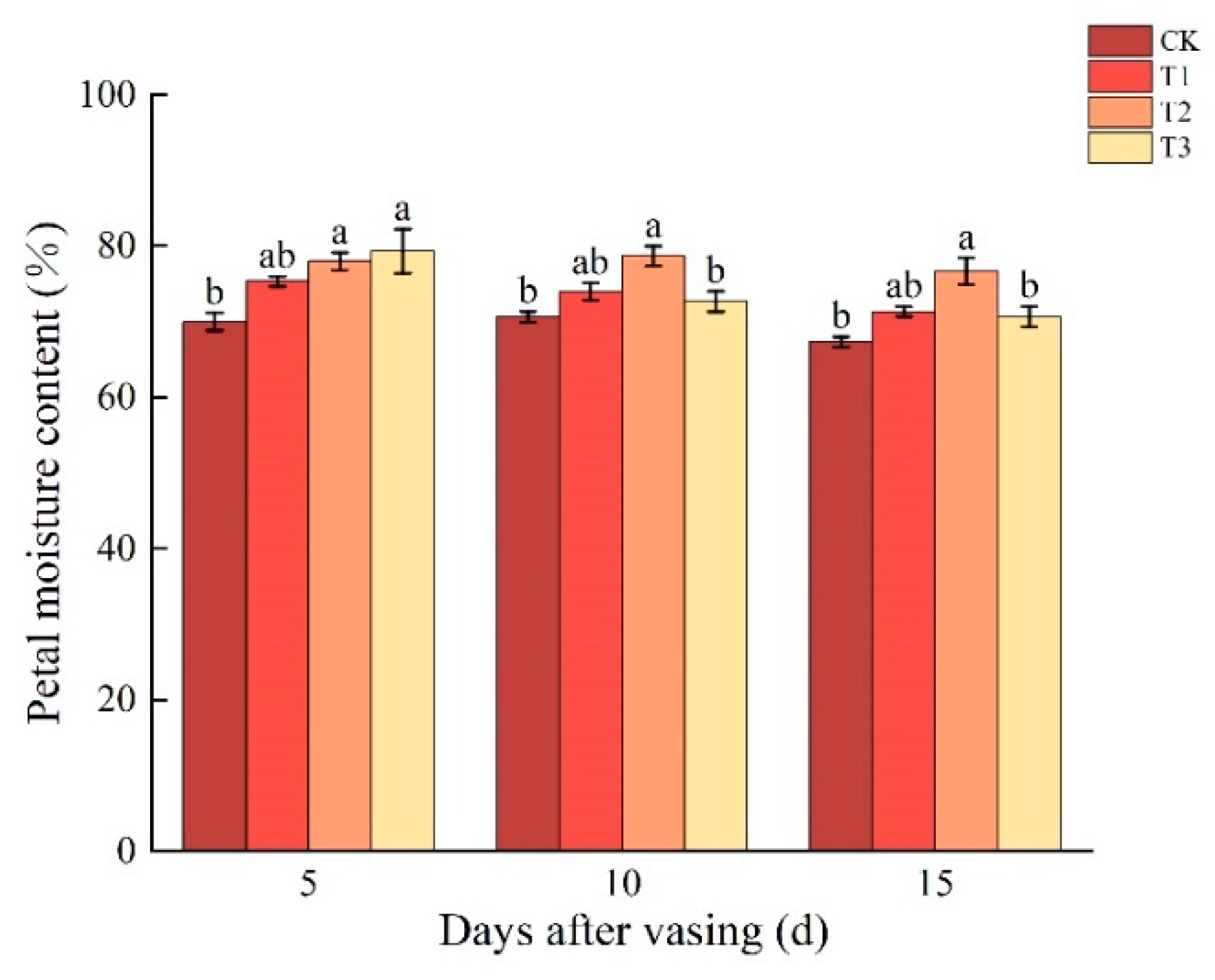
The petal moisture content plays a crucial role in the vase life of cut roses, which reflects the degree of hydration of cut flowers. In this study, the petal moisture content of the T2 group was consistently significantly higher than that of CK from day 5 to 15 (Figure 2). This indicated that the T2 group was able to effectively maintain petal moisture content during the vase life in cut roses. The petal moisture content of the T3 group showed significant differences compared to the CK on day 5, but there were no significant differences on days 10 and 15 (Figure 2).

3.3. Changes in SS and SP Contents of Cut Roses under Different Concentrations of Nano-Se

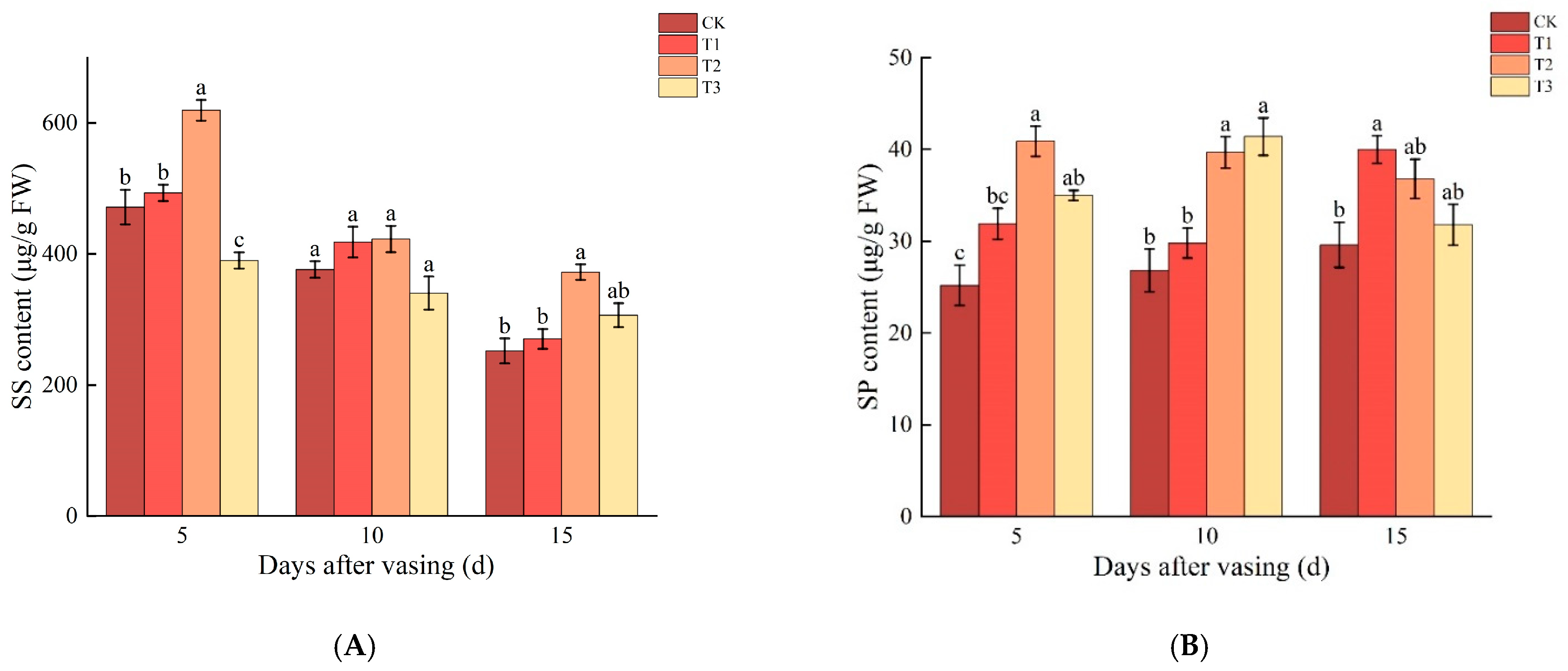
As the vase life extended, the SS content generally decreased. On day 5, the SS content of the T2 group was significantly higher than that of the other groups, while there was no significant difference between T1 and CK. On day 10, there were no significant differences in SS content between the treatment groups and the CK. On day 15, the SS content of the T2 group was significantly higher than that of the CK and T1 groups, indicating that 400 mM of Nano-Se can effectively maintain SS content during the later stages of vase life in cut roses (Figure 3A).
On day 5, the SP content in the T2 and T3 groups was significantly higher than that of CK, but there was no significant difference between T2 and T3. On day 10, the SP content in the T2 and T3 groups was significantly higher than in the CK, with increases of 48.1% and 54.5%, respectively. On day 15, the SP content in the T1 group was significantly higher than that of CK, but there were no significant differences among the T1, T2, and T3 groups (Figure 3B). This indicated that the T2 group was able to effectively maintain SP content during the vase life in cut roses.

3.4. Changes in Total Phenolic Acid and Total Flavonoid Contents of Cut Roses under Different Concentrations of Nano-Se

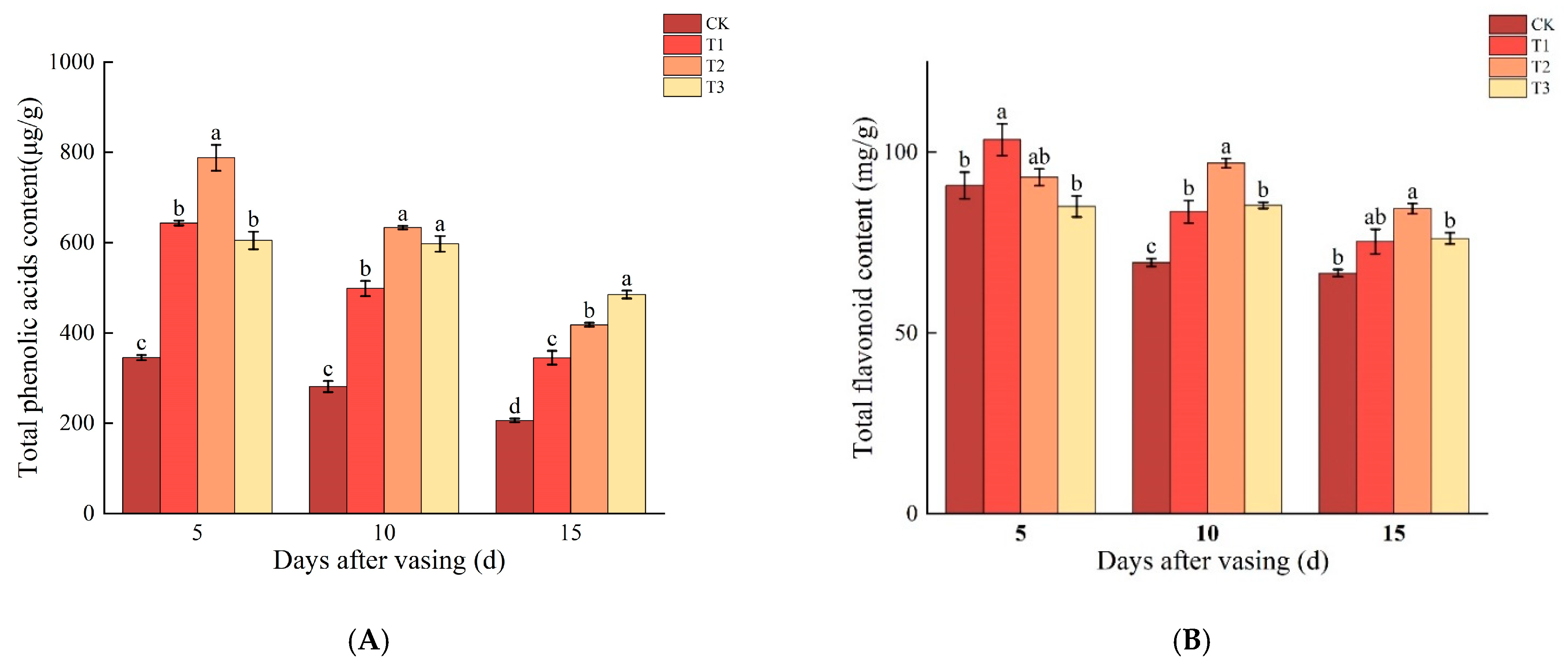
The trend in the total phenolic acid content for the T1, T2, and T3 groups was consistent with that for the CK, with all showing a gradual decrease over the duration of the vase life (Figure 4A). On day 5, the total phenolic acid content in the CK was significantly lower than that in the selenium-treated group, with significant differences between T2 and T1 and T3. The highest phenolic acid content was found in the T2 group at 787.8 µg/g. On day 10, the total phenolic acid content in CK continued to decrease and remained significantly lower than that in the treatment groups T1, T2, and T3, with 281.0 μg/g in CK and 633.2 μg/g in T2. By day 15, the total phenolic acid content in CK decreased further to 206.0 μg/g, whereas the highest level of total phenolic acid was found in T3, with a concentration of 484.6 μg/g (Figure 4A).
The total flavonoid content of the T1 group followed the same trend as that of CK, decreasing over time, while the total flavonoid content of the T2 and T3 groups increased slowly from day 5 to 10, reached a peak on day 10, and then decreased gradually from day 10 to 15 (Figure 4B). The total flavonoid content of T1 was significantly higher than that of CK on day 5, and all selenium-treated groups had significantly higher total flavonoid content than that of CK on day 10. In comparison, the total flavonoid content of the T2 group was still significantly higher than that of CK on day 15 (Figure 4B). This indicated that 400 mM of Nano-Se can effectively maintain total flavonoid contents during the vase life in cut roses.

3.5. Changes in POD, SOD, and CAT Activities of Cut Roses under Different Concentrations of Nano-Se

The POD activities of T1, T2, and CK showed a trend of gradual decrease with the increase in vase time. The POD activity of the T2 group decreased slowly, while the POD activity of the T1 group decreased rapidly from day 10 to 15. The POD activity of the T3 group stayed at a lower level (Figure 5A). On day 5, the POD activity of the T3 and CK groups was significantly lower than that of the T1 and T2 groups. On day 10, the POD activity level was significantly higher for T2 than CK at 46.62 U/g FW. By day 15, the T2 group maintained a high level of POD activity, which was significantly higher than the other treatment groups (Figure 5A).
On day 5, the SOD activity of T3 was significantly higher than that of CK by 14.4%. On day 10, the SOD activity of T2 was significantly higher than that of CK. On day 15, the SOD activity of T3 was significantly higher than both CK and T2. Additionally, the trends in SOD activity varied among treatment groups: the SOD activity of T3 followed a trend similar to that of CK, showing a decrease followed by an increase (Figure 5B).
CAT activity in the T2 and T3 groups followed the same trend as CK, with a gradual decrease in CAT activity over time. From day 5 to 15, CAT activity in CK was significantly lower than in the selenium-treated group. On day 5, there was a significant difference between the groups with CAT activities of T2 > T3 > T1 > CK; on day 10, there was no significant difference between T2 and T3, but they were significantly higher than those of CK, and on day 15, there was also a significant difference between the groups with CAT activities of T1 > T3 > T2 > CK (Figure 5C). This indicated that Nano-Se can effectively maintain CAT activity throughout the vase life of cut roses.

3.6. Changes in MDA and H2O2 Contents of Cut Roses under Different Concentrations of Nano-Se

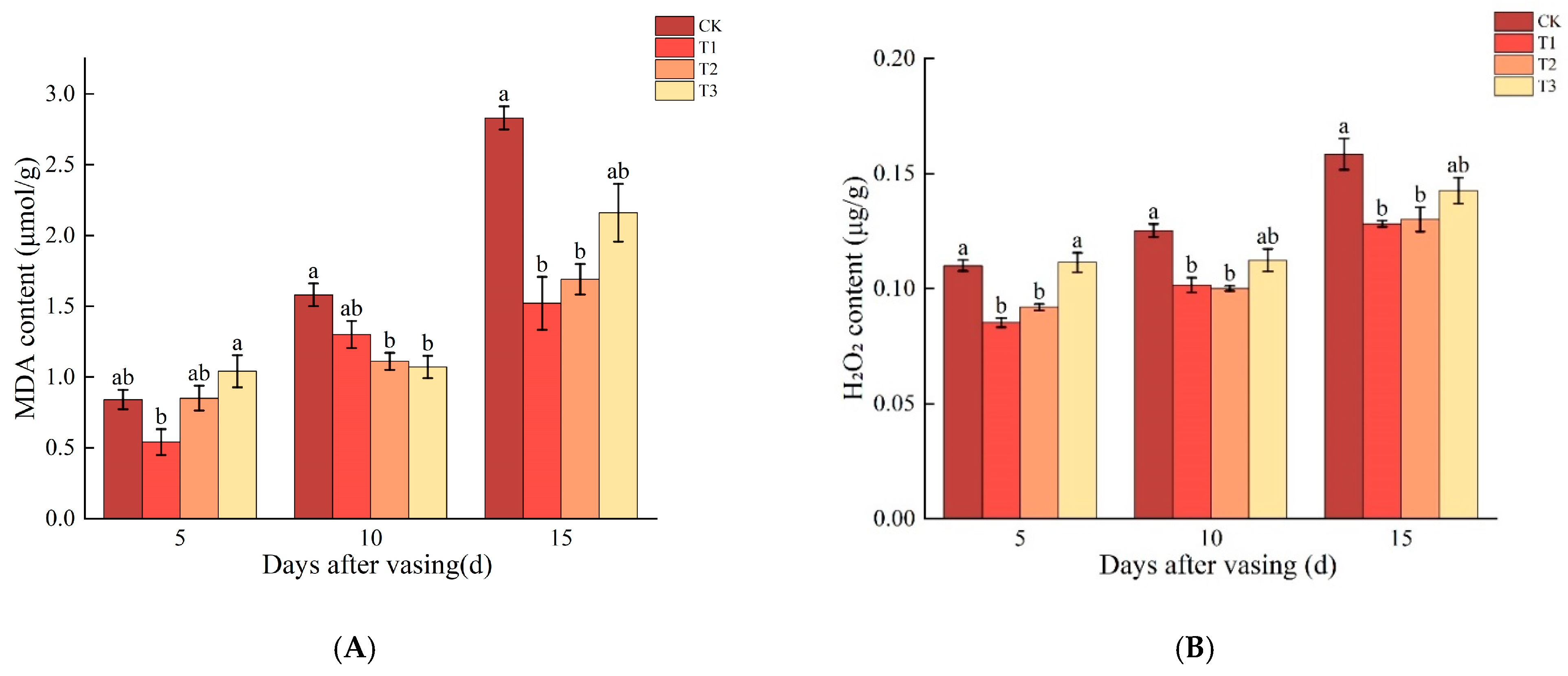
In the T1 and T2 groups, the trend of MDA content was similar to that of CK, with a gradual increase over time (Figure 6A). In the T3 group, the MDA content did not change significantly until day 10 but increased rapidly from day 10 to day 15. On day 5, the MDA content of the T1 group was lower at 1.0 μmol/g, while the differences among the T2, T3, and CK groups were insignificant. On day 10, the MDA content of CK was 1.6 μmol/g, which was significantly higher than that of the T2 and T3 selenium treatment groups. On day 15, the MDA content of CK was 2.8 μmol/g, which was significantly higher than that of the other groups, while the content of T1 and T2 was significantly lower than that of the other groups (Figure 6A).
The trend of H2O2 content in the T1, T2, and T3 groups was consistent with that in CK, with a gradual increase over time (Figure 6B). On day 5, the H2O2 levels in the CK and T3 groups were significantly higher than those in the T1 and T2 groups; on day 10, the H2O2 levels in CK were significantly higher than those in the T1 and T2 groups, reaching 0.1 μg/g. On day 15, the H2O2 levels in CK were still significantly higher than those in the T1 and T2 selenium treatment groups, reaching 0.2 μg/g, and those in the T1 and T2 groups were 20.0% and 18.8% lower than those in CK, respectively.

3.7. Correlation Analysis of Physiological Indicators

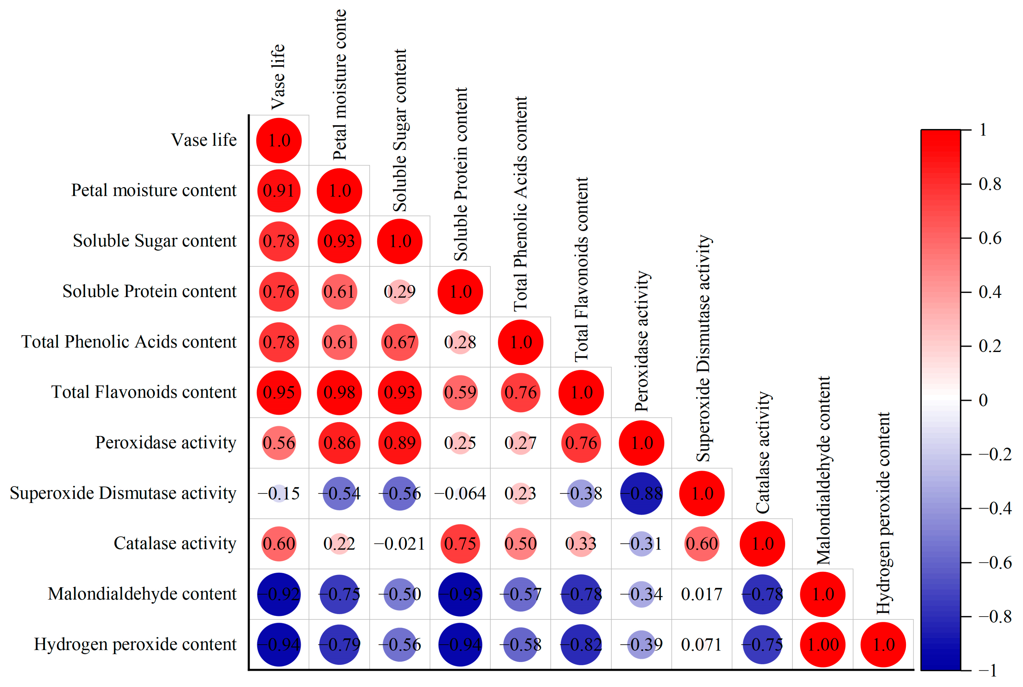
Pearson correlation analysis was conducted on 11 physiological indicators for the CK, T1, T2, and T3 groups. The results showed that the vase life and natural water content of the cut flowers showed a significant positive correlation with the total flavonoid content. The natural water content also had a significant positive correlation with the soluble sugar content and total flavonoid content. Additionally, the soluble sugar content was significantly positively correlated with the total flavonoid content. The vase life of the cut flowers was significantly negatively correlated with the MDA content and H2O2 content, and the MDA content and H2O2 content were significantly negatively correlated with the POD content. Moreover, the MDA content in the cut flowers exhibited a highly significant positive correlation with the H2O2 content (Figure 7).

4. Discussion

4.1. An Appropriate Concentration of Nano-Se Can Significantly Extend the Vase Life of Cut Roses

The vase life of cut flowers is a crucial indicator for assessing the aging and ornamental value of the flowers. Compared to previous study [19], the Nano-Se treatment in this study extended the vase life of cut roses by 3.7 to 5.7 days, showing a more significant effect on vase life extension with reduced variability in the duration of extension. This indicated that adding certain concentrations of Nano-Se to the basic preservative solution can significantly extend the vase life of cut roses, thereby maintaining their ornamental quality and enhancing the economic benefits of fresh-cut flowers during storage, transportation, and shelf life.
The water-holding capacity of petals is a crucial factor in determining the vase life of cut flowers [29]. In this study, the correlation analysis between various physiological indicators and vase life confirmed that the vase life of cut flowers was significantly positively correlated with their petal moisture content. Therefore, maintaining postharvest water content was essential; the longer the water retention and the higher the petal moisture content, the longer the vase life. Our study found that adding Nano-Se to the basic preservative solution helped maintain the high petal moisture content of cut roses, thereby extending their vase life. Cremonini’s experiment demonstrated that Nano-Se effectively degrades the bacterial exopolysaccharide matrix of Pseudomonas aeruginosa, inhibits biofilm synthesis, and exhibits antibacterial properties [30]. We speculated that the properties of the suitable concentration of Nano-Se helped inhibit the proliferation of bacteria and fungi in the vase solution and prevented stem rot and vascular tissue damage, thereby facilitating good water transport, enhancing the water retention capacity of cut flowers, and reducing water loss. Thus, inhibiting the excessive growth of microorganisms in the vase solution effectively prevents vascular blockage in the flower stems, ensures normal water uptake by the cut flowers, prevents water imbalance, and increases water absorption by the flower stems, extending vase life and maintaining the ornamental quality of cut flowers [31].

4.2. Appropriate Concentrations of Nano-Se Delay the Loss of SS and SP Content in Cut Roses

Carbohydrates are the primary energy reserves for cut flowers. After harvest, roses cannot produce energy reserves necessary for sustaining life through their own photosynthesis and must rely on the sugars accumulated before harvest and the energy reserves present in the preservative solution. Over time, energy reserves are gradually depleted, leading to rapid senescence as the flowers struggle to maintain vitality. In this study, the SS content in CK on days 5 and 15 was significantly lower than that in the 400 mM Nano-Se treatment group. This suggests that the preservative solution with added Nano-Se is effective in maintaining the freshness of cut flowers and is significant in extending the vase life of cut flowers.
Under natural conditions, the SP content gradually decreases as cut flowers age [32]. In this study, as the vase life extended, the SP content varied significantly among different treatment groups; the SP content in CK slowly increased, showing minimal overall change and remaining consistently lower than in the selenium-treated groups. The 200 mM Nano-Se treatment group exhibited a decreasing trend before day 10, with a significant increase from day 10 to day 15. The 400 mM Nano-Se treatment group showed a slow decline, while the 600 mM Nano-Se treatment group’s SP content first increased and then decreased. This phenomenon suggested that different concentrations of selenium treatments result in varying physiological changes in the cut flowers, leading to different changes in SP content. However, all treatment groups maintained higher SP levels than CK. Overall, the SP content in Nano-Se-treated cut roses was significantly higher than that of CK. Adding an appropriate concentration of selenium can effectively increase the SP content in cut flowers, providing a theoretical basis for extending the vase life of fresh-cut flowers.

4.3. Adaptability of Vase Roses to Selenium-Enriched Environments

Phenolic compounds and flavonoids are important natural antioxidants in plants, possessing redox properties [33]. In this study, the total phenolic content in cut roses generally decreased over time. However, the total phenolic acid content in the selenium-treated groups was significantly higher than in CK during the vase life. The total flavonoid content in the T2 group varied. Still, on days 10 and 15, the total flavonoid content in CK was consistently significantly lower than that in the selenium-treated groups. These results indicated that selenium treatment significantly promotes the accumulation of antioxidants in cut flowers, thereby extending the vase life of cut flowers.
Most studies suggest that POD plays a dual role in eliminating the toxicity of hydrogen peroxide and phenolic, amine, aldehyde, and benzene compounds, making it an essential enzyme in mitigating the damage caused by free radicals [34]. It serves as a critical physiological and biochemical indicator of plant stress resistance. In this study, the POD activity in cut roses peaked on the 5th day across all treatments and then gradually declined over time. The POD activity in the 600 mM Nano-Se treatment was consistently lower than in the CK and other selenium treatment groups, suggesting that excessive Nano-Se may inhibit POD activity, thereby impairing the antioxidant capacity of cut flowers.
After harvesting, the production of oxygen free radicals significantly increases, exacerbating lipid peroxidation of the cell membrane, disrupting membrane integrity, and leading to substantial leakage of electrolytes and small organic molecules. The extensive leakage of protoplasm ultimately results in cell damage in cut flowers [31]. Plant SOD activity typically exhibits an initial increase followed by a decrease, representing an oxidative defense response [35]. In this study, the trend of SOD activity varied among the different groups, the activity of T2 on day 10 and T3 on days 5 and 15 had significantly higher SOD levels compared to CK. This indicated that Nano-Se enhances the SOD activity in cut roses, thereby mitigating oxidative stress during senescence.
The exposure of cut flowers to environmental stress induces the production of ROS, which damages cellular macromolecules and leads to flower senescence. CAT is a typical antioxidant enzyme that scavenges ROS induced by oxidative stress [26]. During the senescence of cut flowers, CAT activity gradually decreases over time [36]. In this study, the CAT activity in cut flowers treated with Nano-Se was higher than in CK. Specifically, CAT activity was higher in the T2 group on days 5 and 10 compared with the other treatment groups, suggesting that adding appropriate concentrations of Nano-Se could significantly delay the decline in CAT activity in cut flowers.
MDA, as a lipid peroxidation product, compromises cell membrane permeability. Generally, more severe postharvest damage to cut roses leads to greater leakage of substances, higher MDA levels, and more pronounced aging [37]. In this study, the Nano-Se treatment groups exhibited significantly lower MDA levels than CK over extended vase periods, indicating that Nano-Se can delay the increase in MDA content and enhance the stress resistance of cut roses under vase conditions. This finding was consistent with Zahedi’s research, which demonstrated that Nano-Se significantly reduced MDA levels in plants [38]. However, the MDA content in the 600 mM Nano-Se treatment group was slightly higher than in CK, possibly due to variations between individual cut flowers. H2O2 is one of the main ROSs that causes cell damage, leading to lipid peroxidation of cell membranes. This results in compromised membrane integrity and impaired cell function and can even lead to cell death [39]. In this study, the H2O2 content in the T1 and T2 groups was significantly lower than that in the CK and accumulated at a slower rate, suggesting that the selenium treatments reduced the accumulation of H2O2 in the plants and thus were more conducive to the maintenance of membrane stability compared with CK.

4.4. Comprehensive Evaluation of Nano-Se Treatment on the Postharvest Quality of Cut Roses

Preservative solutions with Nano-Se significantly extended the vase life of cut roses by 3.7 to 5.7 days and enhanced the water retention of the flowers, increased essential physiological metabolites such as soluble sugars and soluble proteins, delayed petal loss and aging, and maintained high ornamental value. The correlation analysis of physiological indicators for cut flowers showed that vase life was positively correlated with POD and CAT activities, SS and SP contents, and total phenolic and total flavonoid contents and negatively correlated with MDA and H2O2 levels. These results indicated that applying an appropriate concentration of selenium to cut flowers could effectively maintain water content and soluble sugar levels, support basic metabolism, enhance antioxidant enzyme activity, increase the accumulation of antioxidants, reduce the accumulation of oxidants, and extend the vase life of cut flowers. Therefore, Nano-Se has a great potential to extend the vase life of cut flowers.
Studies have reported that nanomaterials might have adverse effects on the environment [40,41], raising significant concerns about the safety of Nano-Se. Utilizing microorganisms (such as algae, fungi, yeast, and bacteria) to synthesize Nano-Se is a sustainable, environmentally friendly, and cost-effective method [42]. The Nano-Se used in this study was synthesized biologically and exhibited high biological activity with minimal environmental impact. Moreover, Nano-Se has been widely applied in the agri-food sector, where it is used to enrich plants with selenium through direct foliar spraying, thereby providing selenium supplementation for humans [18]. Overall, Nano-Se is expected to see increasing applications across various research fields.

5. Conclusions

This study showed that the use of Nano-Se significantly extended the vase life of cut roses, maintained the petal moisture content, and preserved their ornamental value. The Nano-Se treatment enhanced the activities of SOD, POD, and CAT and effectively reduced the accumulation of MDA and H2O2, thereby protecting the plants from oxidative damage. Additionally, Nano-Se effectively delayed the decline in SS, SP, total phenolics, and total flavonoids in cut roses, thereby postponing flower senescence. Overall, this study provides a solid theoretical foundation for the application of Nano-Se in cut-flower preservation and offers an innovative approach for developing new preservatives for cut flowers, which represents significant economic value for the development of new preservatives not only for roses but for all cut flowers.

Author Contributions

Conceptualization, Q.W.; methodology, Q.W.; validation, Y.C.; resources, Q.W. and F.X.; data curation, Y.W., D.C., and Y.X.; writing—original draft preparation, Y.W.; writing—review and editing, Q.W.; visualization, J.X.; project administration, D.W. All authors have read and agreed to the published version of the manuscript.

Funding

This research was funded by the Open Project Funding of the State Key Laboratory of Biocatalysis and Enzyme Engineering (SKLBEE2022025) and the National Natural Science Foundation of China (3220163).

Data Availability Statement

Data are contained within the article.

Conflicts of Interest

The authors declare no conflicts of interest.

References

  1. Elhindi, K.M. Evaluation of several holding solutions for prolonging vase-life and keeping quality of cut sweet pea flowers (Lathyrus odoratus L.). Saudi J. Biol. Sci. 2012, 19, 195–202. [Google Scholar] [CrossRef] [PubMed]
  2. Ahmad, I.; Dole, J.M.; Clark, E.M.R.; Blazich, F.A. Floral foam and/or conventional or organic preservatives affect the vase-life and quality of cut rose (Rosa × hybrida L.) stems. J. Hortic. Sci. Biotechnol. 2014, 89, 41–46. [Google Scholar] [CrossRef]
  3. Ma, Y. In Vitro Models for Nanotoxicity Testing; Wiley: West Sussex, UK, 2009. [Google Scholar]
  4. Domokos-Szabolcsy, E.; Marton, L.; Sztrik, A.; Babka, B.; Prokisch, J.; Fari, M. Accumulation of red elemental selenium nanoparticles and their biological effects in Nicotinia tabacum. Plant Growth Regul. 2012, 68, 525–531. [Google Scholar] [CrossRef]
  5. Djanaguiraman, M.; Belliraj, N.; Bossmann, S.H.; Prasad, P.V.V. High-Temperature Stress Alleviation by Selenium Nanoparticle Treatment in Grain Sorghum. ACS Omega 2018, 3, 2479–2491. [Google Scholar] [CrossRef] [PubMed]
  6. Bano, I.; Skalickova, S.; Sajjad, H.; Skladanka, J.; Horky, P. Uses of Selenium Nanoparticles in the Plant Production. Agronomy 2021, 11, 2229. [Google Scholar] [CrossRef]
  7. Bai, K.; Hong, B.; He, J.; Hong, Z.; Tan, R. Preparation and antioxidant properties of selenium nanoparticles-loaded chitosan microspheres. Int. J. Nanomed. 2017, 12, 4527–4539. [Google Scholar] [CrossRef]
  8. Alagawany, M.; Qattan, S.; Attia, Y.; El-Saadony, M.; Elnesr, S.; Mahmoud, M.; Madkour, M.; Abd El-Hack, M.; Reda, F.; Ebani, V. Use of Chemical Nano-Selenium as an Antibacterial and Antifungal Agent in Quail Diets and Its Effect on Growth, Carcasses, Antioxidant, Immunity and Caecal Microbes. Animals 2021, 11, 3027. [Google Scholar] [CrossRef]
  9. Vera, P.; Canellas, E.; Nerín, C. New antioxidant multilayer packaging with nanoselenium to enhance the shelf-life of market food products. Nanomaterials 2018, 8, 837. [Google Scholar] [CrossRef]
  10. Samynathan, R.; Venkidasamy, B.; Ramya, K.; Muthuramalingam, P.; Shin, H.; Kumari, P.S.; Thangavel, S.; Sivanesan, I. A Recent Update on the Impact of Nano-Selenium on Plant Growth, Metabolism, and Stress Tolerance. Plants 2023, 12, 853. [Google Scholar] [CrossRef]
  11. Foyer, C.; Descourvières, P.; Kunert, K. Protection against oxygen radicals: An important defence mechanism studied in transgenic plants. Plant Cell Environ. 2006, 17, 507–523. [Google Scholar] [CrossRef]
  12. Kaur, M.; Sharma, S.; Singh, D. Influence of selenium on carbohydrate accumulation in developing wheat grains. Commun. Soil Sci. Plant Anal. 2018, 49, 1650–1659. [Google Scholar] [CrossRef]
  13. Shahraki, B.; Bayat, H.; Aminifard, M.; Azarmi-Atajan, F. Effects of Foliar Application of Selenium and Nano-Selenium on Growth, Flowering, and Antioxidant Activity of Pot Marigold (Calendula officinalis L.) Under Salinity Stress Conditions. Commun. Soil Sci. Plant Anal. 2022, 53, 2749–2765. [Google Scholar] [CrossRef]
  14. Zhang, M.; Tang, S.; Huang, X.; Zhang, F.; Pang, Y.; Huang, Q.; Yi, Q. Selenium uptake, dynamic changes in selenium content and its influence on photosynthesis and chlorophyll fluorescence in rice (Oryza sativa L.). Environ. Exp. Bot. 2014, 107, 39–45. [Google Scholar] [CrossRef]
  15. Morales-Espinoza, M.C.; Cadenas-Pliego, G.; Pérez-Alvarez, M.; Hernández-Fuentes, A.D.; Cabrera de la Fuente, M.; Benavides-Mendoza, A.; Valdés-Reyna, J.; Juárez-Maldonado, A.; Morales-Espinoza, M.C.; Cadenas-Pliego, G.; et al. Se Nanoparticles Induce Changes in the Growth, Antioxidant Responses, and Fruit Quality of Tomato Developed under NaCl Stress. Molecules 2019, 24, 3030. [Google Scholar] [CrossRef]
  16. Manaf, H.H. Beneficial effects of exogenous selenium, glycine betaine and seaweed extract on salt stressed cowpea plant. Ann. Agric. Sci. 2016, 61, 41–48. [Google Scholar] [CrossRef]
  17. Turakainen, M.; Hartikainen, H.; Seppänen, M.M. Effects of selenium treatments on potato (Solanum tuberosum L.) growth and concentrations of soluble sugars and starch. J. Agric. Food Chem. 2004, 52, 5378–5382. [Google Scholar] [CrossRef]
  18. Li, D.; Zhou, C.; Zou, N.; Wu, Y.; Zhang, J.; An, Q.; Li, J.-Q.; Pan, C. Nanoselenium foliar application enhances biosynthesis of tea leaves in metabolic cycles and associated responsive pathways. Environ. Pollut. 2021, 273, 116503. [Google Scholar] [CrossRef]
  19. Ahmadi-Majd, M.; Mousavi-Fard, S.; Rezaei Nejad, A.; Fanourakis, D. Nano-Selenium in the holding solution promotes rose and carnation vase life by improving both water relations and antioxidant status. J. Hortic. Sci. Biotechnol. 2023, 98, 246–261. [Google Scholar] [CrossRef]
  20. Sánchez Díaz, J.M.; Jiménez-Becker, S.; Jamilena, M. A screening test for the determination of cut flower longevity and ethylene sensitivity of carnation. Hortic. Sci. 2017, 44, 14–20. [Google Scholar] [CrossRef]
  21. Wang, H.; Zhang, Y.; Song, Y.; Zhu, J.; Shang, W.; Jiang, L.; Liu, W.; He, S.; Shen, Y.; Shi, L.; et al. ClO2 Prolongs the Vase Life of Paeonia lactiflora ‘Hushui Dangxia’ Cut Flowers by Inhibiting Bacterial Growth at the Stem Base. Horticulturae 2024, 10, 732. [Google Scholar] [CrossRef]
  22. van Doorn, W.G.; Sinz, A.; Tomassen, M.M. Daffodil flowers delay senescence in cut Iris flowers. Phytochemistry 2004, 65, 571–577. [Google Scholar] [CrossRef]
  23. Ul Haq, A.; Lone, M.; Farooq, S.; Parveen, S.; Altaf; Tahir, I.; Hefft, D.; Ahmad, A.; Ahmad, P. Nitric oxide effectively orchestrates postharvest flower senescence: A case study of Consolida ajacis. Funct. Plant Biol. 2021, 50, 97–107. [Google Scholar] [CrossRef]
  24. Labancová, E.; Vivodová, Z.; Šípošová, K.; Kollárová, K. Silicon Actuates Poplar Calli Tolerance after Longer Exposure to Antimony. Plants 2023, 12, 689. [Google Scholar] [CrossRef]
  25. Lou, X.; Anwar, M.; Wang, Y.; Zhang, H.; Ding, J. Impact of inorganic salts on vase life and postharvest qualities of the cut flower of Perpetual Carnation. Braz. J. Biol. 2020, 81, 228–236. [Google Scholar] [CrossRef]
  26. Mazrou, R.; Hassan, S.; Yang, M.; Hassan, F. Melatonin Preserves the Postharvest Quality of Cut Roses through Enhancing the Antioxidant System. Plants 2022, 11, 2713. [Google Scholar] [CrossRef]
  27. Chandra, S.; Khan, S.; Avula, B.; Lata, H.; Yang, M.H.; ElSohly, M.A.; Khan, I.A. Assessment of Total Phenolic and Flavonoid Content, Antioxidant Properties, and Yield of Aeroponically and Conventionally Grown Leafy Vegetables and Fruit Crops: A Comparative Study. Evid. Based Complement. Altern. Med. 2014, 2014, 253875. [Google Scholar] [CrossRef]
  28. McDonald, S.; Prenzler, P.D.; Antolovich, M.; Robards, K. Phenolic content and antioxidant activity of olive extracts. Food Chem. 2001, 73, 73–84. [Google Scholar] [CrossRef]
  29. Lu, N.; Wu, L.; Zhang, C.; Shan, C. Sodium selenite improves the vase life of Eustoma grandiflorum cut flowers. Hortic. Sci. 2022, 49, 248–256. [Google Scholar] [CrossRef]
  30. Cremonini, E.; Zonaro, E.; Donini, M.; Lampis, S.; Boaretti, M.; Dusi, S.; Lleo, M.; Vallini, G. Biogenic selenium nanoparticles: Characterization, antimicrobial activity and effects on human dendritic cells and fibroblasts. Microb. Biotechnol. 2016, 9, 758–771. [Google Scholar] [CrossRef]
  31. Jedrzejuk, A.; Rochala, J.; Zakrzewski, J.; Rabiza-Świder, J. Identification of xylem occlusions occurring in cut clematis (Clematis L., fam. Ranunculaceae Juss.) stems during their vase life. Sci. World J. 2012, 2012, 749281. [Google Scholar] [CrossRef]
  32. Zheng, M.; Guo, Y. Effects of neodymium on the vase life and physiological characteristics of Lilium casa Blanca petals. Sci. Hortic. 2019, 256, 108553. [Google Scholar] [CrossRef]
  33. Aryal, S.; Baniya, M.K.; Danekhu, K.; Kunwar, P.; Gurung, R.; Koirala, N. Total Phenolic Content, Flavonoid Content and Antioxidant Potential of Wild Vegetables from Western Nepal. Plants 2019, 8, 96. [Google Scholar] [CrossRef]
  34. Li, S.; Chen, Z.; Yang, F.; Yue, W. The age of vanadium-based nanozymes: Synthesis, catalytic mechanisms, regulation and biomedical applications. Chin. Chem. Lett. 2024, 35, 108793. [Google Scholar] [CrossRef]
  35. Hashemabadi, D. The role of silver nano-particles and silver thiosulfate on the longevity of cut carnation (Dianthus caryophyllus) flowers. J. Environ. Biol. Acad. Environ. Biol. India 2014, 35, 661–666. [Google Scholar]
  36. Saeed, T.; Hassan, I.; Abbasi, N.A.; Jilani, G. Effect of gibberellic acid on the vase life and oxidative activities in senescing cut gladiolus flowers. Plant Growth Regul. 2014, 72, 89–95. [Google Scholar] [CrossRef]
  37. Kazemi, M.; Shokri, K. Role of salicylic acid in decreases of membrane senescence in cut lisianthus flowers. World Appl. Sci. J. 2011, 13, 142–146. [Google Scholar]
  38. Zahedi, S.M.; Moharrami, F.; Sarikhani, S.; Padervand, M. Selenium and silica nanostructure-based recovery of strawberry plants subjected to drought stress. Sci. Rep. 2020, 10, 17672. [Google Scholar] [CrossRef]
  39. Hassan, F.A.S.; Fetouh, M.I. Does moringa leaf extract have preservative effect improving the longevity and postharvest quality of gladiolus cut spikes? Sci. Hortic. 2019, 250, 287–293. [Google Scholar] [CrossRef]
  40. Ahmadi-Majd, M.; Mousavi-Fard, S.; Rezaei Nejad, A.; Fanourakis, D. Carbon nanotubes in the holding solution stimulate flower opening and prolong vase life in carnation. Chem. Biol. Technol. Agric. 2022, 9, 15. [Google Scholar] [CrossRef]
  41. Far, A.S.; Mousavi-Fard, S.; Nejad, A.R.; Shahbazi, F.; Ahmadi-Majd, M.; Fanourakis, D. Nano Silver and melatonin effectively delay the senescence of cut carnation flowers under simulated vibrational stress. J. Hortic. Sci. Biotechnol. 2024, 99, 597–608. [Google Scholar]
  42. Ghazi, A.A.; El-Nahrawy, S.; El-Ramady, H.; Ling, W. Biosynthesis of Nano-Selenium and Its Impact on Germination of Wheat under Salt Stress for Sustainable Production. Sustainability 2022, 14, 1784. [Google Scholar] [CrossRef]
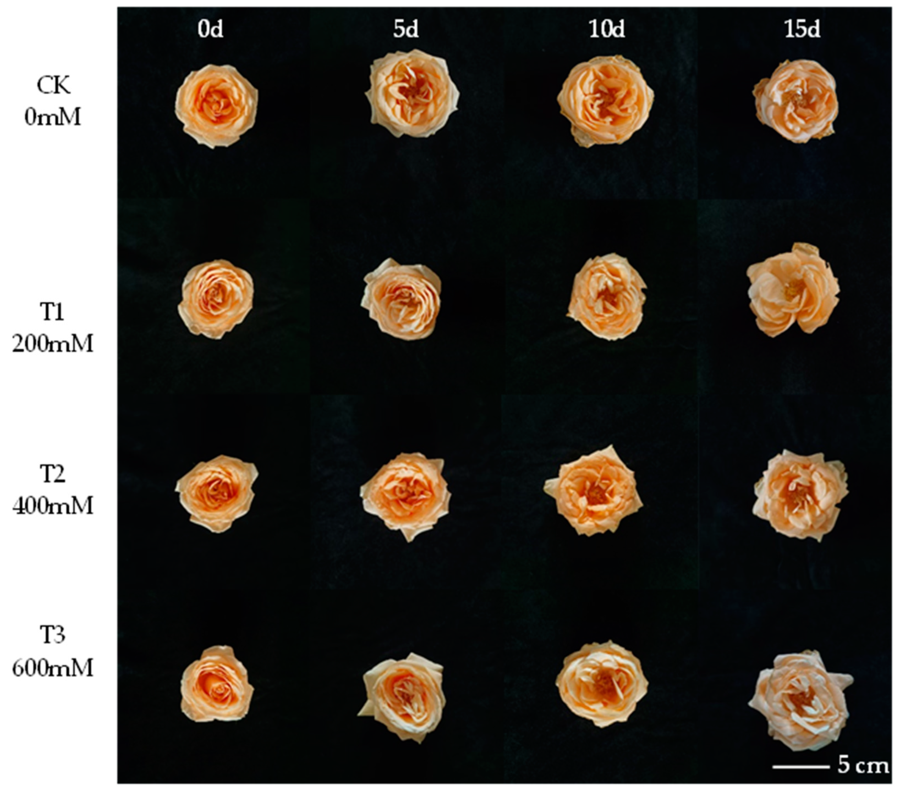
Figure 1. The changes in ornamental quality of R. hybrida under different concentrations of Nano-Se. Bar = 5 cm.
Horticulturae 10 01071 g001
Figure 2. The changes in the petal moisture content of R. hybrida under different concentrations of Nano-Se. Different lowercase letters indicate significant differences between groups (p < 0.05), and the error bars represent the standard errors.
Horticulturae 10 01071 g002
Figure 3. The changes in the SS (A) and SP (B) contents in cut flowers of R. hybrida under different concentrations of Nano-Se. Different lowercase letters indicate significant differences between groups (p < 0.05), and the error bars represent the standard errors.
Horticulturae 10 01071 g003
Figure 4. The changes in the total phenolic acid (A) and total flavonoid content (B) in cut flowers of R. hybrida under different concentrations of Nano-Se. Different lowercase letters indicate significant differences between groups (p < 0.05), and the error bars represent the standard errors.
Horticulturae 10 01071 g004
Figure 5. The changes in POD (A), SOD (B), and CAT (C) activities in cut flowers of R. hybrida under different concentrations of Nano-Se. Different lowercase letters indicate significant differences between groups (p < 0.05), and the error bars represent the standard errors.
Horticulturae 10 01071 g005
Figure 6. The changes in the MDA (A) and H2O2 (B) content in cut flowers of R. hybrida under different concentrations of Nano-Se. Different lowercase letters indicate significant differences between groups (p < 0.05), and the error bars represent the standard errors.
Horticulturae 10 01071 g006
Figure 7. The correlation analysis of 11 physiological indicators in cut flowers of R. hybrida under different concentrations of Nano-Se.
Horticulturae 10 01071 g007
Table 1. The vase life of R. hybrida under different concentrations of Nano-Se.
TreatmentVase Life (d)The Extended Day (d)Vase Life Termination Symptoms
CK13.0 ± 1.0 b050% wilted and the first petal falls off
T117.3 ± 1.5 a4.350% wilted and the first petal falls off
T218.7 ± 0.6 a5.750% wilted and the first petal falls off
T316.7 ± 1.5 a3.750% wilted and the first petal falls off
Different lowercase letters indicate significant differences between groups (p < 0.05).
Disclaimer/Publisher’s Note: The statements, opinions and data contained in all publications are solely those of the individual author(s) and contributor(s) and not of MDPI and/or the editor(s). MDPI and/or the editor(s) disclaim responsibility for any injury to people or property resulting from any ideas, methods, instructions or products referred to in the content.