Article

Effect of Foliar Selenate Supplementation on Biochemical Characteristics of Purslane Weed (Portulaca oleracea L.)

1
Federal Scientific Vegetable Center, Moscow 143072, Russia
2
Chechen Scientific Institute of Agriculture, Lilovaya 1, Gikalo, Grozny 366021, Russia
3
Federal Research Center of Nutrition and Biotechnology, Moscow 109240, Russia
4
Faculty of Pharmacy, The First Moscow State Medical University of I.M. Mechnikov, Moscow 119991, Russia
5
Department of Food Technology, University ‘Ion Ionescu de la Brad’ of Life Sciences of Iasi, 700490 Iasi, Romania
6
Department of Agricultural Sciences, University of Naples Federico II, 80055 Naples, Italy
*
Author to whom correspondence should be addressed.
Horticulturae 2024, 10(7), 708; https://doi.org/10.3390/horticulturae10070708
Submission received: 6 June 2024 / Revised: 30 June 2024 / Accepted: 2 July 2024 / Published: 4 July 2024
(This article belongs to the Special Issue Bioactive Compounds in Horticultural Plants—2nd Edition)

Abstract

:
The high biological activity of cultivated and wild purslane offers broad possibilities for utilizing this plant in medicine and human nutrition. To assess the prospects of obtaining new functional food products based on the wild form of P. oleracea L., foliar biofortification of this species with sodium selenate (VI) was carried out, and the changes in leaf and seed biochemical characteristics were investigated. Selenium significantly enhanced plant yield, photosynthetic pigments and the ascorbic acid content, and showed a tendency to seed productivity increase. The application of selenium augmented quinic acid content in leaves by 1.7 times but did not affect the oxalic acid content. Oxalic acid prevailed in wild purslane and quinic acid in cultivated purslane (cv. Makovey). Seed oil in Se-enriched purslane was characterized by a two-fold decrease in saturated fatty acids and squalene and 2.3-fold decrease in malonic dialdehyde content, along with a 1.4-fold increase in ascorbic acid. Selenium supplementation resulted in an increase in total lipids and mono- and di-unsaturated fatty acids and did not affect the concentration of ω-3 fatty acids and sterol accumulation. Among the identified sterols, only the minor ones (fucosterol, 7-stigmasterol and ∆7-avenosterol) showed a slight decrease upon Se supply. Compared to seeds of cv. Makovey, wild purslane seeds had higher levels of antioxidant activity by a factor of 2 and of polyphenols by a factor of 3.2 but did not differ significantly in oil fatty acid composition. The results indicate the importance of wild purslane leaves/seeds both fortified and not fortified with Se in human nutrition and medicine.

Graphical Abstract

1. Introduction

In conditions of climate change and increase in environmental pollution, the need for new functional food products with high biological activity becomes especially important. In this respect, selenium biofortification is a promising tool for improving plant yield and human health. A significant part of the world population suffers from Se deficiency, which enhances risks of viral, cardiovascular and cancer diseases, and suppresses immunity and brain activity [1].
As an essential micro-element for human organism, Se is beneficial for plants at low-concentration supplementation. The extensive prospects of Se utilization in crop production relate to improvements in crop yield and seed productivity, resistance to biotic and abiotic stresses, the stimulation of photosynthesis and activation of antioxidant biosynthesis, along with an increase in protein and carbohydrate content [2]. In this respect, leafy vegetables, especially medicinal herbs with high nutritional value, are of great interest due to their high content of antioxidants, fast growth and high efficiency of foliar Se biofortification [3].
Recent publications have emphasized purslane importance as one of the most promising medicinal herbs, having high nutritional value, termed a “Global Panacea” by the World Health Organization (WHO) [4]. This species, either wild or cultivated, has never been biofortified with selenium to date.
Common purslane (Portulaca oleracea L.) belongs to the Portulaceae family. It is widespread worldwide due to its high adaptability in conditions of drought, high salinity and low soil fertility [5]. All plant parts are edible and characterized by a high medicinal value; it is highly valued both as a vegetable, a feed in poultry and a decorative plant [6]. Investigations of purslane’s biochemical composition have shown its outstanding ability to accumulate unusually high levels of omega-3 fatty acids [7] and have revealed high prospects of utilization in medicine as a plant with high antioxidant potential, containing significant levels of flavonoids, sterols, squalene, and alkaloids [8]. The biological effect of purslane includes antioxidant, wound-healing, anti-carcinogenic, antifertility, anti-ulcer, anti-inflammatory, bronchodilatory, antidiabetic, hepatoprotective, neuro-pharmacological, cardiotonic and anti-hypertensive properties [9,10,11]. Purslane roots have significant antibacterial properties [12] and seeds high nutritional and medicinal value as a unique source of ω-3 fatty acids, natural antioxidants and low oxalic acid levels [13]. Phenolic compounds including protocatechuic and p-hydroxybenzoic acids and phenolic lipids such as alkyl resorcinols are present in the seeds. The primary components of dried purslane seeds are fats (15.03%), carbohydrates (53.43%) and proteins (27.58%) (w/w) [14,15].
Taking into account the prospect of yield and seed productivity increase, improvement in plant antioxidant status [2,16] and Se protection of seed oil against lipid peroxidation [17] upon the Se biofortification of purslane, the mentioned treatment may open new opportunities to provide functional food products with high Se contents and other antioxidants along with remarkable contents of unsaturated fatty acids, especially ω-3 fatty acid. The utilization of selenates (SeVI) for purslane biofortification is preferable due to their significantly lower toxicity than selenites (SeIV), high mobility and high efficiency of biofortification, especially via foliar supplementation [18].
The present investigation aimed to evaluate the efficiency of foliar selenate supplementation on the biochemical characteristics of wild purslane.

2. Material and Methods

2.1. Experimental Protocol and Growing Conditions

Research was carried out on wild Portulaca oleracea L. grown at the experimental fields of the Chechen Scientific Institute of Agriculture (43°18′43″ N, 45°41′20″ E), in comparison with purslane cultivar Makovey (selection of Federal Scientific Vegetable Center). A sandy-clay-loam soil (38% sand, 36% silt, and 26% clay) with pH 7.4 (1:1 soil/H2O) and 1.3% organic matter content was used for the trial. Soil Se content was 200 ± 20 µg kg−1 d.w. [19]. Plants were sown on 5–8 May and harvested after 120 days, when samples were collected to perform different biochemical analyses. Foliar Se biofortification was performed four times, i.e., before flowering (14 July), during flowering (24 July) and during flowering/fruiting (4, 14 August), at 10-day intervals using 0.26 mmol L−1 (0.05 g L−1) solution of sodium selenate (SeVI; 0.3 L per m2). To minimize the number of factors affecting Se accumulation, no fertilizers were applied during the experiment.
The experimental plot size was 5 m2, replicated thrice, and contained twenty plants. About 200 g of control and Se-treated purslane were harvested in the first 10 days of September, in 2022 and 2023.
The mean values of monthly temperature and rainfall during the growing period are reported in Table 1.
After harvesting, the aboveground part of purslane was washed with distilled water, cut into small pieces and used for the determination of the ascorbic acid and photosynthetic pigment content. The remaining parts of samples were dried at 60 °C to constant weight. The obtained product was homogenized to a fine powder and used in biochemical analysis. Purslane seeds were gathered separately and kept in paper bags at room temperature until the performance of analysis within a month. Common conditions of plant drying were used providing low Se losses [20].

2.2. Photosynthetic Pigments

Photosynthetic pigments in 96% ethanol extract of fresh purslane leaves were determined spectrophotometrically using absorption values of the extracts at 470 nm (A470), 649 nm (A649) and 664 nm (A664) using a Unico 2804 UV spectrophotometer (USA) and equations proposed by Lichtenthaler [21]:
Chl a = 13.36 A664 − 5.19 A649
Chl b = 27.43 A649 − 8.12 A664
Carotene = (1000 A470 − 2.3 Chl a − 97.64 Chl b)/209

2.3. Selenium

Selenium content was analyzed via a microfluorimetric method [22]. The precision of the results was verified using the mitsuba reference standard of Se-fortified stem powder in each determination, with a Se concentration of 1865 µg kg−1 (Federal Scientific Vegetable Center).

2.4. Polyphenols (TP)

The total polyphenol contents in dry purslane leaves and seeds, obtained as previously described (Section 2.1), were determined in 70% ethanol extracts obtained after heating at 80 °C for 1 h using the Folin–Ciocalteu colorimetric method according to Golubkina et al. [23]. Gallic acid was used as an internal standard, and the calibration curve was built using 5 concentrations of gallic acid.

2.5. Antioxidant Activity (AOA)

The antioxidant activity of purslane leaves and seeds was assessed using the titration of the 0.01 M KMnO4 solution with ethanolic extracts of the samples [23]. The values were expressed as mg gallic acid equivalents (mg GAE g−1 d.w.).

2.6. Ascorbic Acid

The ascorbic acid content in purslane leaves and seeds was determined by visual titration method with Tillman’s reagent [24].

2.7. Proline

Proline determination was carried out using 3% sulfur salicylic extract of dry homogenized purslane leaves/seeds spectrophotometrically via a reaction with ninhydrin reagent in glacial acetic acid, as described by Ouertani et al. [25]. The calibration curve was built using 5 different proline (Merck, Rahway, NJ, USA) concentrations.

2.8. Organic Acids

Organic acid content was determined via HPLC (Agilent 1100: column Zorbax Bonus-RP C18, 4.6 × 250 mm, 5 μm; a flow rate of 1.0 mL·min−1; wavelength of 210 nm) using isocratic elution with phosphate buffer at pH 2.5 [26]. The appropriate standards of organic acids (quinic, oxalic) were obtained from Sigma Aldrich (St. Louis, MO, USA). The results were expressed as the mean of three replications.

2.9. Lipid Extraction

Purslane seeds were ground to a fine powder using a grinder (Toos Shekan, Mashhad, Iran). The oil was extracted from purslane seed powder (2 g) with 30 mL of chloroform/methanol mixture (2:1 w/w) by agitation in an orbital shaker (Biosan, Riga, Latvia) at room temperature for 36 h. Then, 10 mL of distilled MilliQ water were added, and the mixture was centrifuged at 1790 g for 5 min. The organic layer was transferred to pre-weighed round bottom flasks, and the extraction was repeated with a new portion of chloroform. The combined extracts were evaporated in a rotary evaporator (Heidolph “Hei-Vap Advantage”, Heidolph Instruments GmbH&Co, Schwabach, Germany) at 75 rpm, 50 °C. The residue was dried at 80 °C in an oven (Binder FED 53, Binder GmbH, Tuttlingen, Germany), cooled to root temperature and weighed. The extracted oil was stored at −18 °C for subsequent analysis.

2.10. Determination of Fatty Acid Composition

Fatty acid composition was determined according to GOST [27] with some modifications, using gas chromatography (GC) and internal standards.
Approximately 10 mg of purslane seed lipophilic fraction were mixed with 2 mL of methanolic solution containing 0.25 mg mL−1 of internal standard undecanoate (C11:0), 0.25 mg mL−1 glyceryl tridecanoate (C13:0; to evaluate the completeness of transesterification), and 0.1 mg mL−1 of BHT, 20 µL of hexane and 20 µL of acetyl chloride (Asros organics, Geel, Belgium). The tube content was mixed intensively and left at 80 °C for 1 h for methylation. After cooling to room temperature, the reaction probes were mixed with 2.5 mL of hexane and 100 µL of distilled water and vortexed for about 10 s. After phase separation, hexane solutions of methyl esters were transferred to GC vials and injected into GC 7890 (Agilent Technologies, Lexington, MA, USA), with a sample injection volume of 1 µL and split mode 50:1. The flow rate of the carrier gas (hydrogen) was 1.65 mL per min−1. The injection block temperature was set to 200 °C and the detector temperature to 240 °C. The analyses were carried out in the programmed temperature mode from 100 to 240 °C with a ramp rate of 3 °C min−1, followed by 5 min hold at 100 °C, 3 min at 200 °C and 6 min at 240 °C.
Agilent ChemStation Rev.B.04.03 and Microsoft Excel 2007 programs were used for collection and processing. The identification of fatty acids was carried out by a comparison of the retention times with those obtained from the analysis of a standard mixture (FAME 37 Component mix in dichloromethane, Supelco, Bellefonte, PA, USA).
The content of fatty acids was calculated according to the internal standard, taking into account the response coefficients of the detector and the stoichiometric coefficients of conversion of fatty acid methyl esters into fatty acids according to Golay and Moulin [28] and Golay and Dong [29].

2.11. Squalene Determination

Squalene content was determined according to Budge and Barry [30] with some modifications. About 100 mg of lipophilic fraction (precise weight) of each sample were weighed into glass tubes and mixed with 200 µL of squalene solution (internal standard, Sigma-Aldrich, St. Louis, MO, USA; 4 mg mL−1) and 1.8 mL of 2 M KOH. The mixture was vortexed for 1 min and then agitated using an orbital shaker for 10 min (Biosan OS-10, Biosan, Riga, Latvia). The probes were mixed intensively in a vortex shaker after supplying 4 mL of hexane and 1 mL of water purified in MilliQ system and centrifuged at 248 g for 3 min. The organic layer was separated, mixed with 1 mL of purified water, centrifuged again and used thereafter for GC-FID analysis.
GC analyses were carried out using Gas Chromatograph 7890 (Agilent Technologies, Lexington, MA, USA) with an FID detector using a HP-5 capillary column (30 m × 320 µm × 0.25 µm) (Agilent J&W GC Columns, Middelburg, The Netherlands). FID gas (hydrogen) flow rate was 1.4 mL min−1 with split injection (60:1). The analyses were carried out according to the programmed temperature mode from 270 to 300 °C with a ramp rate of 10 °C min−1 followed by 10 min hold at 270 °C and 15 min at 300 °C. The detector temperature was 300 °C and the injector temperature −325 °C. Peak identification was performed using an internal standard by comparing the retention time of a sample peak with that of individual squalene standard (Alfa Aesar, Tokyo, Japan).

2.12. Sterol Analysis

Sterol determination was performed via GC-MS analysis of trimethylsilyl ether derivatives according to ISO 12228-1-2014 [31] with some modifications, using GC-MS 7890A gas chromatograph (Agilent Technologies, USA) with flame ionization detector and triple quadrupole mass-spectrometer 7000B (Agilent Technologies, USA) operating in total ion mode. Amounts of 250 mg of extracted lipids (see Section 2.10) were mixed with 1 mL of the internal standard solution (1 mg mL−1 cholestanol in ethanol:isopropanol, 9:1 v/v) and 5 mL of 1 M KOH in ethanol and kept at 90 °C for 60 min. Then, 5 mL of ethanol were added to the reaction mixture and cooled to room temperature. After the addition of hexane (10 mL), the samples were shaken for 10 min, mixed with 20 mL of water and centrifuged at 3756 rpm for 5 min. The upper layer (7 mL) was transferred to a 15 mL glass vial, mixed with water (5 mL) and centrifuged at 939 g for 10 min. The organic fraction was transferred to a 15 mL glass vial and dried over anhydrous Na2SO4. An amount of 4 mL was transferred to a 7 mL glass vial and dried to 1 mL using a gentle stream of nitrogen. After adding the silylation reagent (80 µL of 1% trimethyl chlorosilane (TMCS) solution in N,O-bis-(trimethylsilyl)-2,2,2-trifluoroacetamide (BSTFA), by Farm-Synthesis, corp, Moscow, Russia), the vial was hermetically closed and heated at 70 °C for 30 min.
After cooling to room temperature, 400 µL of isooctane were added, and the vial content was mixed and subjected to GC-MS with a scanning regime 50–500 m/z using HP-5 ms column 30 m × 250 µm × 0.25 µm (Agilent Technologies, Lexington, MA, USA), with helium as a carrier gas and 2.8 mL min−1 flow rate. The gas flow from the column was split between the detector in the ratio 1:1. The temperature program of 230 °C was held for 5 min, ramped at 2 °C min−1 to 280 °C, and held for 15 min. The GC-FID chromatogram of sterol trimethylsilyl derivatives is shown in Figure 1.

2.13. Sterol Identification

Sterol identification was performed via a comparison of the detected mass spectra with those of pure substances: cholestanol (95), campesterol (65%), stigmasterol (95%), β-sitosterol (70%) (Sigma-Aldrich, St. Louis, MO, USA) and literature data [32,33,34,35,36,37]. The results are presented in Table 2.
The retention indexes (RI) of the examined and standard compounds are indicated in Table 3.
Quantitative analysis was performed on the signal from a flame ionization detector, assuming that the response was equal for cholestanol (internal standard) and other sterols. The results were expressed as means of 3 determinations. The total sterol content was calculated using the sum of the areas of all the peaks between cholestanol and Δ7-avenasterol, including unidentified peaks except the cholestanol peak.

2.14. Statistical Analysis

The data were processed by the analysis of variance (ANOVA), and mean separations were performed using Duncan’s multiple range test, with reference to the 0.05 probability level, using SPSS software version 29. The results were expressed as M ± SD (mean ± standard deviation). The data expressed as percentages were subjected to the angular transformation before processing.

3. Results and Discussion

Selenium is known to be an effective growth stimulator [38] at relatively low concentrations. Indeed, Se foliar application provided an increase in purslane biomass by 19%, stem length by 29.8%, number of stems per plant by 20%, and plant diameter by 25.4% (Table 4). As can be seen in Table 4, there was a tendency to seed yield increase due to Se supplementation, which agrees with the reports of a previous investigation about the beneficial effect of Se on lettuce seed productivity [16]. Compared to the results related to cv. Makovey, the wild purslane population showed a three-fold lower weight of 1000 seeds but a thrice higher level of seed production. Selenium biofortification elicited a slight statistically insignificant increase in both weight of 1000 seeds and number of seeds per plant.

3.1. Leaves

3.1.1. Antioxidants and Photosynthetic Pigments

Based on literature reports [39], high variability and significantly higher levels of phenolic acids but lower flavonoids were recorded in wild purslane compared to purslane cultivars. On the contrary, in the present investigation, antioxidant activity as well as ascorbic acid and polyphenol contents in the leaves of wild and cultivated purslane did not significantly differ despite the significant differences in seed weight (Table 5). Furthermore, foliar supplementation of Se led to a significant increase in wild purslane’s ascorbic acid content by 1.2 times and Se level by 6 times. Taking into account the high water content in purslane leaves (about 95%), the measured Se concentration is not sufficient to affect human Se status, as it provides only 3 µg Se with 100 g of serving, which is 23 times lower than the adequate Se consumption level of 70 µg.
In the conditions of our research, Se biofortification did not cause significant changes in the total antioxidant activity attributed to fat-soluble compounds and polyphenol content. Nevertheless, the presented data indicate a high antioxidant status of plant leaves, suggesting their high value for human nutrition.
Wild purslane leaves showed significantly higher levels of chlorophyll and carotene compared to cv. Makovey. The highest differences between Se-biofortified and control leaves were recorded for chlorophyll b content (1.6 times), followed by the total chlorophyll and carotene contents (1.4 and 1.3 times, respectively; Table 5). The mentioned outcome is consistent with the phenomenon of Se participation in chlorophyll biosynthesis [40]. Higher levels of total chlorophyll and carotene in leaves of wild purslane, compared to the corresponding data of cv. Makovey, suggest the higher adaptability of wild purslane accession.

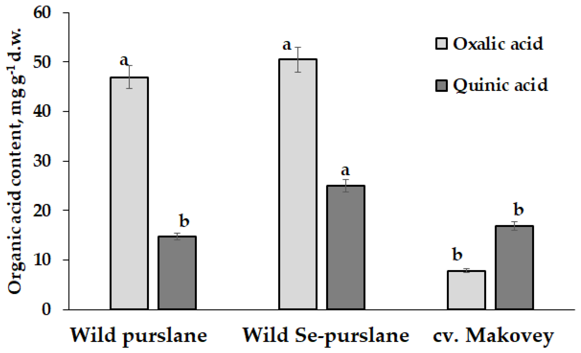
3.1.2. Organic Acids

According to literature reports, the organic acid accumulation in purslane leaves represents one of the most important characteristics of this plant. Among oxalic, quinic, malic and citric acids, the first two compounds predominate in leaves, reflecting both ecological risks connected with high oxalic acid content and high medicinal importance due to the presence of quinic acid [15]. While high oxalic acid content may trigger unfavorable Ca-oxalate precipitation [41], quinic acid is a well-known antidiabetic remedy with significant antibacterial and anticancer activity [42].
The present results indicate significantly lower oxalic acid content in cv. Makovey compared to wild purslane, and similar quinic acid levels (Figure 2). Indeed, while the quinic content in leaves of cultivated purslane was twice higher than the oxalic acid content, the opposite phenomenon was recorded in wild purslane, where the level of oxalic acid exceeded that of quinic acid by 3.2 times.
Moreover, the biofortification of wild purslane with Se led to a 1.7-fold increase in leaf quinic acid, revealing the chance to improve plant medicinal properties and stress tolerance. Literature data indicate the important effect of genotype, soil characteristics and harvesting time on purslane leaf organic acid profile [8,15]. While oxalic acid regulates Ca metabolism, quinic acid is a precursor of chlorogenic acid, thus participating in plant antioxidant defense [43]. In this respect, a beneficial effect of Se on quinic acid accumulation may reflect the improvement in plant immunity and plant resistance against environmental hazards.

3.2. Seeds

3.2.1. Antioxidant Status

Contrary to purslane leaf antioxidant status (Table 4), the total antioxidant activity (AOA) and phenolic content were 2.0- and 3.2-fold higher, respectively, in seeds of wild purslane, compared to cv. Makovey (Table 6). The latter phenomenon agrees with previous findings [44,45], emphasizing the higher biological activity of wild purslane seeds. Such differences may be attributed to higher oxidative stresses of wild purslane, which is known to stimulate secondary metabolite biosynthesis in stress conditions. Indeed, the intensity of lipid peroxidation in seeds of cv. Makovey, reflected in the malonic dialdehyde level, was 2.9-fold lower than in seeds of wild purslane (Table 6). In this respect, Se biofortification of plants provided a significant decrease in the mentioned parameter (Table 6). Furthermore, Se supplementation improved proline biosynthesis in purslane seeds, thus increasing plant tolerance to environmental stresses, in accordance with the known Se effect on the activity of proline synthesis-related enzymes [46].
Overall, the comparison between purslane leaf and seed biochemical characteristics suggests high prospects of purslane seed utilization. Indeed, plant growth and development entirely depend on seed composition and seed nutrients. In this respect, seeds along with nuts, as plant embryos, are highly valued in human nutrition [44]. Epidemiological and clinical trials have demonstrated a beneficial effect of seed and nut consumption on coronary heart disease [47]. The biofortification of seeds with different nutrients was shown to be vital in the production of functional food products with high biological activity [48]. Interestingly, Se supplementation did not significantly affect either AOA or TP levels in wild purslane seeds, but only slightly improved ascorbic acid accumulation and significantly increased Se concentration (Table 6). Taking into account that the proposed daily consumption level of purslane seeds is about 10 g [49], Se-biofortified wild purslane seeds are not able to cause toxicity because they provide only 5.7 µg Se, which is 12.3 times lower than the adequate Se consumption level.
Notably, purslane seeds are most valued for their high lipid content and exclusive composition of seed oil [50]. Indeed, lipid contents in purslane seeds are significantly higher than those of leaves and further of stems [51], which suggests interesting prospects of purslane seed utilization.
In the present work, a tendency to lipid content increase in seeds due to Se supplementation was recorded (Table 6), which is consistent with literature data referring to rice [52], yeast S. cerevisiae [53], chickpea [17] and rapeseed [54].

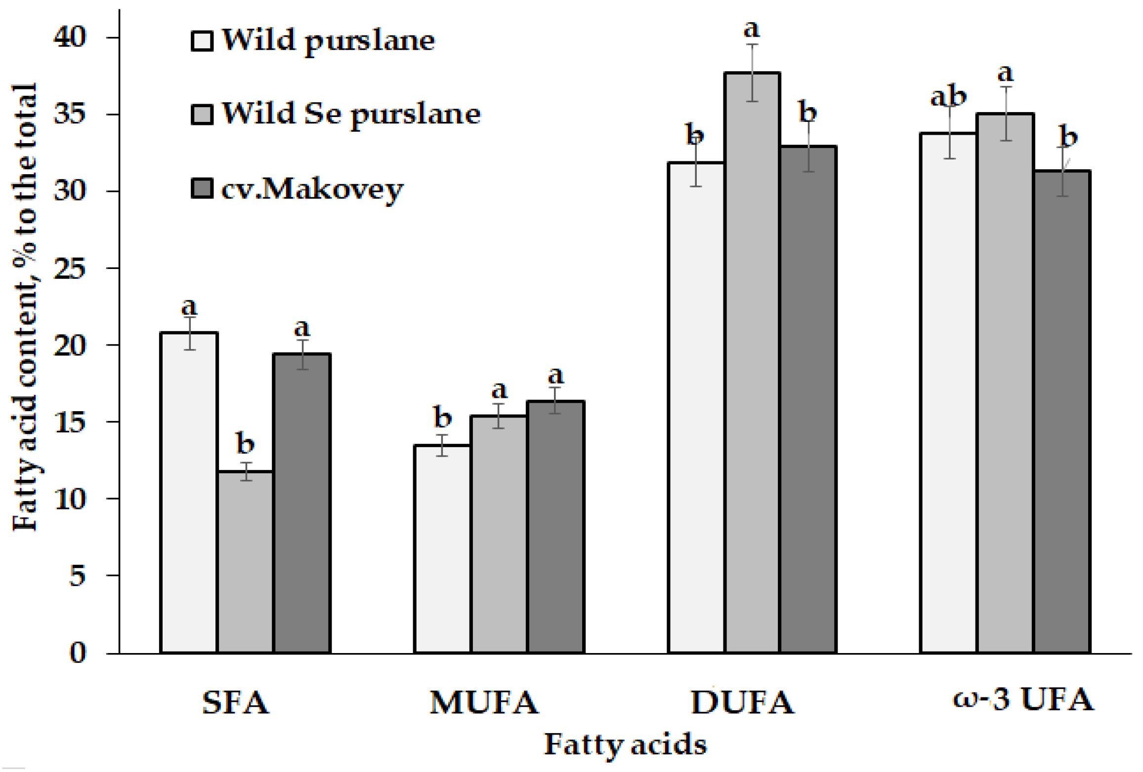
3.2.2. Fatty Acid Profile of Purslane Seed Oil

Purslane seed oil is a fine source of essential fatty acids, including oleic, linoleic and ω-3 linoleic fatty acids [40]. A comparison of seed oil fatty acid profile of wild and cultivated purslane showed similar tendency to the predominant accumulation of C16:0, C18:1, C18:2 and C18:3 fatty acids (Table 7). A previous Iranian report [55] indicated higher levels of palmitic (C16:0) and oleic acids (C18:1) and significantly lower levels of ω-3 linoleic acid (C18:3), which suggests the effect of environmental factors on seed oil composition.
Among different plant species, purslane ranks the first in the content of ω-3 linoleic acid. According to literature data, polyene index (PI—the polyunsaturated/saturated fatty acid ratio), an indicator of oil unsaturation [56], reaches 2.93 in purslane seed oil and is a little bit lower than the corresponding value of soybean oil (3.69) [57]. A high level of PI is known to be extremely favorable for reducing serum cholesterol, decreasing the risk of atherosclerosis and preventing heart diseases [58]. In this respect, the present results indicate that both wild purslane and cv. Makovey seed oils are characterized by high PI, reaching 3.81 and 4.15, respectively, with a two-fold increase in PI value due to Se supplementation (Table 7).
The results of the present research showed a significant reduction in saturated fatty acid content in purslane seeds due to Se biofortification (Figure 3). On the contrary, Se supplementation significantly increased the content of mono- and di-unsaturated fatty acid accumulation but did not affect ω-3 linolenic acid accumulation in seeds. Cultivar Makovey was characterized by increased levels of palmitic acid and mono-unsaturated acids and decreased ω-3 α-linolenic acid levels (Figure 3).
A beneficial effect of Se supplementation on fatty acid accumulation has been intensively investigated in recent years. Indeed, Se biofortification increased C18:1 in S. cerevisiae [53], C18:1 and C18:2 and C16:0 in rice grain [52], and C18:2 and C16:0 in rapeseed oil [54], and it improved the oil stability of seeds in chickpea [17]. The antioxidant properties of Se proved beneficial both in improving the fatty acid profile of purslane seed oil and unsaturated fatty acid stabilization during seed storage.

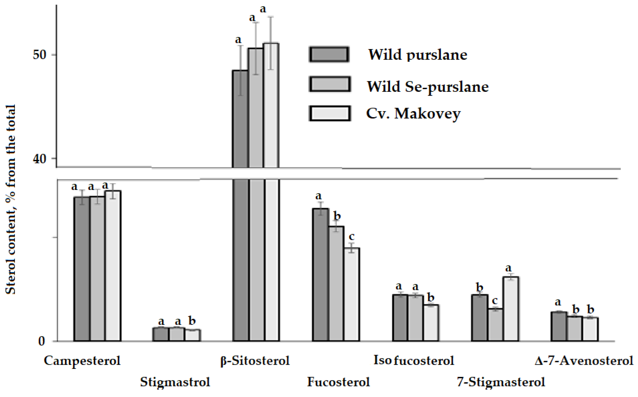
3.3. Phytosterol Composition
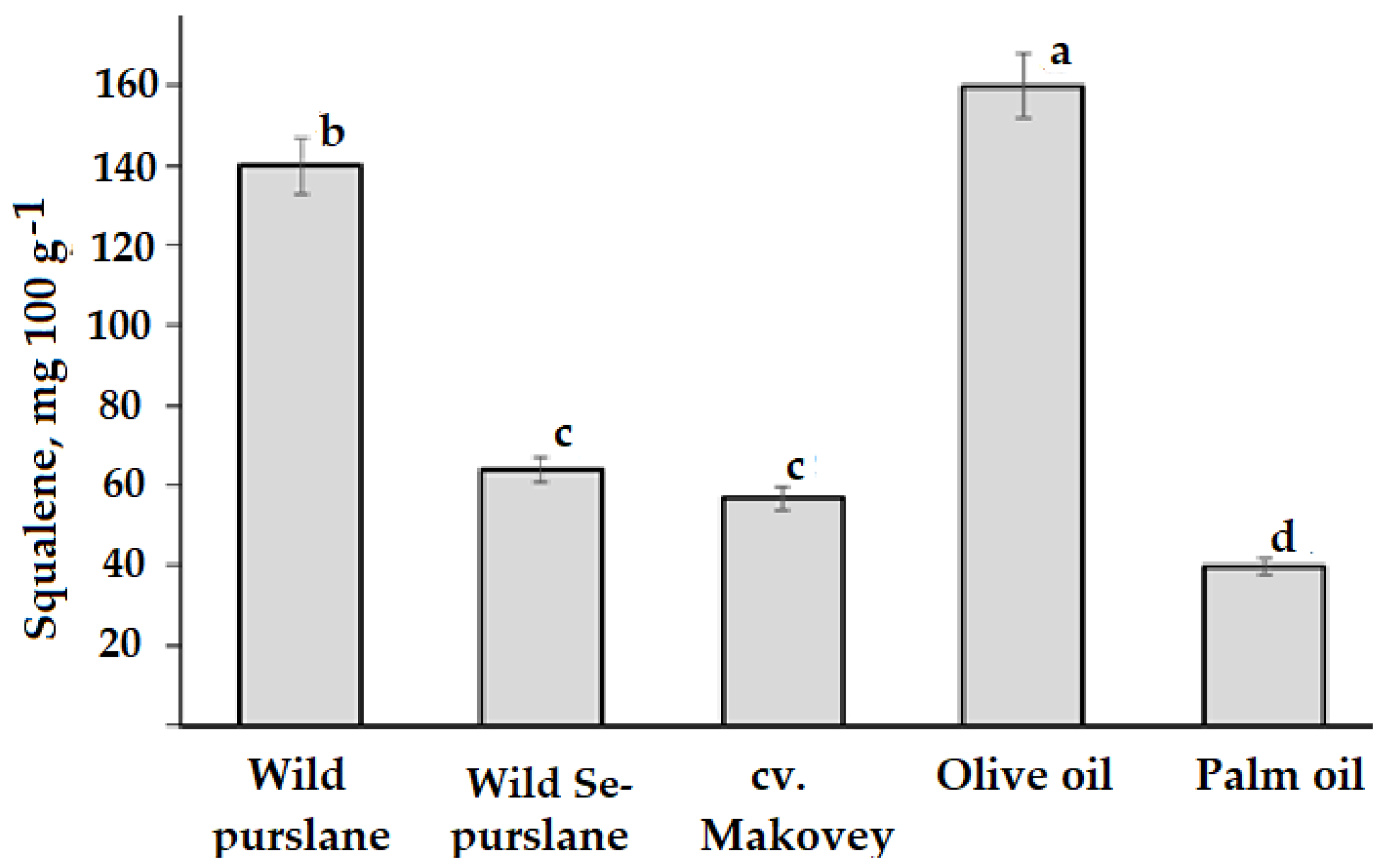
Phytosterols are known as powerful modulators of cholesterol metabolism [59], and their physiological functions include the reduction in cholesterol absorption and decrease in LDL concentrations, participation in antioxidant protection, anti-inflammatory and antipyretic effects, improvement in insulin resistance and lipid metabolism, cancer protection and decrease in atherosclerosis risk. Sitosterol has shown anti-diabetic properties [60].
Though the main phytosterols in higher plants are β-sitosterol, campesterol, and stigmasterol [59], purslane seed oil predominantly contains β-sitosterol > campesterol = fucosterol and relatively low levels of stigmasterol (Table 8, Figure 4). The data presented in Table 8 show the highest levels of campesterol, β-sitosterol and 7-stigmasterol in cv. Makovey but relatively lower levels of isofucosterol. Contrary to the Se effect on fatty acid profile, phytosterol accumulation was only slightly affected by Se supplementation, displaying a slight decrease in fucosterol, 7-stigmasterol and ∆-7-avenosterol levels (Table 8). The total phytosterol accumulation in purslane seed oil was close to that described in olive and walnut oil (234–283 mg 100 g−1), significantly exceeding the value related to coconut (114 mg 100 g−1) but was far lower than the accumulation levels in rapeseed, corn, linseed, sesame and especially wheat germ oil (595–887 and 4240 mg 100 g−1, respectively) [61].

3.4. Squalene

Among the biologically active compounds of medicinal plants, squalene occupies a special place as a precursor for the synthesis of phytosterols [62,63] and as a membrane rigidity regulator [64].
Both squalene and phytosterols participate in plant growth and adaptation to environmental stresses, providing a powerful antioxidant defense [65]. Squalene is a strong antioxidant, antitumor agent and a natural antibiotic with high prospects for utilization in human nutrition, cosmetics and medicine [66]. The present results showed squalene levels in purslane seed oil of 57–140 mg 100 g−1 (Figure 5). These values are much higher than those described for purslane leaves and stems (0.115 mg 100 g−1) [67]. Other natural sources of squalene include olive oil (150–170 mg 100 g−1) [68], palm oil (25–54 mg 100 g−1) [69], and amaranth oil (6000–8000 mg 100 g−1) [70].
Selenium’s effect on squalene accumulation in plants has never previously been studied. Our results demonstrated that Se treatment decreased squalene contents in purslane seed oil by a factor of two (Figure 5).
Taking into account the small differences in the seed production value between control and Se-biofortified wild purslane, the consequences of the mentioned phenomenon may be either the inhibition of sterol biosynthesis from squalene and/or significant changes in cell membrane structure [62,63,64]. A lack of significant Se effect on the sterol level and composition, and insignificant squalene participation in sterol biosynthesis (about 10%) [71] exclude the first hypothesis. On the contrary, the cell membrane structure very likely changed under purslane Se biofortification, as squalene is known to determine membrane rigidity and ion permeability [59,64], while membrane fluidity is directly connected to the unsaturated fatty acid content, which increases with sodium selenate supplementation (Table 7). However, the consequences of the mentioned phenomenon need further investigation, as changes in the squalene contents of purslane seed oil may be connected, at least partly, with changes in squalene levels in other parts of the plant (leaves and roots).

4. Conclusions

The results of the present work indicate significant effects of Se biofortification on purslane yield, seed productivity and seed/leaf quality. The most significant changes were recorded in quinic accumulation in leaves and in squalene/saturated fatty acids levels in seeds. The advantages of wild purslane utilization, compared to common purslane cv. Makovey, include significantly higher levels of seed total antioxidant activity, polyphenol and squalene content, and chlorophyll in leaves of wild purslane. Further investigations are needed to gain further knowledge about the role of Se in the lipid metabolism of purslane.

Author Contributions

Conceptualization, N.G., V.K. and G.C.; data curation, O.C.M.; formal analysis, O.C.M. and N.G.; investigation, N.G., Z.A., Z.K., V.M., M.B., M.M., M.P., A.M. and K.A.; methodology, N.G., G.C. and M.B.; supervision, V.K. and G.C.; validation, G.C.; draft manuscript writing, N.G., M.B. and Z.A.; manuscript revision and final editing, N.G. and G.C. All authors have read and agreed to the published version of the manuscript.

Funding

This research received no external funding.

Data Availability Statement

Data are contained within the article.

Conflicts of Interest

The authors declare no conflicts of interest.

References

  1. Genchi, G.; Lauria, G.; Catalano, A.; Sinicropi, M.S.; Carocci, A. Biological Activity of Selenium and Its Impact on Human Health. Int. J. Mol. Sci. 2023, 24, 2633. [Google Scholar] [CrossRef] [PubMed]
  2. Moulick, D.; Mukherjee, A.; Das, A.; Roy, A.; Majumdar, A.; Dhar, A.; Pattanaik, B.K.; Chowardhara, B.; Ghosh, D.; Upadhyay, M.K.; et al. Selenium—An environmentally friendly micronutrient in agroecosystem in the modern era: An overview of 50-year findings. Ecotoxicol. Environ. Saf. 2024, 270, 115832. [Google Scholar] [CrossRef] [PubMed]
  3. El-Ramady, H.; Faizy, S.E.-D.; Abdalla, N.; Taha, H.; Domokos-Szabolcsy, É.; Fari, M.; Elsakhawy, T.; Omara, A.E.D.; Shalaby, T.; Bayoumi, Y.; et al. Selenium and Nano-Selenium Biofortification for Human Health: Opportunities and Challenges. Soil Syst. 2020, 4, 57. [Google Scholar] [CrossRef]
  4. Chugh, V.; Mishra, V.; Dwivedi, D.V.; Sharma, K.D. Purslane (Portulaca oleracea L.): An underutilized wonder plant with potential pharmacological value. Pharma Innov. J. 2019, 8, 236–246. [Google Scholar]
  5. Yazici, I.; Turkan, A.; Sekmen, H.; Demiral, T. Salinity tolerance of purslane (Portulaca oleracea L.) is achieved by enhanced antioxidative system, lower level of lipid peroxidation and proline accumulation. Environ. Exp. Bot. 2007, 61, 49–57. [Google Scholar] [CrossRef]
  6. Srivastava, R.; Srivastava, V.; Singh, A. Multipurpose Benefits of an Underexplored Species Purslane (Portulaca oleracea L.): A Critical Review. Environ. Manag. 2023, 72, 309–320. [Google Scholar] [CrossRef]
  7. Agil, R.; Gilbert, C.; Tavakoli, H.; Hosseinian, F. Redefining Unusable Weeds to Beneficial Plants: Purslane as a Powerful Source of Omega-3 for the Future. J. Food Res. 2015, 4, 39–47. [Google Scholar] [CrossRef]
  8. Uddin, M.K.; Juraimi, A.S.; Hossain, M.S.; Nahar, M.A.; Ali, M.E.; Rahman, M.M. Purslane Weed (Portulaca oleracea): A Prospective Plant Source of Nutrition, Omega-3 Fatty Acid, and Antioxidant Attributes. Sci. World J. 2014, 2014, 951019. [Google Scholar] [CrossRef]
  9. Masoodi, M.H.; Ahmad, B.; Mir, S.R.; Zargar, B.A.; Tabasum, N. Portulaca oleracea L. A Review. J. Pharm. Res. 2011, 4, 3044–3048. [Google Scholar]
  10. Simopoulos, A.P.; Salem, N. Purslane: A terrestrial source of omega-3 fatty acids. N. Engl. J. Med. 1986, 315, 833. [Google Scholar]
  11. Simopoulos, A.P. Omega-3 fatty acids and antioxidants in edible wild plants. Biol. Res. 2004, 37, 263–277. [Google Scholar] [CrossRef]
  12. Ojah, E.O.; Oladele, E.O.; Chukwuemeka, P. Phytochemical and antibacterial properties of root extracts from Portulaca oleracea Linn. (Purslane) utilised in the management of diseases in Nigeria. J. Med. Plant Econ. Dev. 2021, 5, a103. [Google Scholar] [CrossRef]
  13. Mishra, V.; Chugh, V.; Dwivedi, S.V.; Sharma, K.D. Food and nutraceuticals value of purslane (Portulaca oleracea L.): An overview. Pharma Innov. J. 2020, 9, 419–424. [Google Scholar]
  14. Rayan, A.M.; Swailam, H.M.; Hamed, Y.S. Composition, Structure, and Techno-Functional Characteristics of the Flour, Protein Concentrate, and Protein Isolate from Purslane (Portulaca oleracea L.) Seeds. Plant Food Hum. Nutr. 2023, 7, 117–123. [Google Scholar] [CrossRef]
  15. Petropoulos, S.A.; Fernandes, Â.; Dias, M.I.; Vasilakoglou, I.B.; Petrotos, K.; Barros, L.; Ferreira, I.C.F.R. Nutritional Value, Chemical Composition and Cytotoxic Properties of Common Purslane (Portulaca oleracea L.) in Relation to Harvesting Stage and Plant Part. Antioxidants 2019, 8, 293. [Google Scholar] [CrossRef]
  16. Golubkina, N.; Kharchenko, V.; Moldovan, A.; Antoshkina, M.; Ushakova, O.; Sękara, A.; Stoleru, V.; Murariu, O.C.; Tallarita, A.V.; Sannino, M.; et al. Effect of Selenium and Garlic Extract Treatments of Seed-Addressed Lettuce Plants on Biofortification Level, Seed Productivity and Mature Plant Yield and Quality. Plants 2024, 13, 1190. [Google Scholar] [CrossRef]
  17. Guardado-Félix, D.; Serna-Saldivar, S.O.; Gutiérrez-Uribe, J.A.; Chuck-Hernández, C. Selenium in Germinated Chickpea (Cicer arietinum L.) Increases the Stability of Its Oil Fraction. Plants 2019, 8, 113. [Google Scholar] [CrossRef]
  18. D’Amato, R.; Regni, L.; Falcinelli, B.; Mattioli, S.; Benincasa, P.; Dal Bosco, A.; Pacheco, P.; Proietti, P.; Troni, E.; Santi, C.; et al. Current Knowledge on Selenium Biofortification to Improve the Nutraceutical Profile of Food: A Comprehensive Review. J. Agric. Food Chem. 2020, 68, 4075–4097. [Google Scholar] [CrossRef] [PubMed]
  19. Golubkina, N.; Amagova, Z.; Matsadze, V. Monitoring of the selenium content in the environment of the Chechen repubic. Adv. Mod. Sci. 2017, 2, 28–33. (In Russian) [Google Scholar]
  20. Cipriano, P.E.; da Silva, R.F.; de Lima, F.R.D.; de Oliveira, C.; de Lima, A.B.; Celante, G.; Dos Santos, A.A.; Archilha, M.V.L.R.; Pinatto-Botelho, M.F.; Faquin, V.; et al. Selenium biofortification via soil and its effect on plant metabolism and mineral content of sorghum plants. J. Food Comp. Anal. 2022, 109, 104505. [Google Scholar] [CrossRef]
  21. Lichtenthaler, H.K. Chlorophylls and carotenoids: Pigments of photosynthetic bio-membranes. Methods Enzymol. 1987, 148, 350–382. [Google Scholar]
  22. Alfthan, G.V. A micromethod for the determination of selenium in tissues and biological fluids by single-test-tube fluorimetry. Anal. Chim. Acta 1984, 165, 187–194. [Google Scholar] [CrossRef]
  23. Golubkina, N.A.; Kekina, H.G.; Molchanova, A.V.; Antoshkina, M.S.; Nadezhkin, S.; Soldatenko, A.V. Plants Antioxidants and Methods of Their Determination; Infra-M: Moscow, Russia, 2020; (In Russian). [Google Scholar] [CrossRef]
  24. AOAC Association Official Analytical Chemists. The Official Methods of Analysis of AOAC International; 22 ‘Vitamin C’; AOAC: Rockville, MD, USA, 2012. [Google Scholar]
  25. Ouertani, R.N.; Abid, G.; Karmous, C.; Chikha, M.B.; Boudaya, O.; Mahmoudi, H.; Mejri, S.; Jansen, K.; Ghorbel, A. Evaluating the contribution of osmotic and oxidative stress components on barley growth under salt stress. AoB Plants 2021, 13, plab034. [Google Scholar] [CrossRef] [PubMed]
  26. Guide for the Evaluation of Quality and Safety of Food Biologically Active Additives; Organic Acids Determination; Health Ministry of RF P 4.1.1672-03: Moscow, Russia, 2004; pp. 109–111.
  27. GOST 31663-2012; Vegetable Oils and Animal Fats. Determination of Methyl Esters of Fatty Acids by Gas Chromatography Method. ISO: Moscow, Russia, 2012.
  28. Golay, P.A.; Moulin, J. Determination of labeled fatty acids content in milk products, infant formula, and adult/pediatric nutritional formula by capillary gas chromatography: Collaborative study, final action 2012.13. J. AOAC Int. 2012, 99, 210–222. [Google Scholar] [CrossRef] [PubMed]
  29. Golay, P.A.; Dong, Y. Determination of labeled fatty acids content in milk products, infant formula, and adult/pediatric nutritional formula by capillary gas chromatography: Single-Laboratory Validation, First Action 2012.13. J. AOAC Int. 2015, 98, 1679–1696. [Google Scholar] [CrossRef] [PubMed]
  30. Budge, S.M.; Barry, C. Determination of squalene in edible oils by transmethylation and GC analysis. MethodsX 2018, 6, 15–21. [Google Scholar] [CrossRef]
  31. ISO 12228-1-2014; Determination of Individual and Total Sterols Contents—Gas Chromatographic Method—Part 1: Animal and Vegetable Fats and Oils. ISO: Moscow, Russia, 2014.
  32. National Institute of Standards and Technology. NIST Chemistry WebBook, SRD 69. Available online: https://webbook.nist.gov/chemistry/ (accessed on 26 February 2024).
  33. Goad, L.J.; Akihisa, T. Analysis of Sterols; Blackie Academic and Professional: London, UK, 1997. [Google Scholar]
  34. The Lipid Web. Available online: https://www.lipidmaps.org/resources/lipidweb/lipidweb_html/ms/others/others-arch/index1.htm#sterol (accessed on 26 February 2024).
  35. Cunha, S.S.; Fernandes, J.O.; Oliveira, M.B.P.P. Quantification of free and esterified sterols in Portuguese olive oils by solid-phase extraction and gas chromatography–mass spectrometry. J. Chromatogr. A 2006, 1128, 220–227. [Google Scholar] [CrossRef] [PubMed]
  36. Li, T.S.C.; Beveridge, T.H.J.; Drover, J.C.G. Phytosterol content of sea buckthorn (Hippophae rhamnoides L.) seed oil: Extraction and identification. Food Chem. 2007, 101, 1633–1639. [Google Scholar] [CrossRef]
  37. Semeniuc, C.A.; Cardenia, V.; Mandrioli, M.; Muste, S.; Borsari, A.; Rodriguez-Estrada, M.T. Stability of flavored phytosterol-enriched drinking yogurts during storage as affected by different packaging materials. J. Sci. Food Agric. 2016, 96, 2782–2787. [Google Scholar] [CrossRef]
  38. Márquez, V.G.; Moreno, Á.M.; Mendoza, A.B.; Macías, J.M. Ionic Selenium and Nanoselenium as Biofortifiers and Stimulators of Plant Metabolism. Agronomy 2020, 10, 1399. [Google Scholar] [CrossRef]
  39. Sdouga, D.; Branca, F.; Kabtni, S.; Di Bella, M.C.; Trifi-Farah, N.; Marghali, S. Morphological Traits and Phenolic Compounds in Tunisian Wild Populations and Cultivated Varieties of Portulaca oleracea L. Agronomy 2020, 10, 948. [Google Scholar] [CrossRef]
  40. Liu, H.; Xiao, C.; Qiu, T.; Deng, J.; Cheng, H.; Cong, X.; Cheng, S.; Rao, S.; Zhang, Y. Selenium Regulates Antioxidant, Photosynthesis, and Cell Permeability in Plants under Various Abiotic Stresses: A Review. Plants 2022, 12, 44. [Google Scholar] [CrossRef]
  41. Wang, X.; Zhang, X.; Wang, L.; Zhang, R.; Zhang, Y.; Cao, L. Purslane-induced oxalate nephropathy: Case report and literature review. BMC Nephrol. 2023, 24, 207. [Google Scholar] [CrossRef]
  42. Benali, T.; Bakrim, S.; Ghchime, R.; Benkhaira, N.; El Omari, N.; Balahbib, A.; Bouyahya, A. Pharmacological insights into the multifaceted biological properties of quinic acid. Biotechnol. Genet. Eng. Rev. 2022, 1–30. [Google Scholar] [CrossRef]
  43. Soviguidi, D.R.J.; Pan, R.; Liu, Y.; Rao, L.; Zhang, W.; Yang, X. Chlorogenic acid metabolism: The evolution and roles in plant response to abiotic stress. Phyton Int. J. Exp. Bot. 2022, 91, 239–255. [Google Scholar] [CrossRef]
  44. Cervera-Mata, A.; Sahu, P.K.; Chakradhari, S.; Sahu, Y.K.; Patel, K.S.; Singh, S.; Towett, E.K.; Martín-Ramos, P.; Quesada-Granados, J.J.; Rufián-Henares, J.A. Plant seeds as source of nutrients and phytochemicals for the Indian population. Int. J. Food Sci. Technol. 2022, 57, 525–532. [Google Scholar] [CrossRef]
  45. Jalali Mousavi, S.R.; Niazmand, R.; Shahidi Noghabi, M. Noghabi Antioxidant Activity of Purslane (Portulaca oleracea L.) Seed Hydro-alcoholic Extract on the Stability of Soybean Oil. J. Agric. Sci. Technol. 2015, 17, 1473–1480. [Google Scholar]
  46. Iqbal, M.; Hussain, I.; Liaqat, H.; Ashraf, M.A.; Rasheed, R.; Rehman, A.U. Exogenously applied selenium reduces oxidative stress and induces heat tolerance in spring wheat. Plant Physiol. Biochem. 2015, 94, 95–103. [Google Scholar] [CrossRef]
  47. Ros, E.; Hu, F.B. Consumption of plant seeds and cardiovascular health: Epidemiological and clinical trial evidence. Circulation 2013, 128, 553–565. [Google Scholar] [CrossRef]
  48. Khan, M.A.; Castro-Guerrero, N.; Mendoza-Cozatl, D.G. Moving toward a precise nutrition: Preferential loading of seeds with essential nutrients over non-essential toxic elements. Front. Plant Sci. 2014, 5, 51. [Google Scholar] [CrossRef]
  49. Esmaillzadeh, A.; Zakizadeh, E.; Faghihimani, E.; Gohari, M.; Jazayeri, S. The effect of purslane seeds on glycemic status and lipid profiles of persons with type 2 diabetes: A randomized controlled cross-over clinical trial. J. Res. Med. Sci. 2015, 20, 47–53. [Google Scholar] [PubMed]
  50. Petropoulosa, S.A.; Fernandesb, A.; Arampatzisa, D.A.; Tsiropoulosa, N.G.; Petrovićc, J.; Sokovićc, M.; Barrosb, L.; Ferreirab, I.C.F.R. Seed oil and seed oil byproducts of common purslane (Portulaca oleracea L): A new insight to plant-based sources rich in omega-3 fatty acids. LWT-Food Sci. Technol. 2020, 123, 109099. [Google Scholar] [CrossRef]
  51. Desta, M.; Molla, A.; Yusuf, Z. Characterization of physico-chemical properties and antioxidant activity of oil from seed, leaf and stem of purslane (Portulaca oleracea L.). Biotechnol. Rep. 2020, 27, e00512. [Google Scholar] [CrossRef] [PubMed]
  52. Lidon, F.C.; Oliveira, K.; Ribeiro, M.M.; Pelica, J.; Pataco, I.; Ramalho, J.C.; Leitão, A.E.; Almeida, A.S.; Campos, P.S.; Ribeiro-Barros, A.I.; et al. Selenium biofortification of rice grains and implications on macronutrients quality. J. Cereal Sci. 2018, 81, 22–29. [Google Scholar] [CrossRef]
  53. Kieliszek, M.; Błażejak, S.; Bzducha-Wróbel, A. Effect of Selenium on Lipid and Amino Acid Metabolism in Yeast Cells. Biol. Trace Elem. Res. 2019, 187, 316–327. [Google Scholar] [CrossRef] [PubMed]
  54. Davoudi, B.; Mirshekari, A.; Shirani-Rad, F.; Farahvash, V. Rashidi Effect of selenium foliar application on oil yield, fatty acid composition and glucosinolate content of rapeseed cultivars under late-season thermal stress. Oilseeds Fats Crops Lipids (OCL) 2019, 26, 43. [Google Scholar] [CrossRef]
  55. Mousavi, S.R.J.; Niazmand, R. Fatty Acids Composition and Oxidation Kinetic Parameters of Purslane (Portulaca oleracea) Seed Oil. Agric. Res. 2017, 6, 421–426. [Google Scholar] [CrossRef]
  56. Mahla, H.R.; Rathore, S.S.; Venkatesan, K.; Sharma, R. Analysis of fatty acid methyl esters and oxidative stability of seed purpose watermelon (Citrullus lanatus) genotypes for edible oil. J. Food Sci. Technol. 2018, 55, 1552–1561. [Google Scholar] [CrossRef]
  57. Nehdi, I. Characteristics, chemical composition and utilization of Albizia julibrissin seed oil. Ind. Crops Prod. 2011, 33, 30–34. [Google Scholar] [CrossRef]
  58. Oomah, B.; Dumon, D.; Cardador-Martinez, A.; Godfrey, D. Characteristics of Echinacea seed oil. Food Chem. 2006, 96, 304–312. [Google Scholar] [CrossRef]
  59. Li, X.; Xin, Y.; Mo, Y.; Marozik, P.; He, T.; Guo, H. The Bioavailability and Biological Activities of Phytosterols as Modulators of Cholesterol Metabolism. Molecules 2022, 27, 523. [Google Scholar] [CrossRef] [PubMed]
  60. Babu, S.; Jayaraman, S. An update on β-sitosterol: A potential herbal nutraceutical for diabetic management. Biomed. Pharmacother. 2020, 131, 110702. [Google Scholar] [CrossRef] [PubMed]
  61. Bai, G.; Ma, C.; Chen, X. Phytosterols in edible oil: Distribution, analysis and variation during processing. Grain Oil Sci. Technol. 2021, 4, 33–44. [Google Scholar] [CrossRef]
  62. Lozano-Grande, M.A.; Gorinstein, S.; Espitia-Rangel, E.; Dávila-Ortiz, G.; Martínez-Ayala, A.L. Plant Sources, Extraction Methods, and Uses of Squalene. Int. J. Agron. 2018, 2018, 1829160. [Google Scholar] [CrossRef]
  63. Piironen, V.; Lindsay, D.G.; Miettinen, T.A.; Toivo, J. Plant sterols: Biosynthesis, biological function and their importance to human nutrition. J. Sci. Food Agric. 2000, 80, 939–966. [Google Scholar] [CrossRef]
  64. LoRicco, J.G.; Hoffmann, I.; Caliò, A.; Peters, J. The membrane regulator squalane increases membrane rigidity under high hydrostatic pressure in archaeal membrane mimics. Soft Matter 2023, 19, 6280–6286. [Google Scholar] [CrossRef] [PubMed]
  65. Du, Y.; Fu, X.; Chu, Y.; Wu, P.; Liu, Y.; Ma, L.; Tian, H.; Zhu, B. Biosynthesis and the Roles of Plant Sterols in Development and Stress Responses. Int. J. Mol. Sci. 2022, 23, 2332. [Google Scholar] [CrossRef]
  66. Popa, O.; Băbeanu, N.E.; Popa, I.; Niță, S.; Dinu-Pârvu, C.E. Methods for obtaining and determination of squalene from natural sources. BioMed Res. Int. 2015, 2015, 367202. [Google Scholar] [CrossRef] [PubMed]
  67. Acedo, J.Z.; Reyes, C.T.; Rodriguez, E.B. Health-promoting lipids from purslane (Portulaca oleraceae). Isolation, characterization, quantification and in vivo assay of angiogenic activity. Philipp. Agric. Sci. 2012, 95, 327–334. [Google Scholar]
  68. Akgün, N.A. Separation of squalene from olive oil deodorizer distillate using supercritical fluids. Eur. J. Lipid Sci. Technol. 2011, 113, 1558–1565. [Google Scholar] [CrossRef]
  69. Posada, L.R.; Shi, J.; Kakuda, Y.; Xue, S.J. Extraction of tocotrienols from palm fatty acid distillates using molecular distillation. Sep. Purif. Technol. 2007, 57, 220–229. [Google Scholar] [CrossRef]
  70. Sun, H.; Wiesenborn, D.; Rayas-Duarte, P.; Mohamed, A.; Hagen, K. Bench-scale processing of amaranth seed for oil. J. Am. Oil Chem. Soc. 1995, 72, 1551–1555. [Google Scholar] [CrossRef]
  71. Gilmore, S.F.; Uao, A.I.; Tietel, Z.; Facciotti, K.T.; Parikh, A.N. Role of Squalene in the Organization of Monolayers Derived from Lipid Extracts of Halobacterium salinarum. Langmuir 2013, 29, 7922–7930. [Google Scholar] [CrossRef] [PubMed]
Figure 1. GC-FID chromatogram of trimethylsilyl derivatives of purslane sterols. 1: cholestanol (internal standard); 2: campesterol, 3: stigmasterol, 4: β-sitosterol; 5: isofucosterol; 6: Δ7-stigmastenol; 7: Δ7-avenasterol.
Horticulturae 10 00708 g001
Figure 2. Oxalic and quinic acid content in purslane leaves. For each organic acid, values with the same letters do not differ statistically according to Duncan’s test at p < 0.05. Bars indicate the standard deviation.
Horticulturae 10 00708 g002
Figure 3. Fatty acid profile of cultivated parsnip and wild accession fortified and unfortified with Se. SFA—saturated fatty acids; MUFA—mono-unsaturated fatty acids; DUFA—di-unsaturated fatty acids; ω-3 UFA—ω-3 unsaturated fatty acids. For each group of fatty acids, values with the same letters do not differ significantly according to Duncan’s test at p < 0.05. Bars indicate the standard deviation.
Horticulturae 10 00708 g003
Figure 4. Sterol profile of purslane seed oil. Within each compound, values with the same letters do not differ significantly according to Duncan’s test at p < 0.05. Bars indicate the standard deviation.
Horticulturae 10 00708 g004
Figure 5. Squalene content in seeds of wild and cultivated purslane, olive and palm oil. Values with the same letters do not differ statistically according to Duncan’s test at p < 0.05. Bars indicate the standard deviation.
Horticulturae 10 00708 g005
Table 1. Mean monthly temperatures and rainfall during the experiment in 2022–2023.
Month20222023
Mean Temperature
(°C)
Rainfall
(mm)
Mean Temperature
(°C)
Rainfall
(mm)
May16.697.516.566.0
June21.731.821.2131.0
July23.940.323.780.0
August23.69.026.112.0
September18.131.021.084.0
Table 2. M/z ions, detected and used for sterol trimethylsilyl derivative identification.
Peak NoCompoundMM-15 1M-90 2M-105M-129 3M-122 3M-SC-2H 4Other Ions
1Cholestanol (internal standard)460445370355---215; 75
2Kampesterol472457382367343261-129
3Stigmasterol--394-355--129
4β-Sitosterol486471396381357275-129
5Isofucosterol484469394379355273-386; 296; 129
6Δ7-Stigmastenol486471396381-275343255; 75
7Δ7-Avenasterol-----273343386; 281; 253; 213
1 Ion is formed by the loss of a methyl group; 2 ion is formed by the loss of C3H10OSi fragment; 3 ion is formed as a result of typical fragmentation of Δ5-sterol derivatives; 4 ion is formed by the loss of the side chain (SC), and two hydrogen atoms are typical for Δ7-sterol derivative fragmentation.
Table 3. Retention time (Rt, min) and retention indexes (RI).
Peak NumberCompoundRt, minRIRI in a Mixture
of Standards
Values According to [37]
1Cholestanol (internal standard)20.992314431483169
2Kampesterol23.480323832423253
3Stigmasterol24.316326932743274
4β-Sitosterol25.841332733293348
5Isofucosterol26.2753343--
6Δ7-Stigmastenol27.3003382--
7Δ7-Avenasterol27.7473398--
Table 4. Biometrical characteristics of purslane.
ParameterWild PurslaneWild Se-Purslanecv. Makovey
Plant biomass (g)129 ± 10 b153 ± 11 a110 ± 10 b
Stem length (cm)17.8 ± 1.1 b23.1 ± 1.9 a18.0 ± 1.0 b
Number of stems per plant20.5 ± 1.7 b24.6 ± 1.0 a16.0 ± 1.0 b
Plant diameter (cm)37.4 ± 3.5 b46.9 ± 4.2 a38.0 ± 3.0 b
Weight of 1000 seeds (g)0.123 ± 0.01 b0.118 ± 0.01 b0.336 ± 0.01 a
Seed weight per plant (g)1.79 ab1.84 a1.52 b
Seed productivity (number of seeds per plant)14,553 a15,593 a4524 b
Along each line, values with the same letters do not differ statistically according to Duncan’s test at p < 0.05.
Table 5. Biochemical characteristics of purslane leaves.
ParameterWild PurslaneWild Se-Purslanecv. Makovey
AA (mg 100 g−1 f.w.)25.5 ± 1.5 b30.5 ± 2.5 a24.8 ± 1.9 b
AOA (mg GAE g−1 d.w.)47.7 ± 4.0 a43.2 ± 3.4 a45.3 ± 3.9 a
TP (mg GAE g−1 d.w.)22.4 ± 2.2 a18.1 ± 1.8 a19.3 ± 1.7 a
Se (µg kg−1 d.w.)108 ± 10 b621 ± 56 a93 ± 9 b
Chl a (mg g−1 f.w.)0.66 ± 0.03 b0.84 ± 0.06 a0.51 ± 0.03 c
Chl b (mg g−1 f.w.)0.48 ± 0.03 b0.76 ± 0.06 a0.24 ± 0.02 c
Total Chl (mg g−1 f.w.)1.14 ± 0.1 b1.60 ± 0.1 a0.75 ± 0.04 c
Carotene (mg g−1 f.w.)0.069 ± 0.01 a0.087 ± 0.01 a0.043 ± 0.01 b
Chl a/chl b1.381.112.125
Chl/carotene14.418.417.4
AA: ascorbic acid; AOA: total antioxidant activity; TP: total polyphenols; Chl: chlorophyll. Along each line, values with the same letters do not differ statistically according to Duncan’s test at p < 0.05.
Table 6. Biochemical composition of purslane seeds.
ParameterWild PurslaneWild Se-Purslanecv. Makovey
AOA (mg GAE g−1 d.w.)19.5 ± 1.7 a17.0 ± 1.6 a9.9 ± 1.0 b
TP (mg GAE g−1 d.w.)9.3 ± 0.9 a10.5 ± 0.9 a2.9 ± 0.2 b
AA (mg 100 g−1 d.w.)4.3 ± 0.4 b6.0 ± 0.5 a4.7 ± 0.4 b
Se (µg kg−1 d.w.)87 ± 8.0 b570 ± 50 a78 ± 7.1 b
Total lipids (g 100 g−1)14.8 ± 1.3 a16.2 ± 1.4 a17.4 ± 1.5 a
Pro (mg g−1 d.w.)0.188 ± 0.01 b0.235 ± 0.02 a0.236 ± 0.02 a
MDA (mg g−1 d.w.)0.200 ± 0.02 a0.086 ± 0.008 b0.070 ± 0.007 b
MDA—malonic dialdehyde, Pro—proline, AOA—total antioxidant activity, TP—total polyphenols, AA—ascorbic acid. Along each line, values with the same letters do not differ statistically according to Duncan’s test at p < 0.05.
Table 7. Fatty acid composition of purslane seeds (mg g−1).
Fatty AcidWild
Purslane
Wild
Se Purslane
cv. MakoveyLiterature Data
[55]
Myristic 14:01 a1 a1 a0.7
Pentadecanic 15:01 a0 b0 b0.2
Palmitic 16:0103 b72 c134 a164.3
Margaric 17:00002.2
Stearic 18:026 b23 b32 a30.9
Arachidic 20:064 a5 b6 b1.4
Behenic 22:04 a4 a2 b9.4
Lignoceri 24:03 a3 a2 b1.5
Saturated acids202108177210.9
Hexadecenic 16:11 a1 a1 a4.8
Palmitoleinic 16:1 cis1 a1 a1 a
Elaidic18:1 9-trans1 a1 a0 b
Oleic18:1 9 cis109 a116 a127 a163.7
Vaccenic18:1 11-trans18 a20 a19 a
Gondoic (sum of isomers) 20:1121
Mono-unsaturated131141149168.5
Cis, trans linoleic 18:2 9-cis, 12-trans111
Linoleic 18:2309 b344 a299 b336.3
Di-unsaturated acids310345300336.5
Octadecatrienoic 18:3 cis-9,
trans-12 Trans-15 + 18:3 cis-9.
Cis-12. Trans-15
3 a2 b2 b14.8
α-Linolenic18:3 ω-3325 a320 a283 a267.7
PUSFA 328322285282.5
Total lipids (g 100 g−1)14.816.217.4-
P/S ratio (PI index)3.817.484.152.93
PUSFA: polyunsaturated fatty acids; P/S: polyene index. Along each line, values with the same letters do not differ statistically according to Duncan’s test at p < 0.05.
Table 8. Sterol profile of purslane seed oil.
Sterols (mg 100 g−1)Wild PurslaneWild Se-Purslanecv. Makovey
Campesterol28.5 ± 0.9 b27.8 ± 1.1 b34.2 ± 1.0 a
Stigmasterol2.6 ± 0.1 a2.6 ± 0.1 a2.6 ± 0.1 a
β-Sitosterol99.2 ± 3.0 b100.8 ± 4.0 b120.4 ± 3.6 a
Fucosterol26.1 ± 0.8 a22.1 ± 0.9 b21.2 ± 0.6 b
Isofucosterol9.1 ± 0.3 a8.7 ± 0.3 ab8.2 ± 0.2 b
7-Stigmasterol9.2 ± 0.3 b6.2 ± 0.2 c14.5 ± 0.4 a
∆-7-Avenosterol5.9 ± 0.2 a4.8 ± 0.2 b5.5 ± 0.2 a
Total204.4 ± 6.1 b199.2 ± 8.0 b235.5 ± 7.1 a
Along each line, values with the same letters do not differ statistically according to Duncan’s test at p < 0.05.
Disclaimer/Publisher’s Note: The statements, opinions and data contained in all publications are solely those of the individual author(s) and contributor(s) and not of MDPI and/or the editor(s). MDPI and/or the editor(s) disclaim responsibility for any injury to people or property resulting from any ideas, methods, instructions or products referred to in the content.

