Article

Association of Microbiome Diversity with Disease Symptoms in Brassica oleracea Leaves

by
Héctor Martín-Cardoso
1,
Víctor M. González-Miguel
1,
Luis Soler-López
1,
Sonia Campo
2,* and
Blanca San Segundo
1,3,*
1
Centre for Research in Agricultural Genomics (CRAG) CSIC-IRTA-UAB-UB, Campus Universitat Autònoma de Barcelona (UAB), Bellaterra (Cerdanyola del Vallès), C/de la Vall Moronta, CRAG Building, 08193 Barcelona, Spain
2
Fundació Miquel Agustí/HorPTA, Department of Agri-Food Engineering and Biotechnology, Universitat Politècnica de Catalunya (UPC)-BarcelonaTech, Campus del Baix Llobregat–Edifici D4. C/Esteve Terrades 8, 08860 Barcelona, Spain
3
Consejo Superior de Investigaciones Científicas (CSIC), 08193 Barcelona, Spain
*
Authors to whom correspondence should be addressed.
Horticulturae 2024, 10(7), 765; https://doi.org/10.3390/horticulturae10070765
Submission received: 20 June 2024 / Revised: 10 July 2024 / Accepted: 12 July 2024 / Published: 19 July 2024
(This article belongs to the Section Plant Pathology and Disease Management (PPDM))

Abstract

:
Cabbage (Brassica oleracea), a crop of major economic importance worldwide, is affected by numerous diseases, which are caused by a wide range of microorganisms, including fungi, oomycetes, bacteria, and viruses, which lead to important losses in yield and quality. The increasing availability of reference genomes of plant-associated microbes together with recent advances in metagenomic approaches provide new opportunities to identify microbes linked to distinct symptomatology in Brassica leaves. In this study, shotgun metagenomics was used to investigate the microbial community in leaves of B. oleracea plants from agricultural farmlands. Compared with conventional techniques based on culture-based methods, whole-genome shotgun sequencing allows the reliable identification of the microbial population inhabiting a plant tissue at the species level. Asymptomatic and symptomatic leaves showing different disease symptoms were examined. In the asymptomatic leaves, Xanthomonas species were the most abundant taxa. The relative abundance of bacterial and fungal communities varied depending on disease symptoms on the leaf. The microbiome of the leaves showing mild to severe levels of disease was enriched in bacterial populations (Sphingomonas, Methylobacterium, Paracoccus) and to a lesser degree in some fungal taxa, such as Alternaria and Colletotrichum (e.g., in leaves with high levels of necrotic lesions). Sclerotinia species were highly abundant in severely damaged leaves (S. sclerotium, S. trifolium, S. bolearis), followed by Botrytis species. The common and specific bacterial and fungal species associated to disease symptoms were identified. Finally, the analysis of the gene functions in the metagenomic data revealed enrichment in carbohydrate-active enzymes potentially involved in pathogenicity, whose distribution also varied among disease severity groups. Understanding the B. oleracea leaf microbiome in agricultural ecosystems will pave the way for the efficient management of diseases in this crop.

1. Introduction

The genus Brassica, a member of the family Brassicaceae (previously named Cruciferae), is an economically and scientifically relevant family of dicotyledonous plants (http://www.theplantlist.org/, accessed on 1 January 2024). It contains an important number of crop species that provide edible roots, leaves, stems, buds, flowers, and seeds worldwide. Vegetable species belonging to the Brassica genus are B. oleracea (cabbage, cauliflower, broccoli, Brussels sprouts, kale), B. rapa (turnip, Chinese cabbage), B. napus (rapeseed), B. carinata, and B. juncea (mustards). Among them, B. oleracea, B. napus, and B. rapa are the most widely grown Brassica species. In addition to its agronomical relevance, the Brassicaceae family includes the essential model plant Arabidopsis thaliana [1].
Brassica plants are susceptible to diseases caused by a wide range of microorganisms, such as fungi, oomycetes, bacteria and viruses, which cause important yield losses in both quality and quantity [2]. The most important fungal or oomycete pathogens affecting Brassica species (and diseases they cause) are Sclerotinia sclerotiorum (white mold); Alternaria spp. (Alternaria leaf spot), Hyaloperonospora parasitica (downy mildew), Erysiphe cruciferarum (powdery mildew), Rhizoctonia solani (wirestem), Pythium spp. (damping-off), Fusarium oxysporum (Fusarium wilt), Mycosphaerella brassicicola (ring spot), Plasmodiophora brassicae (clubroot) and Leptosphaeria maculans (blackleg), Colletotrichum spp. (anthracnose), and Phytophthora spp. (Phytophthora root rot). Of them, Alternaria leaf spot, predominantly caused by A. brassicicola, A. brassicae, and occasionally A. alternata, is among the most impactful fungal disease in cabbage (B. oleracea), resulting in important yield losses [3]. Regarding the bacterial pathogens, Xanthomonas campestris (black rot), Erwinia spp. (soft rot), and Pseudomonas spp. (bacterial soft rot) are considered the most impactful bacterial pathogens that can infect Brassica crops. In particular, X. campestris pv. campestris (Xcc) causes black root, one of the most prevalent bacterial diseases in Brassica crops. This disease has been identified in all Brassica-growing regions, causing considerable economical losses [4]. Unfortunately, only a few resistance resources have been identified, thus hindering resistance against this disease in breeding programs.
The bacterial soft rots are a group of diseases commonly found in brassicas that can be caused by several bacteria, most commonly Erwinia spp. (E. carotova, also known as Pectobacterium carotovorum) and certain species of Pseudomonas. Bacteria can enter the host plant through wounds generated by natural causes, insect bites, diseases, and mechanical injuries. The pathogenicity of these bacterial pathogens is determined by the production of plant cell-wall-degrading enzymes. Along with this, soft rots are known for their strong, disagreeable smell that accompanies the breakdown of the plant tissue. Once soft rot bacteria have infected plant tissue, there is no effective treatment. Hence, bacterial soft rot needs to be controlled through cultural practices.
The current practices for the protection of Brassica crops from diseases rely on the use of agrochemicals, which not only has negative economic impacts but also has deleterious side effects on the environment and human health. Climate change, pathogen variation, and continuous and high-intensity farming practices contribute to disease outbreaks, which pose threats to current Brassica production [5]. The considerable rise in Brassica cultivation worldwide has also contributed to novel disease outbreaks, some of which were not previously considered of interest. In recent years, efforts are being made to identify Resistance (R) genes to be used in breeding programs to improve disease resistance in Brassica spp. [6]. By 2000, there was an important increase in the production of vegetable brassicas like cabbage, cauliflower, and broccoli (from 68.1 to 91.1 million tons), but, since then, their production has remained constant at an average of 89.7 million tons per year (from 2000 to 2020) [2]. In addition to breeding new high-yielding cultivars, there is a need to decrease yield losses due to diseases with less input of agrochemicals. Pathogen detection and identification are fundamental for the successful control of Brassica diseases. Forecasting can help inform precise pesticide applications, which also maximizes their effectiveness and reduces environmental impact.
Plants and their associated microbes form functional entities that rely on each other. In this interaction, plant-associated microbiomes can be beneficial or harmful to the host plant in a variety of ways [7,8,9]. They can be beneficial to the plant by improving nutrient acquisition and protecting the plant from pathogen infection through the secretion of antimicrobial compounds, competition for resources and space, or induction of host immune responses [10]. Evidence also supports that healthy and asymptomatic plants coexist with diverse assemblages of microorganisms. The capability of several bacteria to protect the plant from pathogens has been well demonstrated [11]. Most of these bacteria belong to the genera Pseudomonas, Streptomyces, Bacillus, Paenibacillus, Enterobacter, Burkholderia, and Paraburkholderia [12]. Plant-associated microorganism are, however, susceptible to changes in the environment, such as climatic changes and irrigation regimes [13,14]. Our current knowledge of the interactions between plants, microbiomes, and driving forces that affect the microbiome’s dynamics to shape plant performance in the ecosystem is, however, limited. Yet, integrating insights into the microbiome composition and the effects of plant–microbe associations in disease development might contribute to increased production of vegetable Brassica crops. It is believed that, shortly, it will be possible to manipulate microbial communities to improve plant performance and disease resistance as well as to develop new bioinoculants [15].
Traditionally, the research on the microorganisms in plant tissues relied on the identification of cultured organisms under laboratory conditions. Such methods, however, are labor-intensive and are limited by the fact that not all the microorganisms present in a specific environmental niche can be cultured. Alternatively, disease diagnosis based on the observation of symptoms in plant tissues requires expertise and can be challenging as different pathogens might cause similar symptoms. In recent years, metagenomic strategies coupled with high-throughput sequencing technologies have been developed and successfully applied for the characterization of the microbial community residing in plant tissues [7,9,16,17]. Metagenomics has the power to analyze the totality of the genomic material present in a plant tissue without needing to culture the microbes in a laboratory [18,19]. Two main strategies have been developed for the metagenomic analysis of plant microbiomes, namely, metabarcoding of amplified marker DNA sequences (or targeted sequencing) and whole-genome sequencing (or shotgun metagenomics) [17,19]. Targeted sequencing involves PCR amplification of gene-specific DNA segments, typically ribosomal DNAs, to identify the specific taxonomic groups present in the sample, which are then taxonomically profiled using reference databases. However, amplicon-based approaches have some limitations, as universal primers might amplify genes from different taxonomic lineages with different efficiencies. Unlike amplicon metagenomics, shotgun metagenomics explores the total DNA material of plant tissue without the requirement for PCR amplification, providing data into species biodiversity and for minority species, usually providing better phylogenetic resolution than targeted sequencing.
As previously mentioned, the genus Brassica comprises important horticultural crops, such as broccoli, Brussels sprouts, cabbage, cauliflower, kale, and turnips. Due to the economic impact of diseases affecting Brassica species, different molecular approaches were initially developed for the detection and quantification of particular Brassica pathogens while overcoming limitations associated with conventional microbiological, culture-dependent procedures. As an example, PCR-based methods (conventional PCR or real-time PCR) have been applied for detecting the Brassica pathogens A. brassicae and A. brassicicola [3,20]. Advances in metagenomic approaches and bioinformatic tools have provided the opportunity to profile the overall microbial community structure present in a given plant tissue. Yet, few studies have been conducted to characterize the microbiome of Brassica crops, most of them focusing on bacterial communities (e.g., 16S rRNA amplicon sequencing). The microbial communities associated with the roots and seeds in B. napus (canola) have been described [21,22,23,24,25,26,27,28,29]. The endophytic fungi from B. oleracea var. acephala (kale) and B. rapa (Chinese cabbage) have also been reported [30,31,32]. Conversely, few studies have focused on the diversity of the microbial population in B. oleracea leaves, even though Brassica leaves are susceptible to a wide range of pathogens that should be monitored to achieve desirable production. Equally, the association between the microbial community structure and disease symptomatology of B. oleracea leaves has not been explored yet. The potential impact of the microbiome composition of the edible parts of Brassica vegetables on health issues also deserves attention from researchers [33]. Considering the agronomic and economic importance of B. oleracea, further research is still needed to clarify the microbial pathogens associated with the disease symptoms in this crop, with the aim of drastically reducing the use of pesticides in the field.
The primary goal of this study was to characterize the leaf microbiome of field-grown cabbage (B. oleracea var. capitata) plants. Shotgun metagenomics was used to characterize the structure and diversity of the microbial community in nonsymptomatic and symptomatic leaves under natural infection conditions. In addition to exploring microbial diversity, metagenomic data were used to investigate the abundance of carbohydrate-active enzymes (CAZymes) in the microbial communities of B. oleracea leaves. These results provide a global view of the microbial community inhabiting B. oleracea leaves, thus advancing our understanding of plant–pathogen interactions in Brassica crops. Furthermore, the microbial species uniquely associated to particular disease symptoms are described. A better knowledge of the assemblages on B. oleracea leaves will benefit cabbage cultivation via the implementation of effective disease management strategies while fostering the success of breeding strategies for improving disease resistance in this important horticultural crop.

2. Materials and Methods
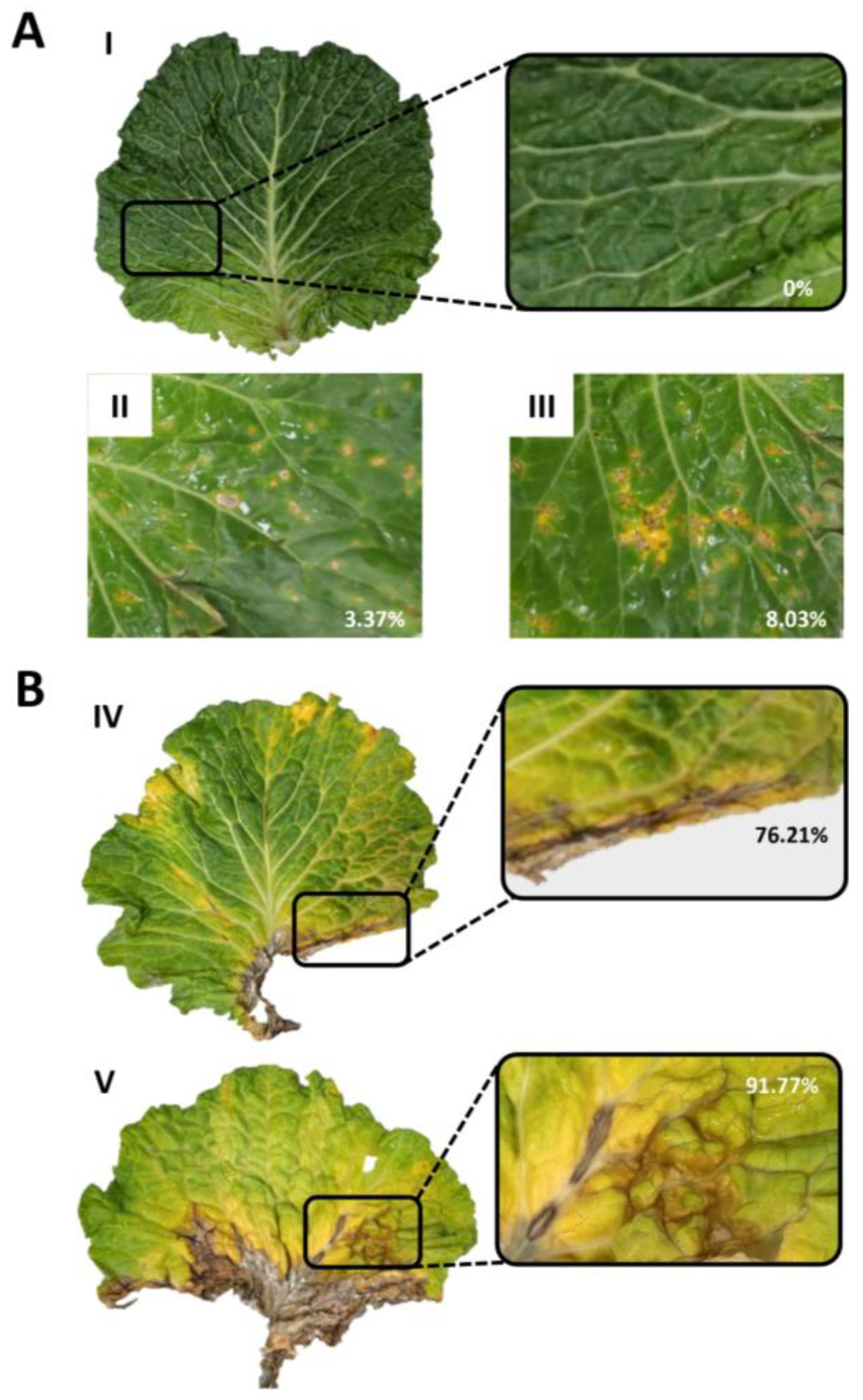
2.1. Plant Material

Cabbage (B. oleracea var. capitata) leaves were collected from naturally infected plants grown in a commercial field located along the eastern Mediterranean coast of Spain (GPS coordinates, 41°38′55.0″ N; 2°45′38.3″ E) in November 2022. Brassica plants had been planted every season in farmers’ fields. The plants were grown under the management practices routinely used by farmers in commercial cabbage fields in the northeastern region of Spain, essentially consisting of insecticide treatments. To obtain the deepest insight into the microbial diversity, healthy (asymptomatic, named Group I) and diseased leaves (named s Group II, Group III, Group IV, and Group V) were collected from plants distributed across the field. For each group, 3 samples were examined (e.g., samples I.1, I.2, and I.3), each sample consisting of a pool of at least 4 leaves. Leaves were not disinfected to preserve both epiphytes and endophytes. Samples were stored at −80 °C until future analysis.

2.2. DNA Extraction, Library Preparation, and Metagenomic Sequencing

Leaf material was pulverized in liquid nitrogen. The same amount (0.3 g) of the pulverized material were used for each DNA extraction. Genomic DNA was obtained using MATAB (100 mM of TRIS-HCl pH 8.0, 1.4 M NaCl, 20 mM EDTA, 2% MATAB, containing 1% PEG 6000 and 0.5% sodium sulfite) as the extraction buffer (6 mL MATAB/g fresh weight) with modifications [34]. Three DNA preparations were made from each group of leaves and further processed. Shotgun metagenomic sequencing was conducted at Novogene (Cambridge, UK). This involved shearing the genomic DNA into short fragments, which were then end-repaired, A-tailored, and ligated with Illumina adapters. The fragments with adapters were PCR-amplified, size-selected, and purified. The library’s quality and size distribution were assessed using Qubit for quantification and a bioanalyzer for size determination. Finally, the quantified libraries were sequenced on Illumina platforms by Novogene, based on the required library concentration and data output.

2.3. Sequencing Data Processing and Metagenome Assembly

Raw reads were filtered to remove low-quality bases (Q value ≤ 38; threshold 40 bp), ambiguous bases at sequence ends (maximal number of ambiguous nucleotides allowed in the sequence after trimming: 10 bp), and adapter sequences (minimum overlap: 15 bp). The phred quality score Q is the standard measure of the quality of the identification of the bases generated by a sequencer. A phred quality score of 30 indicates a base call accuracy of 99.9%, while a Q value of 40 indicates an accuracy of 99.99% (or a probability of incorrect base call of 1 in 10,000 nt). Q 38 means a probability of 1 in 6310 nt. Thus, our trimmed/filtered sequences contained nt that were accurately called by the sequencer at that level or higher. Trimmed and filtered reads were then mapped to the Brassica oleracea var. capitata reference genome [https://www.ncbi.nlm.nih.gov/assembly/GCA_018177695.1] (accessed on 1 January 2024) using Bowtie2 (v2.2.4; http://bowtie-bio.sourceforge.net/bowtie2/index.shtml) (accessed on 1 January 2024), and the positive hits were removed to produce a final set of clean data.
Metagenomes were assembled independently for each sample using the clean datasets. The clean data for each sample were assembled to produce scaffolds using MEGAHIT software (v1.0.4; https://github.com/voutcn/megahit) (accessed on 1 January 2024) with parameter --presets meta-large. The scaffolds were divided into scaftigs (continuous sequences with no Ns within scaffolds). Unassembled read from all samples were pooled to produce a mixed assembly (using same parameters as with the individual assemblies) to obtain information on low-abundance species. Scaftigs with a length shorter than 500 bp were filtered out from the individual and mixed assemblies, and the resulting final set of sequences was selected for further gene prediction and annotation.
Nonmetric dimensional scaling (NMDS) was performed using the ordinate and plot_ordination functions from the phyloseq R package (v1.30.0) using Bray–Curtis distances [35]. The datasets supporting the conclusions of this study are available at the European Nucleotide Archive (ENA) database under accession PRJEB71999.

2.4. Gene Prediction and Taxonomic Annotation

Scaftigs (≥500 bp) from all assemblies were used for open reading frame (ORF) prediction using MetaGeneMark (v2.10; http://topaz.gatech.edu/GeneMark) (accessed on 1 January 2024), not considering predictions shorter than 100 bp. The resulting ORFs were dereplicated using CD-HIT (v4.5.8; http://www.bioinformatics.org/cd-hit) (accessed on 1 January 2024) with parameters -c 0.95, -G 0, -aS 0.9, -g 1, -d 0. For each cluster of overlapping ORFs, the longest one was chosen as the representative gene (unigene) to generate a final set of unigenes (gene catalogue).
Clean data were mapped to the gene catalogue using Bowtie2 with parameters --end-to-end --sensitive -I 200 -X 400 to calculate gene abundances using the formula
G k = r k L k 1 1 = 1 n r i L i
where r is the number of mapping reads, and L is the gene length.
For taxonomic annotation, DIAMOND v0.9.9.110 [32] was used to align the detected unigene sequences to all bacterial, fungal, archaeal, and viral sequences from NCBI’s NR database (as of 01-02-2018), with blastp set to e 1 × 10⁻⁵. Then, from the alignment results of each sequence, the ones with an e value ≤ min. e value ×10 were selected. When multiple alignment results were obtained, the LCA algorithm, applied to systematic taxonomy assignment with MEGAN software v6.13.1, was adopted to determine the species annotation information of the sequence [36]. The relative abundances at different taxonomic levels were imported into R using phyloseq v1.30 [35] and examined using bar plots (kingdom, phylum) and heat maps (species). To represent the data within each group, values were added, and data were normalized to 100%. The relative abundance of taxa across multiple levels of the hierarchy was visualized using Krona plots [37] using the R package psadd v0.1.3 (https://github.com/cpauvert/psadd) (accessed on 1 January 2024).
To associate the enzymatic functions that degraded, modified, or created carbohydrates with specific groups of leaves, the protein coding sequences were mapped against the carbohydrate-active enzymes (CAZymes) database (v2014.11.25) [38]. The database contains six functional classes: glycoside hydrolases, glycosyl transferases, polysaccharide lyases, carbohydrate esterases, auxiliary activities and carbohydrate-binding modules.

3. Results

Environmental factors, such as climatic conditions (e.g., relative humidity, temperature, etc.), are known to affect the microbial community of plant tissues. To avoid the confounding factors associated with the various environmental conditions influencing the microbial population, B. oleracea plants were cultivated at a single location, namely, in a field routinely used for the cultivation of cabbage plants located in the northeast region in Spain. Leaf samples were harvested from plants randomly distributed in the field and then classified into different groups according to disease symptomatology as follows: Group I, leaves with no visual symptoms of infection (named as asymptomatic leaves) (Figure 1A, upper panel); Group II, leaves with scattered yellow spots covering 3–4% of the area (symptomatic-mild leaves); and Group III, leaves with abundant necrotic spots embedded in larger yellow areas covering at least 8% of the area (symptomatic-high leaves) (Figure 1A, lower panel). Two additional groups were examined consisting of leaves with disease symptoms at the central-basal region of the leaf (Figure 1B). They were Group IV, leaves with necrotic lesions along the leaf margin (leaf margin leaves); and Group V, leaves with extensive brown symptoms in the proximity of large necrotic regions and yellowed leaves (symptomatic-severe leaves). Apart from the localized symptoms of infection in their leaves, the cabbage plants did not show apparent differences in plant growth or appearance, which could be indicative of a generalized health problem.

3.1. Data Pre-Processing and Metagenome Assembly

Shotgun metagenomic sequencing was used to examine the composition of the microbial community in B. oleracea leaves. Three independent DNA extractions were made from each group of leaves, which were then used for library preparation (15 libraries) and shotgun metagenomic sequencing.
A total of 106.4 Gb was generated from the 15 libraries, consisting of 19.6 and 29.4 M read pairs (2 × 150 bp) depending on the sample (Table S1). Up to 97.4% of the total number of sequences passed the adapter removal and quality trimming step (Table S1). The cleaned reads from the different samples were assembled independently to produce a collection of scaftigs (Table S2). The proportion of sequence mapping against the B. oleracea genome ranged from 73% to 94% in the different samples (Table S1). Furthermore, sequencing data were mapped to the assembled scaftigs, and the resulting unmapped reads were used to produce a mixed assembly. The effective scaftigs (e.g., those of at least 500 bp) were then identified and further processed for gene prediction (Table S2).
For gene prediction and abundance analysis, scaftigs (≥500 bp) were considered, and ORFs less than 100 nt long were discarded. A final gene catalogue was generated by grouping open reading frames (ORFs) showing at least 95% identity with 90% coverage and selecting the longest ORF from each group as the representative gene (unigenes). Overall, 38.6% of the unigenes in the gene catalogue contain both start and stop codons and 52% contain only one of both codons (three samples/group; Table S3). The gene abundance for each gene in each sample was calculated based on the total number of mapped reads normalized by gene length. The relative abundance of each gene in each sample was computed by dividing the absolute abundance per gene in each sample by the sum of all abundances calculated for that particular sample.

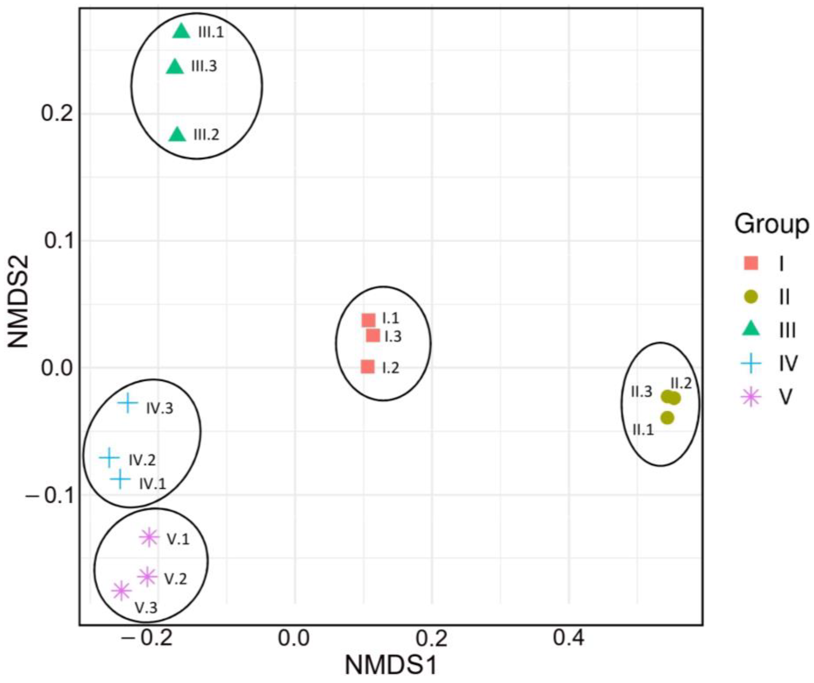
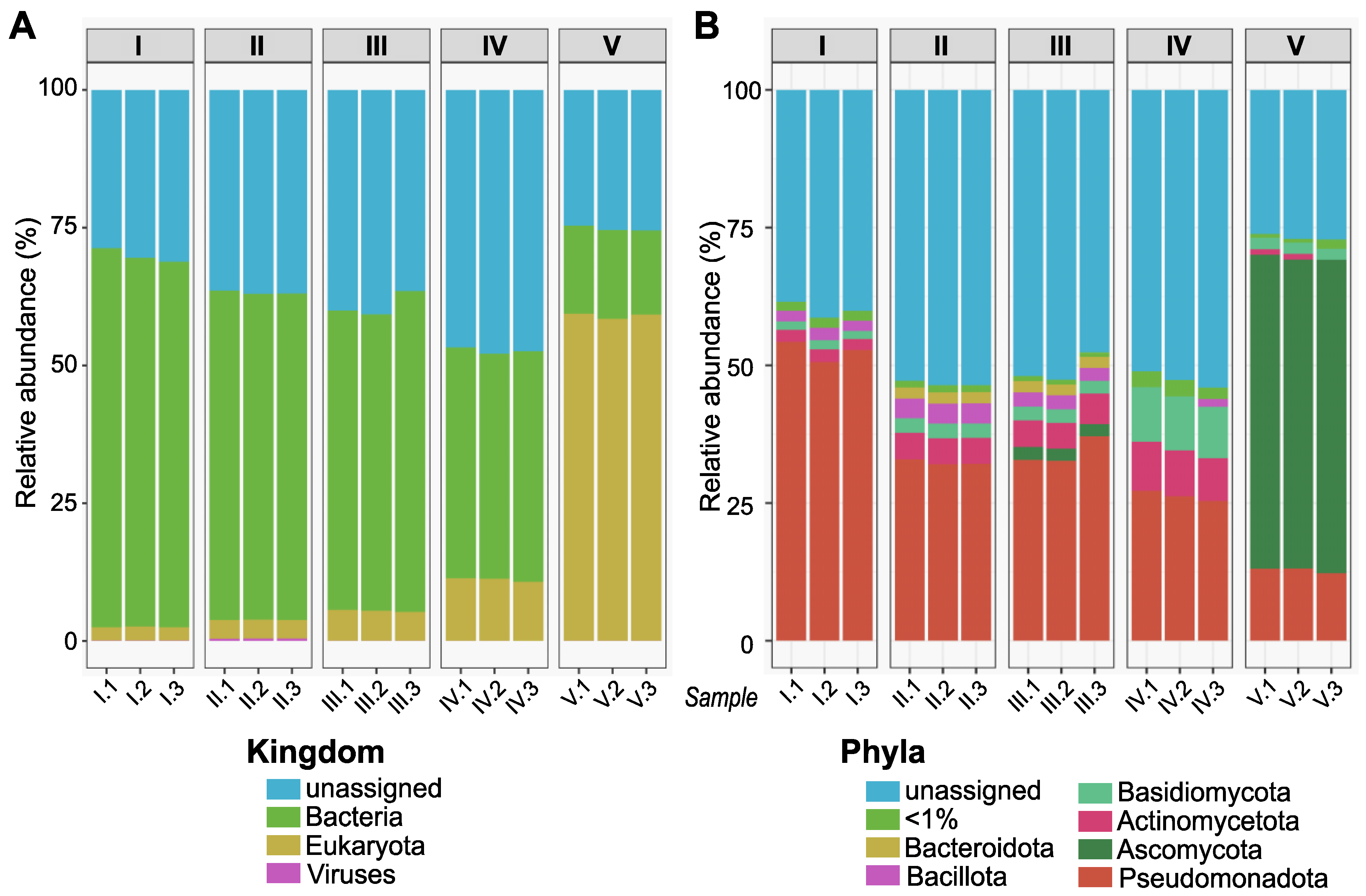
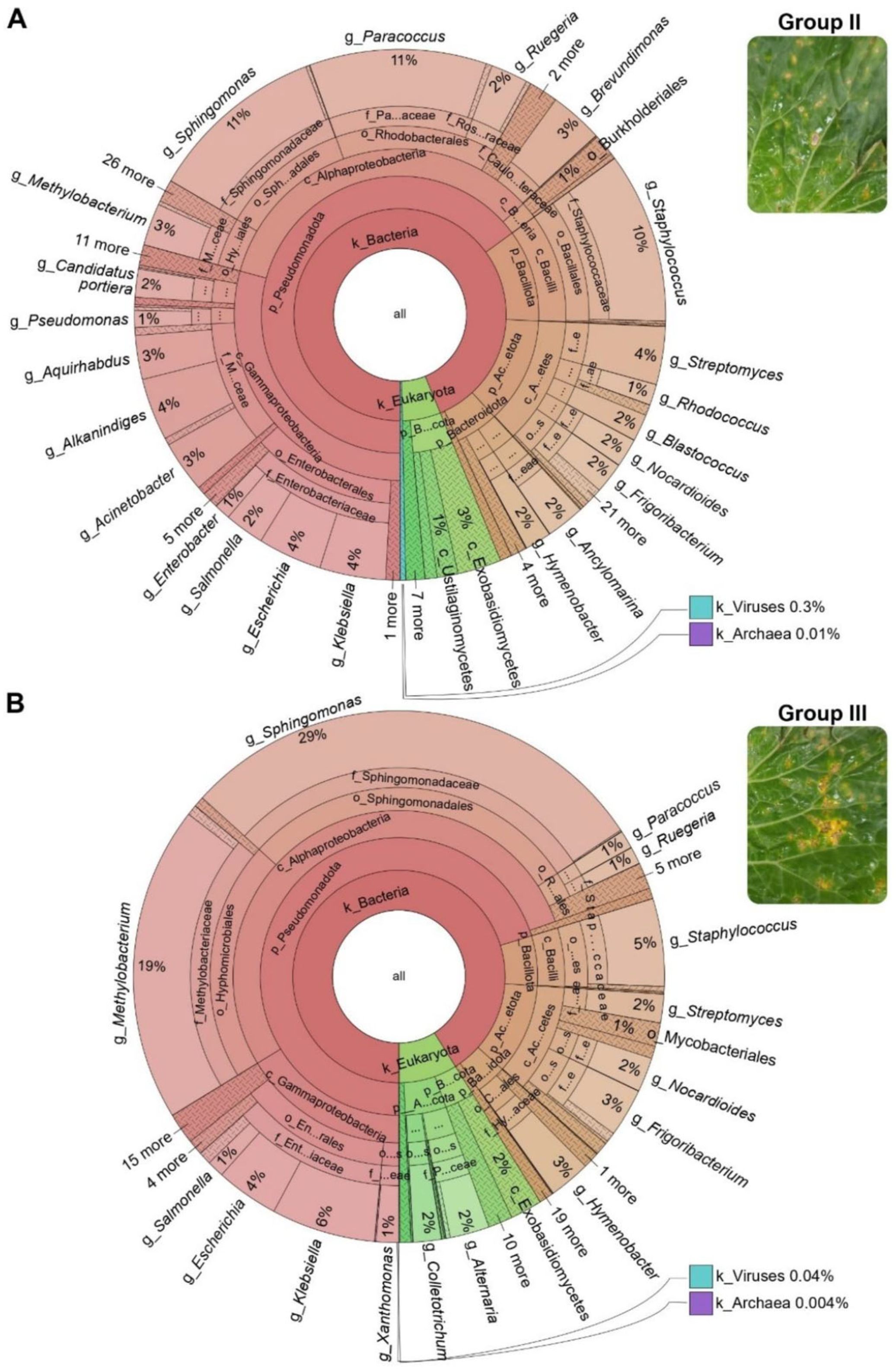
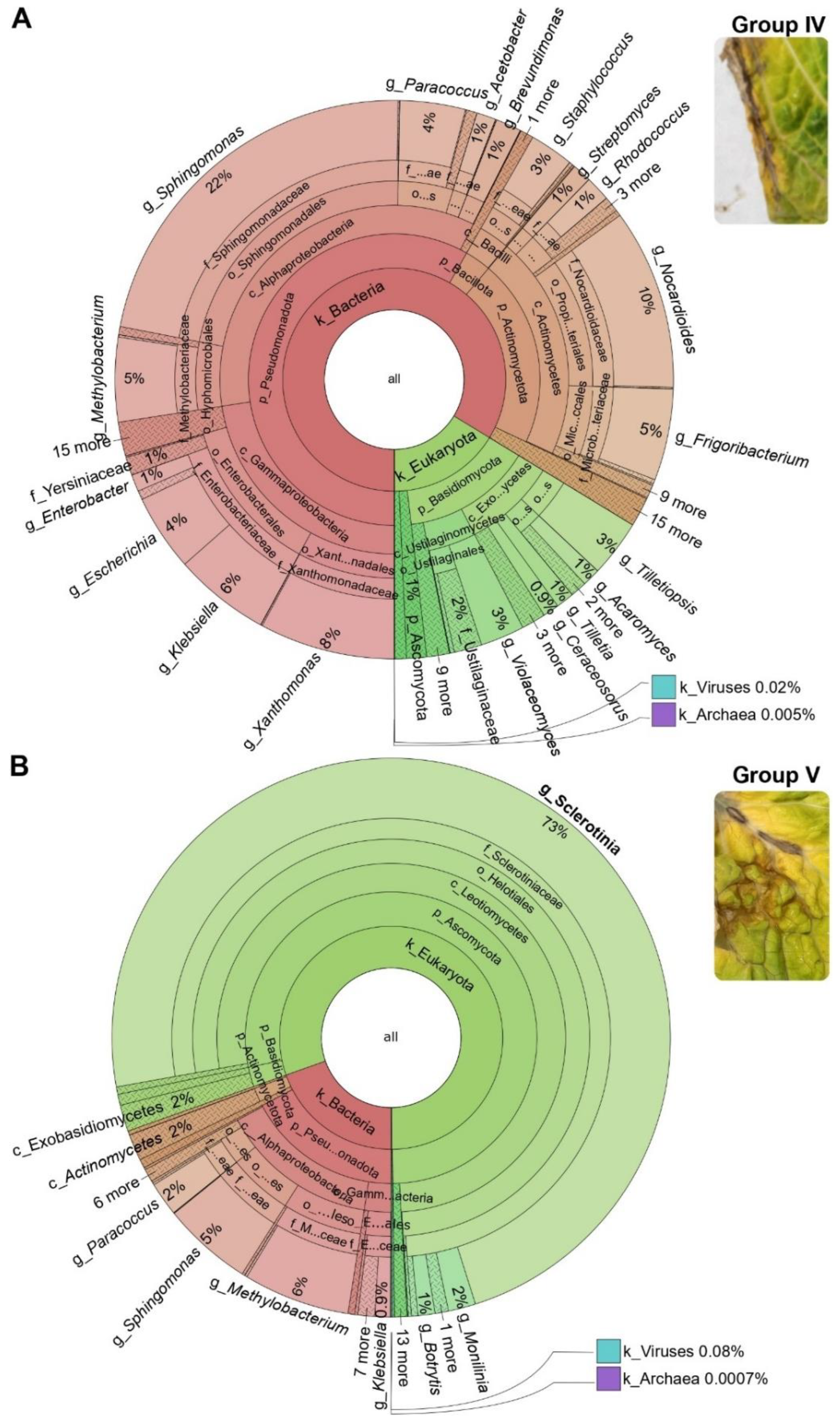
3.2. Microbial Communities in B. oleracea Leaves under Natural Infection Conditions

The unigenes identified in the metagenomic data for the samples of B. oleracea leaves were taxonomically classified using the NCBI’s microbial nonredundant database (https://ftp.ncbi.nlm.nih.gov/blast/db/) (accessed on 1 January 2024). A nonmetric multidimensional scaling (NMDS) plot at the phylum level was then constructed to evaluate if the microbial communities of the samples in a single group (three samples per group) related to each other and to visualize the degree of overall similarity between groups. As shown in Figure 2, the samples within a particular group clustered together, while there was a clear separation between the sets of samples from the different groups.
The microbial community structure of B. oleracea leaves was examined in independent samples in each group of B. oleracea leaves at different taxonomic levels, namely, at the kingdom, phylum (Figure 3), and genus levels (Figure S1). The taxonomic classification of microbial annotations was consistent among independent samples within each group of B. oleracea leaves. Differences among groups of leaves were observed, not only between asymptomatic and symptomatic leaves but also among symptomatic leaves. Thus, the predominant phyla in Groups I to IV were associated with the kingdom Bacteria (e.g., Pseudomonadota followed by Bacillota, Bacteroidota, and Actinomycetota) (Figure 3). However, the major phyla in Group V were linked to Ascomycota and Basidiomycota (kingdom Eukaryota) (Figure 3). To further investigate the microbiomes associated with each group of B. oleracea leaves, the metagenomic datasets from independent samples for each group (three samples each) were combined into a single dataset, excluding unassigned sequences.
Krona visualization was used to investigate in more detail the relative abundances of the microbial taxa within a given group and differences between groups of B. oleracea leaves. Krona uses a radial space-filling display to show hierarchy and abundance simultaneously, thus allowing intuitive exploration of the relative abundances within hierarchies of taxonomic classifications. The Krona graphs for each group of leaves at the phylum level are presented in Figure S2 (the list of taxa in each group of leaves is shown in Table S4). As previously observed, there were important differences between Groups I to IV (enriched in bacterial taxa) and Group V (enriched in fungal taxa) at the phylum level (Figure S2). Pseudomonadota was the predominant bacterial phylum in Groups I, II, III, and IV (accounting for 87%, 69%, 69%, and 55% of the total microbial community, respectively), while contributing 17% of the total microbial community in Group V (Figure S2). Regarding the fungal community, Ascomycota was the most abundant phylum in Group V (77% of the total microbial), while Basidiomycota was the dominant fungal phylum in Group IV (Figure S2). To obtain further insights into the leaf microbiome of B. oleracea leaves, we examined the taxonomic assignments at the genus level across the various groups of leaves, as discussed below.

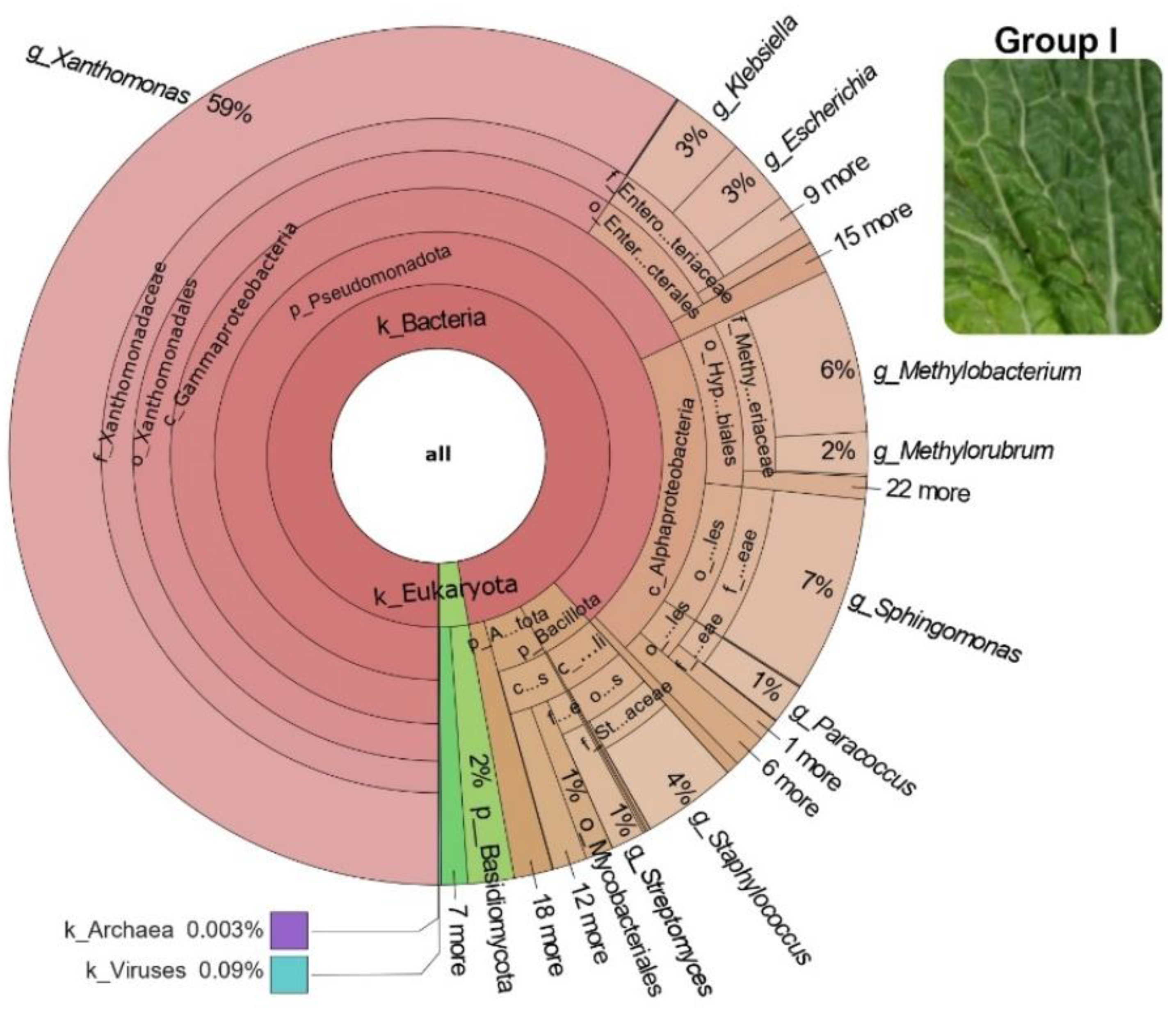
3.3. Bacterial Community in B. oleracea Leaves under Natural Infection Conditions

Krona graphs displaying the taxonomical diversity of the microbial community at the genus level in asymptomatic and diseased B. oleracea leaves are presented in Figure 4 for Group I, Figure 5 for Groups II and III, and Figure 6 for Groups IV and V. The list of bacterial genera used for construction of Krona graphs for each group are shown in Table S5.
The bacteria in the leaves of B. oleracea were diverse in terms of taxonomy. Also, the relative abundance of the bacterial taxa in the various groups of B. oleracea leaves greatly varied. Except for Group V, the most dominant bacterial taxa in B. oleracea leaves were linked to the phylum Pseudomonadota, also in asymptomatic leaves (Group I). Notably, the genus Xanthomonas (phylum Pseudomonadota) was by far the most abundant bacterial taxon in Group I (asymptomatic leaves), accounting for 59% of all genus-level assignments (excluding ‘Others’, i.e., unclassified sequences) (Figure 4). However, Xanthomonas was much less represented in other Groups of leaves (Figure 5 and Figure 6).
A great diversity of bacterial taxa was found in Groups II, III, and IV, whose relative abundance varied distinctly among groups. In Group II, Sphingomonas and Paracoccus (phylum Pseudomonadota) were the most abundant bacterial genera. (Figure 5A). In Group III, the dominant bacterial genera were Sphingomonas and Methylobacterium (phylum Pseudomonadota) (Figure 5B). In Group IV, Sphingomonas, Xanthomonas, and, at a lower abundance, Methylobacterium were the predominant bacterial genera in the phylum Pseudomonadota. Members of the fungal phylum Actinomycetota (e.g., Nocardioides and Frigobacterium) were also detected in Group IV (Figure 6A). Finally, as previously mentioned, Group V showed the lowest abundance of bacterial taxa. They included Methylobacterium and Sphingomonas, followed by Paracoccus (phylum Pseudomonadota) (Figure 6B). From these results, it appears that the bacterial genera Sphingomonas and Methylobacterium were shared among the various groups of leaves, although their contribution to the total microbial community varied among these groups.

3.4. Fungal Community in B. oleracea Leaves under Natural Infection Conditions

The fungal community of B. oleracea leaves (kingdom Eukaryota) was primarily composed of members of the phyla Ascomycota and Basidiomycota, which also showed differential distribution among the different groups of leaves (Figure 4, Figure 5 and Figure 6). The list of fungal genera for each group is shown in Table S5. The contribution of fungal taxa to the total microbial community was found to progressively increase from Groups I to V, being also associated to the symptomatology observed in each group of leaves (e.g., asymptomatic leaves to leaves showing severe symptoms) (Figure 4, Figure 5 and Figure 6). In Group I (asymptomatic leaves), the phyla Basidiomycota and Ascomycota comprised 2% and 0.4% of the total microbial community, respectively (Figure 4). This group also contained various low-abundance fungal species (e.g., Violaceomyces, Tilletiopsis, Puccinia, Tilletia, and Ceraceosorus) in the phylum Basidiomycota and Alternaria and Sclerotinia in the phylum Ascomycota (Table S5). No fungal genus in Group I surpassed 1% of the microbial community.
In the symptomatic leaves, the relative abundance of the fungal taxa increased with disease severity (e.g., from Group II to Group V). In Group II, the fungal community was dominated by members of the phylum Basidiomycota, which constituted 5% of the overall microbial community (Figure 5A). The notable genera within this phylum included Tilletiopsis and Violaceomyces, with lesser contributions from Ceraceosorus, Tilletia, Acaromyces, and Puccinia. The Ascomycota phylum was present at a reduced abundance, comprising 0.5% of the total microbiome (Table S5). In Group III, Alternaria and Colletotrichum were the dominant fungal pathogenic species (Figure 5B), both belonging to the Ascomycota phylum, comprising 5% of the total microbial community. This group also included the phylum Basidiomycota (3% of total microbiome), with Tilletiopsis and Violaceomyces being particularly prevalent (Table S5). Regarding Group IV, a variety of low-abundance fungi were identified in this group, mainly assigned to Basidiomycota, which accounted for 14% of the total microbiome (e.g., Tilletiopsis, Violaceomyces, Ceraceosorus, Acaromyces and Tilletia) (Figure 6A). Meanwhile, Ascomycota members, including Alternaria, represented up to 1% of the total microbial community in this group (Figure 6A and Table S5). As previously mentioned, the fungal community residing in the leaves of Group V was much more abundant than that in the other groups of B. oleracea leaves (80% of the total microbiome). Notably, the genus Sclerotinia (phylum Ascomycota) accounted for most of the fungi identified in Group V (Figure 6B).
Collectively, this study revealed the presence of fungi belonging to genera that are known to cause important diseases in Brassica crops, such as black spot (caused by Alternaria species, mainly A. brassicicola and A. brassicae), white mold (caused by Sclerotinia sclerotiorum), and anthracnose (caused by Colletotrichum species). Of them, Sclerotinia was associated with Group V.

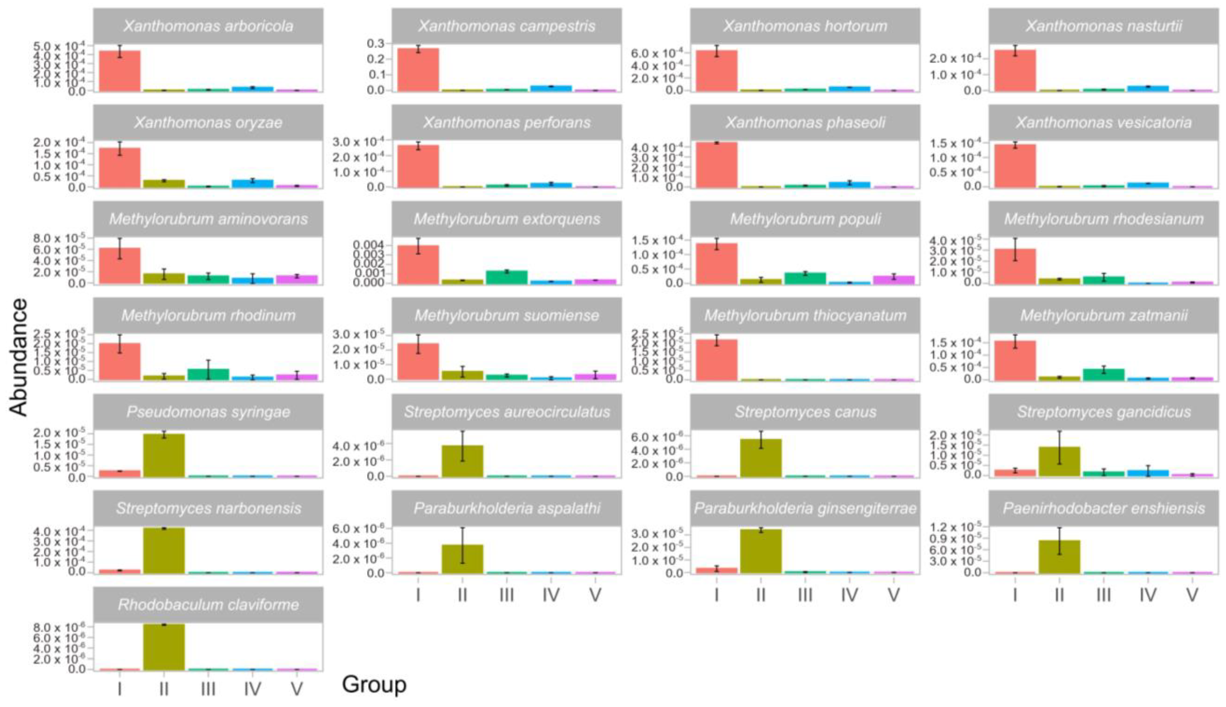
3.5. Common and Specific Microorganisms in B. oleracea Leaves under Natural Infection Conditions

We searched for the common and specific microbial taxa among all five groups of B. oleracea leaves. At the genus level, we identified unique bacterial and fungal (e.g., Eukaryotic) taxa that were either exclusive to a specific group or shared among two or more groups (Figure S3). Specifically, of the asymptomatic leaves in Group I (Figure S3; black dots), the genera Xanthomonas and Methylorubrum were uniquely identified in this group. While certain genera were exclusive to specific groups, a significant proportion was found across multiple groups. Notably, the symptomatic leaves (Groups II to IV) exhibited a notable overlap of bacteria and eukaryotic genera (Figure S3; yellow dots), and a comprehensive distribution across all groups was observed Figure S3; purple dots). This comprehensive distribution encompassed critical pathogenic fungi in the Brassicaceae, such as Alternaria, Sclerotinia, Botrytis, Colletotrichum, and Rhizoctonia, highlighting the intricate microbial landscape present on B. oleracea leaves under natural infection conditions.
To investigate the potential link between pathogenic microorganisms and disease symptoms, we analyzed the relative species-level abundance of the fungal and bacterial taxa across the different groups of B. oleracea leaves. The findings are depicted in Figure 7. Notably, Group III was exclusively enriched in Alternaria species, including A. alternata, A. arborescens, A. atra, A. burnsii, A. gaisen, A. panax, A. postmessia, A. tenuissima, and A. ventricosa (Figure 7). Conversely, Group IV exhibited a more diverse colonization by species such as Pseudozyma flocculosa, Pseudozyma hubeiensis, Rhizoctonia solani, Tilletaria anomala, and Tilletiopsis washingtonensis (Figure 7). In Group V, the exclusive presence of several species from the Sclerotiniaceae family was observed, including Botryotinia species (B. calthae. B. narcissicola, B. convoluta, and B. globose), Botrytis species (B. tulipae, B. byssoidea, B. cinerea, B. elliptica, B. fragariae, B. galanthina, B. hyacinthi, B. paeoniae, B. porri, and B. sinoallii), Stromatinia species (S. cepivora), and Sclerotinia species (S. sclerotiorum, S. trifoliorum, and S. bolearis) (Figure 7).
In the context of bacterial taxa, eight Xanthomonas species (X. arboricola, X. campestris, X. hortorum, X. nasturtii, X. oryzae, X. perforans, X. phaseoli, and X. vesicatoria) and eight Methylorubrum species (M. aminovorans, M. extorquens, M. populi, M. rhodesianum, M. rhodinum, M. suomiense, M. thiocyanatum, and M. zatmanii) were predominantly detected in asymptomatic leaves (Group I), with the majority being absent from symptomatic leaves (Figure 8). The bacterial species that were enriched in Group II demonstrated more heterogeneity, including Pseudomonas syringae, which is known to cause bacterial spot in the Brassicaceae family. Additionally, four Streptomyces species (S. aerocirculatus, S. canus, S. gancidicus, and S. narborensis) were found alongside Paraburkholderia species (P. aspalathi and P. ginsensiterrae), Paenirhodobacter enshiensis, and Rhodobaculum claviforme (Figure 8). Overall, these analyses revealed the associations between certain bacterial and fungal taxa and disease symptoms (Table 1).

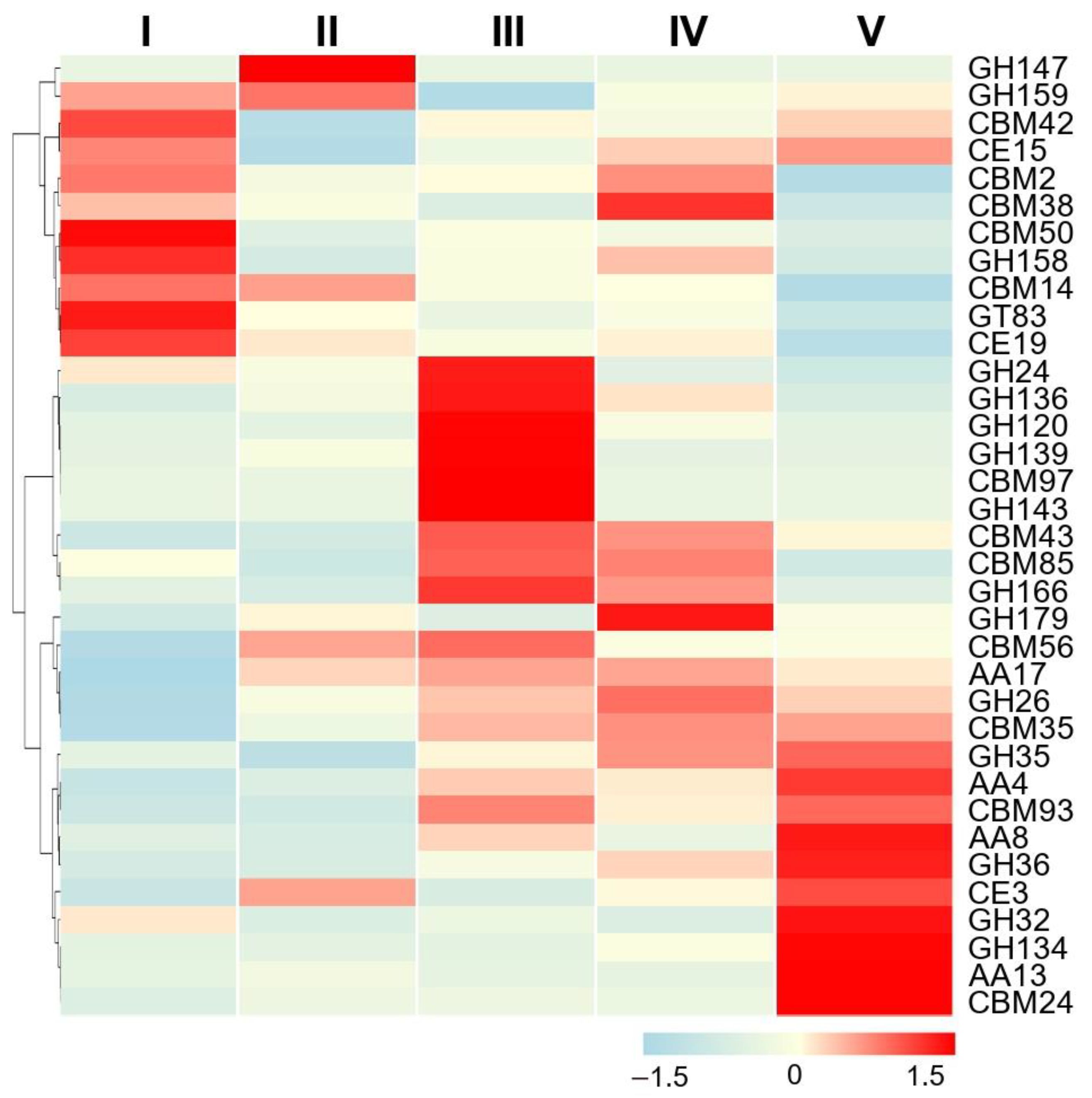
3.6. Functional Analysis of B. oleracea Metagenomes

Shotgun metagenome sequencing is a powerful approach that allows the integration of taxonomic information with microbial community functional profiles. Beyond examining diversity and abundance of microbial assemblies in the various groups of B. oleracea leaves, we aimed to delineate the functional contributions of the microbial taxa in each group of leaves. In this study, we focused on the enzymes that are responsible for breaking down the host cell wall, the first barrier that pathogens encounter in causing infection.
It is well known that phytopathogenic fungi have evolved an arsenal of enzymes responsible for breaking down complex carbohydrate and polysaccharides into smaller products, generally referred to as carbohydrate-active enzymes (or CAZymes) [39,40,41]. CAZymes were assigned to CAZy classes (level 1) and families (level 2) and deposited in the CAZy database (http://www.cazy.org/) (accessed on 1 January 2024). They were classified into different classes, including glycoside hydrolases (GHs), glycosyl transferases (GTs), carbohydrate-binding modules (CBMs), auxiliary activity enzymes (AAs), carbohydrate esterases (CEs), and polysaccharide lyases (PLs).
In the present study, we searched for CAZymes that are encoded by microbial genes in B. oleracea leaves with different disease severity symptoms to obtain insights into how the microbial communities in each group might contribute to infection and pathogenicity. The abundance distribution of the top 35 functions at the family level across the microbiomes of B. oleracea leaves is displayed in a heatmap (Figure 9). Differences in function abundance were clearly observed among the groups of leaves. For instance, Group I was enriched in CBM enzymes CBM2, CBM14, CBM38, CBM42, and CBM50; Group III in GH and CBM (GH24, GH120, GH136, GH139, GH143, and GH166 for GH enzymes; CBM43, CBM85, and CBM97 for CBM enzymes); and Group V in GH, AA, and CBM CAZymes (GH26, GH32, GH35, GH36, and GH134 for GH enzymes; AA4, AA8, and AA13 for AA enzymes; CBM24, CBM35, and CBM93 for CBM enzymes). Such differential abundance profiles offer insights into how microbial communities may influence disease progression and pathogenicity in B. oleracea leaves.

4. Discussion

Brassica crops, including cabbage, have high nutritional value and beneficially affect human health [42]. Because of their importance in food security, they are grown in most countries worldwide. It is well accepted that agricultural production must be increased considerably in the foreseeable future to meet the food and feed demands of a growing human population. During the past decades, however, the production of vegetable Brassicas (e.g., cabbage, cauliflower and broccoli) has remained constant [2]. One of the main reasons for this lack of increased production of Brassica crops is the wide array of microorganisms that infect these species, which substantially diminish their yield and quality. The first step in reducing losses in cabbage production caused by pathogen infection is to identify and describe microbial communities in cabbage plants in agricultural ecosystems. Moreover, the B. oleracea-associated microbiome can be modulated by several factors, such as host genotype and developmental stage, soil type, geographical location and environmental conditions [21,43]. One would expect that cabbage plants acquire a specific set of microorganisms depending on a combination of factors. However, the structure and diversity of the microbial communities in B. oleracea leaves have been poorly described.
In this work, we characterized the microbial community of cultivated cabbage plants showing different symptomatologies under natural infection conditions. Importantly, this study allowed us to obtain species-level resolution of the microbial composition of B. oleracea leaves while providing insights into the potential functional importance of B. oleracea-associated pathogens (e.g., fungal pathogens capable of producing host cell-wall-degrading enzymes to facilitate infection). The cabbage plants were grown in farmer fields where this crop is common in the northeast of Spain (Mediterranean climate conditions). This study was carried out with one cultivar, B. oleracea var. capitata, in plants at the same developmental stage growing in this geographical region. It seems then reasonable to assume that microbial communities are mostly shaped by the geographical location and environmental conditions in which cabbage plants are grown. Even though the application of molecular approaches has made it feasible to obtain large datasets of microorganisms residing in plant tissues, the principles that underlie the assembly of communities in Brassica crops in relationship with disease symptoms remain largely unknown. To move forward in this direction, the information gained in this study shows the existence of different patterns of microbial communities in cabbage leaves. As interactions among community members might affect disease resistance/susceptibility in Brassica crops, this information will allow further explorations of the potential positive relationships between B. oleracea and distinct microorganisms that can be applied for more sustainable cabbage production.
The presented results revealed that the microbial assemblages in cabbage leaves greatly vary in leaves showing different disease symptoms. Notably, certain taxa were exclusive to specific groups of leaves, while others were common across multiple groups. Differences were found not only between asymptomatic and diseased leaves but also between leaves exhibiting various types of symptomatology. These differences were evident across both abundant and rare taxa. These observations further support that microbial communities are complex dynamic systems and that changes in assemblage composition could be associated with different disease symptoms.
The microbial assemblages in B. oleracea leaves contained bacteria and fungi associated with known diseases in Brassica species as well as taxa that are considered to be potentially beneficial for plants. The highest diversity of bacterial taxa was observed in diseased leaves classified in Groups II, III, and IV. The asymptomatic leaves (Group I) and severely affected leaves (Group V) contained an overall low richness of microbiota, these groups being enriched in bacterial and fungal taxa, respectively. Indeed, the microbial community of the asymptomatic leaves (Group I) consisted almost exclusively of bacterial taxa, with Xanthomonas being the dominant bacterial taxon in this group (59% of the microbial community). Up to eight Xanthomonas species were found to be represented in the asymptomatic leaves. Although species in the family Xanthomonadaceae are commonly associated with plants as phytopathogens or endophytes [44], there are also reports demonstrating that certain Xanthomonas species produce secondary metabolites that confer protection against fungal pathogens, these species being considered nonpathogenic to the host plant [45]. This observation suggests that Xanthomonas species might offer a bioprotective effect against B. oleracea pathogens in asymptomatic B. oleracea leaves. Of note, phytopathogenic fungi were found in very low abundance in these asymptomatic leaves. Whether the bacterial species identified in the B. oleracea leaves (Group I) exert a protective effect against pathogens associated with cabbage diseases, in particular against fungal pathogens, requires further investigation. Similar to Xanthomonas, other bacteria species like Pseudomonas or Bacillus species are known to control phytopathogens (e.g., Bacillus subtilis for controlling Fusarium spp. and Pseudomonas fluorescens for controlling Erwinia amylovora) [46,47,48]. Further experimental research is required to ascertain whether a causal relationship exists between certain taxa enriched in symptomatic leaves and disease symptoms or whether specific combinations of microbial taxa are required to cause the observed disease symptoms. This process, however, requires the isolation and culturing of multiple taxa and the preparation of synthetic microbial communities (SymCom approach).
Regarding symptomatic leaves, a great diversity of microbial taxa was found consistently colonizing the leaves classified in Groups II, III, and IV, with differences that were already noticeable at the phylum level. In general, the members of the Pseudomonadota phylum were the more prevalent bacterial genera in Groups II, III, and IV of B. oleracea leaves, pointing to a high degree of adaptation of Pseudomonadota to this ecological niche. Group III was enriched in well-known pathogens of Brassica species, such as Alternaria (black spot) and Colletotrichum (Anthracnose). Meanwhile, the fungi Tilletiopsis and Tilletia were more abundant in Group IV. The fungus Sclerotinia, the causal agent of the white mold, was dominant in severely diseased B. oleracea leaves (Group V), aligning with the visual disease symptoms observed on these leaves.
Through the comparison of bacterial and fungal species’ abundance across the various groups of B. oleracea leaves, it was possible to discern a relationship between the presence or absence of specific microbes and the symptoms of the disease. We identified microbial species uniquely associated with specific leaf groups, revealing a distinct pattern of fungal enrichment in the symptomatic leaves of Groups IV and V. Specifically, Group III was characterized by a significant presence of Alternaria species, such as A. alternata, while Group V showcased a diversity of fungi from the Sclerotiniaceae family, including notable species like Sclerotinia sclerotiorum, Botrytis cinerea, Botryotinia calthae, and Stromatinia cepivora. Meanwhile, Group II, which exhibited milder symptoms, was primarily associated with bacterial inhabitants (e.g., Streptomyces narborensis). This differential symptomatology across the leaf groups suggests a nuanced interplay between the microbial community’s composition, both in terms of relative abundance and specific microbial consortia, and disease expression. These fungal and bacterial taxa might serve as a “core” microbiome in cabbage plants growing in this biogeographical region (e.g., northeast of Spain), which has a typical Mediterranean climate.
Beyond identifying differentially abundant taxonomic groups in diseased B. oleracea leaves, we wanted to determine whether an enrichment in specific microbiota could be related to specific microbial functions. Indeed, this study revealed enrichment in the functional genes encoding the cell-wall-degrading enzymes responsible for breaking down the plant cell during pathogenesis, also known as CAZymes [39]. Among the different classes of CAZy enzymes, GH enzymes can break down the carbohydrates and polysaccharides in cellulose and chitin, acting as endoglucanases, cellulases, xylanases, xyloglucanases, α-amylases, or chitinases. GH enzymes have been characterized mainly in Ascomycetes and Basidiomycetes, many of them acting as virulence factors in promoting host colonization [40,49]. Our metagenomic analysis revealed a significant enrichment of functional genes belonging to different CAZy classes, which was also dependent on the group of B. oleracea leaves. The microbial community residing in Group III and Group V was particularly enriched in GHs, whereas asymptomatic leaves (Group I) harbored more functional genes related to carbohydrate-binding module (CBM) activities. CBMs are known to assist carbohydrate-active enzymes, in particular GHs, by facilitating the interaction between the enzyme and its substrate, thus improving the efficiency of polysaccharide degradation. In our study, the CBM enzymes in asymptomatic leaves could be linked to the activity of GH158 and GH159. Overall, this analysis indicates that microbial community assembly patterns can be associated with distinct carbohydrate–active enzyme functions required for the virulence of fungi.
Fungal pathogens can secrete antimicrobial compounds that can shape the plant microbiome either through the inhibition of plant pathogens or through the regulation of the microbial assembly. On this basis, different residing microbial consortia and interactions with one another (i.e., microbe–microbe competition) might well explain, at least in part, the variations in B. oleracea-associated microbiomes and, hence, those in disease severity. Notably, our observations revealed the presence of Tilletiopsis in Group IV B. oleracea leaves. Tilletiopsis is recognized as an effective biocontrol agent against fungal phytopathogens such as A. alternata [50], which was also detected within this group. Further investigation is, however, needed to better understand the interactions between the components of the B. oleracea microbiome, as well as plant–microbe interactions, in relationship with the health status of the plant.
Finally, our metagenomic analysis of B. oleracea leaves revealed the presence of ubiquitous bacteria that are endemic to surface water and soil that can be pathogenic for humans and animals (e.g., Staphylococcus, Klebsiella, Salmonella, Enterobacter and Escherichia). In line with this, evidence increasingly supports that certain species in these bacterial genera are capable of infecting plant tissues, even being beneficial for the host plant (e.g., by stimulating growth or conferring disease resistance). For instance, Klebsiella pneumoniae (strain JCK-2201) was shown to protect tomato plants from Ralstonia solanacearum, causing tomato bacterial wild [51], whereas Klebsiella sp. (strain KW7-S06) was found to induce systemic resistance to Fusarium oxysporum and Rhizoctonia solani in rice [52]. Furthermore, Staphylococcus aureus and Pseudomonas aeruginosa are capable of infecting tissues (leaves and roots) of the model plant A. thaliana [53,54].

5. Conclusions

Overall, this study described the composition of the microbial communities in the leaves of B. oleracea plants grown in agricultural farmlands. B. oleracea represents an important horticultural crop and an important source of vegetables in many countries. The information gained in this study emphasizes the need to better understand the variations in the structure of leaf microbial communities in relation to disease symptomatology. In future studies, it will be essential to further explore the dynamics of B. oleracea–microbe interactions as well as microbe–microbe interactions in B. oleracea leaves by incorporating microbiome and phytopathological studies. The characterization of the microbial communities in Brassica plants will open new avenues for designing more effective strategies for disease control in cabbage plants while minimizing the environmental impact caused by the indiscriminate use of agrochemicals in cabbage production.

Supplementary Materials

The following are available online at https://www.mdpi.com/article/10.3390/horticulturae10070765/s1, Figure S1: Heatmap analysis of the most abundant genera detected across all samples, Figure S2: Krona graphs showing the diversity of microbial taxa in B. olearacea leaves at the Phylum level, Figure S3: UpSet plots showing shared and unique bacterial (A) and eukaryotic (B) genera in asymptomatic (Group I) and symptomatic (II to V) B. oleracea leaves. Table S1: Statistics for data pre-processing, Table S2: Assembly Statistics, Table S3: Gene Prediction Statistics, Table S4: Taxonomic composition of B. oleracea leaf microbiomes at the phylum level, including asymptomatic leaves (Group I) and leaves showing different disease symptoms (Groups II to V), Table S5: Taxonomic composition of B. oleracea leaf microbiomes at the genus level, including asymptomatic leaves (Group I) and leaves showing different disease symptoms (Groups II to V).

Author Contributions

Conceptualization, B.S.S. and S.C.; Data Curation, V.M.G.-M.; Formal Analysis, H.M.-C., V.M.G.-M., S.C. and B.S.S.; Funding Acquisition, B.S.S.; Investigation, H.M.-C., V.M.G.-M. and L.S.-L.; Methodology, H.M.-C., V.M.G.-M. and L.S.-L.; Project Administration, B.S.S.; Supervision, S.C. and B.S.S.; Writing—Original Draft Preparation, B.S.S., S.C., H.M.-C. and V.M.G.-M.; Writing—Review and Editing, B.S.S., S.C., H.M.-C., V.M.G.-M. and L.S.-L. All authors have read and agreed to the published version of the manuscript.

Funding

This research was supported by the “Severo Ochoa Program for Centres of Excellence in R&D” CEX2019-000902-S, funded by MCIN/AEI/10.13039/501100011033; the CERCA Program/“Generalitat de Catalunya”; and by grant PID2021-128825OB-I00 from the MCIN/AEI/10.13039/501100011033 and by “ERDF A way of making Europe” to BSS. We also acknowledge financial support from grant 2021 SGR 00875 from the Generalitat de Catalunya.

Institutional Review Board Statement

Not applicable.

Informed Consent Statement

Not applicable.

Data Availability Statement

The metagenomic data supporting the conclusions of this work are available at the European Nucleotide Archive (ENA) database under accession PRJEB71999.

Acknowledgments

We thank Cristina Pozo (Federación de Agrupaciones de Defensa Vegetal Selmar) for sample collection from cabbage fields.

Conflicts of Interest

The authors declare no conflicts of interest.

References

  1. OECD. Brassica Crops (Brassica Species). In Safety Assessment of Transgenic Organisms; OECD: Paris, France, 2016; Volume 5. [Google Scholar]
  2. Greer, S.F.; Surendran, A.; Grant, M.; Lillywhite, R. The Current Status, Challenges, and Future Perspectives for Managing Diseases of Brassicas. Front. Microbiol. 2023, 14, 1209258. [Google Scholar] [CrossRef] [PubMed]
  3. Siciliano, I.; Gilardi, G.; Ortu, G.; Gisi, U.; Gullino, M.L.; Garibaldi, A. Identification and Characterization of Alternaria Species Causing Leaf Spot on Cabbage, Cauliflower, Wild and Cultivated Rocket by Using Molecular and Morphological Features and Mycotoxin Production. Eur. J. Plant Pathol. 2017, 149, 401–413. [Google Scholar] [CrossRef]
  4. Vicente, J.G.; Holub, E.B. Xanthomonas Campestris Pv. Campestris (Cause of Black Rot of Crucifers) in the Genomic Era Is Still a Worldwide Threat to Brassica Crops. Mol. Plant Pathol. 2013, 14, 2–18. [Google Scholar] [CrossRef]
  5. Lv, H.; Miyaji, N.; Osabe, K.; Akter, A.; Mehraj, H.; Shea, D.J.; Fujimoto, R. The Importance of Genetic and Epigenetic Research in the Brassica Vegetables in the Face of Climate Change. In Genomic Designing of Climate-Smart Vegetable Crops; Springer International Publishing: Berlin/Heidelberg, Germany, 2020; pp. 161–255. ISBN 9783319974156. [Google Scholar]
  6. Lv, H.; Fang, Z.; Yang, L.; Zhang, Y.; Wang, Y. An Update on the Arsenal: Mining Resistance Genes for Disease Management of Brassica Crops in the Genomic Era. Hortic. Res. 2020, 7, 1–18. [Google Scholar] [CrossRef]
  7. Mendes, R.; Garbeva, P.; Raaijmakers, J.M. The Rhizosphere Microbiome: Significance of Plant Beneficial, Plant Pathogenic, and Human Pathogenic Microorganisms. FEMS Microbiol. Rev. 2013, 37, 634–663. [Google Scholar] [CrossRef] [PubMed]
  8. Vogel, C.M.; Potthoff, D.B.; Schäfer, M.; Barandun, N.; Vorholt, J.A. Protective Role of the Arabidopsis Leaf Microbiota against a Bacterial Pathogen. Nat. Microbiol. 2021, 6, 1537–1548. [Google Scholar] [CrossRef] [PubMed]
  9. Lemanceau, P.; Blouin, M.; Muller, D.; Moënne-Loccoz, Y. Let the Core Microbiota Be Functional. Trends Plant Sci. 2017, 22, 583–595. [Google Scholar] [CrossRef]
  10. Ali, S.; Tyagi, A.; Bae, H.; Ali, S.; Tyagi, A.; Bae, H. Plant Microbiome: An Ocean of Possibilities for Improving Disease Resistance in Plants. Microorganisms 2023, 11, 392. [Google Scholar] [CrossRef]
  11. Berihu, M.; Somera, T.S.; Malik, A.; Medina, S.; Piombo, E.; Tal, O.; Cohen, M.; Ginatt, A.; Ofek-Lalzar, M.; Doron-Faigenboim, A.; et al. A Framework for the Targeted Recruitment of Crop-Beneficial Soil Taxa Based on Network Analysis of Metagenomics Data. Microbiome 2023, 11, 8. [Google Scholar] [CrossRef]
  12. Compant, S.; Samad, A.; Faist, H.; Sessitsch, A. A Review on the Plant Microbiome: Ecology, Functions, and Emerging Trends in Microbial Application. J. Adv. Res. 2019, 19, 29–37. [Google Scholar] [CrossRef]
  13. Bechtold, E.K.; Ryan, S.; Moughan, S.E.; Ranjan, R.; Nüsslein, K. Phyllosphere Community Assembly and Response to Drought Stress on Common Tropical and Temperate Forage Grasses. Appl. Environ. Microbiol. 2021, 87, e00895-21. [Google Scholar] [CrossRef] [PubMed]
  14. Chen, Y.; Yao, Z.; Sun, Y.; Wang, E.; Tian, C.; Sun, Y.; Liu, J.; Sun, C.; Tian, L. Current Studies of the Effects of Drought Stress on Root Exudates and Rhizosphere Microbiomes of Crop Plant Species. Int. J. Mol. Sci. 2022, 23, 2374. [Google Scholar] [CrossRef] [PubMed]
  15. Suman, A.; Govindasamy, V.; Ramakrishnan, B.; Aswini, K.; SaiPrasad, J.; Sharma, P.; Pathak, D.; Annapurna, K. Microbial Community and Function-Based Synthetic Bioinoculants: A Perspective for Sustainable Agriculture. Front. Microbiol. 2022, 12, 805498. [Google Scholar] [CrossRef] [PubMed]
  16. Levy, A.; Salas Gonzalez, I.; Mittelviefhaus, M.; Clingenpeel, S.; Herrera Paredes, S.; Miao, J.; Wang, K.; Devescovi, G.; Stillman, K.; Monteiro, F.; et al. Genomic Features of Bacterial Adaptation to Plants. Nat. Genet. 2017, 50, 138–150. [Google Scholar] [CrossRef] [PubMed]
  17. Regalado, J.; Lundberg, D.S.; Deusch, O.; Kersten, S.; Karasov, T.; Poersch, K.; Shirsekar, G.; Weigel, D. Combining Whole-Genome Shotgun Sequencing and RRNA Gene Amplicon Analyses to Improve Detection of Microbe–Microbe Interaction Networks in Plant Leaves. ISME J. 2020, 14, 2116–2130. [Google Scholar] [CrossRef] [PubMed]
  18. Bhargava, P.; Verma, A.; Singh, A.; Singh, S.; Vats, S.; Khan, M.; Goel, R. Metagenomics as a Tool to Explore New Insights from Plant-Microbe Interface. In Plant Microbe Interface; Springer International Publishing: Berlin/Heidelberg, Germany, 2019; pp. 271–289. ISBN 9783030198312. [Google Scholar]
  19. Nwachukwu, B.C.; Babalola, O.O. Metagenomics: A Tool for Exploring Key Microbiome With the Potentials for Improving Sustainable Agriculture. Front. Sustain. Food Syst. 2022, 6, 886987. [Google Scholar] [CrossRef]
  20. Mourou, M.; Raimondo, M.L.; Lops, F.; Carlucci, A. Brassicaceae Fungi and Chromista Diseases: Molecular Detection and Host–Plant Interaction. Plants 2023, 12, 1033. [Google Scholar] [CrossRef] [PubMed]
  21. Morales Moreira, Z.P.; Helgason, B.L.; Germida, J.J. Environment Has a Stronger Effect than Host Plant Genotype in Shaping Spring Brassica Napus Seed Microbiomes. Phytobiomes J. 2021, 5, 220–230. [Google Scholar] [CrossRef]
  22. Li, Y.; Vail, S.L.; Arcand, M.M.; Helgason, B.L. Contrasting Nitrogen Fertilization and Brassica Napus (Canola) Variety Development Impact Recruitment of the Root-Associated Microbiome. Phytobiomes J. 2023, 7, 125–137. [Google Scholar] [CrossRef]
  23. Etesami, H.; Alikhani, H.A. Rhizosphere and Endorhiza of Oilseed Rape (Brassica Napus L.) Plant Harbor Bacteria with Multifaceted Beneficial Effects. Biol. Control 2016, 94, 11–24. [Google Scholar] [CrossRef]
  24. Mamet, S.D.; Helgason, B.L.; Lamb, E.G.; McGillivray, A.; Stanley, K.G.; Robinson, S.J.; Aziz, S.U.; Vail, S.; Siciliano, S.D. Phenology-Dependent Root Bacteria Enhance Yield of Brassica Napus. Soil Biol. Biochem. 2022, 166, 108468. [Google Scholar] [CrossRef]
  25. Zhang, Q.; Zhang, J.; Yang, L.; Zhang, L.; Jiang, D.; Chen, W.; Li, G. Diversity and Biocontrol Potential of Endophytic Fungi in Brassica Napus. Biol. Control 2014, 72, 98–108. [Google Scholar] [CrossRef]
  26. Rybakova, D.; Mancinelli, R.; Wikström, M.; Birch-Jensen, A.S.; Postma, J.; Ehlers, R.U.; Goertz, S.; Berg, G. The Structure of the Brassica Napus Seed Microbiome Is Cultivar-Dependent and Affects the Interactions of Symbionts and Pathogens. Microbiome 2017, 5, 104. [Google Scholar] [CrossRef] [PubMed]
  27. Links, M.G.; Demeke, T.; Gräfenhan, T.; Hill, J.E.; Hemmingsen, S.M.; Dumonceaux, T.J. Simultaneous Profiling of Seed-Associated Bacteria and Fungi Reveals Antagonistic Interactions between Microorganisms within a Shared Epiphytic Microbiome on Triticum and Brassica Seeds. New Phytol. 2014, 202, 542–553. [Google Scholar] [CrossRef]
  28. Wassermann, B.; Abdelfattah, A.; Wicaksono, W.A.; Kusstatscher, P.; Müller, H.; Cernava, T.; Goertz, S.; Rietz, S.; Abbadi, A.; Berg, G. The Brassica Napus Seed Microbiota Is Cultivar-Specific and Transmitted via Paternal Breeding Lines. Microb. Biotechnol. 2022, 15, 2379–2390. [Google Scholar] [CrossRef]
  29. Yao, Y.; Liu, C.; Zhang, Y.; Lin, Y.; Chen, T.; Xie, J.; Chang, H.; Fu, Y.; Cheng, J.; Li, B.; et al. The Dynamic Changes of Brassica Napus Seed Microbiota across the Entire Seed Life in the Field. Plants 2024, 13, 912. [Google Scholar] [CrossRef] [PubMed]
  30. Lebreton, L.; Guillerm-Erckelboudt, A.Y.; Gazengel, K.; Linglin, J.; Ourry, M.; Glory, P.; Sarniguet, A.; Daval, S.; Manzanares-Dauleux, M.J.; Mougel, C. Temporal Dynamics of Bacterial and Fungal Communities during the Infection of Brassica Rapa Roots by the Protist Plasmodiophora Brassicae. PLoS ONE 2019, 14, e0204195. [Google Scholar] [CrossRef] [PubMed]
  31. Chen, J.; Akutse, K.S.; Saqib, H.S.A.; Wu, X.; Yang, F.; Xia, X.; Wang, L.; Goettel, M.S.; You, M.; Gurr, G.M. Fungal Endophyte Communities of Crucifer Crops Are Seasonally Dynamic and Structured by Plant Identity, Plant Tissue and Environmental Factors. Front. Microbiol. 2020, 11, 528766. [Google Scholar] [CrossRef]
  32. Poveda, J.; Díaz-González, S.; Díaz-Urbano, M.; Velasco, P.; Sacristán, S. Fungal Endophytes of Brassicaceae: Molecular Interactions and Crop Benefits. Front. Plant Sci. 2022, 13, 932288. [Google Scholar] [CrossRef]
  33. Wassermann, B.; Rybakova, D.; Müller, C.; Berg, G. Harnessing the Microbiomes of Brassica Vegetables for Health Issues. Sci. Rep. 2017, 7, 17649. [Google Scholar] [CrossRef]
  34. Escolà, G.; González-Miguel, V.M.; Campo, S.; Catala-Forner, M.; Domingo, C.; Marqués, L.; San Segundo, B. Development and Genome-Wide Analysis of a Blast-Resistant Japonica Rice Variety. Plants 2023, 12, 3536. [Google Scholar] [CrossRef] [PubMed]
  35. McMurdie, P.J.; Holmes, S. Phyloseq: An R Package for Reproducible Interactive Analysis and Graphics of Microbiome Census Data. PLoS ONE 2013, 8, e61217. [Google Scholar] [CrossRef] [PubMed]
  36. Huson, D.H.; Mitra, S.; Ruscheweyh, H.J.; Weber, N.; Schuster, S.C. Integrative Analysis of Environmental Sequences Using MEGAN4. Genome Res. 2011, 21, 1552–1560. [Google Scholar] [CrossRef] [PubMed]
  37. Ondov, B.D.; Bergman, N.H.; Phillippy, A.M. Interactive Metagenomic Visualization in a Web Browser. BMC Bioinform. 2011, 12, 385. [Google Scholar] [CrossRef] [PubMed]
  38. Cantarel, B.I.; Coutinho, P.M.; Rancurel, C.; Bernard, T.; Lombard, V.; Henrissat, B. The Carbohydrate-Active EnZymes Database (CAZy): An Expert Resource for Glycogenomics. Nucleic Acids Res. 2009, 37, D233–D238. [Google Scholar] [CrossRef] [PubMed]
  39. Kameshwar, A.K.S.; Ramos, L.P.; Qin, W. CAZymes-Based Ranking of Fungi (CBRF): An Interactive Web Database for Identifying Fungi with Extrinsic Plant Biomass Degrading Abilities. Bioresour. Bioprocess. 2019, 6, 51. [Google Scholar] [CrossRef]
  40. Rafiei, V.; Vélëz, H.; Tzelepis, G. The Role of Glycoside Hydrolases in Phytopathogenic Fungi and Oomycetes Virulence. Int. J. Mol. Sci. 2021, 22, 9359. [Google Scholar] [CrossRef] [PubMed]
  41. Lombard, V.; Golaconda Ramulu, H.; Drula, E.; Coutinho, P.M.; Henrissat, B. The Carbohydrate-Active Enzymes Database (CAZy) in 2013. Nucleic Acids Res. 2014, 42, D490–D495. [Google Scholar] [CrossRef] [PubMed]
  42. Francisco, M.; Tortosa, M.; Martínez-Ballesta, M.D.C.; Velasco, P.; García-Viguera, C.; Moreno, D.A. Nutritional and Phytochemical Value of Brassica Crops from the Agri-Food Perspective. Ann. Appl. Biol. 2017, 170, 273–285. [Google Scholar] [CrossRef]
  43. Rochefort, A.; Briand, M.; Marais, C.; Wagner, M.H.; Laperche, A.; Vallée, P.; Barret, M.; Sarniguet, A. Influence of Environment and Host Plant Genotype on the Structure and Diversity of the Brassica Napus Seed Microbiota Aude. Phytobiomes J. 2019, 3, 326–336. [Google Scholar] [CrossRef]
  44. An, S.Q.; Potnis, N.; Dow, M.; Vorhölter, F.J.; He, Y.Q.; Becker, A.; Teper, D.; Li, Y.; Wang, N.; Bleris, L.; et al. Mechanistic Insights into Host Adaptation, Virulence and Epidemiology of the Phytopathogen Xanthomonas. FEMS Microbiol. Rev. 2020, 44, 1–32. [Google Scholar] [CrossRef] [PubMed]
  45. Li, T.; Mann, R.; Sawbridge, T.; Kaur, J.; Auer, D.; Spangenberg, G. Novel Xanthomonas Species From the Perennial Ryegrass Seed Microbiome–Assessing the Bioprotection Activity of Non-Pathogenic Relatives of Pathogens. Front. Microbiol. 2020, 11, 553310. [Google Scholar] [CrossRef] [PubMed]
  46. Cabrefiga, J.; Bonaterra, A.; Montesinos, E. Mechanisms of Antagonism of Pseudomonas Fluorescens EPS62e against Erwinia Amylovora, the Causal Agent of Fire Blight. Int. Microbiol. 2007, 10, 123–132. [Google Scholar] [CrossRef] [PubMed]
  47. Khan, N.; Martínez-Hidalgo, P.; Ice, T.A.; Maymon, M.; Humm, E.A.; Nejat, N.; Sanders, E.R.; Kaplan, D.; Hirsch, A.M. Antifungal Activity of Bacillus Species Against Fusarium and Analysis of the Potential Mechanisms Used in Biocontrol. Front. Microbiol. 2018, 9, 401553. [Google Scholar] [CrossRef] [PubMed]
  48. Daulagala, P.W.H.K.P.; Allan, E.J. L-Form Bacteria of Pseudomonas Syringae Pv. Phaseolicola Induce Chitinases and Enhance Resistance to Botrytis Cinerea Infection in Chinese Cabbage. Physiol. Mol. Plant Pathol. 2003, 62, 253–263. [Google Scholar] [CrossRef]
  49. Bradley, E.L.; Ökmen, B.; Doehlemann, G.; Henrissat, B.; Bradshaw, R.E.; Mesarich, C.H. Secreted Glycoside Hydrolase Proteins as Effectors and Invasion Patterns of Plant-Associated Fungi and Oomycetes. Front. Plant Sci. 2022, 13, 853106. [Google Scholar] [CrossRef] [PubMed]
  50. Urquhart, E.J.; Punja, Z.K. Hydrolytic Enzymes and Antifungal Compounds Produced by Tilletiopsis Species, Phyllosphere Yeasts That Are Antagonists of Powdery Mildew Fungi. Can. J. Microbiol. 2002, 48, 219–229. [Google Scholar] [CrossRef] [PubMed]
  51. Kim, B.; Park, A.R.; Song, C.W.; Song, H.; Kim, J.C. Biological Control Efficacy and Action Mechanism of Klebsiella Pneumoniae JCK-2201 Producing Meso-2,3-Butanediol Against Tomato Bacterial Wilt. Front. Microbiol. 2022, 13, 914589. [Google Scholar] [CrossRef] [PubMed]
  52. Ji, S.H.; Gururani, M.A.; Chun, S.C. Isolation and Characterization of Plant Growth Promoting Endophytic Diazotrophic Bacteria from Korean Rice Cultivars. Microbiol. Res. 2014, 169, 83–98. [Google Scholar] [CrossRef]
  53. Walker, T.S.; Bais, H.P.; Déziel, E.; Schweizer, H.P.; Rahme, L.G.; Fall, R.; Vivanco, J.M. Pseudomonas Aeruginosa-Plant Root Interactions. Pathogenicity, Biofilm Formation, and Root Exudation. Plant Physiol. 2004, 134, 320–331. [Google Scholar] [CrossRef]
  54. Prithiviraj, B.; Bais, H.P.; Jha, A.K.; Vivanco, J.M. Staphylococcus Aureus Pathogenicity on Arabidopsis Thaliana Is Mediated Either by a Direct Effect of Salicylic Acid on the Pathogen or by SA-Dependent, NPR1-Independent Host Responses. Plant J. 2005, 42, 417–432. [Google Scholar] [CrossRef] [PubMed]
Figure 1. Representative disease symptoms in groups of B. oleracea leaves: (A) asymptomatic (Group I), mildly symptomatic (Group II), and highly symptomatic (Group III) leaves. (B) Leaves with margin disease (Group IV) and severely symptomatic leaves (Group V), affecting the central-basal region of the leaf. Values (%) represent the mean lesion area affected in each group.
Horticulturae 10 00765 g001
Figure 2. Nonmetric multidimensional scaling (NMDS) analysis of metagenomic data from B. oleracea leaves at the phylum level. Datasets from Group I to Group V are presented. NMDS plots were generated using the ordinate and plot_ordination functions from the phyloseq R package (v1.30.0) using Bray–Curtis distances.
Horticulturae 10 00765 g002
Figure 3. Taxonomic composition of the microbial community of asymptomatic and symptomatic leaves of B. oleracea. Unassigned sequences correspond to the sequences that could not be assigned to a lower level of classification or were else assigned to multiple taxa at all levels. (A) Taxonomic classification at the kingdom level. Archaea accounted for 0.02% to 0.04% in Groups I to IV and 0.004–0.006% in Group V (not depicted in the figure). (B) Taxonomic classification at the phylum level. Bar charts represent the microbial composition of Groups I to V: Group I, asymptomatic leaves; Group II, symptomatic-mild leaves; Group III, symptomatic-high leaves; Group IV, necrotic lesions at the leaf margin, central-basal region); and Group V, symptomatic-severe leaves. Low-abundance phyla (<1%) are shown grouped into a single segment.
Horticulturae 10 00765 g003
Figure 4. Microbial composition of B. oleracea leaves (Group I) at the genus level. Each graph was created from combining the unigenes of all 3 samples (left panel). The rings represent different taxonomic rankings (k, kingdom; p, phylum; c, class; o, order; f, family; and g, genus) and the sectors represent distinct taxa. Representative image of the leaf samples examined in Group I (right panel).
Horticulturae 10 00765 g004
Figure 5. Microbial composition of B. oleracea leaves (Group II (A) and Group III (B)) at the genus level. Each graph was created by combining the unigenes of all 3 samples per group (left panels). The rings represent different taxonomic rankings (k, kingdom; p, phylum; c, class; o, order; f, family; and g, genus) and the sectors represent distinct taxa. Representative image of the leaf samples examined in Groups II and III (right panels).
Horticulturae 10 00765 g005
Figure 6. Microbial composition of B. oleracea leaves (Group IV (A) and Group V (B)) at the genus level. Each graph was created by combining the unigenes of all 3 samples per group (left panels). The rings represent different taxonomic rankings (k, kingdom; p, phylum; c, class; o, order; f, family; and g, genus) and the sectors represent distinct taxa. Representative image of the leaf samples examined in Groups IV and V (right panels).
Horticulturae 10 00765 g006
Figure 7. Fungal species with significantly different relative abundances between groups of B. oleracea leaves. Histograms depict the comparative relative abundance of each identified microbial species in asymptomatic leaves (Group I) versus symptomatic leaves (Groups II to V), demonstrating significant variations between groups.
Horticulturae 10 00765 g007
Figure 8. Bacterial species with significantly different relative abundances between groups of B. oleracea leaves. Histograms depict the comparative relative abundance of each identified microbial species in asymptomatic leaves (Group I) versus symptomatic leaves (Groups II to V), demonstrating significant variations between groups.
Horticulturae 10 00765 g008
Figure 9. Functions of the microbial community in B. oleracea leaves under natural infection conditions. The top 35 CAZy families are represented in terms of gene abundance within each class in the microbiomes of each group of B. oleracea leaves.
Horticulturae 10 00765 g009
Table 1. Bacterial and fungal species associated to B. oleracea leaves according to disease symptom.
Group Kingdom Phylum Family Genus Species
IBacteriaPseudomonadotaXanthomonadaceaeXanthomonasX. arboricola
X. campestris
X. hortorum
X. nasturtii
X. oryzae
X. perforans
X. phaseoli
X. vesicatoria
MethylobacteriaceaeMethylorubrumM. aminovorans
M. extorquens
M. populi
M. rhodesianum
M. rhodinum
M. suomiense
M. thiocyanatum
M. zatmanii
IIBacteriaPseudomonadotaPseudomonadaceaePseudomonasP. syringae
BurkholderiaceaeParaburkholderiaP. aspalathi
P. ginsengiterrae
ParacoccaceaePaenirhodobacterP. enshiensis
RhodobaculumR. claviforme
BacteriaActinomycetotaStreptomycetaceaeStreptomycesS. aureocirculatus
S. canus
S. gancidicus
S. narbonensis
IIIEukaryotaAscomycotaPleosporaceaeAlternariaA. alternata
A. arborescens
A. atra
A. burnsii
A. gaisen
A. panax
A. postmessia
A. tenuissima
A. ventricosa
IVEukaryotaBasidiomycotaUstilaginaceaePseudozymaP. flocculosa
P. hubeiensis
CeratobasidiaceaeRhizoctoniaR. solani
-TilletiopsisT. washingtonensis
TilletiariaceaeTilletiariaT. anomala
VEukaryotaAscomycotaSclerotiniaceaeBotryotiniaB. calthae
B. narcissicola
B. convoluta
B. globosa
BotrytisB. tulipae
B. byssoidea
B. cinerea
B. elliptica
B. fragariae
B. galanthina
B. hyacinthi
B. paeoniae
B. porri
B. sinoallii
StromatiniaS. cepivora
SclerotiniaS. borealis
S. sclerotiorum
S. trifoliorum
Disclaimer/Publisher’s Note: The statements, opinions and data contained in all publications are solely those of the individual author(s) and contributor(s) and not of MDPI and/or the editor(s). MDPI and/or the editor(s) disclaim responsibility for any injury to people or property resulting from any ideas, methods, instructions or products referred to in the content.

