Article

Morphological, Anatomical, Physiological and Biochemical Changes during Adventitious Roots Formation of Bougainvillea buttiana ‘Miss Manila’

1
Collage of Landscape Architecture, Nanjing Forestry University, Nanjing 210037, China
2
Southern Modern Forestry Collaborative Innovation Center, Nanjing Forestry University, Nanjing 210037, China
3
College of Art and Design, Nanjing Forestry University, Nanjing 210037, China
*
Authors to whom correspondence should be addressed.
Horticulturae 2022, 8(12), 1156; https://doi.org/10.3390/horticulturae8121156
Submission received: 15 November 2022 / Revised: 3 December 2022 / Accepted: 5 December 2022 / Published: 6 December 2022

Abstract

:
Bougainvillea, a widely used ornamental plant, is mainly propagated by cuttings and tissue culture. Still, large-scale production of Bougainvillea is often difficult because of rooting issues. Therefore, based on an early establishment of the regeneration system for tissue culture in Bougainvillea by our research team, we further studied its rooting mechanism. It was observed that the morphology and anatomical structure of Bougainvillea buttiana ‘Miss Manila’ contained endogenous hormones, such as indole-3-acetic acid (IAA), abscisic acid (ABA), gibberellic acid (GA3), and zeatin-riboside (ZR), including peroxidase (POD), polyphenol oxidase (PPO), and IAA-oxidase (IAAO) activities. Following the culture, Days 0–15 consisted of the induction and initiation stages, while Days 15–25 included the expression stages. No latent root primordium was found in the Bougainvillea plantlet, which belonged to the induced rooting type. The root primordium was derived from callus cells generated by divisions of parenchyma cells in the basic tissues. It was found that the changes in the POD, PPO, and IAAO activities were closely related to the formation of adventitious roots (AR), in which the highest rooting values occurred during the transition from the initiation stage to the expression stage, whereas the endogenous IAA and ABA contents had negative and positive correlations during the induction, initiation, and expression stages.; The values of GA3 and ZR also peaked during the transition from the initiation to the expression stage. ZR and GA3 were found to promote adventitious root formation, while ABA inhibited it. The IAA/ABA, ABA/ GA3, and IAA/ZR ratios also shifted at the onset of the expression stage of AR, indicating these values were closely related to their occurrence. Overall, this study provides the basis for further research considering AR formation in Bougainvillea, and the propagation of various Bougainvillea varieties.

1. Introduction

Bougainvillea is a flowering plant, native to Brazil, and is one of the most important perennial ornamental shrubs in tropical and subtropical gardens [1] owing to its bright bract colors and wide range of adaptability to various soil and climate conditions, making it suitable as a multi-purpose floriculture crop [2]. Hence, it is one of the most popular flowering plants currently planted in major cities. It belongs to the family Nyctaginaceae and, according to the “The Plant List”, includes approximately 18 species. However, as it has a unique perianth tube structure, its pollen does not easily reach the stigma, which results in low pollination and seed-setting rates. In turn, conventional sexual reproduction is often difficult [3,4]. Further, it also has a slow breeding process, resulting in its failure to meet the demand in local and international markets.
Current practice in Bougainvillea propagation primarily involves tissue and cutting cultures. Here, the formation of adventitious roots (AR) is crucial for transplanting, cutting, and tissue culture, as it is responsible for water and inorganic salt uptake, assimilation, and storage. It also plays an important role in improving the resistance of the plants to stress [5,6]. Hence, successful root formation may not only enable the basic ornamental and ecological functions of Bougainvillea, but it can also support other medical and chemical functions. For instance, previous studies have indicated that the root tissues of Bougainvillea possess flavonoids [7], a rare hydroalcoholic glycerol phosphate composed of phytol, unsaturated fatty acids [8], and antiviral proteins [9], as well as other herbal extracts, such as ethanol [10], which may be used in treatments of hyperglycemia or diabetes [11]. Hence, rooting studies of Bougainvillea are very important for medicinal treatments.
Previous cutting experiments on Bougainvillea found that its varieties had different rooting abilities [12]. Using common plant growth regulators (PGRs) [13,14], alone or in combination [15] with other biological growth regulator products [16] or biosolids [17], including selecting the degree of lignification of the materials [18] and soil matrix formula [19], are common measures to ensure AR formation in Bougainvillea cuttings. A simple cutting propagation can be performed for some varieties, such as the ‘Mary Palmer’ [20] and ‘Crimsonlake’ [21], while others, including ‘Mahatma Gandhi’, ‘Refulgens’ [22], ‘Los Banos Variegata’, and ‘Mary Palmer Special’ [23], can have difficult rooting. It is very important to note that, for these varieties, only tissue culture technology can be used for their easy, rapid, and mass propagation [2]. B. buttiana is a flowering plant, a garden hybrid of B. glabra and B. peruviana. Numerous cultivars of B. buttiana have been developed, several of which have received the Royal Horticultural Society’s Award of Garden Merit: ‘Miss Manila’, ‘Mrs. Butt’, and ‘Poulton’s Special’. In our previous study, we used indole-3-acetic acid (IAA), indole-3-Butyric Acid (IBA), and 1-Naphthaleneacetic acid (NAA) as PGRs for the rooting treatment of B. buttiana ‘Miss Manila’, which is difficult to root, and obtained a good rooting effect (81.97%) with 2.0 mg/L IBA treatment, and a poor (20.28%) effect with 2.0 mg/L IAA treatment [24]. Although a relatively efficient tissue culture propagation system of ‘Miss Manila’ has been established, the rooting effect of this variety, depending on the types and concentrations of PGRs, is very different, and the exact rooting mechanism is still unclear.
Previous studies have shown that the majority of Bougainvillea varieties have significant differences in rooting and survival rates, growth, flowering, as well as total carbohydrate and nitrogen concentration percentages. Specifically, the contents of carbon, water, and nitrogen compounds were higher in the easy-to-root varieties than those in the difficult-to-root varieties [25]. AR originate from the root primordium, the incisions under the buds, and the effect of auxin on AR formation, particularly from the formed root primordium induced by divisions of parenchyma cells [26]. The process of AR development consists of three successive but interdependent physiological stages: induction, initiation, and expression [27]. The dynamic changes of the endogenous hormone levels and enzyme activities are closely related to AR induction and root primordium formation, as well as further root development and growth [28,29]. The method of paraffin sections is widely used to observe cell microstructure and histochemistry during spontaneous or induced AR formation [30,31]. Generally, AR formation in Bougainvillea remains poorly understood; hence, numerous varieties with difficulties in rooting remain unutilized. Leveraging from the established tissue regeneration system for B. buttiana ‘Miss Manila‘ in an early stage, this study investigated AR formation processes by analyzing the morphology, histology, physiology, and biochemistry to determine the AR formation mechanism of tissue culture seedlings of Bougainvillea, thus contributing to better rooting and commercial production, and further providing a reference for rooting difficult-to-root varieties of Bougainvillea.

2. Materials and Methods

2.1. Plant Material and Culture Conditions

The experiment was carried out in the Jiangsu Provincial Key Laboratory of Landscape Architecture, Nanjing Forestry University (118°49′12″ E, 32°4′12″ N). The buds of B. buttiana ‘Miss Manila‘ were used as the plant material. They had a height of 3–5 cm and were obtained from regeneration systems in tissue cultures, particularly from induction, proliferation, and control cultures. Nodal segments were selected as the primary explants. The induction medium consisted of MS+6-BA (2.5 mg/L) + IBA (0.2 mg/L), with a culture cycle of 30 days. Meanwhile, the proliferation medium consisted of MS+6-BA (1.5 mg/L) + NAA (0.1 mg/L), with a subculture cycle of 30 days. The control medium consisted of MS0 (without any PGRs), with a culture cycle of 30 days.
After 30 days, the buds were transferred to the rooting medium (1/2MS + 2.0 mg/L IBA). All the cultured media were then inserted with the explants with the following conditions maintained in the culture room: 3% sucrose concentration, 0.7% agar concentration, pH 5.8–6.0, 12 h/d photoperiod, 25 ± 2 °C temperature, and 2000 Lux light intensity.
Following the rooting culture, samples were randomly taken at five-day intervals until the 25th day. Each treatment was repeated in triplicate, and thereafter washed with water and dried. The bottom of the bud (approximately 0.5 cm) was used as the experimental material, which was frozen with liquid nitrogen and stored at −80 °C for further analysis, except for the materials used for the paraffin sections.

2.2. Morphological and Histological Observations

The paraffin sectioning was conducted based on the methods described by Liu [32], with further improvements involving the following: (1) Fixing: the collected materials were placed into an FAA fixative (70%). (2) Dehydration: the agent concentration was divided into five levels (Table 1) and dehydrated. Prior to dehydration, the materials were softened using a mixed solution of hydrogen peroxide-glacial acetic acid (1:1, v:v) and tert butyl alcohol-glycerol (1:1, v:v) at 50 °C for 1 h and 48 h, respectively. (3) Transparency: the materials were placed into the mixture of pure ethanol and xylene (1:1, v:v) for 2 h, and were then transferred to pure xylene for 4 h. (4) Wax immersion: supersaturated wax was added to the beaker containing the materials. The wax soaking time was then extended from the initial 12 h to 16 h to prevent the material section from producing holes during the sectioning process. Consequently, this allowed the paraffin wax to fully enter and fill the materials during the soaking at 70 °C. The paraffin wax was replaced four times, with each soak lasting for 4 h. (5) Embedding: the wax-impregnated materials were uniformly embedded by the paraffin-embedding machine, with the node section downwards, and slowly transferred to the cooling stage to solidify into a wax block. (6) Sectioning: the embedded wax block was cut into a rectangle using a slicer. The thickness of the section was set at 8–12 μm. (7) Baking: a 1% aqueous solution of gelatin was dropped on a clean glass slide, on which a selected wax ribbon was carefully placed. The slide was placed on the exhibition stand at 40 °C. Upon complete drying of the gelatin, the wax ribbon was uniformly heated. The slide was then incubated overnight at 37 °C. (8) Dewaxing and rehydration: the dewaxing and rehydration agent concentrations were divided into eight levels (Table 2). (9) Dyeing and sealing: the dried slides were taken out and dyed using Safranin-O-Fast Green Staining. A neutral gum was then used as a sealant. (10) Section observation and recording: an electron microscope was used to observe the processed sections; the sections found to be at the stage of adventitious root differentiation and development were selected for imaging.

2.3. Enzyme Assays

To measure the activity of the antioxidant enzymes, the samples with liquid nitrogen were ground and a 0.1 g sample was obtained for each enzyme extraction.
The peroxidase (POD) activity was determined using the guaiacol chromogenic method as described by Zhang [33]. Meanwhile, the polyphenol oxidase polyphenol (PPO) activity was determined using the catechol colorimetry method as described by Lu [34].
The IAA-oxidase (IAAO) activity was determined using the ELISA kit. The IAAO activity in the specimens was also determined using the double antibody sandwich method, in which the absorbance was measured at 450 nm using enzyme standardization. Finally, it was calculated using the standard curve.

2.4. Extraction, Purification, and Quantification of Hormones

To determine the contents of the endogenous hormone, a 0.2 g sample was taken for extraction. The extraction, purification, and quantification of four endogenous hormones were conducted by the method described by Zhang et al. [35]. Here, the following steps were involved: (1) a 2 mL sample of 80% (v/v) methanol supplemented with 1 mmol of butylated hydroxytoluene (BHT) was ground on ice. The extract was transferred to a 10 mL centrifuge tube, and the mortar was washed with 2 mL of the same extraction reagent. It was then combined and mixed with the extract, and left at 4 °C for 4 h to extract the hormone. The extract was then centrifuged at 5000 rpm for 15 min. The supernatant was transferred to another 10 mL centrifuge tube and the pellet was further extracted with 1 mL of 80% (v/v) methanol containing 1 mM of BHT for 1 h. The supernatants of the first and second extractions were combined for analysis. The endogenous hormone contents were determined using ELISA at the China Agricultural University (Beijing, China) [36].

2.5. Statistical Analysis

All the statistical analyses were conducted using Microsoft Excel 2020 and Statistical Package for Social Sciences 20.0 (IBM, Armonk, New York, NY, USA). The one-way ANOVA analysis was used to investigate the significant effect of the treatments. Duncan’s significant difference tests were also performed for the significant differences. The means were presented with standard errors. The graphs were prepared by using Origin 2022 software (Northampton, MA, USA).

3. Results

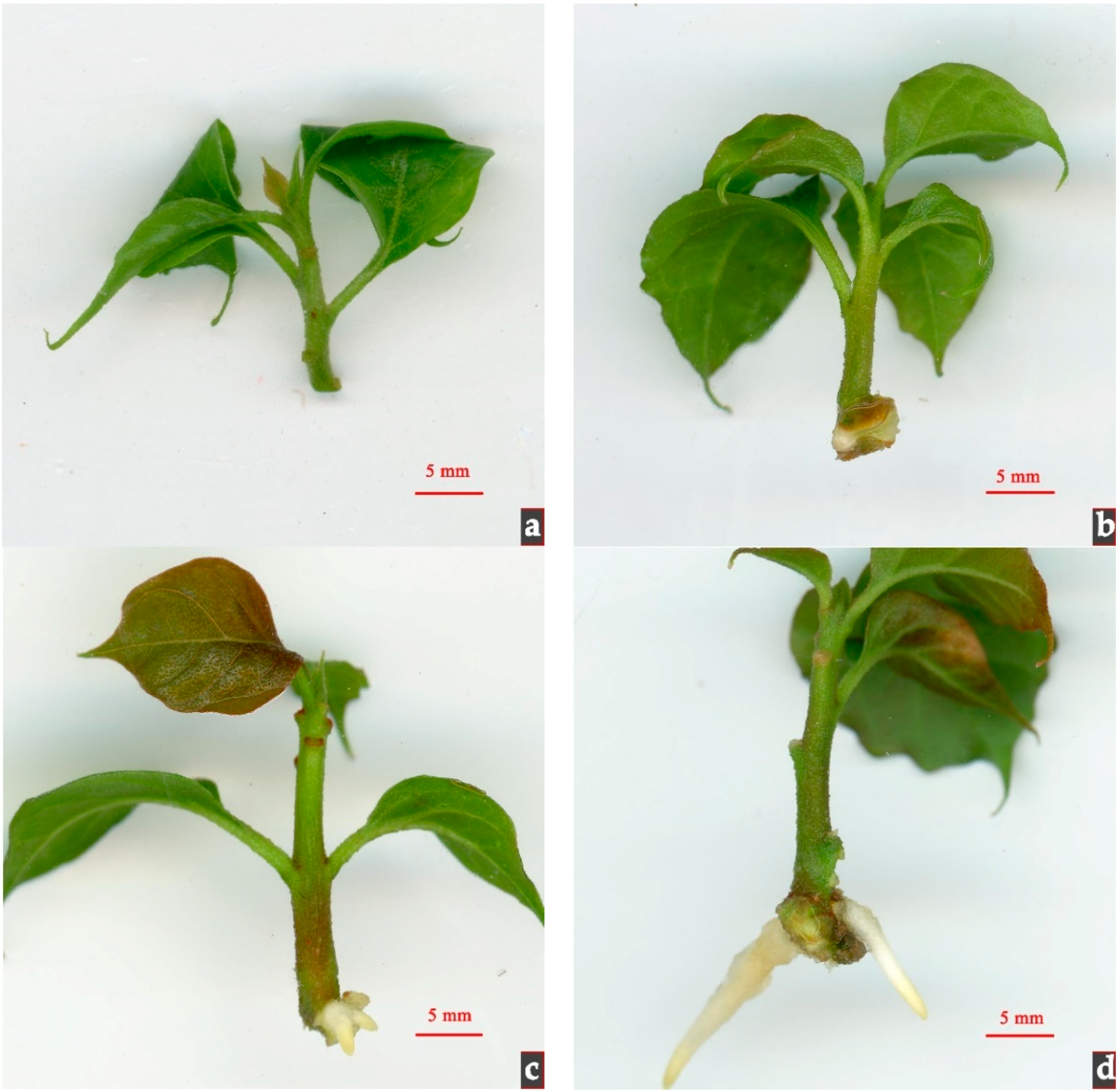
3.1. Morphological and Histological Observations

In the process of the early rooting culture, morphological observations using photos were taken weekly, and sampling was conducted at five-day intervals. It was found that the bud grew relatively well at the beginning (Figure 1a), while its bottom portion gradually expanded after one to two weeks of culture. At approximately three weeks, white roots began to emerge (Figure 1c) and continued to elongate as the culture duration prolonged (Figure 1d). It is inferred that the formation of root primordia occurred between one and three weeks.
According to the observation of the external morphology and anatomy of Bougainvillea, the overall shape is oval and consists of the epidermis, cortex, and vascular cylinder from the outside to the inside (Figure 2a). The epidermal cells are closely arranged and the cell volume is uniform, and the cuticle is relatively developed. The 3–4 layers of cells adjacent to the epidermal cells in the cortex are collenchyma, with small and closely arranged cells, while the cells close to the collenchyma are thin-walled, large, and irregularly arranged. The outer tough collateral bundles are scattered in the basic tissues of the vascular cylinder. The vascular bundles near the pith are arranged in a circle, and other vascular bundles are irregularly arranged in the basic tissues. In general, the vascular bundle becomes larger from the outside to the inside, indicating that the inner vascular bundles are more mature. There are inconspicuous xylem and phloem in the vascular bundles. The vascular cylinder has no obvious pith ray and interfascicular cambium, and the pith has no obvious divisions (Figure 2b).
After the Bougainvillea buds were cultured in the rooting medium, it was found from the continuous tissue slices that no latent root primordium was found, and the AR were formed from the induced root primordium and belong to the callus rooting type. With the prolongation of the tissue culture, the parenchyma cells continuously proliferated to form callus cells, which had different shapes and were closely arranged (Figure 2c). With the continuous divisions of the cells, a callus was induced, and a group of cells with a small size, close arrangement, and strong meristem ability, namely root primordia, were gradually formed in the callus (Figure 2d). With the development of the root primordium, AR gradually formed and passed through the cortex and callus (Figure 2e,f). Finally, the AR passed through the epidermis and formed roots vascularly connected with the stem (Figure 2g). With continued culture, the adventitious roots were further elongated (Figure 2h).

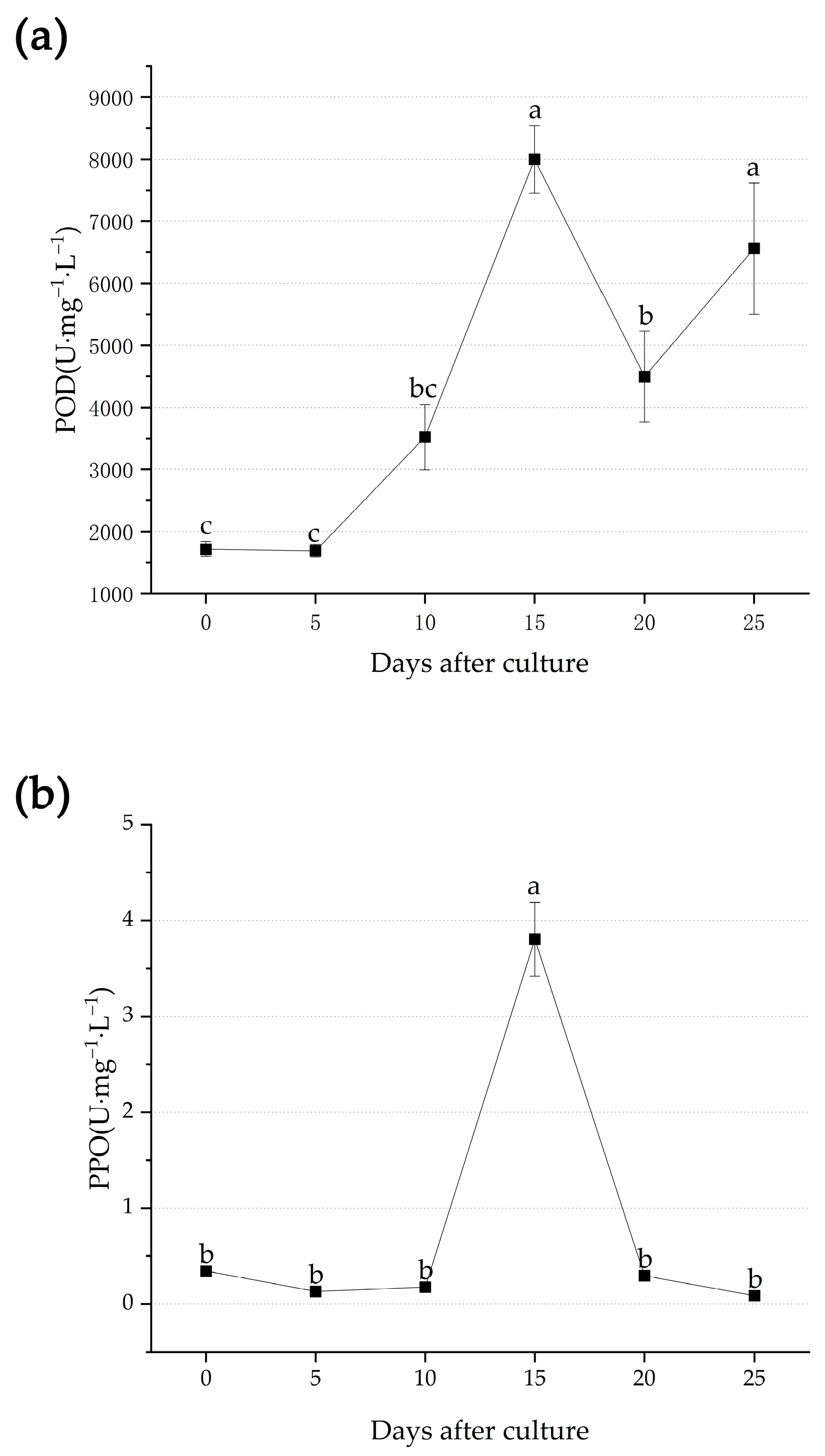
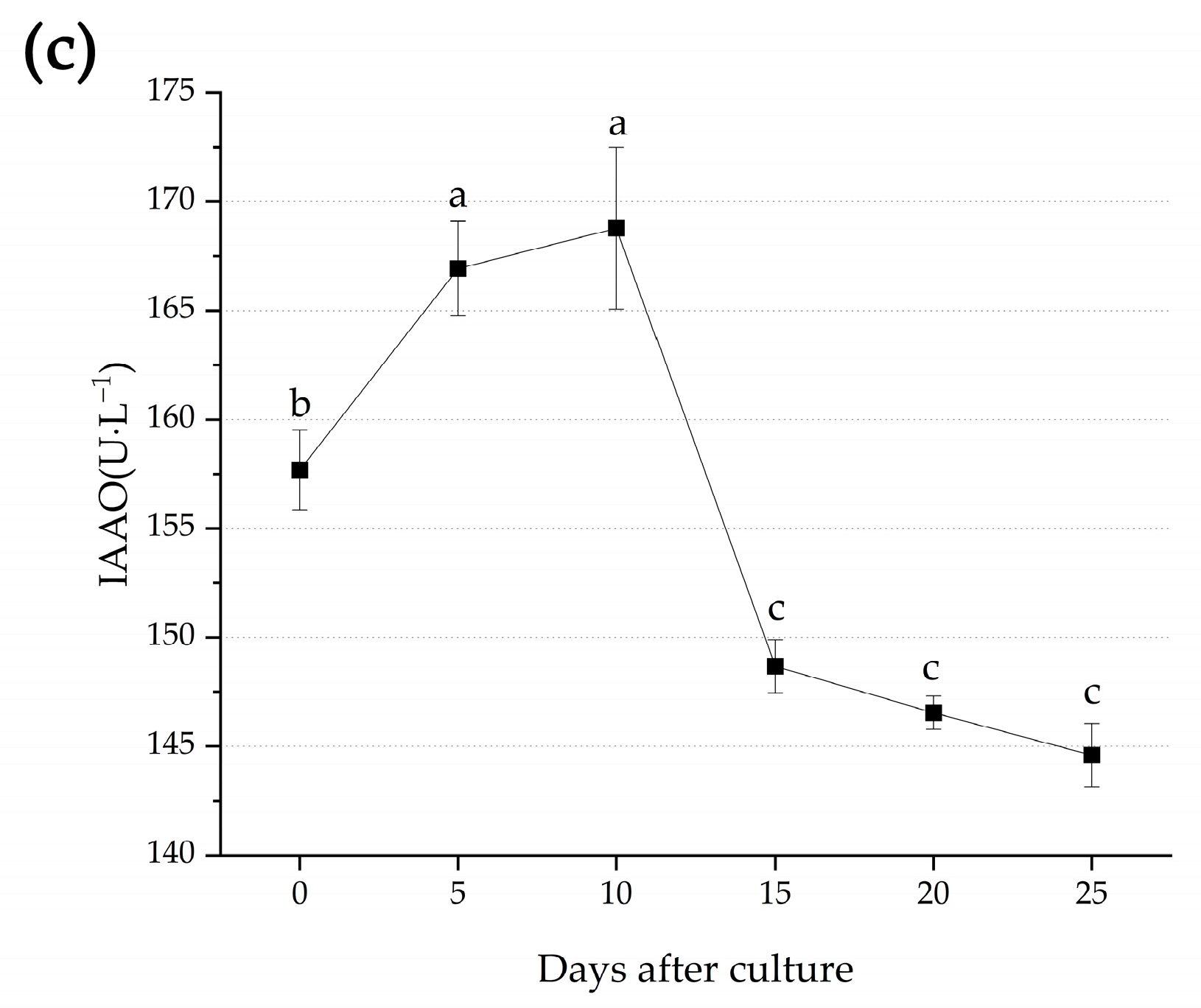
3.2. Metabolic Changes during Adventitious Root Formation

It can be noted that significant differences were observed between the POD, PPO, and IAAO activities during the adventitious root formation. For instance, a fluctuating trend was observed in the POD activities, initially increasing and reaching the peak value on Day 15 (7966.22 U∙mg−1∙L−1). Here, the peak periods were found to be consistent with the initiation phase of the adventitious root formation, on Day 20 (4494.22 U∙mg−1∙L−1). The POD level was also found to increase, with the peak levels achieved on Day 15 (6560.56 U∙mg−1∙L−1) (Figure 3a). Meanwhile, compared with the POD activities, the PPO activities followed an A-shaped trend, in which they initially increased and reached the peak value on Day 15 (3.80 U∙mg−1∙L−1), which was also consistent with the extension phase, until they rapidly decreased as they approached Day 20 (0.30 U∙mg−1∙L−1) (Figure 3b). For the IAAO activities, they also followed an A-shaped trend, in which they initially increased and reached the peak value on Day 10 (168.78 U∙L−1). However, they rapidly decreased towards the end of the culture duration (144.59U∙L−1) (Figure 3c).

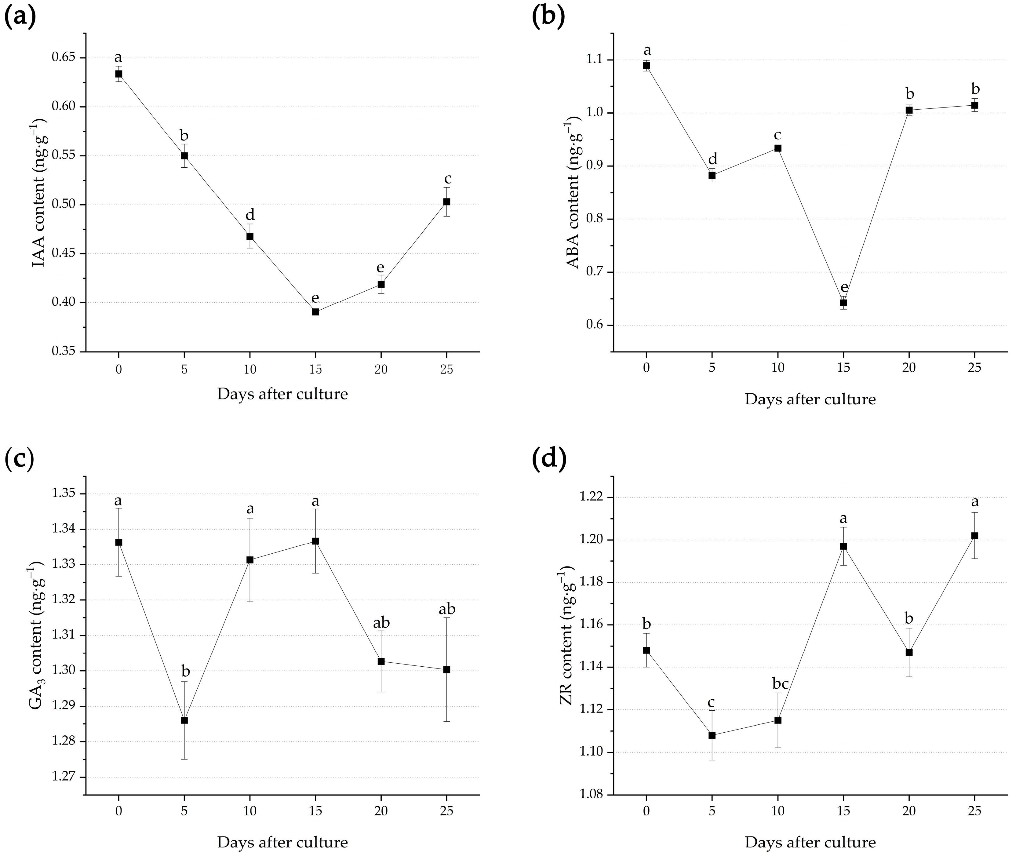
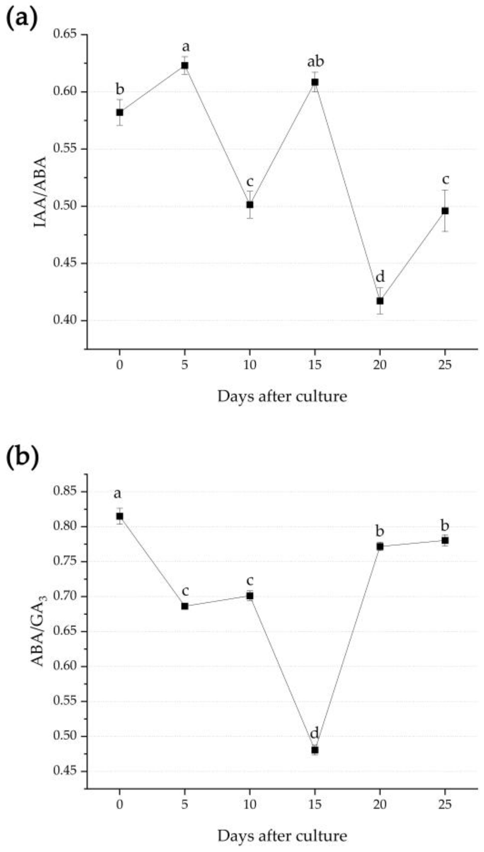
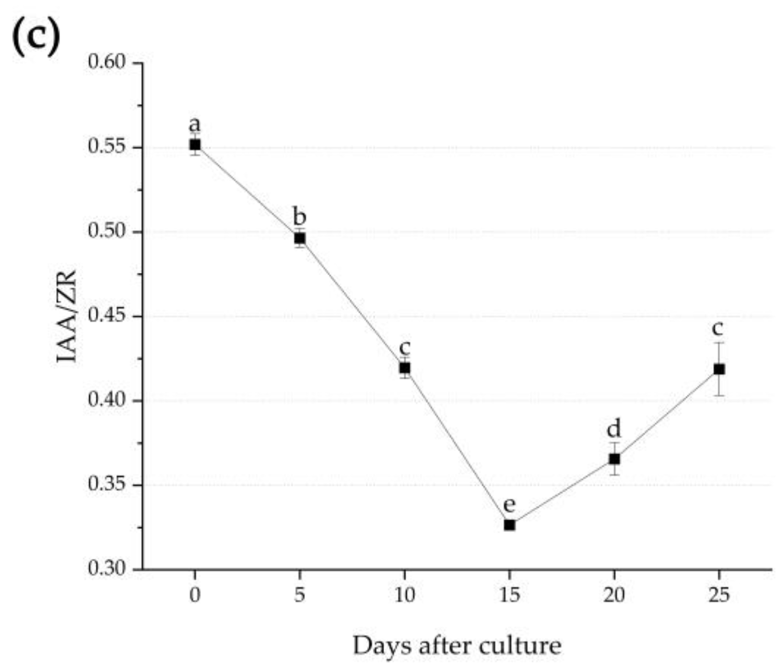
3.3. Endogenous Hormone Changes during Adventitious Root Formation

Significant differences were observed in the endogenous IAA, ABA, GA3, and ZR levels during the adventitious root formation. In the initiation and expression phases, the IAA contents continuously decreased to up to 0.391 ng·g−1 on Day 15. Meanwhile, they increased during the extension phase and peaked on the final day of the tissue culture (0.503 ng·g−1) (Figure 4a).
In the initiation and expression phase, the ABA levels showed a fluctuating trend, in which they initially decreased until Day 5 (0.883 ng·g−1), and then increased to 0.933 ng·g−1 on Day 10. This was then followed by a rapid decrement approaching Day 15 (0.642 ng·g−1). In the extension phase, the ABA levels continuously increased until the final day of the tissue culture (1.015 ng·g−1) (Figure 4b).
Similarly, the GA3 levels also showed fluctuating trends, in which they initially decreased until Day 5 (1.286 ng·g−1) and peaked on Day 15 (1.337 ng·g−1). This was consistent with the extension phase, and towards the final day of the tissue culture, the GA3 levels were continuously decreasing (1.300 ng·g−1) (Figure 4c).
Compared with the other endogenous hormones, the ZR levels showed a W-shaped trend, in which they initially decreased in the initiation and expression phases approaching Day 5 (1.108 ng·g−1) and peaked on Day 15 (1.197 ng·g−1). In the extension phase, they decreased until Day 20 (1.147 ng·g−1) and peaked on the final day of the tissue culture (1.202 ng·g−1) (Figure 4d).
The IAA/ABA ratio during the adventitious root formation showed significant differences. For instance, it demonstrated a double-A-shaped trend, in which it initially increased and peaked on Day 5 (0.623) and rapidly decreased approaching Day 10 (0.501). Thereafter, it increased and peaked on Day 15 (0.609) and rapidly decreased as it approached Day 20 (0.496). On the final day of the tissue culture, it was observed to be continuously increasing (0.496) (Figure 5a). The ABA/GA3 ratio continuously decreased and reached 0.480 on Day 15 in the initiation and expression phases. Further, this ratio increased during the extension phase (0.780) (Figure 5b). Lastly, the IAA/ZR ratio was found to continuously decrease up to 0.326 on Day 15. In the extension phase, it increased as it approached the final day of the tissue culture (0.419) (Figure 5c).

4. Discussion

4.1. Anatomical Evaluation and Root Primordia Development during Adventitious Root Formation

The adventitious root primordia of plant cuttings can be divided into two types: latent and induced root primordia [37]. They can also be divided into the cortex rooting type, callus rooting type, and mixed rooting type, based on the extension part of the AR. Further, the AR formation in tissue culture varies according to the plant species. For instance, Populus tremula × P. tremuloides [38] and Albizia julibrissin [39], both belonging to the cortex rooting type, have adventitious root primordia that can originate from the intersection of the cambium and pith ray, or the vascular cambium between the xylem and phloem. Carya illinoensis [40] forms a callus on the cambium and its root primordia are further differentiated from the developed callus. As a mixed rooting type refers to the ability of plants to simultaneously induce AR by the callus and cortex, the AR primordia, such as those of Amygdalus persica, can be formed in the cortex, phloem, xylem, and callus [41]. In Michelia figo [42], two types of mutual inhibition are exhibited. In Bougainvillea, it was found that its induced root primordium may be similar to that of Torreya grandis [43], as they both belong to the same callus rooting type. Most plant species’ induced root primordia originate from cells in the vascular cambium or parenchyma cells in the primary phloem near the vascular cambium, such as those of chestnut [44]. The AR of Bougainvillea seedlings also originate from the parenchyma, which has great potential to form root primordia [45]. Here, the formation of AR mainly involved the parenchyma cells located in the callus. With the restoration of the meristem ability of these ray parenchyma cells, they differentiate into root primordial cells, and then form root primordia, thus forming AR. These ARs can pass through the cortex and then develop into roots.
As the formation stage of AR involves induction, initiation, and expression [27,46], the AR formation in Bougainvillea is divided into the same three stages, based on the culture date and duration. Days 0–15 consist of the induction and initiation stages, while Days 15–25 represent the expression stage.
In the previous research on the tissue culture system of Nyctaginaceae, the AR that often emerged from the callus achieved a very low survival rate in the transplanting stage, which can be attributed to the growth of induced root primordia through the surrounding tissue layers, including the connection of the vascular bundle system with the stem, which is a pre-requisite for its expression [47]. However, the callus rooting type often lacks this process; hence, the reduction of the callus in the tissue culture system of Bougainvillea is beneficial for their post-transplanting survival as it is regulated by PGRs [48]. It is worth mentioning that, in our tissue culture system for ‘Miss Manila’, we achieved a high survival rate (93.33%) [24], which once again indicates that it is crucial to form a vascular bundle system connected to the stem in the expression stage. The auxin activity modulates the hydraulic properties, including the cell walls of the surrounding root primordia through its control of the expression of its own transporters [49]. In this study, it is inferred that the difference in rooting difficulty between varieties is attributed to their sensitivities to auxin. We reported, for the first time, that the AR induction of tissue-cultured seedlings of Bougainvillea belongs to the type of callus rooting. However, in previous reports considering the rooting of Bougainvillea cuttings, it was found that it belongs to the type of cortex rooting [50]. We speculated that the cuttings had a high degree of lignification and had formed a complete cambium, which can be induced to extend AR. As Bougainvillea is a vine plant, we assume the rooting type is related to the material status, and the microstructure of the material difference of Bougainvillea was also confirmed in Hu [51]. In short, it must be noted that AR formation is complex, as it is influenced by numerous interconnecting external and internal factors [52].
It was found that, in the experimental steps of the paraffin sections in the first experiment, the material tissue fell off during the section process. Generally, many woody plants also exhibit this phenomenon [53]. In this study, this was addressed by softening and increasing the wax soaking time as additional operational steps. Although there were still structural defects in the material when slicing, this test observed that the amount of tissue loss was lower, and the overall slicing effect was more accurate. The results showed that these additional steps were crucial to the slicing of Bougainvillea. Therefore, it is recommended that the paraffin sectioning is improved in future studies.

4.2. Changes of Metabolic Process during Adventitious Root Formation

POD is one of the primary enzymes related to the formation of adventitious roots, as it acts on the cell wall lignification [54] and catabolism of IAA and H2O2 during the rooting period [55]. The PPO participates in the regulation of phenolic precursor synthesis during lignin biosynthesis [56]; hence, it plays an important role during root differentiation and root primordia formation and development. In Bougainvillea, the POD activity was observed to increase on Day 15, which can be attributed to the analogous behavior of low activity at the early stage of rooting, and it regularly increases as the rooting progresses [57], as is also observed in Mucuna pruriens. It is inferred that the low POD activity is an indication of the start of the root induction stage, as this enzyme regulates endogenous IAA catabolism. As it increases and peaks, it signifies the rooting and rooting expression towards the end of the rooting stage [58]. Similarly, this was also exhibited during a cutting experiment in Berberis thunbergia [59], where a similar trend was shown. Generally, the PPO activity during the formation of AR in Bougainvillea continuously increased in the induction and initiation stages, and slowly decreased in the expression stage, which is consistent with the results obtained by Zhang [60].
The IAAO is the key enzyme of IAA catabolism and is closely related to IAA levels [61,62]. In this study, it was found that the IAAO activity increased continuously within 10 days of culture. Hence, it can be considered that the IAA was oxidized, owing to the role of oxidase. The increment can also be attributed to the high content of endogenous IAA and the presence of its derivatives, which stimulated the induction and development of the root primordium [63]. However, this simultaneously reduced the endogenous IAA levels, resulting in the inhibition of root development [64,65], Specifically, the IAAO activity decreased sharply in Days 10–15 to ensure the formation of AR. A positive correlation was found between the rooting and IAAO activity, specifically in the root cuttings of Populus [66]. Hence, it is inferred that the induction and initiation of AR through a dynamic balance results in the initial increment followed by the decrement in the IAAO activity. Studies have shown that the IAAO activity can be high in some plants in which it is difficult to develop roots, which consequently reduces the amount of IAA transferred to the base of cuttings, and such conditions may not be conducive for the induction of AR [67]. In this study, the IAAO activity was relatively high during rooting, hence, not conducive for AR formation. This indicates that the variety is difficult for rooting, which is consistent with the results of previous studies.
Oxidase has the function of oxidizing and decomposing auxin; hence, any change to its levels predominates the process of AR formation [68,69]. Here, the maximum value is often obtained during the root primordium formation. In this study, it was found that oxidase exhibited the same characteristics. The increment of POD and PPO as oxidase resulted in the further oxidation of auxin, which consequently led to the continuous increment of the IAAO levels and decrement of the IAA levels. Through the decreased oxidase content, the IAAO and IAA levels simultaneously decreased and increased, respectively. It was observed that the activity of oxidase can be an indicator of different stages during AR formation [70]. For instance, when the oxidase content was increasing in Days 10–15, the IAA and IAAO were also decreasing, which may be attributed to the regulation of the plant itself by increasing the SOD activity [71] to reach the appropriate IAA content and ensure root formation.

4.3. Changes of Endogenous Hormones during Adventitious Root Formation

In the three stages of AR formation, the induction stage has opposite hormone requirements compared with the subsequent initiation and expression stages [72]. Usually, the induction phase occurs during the first few hours following the cutting removal, without visible cell divisions, and it involves the reprogramming of the target cells. At the stage of AR expression, the cell structure undergoes a qualitative change, in which the cells differentiate from the root primordia into complete root bodies, followed by the emergence of roots.
Auxins are intimately involved in the formation of adventitious roots [73], and interdependent physiological stages in the rooting process are associated with the changes in endogenous auxin concentrations [74]. Indole-3-acetic acid (IAA), the main form of plant auxin, moves between plant cells, either in protonated form (IAAH) by membrane diffusion, or anionic form (IAA−) by carrier-mediated transport [75]. In the process of the AR induction and initiation of Bougainvillea, the IAA content decreased continuously, which was consistent with tree peony cuttings during AR formation [62]. Considering that the IAA content increased during the expression stages, this can be related to the changed oxidase activity. The increased oxidase content decreased the IAA content, and the decreased oxidase content increased the IAA content. It is worth noting that, in previous studies, the increment of the IAA content symbolized rooting [76]; however, on the 15th day after culture, the content of IAA oxidase was very low. At this time, the root primordium might be produced because the oxidation of IAA could promote rooting [63].
Abscisic acid (ABA) is a stress-related hormone, which has been proven to inhibit the AR formation and lateral root development of rice [77] and peanut [78]. During the formation of AR, Bougainvillea promoted rooting by reducing the ABA content to the appropriate level during the induction and initiation stages, and the ABA content kept rising to promote the elongation of AR in the expression stage [79]. This result was consistent with that of Armeniaca sibirica [80].
Gibberellin (GAs) is a complex family of tetracyclic diterpenoid plant growth regulators; some of them, such as GA1, GA3, and GA4, are believed to have the function of bioactive hormones [81]. GA3 is generally considered an inhibitor of AR formation in cutting propagation [82]. However, in our study, we found that GA3 decreased first during AR development, then increased in the induction and initiation stage, and finally decreased in the expression stage. We considered that high GA content could inhibit cell divisions at the initial stage of rooting culture, thereby inhibiting the differentiation and formation of AR, and then completing the differentiation of AR, or that GA could promote the elongation and growth of AR [83].
It can be noted that ABA may interfere with ethylene (ET) and gibberellin (GAs) signal pathways and block cell cycle progression [77], in that it forms an antagonistic effect with GA, as is also shown in this study. It was found that the ABA levels continuously decreased during the induction and initiation stages. The GA3 content also increased following the previous decrement. During the expression stage, the ABA levels increased, whereas the GA3 levels decreased. Therefore, this demonstrates that the interaction between ABA and GA3 affects the occurrence of AR.
Cytokinin plays a key role in the regulation of root development [84], and trans-Zeatin-riboside (ZR) has been identified as an important cytokinin in root exudates [85]. In this study, during the induction and initiation stages, we found that the ZR content decreased slightly 0–5 days after culture. This can be the consequence of transferring the explants to the rooting medium without cytokinin, which made it undergo a dedifferentiation process at the initial stage [72]. The constant increment of ZR content in the 10–15 days after culture indicated the promoted formation and growth of the root primordium, as well as lateral growth by the increased number of cells. We believe that the ZR content does not exceed the threshold value for rooting inhibition. The specific process includes a continuous division of the parenchyma to form a callus, and the gradual formation of cells with a strong ability for differentiation in the callus. During the expression stage, the reduction of the ZR content to an appropriate value made the root primordium further differentiate during the 15–20th days after culture [83]. The increased ZR content in the last stage was speculated to be the result of the continuous elongation of AR after the newly formed vascular bundle reconnection between the roots and stems, and the cell differentiation and proliferation in the phloem, xylem, and cambium areas [86].
Due to the interaction of multiple hormones in plants, a single hormone has a minimal correlation with the production of adventitious roots. Although the concentration of a single hormone meets the requirements for rooting, it may not necessarily result in the production of AR or inhibition of the rooting [87]. The ratio of endogenous hormones is also often used to signify the formation of adventitious roots [88]. The ratios of IAA/ABA and IAA/ZR were used to predict the formation of AR, in which they had a stable correlation with the formation of adventitious roots [62,83,89,90]. In this study, the IAA/ABA ratio peaked at Days 5 and 15, from which it can be inferred that the induction stage began on Day 5 and AR began to differentiate gradually on Day 15, indicating that the ratio was closely related to the formation of the AR, as was also confirmed in the rooting of Pinus bungeana cuttings [91]. In this experiment, the IAA/ZR ratio was the lowest at the initial expression stage of AR on Day 15. However, numerous experiments have proved that endogenous hormonal balances play an important role in plant rooting, in which higher IAA ratios are beneficial for the initiation of rooting [92,93]. As the cytokinins regulate a stage-specific formation of AR, low levels of cytokinins, combined with high auxin levels, are major processes involved in AR induction [86]. In tissue culture, the ratio of auxins and cytokinins is often controlled to regulate the rooting or budding of plant materials [94]. Therefore, the decreased and increased IAA/ZR ratios during AR formation in Bougainvillea were attributed to the high IAAO and low IAA levels in difficult-to-root varieties. Meanwhile, the increased ZR content resulted in the increment of the IAA/ZR ratio following the root primordium differentiation to ensure the growth and further elongation of the AR. The ABA and GAs have similar synthetic precursors, in which they can produce antagonistic effects. Studies have shown that higher ABA/GA3 ratios can promote the formation of AR during the formation of the root primordium and differentiation of adventitious roots [95]. However, in the study of Rhododendron hybridum, a relatively stable ABA/GA3 ratio was consistently demonstrated [90]. In this experiment, it was found that the ABA/GA3 ratio was lowest at the beginning of AR expression. Hence, it is inferred that differences exist between different plant species; in Bougainvillea, the low level was conducive for transformation from the AR initiation stage to expression stage. This finding was also consistent with the study on a root cutting of Malus hupehensis [35].

5. Conclusions

In order to provide an optimal breeding system for healthier and denser roots of B. buttiana ‘Miss Manila’, the morphology, as well as the anatomical, physiology, and biochemistry, of AR formation were investigated. We consider that, during the AR formation of Bougainvillea, the activities of PPO, POD, and IAAO generally increased during the AR induction and initiation stages, and the IAA content decreased with the increase of oxidase content. At the same time, the content of ZR increased continuously, and the callus was produced continuously. The content of ABA decreased, accompanied by the increment of GA content, and finally led to the differentiation of the callus into root primordial cells. In the expression stage, the activities of PPO, POD, and IAAO were generally reduced, which promoted the increment of IAA content, and the increment of ABA content and the decrement of GA content. The comprehensive regulation made the root primordium further grow and develop. This study revealed that the origin of AR formation and its spatial behavior in difficult-to-root varieties of Bougainvillea have rhythmic relationships between the rooting process and physiological and biochemical changes.

Author Contributions

Conceptualization, T.H. and Z.Z.; methodology, T.H.; software, T.H.; validation, T.H.; formal analysis, T.H.; data curation, T.H.; writing—original draft preparation, T.H.; writing—review and editing, H.Z. and Q.S.; supervision, Z.Z.; project administration, Q.S. and Z.Z.; funding acquisition, Q.S. and Z.Z. All authors have read and agreed to the published version of the manuscript.

Funding

This research was funded by the National Natural Science Foundation of China (32101582, 31770752), the Natural Science Foundation of Jiangsu Province of China (BK20210613), and The Natural Science Foundation of the Jiangsu Higher Education Institutions of China (21KJB220008).

Institutional Review Board Statement

Not applicable.

Informed Consent Statement

Not applicable.

Data Availability Statement

The data used to support the findings of this study are available from the corresponding author upon request.

Conflicts of Interest

The authors declare no conflict of interest.

References

  1. Ahmed, A.H. New Flavone from the Aerial Parts of Bougainvillea glabra. Int. J. Comput. Eng. Res. 2014, 4, 1–5. [Google Scholar]
  2. Jain, R.; Janakiram, T.; Swaroop, K.; Kumar, S.; Kumawat, G.L. Standardization of protocol for in vitro multiplication of Bougainvillea. Indian J. Agric. Sci. 2016, 86, 516–521. [Google Scholar]
  3. Christopher, B.C.; James, L.G. Influence of Indolebutyric Acid Potassium Salt on Propagation of Semi-hardwood Stem Cuttings of Bougainvillea. HortScience 2006, 41, 983. [Google Scholar]
  4. Lai, R.Y.; Zhong, Z.H.; Zhang, X.Q.; Lin, L.X.; Su, M.H.; Xie, Z.N. Effects of Remaining Leaf Combining with IBA on Rooting, Physiological and Biochemical Indicators of Leaves from Bougainvillea spectabilis Cuttings. Agric. Sci. Technol. 2010, 11, 120–123. [Google Scholar]
  5. Vidoz, M.L.; Loreti, E.; Mensuali, A.; Alpi, A.; Perata, P. Hormonal interplay during adventitious root formation in flooded tomato plants. Plant J. 2010, 63, 551–562. [Google Scholar] [CrossRef]
  6. Calvo-Polanco, M.; Señorans, J.; Zwiazek, J.J. Role of adventitious roots in water relations of tamarack (Larix laricina) seedlings exposed to flooding. BMC Plant Biol. 2012, 12, 99. [Google Scholar] [CrossRef] [Green Version]
  7. Tran, Y.B.N.; Van-Kieu, N.; Nguyen, P.T.K. Cytotoxic flavonoids from the roots of Bougainvillea spectabilis. Phytochem. Lett. 2021, 42, 117–120. [Google Scholar] [CrossRef]
  8. Singh, S.; Reddu, S.K.; Sharma, S.K.; Ali, M. New Unsaturated Fatty Acid from Roots of Bougainvillea spectabilis Willd. Asian J. Chem. 2009, 21, 4744–4748. [Google Scholar]
  9. Balasaraswathi, R.; Sadasivam, S.; Ward, M.; Walker, J.M. An antiviral protein from Bougainvillea spectabilis roots; purification and characterisation. Phytochemistry 1998, 47, 1561–1565. [Google Scholar] [CrossRef]
  10. Singh, V.; Aggrawal, V. Phytochemical analysis and in vitro antioxidant activities of leaves, stems, flowers, and roots extracts of Bougainvillea spectabilis Willd. Int. J. Green Pharm. 2018, 12, 278–284. [Google Scholar] [CrossRef]
  11. Jawla, S.; Kumar, Y.; Khan, M.S.Y. Isolation of phytoconstituents and antihyperglycemic activity of Bougainvillea spectabilis root bark extracts. Lat. Am. J. Pharm. 2013, 32, 1389–1395. [Google Scholar]
  12. Deshmukh, K.K.; Barad, A.V. Effect of IBA and NAA on Root Initiation of Various Varieties of Bougainvillea Species. Gujarat Agric. Univ. Res. J. 2002, 27, 44–46. [Google Scholar]
  13. Singh, N.; Singh, B.P.; Singh, H.K. Effect of different concentrations of indole butyric acid (IBA) on rooting potential and root growth of Bougainvillea stem cuttings. J. Ornam. Hortic. 2010, 13, 41–44. [Google Scholar]
  14. Kuruwanshi, V.B.; Deo, A.K.; Pal, D.P.; Sarnaik, D.A. Effect of treatment of stem cutting with IBA and NAA on sprouting, rooting and root biomass in Bougainvillea var. Refulgence. Adv. Plant Sci. 2009, 21, 557–558. [Google Scholar]
  15. Singh, P.; Singh, K. Effects of NAA and IBA on Rooting of Stem Cuttings in Bougainvillea CV. Cherry Blossom. Agric. Res. J. 2003, 40, 205–206. [Google Scholar]
  16. Abd, I.A.; Meklef, A.A.; Neamah, S.I. Microbial technique for the production of growth regulator compounds Indole Butyric Acid (IBA) and its role on the rooting and growth of Bougainvillea spectabilis L. stem cuttings. In IOP Conference Series: Earth and Environmental Science; IOP Publishing: Bristol, UK, 2021; Volume 761, p. 012003. [Google Scholar]
  17. Atzmon, N.; Wiesman, Z.; Fine, P. Biosolids improve rooting of bougainvillea (Bougainvillea glabra) cuttings. J. Environ. Hortic. 1997, 15, 1–5. [Google Scholar] [CrossRef]
  18. Costa, E.M.; Loss, A.; Pereira, H.P.N.; Almeida, J.F. Rooting of Bougainvillea spectabilis Willd. stem cuttings using indol butyric acid. Acta Agron. 2015, 64, 205–210. [Google Scholar]
  19. Iddrisu, I.; Adzraku, H.-V.; Tandoh, P.K. Effects of Different Soil-biochar on Physico-Chemical Soil Properties, Rooting and Growth of Bougainvillea glabra and Ficus benjemena Using Stem Cuttings. Asian J. Agric. Hortic. Res. 2018, 1, 1–16. [Google Scholar] [CrossRef]
  20. Kale, P.N.; Bhujbal, B.G. Use of Growth Regulators in Rooting of Cuttings of Bougainvillea Var. Mary Palmer. Indian J. Hortic. 1972, 29, 307–309. [Google Scholar]
  21. Li, C.N.; Deng, J.L.; Zhou, J.Y.; Li, X.L.; Zen, S.J.; Bu, C.Y. lmpact Factors of Root Growth Under Hardwood Cuttage of Bougainvillea glabra. J. Agric. 2016, 6, 47–52. [Google Scholar]
  22. Kumari, P.; Swaroop, K.; Janakiram, T.; Singh, S.K.; Prasad, K.V.; Jain, R. In vitro Sterilization, Rooting and Acclimatization of Difficult-to-root Bougainvillea Cultivars. Int. J. Bio-Resour. Stress Manag. 2016, 7, 412–419. [Google Scholar] [CrossRef]
  23. Datta, S.K.; Mandal, A.K.A. Standardization of in vitro Multiplication of Two Difficult-to-root Bougainvillea Cultivars for Commercial Exploitation. Sci. Cult. 2012, 78, 251–254. [Google Scholar]
  24. Huang, T.; Zhang, H.H.; Zhao, R.N.; Zhu, Z.L. Establishing an Efficient Regeneration System for Tissue Culture in Bougainvillea buttiana ‘Miss Manila’. Plants 2022, 11, 2372. [Google Scholar] [CrossRef] [PubMed]
  25. Kumari, P.; Swaroop, K.; Janakiram, T.; Jain, R.; Singh, S.K.; Singh, N. In vivo propagation of difficult-to-root Bougainvillea cultivars by air layering. J. Ornam. Hortic. 2014, 17, 82–87. [Google Scholar] [CrossRef]
  26. Correa, L.R.; Troleis, J.; Mastroberti, A.A.; Mariath, J.E.; Fett-Neto, A.G. Distinct modes of adventitious rooting in Arabidopsis thaliana. Plant Biol. 2012, 14, 100–109. [Google Scholar] [PubMed]
  27. Pacurar, D.I.; Perrone, I.; Bellini, C. Auxin is a central player in the hormone cross-talks that control adventitious rooting. Physiol. Plant. 2014, 151, 83–96. [Google Scholar] [CrossRef]
  28. Xiao, Z.A.; Xiong, H. Research Progress in Mechanism of Adventitious root Organgenesis for Microporpagated Shoots. Biotechnol. Bull. 2002, 3, 31–34. [Google Scholar]
  29. Zhang, H.X.; Dong, C.J.; Li, F.K.; Wang, H.F.; Shang, Q.M. Progress on the Regulatory Mechanism of Adventitious Rooting. Acta Bot. Boreali-Occident. Sin. 2017, 37, 1457–1464. [Google Scholar]
  30. Xu, A.H.; Wei, C.X. Comprehensive comparison and applications of different sections in investigating the microstructure and histochemistry of cereal kernels. Plant Methods 2020, 16, 8. [Google Scholar] [CrossRef]
  31. Li, S.J.; Yang, N.; Chen, L.Q. Paraffin section observation of flower bud differentiation of Chimonanthus praecox in Kunming and comparison of the differentiation processes in different regions, China. Hortic. Plant J. 2022, 8, 221–229. [Google Scholar] [CrossRef]
  32. Liu, G.B.; Zhao, J.Z.; Zhang, Y.P.; Liao, T.; Guo, L.Q.; Cao, J. Study on the occurrence pattern of Platycladus orientalis. Acta Bot. Boreali-Occident. Sin. 2020, 40, 987–996. [Google Scholar]
  33. Zhang, Z.L.; Qu, W.J. Experimental Course of Plant Physiology; China Forestry Press: Beijing, China, 2003; pp. 88–104. [Google Scholar]
  34. Lu, W.J.; Li, Y.S. Experimental Course of Plant Physiology; China Forestry Press: Beijing, China, 2012; pp. 43–45. [Google Scholar]
  35. Zhang, W.X.; Fan, J.J.; Tan, Q.Q.; Zhao, M.M.; Cao, F.L. Mechanisms Underlying the Regulation of Root Formation in Malus hupehensis Stem Cuttings by Using Exogenous Hormones. J. Plant Growth Regul. 2017, 36, 174–185. [Google Scholar] [CrossRef]
  36. Yang, Y.M.; Xu, C.N.; Wang, B.M.; Jia, J.Z. Effects of plant growth regulators on secondary wall thickening of cotton fibres. Plant Growth Regul. 2001, 35, 233–237. [Google Scholar]
  37. Liu, Y.; Xiao, D.X.; Huang, C.F.; Lei, X.G. The Anatomical Feasture Study on the Rooting of the Tender Branch Cuttings of Castanea mollissima Blume. Acta Hortic. Sin. 1997, 24, 8–12. [Google Scholar]
  38. Zhou, R.T.; Liu, Y.; Leng, S.S.; Wang, N.; Yan, S.P. Morphological and Anatomical Features during the Formation of Adventitious Roots of Populus tremula × P. tremuloides by Micro-cuttage Multiplication. Acta Bot. Boreali-Occident. Sin. 2013, 33, 2184–2188. [Google Scholar]
  39. Zhou, X.M.; Liu, Y.T.; Zhao, X.Z.; Song, Z.W.; Wang, S. Rooting Anatomy of Hardwood Cutting for Albizia julibrissin Duraxx. and Activity Change of Related Enzymes During Rooting Process. Bull. Bot. Res. 2016, 36, 58–61. [Google Scholar]
  40. Zhang, J.H.; Shi, J.J.; Xia, G.H.; Huang, Y.J.; Huang, J.X.; Wang, Z.J. Research of lignified cuttings and rooting mechanism of Carya illinoensis. J. Anhui Agric. Univ. 2014, 41, 203–208. [Google Scholar]
  41. Bai, X.Y.; Wang, L.R.; Wang, X.W.; Zhu, G.R.; Fang, W.C.; Cao, K.; Chen, C.W.; Li, Y. Microscopic observation on adventitious root development of micropropagation and cutting propagation on peach rootstocks. J. Fruit Sci. 2015, 32, 74–78+175. [Google Scholar]
  42. Zheng, Q.Q.; Lan, S.R.; Liu, X.D.; Feng, Y.C.; Peng, D.H. Effects of growth regulators on rooting and correlative enzyme activities of Michelia figo ‘Xiangfei’ cuttings. J. Cent. South Univ. For. Technol. 2020, 40, 67–76. [Google Scholar]
  43. Jin, H.D.; Zheng, C.Y.; Hua, B.; Yu, C.L.; Li, K.Y.; Yu, W.W. Rooting anatomy and physiological enzyme activity of Torreya grandis cuttings. Acta Agric. Zhejiangensis 2022, 34, 1955–1966. [Google Scholar]
  44. Vielba, J.M.; Vidal, N.; José, M.C.S.; Rico, S.; Sánchez, C. Recent Advances in Adventitious Root Formation in Chestnut. Plants 2020, 9, 1543. [Google Scholar] [CrossRef] [PubMed]
  45. Legué, V.; Rigal, A.; Bhalerao, R.P. Adventitious root formation in tree species: Involvement of transcription factors. Physiol. Plant. 2014, 151, 192–198. [Google Scholar] [CrossRef] [PubMed] [Green Version]
  46. Kevers, C.; Hausman, J.F.; Faivre-Rampant, O.; Evers, D.; Gaspar, T. Hormonal control of adventitious rooting: Progress and questions. J. Appl. Bot. 1997, 71, 71–79. [Google Scholar]
  47. Ballester, A.; San-José, M.C.; Vidal, N.; Fernández-Lorenzo, J.L.; Vieitez, A.M. Anatomical and Biochemical Events during in vitro Rooting of Microcuttings from Juvenile and Mature Phases of Chestnut. Ann. Bot. 1999, 83, 619–629. [Google Scholar] [CrossRef]
  48. Sharma, A.K.; Prasad, R.N.; Chaturvedi, H.C. Clonal propagation of Bougainvillea glabra ‘Magnifica’ through shoot apex culture. Plant Cell Tissue Organ Cult. 1981, 1, 33–38. [Google Scholar] [CrossRef]
  49. Banda, J.; Bellande, K.; Wangenheim, D.V.; Goh, T.; Guyomarc, S.; Laplaze, L.; Bennett, M.J. Lateral Root Formation in Arabidopsis: A Well-Ordered LRexit. Trends Plant Sci. 2019, 24, 826–839. [Google Scholar] [CrossRef]
  50. Xie, Z.N.; Lai, R.Y.; Lin, L.X.; Zhong, Z.H. Anatomical observation on the rooting of the cutting of Bougainvillea spectabilis. J. Minxi Vocat. Tech. Coll. 2008, 10, 97–99. [Google Scholar]
  51. Hu, R. Anatomical Structure of the Stem of Bougainvillea glabra. J. Neijiang Norm. Univ. 2015, 30, 15–19. [Google Scholar]
  52. Ibáñez, S.; Ruiz-Cano, H.; Fernández, M.Á.; Sánchez-García, A.B.; Villanova, J.; Micol, J.L.; Pérez-Pérez, J.M. A Network-Guided Genetic Approach to Identify Novel Regulators of Adventitious Root Formation in Arabidopsis thaliana. Front. Plant Sci. 2019, 10, 461. [Google Scholar] [CrossRef]
  53. Zhou, N.F.; Zhang, J.P.; Liu, H.; Cha, W.W.; Pei, D. New Protocols for Paraffin Sections of Heterogeneous Tissues of Woody Plants. Chin. Bull. Bot. 2018, 53, 653–660. [Google Scholar]
  54. Quiroga, M.; Guerrero, C.; Botella, M.A.; Barceló, A.; Amaya, I.; Medina, M.I.; Alonso, F.J.; Forchetti, S.M.; Tigier, H.; Valpuesta, V. A tomato peroxidase involved in the synthesis of lignin and suberin. Plant Physiol. 2000, 122, 1119–1127. [Google Scholar] [CrossRef] [PubMed] [Green Version]
  55. Cheniany, M.; Ebrahimzadeh, H.; Masoudi-nejad, A.; Vahdati, K.; Leslie, C. Effect of endogenous phenols and some antioxidant enzyme activities on rooting of Persian walnut (Juglans regia L.). Afr. J. Plant Sci. 2010, 4, 479–487. [Google Scholar]
  56. Elmongy, M.S.; Zhou, H.; Cao, Y.; Liu, B.; Xia, Y.P. The effect of humic acid on endogenous hormone levels and antioxidant enzyme activity during in vitro rooting of evergreen azalea. Sci. Hortic. 2018, 227, 234–243. [Google Scholar] [CrossRef]
  57. Li, S.W.; Xue, L.G.; Xu, S.J.; Feng, H.Y.; An, L.Z. IBA-induced changes in antioxidant enzymes during adventitious rooting in mung bean seedlings: The role of H2O2. Environ. Exp. Bot. 2009, 66, 442–450. [Google Scholar] [CrossRef]
  58. Porfirio, S.; Calado, M.L.; Noceda, C.; Cabrita, M.J.; da Silva, M.G.; Azadi, P.; Peixe, A. Tracking biochemical changes during adventitious root formation in olive (Olea europaea L.). Sci. Hortic. 2016, 204, 41–53. [Google Scholar] [CrossRef] [Green Version]
  59. Tehranifar, A.; Tabar, S.M.; Selahvarzi, Y. Biochemical changes in barberries during adventitious root formation: The role of indole-3-butyric acid and hydrogen peroxide. Span. J. Agric. Res. 2014, 12, 477–485. [Google Scholar] [CrossRef] [Green Version]
  60. Zhang, W.X.; Fan, J.J.; Tan, Q.Q.; Zhao, M.M.; Zhou, T.; Cao, F.L. The effects of exogenous hormones on rooting process and the activities of key enzymes of Malus hupehensis stem cuttings. PLoS ONE 2017, 12, e0172320. [Google Scholar] [CrossRef] [Green Version]
  61. He, S.L. Dynamic changes in enzyme activities and phenolic content during in vitro rooting of tree peony (Paeonia suffruticosa Andr.) plantlets. Maejo Int. J. Sci. Technol. 2011, 5, 252–265. [Google Scholar]
  62. Meng, X.Y.; Wang, Z.; He, S.L.; Shi, L.Y.; Song, Y.L.; Lou, X.Y.; He, D. Endogenous hormone levels and activities of IAA-modifying enzymes during adventitious rooting of tree peony cuttings and grafted scions. Hortic. Environ. Biotechnol. 2019, 60, 187–197. [Google Scholar] [CrossRef]
  63. Ilczuk, A.; Jacygrad, E. The effect of IBA on anatomical changes and antioxidant enzyme activity during the in vitro rooting of smoke tree (Cotinus coggygria Scop.). Sci. Hortic. 2016, 210, 268–276. [Google Scholar] [CrossRef]
  64. Moncousin, C.H.; Gaspar, T.H. Peroxidase as a Marker for Rooting Improvement of Cynara scolymus L. Cultured in vitro. Biochem. Und Physiol. Pflanz. 1983, 178, 263–271. [Google Scholar] [CrossRef]
  65. Song, J.Y.; He, W.L.; Li, S.B.; Liu, Y.J.; Sun, Y.J. Analysis of physiological and biochemical characteristics related to cutting and rooting of chimera in Populous tomentosa carr. Sci. Silvae Sin. 2001, 37, 64–67. [Google Scholar]
  66. GÜNEŞ, T. Peroxidase and IAA-Oxidase Activities During Rooting in Cuttings of Three Poplar Species. Turk. J. Bot. 2000, 24, 97–102. [Google Scholar]
  67. Tong, Z.Q. Studies on the Softwood Cutting Propagation Technique and Rooting Mechanism of Euonymus alatus (Thunb.) Sieb. Master’s Thesis, Shenyang Agricultural University, Shenyang, China, 12 June 2017. [Google Scholar]
  68. Husen, A.; Pal, M. Metabolic changes during adventitious root primordium development in Tectona grandis Linn. f. (teak) cuttings as affected by age of donor plants and auxin (IBA and NAA) treatment. New For. 2007, 33, 309–323. [Google Scholar] [CrossRef]
  69. Fett-Neto, A.; Teixeira, S.L.; Silva, E.A.A.D.; Anna, R.S. Biochemical and Morphological Changes during in vitro Rhizogenesis in Cuttings of Sequoia sempervirens (D. Don) Endl. J. Plant Physiol. 1992, 140, 720–728. [Google Scholar] [CrossRef]
  70. Mato, M.C.; Rúa, M.L.; Enrique, F. Changes in levels of peroxidases and phenolics during root formation in Vitis cultured in vitro. Physiol. Plant. 1988, 72, 84–88. [Google Scholar] [CrossRef]
  71. Tian, M.; Rao, L.B.; Li, J.Y. Reactive Oxygen Species (ROS)and lts Physiological Functions in Plant Cells. Plant Physiol. J. 2005, 37, 235–241. [Google Scholar]
  72. Druege, U.; Hilo, A.; Pérez-Pérez, J.M.; Klopotek, Y.; Acosta, M.; Shahinnia, F.; Zerche, S.; Franken, P.; Hajirezaei, M.R. Molecular and physiological control of adventitious rooting in cuttings: Phytohormone action meets resource allocation. Ann. Bot. 2019, 123, 929–949. [Google Scholar] [CrossRef] [Green Version]
  73. Rasmussen, A.; Hosseini, S.A.; Hajirezaei, M.R.; Druege, U.; Geelen, D. Adventitious rooting declines with the vegetative to reproductive switch and involves a changed auxin homeostasis. J. Exp. Bot. 2015, 66, 1437–1452. [Google Scholar] [CrossRef] [Green Version]
  74. Osterc, G.; Štampar, F. Differences in endo/exogenous auxin profile in cuttings of different physiological ages. J. Plant Physiol. 2011, 168, 2088–2092. [Google Scholar] [CrossRef]
  75. Kumar, P.; Patel, P.K.; Sonkar, M.K. Propagation through juvenile shoot cuttings in difficult-to-root Dalbergia latifolia—examining role of endogenous IAA in adventitious rooting. Plant Physiol. Rep. 2022, 27, 242–249. [Google Scholar] [CrossRef]
  76. Ahkami, A.H.; Melzer, M.; Ghaffari, M.R.; Pollmann, S.; Javid, M.G.; Shahinnia, F.; Hajirezaei, M.R.; Druege, U. Distribution of indole-3-acetic acid in Petunia hybrida shoot tip cuttings and relationship between auxin transport, carbohydrate metabolism and adventitious root formation. Planta 2013, 238, 499–517. [Google Scholar] [CrossRef] [PubMed] [Green Version]
  77. Steffens, B.; Wang, J.X.; Sauter, M. Interactions between ethylene, gibberellin and abscisic acid regulate emergence and growth rate of adventitious roots in deepwater rice. Planta 2006, 223, 604–612. [Google Scholar] [CrossRef] [PubMed]
  78. Guo, D.L.; Liang, J.H.; Qiao, Y.C.; Yan, Y.C.; Li, L.; Dai, Y.H. Involvement of G1-to-S transition and AhAUX-dependent auxin transport in abscisic acid-induced inhibition of lateral root primodia initiation in Arachis hypogaea L. J. Plant Physiol. 2012, 169, 1102–1111. [Google Scholar] [CrossRef] [PubMed]
  79. Chen, C.W.; Yang, Y.W.; Lur, H.S.; Tsai, Y.G.; Chang, M.C. A novel function of abscisic acid in the regulation of rice (Oryza sativa L.) root growth and development. Plant Cell Physiol. 2006, 47, 1–13. [Google Scholar] [CrossRef] [PubMed] [Green Version]
  80. Dong, S.J.; Liu, M.G.; Dai, F.; Wu, Y.L.; Shan, S.T.; Ding, R.J. Variation of endogenous hormone contents in softwood cuttings of Armeniaca sibirica during adventitious root formation. Non-Wood For. Res. 2013, 31, 108–114. [Google Scholar]
  81. Pacifici, E.; Polverari, L.; Sabatini, S. Plant hormone cross-talk: The pivot of root growth. J. Exp. Bot. 2015, 66, 1113–1121. [Google Scholar] [CrossRef] [Green Version]
  82. Da Costa, C.T.; De Almeida, M.R.; Ruedell, C.M.; Schwambach, J.; Maraschin, F.S.; Fett-Neto, A.G. When stress and development go hand in hand: Main hormonal controls of adventitious rooting in cuttings. Front. Plant Sci. 2013, 4, 133. [Google Scholar] [CrossRef] [Green Version]
  83. Zhu, Z.L.; Qian, Y.P.; Jin, J.B.; Zhou, Q. Physiology and biochemistry dynamics in Carpinus betulus during rooting culture. J. Fujian Agric. For. Univ. Nat. Sci. Ed. 2017, 46, 43–49. [Google Scholar]
  84. Palmer, M.V.; Wong, O.C. Identification of Cytokinins from Xylem Exudate of Phaseolus vulgaris L. Plant Physiol. 1985, 79, 296–298. [Google Scholar] [CrossRef] [Green Version]
  85. Davey, J.E.; Staden, J.V. Cytokinin translocation: Changes in zeatin and zeatin-riboside levels in the root exudate of tomato plants during their development. Planta 1976, 130, 69–72. [Google Scholar] [CrossRef] [PubMed]
  86. Agulló-Antón, M.Á.; Ferrández-Ayela, A.; Fernández-García, N.; Nicolás, C.; Albacete, A.; Pérez-Alfocea, F.; Sánchez-Bravo, J.; Pérez-Pérez, J.M.; Acosta, M. Early steps of adventitious rooting: Morphology, hormonal profiling and carbohydrate turnover in carnation stem cuttings. Physiol. Plant. 2014, 150, 446–462. [Google Scholar] [CrossRef] [PubMed]
  87. Ao, H.; Wang, K.; Feng, Y.L. Endogenous hormones levels in cuttings of Larix olgensis and their relations to rooting. Bull. Bot. Res. 2002, 22, 190–195. [Google Scholar]
  88. He, H.W. Studies on The Changes of Structure, Physiology and GeneExpression in Chromolaena odorata Adventitious Rooting. Master’s Thesis, Guangxi University, Nanning, China, 25 May 2015. [Google Scholar]
  89. Ross, A.R.S.; Ambrose, S.J.; Cutler, A.J.; Feurtado, J.A.; Kermode, A.R.; Nelson, K.; Zhou, R.; Abrams, S.R. Determination of endogenous and supplied deuterated abscisic acid in plant tissues by high-performance liquid chromatography-electrospray ionization tandem mass spectrometry with multiple reaction monitoring. Anal. Biochem. 2004, 329, 324–333. [Google Scholar] [CrossRef]
  90. Chen, L.Y.; Zheng, Y.; Chen, L.G.; Rong, J.D.; Zheng, Y.S. Study on Rhododendron hybridum’s rooting culture and hormones contents changes. J. For. Environ. 2011, 31, 131–135. [Google Scholar]
  91. Guo, S.J.; Ling, H.Q.; Li, F.L. Physiological and biochemical basis of rooting of Pinus bungeana cuttings. J. Beijing For. Univ. 2004, 26, 43–47. [Google Scholar]
  92. Cline, M.; Wesse, T.; Iwamura, H. Cytokinin/Auxin Control of Apical Dominance in Ipomoea nil. Plant Cell Physiol. 1997, 38, 659–667. [Google Scholar] [CrossRef] [Green Version]
  93. Nag, S.; Saha, K.; Choudhuri, M. Role of Auxin and Polyamines in Adventitious Root Formation in Relation to Changes in Compounds Involved in Rooting. J. Plant Growth Regul. 2001, 20, 182–194. [Google Scholar] [CrossRef]
  94. Dagla, H. Plant tissue culture. Resonance 2012, 17, 759–767. [Google Scholar] [CrossRef]
  95. Feng, X. The Study of Basis of Adventitious Roots on (P. simonii × P. nigra) × P.15 ACL Tissue Culuture Leaves. Master’s Thesis, Northeast Forestry University, Harbin, China, 2014. [Google Scholar]
Figure 1. Morphological observation of the in vitro rooting of B. buttiana ‘Miss Manila’. (a) Morphological structure on Day 0. (b) Morphological structure on Day 7,14. The bottom of the bud kept expanding. (c) Morphological structure on Day 21 in tissue culture. The white root protruded from the bottom. (d) Morphological structure on Day 28 in tissue culture. Further elongation of adventitious roots.
Horticulturae 08 01156 g001
Figure 2. Anatomical analysis of in vitro rooting of B. buttiana ‘Miss Manila’. (a) Histological structure on Day 0. (b) Histological structure on Day 5. (c) Histological structure on Day 10. The parenchyma cells of the basic tissue continuously proliferate and form callus tissue. (d) Histological structure on Day 15. Root primordia were gradually formed in the callus (e) The differentiated root primordia continuously pushed outward in the cortex. (f) Adventitious roots, formed from root primordia, penetrated the cortex and callus. (g) The adventitious roots pass through the epidermis and form roots vascularly connected with the stem. (h) Constant elongation of adventitious roots. The red frame shows precursor cells of adventitious root primordia and root primordium, and the red arrow shows growth direction of root primordium and adventitious root. Abbreviations: ep = epidermis; co = cortex; col = collenchyma; par = parenchyma; vc = vascular cylinder; vb = vascular bundle; prp = precursor of adventitious root primordia; cal = callus tissue; pi = pith.
Horticulturae 08 01156 g002
Figure 3. Changes in enzymatic activities and soluble protein contents during rooting of B. buttiana ‘Miss Manila’. (a) POD activities. (b) PPO activities. (c) IAAO activities. Data present the means of three replicates ± SE. Letters on the top of each point show p < 0.05; the same letters show a non-significant difference, while different letters show a significant difference between different days after culture.
Horticulturae 08 01156 g003aHorticulturae 08 01156 g003b
Figure 4. Changes in IAA, ABA, GA3, and ZR quantifications during rooting of B. buttiana ‘Miss Manila’. (a) IAA contents. (b) ABA contents. (c) GA3 contents. (d) ZR contents. Data present the means of three replicates ± SE. Letters on the top of each point show p < 0.05; the same letters show the non-significant difference, while different letters show a significant difference between different days after culture.
Horticulturae 08 01156 g004
Figure 5. Changes in ratios of IAA/ABA, ABA/GA3, and IAA/ZR during rooting of B. buttiana ‘Miss Manila’. (a) IAA/ABA ratio. (b) ABA/GA3 ratio. (c) IAA/ZR ratio. Data present the means of three replicates ± SE. Letters on the top of each point show p < 0.05; the same letters show the non-significant difference, while different letters show a significant difference between different days after culture.
Horticulturae 08 01156 g005aHorticulturae 08 01156 g005b
Table 1. The specific ratios of concentration grades used as dehydrating agents preceding preparation of paraffin sections of Bougainvillea.
Proportioning and Dewatering OrderLevel 1Level 2Level 3Level 4Level 5
Distilled water:ethanol1:13:73:171:19Only ethanol
Dehydration time/h0.50.50.50.51.0
Table 2. The specific ratios of concentration grades used for dewaxing and rehydration of paraffin sections of Bougainvillea.
Proportioning and Dewatering OrderLevel 1Level 2Level 3Level 4Level 5
xylene:Distilled water:ethanolOnly xylene1:1:0Only ethanol0:1:190:3:17
Dehydration time/min60–905–105–105–105–10
Publisher’s Note: MDPI stays neutral with regard to jurisdictional claims in published maps and institutional affiliations.

