Article

A Preliminary Investigation on the Functional Validation and Interactions of PoWOX Genes in Peony (Paeonia ostii)

1
International Center for Bamboo and Rattan, No. 8, Futong Eastern Avenue, Wangjing Area, Chaoyang District, Beijing 100102, China
2
Key Laboratory of National Forestry and Grassland Administration/Beijing for Bamboo & Rattan Science and Technology, No. 8, Futong Eastern Avenue, Wangjing Area, Chaoyang District, Beijing 100102, China
*
Authors to whom correspondence should be addressed.
These authors contributed equally to this work.
Horticulturae 2022, 8(3), 266; https://doi.org/10.3390/horticulturae8030266
Submission received: 1 February 2022 / Revised: 14 March 2022 / Accepted: 17 March 2022 / Published: 20 March 2022
(This article belongs to the Section Genetics, Genomics, Breeding, and Biotechnology (G2B2))

Abstract

:
As a woody plant, peony (Paeonia suffruticosa) has a long growth cycle and inefficient traditional breeding techniques. There is an urgent need in peony molecular breeding to establish an efficient and stable in vitro regeneration and genetic transformation system, in order to overcome the recalcitrant characteristics of peony regeneration and shorten the breeding cycle. The development of plant somatic embryos is an important way to establish an efficient and stable in vitro regeneration and genetic transformation system. Plant-specific WUSCHEL-related homeobox (WOX) family transcription factors play important roles in plant development, from embryogenesis to lateral organ development. Therefore, in this research, four PoWOX genes of “Fengdan” (Paeonia ostii) were cloned from the peony genome and transcriptome data of preliminary peony somatic embryos. The sequence characteristics and evolutionary relationships of the PoWOX genes were analyzed. It was demonstrated that the four PoWOX genes, named PoWOX1, PoWOX4, PoWOX11, and PoWOX13, belonged to three branches of the WOX gene family. Their expression patterns were analyzed at different stages of development and in different tissues of peony seedlings. The expression localization of the PoWOX genes was determined to be the nucleus via subcellular localization assay. Finally, the interaction protein of the PoWOX genes was identified via yeast two-hybrid assay combined with bimolecular fluorescence complementation assay. It was shown that PoWOX1 and PoWOX13 proteins could form homodimers by themselves, and PoWOX11 interacted with PoWOX1 and PoWOX13 to form heterodimers. Peony stem cell activity may be regulated from PoWOX1 and PoWOX13 by forming dimers and moving to peony stem cells through plasmodesmata. Additionally, PoWOX11–PoWOX1 and PoWOX11–PoWOX13 may play important regulatory functions in promoting the proliferation of stem cells and maintaining the homeostasis of stem cells in the SAM of peony stems. Exploring the critical genes and regulatory factors in the development of the peony somatic embryo is beneficial not only to understand the molecular and regulatory mechanisms of peony somatic embryo development but also to achieve directed breeding and improvements in efficiency through genetic engineering breeding technology to accelerate the fundamental process of molecular breeding in peony.

1. Introduction

Peony (Paeonia suffruticosa) is native to China and belongs to the peony group (Section Moutan DC) of the genus Paeonia in the family Paeoniaceae. It is one of the ten most famous traditional flowers in China, famous for its large and colorful flowers, as well as an important economic plant with both medicinal and oil value [1,2,3,4,5]. The long growth cycle and low efficiency of traditional breeding techniques of peony have severely limited the research on new variety breeding and molecular breeding of peony [6,7], resulting in a contradiction between the demand for new peony varieties with “novel” characteristics in the peony market and the insufficient renewal of existing peony varieties in terms of important ornamental traits [8,9]. Therefore, there is an urgent need in peony molecular breeding to establish an efficient and stable in vitro regeneration and genetic transformation system in order to overcome the recalcitrant characteristics of peony regeneration and shorten the breeding cycle.
The WUSCHEL-related homeobox (WOX) family is a superfamily of homeobox (HB) transcription factors in eukaryotes. It belongs to a plant-specific transcription factor, and all members of this family contain a conserved homeodomain (HD). The HD can be recognized and bound by specific DNA sequences and consists of 60–66 residues folded into a helix-turn-helix structure [10,11,12]. After a comprehensive study of the Arabidopsis genome, Haecker found that the Arabidopsis WOX family contains a total of 15 genes [13]. These genes were classified into three clades: the modern/WUS clade (WUS and WOX1-7), the intermediate clade (WOX8-9 and WOX11-12), and the ancient clade (WOX10 and WOX13-14). In addition to HD, members of each clade also contain other functional elements. For example, members of the modern/WUS clade contain a specific WUS-box (T-L-X-L-F-P-X-X, X represents any amino acid) with TL as the initial amino acid, but the starting amino acid is not fixed in the ancient clade and intermediate clade [12].
Studies have shown that the members of the WOX family have a wide range of functions and play an important role in the maintenance of the apical meristem tissue and stem cells, the formation of lateral and floral organs, embryo development, hormone signaling, and resistance metabolism in plants [13,14,15,16,17,18,19]. In particular, they play an important role in regulating the proliferation and differentiation of cells [19]. The WOX1 gene regulates leaf development, the Arabidopsis AtWOX1 gene mainly regulates the proliferation of lateral organs, and AtWOX1 and AtWOX3 together regulate the lateral growth of leaves [20,21]. The WOX1 homologous gene SlLAM1 in tomatoes promotes several types of leaf expansion and regulates leaf outgrowth, especially mid-lateral axis leaves, as well as the initiation of secondary leaflet growth; it also acts on the growth of floral organs and affects the fertility of gametophytes [22,23]. Arabidopsis AtWOX4 promotes the development of the primordial formative layer [24], regulates the lateral growth of plants [25], and is involved in regulating vascular cell division [26]. Poplar WOX11/12a promotes salt tolerance in poplar by enhancing ROS scavenging [27], and WOX11 in rice recruits histone H3K27me3 demethylase to promote gene expression related to rice shoot development [28]. The AtWOX13 gene is involved in membrane formation during fruit development [29], and wound-induced AtWOX13 in Arabidopsis plays a role in callus formation and organ reconnection [30]. AtWOX14 is mainly involved in the lignification process of Arabidopsis [31].
In ornamental plants, overexpression of the Rosa canina RaWUS gene was found to promote the transformation of the apical parenchyma cells of tobacco into meristem stem cells and form adventitious buds [32]. RcWOX1 in Rosa canina was found to be expressed throughout the process of callus formation, and ectopic overexpression of RcWOX1 could significantly increase lateral root production in transgenic Arabidopsis [33]. In four Rosaceae plants (strawberry, peach, plum, and pear), 14, 10, 10, and 9 WOX genes have been identified, respectively, as well as RoWUS obtained in callus tissue of Rhododendron ovatum Planch, but no studies related to the functional validation of their genes have been seen [34,35]. JsWOX1 and JsWOX4 were both found to be expressed in the callus tissues of Jasmine, and overexpression of the JsWOX1 gene could induce the root differentiation of the callus tissues [36]. At present, there are no reports on the WOX gene family in peony at home and abroad.
WOX has been researched deeply on the meristem and the development of various organs in model plants [19]. However, the molecular mechanism of the WOX gene family in ornamental plants, especially in the woody plant peony, is not clear. Therefore, in this study, four PoWOX genes were cloned from the peony genome and transcriptome data of pre-existing peony somatic embryos. Their sequence characteristics and evolutionary relationships were analyzed, and their expression patterns were analyzed via quantitative fluorescence expression. The expression locations of these genes were determined via subcellular localization assay. Finally, the interaction proteins of the PoWOX genes were identified via yeast two-hybrid assay combined with bimolecular fluorescence complementation assay. The purpose of this study was to analyze the expression pattern of the PoWOX gene family and the molecular mechanism of the regulatory pathway in peony. It will provide a theoretical basis for obtaining an efficient in vitro regeneration and genetic transformation system of peony by molecular biology.

2. Materials and Methods

2.1. Plant Materials

The experimental material was selected from “Fengdan” (P. ostii), a cultivated variety formed by the long-term cultivation and evolution of the wild peony species, YangShan Peony (P. ostii). It has ornamental, medicinal, and oil properties and stable genetic traits. The seeds of “Fengdan” came from the peony nursery in Heze city in Shandong Province. The tissue-cultured seedlings of peony were cultured from the explants of the seed embryo. After 7 days of dark culture at 24 ± 1 °C, the explants were transferred to alternating culture under light for 16 h and dark for 8 h with a light intensity of 40 μmol·m−2·s−1 and 24 ± 1 °C. The successions were carried out every 7–15 days. The seeds and the root, stem, leaf, and callus tissue from the tissue-cultured seedlings of peony were quick-frozen in liquid nitrogen for fluorescence quantification experiments.
Nicotiana benthamiana was also grown in a lighted incubator at 24 °C with a light intensity of 40 μmol·m−2·s−1 and alternating light for 16 h and dark for 8 h.

2.2. Screening and Cloning of PoWOX Genes

Third-generation sequencing full-length transcriptome data obtained from somatic embryos “Fengdan” at various developmental stages from the preliminary work of our research group and published peony genome data were chosen as the database [37]. In addition, the AtWOX gene family of A. thaliana in the public database (TAIR10, https://www.arabidopsis.org/, accessed on 22 October 2020) was chosen as the query sequence (Table S1). The basic local alignment search tool (BLAST) was used to retrieve homologous PoWOX gene sequences. HMMER (hmmsearch search|HMMER (ebi.ac.uk), accessed on 27 October 2020)) was used to confirm the integrity of the HD in the candidate PoWOX proteins and remove those missing the HD domain [38]. The sequences with the correct HD were selected as the amplification sequence of the subsequent gene.
RNA was extracted from the somatic embryos of sterile peony seedlings at various developmental stages using the Quick RNA Isolation Kit (Hua Yueyang, Beijing, China). The RNA concentration was determined by spectrophotometer and the band integrity was determined by 1% agarose gel electrophoresis. The RNA was stored in a −80 °C refrigerator for later use. The cDNA was reverse transcribed using the above-extracted RNA as a template via a reverse transcription kit (A3500, Promega). Primers were designed according to the primer design principles using SnapGene software (V2.3.2) (Table 1). The cDNA was used as a template for the amplification of the target sequence via the LA Taq kit (TaKaRa, Kusatsu, Japan). In addition, the target sequence was obtained via 1% agarose gel electrophoresis when the target band matched the corresponding length of the marker sequence. The DNA products were recycled using a DNA purification and recovery kit (TIANprep Mini Plasmid Kit, DP103-03, Tiangen, Beijing, China). The target sequence was ligated to the pMD19-T vector at room temperature using the pMDTM19-T Vector Cloning Kit (TaKaRa, Kusatsu, Japan) and then transferred into DH5α Escherichia coli. The plasmids were extracted via a plasmid extraction kit (Tiangen, DP103), and 10 μL plasmid solution was sent for sequencing to verify the correctness of the target sequence (Anshengda, Beijing, China).

2.3. Analysis of Physicochemical Properties and Phylogenetic Tree of PoWOX Genes

The basic physicochemical properties of the PoWOX proteins sequences were analyzed using ProtParam (ExPASy-ProtParam tool) [39]. The secondary structures of the PoWOX proteins were analyzed using SOPMA (https://npsa-prabi.ibcp.fr/cgi-bin/npsa_automat.pl?page=npsa_sopma.html, accessed on 19 May 2021)). The subcellular localizations of the PoWOX proteins were predicted using Plant-mPLoc (Plant-mPLoc server (sjtu.edu.cn, accessed on 19 May 2021)) [40].
Homology searches were performed from Ensembl Plants, the Plant Transcription Factor Database (PlantTFDB, PlantTFDB-Plant Transcription Factor Database @ CBI, PKU (gao-lab.org, accessed on 19 May 2021)) [41], and NCBI public databases (National Center for Biotechnology Information (nih.gov, accessed on 19 May 2021)). The WOX protein sequences from A. thaliana, Oryza sativa, Juglans regia, Vitis vinifera, Populus trichocarpa, Amborella trichopoda, Theobroma cacao, Picea abies, Selaginella moellendorffii, Ceratopteris richardii, Ginkgo biloba, Physcomitrella patens, and Ostreococcus lucimarinus were used for multiple sequence alignment using the software of Clustal W [42]. Using the MEGA 7 software with the neighbor-joining method and 1000 bootstrap replicates [43,44], the phylogenetic trees were constructed by the PoWOX protein sequences and the above WOX protein sequences of all species. Other parameters were set as default. Then, each PoWOX gene was given a unique name based on the complete WOX amino acid sequence according to the phylogenetic tree branching results.

2.4. Multiple Sequence Alignment and Conserved Structural Domain Analysis of PoWOX Genes

The multiple sequence alignment was analyzed among the amino acid sequences of WOX1, WOX4, WOX11, and WOX13 proteins from A. thaliana, O. sativa, J. regia, V. vinifera, P. trichocarpa, and P. suffruticosa using DNAMAN software. The obtained PoWOX protein sequences were submitted to Multiple Em for Motif Elicitation (MEME Version 5.4.1, http://meme-suite.org/tools/meme, accessed on 25 May 2021) for motif analysis along with the WOX protein sequences of the homologous species [45], with parameters selected to expect a motif site distribution of 0 or 1, the number of 10, and a width of 5 to 200. The MAST XML file saved and the Newick file obtained from the tree building were entered together into TBtools software under the Gene Structure View (Advanced) window for visualization and analysis [46]. The sequence identity map was drawn using the online website, WEBLOGO (http://weblogo.berkeley.edu/logo.cgi, accessed on 12 November 2021) [47].

2.5. Quantitative Fluorescence Analysis

Seven periods of somatic embryo development of peony (0; 5; 10; 15; 20; 30; 60 days) and five different tissues of peony (seed, root, stem, leaf, and callus) were taken and snap-frozen in liquid nitrogen. The total RNA was extracted for reverse transcription of cDNA and diluted ten times with deionized water as the template. The specific primers of quantitative fluorescence for the PoWOX genes were designed using Primer Premier 6.0 software (Table 2).
Reverse transcription quantitative real-time PCR (RT-qPCR) was performed using the TB Green Premix Ex Taq II fluorescence quantification kit (Tli RNaseH Plus, TaKaRa Kusatsu, Japan) on a QTOWER real-time fluorescence PCR instrument (Analytik Jena, Germany). The PCR reaction system was as follows: TB Green Premix Ex Taq for 5 μL, cDNA template for 1 μL, primer (F+R) for 0.4 μL, and ddH2O supplemented to 10 μL. Three biological replicates were set up, and the reaction conditions were 95 °C pre-denaturation for 90 s, 95 °C denaturation for 5 s, melting at 60 °C for 30 s, and 40 cycles. The melting curve was 60 to 95 °C and the temperature was increased by 1 °C every 15 s. Using peony ubiquitin ligase as the internal reference gene, the relative expressions of PoWOX1, PoWOX4, PoWOX11, and PoWOX13 in each tissue were analyzed via the 2−ΔΔCT [48], with the expression of Day 0 peony somatic embryos and peony root tissue being the control group.

2.6. Subcellular Localization Assay of PoWOX Proteins in Tobacco

The plant expression vector was constructed by ligating the CDS sequence of PoWOX to the PHG vector (Shanghai Weidi Biotechnology Co., Ltd., Shanghai, China) containing the GFP tag. Restriction endonucleases (New England BioLabs, UK) BamHI and PstI were used for double digestion of the PHG plasmid to obtain single-stranded vectors. The double digestion reaction system was as follows: PHG vector for 1 µg, Cut Smart Buffer for 5 µL, BamHI for 1 µL, PstI for 1 µL, and ddH2O supplemented to 50 µL. The double digestion reaction system was placed on a PCR instrument for 15 min digestion at 37 °C and 20 min heat inactivation at 65 °C. The splice primers were designed on SnapGene software (V2.3.2) (Table 3). PCR amplification was performed using the pMD19-T vector ligated to PoWOX as a template with the LA Taq kit’s high-fidelity enzyme (TaKaRa, Kusatsu, Japan).
The plant expression vector 35S::PoWOX-GFP was constructed by homologous cloning of the target fragment using the 2x M5 superfast seamless cloning mix kit (Mei5 Bioservices Co., Ltd., Beijing, China). The 10 µL ligation system included 2× M5 superfast seamless cloning mix for 5 µL, the linear reaction conditions were 50 °C for 25 min. The ligated product was transformed into E. coli competent DH5α and incubated on LB solid medium containing kanamycin (50 µg/mL) at 37 °C for 12~16 h in inverted mode. The positive clones identified by the colony PCR were sequenced from Beijing Anshengda. Plasmids of the correctly sequenced colonies were extracted and stored at −20 °C in the refrigerator.
The plant expression vector plasmid 35S::PoWOX-GFP was transferred into A. tumefaciens competent cells GV3101 (Shanghai Weidi Biotechnology Co., Ltd., Shanghai, China) by heat stimulation. The single clones were cultured for 2~3 days. The single colonies identified as positive by PCR were picked and placed into 5 mL of LB liquid medium (Kan, 50 µg/mL; Rif, 50 µg/mL; Gen, 50 µg/mL) and incubated at 28 °C with shaking at 200 rpm for 16 h. When the OD600 of the cultured bacteria grew to 0.1, the sample was centrifuged at 4000 rpm for 10 min at room temperature to harvest the bacteria. The supernatant was discarded to collect the bacteria, which were resuspended in an infection solution (10 mM MES-KOH, 10 mM MgCl2, 100 μM acetosyringone, pH 5.6) until OD600 = 0.1. The cells were washed twice and allowed to stand for 2 h at room temperature in the dark.
A 1 mL syringe (with the needle removed) was used to inject the infection solution into the back of the tobacco leaves. Then, the leaves were incubated in a 22 °C lighted incubator for 2 to 3 days. Infected tobacco leaves were cut, stained with DAPI solution for 5–10 min at room temperature, rinsed 2 to 3 times with PBS solution, and incubated for 2 to 3 days at 22 °C in a lighted incubator. The fluorescence signals of EGFP and DAPI were observed using an Axio Imager M2 microscope (Zeiss, Jena, Germany) under 509 nm and 465 nm excitation light, respectively. Then, photographs were taken.

2.7. Yeast Two-Hybrid Assay

2.7.1. Vector Construction

The full-length CDSs of the PoWOX genes were constructed into the yeast vectors pGADT7 and pGBKT7 (Clontech, USA) (Table 4). The ligated products were transformed into E. coli DH10B. Before plasmid extraction the positive bacteria were sequenced and compared without errors and stored at −20 °C. The amplification of the target fragment and enzymatic cleavage of the vector were performed in the same way as subcellular localization. The target fragments were cloned homologously via the Seamless Assembly Cloning Kit (Clone Smarter). A 10 μL ligating system was as follows: 2× Seamless Master Mix for 5 μL, DNA fragment and linearized vector for 2 μL, and ddH2O supplemented to 10 μL. The reaction conditions were 50 °C for 15 min. Then, the recombinant vector was transferred to Y2H Gold yeast (Hua Yueyang, Beijing, China) and incubated in an inverted incubator at 30 °C for 2~3 days.

2.7.2. Self-Activation Verification

The transformed Y2H Gold with bait pGBKT7-gene was taken and self-activation was detected on SD/-Trp and SD/-Trp-Ade-His plates incubated upside down at 30 °C for 3 days in the incubator. The bait proteins were observed in order to ensure their self-activation activity. The transformed Y2H Gold with bait pGBKT7-gene and the empty pGBKT7 were cultivated on SD/-Trp plates and incubated upside down at 30 °C for 3 days. The growth rate of the yeast was observed to determine whether the bait proteins were toxic or not.

2.7.3. Yeast Co-Transformation

pGBKT7-gene and pGADT7-gene were co-transformed into Y2H Gold strain to study the protein interactions between PoWOX transcription factors. The constructed pGBKT7-gene and pGADT7-gene vector plasmids were separately co-transformed into the Y2H Gold strain at 500 ng each as the experimental group. pGBKT7-53 and pGADT7-T were co-transformed as the positive control group, and pGBKT7-LAM and pGADT7-T were co-transformed as the negative control group. A total of 10 μL of each bacterial solution was diluted 10-fold with 0.9% NaCl solution and then applied on SD/-Trp-Leu plates and incubated upside down in an incubator at 30 °C for 3 days. Monoclonal clones were picked and dissolved in sterile ddH2O and diluted 10-, 100-, and 1000-fold. A total of 10 μL of every diluted solution was dropped on SD/-Trp-Leu and SD/-Trp-Leu-Ade-His/X-α-gal/Aureobasidin (SD/-Trp-Leu-Ade-His/X-α-gal/AbA) plates and incubated in an inverted incubator at 30 °C for 3 days. The growth conditions were observed and photographed with a Stemi 305 microscope (Zeiss, Jena, Germany).

2.8. Bimolecular Fluorescence Complementation Experiments

According to the bimolecular fluorescence complementation (BiFC), the restriction sites were selected according to the vector sequences of pSPYNE(R)173 (YNE) and pSPYCE(MR) (YCE). The primers were designed using SnapGene software (V2.3.2) (Table 5). The target fragments were amplified using the method of subcellular localization, and the methods of enzymatic cleavage and ligation of the linear vector were used for yeast two-hybrid assay (Y2H).
The procedure for tobacco leaf infection in the BiFC is referred to the subcellular localization. In this experiment, it was necessary to calculate the volume of the required bacterial solution so that the final OD600 was 0.5. The bacterial was resuspended with bacteria solution and the bacterial solution was combined two by two according to the experimental group in the required amount. The bacterial solution was injected with a 1 mL syringe into the abaxial surface of 5- to 6-week-old robust and rich green, thick tobacco leaves, which were then incubated for 2 to 3 days in a lighted incubator. The infested tobacco leaf parts were cut and stained with DAPI solution for 5–10 min and rinsed with PBS solution 2 to 3 times. Then the EGFP and DAPI fluorescence signals were observed and photographed using a Zeiss Axio Imager M2 microscope under 509 nm and 465 nm excitation light, respectively.

3. Results and Analysis

3.1. Cloning and Phylogenetic Analysis of PoWOX Genes in Peony

Using AtWOX gene family sequences of A. thaliana as query sequences, a total of four PoWOX gene sequences were screened from the peony genome database and the third-generation full-length transcriptome database of peony was obtained by the research group earlier [37]. Four transcripts were identified with complete HD by HMMER and belonged to the WOX gene family. The CDS sequences of the four PoWOX genes, named PoWOX1, PoWOX4, PoWOX11, and PoWOX13, were amplified and obtained using the total cDNA from different developmental periods of peony somatic embryos.
The basic physicochemical properties of the four obtained PoWOX genes were analyzed, including protein length, relative molecular weight, isoelectric point, instability coefficient, average hydrophilicity number, and aliphatic index (Table 6). The results revealed that the length range of the PoWOX proteins was 211~317 aa and the relative molecular weight was about 24~37 kDa. All the proteins were unstable hydrophilic proteins. Subcellular localization prediction of the PoWOX proteins found that all the PoWOX proteins were localized in the nucleus (Table 6). The secondary structure of the PoWOX proteins consisted of α-helix, β-sheet, extended chain, and random coil, and the structures of these amino acids were “helix-loop-helix-turned-helix” (Table 7).
To further understand the evolutionary relationships of the WOX gene family of peony, the four obtained PoWOX genes were compared with a total of 123 sequences from 13 species by multiple sequences and an evolutionary tree was constructed (Table S1). The results revealed that all WOX members were divided into three clades: modern/WUS clade (WC-WOX), intermediate clade (IC-WOX), and ancient clade (AC-WOX). Among them, PoWOX1 and PoWOX4 were in the modern clade, which were more closely related to T. cacao and P. trichocarpa, respectively. PoWOX11 was in the intermediate clade, which was more closely related to V. vinifera. PoWOX13 was in the ancient clade, which was more closely related to A. trichopoda. WC-WOX only contains gymnosperms and angiosperms generally, but we detected that CrWOXB and CrWUS of C. richardii are also distributed in the modern branch, which may represent an evolutionary stage of transition from IC-WOX (Figure 1).

3.2. Multiple Sequence Alignment and Sequence Characterization Analysis of PoWOX Proteins in Peony

The multiple sequence alignment was analyzed among the four PoWOX proteins and the WOX1, WOX4, WOX11, and WOX13 proteins distributed the same subclade from the model species A. thaliana, O. sativa, and three closely related species, namely, J. regia, V. vinifera, and P. trichocarpa. The results showed that PoWOX proteins also contain HD and they were highly conserved (Table S1). The results of the homologous structural domain comparison showed that the highly conserved residue sites in the HD included R, W, P, Q, L, I, G, V, F, and N (Figure 2).
A phylogenetic tree was built for over 31 WOX sequences (Figure 3a), and it was revealed that they were still divided into three clades, WC-WOX, IC-WOX, and AC-WOX, where PoWOX1 and PoWOX4 clustered in the WC-WOX, PoWOX11 in the IC-WOX, and PoWOX13 in the AC-WOX. Based on the amino acid sequences, the conserved motifs of the six species (A. thaliana, O. sativa, P. suffruticosa, J. regia, V. vinifera, and P. trichocarpa) were analyzed and a total of 10 conserved motifs were identified (Figure 3b). Among them, blue motif 1 was HD, which was presented in all branches. Red motif 5 was the WUS motif with sequence TLELFPLH, which was presented only in the WC-WOX including WOX1 and WOX4 (except JrWOX1). For most of the remaining motifs, no function has been characterized yet. Yellow motif 2 was highly conserved and was presented in every clade. Dark gray motif 6 and light green motif 7 were unique to WOX4 proteins and may exercise unique functions in WOX4 proteins. Purple motif 9 was presented in WOX4 sub-branches and AtWOX13. Orange motif 10 and light gray motif 3 were only present in IC-WOX, suggesting that they may have special functions in vascular plants. Dark green motif 4 and orange motif 8 also presented separately in AC-WOX.

3.3. Analysis of the Expression Pattern of PoWOX Genes in Peony

To verify the function of the WOX gene in peony seed embryo formation and tissue development, the expression pattern of PoWOX genes in peony was analyzed by RT-qPCR. According to the expression pattern of PoWOX in the peony seed embryo at different stages of development in vitro (Figure 4), the expression level of PoWOX1 was highest at 0 days of somatic embryo development and dropped to the lowest at 30 days. Then, the expression level increased abruptly at 60 days. It indicated that PoWOX1 plays a role in the whole period of development of the peony somatic embryo. The expression level of PoWOX4 was highest at 15 days of somatic embryo development. The results of RT-qPCR also showed that PoWOX11 and PoWOX13 presented similar expression patterns, with the highest expression in leaves at 5 days of somatic embryo development and the lowest expression at 60 days. According to the expression pattern of PoWOX in different tissues of peony seedlings (Figure 5), it was found that PoWOX1 had the highest expression level in leaves, followed by seeds and callus. PoWOX4 in callus obtained the highest level, whereas PoWOX11 in seeds obtained the highest. The expression level of PoWOX13 in seeds was the highest and in roots it was higher.

3.4. Subcellular Localization of PoWOX Proteins

Prediction of the PoWOX proteins by the Plant-mPLoc server revealed that the PoWOX1, PoWOX4, PoWOX11, and PoWOX13 proteins were localized in the nucleus (Table 6). In this study, four 35::PoWOX-GFP fusion expression vectors were constructed using PHG plant expression vectors to detect the subcellular localization of the PoWOX proteins. It was found that the positive control 35S::GFP fusion proteins had fluorescent signals in both the nucleus and cytoplasm, and the green fluorescent signals of all the PoWOX proteins were observed only in the nucleus of epidermal cells in tobacco (Figure 6). This provided further validation that all four PoWOX proteins are localized in the nucleus, and it is speculated that the PoWOX proteins may function as transcription factors. Meanwhile, subcellular localization experiments showed that the PoWOX proteins have common expression sites in cells, which could be detected further for protein interactions.

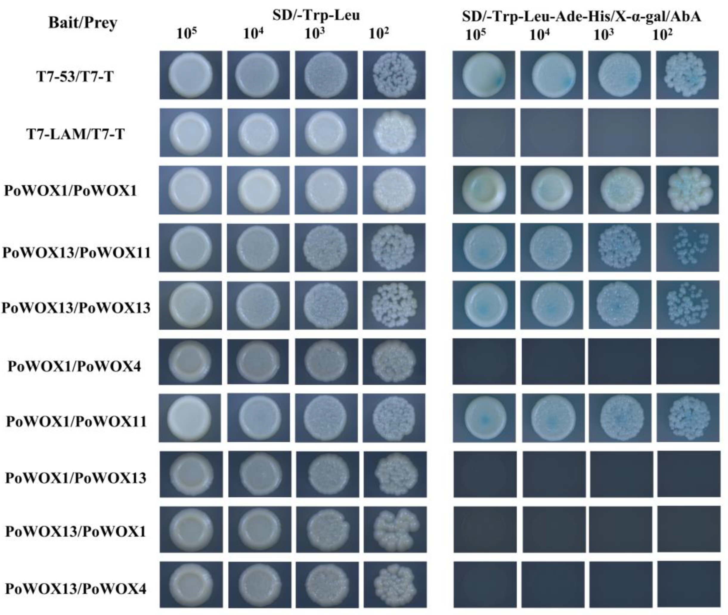
3.5. Results of Interactions between PoWOX Proteins

The Y2H-Gold-GAL4 yeast two-hybrid system was used to identify protein interactions between PoWOX1, PoWOX4, PoWOX11, and PoWOX13. First, the PoWOX proteins were tested for toxicity and self-activation. Their CDS sequences were cloned into the pGBKT7 vector as baits and then transferred into Y2H Gold yeast cells and cultured on SD/-Trp plates. It was noticed that the yeast strains containing the pGBKT7-WOX vector could all grow normally on SD/-Trp plates and did not differ from the control group in terms of growth. This indicates that the recombinant vectors were not toxic and did not affect the normal growth of the yeast strain. In addition, the pGBKT7-WOX1 and pGBKT7-WOX13 vector strains could not grow on the SD/-Trp-Ade-His plates, but pGBKT7-WOX4 and pGBKT7-WOX11 could (Figure 7). This showed that the PoWOX1 and PoWOX13 proteins do not have the ability to self-activate and, therefore, could be used as both bait and prey for the subsequent yeast interaction experiments. However, PoWOX4 and PoWOX11 proteins can only be used as prey proteins for co-transformation experiments because of their self-activating ability.
According to the above results, eight combinations of the yeast two-hybrid assays were performed using PoWOX1 and PoWOX13 as bait and prey, and PoWOX4 and PoWOX11 as prey in two-by-two interactions. All normally growing yeasts on non-selective SD/-Trp-Leu plates showed that all bait vector pGBKT7-WOX and prey vector pGADT7-WOX plasmids were successfully transferred into the Y2H Gold strain (Figure 8). Then, based on the analysis of the growth condition of the co-transformed yeast strains on selective SD/-Trp-Leu-Ade-His/X-α-gal/AbA plates, PoWOX11 could interact as prey to form heterodimers with PoWOX1 and PoWOX13, and PoWOX1 and PoWOX13 could also form homodimers. As a prey protein, PoWOX4 could not form dimers with other PoWOX proteins, and the rest of the combinations did not have direct interactions.

3.6. BiFC to Verify the Interactions between PoWOX Proteins

The interactions between the PoWOX proteins were verified by BiFC experiments. The fluorescence signals were visualized under the microscope based on the results of previous Y2H co-transformation experiments in two-by-two combinations. The results showed that green fluorescent signals were observed for the experimental groups YCE-PoWOX1/YNE-PoWOX1, YCE-PoWOX1/YNE-PoWOX11, YCE-PoWOX11/YNE-PoWOX13, and YCE-PoWOX13/YNE-PoWOX13 (Figure 9), while the negative control group had no green fluorescent signal. This inferred that there is an interaction relationship between the four groups of PoWOX proteins, PoWOX1–PoWOX1, PoWOX1–PoWOX11, PoWOX11–PoWOX13, and PoWOX13–PoWOX13.

4. Discussion

4.1. Evolution Analysis of PoWOX Gene Family

The WOX gene family is associated with the diversity of stem cell types during plant evolution. From low mosses and lycopodiopbyta plants to gymnosperms and angiosperms, stem cells play an important role in individual development. The conserved function of the WOX gene family in plant evolution corroborates that the origin of the stem cell regulation mechanism is ancient and conservative in evolution [49]. For example, the presence of WOX homologs in green algae, the oldest species in the plant kingdom, suggests that the WOX family may have originated in green algae. In the moss P. patens, PpWOX13L (WOX13 homologous gene) was found to play an important regulatory role in the initiation and maintenance of stem cells [50]. The expression of WOX genes was also shown to be involved in the regeneration process of organs in lycophytes [51].
From the phylogenetic tree (Figure 1), it can be seen that the ancient clade of the plant WOX family contains at least one WOX member from the lower plants (green algae and mosses) to the vascular plants (lycophytes and euphyllophytes). However, the WOX members of the non-vascular plants green algae and mosses are only presented in the ancient clade. The members of the vascular plants only contained in the intermediate clade of the WOX family imply the specificity on the IC-WOX. The study suggested that the IC-WOX is specific to vascular plants and that extant vascular plants are descendants of two early ancestral lineages, the euphyllophytes lineage, which included ferns and seed plants, and the lycophytes lineage [52]. From the evolutionary tree it can be seen that IC-WOX is present in lycophytes (S. moellendorffii), extant ferns (C. richardii), gymnosperms (G. biloba and P. abies), and angiosperms (A. trichopoda, A. thaliana, J. regia, V. vinifera, T. cacao, P. trichocarpa, O. sativa, and P. suffruticosa). It has been suggested that the evolution of WOX genes may accompany the adaptation of plants from aquatic to terrestrial environments. In general, WOX is a relatively old gene family involved in plant evolution. However, current studies on the gene functions of WOX family members in seed plants, especially in woody plant stem cells, are still unclear. The four PoWOX genes cloned from peony were distributed among the ancient, intermediate, and modern clades of the WOX family, providing an entry point for subsequent studies on stem cell diversification in peony.

4.2. Expression Pattern Analysis of PoWOX Gene Family

The fluorescence quantification of the PoWOX genes showed different expression patterns in different tissues of peony. According to the expression patterns of PoWOX at the various stages of somatic embryo development in vitro (Figure 4), it was observed that PoWOX1 had the highest expression at 0 days and PoWOX4 at 15 days. Both PoWOX11 and PoWOX13 were the most highly expressed at 5 days. It was hypothesized that PoWOX may be expressed in the stem apical meristem, which could improve the differentiation and development of the vegetative organs of peony by enhancing the activity of shoot apical stem cells. Meanwhile, according to the expression pattern of PoWOX genes in different tissues of peony (Figure 5), PoWOX1 obtained the highest expression level in leaves, followed by seeds and callus. This showed that the PoWOX1 gene may be related to leaf morphogenesis. PoWOX4 with its highest expression level in callus may promote callus proliferation or differentiation. PoWOX11 and PoWOX13 both had the richest expression in seeds and were considered to play a major function in seed development. At the same time, the expression of PoWOX13 in roots was second only to that in seeds, and it is speculated that PoWOX13 may be involved in both seed and root development.
The functions of WOX genes may be variable in annuals and woody plants [12]. In Arabidopsis, AtWOX4 mainly promotes the development of the primitive cambium and modulates vascular cell division [24,26]. PoWOX4, however, was highly expressed in the callus, which may facilitate the proliferation or differentiation of peony callus. Gene duplication and differentiation may result in an increase in the number of WOX gene families in different species. Therefore, there are generally functional redundancies in WOX gene families and the different expression patterns of WOX genes in various species.
The WOX protein family participates in the maintenance and differentiation of all stem cells in plants, including the primary meristem (shoot apical meristem (SAM) and root apical meristem (RAM)) and the secondary meristem (vascular meristem), through similar or specific regulatory networks. It has been demonstrated that WOX family members can express from postembryonic development to lateral organs in plants and have functions in embryonic development, vascular formation, and leaf morphogenesis [17,19,53]. Further elucidation of the regulatory mechanism of the WOX gene family in peony is beneficial to solve the bottlenecks of tissue culture, such as low induction rate of callus and difficult rooting in peony, in molecular breeding.

4.3. PoWOX Genes May Form a Dimer to Play a Transcriptional Regulatory Role in Peony Growth and Development

Different from the embryonic development of animals, all new organs of many plants develop during postembryonic development. In addition, the new organs are derived from the division and differentiation of the apical meristem and various external environmental factors and internal genetic influences [54]. Plant stem cells located in the central zone of the apical meristematic tissue have the capacity of self-renewal and multiple differentiation and are the core of the development regulation processes of plants. The organization center provides the important microenvironmental signals for stem cell development and is necessary for maintaining stem cell function [55]. It has been shown that CLV3, the dodecapeptide secreted from stem cell expression, can receive the signals from CLV1, which is a receptor kinase rich in the leucine. In addition, CLV3 with the members of the CLV1 family can downregulate the expression of WUS from the WOX gene family together. The WUS protein can be expressed in the organization center and move to the apical part of the meristematic tissue to activate the stem cell marker gene CLV3 and maintain the properties of stem cells. The expression of WUS and CLV3 together constitutes a negative feedback regulatory loop [56,57,58].
The WOX transcription factor family is a subfamily of the homeobox (HOX) transcription factor superfamily, and it encodes the homeodomain with 60–66 amino acids [13]. The typical secondary structure of WOX proteins consists of α-helix, β-fold, extended chain, and random coil. These amino acids form a structure, named “helix-loop-helix-turn-helix”, to help the interactions between mediated proteins. Therefore, WOX transcription factors are prone to forming homodimers or heterodimers. These dimers were found to promote the regulation of stem cell activity from the WUS protein movement to stem cells through plasmodesmata [57]. Studies of the mutation, deletion, and substitution of WUS protein sequences have revealed that the homeodomain of WUS and the non-structural region between it and the WUS-box play an important role in WUS homodimerization [57,59]. The interactions between WUS and DNA could be stabilized by the WUS dimers to combine with the different DNA motifs in order to activate and repress the expression of the downstream genes of WUS directly or indirectly [60]. In this work, we demonstrated that the PoWOX1 and PoWOX13 proteins in the peony WOX family can form homodimers by yeast two-hybrid and BiFC assays individually. Peony stem cell activity may be regulated from PoWOX1 and PoWOX13 by forming dimers and moving to peony stem cells through plasmodesmata like the WUS proteins in Arabidopsis.
As transcription factors, WUS proteins can play a regulatory role by interacting with other proteins. The heterodimer can be formed by the interaction between WUS proteins in Arabidopsis and STM. It co-binds to the promoter of CLV3 in order to enhance the binding strength between WUS and the CLV3 promoter, activating the expression of the CLV3 gene, and enhancing stem cell activity at the stem apex [61]. WUS proteins and the family members of transcription factors HAIRY MERISTEM (HAM) can interact to promote stem cell proliferation and maintain the homeostasis of stem cells in the SAM [62]. It has been found that WOX4 and WOX5 of the WOX family can interact with HAM proteins, and the regulation of WUS-HAM-CLV3 on the polarity of plant apical meristem tissue was demonstrated by a combination of computer models and experimental evidence [63]. By using the yeast two-hybrid assay and the BiFC assay, it has been shown that the formation of rice epidermal hairs can be modulated by the interaction between OsWOX3B and the AP2/ERF transcription factor Hairy Leaf 6 (HL6) in rice [64]. Subsequently, a protein complex was formed to enhance the binding between HL6 and the growth hormone-related gene OsYUCCA5 [64]. In this research, PoWOX11 interacted with PoWOX1 and PoWOX13 to form heterodimers. The two pairs of interacted proteins, PoWOX11–PoWOX1 and PoWOX11–PoWOX13, may play important regulatory functions in promoting the proliferation of stem cells and maintaining the homeostasis of stem cells in the SAM of peony stems.
At present, an efficient in vitro regeneration and genetic transformation system of peony has not been reported, hindering the process of peony genetic engineering breeding. By exploring the critical genes and regulatory factors in the development of the peony somatic embryo, the bottleneck of peony genetic transformation would be broken by improving the efficiency of in vitro regeneration of peony using molecular means. This is beneficial not only to understand the molecular and regulatory mechanisms of peony somatic embryo development but also to achieve directed breeding and improvements in efficiency through genetic engineering breeding technology to accelerate the fundamental process of molecular breeding in peony.

5. Conclusions

In this research, four PoWOX genes, named PoWOX1, PoWOX4, PoWOX11, and PoWOX13, were cloned from the somatic embryo in peony “Fengdan” by PCR and identified by bioinformatics. PoWOX1, PoWOX4, PoWOX11, and PoWOX13 were shown to have vital functions in the development of peony somatic embryos, and the expression of each gene in different parts of peony was tissue specific. The PoWOX were all localized in the nucleus by subcellular localization assays. Using yeast two-hybrid and BiFC methods, it was shown that the PoWOX1 and PoWOX13 proteins could form homodimers by themselves, and PoWOX11 interacted with PoWOX1 and PoWOX13 to form heterodimers. Different protein complexes may play roles in different regulatory pathways, such as peony stem cell maintenance and meristem development.

Supplementary Materials

The following supporting information can be downloaded at: https://www.mdpi.com/article/10.3390/horticulturae8030266/s1. Table S1: The accessions and sequences of WOX proteins used in Figure 1.Table S2: The WOX-HD multiple sequence comparison results sequence used in Figure 2.

Author Contributions

Conceptualization, M.X., W.Z., Z.J. and T.H.; methodology, M.X., Y.M. and Y.D.; software, M.X. and Y.C.; validation, Y.C. and Y.D.; data curation, K.F. and X.Z.; writing—original draft preparation, M.X. and W.Z.; writing—review and editing, M.X., W.Z. and T.H. All authors have read and agreed to the published version of the manuscript.

Funding

This research was supported by ICBR Fundamental Research Funds Grant (NO. 1632019009), ICBR Fundamental Research Funds Grant (NO. 1632021005) and ICBR Fundamental Research Funds Grant (NO. 1632020001).

Institutional Review Board Statement

Not applicable.

Informed Consent Statement

Not applicable.

Data Availability Statement

All data in this study could be found in the manuscript or Supplementary Materials.

Acknowledgments

We thank all the colleagues that helped with the development of different parts of this manuscript.

Conflicts of Interest

The authors declare no conflict of interest.

References

  1. Gao, J.; Xue, J.; Xue, Y.; Liu, R.; Ren, X.; Wang, S.; Zhang, X. Transcriptome Sequencing and Identification of Key Callus Browning-Related Genes from Petiole Callus of Tree Peony (Paeonia suffruticosa Cv. Kao) Cultured on Media with Three Browning Inhibitors. Plant Physiol. Biochem. 2020, 149, 36–49. [Google Scholar] [CrossRef] [PubMed]
  2. Zhang, H.; Li, X.; Wu, K.; Wang, M.; Liu, P.; Wang, X.; Deng, R. Antioxidant Activities and Chemical Constituents of Flavonoids from the Flower of Paeonia Ostii. Molecules 2016, 22, 5. [Google Scholar] [CrossRef] [PubMed] [Green Version]
  3. Wang, Y.; Dong, C.; Xue, Z.; Jin, Q.; Xu, Y. De Novo Transcriptome Sequencing and Discovery of Genes Related to Copper Tolerance in Paeonia Ostii. Gene 2016, 576, 126–135. [Google Scholar] [CrossRef]
  4. Huliang, C.; Fangyun, C.; Liping, P. Determination of the Fatty Acid Composition in Tree Peony Seeds Using Near-Infrared Spectroscopy. J. Am. Oil. Chem. Soc. 2016, 93, 943–952. [Google Scholar] [CrossRef]
  5. Xie, L.; Niu, L.; Zhang, Y.; Jin, M.; Ji, D.; Zhang, X. Pollen Sources Influence the Traits of Seed and Seed Oil in Paeonia Ostii ‘Feng Dan’. HortScience 2017, 52, 700–705. [Google Scholar] [CrossRef]
  6. Yu, X.; Zhao, R.; Cheng, F. Seed Germination of Tree and Herbaceous Peonies: A Mini-Review. Seed Sci. Biotechnol. 2007, 1, 11–14. [Google Scholar]
  7. Zhang, K.; Yao, L.; Zhang, Y.; Baskin, J.M.; Baskin, C.C.; Xiong, Z.; Tao, J. A Review of the Seed Biology of Paeonia Species (Paeoniaceae), with Particular Reference to Dormancy and Germination. Planta 2019, 249, 291–303. [Google Scholar] [CrossRef] [PubMed]
  8. Zhong, Y.; Cheng, F.; Wu, J. The Study Advances on Functional Genes in Tree Peony (Paeonia sect. Moutan). Mol. Plant Breed. 2016, 14, 2353–2364. [Google Scholar] [CrossRef]
  9. Yang, Y.; Sun, M.; Li, S.; Chen, Q.; Teixeira da Silva, J.A.; Wang, A.; Yu, X.; Wang, L. Germplasm Resources and Genetic Breeding of Paeonia: A Systematic Review. Hortic. Res. 2020, 7, 107. [Google Scholar] [CrossRef] [PubMed]
  10. Mayer, K.F.; Schoof, H.; Haecker, A.; Lenhard, M.; Jürgens, G.; Laux, T. Role of WUSCHEL in Regulating Stem Cell Fate in the Arabidopsis Shoot Meristem. Cell 1998, 95, 805–815. [Google Scholar] [CrossRef] [Green Version]
  11. Kamiya, N.; Nagasaki, H.; Morikami, A.; Sato, Y.; Matsuoka, M. Isolation and Characterization of a Rice WUSCHEL-Type Homeobox Gene That Is Specifically Expressed in the Central Cells of a Quiescent Center in the Root Apical Meristem. Plant J. 2003, 35, 429–441. [Google Scholar] [CrossRef]
  12. Van der Graaff, E.; Laux, T.; Rensing, S.A. The WUS Homeobox-Containing (WOX) Protein Family. Genome Biol. 2009, 10, 248. [Google Scholar] [CrossRef]
  13. Haecker, A.; Gross-Hardt, R.; Geiges, B.; Sarkar, A.; Breuninger, H.; Herrmann, M.; Laux, T. Expression Dynamics of WOX Genes Mark Cell Fate Decisions during Early Embryonic Patterning in Arabidopsis Thaliana. Development 2004, 131, 657–668. [Google Scholar] [CrossRef] [Green Version]
  14. Laux, T.; Mayer, K.F.; Berger, J.; Jürgens, G. The WUSCHEL Gene Is Required for Shoot and Floral Meristem Integrity in Arabidopsis. Development 1996, 122, 87–96. [Google Scholar] [CrossRef]
  15. Lin, H.; Niu, L.; McHale, N.A.; Ohme-Takagi, M.; Mysore, K.S.; Tadege, M. Evolutionarily Conserved Repressive Activity of WOX Proteins Mediates Leaf Blade Outgrowth and Floral Organ Development in Plants. Proc. Natl. Acad. Sci. USA 2013, 110, 366–371. [Google Scholar] [CrossRef] [Green Version]
  16. Dolzblasz, A.; Nardmann, J.; Clerici, E.; Causier, B.; van der Graaff, E.; Chen, J.; Davies, B.; Werr, W.; Laux, T. Stem Cell Regulation by Arabidopsis WOX Genes. Mol. Plant 2016, 9, 1028–1039. [Google Scholar] [CrossRef]
  17. Costanzo, E.; Trehin, C.; Vandenbussche, M. The Role of WOX Genes in Flower Development. Ann. Bot. 2014, 114, 1545–1553. [Google Scholar] [CrossRef] [Green Version]
  18. Zhang, X.; Zong, J.; Liu, J.; Yin, J.; Zhang, D. Genome-Wide Analysis of WOX Gene Family in Rice, Sorghum, Maize, Arabidopsis and Poplar. J. Integr. Plant Biol. 2010, 52, 1016–1026. [Google Scholar] [CrossRef]
  19. Jha, P.; Ochatt, S.J.; Kumar, V. WUSCHEL: A Master Regulator in Plant Growth Signaling. Plant Cell Rep. 2020, 39, 431–444. [Google Scholar] [CrossRef]
  20. Zhang, Y.; Wu, R.; Qin, G.; Chen, Z.; Gu, H.; Qu, L.-J. Over-Expression of WOX1 Leads to Defects in Meristem Development and Polyamine Homeostasis in Arabidopsis. J. Integr. Plant. Biol. 2011, 53, 493–506. [Google Scholar] [CrossRef]
  21. Nakata, M.; Matsumoto, N.; Tsugeki, R.; Rikirsch, E.; Laux, T.; Okada, K. Roles of the Middle Domain-Specific WUSCHEL-RELATED HOMEOBOX Genes in Early Development of Leaves in Arabidopsis. Plant Cell 2012, 24, 519–535. [Google Scholar] [CrossRef] [Green Version]
  22. Du, F.; Mo, Y.; Israeli, A.; Wang, Q.; Yifhar, T.; Ori, N.; Jiao, Y. Leaflet Initiation and Blade Expansion Are Separable in Compound Leaf Development. Plant J. 2020, 104, 1073–1087. [Google Scholar] [CrossRef]
  23. Wang, C.; Zhao, B.; He, L.; Zhou, S.; Liu, Y.; Zhao, W.; Guo, S.; Wang, R.; Bai, Q.; Li, Y.; et al. The WOX Family Transcriptional Regulator SlLAM1 Controls Compound Leaf and Floral Organ Development in Solanum Lycopersicum. J. Exp. Bot. 2021, 72, 1822–1835. [Google Scholar] [CrossRef]
  24. Ji, J.; Strable, J.; Shimizu, R.; Koenig, D.; Sinha, N.; Scanlon, M.J. WOX4 Promotes Procambial Development. Plant Physiol. 2010, 152, 1346–1356. [Google Scholar] [CrossRef] [Green Version]
  25. Suer, S.; Agusti, J.; Sanchez, P.; Schwarz, M.; Greb, T. WOX4 Imparts Auxin Responsiveness to Cambium Cells in Arabidopsis. Plant Cell 2011, 23, 3247–3259. [Google Scholar] [CrossRef] [Green Version]
  26. Etchells, J.P.; Provost, C.M.; Mishra, L.; Turner, S.R. WOX4 and WOX14 Act Downstream of the PXY Receptor Kinase to Regulate Plant Vascular Proliferation Independently of Any Role in Vascular Organisation. Development 2013, 140, 2224–2234. [Google Scholar] [CrossRef] [Green Version]
  27. Wang, L.; Wen, S.; Wang, R.; Wang, C.; Gao, B.; Lu, M. PagWOX11/12a Activates PagCYP736A12 Gene That Facilitates Salt Tolerance in Poplar. Plant Biotechnol. J. 2021, 19, 2249–2260. [Google Scholar] [CrossRef]
  28. Cheng, S.; Tan, F.; Lu, Y.; Liu, X.; Li, T.; Yuan, W.; Zhao, Y.; Zhou, D.-X. WOX11 Recruits a Histone H3K27me3 Demethylase to Promote Gene Expression during Shoot Development in Rice. Nucleic Acids Res. 2018, 46, 2356–2369. [Google Scholar] [CrossRef] [Green Version]
  29. Romera-Branchat, M.; Ripoll, J.J.; Yanofsky, M.F.; Pelaz, S. The WOX13 Homeobox Gene Promotes Replum Formation in the Arabidopsis Thaliana Fruit. Plant J. 2013, 73, 37–49. [Google Scholar] [CrossRef]
  30. Ikeuchi, M.; Iwase, A.; Ito, T.; Tanaka, H.; Favero, D.S.; Kawamura, A.; Sakamoto, S.; Wakazaki, M.; Tameshige, T.; Fujii, H.; et al. Wound-Inducible WUSCHEL RELATED HOMEOBOX 13 Is Required for Callus Growth and Organ Reconnection. Plant Physiol. 2021, 188, 425–441. [Google Scholar] [CrossRef]
  31. Denis, E.; Kbiri, N.; Mary, V.; Claisse, G.; Conde, E.; Silva, N.; Kreis, M.; Deveaux, Y. WOX14 Promotes Bioactive Gibberellin Synthesis and Vascular Cell Differentiation in Arabidopsis. Plant J. 2017, 90, 560–572. [Google Scholar] [CrossRef] [PubMed] [Green Version]
  32. Jiang, F.; Liu, F.; Zhao, L. Overexpression of RaWUS Gene of Rosa canina Inducing Shoot Regeneration from Root Tip of Transgenic Tobacco. Sci. Silvae Sin. 2011, 47, 43–52. [Google Scholar]
  33. Gao, B.; Wen, C.; Fan, L.; Kou, Y.; Ma, N.; Zhao, L. A Rosa Canina WUSCHEL-Related Homeobox Gene, RcWOX1, Is Involved in Auxin-Induced Rhizoid Formation. Plant Mol. Biol. 2014, 86, 671–679. [Google Scholar] [CrossRef] [PubMed]
  34. Kuang, H. Tissue Culture and Stem Cell Related Gene Cloning and Expression of Rhododendron Equisetum. Master’s Thesis, Fujian Agriculture and Forestry University, Fuzhou City, China, 2014. [Google Scholar]
  35. Cao, Y.; Han, Y.; Meng, D.; Li, G.; Li, D.; Abdullah, M.; Jin, Q.; Lin, Y.; Cai, Y. Genome-Wide Analysis Suggests the Relaxed Purifying Selection Affect the Evolution of WOX Genes in Pyrus bretschneideri, Prunus persica, Prunus mume, and Fragaria vesca. Front. Genet. 2017, 8, 78. [Google Scholar] [CrossRef] [PubMed] [Green Version]
  36. Lu, Y.; Liu, Z.; Lyu, M.; Yuan, Y.; Wu, B. Characterization of JsWOX1 and JsWOX4 during Callus and Root Induction in the Shrub Species Jasminum Sambac. Plants 2019, 8, 79. [Google Scholar] [CrossRef] [PubMed] [Green Version]
  37. Lv, S.; Cheng, S.; Wang, Z.; Li, S.; Jin, X.; Lan, L.; Yang, B.; Yu, K.; Ni, X.; Li, N.; et al. Draft Genome of the Famous Ornamental Plant Paeonia suffruticosa. Ecol. Evol. 2020, 10, 4518–4530. [Google Scholar] [CrossRef] [PubMed]
  38. Potter, S.C.; Luciani, A.; Eddy, S.R.; Park, Y.; Lopez, R.; Finn, R.D. HMMER Web Server: 2018 Update. Nucleic Acids Res. 2018, 46, W200–W204. [Google Scholar] [CrossRef] [PubMed] [Green Version]
  39. Wilkins, M.R.; Gasteiger, E.; Bairoch, A.; Sanchez, J.C.; Williams, K.L.; Appel, R.D.; Hochstrasser, D.F. Protein Identification and Analysis Tools in the ExPASy Server. Methods Mol. Biol. 1999, 112, 531–552. [Google Scholar] [CrossRef] [PubMed]
  40. Chou, K.-C.; Shen, H.-B. Plant-MPLoc: A Top-down Strategy to Augment the Power for Predicting Plant Protein Subcellular Localization. PLoS ONE 2010, 5, e11335. [Google Scholar] [CrossRef] [PubMed] [Green Version]
  41. Jin, J.; Tian, F.; Yang, D.-C.; Meng, Y.-Q.; Kong, L.; Luo, J.; Gao, G. PlantTFDB 4.0: Toward a Central Hub for Transcription Factors and Regulatory Interactions in Plants. Nucleic Acids Res. 2017, 45, D1040–D1045. [Google Scholar] [CrossRef] [PubMed] [Green Version]
  42. Thompson, J.D.; Higgins, D.G.; Gibson, T.J. CLUSTAL W: Improving the Sensitivity of Progressive Multiple Sequence Alignment through Sequence Weighting, Position-Specific Gap Penalties and Weight Matrix Choice. Nucleic Acids Res. 1994, 22, 4673–4680. [Google Scholar] [CrossRef] [PubMed] [Green Version]
  43. Kumar, S.; Stecher, G.; Tamura, K. MEGA7: Molecular Evolutionary Genetics Analysis Version 7.0 for Bigger Datasets. Mol. Biol. Evol. 2016, 33, 1870–1874. [Google Scholar] [CrossRef] [PubMed] [Green Version]
  44. Saitou, N.; Nei, M. The Neighbor-Joining Method: A New Method for Reconstructing Phylogenetic Trees. Mol. Biol. Evol. 1987, 4, 406–425. [Google Scholar] [CrossRef] [PubMed]
  45. Bailey, T.L.; Johnson, J.; Grant, C.E.; Noble, W.S. The MEME Suite. Nucleic Acids Res. 2015, 43, W39–W49. [Google Scholar] [CrossRef] [PubMed] [Green Version]
  46. Chen, C.; Chen, H.; Zhang, Y.; Thomas, H.R.; Frank, M.H.; He, Y.; Xia, R. TBtools: An Integrative Toolkit Developed for Interactive Analyses of Big Biological Data. Mol. Plant 2020, 13, 1194–1202. [Google Scholar] [CrossRef] [PubMed]
  47. Crooks, G.E.; Hon, G.; Chandonia, J.-M.; Brenner, S.E. WebLogo: A Sequence Logo Generator. Genome Res. 2004, 14, 1188–1190. [Google Scholar] [CrossRef] [PubMed] [Green Version]
  48. Livak, K.J.; Schmittgen, T.D. Analysis of Relative Gene Expression Data Using Real-Time Quantitative PCR and the 2−ΔΔCT Method. Methods 2001, 25, 402–408. [Google Scholar] [CrossRef] [PubMed]
  49. Hedman, H.; Zhu, T.; von Arnold, S.; Sohlberg, J.J. Analysis of the WUSCHEL-RELATED HOMEOBOX Gene Family in the Conifer Picea abies Reveals Extensive Conservation as Well as Dynamic Patterns. BMC Plant. Biol. 2013, 13, 89. [Google Scholar] [CrossRef] [PubMed] [Green Version]
  50. Sakakibara, K.; Reisewitz, P.; Aoyama, T.; Friedrich, T.; Ando, S.; Sato, Y.; Tamada, Y.; Nishiyama, T.; Hiwatashi, Y.; Kurata, T.; et al. WOX13-like Genes Are Required for Reprogramming of Leaf and Protoplast Cells into Stem Cells in the Moss Physcomitrella Patens. Development 2014, 141, 1660–1670. [Google Scholar] [CrossRef] [PubMed] [Green Version]
  51. Ge, Y.; Liu, J.; Zeng, M.; He, J.; Qin, P.; Huang, H.; Xu, L. Identification of WOX Family Genes in Selaginella Kraussiana for Studies on Stem Cells and Regeneration in Lycophytes. Front. Plant Sci. 2016, 7, 93. [Google Scholar] [CrossRef] [PubMed] [Green Version]
  52. Liu, W.; Xu, L. Recruitment of IC-WOX Genes in Root Evolution. Trends Plant Sci. 2018, 23, 490–496. [Google Scholar] [CrossRef] [PubMed]
  53. Wójcik, A.M.; Wójcikowska, B.; Gaj, M.D. Current Perspectives on the Auxin-Mediated Genetic Network That Controls the Induction of Somatic Embryogenesis in Plants. Int. J. Mol. Sci. 2020, 21, 1333. [Google Scholar] [CrossRef] [PubMed] [Green Version]
  54. Weigel, D.; Jürgens, G. Stem Cells That Make Stems. Nature 2002, 415, 751–754. [Google Scholar] [CrossRef] [PubMed]
  55. Sablowski, R. The Dynamic Plant Stem Cell Niches. Curr. Opin. Plant Biol. 2007, 10, 639–644. [Google Scholar] [CrossRef] [PubMed]
  56. Yadav, R.K.; Perales, M.; Gruel, J.; Girke, T.; Jönsson, H.; Reddy, G.V. WUSCHEL Protein Movement Mediates Stem Cell Homeostasis in the Arabidopsis Shoot Apex. Genes Dev. 2011, 25, 2025–2030. [Google Scholar] [CrossRef] [PubMed] [Green Version]
  57. Daum, G.; Medzihradszky, A.; Suzaki, T.; Lohmann, J.U. A Mechanistic Framework for Noncell Autonomous Stem Cell Induction in Arabidopsis. Proc. Natl. Acad. Sci. USA 2014, 111, 14619–14624. [Google Scholar] [CrossRef] [PubMed] [Green Version]
  58. Schlegel, J.; Denay, G.; Wink, R.; Pinto, K.G.; Stahl, Y.; Schmid, J.; Blümke, P.; Simon, R.G. Control of Arabidopsis Shoot Stem Cell Homeostasis by Two Antagonistic CLE Peptide Signalling Pathways. Elife 2021, 10, e70934. [Google Scholar] [CrossRef] [PubMed]
  59. Rodriguez, K.; Perales, M.; Snipes, S.; Yadav, R.K.; Diaz-Mendoza, M.; Reddy, G.V. DNA-Dependent Homodimerization, Sub-Cellular Partitioning, and Protein Destabilization Control WUSCHEL Levels and Spatial Patterning. Proc. Natl. Acad. Sci. USA 2016, 113, E6307–E6315. [Google Scholar] [CrossRef] [PubMed] [Green Version]
  60. Sloan, J.; Hakenjos, J.P.; Gebert, M.; Ermakova, O.; Gumiero, A.; Stier, G.; Wild, K.; Sinning, I.; Lohmann, J.U. Structural Basis for the Complex DNA Binding Behavior of the Plant Stem Cell Regulator WUSCHEL. Nat. Commun. 2020, 11, 2223. [Google Scholar] [CrossRef] [PubMed]
  61. Su, Y.H.; Zhou, C.; Li, Y.J.; Yu, Y.; Tang, L.P.; Zhang, W.J.; Yao, W.J.; Huang, R.; Laux, T.; Zhang, X.S. Integration of Pluripotency Pathways Regulates Stem Cell Maintenance in the Arabidopsis Shoot Meristem. Proc. Natl. Acad. Sci. USA 2020, 117, 22561–22571. [Google Scholar] [CrossRef] [PubMed]
  62. Zhou, Y.; Liu, X.; Engstrom, E.M.; Nimchuk, Z.L.; Pruneda-Paz, J.L.; Tarr, P.T.; Yan, A.; Kay, S.A.; Meyerowitz, E.M. Control of Plant Stem Cell Function by Conserved Interacting Transcriptional Regulators. Nature 2015, 517, 377–380. [Google Scholar] [CrossRef] [Green Version]
  63. Zhou, Y.; Yan, A.; Han, H.; Li, T.; Geng, Y.; Liu, X.; Meyerowitz, E.M. HAIRY MERISTEM with WUSCHEL Confines CLAVATA3 Expression to the Outer Apical Meristem Layers. Science 2018, 361, 502–506. [Google Scholar] [CrossRef] [Green Version]
  64. Sun, W.; Gao, D.; Xiong, Y.; Tang, X.; Xiao, X.; Wang, C.; Yu, S. Hairy Leaf 6, an AP2/ERF Transcription Factor, Interacts with OsWOX3B and Regulates Trichome Formation in Rice. Mol. Plant 2017, 10, 1417–1433. [Google Scholar] [CrossRef] [Green Version]
Figure 1. Phylogenetic analysis of WOX genes in plants. Evolutionary analysis of the full-length sequences of WOX proteins was performed by the NJ method using MEGA 7. A total of 127 WOX sequences from 14 species were classified into three clades: modern clade (WC-WOX), intermediate clade (IC-WOX), and ancient clade (AC-WOX). PoWOX1 and PoWOX4 were highlighted with green long triangular tags; PoWOX11 was highlighted with dark blue long triangular tags; PoWOX13 was highlighted with red long triangular tags.
Horticulturae 08 00266 g001
Figure 2. Multiple sequence alignment and sequence identification of WOX1, WOX4, WOX11, and WOX13 proteins. (a) Multiple sequence alignment of WOX1, WOX4, WOX11, and WOX13 proteins in six species (A. thaliana, O. sativa, J. regia, V. vinifera, P. trichocarpa and P. suffruticosa). The amino acid sequence similarity indicated in red was 100%, and the amino acid sequence similarity in purple was greater than or equal to 50%. (b) Sequence identification map of WOX-HD.
Horticulturae 08 00266 g002
Figure 3. Conserved motif analysis of WOX proteins and sequence identification of ten conserved motifs. (a) the phylogenetic tree and conserved motif analysis of six species (A. thaliana, O. sativa, P. suffruticosa, J. regia, V. vinifera, and P. trichocarpa), the red rectangle circles the peony PoWOX protein. A total of ten conserved motifs were identified, and each motif was represented by a separate number and color. Blue motif 1 represents HD, red motif 5 represents that the WUS motif is only present in the WC-WOX, yellow motif 2 is present in every clade, dark gray motif 6 and light green motif 7 are unique to WOX4 protein, purple motif 9 is present in the WOX4 subclade and AtWOX13, orange motif 10 and light grey motif 3 belong to the IC-WOX, and dark green motif 4 and orange motif 8 exist alone in the AC-WOX. (b) Sequence identification of the predicted 10 motifs of WOX protein.
Horticulturae 08 00266 g003
Figure 4. Expression of PoWOX genes of peony in the developmental stage of somatic embryos. Expression levels of peony somatic embryos in seven developmental stages (0; 5; 10; 15; 20; 30; 60 days). The expression of Day 0 peony somatic embryos was the control group.
Horticulturae 08 00266 g004
Figure 5. Expression of PoWOX genes in peony tissues. The expression of PoWOX gene in five different of peony (seed, root, stem, leaf, and callus). The expression of peony root tissues was the control group, respectively.
Horticulturae 08 00266 g005
Figure 6. Subcellular localization analysis of 35::PoWOX-GFP fusion proteins. The 35::PoWOX-GFP vector was infected with N. benthamiana mediated by Agrobacterium GV3101, and the expression of the fusion protein was observed under the Zeiss Axio Imager M2 microscope. Positive control 35S::GFP fusion protein was expressed in the nucleus and cytoplasm, and PoWOX protein was expressed in the nucleus. Tobacco leaf cells were stained. Green fluorescence shows the fusion protein location and blue fluorescence is the nuclear signal localized by DAPI for bright field, DAPI, GFP, and merge, respectively. Positive control is 35S::GFP empty. The white arrows indicate the location of the nucleus and the cell membrane. The scale bar is 20 μm.
Horticulturae 08 00266 g006
Figure 7. Yeast self-activating analysis. All yeasts grew normally on SD/-Trp plates, and colonies of PoWOX4 and PoWOX11 appeared on SD/-Trp-Ade-His plate.
Horticulturae 08 00266 g007
Figure 8. Protein interactions analysis between PoWOX members in yeast two-hybrid assay. The CDS sequences of PoWOX1, PoWOX4, PoWOX11, and PoWOX13 were cloned into pGBKT7 (bait) vector and pGADT7 (prey) vector, respectively. Co-transformation to SD/-Trp-Leu plates and SD/- Trp-Leu-Ade-His/X-α-gal/AbA plates was performed to verify the interactions between PoWOX members. pGBKT7-53/pGADT7-T (T7-53/T7-T) was co-transformed as a positive control and pGBKT7-LAM/pGADT7-T (T7-LAM/T7-T) was co-transformed as a negative control. The pictures were taken by Zeiss Stemi 305 microscope to preserve.
Horticulturae 08 00266 g008
Figure 9. Subcellular localization analysis of 35::PoWOX-GFP fusion protein. 35::PoWOX-GFP vector was used to infiltrate the lower epidermis of tobacco mediated by A. tumefaciens GV3101. Fusion protein expression was observed under Zeiss Axio Imager M2 microscope. Tobacco leaf cells were stained with DAPI (1 μg/mL). Green fluorescence shows the fusion protein location and blue fluorescence is the nuclear signal localized by DAPI for bright field, DAPI, GFP, and merge, respectively. Positive control is 35S::GFP empty. The white arrows indicate the location of the nucleus and the cell membrane. The scale bar is 20 μm.
Horticulturae 08 00266 g009
Table 1. Primer sequences of PCR amplification.
GenesPrimer Sequence F (5’-3’)Primer Sequence R (5’-3’)
PoWOX1GCTCTAGAATGTATATGATGGGTTATAATGATGGCGGAGTCCCCCCGGGATTCCTCAACGGAAGGAACTCAAAATACT
PoWOX4CGTCTAGAATGGGAAACATGAAGGTTCATCAGTTCTCCCCCCGGGTCTTCCTTCCGGGTGTAATGGAA
PoWOX11GCTCTAGAATTCATGTGTTTTATCTTTTTTCTCTCAACTCTTCCCCCCGGGAGTGGTTCTTGAAACTAGGAAATAGCTTTC
PoWOX13GCTCTAGAATGGGGTTAGCAAAAAAGATTTTAGAAAGTTCCCCCCGGGCGAAGAATGTCAAATTCTTCCTCCTG
Table 2. Primer sequences of PoWOX genes for real-time quantitative.
Primers NamePrimers Sequence (5′-3′)
Q-PoWOX1-FCGTTGGCGGCAATGAAGAAGAATC
Q-PoWOX1-RGGCAATTAGGAGGACTCAAGTTGGTAT
Q-PoWOX4-FCCGCAACAGTCTTGGTCTTAGCC
Q-PoWOX4-RTTCCTCATCTCTACATCTCACCTCTTCC
Q-PoWOX11-FGCAACGCCAGATTCAAGCAAGTC
Q-PoWOX11-RAAGAGGAACCAGCAAGACAAGAAGATG
Q-PoWOX13-FATGACGGACGAGCAAATAGAGGAACTT
Q-PoWOX13-RCCGCTGCCTGGTAGTGATCTTCTG
Q-Poubiquitin-FTCCTCCACCTCCTACCTTCCGACTC
Q-Poubiquitin-RCGATCCTCCTGAGCCAAGCGTCAT
Table 3. Primer sequences of plant expression vector 35S::PoWOX-GFP.
Primers NamePrimers Sequence (5′-3′)
PHG-PoWOX1-FAGTCTCTCTCTCAAGCTTGATGTATATGATGGGTTATAATGATGGC
PHG-PoWOX1-RCGGGTCATGAGCTCCTGCAATTCCTCAACGGAAGGAACT
PHG-PoWOX4-FAGTCTCTCTCTCAAGCTTGATGGGAAACATGAAGGTTCATCA
PHG-PoWOX4-RCGGGTCATGAGCTCCTGCATCTTCCTTCCGGGTGTAATG
PHG-PoWOX11-FAGTCTCTCTCTCAAGCTTGATGGAAGATCATGACCCTAACA
PHG-PoWOX11-RCGGGTCATGAGCTCCTGCAAGTGGTTCTTGAAACTAGGAAATAG
PHG-PoWOX13-FAGTCTCTCTCTCAAGCTTGATGGGGTTAGCAAAAAAGATTTTAG
PHG-PoWOX13-RCGGGTCATGAGCTCCTGCACCGAAGAATGTCAAATTCTTCCT
Table 4. Primer sequences for the construction of pGADT7 and pGBKT7 recombinant vectors on PoWOX genes in Y2H.
Primer NameUpstream Primer F (5′-3′) Downstream Primer R (5′-3′)
BK-PoWOX1ATCTCAGAGGAGGACCTGCAATGTATATGATGGGTTATAANdeIAGGGGTTATGCTAGTTATGCATTCCTCAACGGAAGGAACTNotI
BK-PoWOX4ATCTCAGAGGAGGACCTGCAATGGGAAACATGAAGGTTCANdeIAGGGGTTATGCTAGTTATGCTCTTCCTTCCGGGTGTAATGNotI
BK-PoWOX13ATCTCAGAGGAGGACCTGCAATGGGGTTAGCAAAAAAGATNdeIAGGGGTTATGCTAGTTATGCCCGAAGAATGTCAAATTCTTNotI
BK-PoWOX11ATCTCAGAGGAGGACCTGCAATGGAAGATCATGACCCTAANdeIAGGGGTTATGCTAGTTATGCAGTGGTTCTTGAAACTAGGANotI
AD-PoWOX1GACGTACCAGATTACGCTCAATGTATATGATGGGTTATAANdeITATTAAGGGTTCCGGATCGCATTCCTCAACGGAAGGAACTNotI
AD-PoWOX4GACGTACCAGATTACGCTCAATGGGAAACATGAAGGTTCANdeITATTAAGGGTTCCGGATCGCTCTTCCTTCCGGGTGTAATGNotI
AD-PoWOX13GACGTACCAGATTACGCTCAATGGGGTTAGCAAAAAAGATNdeITATTAAGGGTTCCGGATCGCCCGAAGAATGTCAAATTCTTNotI
AD-PoWOX11GACGTACCAGATTACGCTCAATGGAAGATCATGACCCTAANdeITATTAAGGGTTCCGGATCGCAGTGGTTCTTGAAACTAGGANotI
Table 5. Primer sequences for the construction of pSPYNE(R)173 and pSPYCE(MR) recombinant vectors on PoWOX genes in BiFC.
Gene NameUpstream Primer F (5′-3′) Downstream Primer R (5′-3′)
YCE-PoWOX1TTACGCTGGGCCCAGGCCTAATGTATATGATGGGTTATAASpeICGGTACCCTCGAGGTCGACGATTCCTCAACGGAAGGAACTBamHI
YCE-PoWOX4TTACGCTGGGCCCAGGCCTAATGGGAAACATGAAGGTTCASpeICGGTACCCTCGAGGTCGACGTCTTCCTTCCGGGTGTAATGBamHI
YCE-PoWOX13TTACGCTGGGCCCAGGCCTAATGGGGTTAGCAAAAAAGATSpeICGGTACCCTCGAGGTCGACGCCGAAGAATGTCAAATTCTTBamHI
YCE-PoWOX11TTACGCTGGGCCCAGGCCTAATGGAAGATCATGACCCTAA SpeICGGTACCCTCGAGGTCGACGAGTGGTTCTTGAAACTAGGABamHI
YNE-PoWOX1GGATCTTGGGCCCAGGCCTAATGTATATGATGGGTTATAASpeICGGTACCCTCGAGGTCGACGATTCCTCAACGGAAGGAACTBamHI
YNE-PoWOX4GGATCTTGGGCCCAGGCCTAATGGGAAACATGAAGGTTCASpeICGGTACCCTCGAGGTCGACGTCTTCCTTCCGGGTGTAATGBamHI
YNE-PoWOX13GGATCTTGGGCCCAGGCCTAATGGGGTTAGCAAAAAAGATSpeICGGTACCCTCGAGGTCGACGCCGAAGAATGTCAAATTCTTBamHI
YNE-PoWOX11GGATCTTGGGCCCAGGCCTAATGGAAGATCATGACCCTAASpeICGGTACCCTCGAGGTCGACGAGTGGTTCTTGAAACTAGGABamHI
Table 6. Basic physicochemical properties of PoWOX genes.
GeneGenBank
Accession
ORF
(bp)
Protein
Length (aa)
MW
(Da)
pIInstability CoefficientGRAVYAliphatic IndexSubcellular Localization
PoWOX1OM45704795131736562.688.7155.93−0.9360.63Nucleus
PoWOX4OM45705063321124300.559.3647.58−0.9160.52Nucleus
PoWOX11OM45704976525528165.356.1367.96−0.4661.88Nucleus
PoWOX13OM45704885828632543.314.9264.70−0.8069.27Nucleus
Notes: ORF: open reading frame; bp: Base pair; aa: amino acid; MW: molecular weight; Da: Dalton; pI: isoelectric point; GRAVY: Grand average of hydropathicity.
Table 7. Predicted secondary structure of PoWOX proteins.
Proteinα-Helixβ-SheetExtended ChainRandom Coil
PoWOX129.65%12.62%3.15%54.57%
PoWOX425.41%6.63%4.42%63.54%
PoWOX1120.11%15.76%9.24%54.89%
PoWOX1339.51%8.39%3.15%48.95%
Publisher’s Note: MDPI stays neutral with regard to jurisdictional claims in published maps and institutional affiliations.

