Article

PpMYB39 Activates PpDFR to Modulate Anthocyanin Biosynthesis during Peach Fruit Maturation

1
Zhengzhou Fruit Research Institute, Chinese Academy of Agricultural Sciences, Zhengzhou 450009, China
2
Centre for Plant Science and Biodiversity, Faculty of Life Sciences, University of Swat, Mingora 19200, Pakistan
3
Department of Botany, Kohat University of Science and Technology, Kohat 26000, Pakistan
4
Agriculture Research Institute Swat, Agriculture, Livestock and Cooperative Department, Government of Khyber Pakhtunkhwa, Mingora 19130, Pakistan
5
Department of Horticulture, Faculty of Agricultural Sciences, The University of Haripur, Haripur 22620, Pakistan
*
Authors to whom correspondence should be addressed.
Horticulturae 2022, 8(4), 332; https://doi.org/10.3390/horticulturae8040332
Submission received: 8 March 2022 / Revised: 11 April 2022 / Accepted: 12 April 2022 / Published: 14 April 2022
(This article belongs to the Topic Plant Breeding, Genetics and Genomics)

Abstract

:
Anthocyanins are a class of water-soluble flavonoids widely present in fruits and vegetables responsible for the red flesh formation of peach fruit. Previously, several genes of the MYB family have been reported as transcriptional regulators of the anthocyanin biosynthetic pathway of structural genes in plants. In this study, through comparative transcriptome analysis of the white and red flesh peach cultivars of Harrow Blood and Asama Hakuto, a predicted transcription factor of the R2R3MYB family, PpMYB39, was identified to be associated with anthocyanin biosynthesis in peach fruit. In red-fleshed peach cultivars, the maximum amount of anthocyanin accumulated 95 days after full bloom (DAFB), at full maturity near ripening. Our results showed that, at this stage, PpMYB39 had the highest expression level among the 13 differentially expressed genes (DEGs) found in both red- and white-fleshed fruits, as well as a high correlation with total anthocyanin content throughout fruit development. Moreover, the expression analysis of the structural genes of the anthocyanin biosynthetic pathway in peach fruit revealed that Prunus persica Dihydroflavonol-4-reductase (PpDFR) was co-expressed and up-regulated with PpMYB39 at 95 DAFB, suggesting its possible role as a transcriptional activator of MYB39. This was further confirmed by a yeast one-hybrid assay and a dual luciferase reporter assay. Our results will be helpful in the breeding of peach cultivars and the identification and significance of color in peaches and related fruit species, in addition to providing an understanding of color formation in peach fruit for future research.

1. Introduction

Peach (Prunus persica L. Batsch) is a major and widely cultivated fruit tree of the Rosaceae family. Currently, there are more than 3000 peach varieties throughout the world, of which 1000 are found in China [1]. For a long time, peach has been one of the model species for studying Rosaceae genetics. It has remained an ideal target for plant breeders to improve fruit quality due to its unique characteristics [2,3]. Flesh color serves as an important commercial trait in peach fruit to increase its market value. Based on color, peach varieties are divided into white peaches, yellow peaches, and red peaches [4]. White peaches show reduced or absent carotenes and anthocyanin, while yellow peaches are rich in carotenes and xanthophylls. However, red flesh peaches have a distinct flavor compared to yellow peaches because of their high anthocyanin content.
Mainly, red color in peach flesh is because of the over accumulation of anthocyanin [5], which plays a major role in color formation throughout the development process of fruit [6]. Anthocyanin, a pigment responsible for the red coloration of several fruits [7], is in fact a functional compound capable of increasing antioxidant defenses, reducing free radical damage, chronic inflammation, and mutation risk, and even attenuating the development and progression of many non-infectious and degenerative chronic diseases, e.g., atherosclerosis, metabolic syndrome, eye and kidney complications, several types of cancer, and weight management in human and animals [8].
In peach fruit, the main constituent of anthocyanin is cyanidin-3-glucoside, and a small amount of cyanidin 3-rutinoside [9]. Anthocyanin biosynthesis in plants, particularly in species of the Rosaceae family, including apples, strawberries and peaches, is regulated by the transcriptional activation of the enzymatic genes of the anthocyanin biosynthetic pathway in plants [10], and controlled by MYB, basic helix-loop-helix (bHLH), and WD40 transcriptional regulators at the transcriptional level [11]. These transcription factors of MYB, basic helix-loop-helix (bHLH), and WD40 families form a complex called a MBW complex that binds to the promoters of the anthocyanin pathway genes during phenylalanine metabolism [12]. After biosynthesis on the cytoplasmic surface of the endoplasmic reticulum, anthocyanin is transported by GSTs (Glutathione S-transferases) to the cell vacuole [13]. Previously, researchers have demonstrated that a homolog of Arabidopsis MYB75 regulated anthocyanin biosynthesis in various fruits such as bayberry [14], grapes [15], pears [16], apples [17], and citrus [18]. Most recently, Malus × domestica CONSTITUTIVELY PHOTOMORPHOGENIC 1 (MdCOP1) has been reported to be associated with the ubiquitination as well as the degradation of MdMYB1 proteins in apples under dark conditions [19]. Promoter methylation of the R2R3 MYB gene, as well as other epigenetic mechanisms, have also been reported to have an important role in anthocyanin biosynthesis in pears [20] and apples [21]. Similarly the promoter activation of Prunus persica dihydroflavonol 4-reductase (PpDFR) and UDP-glucose flavonoid 3-O-glucosyltransferase (UFGT) by PpMYB10 during anthocyanin biosynthesis in peach fruit skin has uncovered the transcriptional activator function of MYB transcription factors [22].
More recently, several studies have also been carried out on peaches to study the mechanism of anthocyanin biosynthesis in the fruit flesh [5,23], the skin [22,24], endocarp [25], leaves [26], and flowers [27]. The R2R3 MYB genes in the peach genome, termed MYB10.1 to MYB10.3, have also been reported, and their relationship to the color of the skin and flesh around the stone (Cs) has been described as well [28]. Arabidopsis thaliana MYB39 homologue also plays a part in a variety of biological processes, including the anthocyanin biosynthesis of other plant species [29].
However, a specific MYB transcription factor that regulates a particular structural gene of the anthocyanin biosynthetic pathway in the fruit flesh of peach has not yet been identified. The identification of a specific MYB regulator gene can lead to the development of anthocyanin and color-related markers in peach fruits for the purposes of identification, selection, and the breeding of peach cultivars. Considering the fact that the anthocyanin biosynthetic pathway is the main phenomenon behind the red color formation of peach flesh, we performed a comparative transcriptome analysis of red and white flesh from peach fruits. We identified an R2R3 family transcription factor MYB39 that participates in the process of anthocyanin biosynthesis in peach flesh by activating the key gene of the anthocyanin pathway that encodes for enzyme DFR during phenylalanine metabolism. This study will provide new insights into the red color formation of peach fruit.

2. Materials and Methods

2.1. Plant Materials and Sample Collection

Fresh fruit samples of the peach cultivars Harrow Blood and Asama Hakuto were collected from the experimental orchard of the Agriculture Research Institute (ARI), Swat, Pakistan. The maturity of the peach fruit was counted according to the number of days after the full bloom of the peach flowers. Fruits were collected at 35, 65 and 95 days after full bloom. These time points represent the three developmental stages of peach fruit. At SI, the fruit is immature, the skin color is green, and the flesh is completely white. SII is the start of maturity phase: both the skin and flesh start turning red. SIII is the stage of full maturity just before ripening, where the skin is partially red and the flesh turns completely red. We collected three biological replicates for each sample at every stage. Peels were immediately removed and the flesh samples were directly frozen with liquid nitrogen. Samples were then ground into a fine powder with the help of an electrified grinder MM400 (Retsch, Newtown, PA, USA) at a frequency of 45 for 90 s and then preserved at −80 °C before use.

2.2. RNA Extraction and Library Construction

Total RNA was extracted from 18 fresh fruit samples. Each sample comprised 100 mg of two peach cultivars—Harrow Blood and Asama Hakuto—representing the three stages of fruit development with three biological replications for each sample, using a rapid RNA extraction kit (Waryong, Beijing, China). The samples were HBSI-R1, HBSI-R2, HBSI-R3, HBSII-R1, HBSII-R2, HBSII-R3, HBSIII-R1, HBSIII-R2, and HBSIII-R3 for Harrow Blood, and AHSI-R1, AHSI-R2, AHSI-R3 AHSII-R1 AHSII-R2, AHSII-R3, AHSIII-R1, AHSIII-R2, and AHSIII-R3 for Asama Hakuto. To remove DNA residue, RNA was then digested with DNase I. Before usage, DNase I was preheated for 30 min at 37 °C. The integrity and concentration were subsequently confirmed by gel electrophoresi and NanoPhotometer (IMPLEN, Los Angeles, CA, USA). For sequencing and constructing the RNA-seq libraries, 20 µg RNA was taken individually from each sample. To isolate mRNA from total RNA, we used the Oligotex mRNA Midi Kit (Qiagen, Shanghai, China). Thereafter, the mRNA was divided into short fragments of approximately 300 bp. Then, we used a cDNA synthesis kit (TOYOBO, Osaka, Japan) to synthesize the first and second cDNA strands according to the manufacturer’s protocol. Adaptors were linked to a short fragment after the purification of the double-stranded cDNA. The Illumina HiSeq 2000 platform in 150 bp pairing mode was use for the sequencing of built-in RNA-seq libraries. We used the FASTX tool kit [29] to the remove low-quality readings from the raw readings, according to the value of Q20 per base.

2.3. Mapping of Reads and Gene Expression Quantification

Following the previously reported method by Guo, [30] HISAT2 software was used for the mapping of reads to the peach reference genome V2.0 a1. (https://www.rosaceae.org/species/prunus_persica/genome_v2.0.a1 accessed on 15 June 2021) using the standard parameters described by Kim [31]. For the assembly of transcripts and the calculation of the transcript abundance estimation we used HTSeq V0.6.1, following the method developed by Simon Anders (Genome Biology Unit, EMBL Heidelberg, Germany) [32].

2.4. Analysis of Differential Expressions of Genes (DEGs)

To identify genes differentially expressed between white and red flesh peach cultivars, we followed the method of Frazee using the R ballgown package [33]. Read counts and genes lengths were used to calculate FPKM values. The statistical significance of differential expression was measured according to p-values < 0.05, previously adjusted and described by Benjamini and Hochberg [34]. The total number of differentially expressed genes (DEGs) between white and red flesh peaches at each developmental stage were illustrated with the help of Venn diagrams.

2.5. Selected DEGs Validation by RT-qPCR

The full coding regions of selected DEGs were obtained from GDR (Genome Database for Rosaceae www.rosaceae.org accessed on 6 August 2021) for designing gene-specific primers. We used the Primer-BLAST tool of NCBI (National Center for Biotechnology Information, Bethesda, MD, USA) to design the primers. The cDNA template used for the construction of the RNA libraries was used as an RT-qPCR template for the expression analysis of selected DEGs through RT-qPCR using the previously described method by Cao [35]. We used the actin gene as an internal control. Three biological replicates were maintained for each treatment.

2.6. Anthocyanin Quantification in Red- and White-Fleshed Fruits

We followed the previously described methods by Liu [36] for the selection of fruits to measure anthocyanin. The fruits of the peach cultivars of Harrow Blood and Asama Hakuto at each developmental stages were obtained and cut into small pieces. Then, 5 g of each flesh sample was extracted with 25 mL 80% methyl alcohol containing 2% formic acid using ultrasound for 20 min (25 °C, 40 Hz, 100 W). Then, we used the pH differential method of Cheng and Breen [37] for the determination of total anthocyanin content. The weight (mg) of cyanidin 3-glucoside per kg of fresh weight (mg kg−1 FW) was used to express total anthocyanin content. Three biological replicates were maintained for each sample.

2.7. Dual Luciferase Reporter Assay

To confirm the activation of PpDFR by PpMYB39, the vectors pBI121-GFP and vector pGreenII0800-LUC were subsequently digested with restriction enzymes Xbal and BamH1. Thereafter, the 1041 bp coding region of PpMYB39 was obtained from the Peach reference genome (Rosaceae.org accessed on 27 August 2021) and amplified with gene-specific primers designed using the NCBI primer tool. The plasmid was then cloned into the vector pBI121-GFP downstream of CaMV35S promoter, which was used as effector plasmid. In a similar way, the 2 kb promoter region of PpDFR was cloned into the vector pGreenII0800-LUC, which was used as a reporter. Both constructs were separately transformed into Agrobacterium tumefaciens strain GV3101. The strains were incubated at 28 °C for two days and then co-transformed to the tobacco leaves. The leaves samples were harvested two days after the infiltration to calculate the LUC to REN activity ratio using dual-LUC reaction reagents with the Promega GloMax detection system (Promega, Madison, WI, USA). Three biological replicates were performed for each treatment.

2.8. Yeast One-Hybrid Assay

To further confirm the transcriptional activation of DFR by MYB39, we conducted a yeast one-hybrid assay following the previously described method by Wang [38]. The coding region of MYB39 was cloned into the pGADT7 vector to generate the recombined constructs of AD-MYB39. The DFR promoter was amplified and inserted into the pHIS2 vector to generate the recombinant pHis2-DFR construct. The Y1H assays were conducted according to the manufacturer’s instructions (Matchmaker Gold Y1H Library Screening System; Clontech Laboratories, Mountain View, CA, USA). The pHis2-DFR combined with the AD empty vector was used as a negative control. The pGADT7-p53 and p53-His2 were provided in the kit as a positive control. The plasmids were co-transformed to Y187 Gold yeast strains and were plated on synthetic-defined (SD) medium with tryptophan, leucine and histidine (–TLH) at 30 °C for 3 days, and then incubated on the same SD medium supplemented with 60 mM 3-AT (3-amino-1, 2, 4-triazole) at 30 °C for 3–5 days. Three biological replicates were maintained for each combination.

3. Results

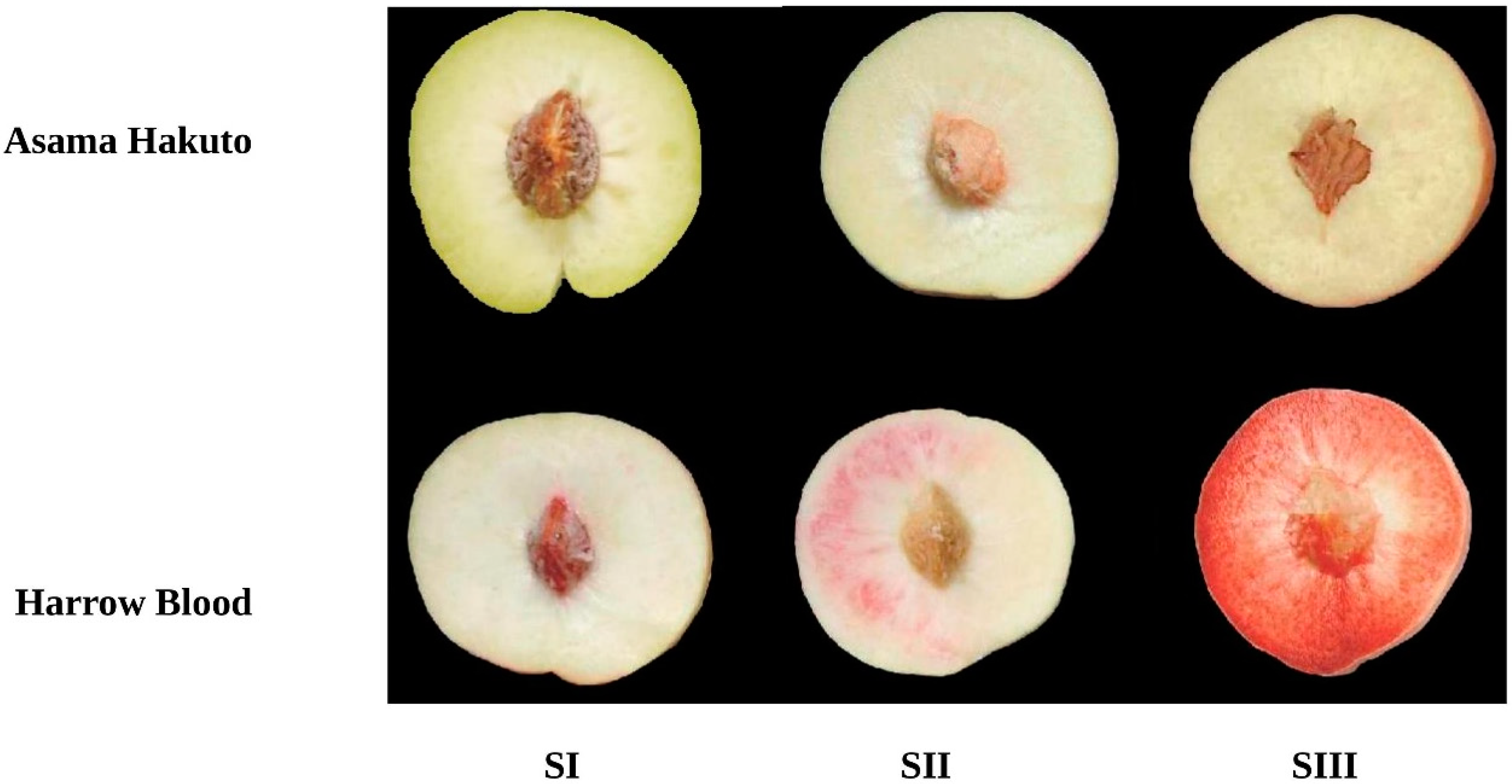
3.1. Anthocyanin Accumulation during Peach Fruit Development

The flesh of the red and white peach varieties of Harrow Blood (HB) and Asama Hakuto (AH) were used to determine the anthocyanin content in the fruit at different developmental stages. The flesh of Harrow Blood remained light green and white during the first two stages of fruit development, and gradually turned red after 95 days of blooming. (Figure 1). In contrast, the flesh of Asama Hakuto also remained light green in the early stage of fruit development, but turned white 95 days after full bloom. The anthocyanin accumulation in Harrow Blood started at SII; however, the amount of anthocyanin at this stage was both visibly and significantly lower than at the later stage, and the flesh color largely remained white at this stage. A high amount of total anthocyanin was detected in Harrow Blood at SIII and the flesh color was completely dark red; on other hand no anthocyanin accumulated in Asama Hakuto at SI and SII (Figure 2C). Red flesh fruits began to synthesize anthocyanin nearly 2 weeks before maturity, and showed a rapid increase in anthocyanin content 10 days before maturity (Figure 1). On the other hand, the white flesh fruits of AH did not show any visible anthocyanin in the early stages of fruit development; however, a small amount of anthocyanin was detected at SIII near the mature stage. In general, the total anthocyanin content in red flesh fruits was both visibly and significantly higher than in white flesh fruits during all three stages of fruit development.

3.2. Transcriptome Assembly

Two transcriptomes were sequenced (18 libraries) for samples from three fruit developmental stages (Table 1). Clean data of 24 GB were obtained from the 18 cDNA libraries. The lowest amount of reads of 40.5 million per library was recorded for AH–SII, while the greatest number of reads per library was 47.6 million for HB–SII. The read length was 150 bp. About 93% of total reads were successfully mapped to peach genome V2.0 a1 (Table S1). Our main focus, considering the current comparative transcriptome study, was to identify the presence of genes differentially expressed across three stages of fruit development in both red- and white-fleshed fruit. We subsequently identified 21,449 genes expressed in red- and white-fleshed fruits.

3.3. Differentially Expressed Genes (DEGs between Red- and White-Fleshed Fruit

We compared transcriptomes of HB and AH at the three stages of fruit development to identify DEG during peach fruit development. DEGs were filtered based on expression level |log2 (fold change)|> 2 and adjusted p value < 0.05 in each pairwise comparison. At each developmental stage, the up-regulated DEGs were more abundant than the down-regulated DEGs (Table 2). After removing 20,664 genes with low expression variance (p < 0.5 variance), 785 common genes were found between the red-fleshed and white-fleshed fruit at all three stages of development (Figure 2A).

3.4. Identification of Genes Associated with Anthocyanin Biosynthesis

Based on the high anthocyanin presence at SIII, we further selected the genes that were only differentially expressed at SIII. Out of 785 commonly shared genes, only 13 genes were differentially expressed at SIII. These 785 DEGs were used to construct Venn diagrams to illustrate their differential expression at each developmental stage. Most common DEGs were found at the early stage of fruit development stage SI (35DAFB) (381 DEGs), followed by SII (65 DAFB) (219 DEGS). The lowest number of DEGs (185 DEGs) were expressed at SIII, with 95 DAFB (Table 2). To further search for key DEGs that were associated with the anthocyanin biosynthesis, we focused on the DEGs that were differentially expressed only at SIII, which is a crucial stage of anthocyanin accumulation in red-fleshed peaches according to the previous studies [39].

3.5. Selection of Key Genes Controlling Flesh Color

To identify the genes associated with the red flesh color and anthocyanin biosynthesis, the annotation information of these 13 genes was extracted from the peach reference genome annotation database, and their relative expression analysis was performed at all three developmental stages. The expression levels of these 13 genes from the RNA-seq were subsequently verified by RT-qPCR and presented through a heatmap (Figure 2B) to accurately identify key candidate genes. All of these genes had significantly high expression values at SIII in the red flesh peach fruit tissues. Among these genes, one gene, Prupe.3G228500, which was previously coded for PpMYB39, was reported to be co-expressed with the anthocyanin pathway biosynthetic genes [28]. Results obtained from the RT-qPCR were highly consistent with the RNA-seq data results (Figure 2D). The relative expression levels of PpMYB39 were correlated with anthocyanin levels at all three stages of fruit development in the red-fleshed fruits, but its expression levels in white-fleshed fruits were significantly lower. Finally, based on its higher expression levels and correlation with anthocyanin content, this gene was selected as a key candidate associated with the red flesh color of peach fruit.

3.6. PpMYB39 Plays a Crucial Role in Red Flesh Formation in Peaches

To further confirm the association of PpMYB39 with red flesh peach characteristics, the expression levels of PpMYB39 were analyzed at the full maturity stage in five different peach cultivars with different flesh colors: Asama Hakuto (white flesh), Zao Yan (creamy white flesh), Phillips (light-yellow flesh), Fay Elberta (yellow flesh), Indian Blood (blood-red flesh) and Harrow Blood (deep-red flesh) (Figure 3A). PpMYB39 was highly expressed in the fruit flesh of two red cultivars of Indian Blood and Harrow Blood, while the expression was very low in both the white- and yellow-fleshed cultivars (Figure 3B). To confirm that this higher expression was only found in flesh tissues, PpMYB39 expression analysis was also performed in different tissues of Harrow Blood. Flower sepals and petals were collected at the full bloom stage of the peach flower, and young leaf samples were collected at the ending bloom stage at onset of fruit. Similarly, fruit flesh and fruit skin samples were obtained at full maturity stage. Roots samples were collected from a four-year-old tree. The results showed that the expression levels of PpMYB39 were significantly higher in the flesh tissue compared to the fruit skin, leaves, roots, flower sepals and petals (Figure 3C). However, the expression levels in the fruit skin and leaves were significantly higher than the roots and stem. Furthermore, the relative expression patterns of PpMYB39 were highly correlated with the anthocyanin content of peach flesh throughout fruit development. All these findings suggested that PpMYB39 is associated with the red flesh coloration of peach fruit.

3.7. PpMYB39 Function as a Transcriptional Activator of PpDFR

To study the role of PpMYB39 during anthocyanin biosynthesis, which is the main pigment behind red color formation in peach flesh, we therefore analyzed the expression levels of the late biosynthetic genes of the anthocyanin pathway in peach fruit. Interestingly, a key structural gene of the anthocyanin pathway PpDFR (Prupe.1G376400) was co-expressed with PpMYB39 at all three stages of fruit development (Figure 4A). It is also important to mention that some previous studies, [40,41], suggested that MYB39 is not directly involve in anthocyanin biosynthesis, but is up-regulated together with the biosynthetic genes of the anthocyanin pathway in red strawberry fruits. Therefore, we believe that PpMYB39 might be the transcriptional activator for PpDFR, which activates the enzyme DFR of the anthocyanin pathway from where the actual process towards anthocyanin biosynthesis starts during phenylalanine metabolism in plants. PpMYB39 was identified as being up-regulated in peach flesh in the KEGG enrichment analysis (Figure 4C).
Initially, to confirm whether PpMYB39 could activate the promoter of PpDFR, we conducted dual luciferase reporter assays. PpMYB39 CDS cloned into the vector pBI121-GFP under the control of a CaMV35S promoter was used as the effector plasmid, while a PpDFR promoter cloned into the vector pGreenII0800-LUC was used as a reporter. A pBI121-GFP empty vector, driven by the CaMV25S promoter co-infiltrated with pGreen0800-LUC, was used as a negative control. Constructs were separately transformed into the Agrobacterium tumefaciens strain GV3101 and incubated at 28 °C for 48 h, before being infiltrated into the tobacco leaves using a transformation buffer. The leaf samples were harvested two days after the infiltration. The highest activity for DFR was observed when co-infiltrated with MYB39, compared to the negative control treatment where DFR was con-transformed with the empty effector plasmid (Figure 4B). Similarly, to examine the transcriptional activity of MYB39, we conducted a yeast one-hybrid assay. The promoter of PpDFR, carried by the pHIS vector, and PpMYB39 CDS, carried by pGADT7, were transformed into Y187 gold yeast strains and were plated with a solid medium for three days. The results showed that yeast cells co-transformed with pHIS-PpDFR and AD-PpMYB39 grew well on the SD medium. The yeast cells co-transformed with pHIS2 and AD empty vectors provided by the kit as a positive control also grew well on the medium, whereas no growth was observed for the yeast cells co-transformed with pHIS2-DFR and the AD empty vector as a negative control (Figure 5). Therefore, we believe that MYB39 plays a major role during anthocyanin biosynthesis in peach flesh, initiating the transcription of the structural gene of the anthocyanin pathway that codes for the key enzyme DFR responsible for anthocyanin biosynthesis.

4. Discussion

Generally, the fruit color, taste, and aroma determine its acceptance by consumers. Flesh color is one of the most important commercial characteristics of peach fruits. Peach varieties are usually divided into two categories: white peaches and yellow peaches. Red-fleshed peaches are the third flesh color phenotype of the peach germplasm [4]. Anthocyanin biosynthesis plays an important role in the coloration of red-fleshed peaches. Compared to white and yellow peaches, red-fleshed peaches have a high anthocyanin content and have a unique flavor. It has been found that anthocyanin-rich foods have antiaging, antioxidant and anticancer properties, and are beneficial to human health, helping to promote blood circulation, reduce free radicals, and prevent several cerebrovascular and cardiovascular diseases [42]. Therefore, anthocyanin-rich peaches are becoming increasingly popular among consumers due to their antioxidant properties, pleasant taste, and enchanting color. In the past, the anthocyanin biosynthesis pathway has been extensively studied in fruit trees and found to be regulated by MYB transcription factors [10,43]. The key genes associated with flesh color phenotypes have been previously identified as PpMYB9, PpMYB17, PpMYB18, PpMYB19, PpMYB20 [44] and PpMYB10.1 [22]. However, a particular MYB regulator gene that controls the transcriptional activation of the key anthocyanin biosynthetic pathway genes has not yet been identified. Therefore, we conducted a comparative transcriptome analysis of red- and white-fleshed peach fruits to identify an anthocyanin regulator gene of the anthocyanin biosynthetic pathway.
The maturity of the peach fruit was counted according to the number of days after the full bloom of the peach flower. Fruits were collected at 35, 65 and 95 days after full bloom. These time points represent the three developmental stages of peach fruit. At SI, the fruit is immature, the skin color is green, and flesh is completely white. SII is the start of the maturity phase, where both the skin and flesh start turning red. SIII is the stage of full maturity, just before ripening, where the skin is partially red and the flesh turns completely red. It is also important to mention here that fruit color is a visible parameter, and the red color of peach fruit is due to anthocyanin accumulation, which is a well-reported and well-established hypothesis; therefore, the accumulation of anthocyanin in peach flesh at SIII and the DEGs at this stage must have a strong relationship with each other. Previously, Guo used a similar approach to identify DEGs associated with peach fruit shape [30].
In the present study, red-fleshed peaches showed an abundant accumulation of anthocyanin at the critical developmental stage III. Therefore, based on the differential expressions and functional annotations of the DEGs at SIII, we further selected and analyzed the candidate regulator gene associated with anthocyanin biosynthesis and flesh color in peach fruit. Our results were also consistent with previous studies in which researchers have reported a higher accumulation of anthocyanin in the later stages of fruit development, near 90 days [45]. The lower concentration of anthocyanin in the early stage of fruit development and its higher concentration in the later stage suggest its importance in the anthocyanin biosynthesis of red-fleshed peaches. Furthermore, the coloration of red-fleshed peaches at the later stage of fruit development—compared with no coloration in white-fleshed peaches at any developmental stage of the fruit—might be caused by the differential expression of anthocyanin biosynthesis genes.
Anthocyanin accumulation is mainly controlled by the expression of genes encoding specific enzymes involved in the anthocyanin biosynthesis pathway. Previously, regulatory genes controlling the expression of structural genes involved in the anthocyanin biosynthetic pathway have been identified in many plants, most recently in [46]. It has been reported that the anthocyanin biosynthesis pathway is transcriptionally regulated by a MBW complex in which MYB transcription factors determine the activation of this complex by binding with the promoter region of the structural genes [47]. In the current study, we identified several MYB transcription factors and assessed their expression levels at different developmental stages of fruit. The results showed higher expression levels of MYB transcription factors in the later developmental stages of peach fruit. Previously, researchers have shown similar higher expression levels of MYB transcription factors at the later developmental stages of different fruits [44]. Our results suggest that the higher expression levels of MYB transcription factors found in the later developmental stages of fruit activate anthocyanin biosynthetic genes, and boost the accumulation of anthocyanin in red-fleshed peaches (Figure 6).
It has been previously reported that several transcription factors from the MYB family can regulate the biosynthetic genes of the anthocyanin pathway in different plant species [48]. Among them, it is reported that MYB39 can regulate the accumulation of anthocyanin in several plants. Capsicum annum CA10g03650 is homologous to MYB39 from Arabidopsis thaliana, encoding MYB39 TF, and was found to be associated with anthocyanin biosynthesis in pepper fruit [29]. Similarly, Fragaria vesca MYB39 is up-regulated in red strawberry fruits and down-regulated in yellow mutants [40].
Other studies have further demonstrated the association of MYB transcription factors with red flesh phenotypes in apples. The R2R3 MYB transcription factor can also regulate the expression of anthocyanin biosynthesis genes, as well as other transcription factor co-activators, such as the basic-helix-loop-helix (bHLH) and the WD40 repeat family [17]. In addition, researchers have demonstrated that the overexpression of the combination of MYB10.1/bHLH3 and MYB10.3/bHLH3 can activate the production of anthocyanin by up-regulating the expression of the anthocyanin biosynthesis genes NtCHS, NtDFR and NtUFGT in peach fruits [27]. Dihydroflavonol 4-reductase (DFR) catalyzes the reduction of dihydroflavonol to leuco-anthocyanins, which is a key “late” step in the biosynthesis of anthocyanins [49,50]. At this stage, DFR and FLS also catalyze a competitive reaction to produce products that lead to anthocyanin or flavonols [51,52]. In the current study, the results of the expression analysis showed that the expression levels of PpMYB39 and PpDFR were positively correlated with each other, as well as with anthocaynin content in the later stages of fruit development of red-fleshed peaches. The interaction between them during anthocyanin biosynthesis was confirmed in yeast one-hybrid and dual luciferase assays. Our results indicate that PpMYB39 initiates the transcription of PpDFR by binding to its promoter, and therefore red-fleshed peaches accumulate a high anthocyanin content (Figure 6).

5. Conclusions

We performed transcriptome profiling and screening to identify candidate genes associated with anthocyanin biosynthesis in peach fruit. We identified 13 DEGs as possible candidate genes for flesh color. Among these 13 genes, the expression of Prupe.3G228500, which encodes for PpMYB39, was found to be positively correlated with anthocyanin content in the red-fleshed peach cultivars of Harrow Blood, and correlated with Prupe.1G376400, which encodes for Dihydroflavonol 4-reductase (PpDFR). Yeast one-hybrid and dual luciferase reporter assays confirmed the activation of PpDFR by PpMYB39. These findings allow us to hypothesize that PpMYB39 plays a role as a transcription activator of PpDFR during the process of anthocyanin biosynthesis in peach fruit. Our results might be helpful in peach breeding as well as in identifying anthocyanin regulator genes in related fruit species.

Supplementary Materials

The following supporting information can be downloaded at: https://www.mdpi.com/article/10.3390/horticulturae8040332/s1, Table S1: Report on sequencing analysis of 6 Peach Fruit Samples.

Author Contributions

Conceptualization, I.A.K. and S.S.; methodology, M.U.R.; software, A.A.K.; formal analysis, I.A.K.; investigation, S.M.K., T.A. and M.A.; resources, A.A.K.; data curation, M.U.R.; writing—original draft preparation, M.U.R.; writing—review and editing, I.A.K., G.N. and S.M.K.; supervision, S.S.; project administration, S.S. All authors have read and agreed to the published version of the manuscript.

Funding

This research was supported by the Center for plant biodiversity university of Swat, Pakistan.

Institutional Review Board Statement

Not applicable.

Informed Consent Statement

Not applicable.

Data Availability Statement

The sequencing data have been deposited in the NCBI Sequence Read Archive (http://www.ncbi.nlm.nih.gov/sra/, accessed on 11 April 2022), with the accession number PRJNA826334.

Conflicts of Interest

The authors declare no conflict of interest.

References

  1. Zhao, X.; Zhang, W.; Yin, X.; Su, M.; Sun, C.; Li, X.; Chen, K. Phenolic composition and antioxidant properties of different peach [Prunus persica (L.) Batsch] cultivars in China. Int. J. Mol. Sci. 2015, 16, 5762–5778. [Google Scholar] [CrossRef] [PubMed] [Green Version]
  2. Folta, K.M.; Gardiner, S.E. Genetics and Genomics of Rosaceae; Springer: New York, NY, USA, 2009; Volume 6, pp. 411–506. [Google Scholar] [CrossRef]
  3. Arús, P.; Verde, I.; Sosinski, B.; Zhebentyayeva, T.; Abbott, A.G. The peach genome. Tree Genet. Genomes 2012, 8, 531–547. [Google Scholar] [CrossRef]
  4. Werner, D.J.; Creller, M.A.; Chaparro, J.X. Inheritance of the blood-flesh trait in peach. HortScience 1998, 33, 1243–1246. [Google Scholar] [CrossRef] [Green Version]
  5. Cao, K.; Ding, T.; Mao, D.; Zhu, G.; Fang, W.; Chen, C.; Wang, L. Transcriptome analysis reveals novel genes involved in anthocyanin biosynthesis in the flesh of peach. Plant Physiol. Biochem. 2018, 123, 94–102. [Google Scholar] [CrossRef]
  6. Kong, J.M.; Chia, L.S.; Goh, N.K.; Chia, T.F.; Brouillard, R. Analysis and biological activities of anthocyanins. Phytochemistry 2003, 64, 923–933. [Google Scholar] [CrossRef]
  7. Zhao, Y.; Dong, W.; Wang, K.; Zhang, B.; Allan, A.C.; Lin-Wang, K.; Xu, C. Differential sensitivity of fruit pigmentation to ultraviolet light between two peach cultivars. Front. Plant Sci. 2017, 8, 1552. [Google Scholar] [CrossRef]
  8. Gonçalves, A.C.; Nunes, A.R.; Falcão, A.; Alves, G.; Silva, L.R. Dietary effects of anthocyanins in human health: A comprehensive review. Pharmaceuticals 2021, 14, 690. [Google Scholar] [CrossRef]
  9. Orazem, P.; Stampar, F.; Hudina, M. Fruit quality of Redhaven and Royal Glory peach cultivars on seven different rootstocks. J. Agric. Food Chem. 2011, 59, 9394–9401. [Google Scholar] [CrossRef]
  10. Lin-Wang, K.; Bolitho, K.; Grafton, K.; Kortstee, A.; Karunairetnam, S.; McGhie, T.K.; Allan, A.C. An R2R3 MYB transcription factor associated with regulation of the anthocyanin biosynthetic pathway in Rosaceae. BMC Plant Biol. 2010, 10, 50. [Google Scholar] [CrossRef] [Green Version]
  11. Grotewold, E. The genetics and biochemistry of floral pigments. Annu. Rev. Plant Biol. 2006, 57, 761–780. [Google Scholar] [CrossRef]
  12. Dare, A.P.; Schaffer, R.J.; Lin-Wang, K.; Allan, A.C.; Hellens, R.P. Identification of a cis-regulatory element by transient analysis of co-ordinately regulated genes. Plant Methods 2008, 4, 17. [Google Scholar] [CrossRef] [Green Version]
  13. Jiang, S.; Chen, M.; He, N.; Chen, X.; Wang, N.; Sun, Q.; Chen, X. MdGSTF6, activated by MdMYB1, plays an essential role in anthocyanin accumulation in apple. Hortic. Res. 2019, 6, 40. [Google Scholar] [CrossRef] [PubMed] [Green Version]
  14. Niu, S.S.; Xu, C.J.; Zhang, W.S.; Zhang, B.; Li, X.; Lin-Wang, K.; Chen, K.S. Coordinated regulation of anthocyanin biosynthesis in Chinese bayberry (Myrica rubra) fruit by a R2R3 MYB transcription factor. Planta 2010, 231, 887–899. [Google Scholar] [CrossRef] [PubMed]
  15. Kobayashi, S.; Goto-Yamamoto, N.; Hirochika, H. Retrotransposon-induced mutations in grape skin color. Science 2004, 304, 982. [Google Scholar] [CrossRef]
  16. Feng, S.; Wang, Y.; Yang, S.; Xu, Y.; Chen, X. Anthocyanin biosynthesis in pears is regulated by a R2R3-MYB transcription factor PyMYB10. Planta 2010, 232, 245–255. [Google Scholar] [CrossRef]
  17. Chagné, D.; Lin-Wang, K.; Espley, R.V.; Volz, R.K.; How, N.M.; Rouse, S.; Allan, A.C. An ancient duplication of apple MYB transcription factors is responsible for novel red fruit-flesh phenotypes. Plant Physiol. 2013, 161, 225–239. [Google Scholar] [CrossRef] [PubMed] [Green Version]
  18. Butelli, E.; Licciardello, C.; Zhang, Y.; Liu, J.; Mackay, S.; Bailey, P.; Reforgiato-Recupero, G.; Martin, C. Retrotransposons control fruit-specific, cold-dependent accumulation of anthocyanins in blood oranges. Plant Cell 2012, 24, 1242–1255. [Google Scholar] [CrossRef] [Green Version]
  19. Li, Y.Y.; Mao, K.; Zhao, C.; Zhao, X.Y.; Zhang, H.L.; Shu, H.R.; Hao, Y.J. MdCOP1 ubiquitin E3 ligases interact with MdMYB1 to regulate light-induced anthocyanin biosynthesis and red fruit coloration in apple. Plant Physiol. 2012, 160, 1011–1022. [Google Scholar] [CrossRef] [Green Version]
  20. Wang, Z.; Meng, D.; Wang, A.; Li, T.; Jiang, S.; Cong, P.; Li, T. The methylation of the PcMYB10 promoter is associated with green-skinned sport in Max Red Bartlett pear. Plant Physiol. 2013, 162, 885–896. [Google Scholar] [CrossRef] [Green Version]
  21. Telias, A.; Lin-Wang, K.; Stevenson, D.E.; Cooney, J.M.; Hellens, R.P.; Allan, A.C.; Bradeen, J.M. Apple skin patterning is associated with differential expression of MYB10. BMC Plant Biol. 2011, 11, 93. [Google Scholar] [CrossRef] [Green Version]
  22. Ravaglia, D.; Espley, R.V.; Henry-Kirk, R.A.; Andreotti, C.; Ziosi, V.; Hellens, R.P.; Allan, A.C. Transcriptional regulation of flavonoid biosynthesis in nectarine (Prunus persica) by a set of R2R3 MYB transcription factors. BMC Plant Biol. 2013, 13, 68. [Google Scholar] [CrossRef] [PubMed] [Green Version]
  23. Zhu, Y.C.; Zhang, B.; Allan, A.C.; Lin-Wang, K.; Zhao, Y.; Wang, K.; Chen, K.S.; Xu, C.J. DNA demethylation is involved in the regulation of temperature-dependent anthocyanin accumulation in peach. Plant J. 2020, 102, 965–976. [Google Scholar] [CrossRef] [PubMed]
  24. Tsuda, T.; Yamaguchi, M.; Honda, C.; Moriguchi, T. Expression of anthocyanin biosynthesis genes in the skin of peach and nectarine fruit. J. Am. Soc. Hortic. Sci. 2004, 129, 857–862. [Google Scholar] [CrossRef] [Green Version]
  25. Dardick, C.; Callahan, A.M. Evolution of the fruit endocarp: Molecular mechanisms underlying adaptations in seed protection and dispersal strategies. Front. Plant Sci. 2014, 5, 284. [Google Scholar] [CrossRef] [PubMed]
  26. Zhou, Y.; Zhou, H.; Lin-Wang, K.; Vimolmangkang, S.; Espley, R.V.; Wang, L.; Han, Y. Transcriptome analysis and transient transformation suggest an ancient duplicated MYB transcription factor as a candidate gene for leaf red coloration in peach. BMC Plant Biol. 2014, 14, 388. [Google Scholar] [CrossRef] [PubMed] [Green Version]
  27. Uematsu, C.; Katayama, H.; Makino, I.; Inagaki, A.; Arakawa, O.; Martin, C. Peace, a MYB-like transcription factor, regulates petal pigmentation in flowering peach ‘Genpei’bearing variegated and fully pigmented flowers. J. Exp. Bot. 2014, 65, 1081–1094. [Google Scholar] [CrossRef] [Green Version]
  28. Rahim, M.A.; Busatto, N.; Trainotti, L. Regulation of anthocyanin biosynthesis in peach fruits. Planta 2014, 240, 913–929. [Google Scholar] [CrossRef]
  29. Wang, G.; Chen, B.; Du, H.; Zhang, F.; Zhang, H.; Wang, Y.; Zhang, X. Genetic mapping of anthocyanin accumulation-related genes in pepper fruits using a combination of SLAF-seq and BSA. PLoS ONE 2018, 13, e0204690. [Google Scholar] [CrossRef]
  30. Guo, J.; Cao, K.; Li, Y.; Yao, J.L.; Deng, C.; Wang, Q.; Zhu, G.; Fang, W.; Chen, C.; Wang, X.; et al. Comparative transcriptome and microscopy analyses provide insights into flat shape formation in peach (Prunus persica). Front. Plant Sci. 2018, 8, 2215. [Google Scholar] [CrossRef] [Green Version]
  31. Kim, D.; Langmead, B.; Salzberg, S.L. HISAT: A fast spliced aligner with low memory requirements. Nat. Methods 2015, 12, 357–360. [Google Scholar] [CrossRef] [Green Version]
  32. Anders, S.; Pyl, P.T.; Huber, W. HTSeq—A Python framework to work with high-throughput sequencing data. Bioinformatics 2015, 31, 166–169. [Google Scholar] [CrossRef] [PubMed]
  33. Frazee, A.C.; Pertea, G.; Jaffe, A.E.; Langmead, B.; Salzberg, S.L.; Leek, J.T. Ballgown bridges the gap between transcriptome assembly and expression analysis. Nat. Biotechnol. 2015, 33, 243–246. [Google Scholar] [CrossRef] [PubMed] [Green Version]
  34. Benjamini, Y.; Hochberg, Y. Controlling the false discovery rate: A practical and powerful approach to multiple testing. J. R. Stat. Soc. Ser. B (Methodol.) 1995, 57, 289–300. [Google Scholar] [CrossRef]
  35. Cao, K.; Zhou, Z.; Wang, Q.; Guo, J.; Zhao, P.; Zhu, G.; Fang, W.; Chen, C.; Wang, X.; Wang, X.; et al. Genome-wide association study of 12 agronomic traits in peach. Nat. Commun. 2016, 7, 13246. [Google Scholar] [CrossRef] [PubMed] [Green Version]
  36. Liu, H.; Cao, K.; Zhu, G.; Fang, W.; Chen, C.; Wang, X.; Wang, L. Genome-wide Association Analysis of Red Flesh Character Based on Resequencing Approach in Peach. J. Am. Soc. Hortic. Sci. 2019, 144, 209–216. [Google Scholar] [CrossRef] [Green Version]
  37. Cheng, G.W.; Breen, P.J. Activity of phenylalanine ammonia-lyase (PAL) and concentrations of anthocyanins and phenolics in developing strawberry fruit. J. Am. Soc. Hortic. Sci. 1991, 116, 865–869. [Google Scholar] [CrossRef]
  38. Wang, Y.; Zhou, L.J.; Wang, Y.; Geng, Z.; Ding, B.; Jiang, J.; Chen, S.; Chen, F. An R2R3-MYB transcription factor CmMYB21 represses anthocyanin biosynthesis in color fading petals of chrysanthemum. Sci. Hortic. 2022, 293, 110674. [Google Scholar] [CrossRef]
  39. Guo, J.; Cao, K.; Deng, C.; Li, Y.; Zhu, G.; Fang, W.; Chen, C.; Wang, X.; Wu, J.; Guan, L.; et al. An integrated peach genome structural variation map uncovers genes associated with fruit traits. Genome Biol. 2020, 21, 1–19. [Google Scholar] [CrossRef]
  40. Zhang, Y.; Li, W.; Dou, Y.; Zhang, J.; Jiang, G.; Miao, L.; Zhang, Z. Transcript quantification by RNA-seq reveals differentially expressed genes in the red and yellow fruits of Fragaria vesca. PLoS ONE 2015, 10, e0144356. [Google Scholar] [CrossRef] [Green Version]
  41. Zhang, L.; Wang, L.; Zeng, X.; Chen, R.; Yang, S.; Pan, S. Comparative transcriptome analysis reveals fruit discoloration mechanisms in postharvest strawberries in response to high ambient temperature. Food Chem. X 2019, 2, 100025. [Google Scholar] [CrossRef]
  42. Chen, P.N.; Chu, S.C.; Chiou, H.L.; Kuo, W.H.; Chiang, C.L.; Hsieh, Y.S. Mulberry anthocyanins, cyanidin 3-rutinoside and cyanidin 3-glucoside, exhibited an inhibitory effect on the migration and invasion of a human lung cancer cell line. Cancer Lett. 2006, 235, 248–259. [Google Scholar] [CrossRef] [PubMed]
  43. Tuan, P.A.; Bai, S.; Yaegaki, H.; Tamura, T.; Hihara, S.; Moriguchi, T.; Oda, K. The crucial role of PpMYB10. 1 in anthocyanin accumulation in peach and relationships between its allelic type and skin color phenotype. BMC Plant Biol. 2015, 15, 280. [Google Scholar] [CrossRef] [PubMed] [Green Version]
  44. Zhou, H.; Peng, Q.; Zhao, J.; Owiti, A.; Ren, F.; Liao, L.; Han, Y. Multiple R2R3-MYB transcription factors involved in the regulation of anthocyanin accumulation in peach fruit. Front. Plant Sci. 2016, 7, 1557. [Google Scholar] [CrossRef] [PubMed]
  45. Yan, J.; Cai, Z.X.; Shen, Z.J.; Ma, R.J.; Yu, M.L. Proanthocyanidin monomers and cyanidin 3-o-glucoside accumulation in blood-flesh peach (Prunus persica (l.) Batsch) fruit. Arch. Biol. Sci. 2017, 69, 611–617. [Google Scholar] [CrossRef]
  46. Zhao, Y.; Dong, W.; Zhu, Y.; Allan, A.C.; Lin-Wang, K.; Xu, C. PpGST1, an anthocyanin-related glutathione S-transferase gene, is essential for fruit coloration in peach. Plant Biotechnol. J. 2020, 18, 1284–1295. [Google Scholar] [CrossRef] [PubMed] [Green Version]
  47. Liu, Y.; Tikunov, Y.; Schouten, R.E.; Marcelis, L.F.; Visser, R.G.; Bovy, A. Anthocyanin biosynthesis and degradation mechanisms in Solanaceous vegetables: A review. Front. Chem. 2018, 6, 52. [Google Scholar] [CrossRef]
  48. Khan, I.A.; Cao, K.; Guo, J.; Li, Y.; Wang, Q.; Yang, X.; Wang, L. Identification of key gene networks controlling anthocyanin biosynthesis in peach flower. Plant Sci. 2022, 316, 111151. [Google Scholar] [CrossRef]
  49. Moyano, E.; Portero-Robles, I.; Medina-Escobar, N.; Valpuesta, V.; Munoz-Blanco, J.; Luis Caballero, J. A fruit-specific putative dihydroflavonol 4-reductase gene is differentially expressed in strawberry during the ripening process. Plant Physiol. 1998, 117, 711–716. [Google Scholar] [CrossRef] [Green Version]
  50. Piero, A.R.L.; Puglisi, I.; Petrone, G. Gene characterization, analysis of expression and in vitro synthesis of dihydroflavonol 4-reductase from [Citrus sinensis (L.) Osbeck]. Phytochemistry 2006, 67, 684–695. [Google Scholar] [CrossRef]
  51. Tian, J.; Han, Z.Y.; Zhang, J.; Hu, Y.; Song, T.; Yao, Y. The balance of expression of dihydroflavonol 4-reductase and flavonol synthase regulates flavonoid biosynthesis and red foliage coloration in crabapples. Sci. Rep. 2015, 5, 12228. [Google Scholar] [CrossRef]
  52. Luo, P.; Ning, G.; Wang, Z.; Shen, Y.; Jin, H.; Li, P.; Bao, M. Disequilibrium of flavonol synthase and dihydroflavonol-4-reductase expression associated tightly to white vs. red color fruit formation in plants. Front. Plant Sci. 2016, 6, 1257. [Google Scholar] [CrossRef] [PubMed] [Green Version]
Figure 1. Color morphology of white flesh (Asama Hakuto) and red flesh (Harrow Blood) peach cultivars during peach fruit development. Fruits were collected at three developmental stages: SI, SII and SIII; 35, 65, and 95 days after full bloom (DAFB).
Horticulturae 08 00332 g001
Figure 2. Genes associated with flesh color: (A) Venn diagram of differentially expressed genes of the red and white flesh fruits. (B) Expression heatmap of 13 selected DEGs differentially expressed at SIII. (C) Anthocyanin content of peach fruit at different fruit developmental stages MYB39. (D) The expression levels of PpMYB39 at different developmental stages of red- and white-fleshed peach fruits obtained from RNA−seq and validated by RT−qPCR. The error bars show the SE of the three biological replicates while * indicates the significant difference at p < 0.05 in the Student’s test. While ** indicates significant different at p < 0.01 in student’s test.
Horticulturae 08 00332 g002
Figure 3. Expression analysis of PpMYB39 in peach fruit. (A) Phenotypic variation among five peach cultivars used in this study at full maturity. (B) The RT-qPCR expression analysis of PpMYB39 in five peach cultivars differing in flesh color. (C) RT-qPCR expression analysis of PpMYB39 in different tissues of peach plant. The actin gene was used as the internal control. The error bars show the SE of three biological replicates, while * indicates the significant difference in the Student’s test at p < 0.01, while ** indicates significant different at p < 0.05 in student’s test.
Horticulturae 08 00332 g003
Figure 4. Functional validation of PpMYB39 in peach flesh. (A) The expression pattern correlation between PpMYB39 and the structural genes of the anthocyanin biosynthetic pathway. (B) Transient dual luciferase reporter assay of PpMYB39 on the promoter of PpDFR. (C) The enrichment analysis of DEGs detected in fruit flesh. Error bars show SE of three biological replicates, while ** indicates the significant difference in the Student’s test at p < 0.01.
Horticulturae 08 00332 g004
Figure 5. Interaction between PpMYB39 and PpDFR in yeast one-hybrid assay. Yeast one-hybrid assay shows the promoter activation of PpDFR by PpMYB39. For positive control treatment, the co-transformed yeast cells carrying AD and pHIS2 empty vectors were used. For negative control treatment, transformed yeast cells carrying AD empty vectors and pHIS2-DFR were used.
Horticulturae 08 00332 g005
Figure 6. Proposed anthocyanin biosynthetic pathway of peach fruit. The role of PpMYB39 as transcriptional activator of PpDFR has been highlighted. The red and blue colors of the heat maps represent the expression levels of the structural genes of the anthocyanin pathway in the red- and white-fleshed tissues of peach fruit. SI, SII and SIII represent the three stages of peach fruit development in Asama Hakuto and Harrow Blood.
Horticulturae 08 00332 g006
Table 1. Transcriptome assembly and Illumina HiSeq quality control information. Three biological replicates were maintained for each sample.
SampleRaw ReadsClean ReadsClean Reads Rate (%)Q30 (%)
HB-SI51,170,55647,624,06493.0794.05
HB-SII50,971,60046,185,79290.6194.3
HB-SIII47,834,47242,998,91689.8994.2
AH-SI46,768,71842,826,84291.5793.76
AH-SII48,688,72845,267,66692.9794.4
AH-SIII50,232,67446,087,39691.7594.14
Table 2. Differentially expressed genes shared between red- and white-fleshed fruits during fruit development.
GroupsTotal DEGsUp-RegulatedDown-Regulated
HB SI_V_AS SI38269112
HB SII_V_AS SII21914871
HB SIII_V_AS SIII18512560
Publisher’s Note: MDPI stays neutral with regard to jurisdictional claims in published maps and institutional affiliations.

