Article

An Efficient Agrobacterium-Mediated Genetic Transformation System for Persimmon (Diospyros kaki Thunb.)

1
Key Laboratory of Horticultural Plant Biology, Huazhong Agricultural University, Wuhan 430070, China
2
Horticultural Institute, Jiangxi Academy of Agricultural Sciences, Nanchang 330200, China
*
Author to whom correspondence should be addressed.
Horticulturae 2022, 8(5), 422; https://doi.org/10.3390/horticulturae8050422
Submission received: 30 March 2022 / Revised: 25 April 2022 / Accepted: 6 May 2022 / Published: 10 May 2022
(This article belongs to the Section Genetics, Genomics, Breeding, and Biotechnology (G2B2))

Abstract

:
Persimmon (Diospyros kaki Thunb.) is an important fruit crop in East Asia and has great commercial potential due to the high demand for fresh edible fruits or processed dried persimmon, as well as its being a source of raw materials for the chemical industry. Genetic engineering has shown great potential in persimmon fruit improvement and compensated for the limitations of conventional breeding. In this study, we developed an efficient transformation system and in vivo regeneration protocol by using the leaf disc culture of ‘Gongcheng Shuishi’ persimmon through the optimization of different basic media and hormone combinations. Based on the secondary induction system on 1/2DKW (DKW medium) + ZT (2.0 mg/L) + TDZ (0.5 mg/L) and MS (1/2 N) + ZT (2.0 mg/L) + IAA (0.1 mg/L), the callus induction rate and adventitious bud induction rate were 96.0% and 94.0%, respectively, and RNA interference of DkANR (ANR, anthocyanidin reductase) was achieved after transformation, thus resulting in a reduction in proanthocyanidin accumulation. The GFP (green fluorescent protein) fluorescence signal was visualized in the transgenic seedlings with GFP gene cotransformation. A total of 17.9% of the positive regenerated transgenic seedlings with empty vector were confirmed by both hyg gene and 35S promoter PCR analysis. Southern blotting assays showed that 20 positive transgenic persimmon seedlings were obtained from 135 regenerated transformants derived from DkANRi. Altogether, a reliable and highly efficient transformation system was achieved in persimmon, which may contribute to gene function identification and genetic improvement in the future.

1. Introduction

Persimmons (Diospyros kaki Thunb.; 2n = 6x = 90 or 2x = 9x = 135) are a kind of deciduous perennial woody fruit tree that originated in China and have a 2000-year cultivation history. Persimmons belong to the family Ebenaceae, which contains nearly 500 species [1]. Persimmon production in China accounts for 76.04% of the world’s production (FAOSTAT, 2021). Persimmon cultivars are classified into two groups according to the genetic characteristics of natural deastringency: pollination constant and nonastringent (PCNA) persimmons that are controlled by qualitative loci, and non-PCNA types that are controlled by quantitative loci. The former is divided into Chinese PCNA (C-PCNA) and Japanese PCNA (J-PCNA), and the trait of natural deastringency of C-PCNA is dominant but recessive in J-PCNA. Non-PCNA types include the pollination variant and nonastringent (PVNA), the pollination variant and astringent (PVA) and the pollination constant and astringent (PCA) type [2]. At present, most commercial cultivars belong to astringent (PCA type) landraces in China. PCNA-type persimmon is attractive because their fruits can lose their astringency on the tree naturally and be eaten directly without extra artificial deastringency treatment after harvesting. PCNA-type persimmon fruits are suitable for fresh consumption, long-term storage, and transportation. As conventional breeding is seriously hindered by the long juvenile period and polyploidy properties of persimmon, genetic transformation presents an advantage in the rapid introduction of foreign genes to existing cultivars with small specific changes in the genome [1].
Proanthocyanidins (PAs) are important secondary metabolites in higher plants. As there is a large amount of PAs accumulation in persimmon fruits compared with other fruit trees, the PAs biosynthetic pathway and the process of natural deastringency have become interesting issues in persimmon studies [2]. With the increasingly abundant data from genome [3], transcriptome, proteome [4,5], and miRNA sequencing [6] in persimmon, a large number of important differentially expressed genes (DEGs) have been screened and isolated, but their functions need to be further verified. Some research on in vitro regeneration and genetic transformation has been carried out by explants of Japanese persimmon [7,8,9] and rootstock D. lotus [10]. The phenotype of the transgenic lines presented remarkable changes, such as plant dwarfing [7,11] and rooting ability reduction [7], salt stress or other environmental stress tolerance enhancements [12,13,14], and the down-regulation of polygalacturonase biosynthesis [15]. To date, only a few reports on heterozygous genes from other plant species have been introduced into J-PCNA persimmon by stable transformation. There are few functional identifications of genes derived from persimmon itself by a stable genetic transformation in persimmons. The regeneration rate and positive rate concerning transformation in persimmon need to be improved because the high PAs content notably results in brown calli and hindered plantlet growth (Table 1).
To some extent, the transformation capabilities for woody plants are largely restricted to successful efficient regeneration systems. In previous reports, explants from somatic embryogenesis [19], leaf primordia, stem internodes, and the young leaves of adult Japanese persimmon ‘Jiro’ showed a high frequency of regeneration on Murashige and Skoog (MS) medium with half of the normal concentrations of nitrogen-containing 10 µM zeatin (ZT), 1 µM 4PU-30 (forchlorfenuron) in combination with 0.1 µM indole-3-acetic acid (IAA), or MS (1/2N) medium containing 0.03 to 0.1 µM IAA or 0.01 to 0.03 µM 1-naphthylacetic acid (NAA) combined with 10 µM ZT in the dark [20]. The J-PCNA ‘Jiro’ persimmon adventitious buds could be generated spontaneously and directly from the roots [21]. Of all the tested plant growth regulators, 5–10 μM ZT in combination with 8–10 μM IAA exhibited the best influence on stimulating the generation of adventitious buds in Japanese persimmon micropropagation.
There are two leading commercial PCA persimmon cultivars, ‘Mopanshi’ and ‘Gongcheng Shuishi’, distributed in northern and southern China, respectively. Although the study of adventitious bud regeneration has been reported in ‘Mopanshi’ (PCA type) from China [22], the callus easily turns brown or black before generating adventitious buds, which tremendously hinders the application of ‘Mopanshi’ for stable transformation. Currently, the PCA persimmon ‘Gongcheng Shuishi’ is an important commercial cultivar in southern China and accounts for nearly 30% of persimmon production in China. To date, there have been no reports on regeneration and genetic transformation related to ‘Gongcheng Shuishi’ persimmon, which is important for fresh or dried consumption. Meaningfully, accompanying the development of molecular breeding and functional genomics research on persimmon, the establishment of a stable genetic transformation system for traditional Chinese persimmon cultivars is needed. A key gene, ANR (anthocyanidin reductase), which is involved in PAs biosynthesis in plants [23,24,25], was used as an exogenous gene in this study. The present work aimed to develop a practical in vitro regeneration and Agrobacterium tumefaciens-mediated genetic transformation on ‘Gongcheng Shuishi’ persimmon, which will be further applied to identify the functions of the genes involved in PAs metabolism or fruit development and the genetic improvement of persimmon.

2. Materials and Methods

2.1. Plant Materials

To remove endophytes and obtain in vitro shoots to induce adventitious buds and leaves for gene transformation, the burgeon (5–10 cm) from vegetative shoots of ‘Gongcheng Shuishi’ (Diospyros kaki Thunb.; PCA type) in the field were sampled and washed thoroughly under running tap water for 15 min. Explants were further sterilized with 75% ethanol for 30 s, followed by 10 min of treatment with 1% NaClO. After surface sterilization, the buds were rinsed three times with autoclaved deionized water to remove disinfectant residue and then cultured onto two kinds of basal medium: MS (1/2 N) [7] or DKW [26] medium plus different concentrations of ZT (zeatin) to induce adventitious buds (Table S1). The pH of all media was adjusted to 5.8 before autoclaving at 121 °C for 15 min. The buds were cultured under a 16 h/day photoperiod at 25 ± 2 °C.

2.2. Plant Growth Regulator Combinations for Shoot Multiplication

The buds gradually elongated, and when the shoot lengths reached 1.0 cm, they were provided fresh-cut ends and transferred to MS (1/2N), or DKW medium supplemented with 0.5 mg/L or 1.0 mg/L ZT and 0 or 0.1 mg/L IAA [10]. All the media contained 0.7% agar, 3.0% sucrose, and 0.06% PVP-40 (Table S1). Each treatment was comprised of 50 tissue culture seedlings and repeated three times. After two weeks, the plantlets were moved into a new fresh subculture medium containing the same additive composition. After 30 days of culture, the effectiveness of shoot induction for each treatment was determined by recording the average number of shoots per explant and the number of differentiated buds. The number of effective shoots was calculated by the formula: shoot number/cultured seedling number. The leaves of those tissue culture seedlings were used for inducing callus and adventitious bud regeneration and stable genetic transformation.

2.3. Regeneration and Rooting of Adventitious Buds

After pre-culture of leave by MS (1/2N) + 1.0 mg/L ZT + 0.1 mg/L IAA from in vitro cultured shoot derive from burgon, two different solid callus induction media, MS (1/2N) and 1/2DKW [26]. The infected leaf discs were subcultured in a medium containing ZT (1.0 mg/L, 2.0 mg/L) and thidiazuron (TDZ) (0.5 mg/L) for 1 month for callus induction, then transferred to fresh medium for adventitious bud regeneration (Table S1). After 40 days of culture, ZT (1.0 mg/L, 2.0 mg/L) and IAA (0.1 mg/L) were also examined to obtain the optimal combination for adventitious bud induction, then the callus formation rate and adventitious bud regeneration rate were recorded (Table S1). After the regenerated shoots elongated to approximately 1.5 cm to 3.0 cm, they were transferred to a subculture medium (Table S1) for positive detection.
After more than 3 to 5 subcultures, individual shoots approximately 2.0 cm in length were excised and transferred into 250 mL glass culture vessels containing 50 mL of MS (1/2N) rooting induction medium supplemented with 2.0 mg/L IAA at 25 ± 2 °C in the dark for 1 month and then cultivated in an experimental greenhouse under a 16 h photoperiod at 25 ± 2 °C.

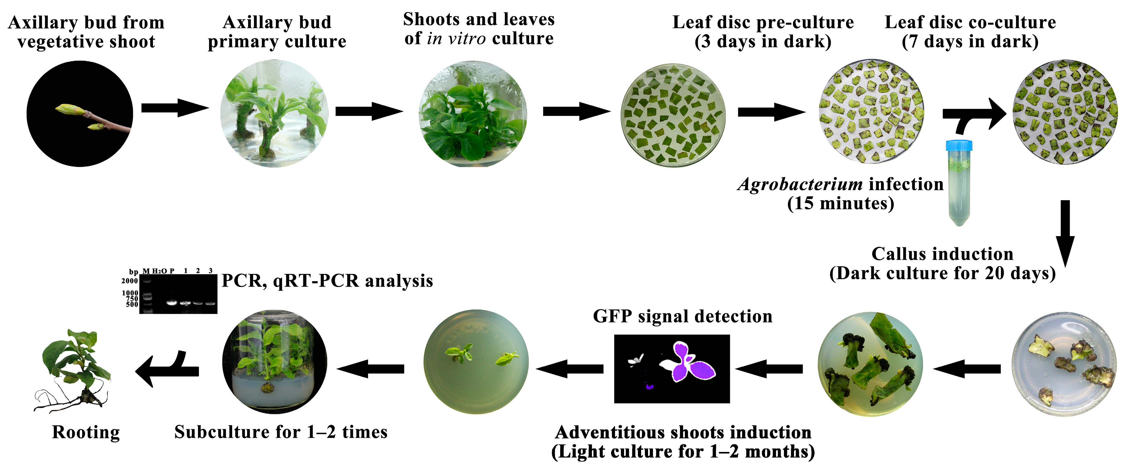
2.4. Agrobacterium tumefaciens-Mediated Gene Transformation

DkANRi constructs contain the silencing vector pH7GWIWG2 with the 5′ end sequences of the DkANR (accession no. AB195284.1) or EV (empty vector), and the pMS4 vector (containing the GFP gene) was transformed into Agrobacterium tumefaciens strain GV3101 and used to infect explants [16]. The primers used for cloning are listed in Table S2. The spectinomycin (spe)-resistance and hygromycin (hyg)-resistance loci labelled in pH7GWIWG2 were used to select positive transgenic seedlings.
The leaves of ‘Gongcheng Shuishi’ in vitro culture shoots that were continuously subcultured 4 times were sliced into leaf discs (1.0 cm × 1.0 cm) and cultured on pre-culture leaf medium (Table S1) in darkness for 3 days at 25 ± 2 °C. The vector Agrobacterium GV3101 containing the target vector DkANRi was added to 50 mL of Luria Bertani liquid medium (pH 7.2) containing the corresponding antibiotic spectinomycin (no. CS10421, Coolaber, Beijing, China) (50 mg/mL) and cultured at 28 °C in a shaker for 12–14 h at 220 r/min. Then, the overnight Agrobacterium culture was harvested at an OD600 of 1.6–1.8 and centrifuged at 5000× g for 5 min before being resuspended to an OD600 of 0.75 with liquid infiltration medium [10 mmol MgCl2, 10 mmol 2-(N-morpholino) ethanesulfonic acid (MES), and 150 μmol acetosyringon]. The cultures were incubated for 1–2 h at room temperature before infiltration [16]. Then, leaf discs (1.0 cm × 1.0 cm) were transferred into suspensions of Agrobacterium tumefaciens strain GV3101 containing DkANRi for 15 min. Later, leaf discs were drained on sterile filter paper and moved into co-cultivation medium (Table S1). After co-cultivation with Agrobacterium for 7 days at 25 ± 2 °C in the dark, they were washed with sterilized water containing 400 mg/L cefotaxime sodium salt (no. CC3251, Coolaber Ltd., Beijing, China) (Cef) 3 times. Finally, similar to the method in “2.3 Regeneration and Rooting of Adventitious Buds”, the leaf discs were moved to a callus induction medium containing 400 mg/L Cef and 5 mg/L spectinomycin for callus induction for 20 days and an adventitious bud regeneration medium containing 400 mg/L Cef and 5 mg/L spectinomycin for 1–2 months (Table S1). Adventitious buds were transferred to a fresh selection medium every month, and they were sampled for positive detection and PAs content analysis.

2.5. Green Fluorescent Protein Detection

The transgenic lines of pMS4 and control plants were used for detecting the GFP fluorescence signal. The robust GFP fluorescence signal in the transgenic callus and seedlings can be visualized under an in vivo imaging system (LB 985 Night SHADE). The excitation/emission wavelengths for GFP are 488 nm/505–550 nm.

2.6. Molecular Detection of Transformants

The regenerated seedlings transformed with DkANRi and EV were subcultured 1–2 times for multiplication in subculture medium containing Cef (400 mg/L) and spe (5 mg/L). Genomic DNA from the fresh leaves of putatively transformed plantlets and control plants was isolated by a Dzup (Plant) Genomic DNA Isolation Reagent kit (Sangon Biotech, Shanghai, China), and they were used for polymerase chain reaction (PCR) detection to confirm whether the exogenous gene was integrated into the chromosome of transformed ‘Gongcheng Shuishi’. The 711-bp coding region of the hyg gene was amplified using the primers Hyg-F and Hyg-R (Table S2). The 485-bp coding region of the 35S promoter was amplified using the primers 35S-F and 35S-R (Table S2). In addition, two primers, LB (LB-F, LB-R) and RB (RB-F, RB-R), the site that could not be recombined into the plant genome for PCR detection to avoid false positives determination of Agrobacterium and tissue culture seedlings (Figure S1). The PCR conditions were as follows: 1 cycle of 94 °C for 5 min; 30 cycles of 94 °C for 30 s, 60 °C for 30 s and 72 °C for 30 s; and a final extension at 72 °C for 10 min (1 cycle). The amplified products were separated by electrophoresis in a 1.5% agarose gel and stained with ethidium bromide.

2.7. Southern Blotting Assay to Identify Positive Transgenic Plants

To further confirm positive transgenic plants, total genomic DNA was extracted from plant leaves after transformation using the CTAB method [27]. Southern blotting was conducted by the DIG-High Prime DNA Labelling and Detection Starter Kit ⅠⅠ (11585614910, Roche, Switzerland). Genomics DNA (15 µg each) of the transformed persimmon seedlings and nontransformed persimmon seedlings were digested with Xba I. The digests were separated on a 0.8% agarose gel and blotted to a nylon membrane (Hybond-N+, Amersham, USA). The filter was probed with digoxigenin (DIG)-labelled coding regions of the hyg gene [15].

2.8. RNA Extraction and cDNA Synthesis

Total RNA was isolated from frozen leaf samples using an RNAiso Plus* kit (TaKaRa, Dalian, China). Three biological replicates were performed for each sample. cDNA was synthesized from 1.0 μg of each RNA sample using the PrimeScript RT Kit with gDNA Eraser (TaKaRa, Dalian, China).

2.9. Quantitative Reverse Transcription PCR (qRT–PCR)

The PCR mixture (10 μL total volume) included 5 μL of SYBR Premix Ex Taq II (TaKaRa, Dalian, China), 3.5 μL of ultrapure deionized H2O, 1.0 μL of diluted cDNA (200 ng/μL), and 0.25 μL of each primer (10 μM) (Table S2). The thermal profiles were 95 °C for 5 min, followed by 45 cycles of 95 °C for 5 s, 58 °C for 10 s, and 72 °C for 15 s. Each sample was assayed in quadruplicate, and DkActin (accession no. AB473616) was used as the internal control.

2.10. PAs Content Determination

Both soluble and insoluble PAs contents were measured by the Folin–Ciocalteu method [28]. The absorbance spectra of the samples were detected using a UV spectrophotometer at 725 nm, and a standard curve of tannic acid at various concentrations was drawn to quantify the PAs content of the samples.
In addition, the PAs content of persimmon fruit discs was also detected by staining with DMACA (4-dimethylaminocinnamaldehyde) solution (0.6% DMACA and 1% 6 N HCl in methanol) [29]. The transformed discs were decolorized for 2 h in 30% (v/v) acetic acid in ethanol, washed with 75% ethanol, and then dyed blue in DMACA solution for 2 min. A visual deeper blue colour indicated a higher PAs content.

3. Results and Discussion

3.1. The Primary Culture and Subculture Proliferation of ‘Gongcheng Shuishi’

In vitro tissue culture is a tool for enhancing cultivation through massive vegetative propagation (e.g., direct regeneration from dormant buds and shoot tips) and indirect organogenesis from calli obtained from meristematic tissue and leaves. In this study, to obtain the culture tissue seedlings of ‘Gongcheng Shuishi’, the axillary buds sampled from vegetative shoots of ‘Gongcheng Shuishi’ were cultured in primary culture medium for one month. The axillary bud was a common explant for the primary culture in persimmon [30]. The frequency of the surviving buds indicated that the ZT concentration had no obvious influence on the germination rate in MS (1/2N) medium (Figure 1A, B). When the concentration of ZT was 1.0 mg/L, the germination rate of the axillary bud was higher in DKW medium than in MS (1/2N) medium. The bud germination rate in the DKW medium with ZT 1.0 mg/L (63.7%) was significantly higher than in MS (1/2N) medium containing ZT (1.0-4.0 mg/L) (Figure 1A,B). Moreover, the callus grew slowly and was smaller in the DKW medium than in the MS (1/2N) medium (Figure 1A), suggesting that DKW was more suitable for bud germination.
The frequency of bud germination and growth was greatly influenced by the basic medium, auxin, and cytokinin combination in the subculture. The multiplication rates and growth potential of the seedlings in DKW were all higher than in MS (1/2N), and the seedlings grew robustly with more green leaves (Figure 1C). The number of effective shoots was 1.42 on DKW medium with 1.0 mg/L ZT and 0.1 mg/L IAA (Figure 1D). This was consistent with previous reports [31,32] that the basic medium is the key factor influencing the proliferation culture of persimmon, and DKW is the most suitable medium for the proliferation of ‘Mopanshi’ (non-PCNA) [26]. Moreover, cytokinin is an important phytohormone to promote the proliferation of the tissue culture seedlings, high concentrations of zeatin were required for shoot growth, and the optimum range in concentration was 10−4–10−5 M, as due to the cytokinin in xylem sap increases in the spring and causes bud sprouting [33]. However, a higher concentration of ZT also induced a large callus block around the basal part of the seedlings, and a negative correlation between shoot length and shoot number indicated that the low concentration of ZT (1.0 mg/L) was suitable for the growth of ‘Gongcheng Shuishi’ persimmon.

3.2. ‘Gongcheng Shuishi’ Persimmon Shoots Formation from Leaf Explants

After subculturing 4 times, the leaves of the ‘Gongcheng Shuishi’ persimmon plantlets were used to induce callus and adventitious buds on MS (1/2N) or 1/2DKW media comprised of different concentrations of ZT and IAA. 1/2DKW with ZT (2.0 mg/L) and TDZ (0.5 mg/L) was optimal for callus induction, followed by MS (1/2 N) with ZT (2.0 mg/L) and IAA (0.1 mg/L) for adventitious bud induction (Figure 2A). The callus and bud formation rates were 96.0% and 94.0%, respectively (Figure 2B).
Moreover, the presence of phytohormones is regarded as an essential factor for shoot growth, callus induction, and adventitious bud differentiation. Our results indicated that TDZ (0.5 mg/L) and ZT (2.0 mg/L) were better at inducing calli, and ZT (2.0 mg/L) in combination with IAA (0.1 mg/L) showed a similar effect in promoting the differentiation of adventitious buds (Figure 2A,B). TDZ and several substituted pyridyl phenylurea compounds have been demonstrated to have strong cytokinin-like activity and stimulate callus growth at relatively lower concentrations [34]. TDZ seems to induce meristematic nodules more effectively than other phytohormones but inhibits adventitious bud differentiation. Most of the adventitious buds induced by TDZ did not grow further unless they were transferred to a medium containing zeatin [35]. A significant increase in persimmon callus and shoot formation in different media was observed, and we obtained the highest proportion of leaf calli (96.0%, 0.5 mg/L TDZ+ 2.0 mg/L ZT) and adventitious buds (94.0%, 2.0 mg/L ZT + 0.1 mg/L IAA) compared with a previous report.

3.3. Rooting of Regenerated Shoots

After the in vitro seedling of ‘Gongcheng Shuishi’ was subcultured in DKW + ZT (1.0 mg/L) + IAA (0.1 mg/L) more than 4 times, the seedlings were moved to MS (1/2N) + IAA (2.0 mg/L) culture medium for 7 days in the dark to induce rooting. Roots gradually emerged from the side of the callus after 1 month of culture, and 35.7% of the plantlets rooted well (Figure 2C). Some experiments were conducted on persimmon cultivars in tissue culture to induce adventitious roots. Most of those studies presented difficulty in rooting, and IAA, indole-3-butyric acid (IBA), and NAA could affect the rooting ability of persimmons [36]. IAA exerts a significant influence on the rooting of ‘Uenishiwase’ persimmon, with the highest rooting rate (32.4%) and number (0.6) of adventitious roots [37]. In this study, our results also supported that IAA (2.0 mg/L) promotes the rooting of ‘Gongcheng Shuishi’ with 35.7% rooting rate.

3.4. Transgenic Persimmon Plants with GFP

Based on the regeneration system, the Agrobacterium-mediated leaf disc infection protocol was established to transfer the pMS4 vector (containing the green fluorescent protein gene, GFP) into ‘Gongcheng Shuishi’ persimmon. As a result, the callus and adventitious buds were generated after one month and two months of culture, respectively (Figure 3A). Likewise, a robust GFP fluorescence signal in the transgenic callus and seedlings was detected by an in vivo imaging system (LB 985 Night SHADE) (Figure 3B). This meant that the GFP gene was introduced into ‘Gongcheng Shuishi’ seedlings and that successful expression could be visualized.

3.5. Identification of Stable Regenerated Transgenic Seedlings

A key structural gene ANR encoding anthocyanidin reductase participate in the PAs biosynthesis (Figure 4A). Through the regeneration system, we also transferred the EV and DkANRi into ‘Gongcheng Shuishi’ persimmon leaves. To verify the stable transformation system, we identified positive seedlings transformed with DkANRi and performed a comparative analysis of the expression levels of DkANR and PAs between the transgenic lines and the control.
In the present study, to obtain stable transgenic seedlings of DkANRi of ‘Gongcheng Shuishi’ in vitro, 80 and 250 leaf discs were immersed in Agrobacterium infection solution containing the empty vectors and DkANRi in vitro, respectively. As a result, a total of 39 and 135 regenerated seedlings inoculated with EV and DkANRi were obtained (Table S3), and the callus induction rates and adventitious bud information rates reached 91.6% and 63.3%, respectively. To confirm the transformed nature of the regenerants, after 1–2 subcultures of those seedlings, DNA was isolated from all transgenic plantlets and a nontransformed ‘Gongcheng Shuishi’ persimmon plant, and hyg gene- and 35S-specific primers (Table S2) were used for PCR detection. Target fragments of 711 bp and 485 bp were amplified from the positive samples using hyg-specific primers and 35S primers, respectively, and the sequencing analysis of the PCR products was consistent with the hyg and 35S sequences (Figure S2). The target band was amplified in the EV and recombinant plasmids but not in the transgenic plants by the LB and RB primers (Figure S3). These results indicated that the hyg gene and 35S were successfully integrated into transgenic ‘Gongcheng Shuishi’ plants. There were seven transgenic positive seedlings inoculated with EV (Figure 4B,C) and 20 positive seedlings inoculated with DkANRi (Figure 4D, E) by PCR analysis. The transformation efficiency was 17.9% and 14.8%, respectively. Consistently, the stable genetic transformation was confirmed by Southern blot analysis. The expected fragments were detected in the transgenic lines obtained from transformation with DkANRi but not in the control seedlings (Figure 4F).
The expression of DkANR was determined in DkANRi seedling lines and WT (wild type) seedlings for 4 biological duplications (4 plants derived from each transgenic line). The results showed that the transcript level of DkANR in the DkANRi lines was significantly lower than in the WT seedlings and empty vector plasmid-infected plantlets (Figure 4H).
DMACA staining of the leaves from positive DkANRi transgenic lines (#31, #52, #53, #57) presented a distinctly weaker blue colour than the EV control (Figure 4G). Correspondingly, the total PAs concentration was significantly reduced in the DkANRi lines (0.61%, 0.81%, 0.50%, 0.32%) compared with the WT lines (1.67%) and EV plasmid-transformed plantlets (1.56%) (Figure 4I). Compared with the callus of the transformed seedlings and nontransformed seedlings, we found that the colour shown in the transgenic callus was also light against the WT (Figure S4). In summary, DkANRi was successfully introduced into ‘Gongcheng Shuishi’ in vitro culture seedlings, which led to the downregulation of DkANR expression and a decrease in PAs content. To preserve materials, the positive seedlings of ‘Gongcheng Shuishi’ were subcultured in DKW + ZT (1.0 mg/L) + IAA (0.1 mg/L) + spe (5 mg/L), and some of the positive seedlings were screened continuously through spe in the root induction stage. Positively rooted plants were obtained (Figure 5).
Conventional cross-breeding of woody fruit trees is constrained by their extensive reproductive cycle with long juvenile periods, complex reproductive biology, and high degree of heterozygosity [38]. To overcome the limitations of conventional cross-breeding, genetic transformation provides a promising approach to accelerate the production of persimmon fruit cultivars with improved traits [39]. Agrobacterium-mediated gene transfer remains the most conventional method for fruit species, including apple, almond, banana, citrus, grapevine, and melon. Regarding persimmon, the Agrobacterium-mediated transient transformation system in the leaves in vivo [16,17] and fruit discs in vitro have been established [18], which provide simple and rapid tools for the primary functional identification of genes. The functional verification of MYB, which is related to persimmon proanthocyanidin synthesis, and ERF (the hypoxia response transcription factor) employed transient expression in persimmon leaves and fruit discs [40,41], and DkMYB4 was also transformed into kiwifruit for functional analysis [24]. However, the instability and uncertainty of the transient expression system cannot guarantee the confirmation of gene function.
GFP is usually used as a selection reporter, detection of the GFP fluorescence signal and monitoring of GFP expression at different developmental stages in transgenic plants assist in establishing stable genetic transformation in cotton [42]. In our study, robust fluorescence could be detected inside the callus and regenerated seedlings. Intensive GFP signals were derived from calli and plants after four to eight weeks of culture (Figure 3). Thus, we established and optimized the regeneration protocol for the commercially important non-PCNA ‘Gongcheng Shuishi’ (Figure 5) and silenced the DkANR gene, which is related to PAs biosynthesis, to confirm its function.
During the process of stable genetic transformation on non-PCNA persimmon, the fruit easily turns brown due to the high PAs content. Compared with the material ‘Mopanshi’ (non-PCNA persimmon) (the regeneration rate was 41.4%, but the positive rate was less than 9%), another important leading persimmon cultivar, ‘Gongcheng Shuishi’, had a higher regeneration efficiency (94.0%) and transgenic survival (17.9%) due to traits that were not easy to brown. Through current optimization, the process and period of stable transformation was shortened, with transformation efficiency increasing with a positive rate up to 18%, which was higher than most previous reports on persimmon and similar to the results of Gao et al. [12] (Table 1). Based on this effective protocol, we obtained transgenic lines of DkMYB19 and DkMYB20 with a positive rate of 28.8% [43].

4. Conclusions

An efficient Agrobacterium-mediated persimmon genetic transformation protocol was successfully established in this study. The appropriate medium for subculture proliferation of ‘Gongcheng Shuishi’ tissue culture seedlings was DKW medium with 1.0 mg/L ZT and 0.1 mg/L IAA. The callus induction rate reached 96.0% based on solid callus induction medium [1/2DKW + TDZ (0.5 mg/L) + ZT (2.0 mg/L) + Cef (400 mg/L)], and the adventitious bud regeneration rate was 94% in medium [MS (1/2)N + ZT (2.0 mg/L) + IAA (0.1 mg/L) + Cef (400 mg/L)]. Through 1–2 months of culture, we achieved a rate of 17.8% positive seedlings. Our regeneration and transformation protocol offers a promising approach for gene function verification and the genetic improvement of Chinese persimmon.

Supplementary Materials

The following supporting information can be downloaded at: https://www.mdpi.com/article/10.3390/horticulturae8050422/s1, Figure S1: PH7GWIWG2 vector map containing DkANRi by Snapgene 6.0; Figure S2: Sequence alignment of 35S (A) and hyg (B) PCR products from DkANRi positive lines and vector; Figure S3: PCR amplification of putative positive transformed ‘Gongcheng Shuishi’ persimmon seedlings; Figure S4: The status of the transgenic lines of ‘Gongcheng Shuishi’ persimmon; Table S1: Composition of the medium used in this study; Table S2: The primers used in this study; Table S3: Regeneration seedlings and positive detection of transformed ‘Gongcheng Shuishi’ persimmon.

Author Contributions

Investigation: formal analysis, writing—original draft, methodology, M.Z.; formal analysis, writing—original draft, S.Y.; Investigation, W.C., and L.X. and D.G.; conceptualization, resources, Z.L.; project administration, formal analysis, supervision, writing—review & editing, Q.Z. All authors have read and agreed to the published version of the manuscript.

Funding

This research was supported by the National Key R & D Program of China (2019YFD1000600, 2019YFD1001200) and the National Natural Science Foundation of China (31471846 and 32102324).

Institutional Review Board Statement

Not applicable.

Informed Consent Statement

Not applicable.

Data Availability Statement

Data are contained within the article.

Conflicts of Interest

The authors declare no conflict of interest.

References

  1. Tao, R.; Dandekar, A.M. Genetic Transformation of Diospyros kaki L. (Japanese Persimmon). In Transgenic Trees; Bajaj, Y.P.S., Ed.; Springer: Berlin/Heidelberg, Germany, 2000; Volume 44, pp. 77–87. [Google Scholar] [CrossRef]
  2. Akagi, T.; Katayama-Ikegami, A.; Yonemori, K. Proanthocyanidin biosynthesis of persimmon (Diospyros kaki Thunb.) fruit. Sci. Hort. 2011, 130, 373–380. [Google Scholar] [CrossRef]
  3. Akagi, T.; Shirasawa, K.; Nagasaki, H.; Hirakawa, H.; Tao, R.; Comai, L.; Henry, I.M. The persimmon genome reveals clues to the evolution of a lineage-specific sex determination system in plants. PLoS Genet. 2020, 16, e1008566. [Google Scholar] [CrossRef] [Green Version]
  4. Luo, C.; Zhang, Q.L.; Luo, Z.R. Genome-wide transcriptome analysis of Chinese pollination-constant nonastringent persimmon fruit treated with ethanol. BMC Genom. 2014, 15, 112. [Google Scholar] [CrossRef] [Green Version]
  5. Chen, W.X.; Xiong, Y.L.; Xu, L.Q.; Zhang, Q.L.; Luo, Z.R. An integrated analysis based on transcriptome and proteome reveals deastringency-related genes in CPCNA persimmon. Sci. Rep. 2017, 7, 44671. [Google Scholar] [CrossRef] [Green Version]
  6. Luo, Y.J.; Zhang, X.N.; Luo, Z.R.; Zhang, Q.L.; Liu, J.H. Identification and characterization of microRNAs from Chinese pollination constant non-astringent persimmon using high-throughput sequencing. BMC Plant Biol. 2015, 15, 11. [Google Scholar] [CrossRef] [Green Version]
  7. Tao, R.; Handa, T.; Tamura, M.; Sugiura, A. Genetic transformation of Japanese persimmon (Diospyros kaki L.) by Agrobacterium rhizogenes wild type strain A4. J. Jpn. Soc. Hort. Sci. 1994, 63, 283–289. [Google Scholar] [CrossRef] [Green Version]
  8. Tao, R.; Dandekar, A.M.; Uratsu, S.L.; Vail, P.V.; Tebbts, J.S. Engineering genetic resistance against insects in Japanese persimmon using the cryIA(c) gene of Bacillus thuringiensis. J. Amer. Soc. Hort. Sci. 1997, 122, 764–771. [Google Scholar] [CrossRef] [Green Version]
  9. Nakamura, Y.; Kobayashi, S.; Nakajima, I. Agrobacterium-mediated transformation and plant regeneration from hypocotyl segments of Japanese persimmon (Diospyros kaki Thunb). Plant Cell Rep. 1998, 17, 435–440. [Google Scholar] [CrossRef]
  10. Li, X.H.; Jiang, Z.Y.; Shen, Y.Y.; Li, F.H.; Yu, X.Y.; Qu, S.C. In vitro regeneration and Agrobacterium tumefaciens-mediated genetic transformation of D. lotus (Diospyros lotus L.). Sci. Hort. 2018, 236, 229–237. [Google Scholar] [CrossRef]
  11. Koshita, Y.; Nakamura, Y.; Kobayashi, S.; Morinaga, K. Introduction of the rolC gene into the genome of the Japanese persimmon causes dwarfism. J. Jpn. Soc. Hort. Sci. 2002, 71, 529–531. [Google Scholar] [CrossRef] [Green Version]
  12. Gao, M.; Sakamoto, A.; Miura, K.; Murata, N.; Sugiura, A.; Tao, R. Transformation of Japanese persimmon (Diospyros kaki Thunb.) with a bacterial gene for choline oxidase. Mol. Breed. 2000, 6, 501–510. [Google Scholar] [CrossRef]
  13. Gao, M.; Tao, R.; Miura, K.; Dandekar, A.M.; Sugiura, A. Transformation of Japanese persimmon (Diospyros kaki Thunb.) with apple cDNA encoding NADP-dependent sorbitol-6-phosphate dehydrogenase. Plant Sci. 2001, 160, 837–845. [Google Scholar] [CrossRef]
  14. Deguchi, M.; Koshita, Y.; Gao, M.; Tao, R.; Tetsumura, T.; Yamaki, S.; Kanayama, Y. Engineered sorbitol accumulation induces dwarfism in Japanese persimmon. J. Plant Physiol. 2004, 161, 1177–1184. [Google Scholar] [CrossRef]
  15. Tamura, M.; Gao, M.; Tao, R.; Labavitch, J.M.; Dandekar, A.M. Transformation of persimmon with a pear fruit polygalacturonase inhibiting protein (PGIP) gene. Sci. Hort. 2004, 103, 19–30. [Google Scholar] [CrossRef]
  16. Mo, R.L.; Huang, Y.M.; Yang, S.C.; Zhang, Q.L.; Luo, Z.R. Development of Agrobacterium-mediated transient transformation in persimmon (Diospyros kaki Thunb.). Sci. Hort. 2015, 192, 29–37. [Google Scholar] [CrossRef]
  17. Mo, R.L.; Yang, S.C.; Zhang, Q.L.; Xu, L.Q.; Luo, Z.R. Vacuum infiltration enhances the Agrobacterium-mediated transient transformation for gene functional analysis in persimmon (Diospyros kaki Thunb.). Sci. Hort. 2019, 251, 174–180. [Google Scholar] [CrossRef]
  18. Zhang, M.; Wang, S.D.; Xu, L.Q.; Luo, Z.R.; Zhang, Q.L. Development of Agrobacterium-mediated transient transformation for fruit discs in persimmon (Diospyros kaki Thunb.). Eur. J. Hort. Sci. 2021. [Google Scholar] [CrossRef]
  19. Sugiura, A.; Matsuda-Habu, Y.; Gao, M.; Esumi, T.; Tao, R. Somatic embryogenesis and plant regeneration from immature persimmon (Diospyros kaki Thunb.) embryos. HortScience 2008, 43, 211–214. [Google Scholar] [CrossRef] [Green Version]
  20. Tao, R.; Sugiura, A. Adventitious bud formation from callus cultures of Japanese persimmon. HortScience 1992, 27, 259–261. [Google Scholar] [CrossRef] [Green Version]
  21. Tetsumura, T.; Yukinaga, H. High-frequency shoot regeneration from roots of Japanese persimmon. HortScience 1996, 31, 463–464. [Google Scholar] [CrossRef] [Green Version]
  22. Yin, H.; Song, C.L.; Ma, J.L.; Zhang, Z.D.; Tang, X. Optimization of Agrobacterium tumefaciens mediated genetic transformation system of Mopanshi. Acta Agric. Boreali-Sin. 2007, 22, 56–59. Available online: https://kns.cnki.net/kcms/detail/detail.aspx?FileName=HBNB200702014&DbName=CJFQ2007 (accessed on 1 March 2022).
  23. Ikegami, A.; Eguchi, S.; Kitajima, A.; Inoue, K.; Yonemori, K. Identification of genes involved in proanthocyanidin biosynthesis of persimmon (Diospyros kaki) fruit. Plant Sci. 2007, 172, 1037–1047. [Google Scholar] [CrossRef]
  24. Akagi, T.; Ikegami, A.; Tsujimoto, T.; Kobayashi, S.; Sato, A.; Kono, A.; Yonemori, K. DkMyb4 is a Myb transcription factor involved in proanthocyanidin biosynthesis in persimmon fruit. Plant Physiol. 2009, 151, 2028–2045. [Google Scholar] [CrossRef] [PubMed] [Green Version]
  25. Han, Y.P.; Vimolmangkang, S.; Soria-Guerra, R.E.; Korban, S.S. Introduction of apple ANR genes into tobacco inhibits expression of both CHI and DFR genes in flowers, leading to loss of anthocyanin. J. Exp. Bot. 2012, 63, 2437–2447. [Google Scholar] [CrossRef] [PubMed] [Green Version]
  26. Shi, X.X.; Du, G.Q.; Ma, J.L.; Gao, Y.; Wang, L. Callus formation and adventitious bud regeneration from leaves in vitro in persimmon (Diospyros kaki). J. Fruit Sci. 2004, 21, 376–378. [Google Scholar] [CrossRef]
  27. Doyle, J.I.; Doyle, J.L. A rapid DNA isolation procedure for small quantities of fresh leaf tissue. Phytochem. Bull. 1987, 19, 11–15. Available online: https://www.scienceopen.com/document?vid=9f54eaa3-acbe-4864-a21c-7ab44385e409 (accessed on 1 March 2022).
  28. Oshida, M.; Yonemori, K.; Sugiura, A. On the nature of coagulated tannins in astringent-type persimmon fruit after an artificial treatment of astringency removal. Postharvest Biol. Technol. 1996, 8, 317–327. [Google Scholar] [CrossRef]
  29. Li, Y.G.; Tanner, G.; Larkin, P. The DMACA-HCl protocol and the threshold proanthocyanidin content for bloat safety in forage legumes. J. Sci. Food Agric. 1996, 70, 89–101. [Google Scholar] [CrossRef]
  30. Giordani, E.; Naval, M.; Benelli, C. In vitro propagation of persimmon (Diospyros kaki Thunb.). In Protocols for Micropropagation of Selected Economically-Important Horticultural Plants, Methods in Molecular Biology; Lambardi, M., Ozudogru, E.A., Jain, S.M., Eds.; Springer: New York, NY, USA, 2013; Volume 994, pp. 89–98. [Google Scholar] [CrossRef] [Green Version]
  31. Witte, C.P.; Tiller, S.A.; Taylor, M.A.; Davies, H.V. Addition of nickel to Murashige and Skoog medium in plant tissue culture activates urease and may reduce metabolic stress. Plant Cell Tiss. Org. 2002, 68, 103–104. [Google Scholar] [CrossRef]
  32. Li, J.; Luo, Y.J.; Zhang, Q.L.; Luo, Z.R.; Liu, J.H. In vivo culture system optimization and regeneration of date plum (Diospyros lotus Linn.) dormant buds and leaves. J. Huazhong Agric. Univ. 2016, 35, 14–19. [Google Scholar] [CrossRef]
  33. Fukui, H.; Sugiyama, M.; Nakamura, M. Shoot tip culture of Japanese persimmon (Diospyros kaki Thunb.). J. Jpn. Soc. Hort. Sci. 1989, 58, 43–47. [Google Scholar] [CrossRef] [Green Version]
  34. Fellman, C.D.; Read, P.E.; Hosier, M.A. Effects of thidiazuron and CPPU on meristem formation and shoot proliferation. HortScience. 1987, 22, 1197–1200. Available online: http://pascal-francis.inist.fr/vibad/index.php?action=getRecordDetail&idt=7678477 (accessed on 1 March 2022).
  35. Yokoyama, T.; Moriyasu, Y.; Sugawara, Y. Adventitious bud formation through nodule induction by thidiazuron in cultured leaf segments of the Japanese persimmon (Diospyros kaki Thunb.). Plant Biotechnol. 2011, 28, 339–344. [Google Scholar] [CrossRef] [Green Version]
  36. Delia, G.; Farina, L.; Buccheri, M.; Rosato, S.; Insero, O.; Rega, P.; Damiano, C. Proteomic analysis of in vitro rooted persimmon (cv. ‘Kaki Tipo’) plants in response to various auxins and light conditions. Acta Hort. 2009, 833, 171–176. [Google Scholar] [CrossRef]
  37. Ma, J.L.; Liu, X.N.; Zhang, Z.D.; Yuan, S.Q. Rooting and adventitious shoot regeneration from root of Uenishiwase persimmon. Acta Agric. Boreali-Sin. 2004, 19, 115. Available online: https://kns.cnki.net/kcms/detail/detail.aspx?FileName=HBNB200404033&DbName=CJFQ2004 (accessed on 1 March 2022).
  38. Gambino, G.; Gribaudo, I. Genetic transformation of fruit trees: Current status and remaining challenges. Transgenic Res. 2012, 21, 1163–1181. [Google Scholar] [CrossRef]
  39. Tao, R. Agrobacterium-mediated genetic transformation in Japanese persimmon. Acta Hort. 2003, 601, 57–63. [Google Scholar] [CrossRef]
  40. Chen, W.X.; Zheng, Q.Y.; Li, J.W.; Liu, Y.; Xu, L.Q.; Zhang, Q.L.; Luo, Z.R. DkMYB14 is a bifunctional transcription factor that regulates the accumulation of proanthocyanidin in persimmon fruit. Plant J. 2021, 106, 1708–1727. [Google Scholar] [CrossRef]
  41. Zhu, Q.G.; Gong, Z.Y.; Wang, M.M.; Li, X.; Grierson, D.; Yin, X.R.; Chen, K.S. A transcription factor network responsive to high CO2/hypoxia is involved in deastringency in persimmon fruit. J. Exp. Bot. 2018, 69, 2061–2070. [Google Scholar] [CrossRef] [Green Version]
  42. Cui, M.L.; Liu, C.; Piao, C.L.; Liu, C.L. A stable Agrobacterium rhizogenes-mediated transformation of cotton (Gossypium hirsutum L.) and plant regeneration from transformed hairy root via embryogenesis. Front. Plant Sci. 2020, 11, 604255. [Google Scholar] [CrossRef]
  43. Yang, S.C.; Zhang, M.; Xu, L.Q.; Luo, Z.R.; Zhang, Q.L. MiR858b inhibits proanthocyanidin accumulation by the repression of DkMYB19 and DkMYB20 in persimmon. Front. Plant Sci. 2020, 11, 576378. [Google Scholar] [CrossRef] [PubMed]
Figure 1. Establishment of the primary culture and subculture of ‘Gongcheng Shuishi’ persimmon. (A) Primary culture of ‘Gongcheng Shuishi’ in different media for 2 months. (B) The bud germination rates of ‘Gongcheng Shuishi’ in different media for 2 months. (C) The subculture of ‘Gongcheng Shuishi’ in different media for 1 month. (D) The number of effective shoots of ‘Gongcheng Shuishi’ in different media for 1 month. Bar = 1.0 cm. The unit of hormone concentration is mg/L, followed by the number in (A,C). Means followed by different letters indicate significant differences at p ≤ 0.05 by Tukey’s test.
Horticulturae 08 00422 g001
Figure 2. In vitro propagation of ‘Gongcheng Shuishi’ persimmon leaves. (A) Callus induction (20 days) and adventitious bud regeneration (1–2 months) of ‘Gongcheng Shuishi’ in different media. (B) The rate of callus formation and bud formation of ‘Gongcheng Shuishi’ in different media. (C) The process of adventitious bud regeneration and rooting for ‘Gongcheng Shuishi’ persimmon. C1–C2 callus parenchyma cells began to form shoot tips. C3 demonstrates adventitious bud germination. In C4 adventitious buds grew into seedlings. C5–C6 demonstrate the rooting of the seedlings. Bar = 1.0 cm. The unit of plant growth regulator concentration is mg/L, followed by the number in (A,B). Means followed by different letters indicate significant differences at p ≤ 0.05 by Tukey’s test.
Horticulturae 08 00422 g002
Figure 3. Detection of GFP fluorescence in a transgenic ‘Gongcheng Shuishi’ persimmon plantlet with the GFP gene. (A) Morphology of transgene-positive and control plants from the small to large stages. (B) Visualization of the GFP fluorescence signal in the transgenic plantlet by an in vivo imaging system (LB 985 Night SHADE). Bar = 2.0 cm.
Horticulturae 08 00422 g003
Figure 4. PCR amplification, DMACA staining of leaves, and DkANR expression and PAs contents of putatively positively transformed ‘Gongcheng Shuishi’ persimmon seedlings. (A) Brief scheme for the biosynthesis of proanthocyanidin. (B) PCR analysis of the transformed plants with EV using the hyg primer. (C) PCR analysis of the positively transformed plants with EV using the 35S primer. (D) PCR analysis of the transformed plants with DkANRi using the hyg primer. (E) PCR analysis of the positively transformed plants with DkANRi using the 35S primer. (F) Southern blot analysis of the positively transformed plants with DkANRi. (G) Analysis of PAs content in the leaves of the transgenic plants by 4-dimethylaminocinnamaldehyde (DMACA) staining. (H) Transcript level of DkANR in the transgenic plants with EV and DkANRi. (I) PAs content of WT, EV, and DkANRi transgenic plants. WT and EV represent the wild-type persimmon DNA and empty vector transgenic plant DNA, respectively. P represents the empty vector plasmid DNA. Asterisks (*) indicate significantly different values, **, p ≤ 0.01; ***, p ≤ 0.001. Bar = 1.0 cm.
Horticulturae 08 00422 g004
Figure 5. Illustration of the experimental procedure for regeneration of sprouting buds and genetic transformation in persimmon leaves.
Horticulturae 08 00422 g005
Table 1. Advancements of transgenic persimmon.
Donor GeneAcceptor GenotypeExplantsTotal Time of Callus FormationRegeneration Rate (Positive)%References
cryIA(c)J-PCNA ‘Jiro’Leaf disc2 months2.1 (1.5)[8]
-J-PCNA ‘Saijo’Hypocotyl segments4–5 months27.0 (11.1)[9]
codAJ-PCNA ‘Jiro’Leaf disc-20.0[12]
S6PDHJ-PCNA ‘Jiro’Leaf disc-3.5 (2.5)[13]
PGIPJ-PCNA ‘Jiro’Leaf disc1 month7.8 (5.5)[15]
LACC-PCNA‘Baogai Tianshi’, ‘Eshi 1’, non-PCNA ‘Mopanshi’Leaves in vivo [16]
LARC-PCNA ‘Xiaoguo Tianshi’, non-PCNA ‘Mopanshi’Leaves in vivo [17]
ANRNon-PCNA ‘Gongheng Shuishi’Fruit discs [18]
Publisher’s Note: MDPI stays neutral with regard to jurisdictional claims in published maps and institutional affiliations.

