Article

Leaf Epidermal Morphology of Ten Wild Tree Peonies in China and Its Taxonomic Significance

1
Postdoctoral Research Base, Henan Institute of Science and Technology, Xinxiang 453003, China
2
College of Landscape Architecture and Art, Henan Agricultural University, Zhengzhou 450002, China
*
Author to whom correspondence should be addressed.
Horticulturae 2022, 8(6), 502; https://doi.org/10.3390/horticulturae8060502
Submission received: 3 April 2022 / Revised: 28 May 2022 / Accepted: 2 June 2022 / Published: 5 June 2022

Abstract

:
This research reports for the first time the epidermal characteristics of 10 wild tree peonies belonging to the sect. Moutan group. The leaf micromorphology of 10 wild tree peonies—P. qiui, P. decomposita, P. jishanensis, P. ostii, P. delavayi, P. lutea, P. potanini, P. rockii, P. ludlowii and P. cathayana—were investigated by light and scanning electron microscopy. The characteristics of the leaf epidermis were found to be constant at the species level, but variable among species. Patterns of anticlinal walls were slightly wavy to wavy, very wavy, and straight to curved. All studied tree peonies presented stomata only on the abaxial surfaces of the leaves. The stomatal apparatus was elliptical or rectangular. Stomatal density was highest on the abaxial surface in P. qiui (300.25 per mm2), and lowest in P. jishanensiss (198.41 per mm2). Stomatal Index was highest on the adaxial surface of P. potanini (27.30%), and lowest in P. ludlowii (18.35%). Trichomes were observed in four studied peonies, which appeared only on the abaxial surface of the leaves, and three basic types of trichomes were identified. The long conical trichomes were distinctive of P. jishanensis, and the flat-shaped trichomes were characteristic of P. qiui, P. rockii and P. cathayana, and commonly occurred in the intervenous region and leaf veins. Stomatal shape, stomatal density, anticlinal wall patterns and trichomes may be of great value and significance for the classification of wild tree peonies. The principal component analysis showed that the component 1, component 2 and component 3 were the most components and nearly 79% of the observed variation. The key to the identification of wild tree peonies based on trichomes and stomata diversity was provided.

1. Introduction

Tree peony (Paeonia suffruticosa Andrews) (Paeoniaceae) is an important traditional flower in China. They are either shrubs or subshrubs belonging to sect. Moutan group of the genus Paeonia [1], which comprises eight to nine species and is mainly distributed in the mountains of eastern, central and southwestern China. They play an important role in the hybrid breeding of tree peonies [2,3].
It is well known that sect. Moutan DC includes subsect. Vaginatae F. C. Stern and subsect. Delavayanae F. C. Stern, the fleshy disc subgroup and the leathery disc subgroup. P. suffruticosa was first documented scientifically in 1804 by H. Andrews. The classification of the wild resources for tree peony has long been controversial. Stern and Taylor [4,5] described the wild tree peony (P. ludlowii (Stern and G. Taylor) D. Y. Hong) cultivated from seeds collected in the Brahmaputra valley of Milin in southeastern Tibet. They treated it as a variety, P. lutea var. ludlowii Stern et Taylor. Fang [6] recorded six species in sect. Moutan; two species were recorded in the subsect. Vaginatae FC Stern, and he recorded four species in subsect. Delavayana FC Stern. Shen [7] believes that there are seven species of tree peony group, and Wang et al. [8] believe that in addition to cultivated tree peonies, there are eight species. Hong et al. [9,10] conducted a systematic study on this group of plants and believes that there are nine species, two subspecies and two hybrids. Even now, whether P. cathayana D. Y. Hong and K. Y. Pan [9,11] is a species, its relationship with cultivated tree peonies and P. ostii are still not fully resolved.
The species of Paeonia sect. Moutan are characterized by yellow and orange flowers, red leaves in spring, scarlet leaves in autumn, and dwarfed phenotype (dwarfism), which is rare among cultivated tree peonies. In recent years, due to the deterioration of the ecological environment and human disturbance, the distribution of Paeonia sect. Moutan communities has been greatly reduced. Some wild tree peonies are listed as rare and endangered plants [2], which have become one of the key groups for biodiversity conservation in China [10].
Identification and classification of Paeonia sect. Moutan germplasm resources are the basis for conservation and utilization. The micromorphological features of plant leaves served as a promising tool for plant species identification and classification [12,13]. It has been applied to the systematic classification of species such as bryophytes [14], ferns [15,16,17], gymnosperms [18,19], and angiosperms [20,21,22]. Currently, research on tree peonies mainly focuses on morphology [23,24], palynology [2], cytology [25], molecular systematics [26,27]. Systematic observation on epidermal features of wild tree peonies leaves has not been reported. The main objective of this study was to evaluate the diversity in leaf micromorphology of ten wild tree peonies. It also contributes some useful information for systematic classification of the sect. Moutan group of the genus Paeonia.

2. Materials and Methods

The leaf samples at the flowering stage in 2020 were collected from ten wild tree peonies from different sites in China (Figure 1, Table 1) and were identified by the authors with the assistance of Professor Huichao Liu, Professor Lixia He and Professor Gaixiou Liu. Voucher specimens of the ten wild tree peonies were deposited in the Herbarium at the Henan Institute of Science and Technology and Luoyang National Peony Garden, Henan Province (Table 1).

2.1. Light Microscope

Fragments of 1 cm2 were cut out from the central part of the collected leaves and boiled in water for 3 to 5 min, then soaked in hydrogen peroxide and glacial acetic acid = 1:1 solution until the epidermis was completely separated from the mesophyll tissues. Subsequently, the leaf epidermis fragments were stained with the (I2 + KI) aqueous solution for 5 to 10 min before being mounted on the glass slides with Canadian gum, and then observed under Olympus BH3 light microscope. Epidermal characteristics including the number and size of stomata, stomatal density and stomatal index were recorded. The photographs (200× magnification) were taken by using a digital camera (Olympus DP22, Tokyo, Japan) fitted on Olympus BH3 light microscope, and obtained with the help of Olympus cellSens standard software. To assess the stability of the epidermal structure, the leaves of three plants, which are 8–10 years old, of each species were sampled. Four leaves were chosen per plant, which were fully developed at different parts of each tree peony. For each leaf, four slides were made from the central parts. The width and length of epidermal cells were measured by counting 100 cells for each wild tree peony.

2.2. Scanning Electron Microscope

For the scanning electron microscope observation, fragments of 0.25 cm2 were cut out from the middle part of the leaves from ten wild tree peonies and washed with 90% ethanol to remove impurities, and then dried at room temperature. Approximately 10~20 small leaves of about 5 mm × 5 mm from the central part of each tree peony leaf were taken, and they were glued on the sample table with double-sided black conductive tape. The samples were sputtered with gold-palladium. After that, the samples were observed and photographed under an HEE-7 scanning electron microscope (made in the USA). The specimens were analyzed under the scanning electron microscope to observe micromorphological features (trichomes, stomata, sculptures and epidermal cells) of leaves. The analysis of the epidermis sculptures involved the observation and description of the epidermal cells, and stomatal index was calculated by using the formula of Wang et al. [28].
SI = S × 100/(E + S)
where SI = stomatal index, S = number of stomata, and E = number of epidermal cells.
The description of leaf morphology was mainly based on the glossaries described by Wang et al. [28].

2.3. Statistical Analysis

Statistical analyses for leaf micromorphology were performed by SPSS 19.0 software (SPSS Inc., Chicago, IL, USA). Duncan’s multiple comparisons were used to evaluate the differences in leaf micromorphology of 10 wild tree peonies at a 95% confidence interval (p < 0.05). Furthermore, Microsoft Excel 2016 software (Microsoft, Redmond, Washington D.C., USA) was used to generate tables and plots.

3. Results

Foliar epidermal micromorphological characters and the results of the qualitative and quantitative analysis of all the ten wild tree peonies investigated under light microscope (LM) (Figure 2 and Figure 3) and scanning electron microscope (SEM) (Figure 4, Figure 5 and Figure 6) are summarized in Table 2 and Table 3.

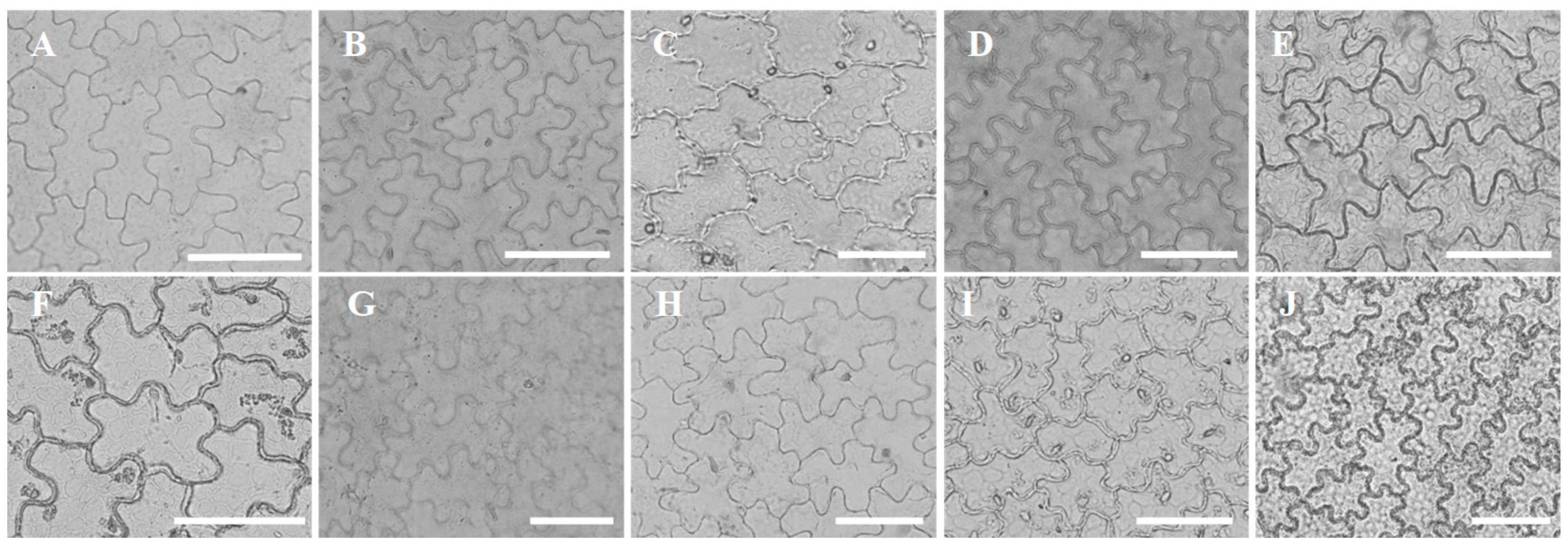
3.1. Leaf Epidermis Cell Shape and Anticlinal Wall Pattern

The shape of the epidermal cells was observed to be irregular on the adaxial and abaxial surfaces of leaves (Figure 2A–J and Figure 3A–J). The patterns formed by the anticlinal walls of the epidermal cells were divided into three types: (1) slightly wavy to wavy (Table 2), such as the abaxial epidermis cells and the adaxial epidermis cell in P. qiui (Figure 2A and Figure 3A), P. ostii (Figure 2D and Figure 3D), P. delavayi (Figure 2E and Figure 3E), P. potanini (Figure 2G and Figure 3G), P. rockii (Figure 2H and Figure 3H), and the adaxial epidermis cell in P. decomposita (Figure 2B); (2) very wavy (Table 2), such as the adaxial epidermis cell in P. cathayana (Figure 2J); (3) straight to curved (Table 2), such as the abaxial epidermis cell in P. cathayana (Figure 3J), P. decomposita (Figure 3B); the adaxial and abaxial epidermis cell in P. ludlowii (Figure 2I and Figure 3I), P. lutea (Figure 2F and Figure 3F) and P. jishanensis (Figure 2C and Figure 3C).
The length and width of epidermal cells in different tree peonies were significantly different (p < 0.05) (Table 2). The greatest epidermal cell density on the adaxial surface was recorded in P. lutea, and the minimum one was recorded in P. qiui. The maximum epidermal cell density was observed on the abaxial surface in P. qiui, while the minimum one was reported in P. jishanensis (Figure 2 and Figure 3).

3.2. Cuticular Wax

The adaxial epidermal cells were jigsaw-puzzle-like or ridged in shape (Figure 4A–G). Their surface pattern was flat with a smooth waxy layer in all studied wild tree peonies except P. cathayana (Figure 4G). The smooth waxy layer on the abaxial epidermis was found in most wild tree peonies investigated, with a striate variation only on the lower epidermis in P. delavayi (Figure 5E) and P. potanini (Figure 5G).

3.3. Micromorphology of Stomata

The leaves in all observed wild tree peonies were hypostomatic, with stomata appearing only on the abaxial epidermis of leaves. All studied wild tree peonies possessed a single type of stomata (paracytic or rubiaceous) consisting of two small epidermal cells on the inner side and two large epidermal cells on the outer side, and the long axis of the small cells was parallel to the long axis of the large cells (Figure 6). As for surface ornamentation of stomata, smooth stomatal surface was the most common in most of studied species, while rough stomatal surface was only present in P. decomposita, P. jishanensis and P. rockii. Two main types of stomata were observed in this study. The shape of stomatal apparatus was found to be elliptical in P. qiui, P. decomposita, P. jishanensis, P. delavayi, P. potanini and P. ludlowii; Rectangular stomata was only recorded in P. ostii, P. lutea, P. rockii and P. cathayana. In this study, the shape of the stomata was useful for identifying wild tree peonies.
The stomatal pore was surrounded by a pair of guard cells, which were deeply sunken and surrounded by subsidiary cells, which are narrowly elliptical. The inner margins of stomata were observed to be of three types, which included smooth, nearly smooth and erose. The stomata inner margins were smooth in P. qiui and P. decomposita, nearly smooth in P. delavayi and P. ludlowii, and erose in other six wild tree peonies.
The stomata regularly appeared on the abaxial epidermis of leaves, and the outer arch and inner edge of stomata were wavy. The stomatal apparatus and epidermal cells of some wild tree peonies were in the same plane, such as P. qiui (Figure 6A), P. decomposita (Figure 6B), P. jishanensis (Figure 6C), P. delavayi (Figure 6E), P. lutea (Figure 6F), P. potanini (Figure 6G), P. rockii (Figure 6H). The stomatal apparatus of some tree peonies is trapped in epidermal cells, such as P. ostii (Figure 6D), P. ludlowii (Figure 6I) and P. cathayana (Figure 6J).
The stomata size varied significantly (p < 0.05) with different wild tree peonies (Table 3). The largest length (28.12 μm) of the stomata was found in P. jishanensis, and lowest (19.05 μm) was in P. ludlowii. Whereas as the maximum width of the stomata apparatus (19.56 μm) were reported in P. jishanensis and minimum (13.85 μm) in P. ludlowii (Table 3).
The mean stomatal density varied from 198 to 300 per square of 1 mm. The polar orientation of stomata was found to be random. The stomatal index varied significantly in wild tree peonies (Table 3); the highest stomatal index (27.30%) was observed in P. potanini, followed by P. qiui (26.35%), and the lowest stomatal index (18.35%) was in P. ludlowii. The maximum stomatal density was recorded for P. qiui (300.25 per mm2), followed by P. cathayana (287.50 per mm2) and the minimum in P. jishanensis (198.41 per mm2) (Table 3). The stomatal index of the wild tree peonies was higher on the abaxial surface than on the adaxial surface (Table 3).

3.4. Principal Component Analysis

The principal component analysis calculated that the first four components contributed 0.332, 0.284, 0.172 and 0.107 of the variance, respectively (Table 4). The variables which mainly correlate with component 1 were stomata width, stomata length and ADCL (positive) and ABCL/ABCW (negative). The parameters such as ABCL, ABCW, SL/SW and stomata length were mainly positively loaded on component 2, while stomatal density, stomatal index and ADCW were negatively loaded on component 2. Component 3 was positively correlated with ADCW, ABCL and stomatal index, but negatively correlated with ADCL/ADCW and ADCL. The most influential variables on component 4 were SL/SW, stomatal index and ADCL (positive) and stomata width (negative). Table 3 indicated that stomata width, stomata length, ADCL, ABCW, stomatal density and stomatal index contributed their maximum part in the identification of wild tree peonies. A scatter plot is often helpful for finding patterns of variation; it is apparent in the plot (Figure 7) that the first and second components contributed to the identification of wild tree peonies.

3.5. Trichomes

Trichomes and their distributions in different species were observed and summarized in SEM micrographs (Figure 8). These trichomes were identified as simple, single, nonglandular and unicellular in type. The length of these trichomes was between 400 μm and 900 μm. There were three types of trichomes observed: flat-shaped, conic, and long conic. Flat-shaped trichomes were commonly found on the abaxial leaf surface and mainly occurred in the leaf intercostal region of P. qiui, P. rockii and P. cathayana, as well as the midrib or veins of P. rockii and P. cathayana. The number of flat-shaped trichomes varied among species, and the density of trichomes in P. qiui was high with a twisted appearance. Conic trichomes were restricted on the abaxial leaf surface in P. qiui and P. rockii, and commonly present in the intercostal region. Long conic trichomes were only observed on the abaxial leaf surface in P. jishanensis.
Based on the results of this study, the new key of ten wild peonies has also been edited as below:
Leaf epidermis with hairs
  Only with long conic trichomes or flat-shaped trichomes
    Only with long conic trichomes----------------------------------P. jishanensi
    Only with flat-shaped trichomes---------------------------------P. cathayana
  Leaf epidermis with flat-shaped trichomes and conic trichomes
    The trichomes only distributed on the areoles----------------P. qiui
    The trichomes distributed on the areoles and the veins----P. rockii
Leaf epidermis glabrous
  Elliptic stomata
    Stomatal density (≥250)---------------------------------------------P. ludlowii
    Stomatal density (<250)
      Stomatal index (>25)------------------------------------------P. potanini
  Stomatal index (<25)
        Stomatal index (<20) -----------------------------------P. decomposita
        Stomatal index (≥20, <25) -----------------------------P. delavayi
  Rectangular stomata
    Stomatal density (≥250) --------------------------------------------P. ostii
    Stomatal density (<230) -------------------------------------------P. lutea

4. Discussion

The taxonomic value of epidermal morphology varies depending on the plant family and genus. For families such as Anacardiaceae [29], Rhododendron (Ericaceae) [30], Lamiaceae [31]; Combretaceae [32], Gramineae [33], Stemonaceae [34], Euphorbiaceae [35], etc., the morphological characteristics of leaf epidermis are helpful for the identification of species or genus. Moreover, these characteristics are relatively stable and can reveal the difference between plant groups, but they were less helpful for the taxonomic identification in Winteraceae [36]. The epidermal characteristics, such as stomatal type, size and distribution; epidermal cell shape; anticlinal wall pattern; trichome type and distribution are useful for the identification and classification of many plants [37,38]. Saba’s [39] study on micromorphology of leaf epidermis of 22 species in Lamiaceae by LM concluded that the characteristics of trichomes, the morphology of epidermal cells, and stomata types have important taxonomic value in Lamiaceae. Wang et al. [12] recognized that cell boundaries, stomatal distribution, relative height of stomatal apparatus, epidermal cell length, epidermal cell area, guard cell length and guard cell area are of great value and significance for the classification of 15 species of Pleione. Epidermal cell shape, anticlinal wall pattern, stomata types and trichome types were of certain taxonomic value in Amaranthaceae [22,40]. The results acquired from the present study showed that the microscopic characteristics of leaves were of great value for the classification and identification of ten wild peonies. The survey of leaf epidermis indicated that the anticlinal cell walls could be used as the basis for identification of wild tree peonies. For example, the anticlinal cell walls on both surfaces in P. ludlowii, P. lutea and P. jishanensis were straight to curved, while the anticlinal cell wall on both surfaces were slightly wavy to wavy in five wild tree peonies: P. qiui, P. ostii, P. delavayi, P. potanini and P. rockii. The anticlinal cell walls on the adaxial epidermis in P. cathayana were very wavy and different from those of other nine wild tree peonies. These results supported the establishment of the new species status of P. cathayana [10].
Previous studies have also found that trichomes were important and useful for the classification of genera and species in Asteraceae [10], Convolvulaceae [41], Actinidia (Actinidiaceae) [42], Houpoa officinalis [43] and Zingiber (Zingiberaceae) [44]. Boyko’s [45] study of 310 species in Asteraceae concluded that four types of trichomes have important taxonomic significance for the family and genus level. In most cases, trichomes provide important and useful diagnostic features at the species level. The present study showed that trichomes were only present in P. qiui, P. jishanensis, P. ostii and P. rockii. These four wild peonies could be distinguished from the other six wild tree peonies by their possession of trichomes on the abaxial leaf surface. This indicated that epidermal hairs had taxonomic significance in wild tree peonies. These trichome characters have not been previously recorded for the sect. Moutan group of the genus Paeonia in the Flora of China.
The stomatal shape and stomatal index on the plant epidermis are relatively stable and do not show significant differences for different environmental changes, which can be used as a tool for plant classification [46,47,48]. Sun’s [49] study based on the leave characteristics of five wild tree peonies found that the stomata are surrounded by two kidney-shaped guard cells, with a thinner cell wall on the side connected to the epidermal cells and a thicker cell wall on the side of the stomatal gap. These stomatal features provide enriched evidence in distinguishing different populations. Shi et al. [50] also recognized that the number of stomata is valuable for analyzing the genetic relationship between various varieties of tree peonies. Our observation of stomata in the leaf epidermis of ten wild tree peonies showed that each species has its unique stomatal characteristics, including stomatal shape, size and density; guard cell size, stomatal index and stomatal density are stable within a species. The studied wild tree peonies can be classified into two distinct types based on stomatal shape. Type1: elliptical stomata, P. qiui, P. decomposita, P. jishanensis, P. delavayi P. potanini, P. ludlowii. Type2: rectangular stomata, P. ostii, P. lutea, P. rockii P. cathayana. Furthermore, this paper reported for the first time that the stomatal apparatus of tree peony leaves is composed of a pair of guard cells and a pair of accessory guard cells, which was different from Sun’s [49] finding that the stomata was surrounded by only one pair of guard cells and only appeared on the lower surface of leaves in the wild tree peonies. A possible explanation is that the subsidiary cells are mistaken for guard cells because the guard cells are small, deeply sunken, and surrounded by accessory cells, which are difficult to observe by light microscopy.
Our results showed that there were significant differences in leaf micromorphology among four wild tree peonies: P. decomposita, P. jishanensis, P. ludlowii and P. cathayana. The principal component analysis revealed that they are four species with farther genetic distances. This was in consensus with Hong’s study [10] on wild tree peonies, in which P. decomposita, P. jishanensis, P. ludlowii and P. cathayana were identified as four different species based on flower and leaf analysis.
The results of this study showed that the number and structure of leaf epidermal appendages showed rich diversity, such as epidermal wall pattern, epidermal cell density, stomatal density and stomatal index, etc. There were some significant differences among the ten wild peonies in the epidermal morphology, which could be used to identify the wild tree peonies even if they have no flowers or fruits [46,51,52]. Besides, the morphological characteristics of the epidermis, especially the epidermal hairs and stomata, had a certain taxonomic value for the classification of wild tree peonies. These may help us to solve the problem of interspecific classification that is difficult to solve by macroscopic morphology. However, according to the research by Hong’s [10] and Wang’s [8] studies, the number of Chinese peony plants is more than the ten wild peonies described in this paper. Thus, the leaf epidermis of these peonies remains to be studied.

5. Conclusions

The leaf epidermal cells of wild tree peonies were irregular in shape. The stomatal apparatus was only distributed on the abaxial leaf surface, and surrounded by two guard cells and two kidney-shaped subsidiary cells. The studied wild tree peonies can be distinguished by their epidermal features, and many characteristics such as the anticlinal wall pattern, the stomatal shape, and the presence or absence of epidermal hairs were significant for distinguishing some wild tree peonies. This study revealed that the principal component analysis approach is a useful technique for the identification of ten wild tree peonies. Furthermore, our present study supports the previous subgroup classification of Hong [10]; the data do provide evidence for taxonomic treatment of some controversial species. In general, the results presented here will provide useful information for future revisionary studies of sect. Moutan group classification.

Author Contributions

Formal analysis, W.J. and Q.Q.; investigation, W.J., Y.W., Z.M. and X.Z.; resources, W.J. and S.H.; data curation, W.J. and Q.Q.; writing—original draft preparation, W.J. and Q.Q.; writing—review and editing, W.J. and S.H.; visualization, X.Z.; project administration, W.J. and S.H.; funding acquisition, W.J. and S.H. All authors have read and agreed to the published version of the manuscript.

Funding

This research was funded by the National Key Research and Development Program of China (2018YFD1000401) and the Plan Project Science and Technology of Henan Province (202102110056; 202102110233).

Institutional Review Board Statement

Not applicable.

Informed Consent Statement

Not applicable.

Data Availability Statement

Not applicable.

Conflicts of Interest

The authors declare no conflict of interest.

References

  1. Sun, J.; Chen, T.; Tao, J. Single molecule, full-length transcript sequencing provides insight into the TPS gene family in Paeonia ostii. Peer J. 2021, 9, e11808. [Google Scholar] [CrossRef] [PubMed]
  2. Zhai, L.-J.; Shi, Q.Q.; Niu, L.-X.; Zhang, Y.-L. Research progress of wild tree peony resources of Subsect. Vagintae. North Hortic. 2018, 10, 167–174. [Google Scholar]
  3. Wang, E.-Q.; Pang, J.-J.; Ji, H.-L. Analysis of using value and protection measures of wild peony sources of Subsect. Delavayanae. Hubei Agric. Sci. 2020, 59, 82–85. [Google Scholar]
  4. Stern, F.C.; Taylor, G. A new peony from S. E. Tibet. J. Roy. Hort. Soc. 1951, 76, 216–217. [Google Scholar]
  5. Stern, F.C.; Taylor, G. Paeonia lutea var. ludlowii. Curtis’ Bot. Mag. 1953, 169, 209. [Google Scholar]
  6. Fang, W.-P. Notes on Chinese Paeonies. Acta. Phytotax. Sin. 1958, 7, 297–323. [Google Scholar]
  7. Shen, B.-A. The origin of the tradional Chinese Medicine, “Mudanpi”—A new variety of Paeonia. J. Syst. Evol. 1997, 35, 360–361. [Google Scholar]
  8. Wang, L.-Y. Pictorial Record of Chinese Tree Peony Varieties, 1st ed.; China Forestry Publishing House Press: Beijing, China, 1997; pp. 1–30. [Google Scholar]
  9. Hong, D.-Y.; Pan, K.-Y. Paeonia cathayana D. Y. Hong &K. Y. Pan. A new tree peony, with revision of P. suffruticosa ssp. yinpingmudan. Acta Phytotaxon. Sin. 2007, 45, 285–288. [Google Scholar]
  10. Hong, D.-Y.; Zhou, S.-L.; He, X.-J.; Yuan, J.-H.; Zhang, Y.-L.; Cheng, F.-Y.; Zeng, X.-L.; Wang, Y.; Zhang, X.-X. Current status of wild tree peony species with special reference to conservation. Biodivers. Sci. 2017, 59, 82–85. [Google Scholar] [CrossRef]
  11. Zhou, S.-L.; Zou, X.-H.; Zhou, Z.-Q.; Liu, J.; Xu, C.; Yu, J.; Wang, Q.; Zhang, D.-M.; Wang, X.-Q.; Ge, S.; et al. Multiple species of wild tree peonies gave rise to the “king of flowers” Paeonia suffruticosa Andrews. Proc. R. So. B. 2014, 281, 0141687. [Google Scholar] [CrossRef] [Green Version]
  12. Wang, Z.-X.; Zhang, H.; Chen, L.; Zhao, Y.-M.; Zhai, J.-W.; Chen, S.-P.; Wu, S.-S. Leaf epidermal micro-morphology and taxonomic significance of 15 species of Pleione. Acta Bot. Boreali-Occident. Sin. 2020, 40, 1527–1538. [Google Scholar]
  13. Gang, Z.; Liu, B.; Rohwer, J.G.; Ferguson, D.K.; Yang, Y. Leaf epidermal micromorphology defining the clades in Cinnamomum (Lauraceae). PhytoKeys 2021, 182, 125. [Google Scholar] [CrossRef] [PubMed]
  14. Khan, A.S.; Ahamd, M.; Zafar, M.; Athar, M.; Ozdemir, F.A.; Gilani, S.A.A.; Sultana, S.; Ahmad, S.; Butt, M.A.; Majeed, S.; et al. Morphological characterization of Hypnaceae (Bryopsida, Hypnales): Investigating four genera from Western Himalayas by using LM and SEM techniques. Microsc. Res. Techniq. 2020, 83, 676–690. [Google Scholar] [CrossRef] [PubMed]
  15. Shah, S.N.; Ahamd, M.; Zafar, M.; Malik, K.; Rashid, N.; Ullah, F.; Zaman, W.; Ali, M. A light and scanning electron microscopic diagnosis of leaf epidermal morphology and its systematic implications in Dryopteridaceae: Investigating 12 Pakistani taxa. Micron 2018, 111, 36–49. [Google Scholar] [CrossRef] [PubMed]
  16. Shah, S.N.; Ahamd, M.; Zafar, M.; Razzaq, A.; Malik, K.; Rashid, N.; Ullah, F.; Iqbal, M.; Zaman, W. Foliar epidermal micromorphology and its taxonomic implications in some selected species of Athyriaceae. Microsc. Res. Techniq. 2018, 81, 902–913. [Google Scholar] [CrossRef]
  17. Shah, S.N.; Celik, A.; Ahmad, M.; Ullah, F.; Zaman, W.; Zafar, M.; Malik, K.; Rashid, N.; Iqbal, M.; Sohail, A.; et al. Leaf epidermal micromorphology and its implications in systematics of certain taxa of the fern family Pteridaceae from northern Pakistan. Microsc. Res. Techniq. 2019, 82, 317–332. [Google Scholar] [CrossRef] [PubMed]
  18. Khan, R.; Abidin, S.Z.; Ahmad, M.; Zafar, M.; Liu, J.; Jamshed, S.; Kiliç, Ö. Taxonomic importance of SEM and LM foliar epidermal micro-morphology: A tool for robust identification of gymnosperms. Flora 2019, 255, 42–68. [Google Scholar] [CrossRef]
  19. Woodenberg, W.; Govender, J.; Murugan, N.; Ramdhani, S. Cycad forensics: Leaflet micromorphology as a taxonomic tool for South African cycads. Plant Syst. Evol. 2019, 305, 445–457. [Google Scholar] [CrossRef]
  20. Fernandes, L.J.; Batista, L.K.; Clark, L.G.; Patricia, D.O.R. Leaf micromorphology in Poaceae subtribe Olyrinae Bambusoideae and its systematic implications. Bot. J. Linn. Soc. 2020, 192, 184–207. [Google Scholar]
  21. Moon, H.K.; Hong, S.P.; Smets, E.; Huysmans, S. Phylogenetic significance of leaf micromorphology and anatomy in the tribe Mentheae (Nepetoideae: Lamiaceae). Bot. J. Linn. Soc. 2009, 160, 211–231. [Google Scholar] [CrossRef]
  22. Wu, Y.; Yang, W.-Q.; Liu, Y.; Cao, C.-L.; Kou, J.-C. Studies on the Leaf Epidermal Micromorphology of 12 Species of the Genus Lespedeza. Chin. J. Gras. 2019, 41, 57–65. [Google Scholar]
  23. Hao, J.-L.; Zhao, N.; Shi, Y.-T.; Wang, F.; Yuan, T.; Wang, L.-Y. Distant hybridization compatibility of Paeonia lutea and morphological analysis of its progenies. Acta Hortic. Sinica. 2014, 41, 1651–1662. [Google Scholar]
  24. Li, S.F.; Cai, Y.-F.; Zhang, X.-X.; Xue, J.-Q.; Qu, S.-P. Phenotypic diversity of natural populations of Paeonia delavayi. Southwest China J. Agric. Sci. 2016, 29, 2470–2478. [Google Scholar]
  25. Zhang, X.-Y.; Fang, Z.-W.; Liu, H.-N.; Zhao, D.-Q.; Tao, J. Exogenous calcium-induced physiological and biochemical changes in tree peony (Paeonia section Moutan DC.) under drought stress. Photosynthetica 2019, 57, 904–911. [Google Scholar] [CrossRef] [Green Version]
  26. Hou, X.G.; Guo, D.L.; Cheng, S.P.; Zhang, J.Y. Development of thirty new polymorphic microsatellite primers for Paeonia suffruticosa. Biologia Plant. 2011, 55, 708–710. [Google Scholar] [CrossRef]
  27. Wu, J.; Cai, C.-F.; Cheng, F.-Y.; Cui, H.-L.; Zhou, H. Characterisation and development of EST-SSR markers in tree peony using transcriptome sequences. Mol. Breed. 2014, 34, 1853–1866. [Google Scholar] [CrossRef]
  28. Wang, Y.F.; Tao, J.R. An introduction to a new system of terminology for plant cuticular analysis. Chin. Bull. Bot. 1991, 8, 6–13. [Google Scholar]
  29. Abdulrahman, A.A.; Kolawole, O.S.; Oladele, F.A. Leaf epidermal features as taxonomic characters in some Lannea spieces (Anacardiaceae) from Nigeria. Phytol. Balcan. 2014, 20, 227–231. [Google Scholar]
  30. Sosnovsky, Y.; Nachychko, V.; Prokopiv, A.; Honcharenko, V. Leaf architecture in Rhododendron Subsection Rhododendron (Ericaceae) from the Alps and Carpathian Mountains: Taxonomic and evolutionary implications. Flora 2017, 230, 26–38. [Google Scholar] [CrossRef]
  31. Gul, S.; Ahmad, M.; Zafar, M.; Bahadur, S.; Celep, F.; Sultana, S.; Begum, N.; Hanif, U.; Zaman, W.; Shuaib, M.; et al. Taxonomic significance of foliar epidermal morphology in Lamiaceae from Pakistan. Microsc. Res. Techniq. 2019, 82, 1507–1528. [Google Scholar] [CrossRef]
  32. Stace, C.A. The significance of the leaf epidermis in the taxonomy of the Combretaceae V: The genus Combretum subgenus cacoucia in Africa. Bot. J. Linn. Soc. 1980, 81, 185–203. [Google Scholar] [CrossRef]
  33. Ogundipe, O.T.; Olatunji, O.A. Vegetative Anatomy of Brachiaria obtussiflora (Hochst. ex A. Rich.) Stapf and Brachiaria callopus (Pilg.) Stapf (Poaceae). Feddes Repert 1991, 102, 159–166. [Google Scholar] [CrossRef]
  34. Qin, Y.; Wang, Y.-H.; Sun, W.-B.; Chen, G. Characters of the leaf epidermis of Stemonaceae and their taxonomical significance. J. Plant Sci. 2018, 36, 487–500. [Google Scholar]
  35. Dehgan, B. Application of epidermal morphology to taxonomic delimitations in the genus Jatropha L. (Euphorbiaceae). Bot. J. Linn. Soc. 1980, 80, 257–278. [Google Scholar] [CrossRef]
  36. Bailey, I.W.; Nast, C.G. The comparative morphology of Winteraceae V. Foliar epidermis and sclerenchyma. J. Arnold. Arbor. 1944, 25, 342–348. [Google Scholar] [CrossRef]
  37. Maroof, A.; Saraj, B.; Abrar, H.; Sara, S.; Izaz, K.; Manzoor, U.; Shao, J.W.; Akhtar, N. Foliar epidermal micromorphology and its taxonomic significance in Polygonatum (Asparagaceae) using scanning electron microscopy. Microsc. Res. Techniq. 2020, 83, 1381–1390. [Google Scholar]
  38. Zeng, N.; Zhang, J.-R.; Chang, Z.Y. Micromorphological characteristics of leaf epidemis and systematic significance of Rosa L. Guihaia 2017, 37, 169–185. [Google Scholar]
  39. Saba, G.; Mushtaq, A.; Muhammad, Z.; Saraj, B.; Shazia, S.; Fazal, O.; Fayyaz, U.H.; Zafar, S. Foliar epidermal anatomy of Lamiaceae with special emphasis on their trichomes diversity using scanning electron microscopy. Microsc. Res. Techniq. 2019, 82, 206–223. [Google Scholar]
  40. Nazish, M.; Ahmad, M.; Ullah, R. Taxonomic implications of leaf epidermis in halophytes of Amaranthaceae from salt range of punjab, Pakistan. Plant Biosyst. 2020, 2, 1–12. [Google Scholar] [CrossRef]
  41. Ashfaq, S.; Ahmad, M.; Zafar, M. Foliar micromorphology of Convolvulaceous species with special emphasis on trichome diversity from the arid zone of Pakistan. Flora 2019, 255, 110–124. [Google Scholar] [CrossRef]
  42. He, Z.-C.; Zhong, Y.; Liu, H.-T.; Tang, X.-H.; Ye, L.; Huang, D.-S.; Xu, L.-M. Quantitative taxonomic analyses of Actinidia (Actinidiaceae) in China based on micromorphological characters of foliar trichomes. J. Syst. Evol. 2000, 38, 121–136. [Google Scholar]
  43. Yang, X.; Yang, Z.; Mai, J.; Pan, W.; Li, H. Comparison of characteristics of leaf trichomes in Houpo a officinalis and their taxonomical significances. Guihaia 2016, 36, 1335–1343. [Google Scholar]
  44. Zhao, H.; Xiao, M.-H.; Zhong, Y.; Wang, Y.-Q. Leaf epidermal micromorphology of Zingiber (Zingiberaceae) from China and its systematic significance. PhytoKeys 2022, 190, 131–146. [Google Scholar] [CrossRef] [PubMed]
  45. Boyko, E.V. Trichomes of achenes of Asteraceae.I. Covering hairs. Turczaninowia 2011, 14, 130–144. [Google Scholar]
  46. Ugbogu, O.A.; Olowokudejo, J.D.; Ogundipe, O.T. Leaf epidermal morphology of Diospyros (Ebenaceae) in Nigeria. Phytologia balcanica 2016, 22, 377–385. [Google Scholar]
  47. Guyot, M. Phylogenetic and systematic value of stomata of Umbelliferae. In The Biology and Chemistry of Umbelliferae; Heywood, V.H., Ed.; Academic Press: London, UK, 1971; pp. 199–214. [Google Scholar]
  48. Ma, Q.W.; Li, F.-L.; Li, C.-S. Variation coefficient and influencing factors of stomatal parameters. J. Beijing For. Univ. 2005, 27, 19–23. [Google Scholar]
  49. Sun, H.Z.; Hou, X.G.; Liu, G.X.; Qiao, Q.; Zhang, Y.F. Comparative study on the anatomical leaf structure of five wild peony species. J. Fujian Agric. For. Univ. 2012, 41, 24–28. [Google Scholar]
  50. Shi, D.-Y.; Fang, C.-J.; Guo, M.-L. Micromorphological characteristics of leaf epidermis of different varieties of peony. J. Heze Univ. (Nat. Sci.) 2018, 40, 96–100. [Google Scholar]
  51. Xu, X.-X.; Cheng, F.-Y.; Peng, L.-P.; Xian, H.-L. Suggestions on conservation and utilization of wild tree peony resources of Subsect. Vagintae based on recent investigation. J. Plant Genet. Resour. 2017, 18, 46–55. [Google Scholar]
  52. Vislobokov, N.A.; Fu, L.-F.; Wei, Y.-G.; Nuraliev, M.S. Leaf epidermal micromorphology in Aspidistra (Asparagaceae): Diversity and taxonomic significance. PhytoKeys 2021, 185, 65–86. [Google Scholar] [CrossRef]
Figure 1. Collection sites of ten wild tree peonies leaves. Luoyang: P. qiui; P. decomposita; P. jishanensis; P. ostii; P. cathayana. Lanzhou: P. rockii; P. delavayi. Dali: P. lutea. Linzhi: P. ludlowii.
Horticulturae 08 00502 g001
Figure 2. Characteristics of epidermal cells under light microscope in ten wild tree peonies, all adaxial. (A) P. qiui; (B) P. decomposita; (C) P. jishanensis; (D) P. ostii; (E) P. delavayi; (F) P. lutea; (G) P. potanini; (H) P. rockii; (I) P. ludlowii; (J) P. cathayana. (bar = 50 μm).
Horticulturae 08 00502 g002
Figure 3. Characteristics of epidermal cells under light microscope in ten wild tree peonies, all abaxial. (A) P. qiui; (B) P. decomposita; (C) P. jishanensis; (D) P. ostii; (E) P. delavayi; (F) P. lutea; (G) P. potanini; (H) P. rockii; (I) P. ludlowii; (J) P. cathayana. (bar = 50 μm).
Horticulturae 08 00502 g003
Figure 4. Characteristics of the adaxial epidermal cells under scanning electron microscope. (A) P. qiui; (B) P. decomposita; (C) P. jishanensis; (D) P. ostii; (E) P. delavayi; (F) P. lutea; (G) P. potanini; (H) P. rockii; (I) P. ludlowii; (J) P. cathayana. (bar = 50 μm).
Horticulturae 08 00502 g004
Figure 5. Characteristics of the abaxial epidermal cells under scanning electron microscope. (A) P. qiui; (B) P. decomposita; (C) P. jishanensis; (D) P. ostii; (E) P. delavayi; (F) P. lutea; (G) P. potanini; (H) P. rockii; (I) P. ludlowii; (J) P. cathayana. (bar = 50 μm).
Horticulturae 08 00502 g005
Figure 6. Stomata micrographs under scanning electron microscope in ten wild tree peonies, all abaxial. (A) P. qiui; (B) P. decomposita; (C) P. jishanensis; (D) P. ostii; (E) P. delavayi; (F) P. lutea; (G) P. potanini; (H) P. rockii; (I) P. ludlowii; (J) P. cathayana. Abbreviations: sg: subsidiary cell; gc: guard cells; st: stomatal pore. (bar = 10 μm).
Horticulturae 08 00502 g006
Figure 7. Scatter plot based on the first component and the second component.
Horticulturae 08 00502 g007
Figure 8. Trichomes under scanning electron microscopy. (A,B) P. qiui; (C,D) P. rockii; (E,F) P. jishanensis; (G,H) P. cathayana. Abbreviations: ft: flat-shaped trichomes; lt: long conic trichomes; ct: conic trichomes. (bar = 100 μm).
Horticulturae 08 00502 g008
Table 1. Sources of ten wild tree peonies used for the study.
TaxonCollection InformationVoucher NumbersStorage Location
P. qiui Y. L. Pei and D. Y. HongLuoyang National Tree Peony Garden, Luoyang, Henan province, ChinaJia 20200058Henan Institute of Science and Technology
P. decomposita Hand.-MazzLuoyang Academy of Agriculture and Forestry Sciences, Luoyang, Henan province, ChinaJia 20200056Henan Institute of Science and Technology
P. jishanensis T. Hong and W. Z. ZhaoLuoyang National Tree Peony Garden, Luoyang, Henan province, ChinaJia 20200052Henan Institute of Science and Technology
P. ostii T. Hong and J. X. ZhangLuoyang National Tree Peony Garden, Luoyang, Henan province, ChinaJia 20200050Henan Institute of Science and Technology
P. delavayi FranchGansu Forestry Science and Technology Promotion Station, Lanzhou, Gansu province, chinaJia 20200060Henan Institute of Science and Technology
P. lutea Delavay ex FranchLuoyang Academy of Agriculture and Forestry Sciences, Luoyang, Henan province, ChinaJia 20200062Henan Institute of Science and Technology
P. potanini KomCangshan Erhai National Nature Reserve, Dali, Yunnan province, ChinaWang 2020004 Luoyang National Peony Garden
P. rockii (S. G. Haw and Lauener) T. Hong and J. J. Li ex D. Y. HongGansu Forestry Science and Technology Promotion Station, Lanzhou, Gansu province, ChinaWang 2020005 Luoyang National Peony Garden
P. ludlowii (Stern and G. Taylor) D. Y. HongTibet Agricultural and Animal Husbandry University, Linzhi, Tibet, ChinaWang 2020006 Luoyang National Peony Garden
P. cathayana D. Y. Hong and K. Y. PanLuoyang National Tree Peony Garden, Luoyang, Henan province, ChinaWang 2020007 Luoyang National Peony Garden
Table 2. Characteristics of leaf epidermis cells of ten wild tree peonies.
Code Adaxial Epidermis Abaxial Epidermis
SAAWPCell Length
(μm)
Cell Width
(μm)
L/WSAAWPCell Length
(μm)
Cell Width
(μm)
L/W
P. qiuiAW58.17 ± 1.38 c20.38 ± 0.65 b2.88PW36.25 ± 2.01 g14.50 ± 0.50 ef2.50
P. decompositaAW53.73 ± 1.23 d14.80 ± 0.35 d3.60PSC45.70 ± 1.15 e20.53 ± 0.27 b2.25
P. jishanensisASC60.25 ± 1.00 b12.83 ± 0.21 e4.75PSC56.40 ± 1.04 b24.80 ± 0.60 a2.30
P. ostiiAW48.25 ± 2.08 ef14.83 ± 0.46 d3.22PW50.20 ± 1.27 d17.15 ± 0.75 cd2.90
P. delavayiAW49.32 ± 1.09 e14.35 ± 0.58 d3.42PW50.30 ± 1.58 d15.40 ± 0.58 e3.29
P. luteaASC42.12 ± 1.12 f12.50 ± 0.35 e3.41PSC51.15 ± 0.89 d15.30 ± 0.39 e3.36
P. potaniniAW53.79 ± 2.05 d22.70 ± 1.02 a2.37PW59.85 ± 1.02 a23.40 ± 0.84 a2.50
P. rockiiAW46.65 ± 2.16 f13.64 ± 0.55 de3.40PW54.31 ± 1.55 c16.28 ± 0.29 d3.35
P. ludlowiiASC40.52 ± 1.87 g15.86 ± 0.35 c2.52PSC41.76 ± 2.32 f15.60 ± 0.38 de2.70
P. cathayanaAVW73.15 ± 1.34 a14.72 ± 0.46 d4.50PSC40.21 ± 1.28 f17.40 ± 0.29 c2.32
Abbreviations: SA: stomatal apparatus; AWP: anticlinal wall patterns; L/W: cell length/cell width; A: absent; P: present; W: slightly wavy to wavy; VW: very wavy; SC: straight to curved. Note: The data with different lowercase letter indicate significant differences at the 0.05 level within a column, the same below.
Table 3. Characteristics of stomatal apparatus.
TaxonShape of StomataStomata Length (μm)Stomata Width (μm)L/WStomatal Density (Num per mm2)Stomatal
Index (%)
P. qiuiElliptic25.64 ± 0.52 b19.25 ± 0.24 a1.07300.25 ± 5.25 a26.35 ± 0.85 a
P. decompositaElliptic23.09 ± 0.36 c19.50 ± 0.15 a1.18201.50 ± 2.25 g19.10 ± 0.35 d
P. jishanensisElliptic28.12 ± 0.28 a19.56 ± 0.20 a1.42198.30 ± 2.20 g19.20 ± 0.52 d
P. ostiiRectangular21.69 ± 0.35 cd16.75 ± 0.25 c1.29267.25 ± 5.24 d21.20 ± 0.50 c
P. delavayiElliptic23.22 ± 0.52 c15.41 ± 0.21 cd1.53207.50 ± 3.65 f23.65 ± 1.20 b
P. luteaRectangular23.81 ± 0.24 bc15.65 ± 0.35 cd1.52225.50 ± 4.25 f20.55 ± 1.02 c
P. potaniniElliptic25.55 ± 0.19 b18.16 ± 0.25 b1.41242.20 ± 3.26 e27.30 ± 0.50 a
P. rockiiRectangular22.51 ± 0.28 c16.54 ± 0.24 c1.36215.30 ± 2.55 g20.40 ± 0.58 c
P. ludlowiiElliptic19.05 ± 0.38 d13.85 ± 0.20 e1.38275.20 ± 2.35 c18.35 ± 0.55 d
P. cathayanaRectangular21.82 ± 0.54 cd15.88 ± 0.21 c1.41287.50 ± 4.20 b24.20 ± 0.48 b
Abbreviations: L/W: stomata length/stomata width. Note: The data with different lowercase letter indicate significant differences at the 0.05 level within a column.
Table 4. Eigen analysis of the correlation matrix.
VariablesPC1PC2PC3PC4PC5PC6PC7PC8PC9
ADCL0.383−0.052−0.3330.432−0.017−0.3260.1100.3740.381
ADCW0.249−0.3480.4370.033−0.178−0.053−0.1520.5030.105
ADCL/ADCW0.1460.316−0.5180.2570.1610.0100.1620.073−0.319
ABCL0.0260.4220.4350.076−0.140−0.0900.5970.056−0.021
ABCW0.3590.3120.126−0.060−0.4970.0450.107−0.056−0.196
ABCL/ABCW−0.4180.0980.2340.1270.503−0.0980.3030.2420.158
Stomata length0.3830.2220.1890.0680.4440.621−0.2180.238−0.129
Stomata width0.4580.0900.043−0.3280.306−0.0920.102−0.4390.550
SL/SW −0.2390.3200.1170.559−0.2430.234−0.346−0.2380.444
Stomatal density0.017−0.504−0.1190.189−0.1360.5790.547−0.1530.117
Stomatal index0.227−0.2780.3240.5110.236−0.296−0.045−0.456−0.386
Eigenvalue3.6533.1291.8931.1750.7590.2060.1690.0140.002
Contribution rate0.3320.2840.1720.1070.0690.0190.0150.0010
CVCR0.3320.6170.7890.8960.9640.9830.9991.0001
Abbreviation: ADCL: adaxial epidermal cell length. ADCW: adaxial epidermal cell width. ABCL: abaxial epidermal cell length. ABCW: abaxial epidermal cell width. SL/SW: stomata length/stomata width. CVCR: cumulative variance contribution rates.
Publisher’s Note: MDPI stays neutral with regard to jurisdictional claims in published maps and institutional affiliations.

