Article

Efficacy and Potential Mechanism of Essential Oils of Three Labiatae Plants against the Pathogenic Fungi of Root Rot Disease in Atractylodes chinensis

1
National Engineering Laboratory for Resource Development of Endangered Chinese Crude Drugs in Northwest of China, Shaanxi Normal University, Xi’an 710119, China
2
Key Laboratory of Medicinal Resources and Natural Pharmaceutical Chemistry, Shaanxi Normal University, The Ministry of Education, Xi’an 710119, China
3
College of Life Sciences, Shaanxi Normal University, Xi’an 710119, China
4
College of Life Sciences, Yulin University, Yulin 718000, China
*
Authors to whom correspondence should be addressed.
Horticulturae 2023, 9(10), 1136; https://doi.org/10.3390/horticulturae9101136
Submission received: 20 September 2023 / Revised: 7 October 2023 / Accepted: 13 October 2023 / Published: 15 October 2023
(This article belongs to the Section Plant Pathology and Disease Management (PPDM))

Abstract

:
Atractylodes chinensis has a high medicinal value and is widely cultivated. However, root rot disease seriously affects the yield and quality of A. chinensis. To develop green and safe pesticides, the inhibitory effect of essential oils (EOs) of three Labiatae plants on the pathogenic fungi that causes root rot disease in Atractylodes chinensis was investigated. The results showed that the Origanum vulgare EO and Thymus mongolicus EO exhibited strong inhibitory effects on Fusarium oxysporum, Fusarium solani, and Fusarium redolens, with 100% inhibition rate. The low MIC values of EOs and their main components against the three pathogenic fungi indicated that all of them showed strong fungicidal effects. The MIC values of O. vulgare EO against F. oxysporum, F. solani, and F. redolens were 2.60 mg/mL, 3.13 mg/mL, and 1.56 mg/mL, respectively. Analyses using scanning electron microscopy (SEM) and transmission electron microscopy (TEM) showed that the O. vulgare EO severely damaged the cell wall and cell membrane of mycelial cells. The O. vulgare EO increased cell permeability, leading to a large leakage of cell contents (DNA and proteins). In addition, O. vulgare EO inhibited F. oxysporum by inducing ROS production and reducing the amount of intracellular GSH, leading to a large accumulation of ROS. This study showed that plant EOs have excellent fungicidal activity and can be used as novel natural and environmentally friendly pesticides for the control of root rot in A. chinensis.

1. Introduction

Atractylodes chinensis is a perennial herbal plant of the Asteraceae family, mainly distributed in East Asian countries [1]. A. chinensis is one of the commonly used bulk herbs, and its dried rhizomes are used as a medicine, and A. chinensis has a high medicinal value [2,3]. A. chinensis has been traditionally used to treat digestive disorders, rheumatic diseases, and visual disorders [4]. The main active constituents of A. chinensis are atractylodin, atractylon, and β-eudesmol [5]. Modern pharmacological studies have shown that these substances have excellent pharmacological properties, such as anti-inflammatory, anti-cancer, anti-tumor, hypoglycemic, and neuroprotective [2,6,7,8]. In addition to its medicinal value, A. chinensis has been widely used in food, feed, and cosmetic industries [3].
Root rot is a highly prevalent disease in agricultural production and is known as “plant cancer” [9,10]. It is highly infectious, lethal, and difficult to control [11,12]. Root rot is the most threatening disease observed in A. chinensis and can occur at all stages of its growth. In recent years, the market demand for A. chinensis has been expanding, and wild A. chinensis resources have not been able to meet people’s needs [13]. However, with the increasing planting area and cultivation density, the root rot disease of A. chinensis has been becoming more and more serious in each cultivation area. Previous studies have shown that root rot of A. chinensis is mainly caused by Fusarium spp. [14]. The pathogenic fungus first invades the roots and keeps spreading to the above-ground parts, and at a later stage, the plant wilts completely [15]. Root rot causes 20% economic losses to the A. chinensis industry each year, with yield reductions of up to 50–80% in areas with severe disease [16].
Currently, the main method of controlling root rot of A. chinensis is chemical control, which mainly relies on pesticides, such as carbendazim and hymexazol [17]. Although there are obvious benefits of their use in the short term, the long-term use of chemicals will make the pathogens resistant and reduce the effectiveness of control [18,19,20]. Meanwhile, synthetic pesticides cause large amounts of residues and environmental pollution, which result in serious harm to the ecology, non-target organisms, and even human beings [21,22]. There is an urgent need to find safe and harmless methods of plant disease control.
Plant-derived pesticides have received extensive attention from scholars due to their easy degradation, low toxicity, and non-induced resistance [23,24]. Plant essential oils are a group of secondary metabolites of plants, which are fat-soluble natural mixtures with a volatile, strong aroma [25]. Plants naturally use these ingredients to fight off various pests and pathogens [26]. Plant EOs are fully degradable, leaving no residue and are highly selective and have no toxic effects on non-target organisms [27]. Therefore, plant EOs can be used as natural plant protectors as an alternative to synthetic pesticides [28].
Labiatae are annual or perennial herbs with a wide global distribution [29]. There are more than 7000 species in 236 genera of the family Labiatae that are rich in EOs, such as Origanum vulgare, Thymus mongolicus, Mentha canadensis, and Lavandula angustifolia [29]. Previous studies have shown that EOs of Labiatae have excellent inhibitory activities against bacteria and fungi [30,31,32]. Xiao et al. found that O. vulgare and T. mongolicus significantly inhibited Staphylococcus aureus [33]. A previous study showed that the EO of M. piperita and its major volatile components inhibited the growth of Fusarium sambucinum, effectively controling soft rot of Capsicum pubescens [34]. Nafis et al. found that lavender essential oil could inhibit a wide range of foodborne bacteria [35]. Currently, EOs of Labiatae have been widely used in food, feed, and pharmaceuticals and for crop protection [36]. However, there are no previous reports on controlling the root rot of A. chinensis by using Labiatae EOs.
In the study, the inhibitory activities of EOs from three Labiatae plants against root rot pathogens of A. chinensis were evaluated. The O. vulgare EO was selected to further investigate the mechanism of action against F. oxysporum. This study provides a theoretical basis for the development of green and environmentally friendly pesticides for the control of root rot disease of A. chinensis.

2. Materials and Methods

2.1. Essential Oils and Chemicals

O. vulgare EO, T. mongolicus EO, and P. cablin EO were purchased from Jiangxi Jianmin Natural Spice Co., Ltd. (Ji’an, China). Carbendazim (≥97%), carvacrol (≥98%), thymol (≥98%), and NAC (≥99%) were purchased from Shanghai Aladdin Biochemical Technology Co., Ltd. (Shanghai, China). O-Cymene (≥98%) and γ-terpinene (≥97%) were purchased from Sigma Aldrich Co., Ltd. (St. Louis, MO, USA). Hymexazol (≥98%) was purchased from Shanghai Acmec Biochemical Co., Ltd. (Shanghai, China). MAN (≥98%) was purchased from Shanghai Yuanye Bio-Technology Co., Ltd. (Shanghai, China). GSH (≥98%) was purchased from Beijing Solarbio Science & Technology Co., Ltd. (Beijing, China). Reactive Oxygen Species (ROS) Fluorometric Assay Kit was purchased from Elabscience Biotechnology Co., Ltd. (Wuhan, China). Total Glutathione Assay Kit was purchased from Beyotime Biotechnology Co., Ltd. (Shanghai, China).

2.2. Pathogenic Fungi

The pathogenic fungi were isolated from rotten roots of A. chinensis and identified as F. oxysporum, F. solani, and F. redolens by Tsingke Biotechnology Co., Ltd. (Beijing, China).

2.3. Measurement of In Vitro Antifungal Activity of Three EOs

The in vitro antifungal activity of three EOs was determined according to the Oxford cup method [37]. A mixture of 1% DMSO and 0.1% Tween-80 (1-DMSO-T) was used to dissolve three EOs and two chemical pesticides. The final concentrations of EOs and chemical pesticides were 50 mg/mL and 5 mg/mL, respectively. Under sterile conditions, all solutions were filtered through a 0.22 μm microporous filter membrane. After the activation of the pathogenic fungi, they were incubated at 28 °C for 7 days. Mycelial plugs were taken along with a 5 mm sterile punch and placed in the center of a new PDA plate. Four sterilized Oxford cups were placed symmetrically at 2 cm from the mycelial plug, and 200 µL of EO dilution was added to each Oxford cup. 1-DMSO-T was used as a negative control, and two chemical pesticides (carbendazim and hymexazol) were used as positive controls. Each group was repeated five times. The PDA plates were incubated in a constant temperature incubator at 28 °C for 7 days. The diameter of the colony was determined based on the average of two vertical diameters. The inhibition rate was calculated as follows:
The inhibition rate = (the diameter of negative control − the diameter of treatment group)/the diameter of negative control × 100%

2.4. Measurement of In Vitro Antifungal Activity of Three EOs

The fungal inhibitory activity of EO volatiles was determined with minor modifications according to the methods previously reported in the literature [26]. A 5 mm fungal mycelial plug was placed in the center of the PDA plate. EOs (20 μL) were added to the Petri dish lids, and the PDA plates with mycelial plugs were placed upside down on the lids. The control plates were not amended with EO. The control group was not treated with EO. Each group was repeated five times. The PDA plates were incubated in a constant temperature incubator at 28 °C for 7 days. The diameter of the colony was determined based on the average of two vertical diameters. The inhibition rate was calculated as described in Section 2.3.

2.5. GC-MS Determination of O. vulgare EO and T. mongolicus EO

The chemical compositions of the O. vulgare EO and the T. mongolicus EO were analyzed using GC-MS. The GC apparatus was a Thermo TRACE 1300 with a nonpolar Agilent DB-5 capillary column (30 m × 0.25 mm × 0.25 μm). The ionization potential was 70 ev. The inlet temperature, gasification temperature, and ion source temperature were all set to 280 °C. Samples were injected manually. The carrier gas was helium at a flow rate of 1.5 mL/min. The split ratio was 10:1. The scanning range was 30–550 AMU. The retention indices (RI) were determined according to previous literature methods [38]. Based on the RI, the chemical composition was determined by comparing with n-alkanes. The chemical composition of the essential oils was determined by comparing GC-MS mass spectra with previous data from the literature and computerized databases (Wiley/NIST) [39]. The relative content of each component was then calculated based on the relative peak area ratio [40].

2.6. Measurement of Fungal Inhibitory Activities of Main Components

The inhibitory activities of carvacrol, thymol, γ-terpinene, and o-cymene against the three pathogenic fungi were determined. The four main components were dissolved using 1-DMSO-T to a final concentration of 5 mg/mL. Under sterile conditions, all the solutions were passed through a 0.22 μm microporous filter membrane to obtain a sterile filtrate. A Ø5 mm mycelial plug was placed in the center of the PDA plate, and four sterilized Oxford cups were placed symmetrically at 2 cm from the plug, and 200 µL of sterile filtrate was added to each Oxford cup. The PDA plates were incubated in a constant temperature incubator at 28 °C for 7 d. The colony diameters and inhibition rates were calculated as described in Section 2.3.

2.7. Determination of MIC

The pathogen was incubated at 28 °C for 7 days, and the spores were rinsed with 15 mL of 1/4 Potato Dextrose Broth (PDB). Spore suspensions were counted with a hemocytometer plate and diluted to a concentration of 1 × 10⁵ spores/mL with 1/4 PDB. The O. vulgare EO and the T. mongolicus EO were dissolved using a mixture of 2% DMSO and 0.1% Tween-80 (2-DMSO-T). The initial concentration of the EOs were 50 mg/mL, and their solution was diluted 9 times with 2-DMSO-T using double dilution to obtain a concentration range of 50–0.098 mg/mL. Carbendazim and the main components (carvacrol and thymol) of the O. vulgare EO were also dissolved and diluted using the same method to obtain 10 concentrations ranging from 5 to 0.0098 mg/mL. All solutions were filtered using a 0.22 µm microporous membrane to obtain sterile filtrates. The 150 µL spore suspension and 50 µL sterile filtrate were added to the 96-well plate. A mixture of 150 µL spore suspension and 50 µL 2-DMSO-T was used as a negative control. The 96-well plates were incubated in a constant temperature incubator at 28 °C for 36 h. The absorbance at 595 nm of each well was detected using a multimode microplate reader (Tecan, Spark 30086376, Männedorf, Switzerland). Each group was repeated eight times, and the experiment was performed three times.

2.8. Effect of O. vulgare EO on the Mycelial Morphology of F. oxysporum

2.8.1. Optical Microscope Observation

The F. oxysporum colonies were rinsed with sterile water, and the spore concentration was adjusted to a concentration of 1 × 10⁶ spores/mL. PDB (24 mL) and the spore suspension (1 mL) were added to a 50 mL centrifuge tube and incubated at 28 °C at 180 rpm for 3 d. The EO of O. vulgare was diluted with 2-DMSO-T and added to centrifuge tubes to produce a final concentration of 1/4 MIC and MIC, and incubated at 28 °C at 180 rpm for 24 h, with 2-DMSO-T as a negative control. The mycelial suspension was centrifuged at 10,000× g for 10 min and then rinsed three times with PBS. The morphology of the mycelia was observed using an optical microscope (Leica, DM6000B, Wetzlar, Germany).

2.8.2. Scanning Electron Microscopy (SEM)

The F. oxysporum colonies were treated with 1/4 MIC and MIC of O. vulgare EO for 6 h. Mycelium was fixed using 3% glutaraldehyde, post-fixed using 1% osmium tetroxide, and then dehydrated using gradient ethanol. The samples were glued to the sample holders with conductive adhesive, and the holders were placed into the ion sputterer for the spray treatment. Samples were observed using a scanning electron microscope (JEOL, JSM-IT700HR, Akishima, Japan).

2.8.3. Transmission Electron Microscopy (TEM)

The mycelial preparation method for TEM was the same as described in Section 2.8.1. The mycelium was pretreated with 3% glutaraldehyde, post-treated with 1% osmium tetroxide, dehydrated in a series of acetone treatments, infiltrated for a longer period in Epox 812, and then embedded. Ultrathin sections were sliced with a diamond knife and stained with uranyl acetate and lead citrate. Sections were observed using a transmission electron microscope (JEOL, JEM-1400-FLASH, Akishima, Japan).

2.9. Effect on Leakage of Cell Contents of F. oxysporum

A 20 mL volume of 1/4 MIC and MIC of the O. vulgare EO was added to 50 mL centrifuge tubes, and 0.5× g of mycelia was added to the tubes, respectively. Centrifuge tubes were incubated at 28 °C at 180 rpm for 12 h. 2-DMSO-T was used as a negative control. The mycelial suspensions were centrifuged at 10,000× g for 5 min. The DNA concentration and soluble protein concentration in the supernatant were measured using a spectrophotometer (Thermo scientific, ND-2000C, Waltham, MA, USA). Each treatment was replicated five times.

2.10. Measurement of Intracellular Reactive Oxygen Species (ROS) Production

A 24 mL volume of PDB with 1 mL of F. oxysporum spore suspension were added to a 50 mL centrifuge tube and incubated at 28 °C at 180 rpm for 3 days. The O. vulgare EO diluted with 2-DMSO-T was added to the centrifuge tube to adjust the concentration of the solution to 1/2 MIC and incubated for 3 h. Mycelia were collected and washed three times using pre-cooled PBS. The mycelia were incubated with 10 μM DCFH-DA at 37 °C for 30 min in the dark. Mycelia were washed three times with PBS and observed under a high-resolution laser confocal microscope (Leica, TCSSP8, Wetzlar, Germany).

2.11. Effects of Exogenous ROS Scavengers on the Activity of O. vulgare EO

ROS scavengers (GSH, NAC, and MAN) were used to further analyze the relationship between the inhibition mechanism of the O. vulgare EO and ROS accumulation. A 5 mm fungal mycelial plug was placed on the PDA plate supplemented with 2 mM ROS scavengers. The O. vulgare EO (2 μL) was added to the Petri dish lids, and the PDA plates with mycelial plugs were placed upside down on the lids. Each group was repeated five times. The PDA plates were incubated in a constant temperature incubator at 28 °C for 7 days. The diameter of the colony was determined based on the average of two vertical diameters. The inhibition rate was calculated according to a previous method reported in the literature [41].

2.12. Intracellular GSH Detection

F. oxysporum mycelia were collected and treated with 1/2 MIC O. vulgare EO for 0.5 h, 1 h, and 2 h. 2-DMSO-T was used as negative control. A total of 1 g of mycelia was obtained, and the intracellular glutathione content was measured using a Total Glutathione Assay Kit [41]. Each group was repeated five times.

3. Results

3.1. Inhibitory Effect of EOs on Pathogenic Fungi

The inhibitory effects of the three EOs on the growth of root rot pathogens are shown in Figure 1. Both the O. vulgare EO and the T. mongolicus EO showed 100% inhibition of the three fungi, indicating that both EOs exhibited strong antifungal activities. The fungal inhibitory effect of the P. cablin EO was weak, with 35.47%, 11.48%, and 12.90% inhibition rates against F. oxysporum, F. solani, and F. redolens, respectively. Both chemical pesticides showed a certain inhibitory activity against the three pathogenic fungi. The inhibition rates of carbendazim against F. oxysporum, F. solani, and F. redolens were 65.59%, 6.81%, and 40.32%, respectively. Hymexazol exhibited the strongest inhibitory effect on F. oxysporum (76.52%), followed by F. redolens (50.21%) and F. solani (30.15%). The inhibitory effect of chemical pesticides on F. solani was weak.

3.2. Inhibitory Effects of EO Volatiles

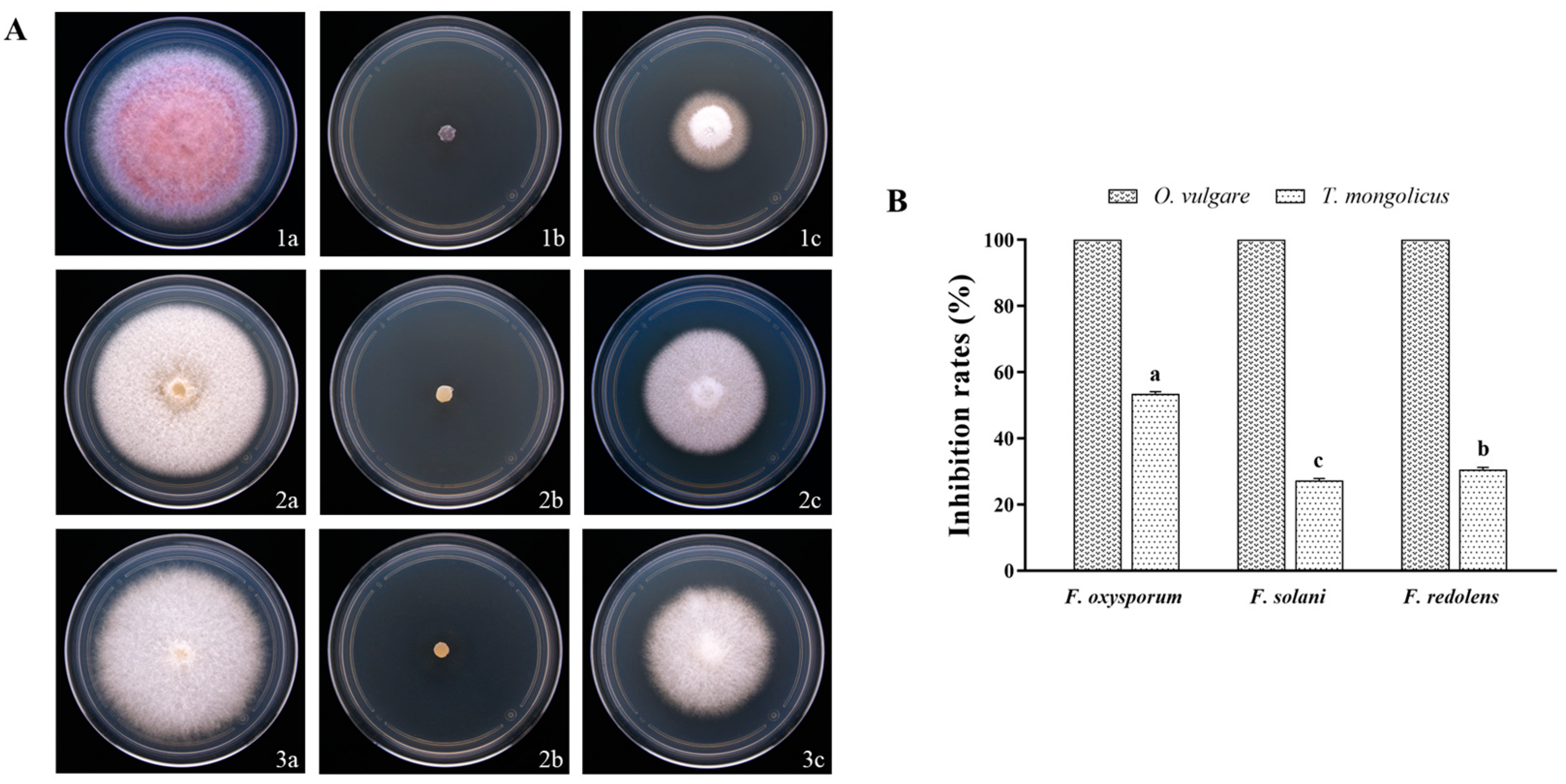
The fungal inhibitory effects of the volatiles of the O. vulgare EO and the T. mongolicus EO were further measured to evaluate the indirect activity of the EOs (Figure 2). Volatiles from the O. vulgare EO inhibited 100% of the mycelial growth in the three pathogenic fungi. The volatiles of T. mongolicus EO inhibited the mycelial growth of F. oxysporum, F. solani, and F. redolens by 53.79%, 27.25%, and 30.86%, respectively.

3.3. Chemical Compositions

The chemical compositions of the O. vulgare EO and the T. mongolicus EO were analyzed with GC-MS, and the results are shown in Tables S1 and S2. The GC-MS chromatograms of the O. vulgare EO and the T. mongolicus EO are shown in Figures S1 and S2. The main components of the O. vulgare EO were carvacrol (86.23%) and thymol (12%). The most abundant component of the T. mongolicus EO was o-cymene (56.63%), followed by γ-terpinene (21.96%) and thymol (12.05%). The main component common to both EOs was thymol.

3.4. Antifungal Activities of Predominant Constituents

The results revealed that carvacrol and thymol had excellent inhibitory effects against the three pathogenic fungi (Figure 3). The inhibition rates of carvacrol against F. oxysporum, F. solani, and F. redolens were 67.52%, 54.46%, and 75.29%, respectively. Thymol showed the strongest inhibitory effect on F. redolens (73.99%), followed by F. oxysporum (63.59%), and F. solani (50.36%). The inhibition rates of γ-pinene against F. oxysporum, F. solani, and F. redolens were 9.84%, 5.55%, and 3.81%, which were significantly lower than those of the two phenols. The inhibitory effects of o-cymene on F. oxysporum, F. solani, and F. redolens were very weak, with inhibition rates of 4.11%, 3.75%, and 8.09%, respectively.

3.5. Determination of MIC

The MIC values of the EOs and the main compounds against the fungi are shown in Table 1. The MIC values of the O. vulgare EO against F. oxysporum, F. solani, and F. redolens were 2.60 mg/mL, 3.13 mg/mL, and 1.56 mg/mL, respectively. The MIC values of the T. mongolicus EO against the three pathogenic fungi were 3.13 mg/mL, 2.60 mg/mL, and 2.60 mg/mL, respectively. The main components of the O. vulgare EO exhibited a higher antifungal activity than the O. vulgare EO and had a lower MIC value. The MIC values of carvacrol and thymol against the three pathogenic fungi were 0.83–1.04 mg/mL and 0.83–1.25 mg/mL, respectively. Carbendazim had a strong antifungal effect, with low MIC values of 0.05–0.07 mg/mL.

3.6. Effects of O. vulgare EO on Morphology and Ultrastructure of F. oxysporum

3.6.1. Optical Microscopy Observations

The optical microscopy showed that that the surface of mycelium in the control group was smooth, and the structure of mycelium was intact (Figure 4A). However, after treatment with 1/4 MIC O. vulgare EO, cavities appeared on the surface of the mycelium, and mycelia were locally deformed (Figure 4B). The mycelium treated with MIC O. vulgare EO degraded, and a large number of cavities appeared on the surface of the mycelium (Figure 4C). These results indicated that the O. vulgare EO might cause damage to the cell membrane of F. oxysporum.

3.6.2. Scanning Electron Microscopy (SEM)

Scanning electron microscopy showed that the mycelium of the control group had a smooth surface and a complete structure (Figure 5(1A,2A)). However, after treatment with 1/4 MIC concentration of O. vulgare EO, the mycelium was deformed and appeared folded and wrinkled (Figure 5(1B,2B)); after treatment with MIC concentration of O. vulgare EO, the mycelium deformation was more severe, and some mycelia were ruptured (Figure 5(1C,2C)).

3.6.3. Transmission Electron Microscopy (TEM)

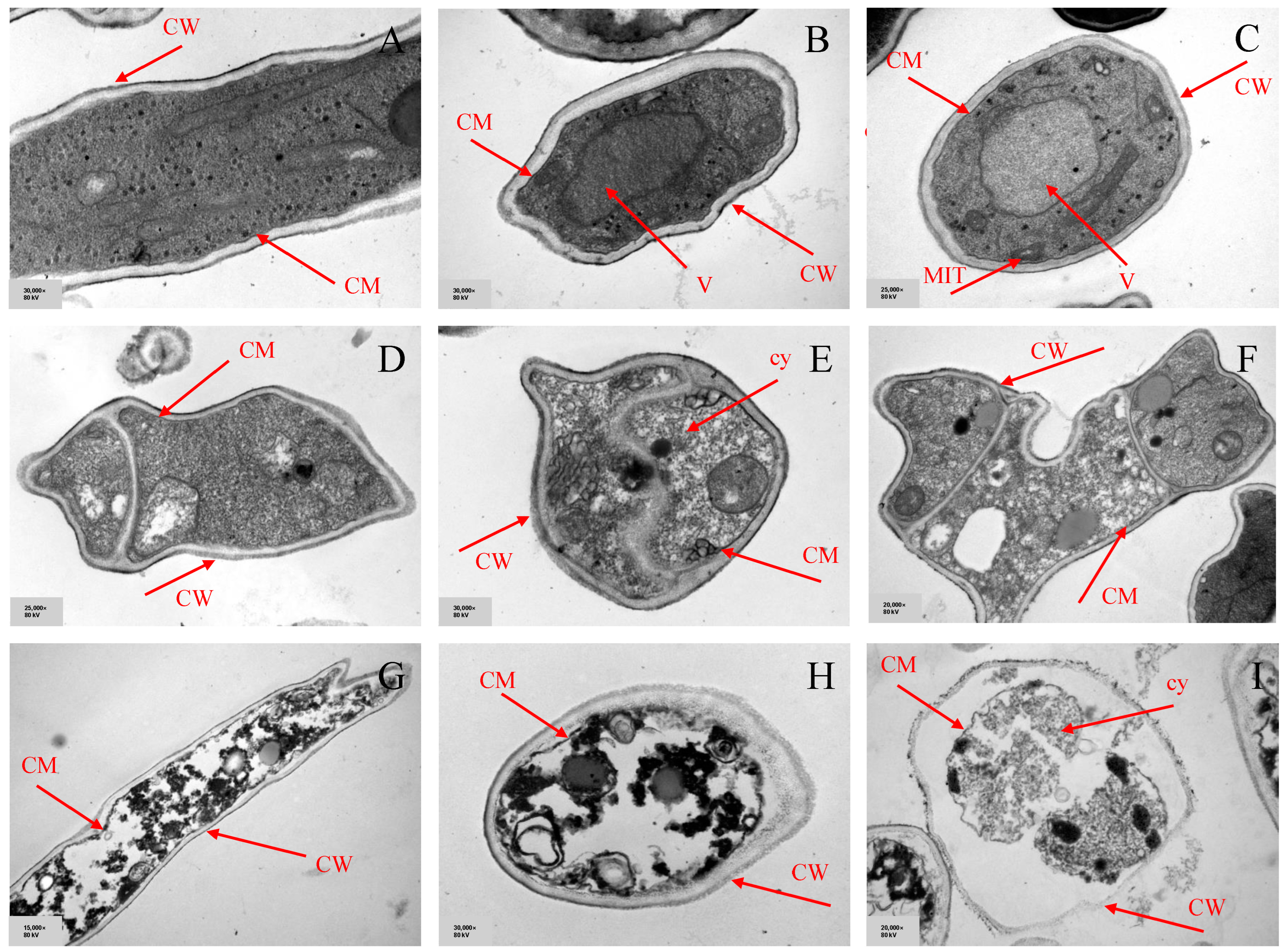
The ultrastructure of mycelium was observed with transmission electron microscopy (TEM) (Figure 6). In the control group, the mycelial cell wall and plasma membrane were intact. Mycelial cell walls were thick and uniform, regular in shape, round or oval. The organelles were structurally intact and evenly distributed in the cytoplasm. After treatment with 1/4 MIC O. vulgare EO, the mycelial cell morphology was found to be highly irregular, and the cell wall deformation appeared as protrusions or depressions. Mycelial cells appeared to be adherent, and the cytoplasm was unevenly distributed. Moreover, the hyphae treated with MIC O. vulgare EO exhibited significant structural changes. Mycelial cell walls were thinner, and even their lysis occurred. The mycelial cells exhibited obvious plasmolysis, and the internal organelles were blurred and unrecognizable.

3.7. Effect on Leakage of Cell Contents of F. oxysporum

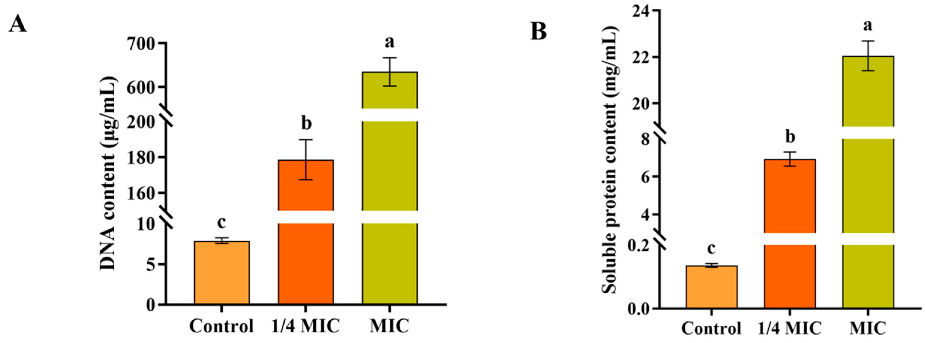
As shown in Figure 7, the O. vulgare EO caused a significant leakage of intracellular DNA and soluble proteins, and this effect exhibited concentration dependence. The DNA contents of 1/4 MIC and MIC were 179.48 μg/mL and 737.46 μg/mL, respectively, while that of the control group was only 8.1 μg/mL (Figure 7A). When treated with 1/4 MIC and MIC O. vulgare EO, the soluble protein contents were 51.07 and 162.22 times higher than the control group, respectively (Figure 7B).

3.8. The Effect of O. vulgare EO on Endogenous ROS in F. oxysporum

The DCFH-DA fluorescent probe was used to determine the intracellular ROS content of mycelium. As shown in Figure 8, the mycelium in the control group did not fluoresce. When treated with 1/2 MIC O. vulgare EO, clear fluorescence appeared within the mycelium. The results showed that the O. vulgare EO caused the production of ROS in the mycelial cells of F. oxysporum.

3.9. Effects of Exogenous ROS Scavengers on the Activity of O. vulgare EO

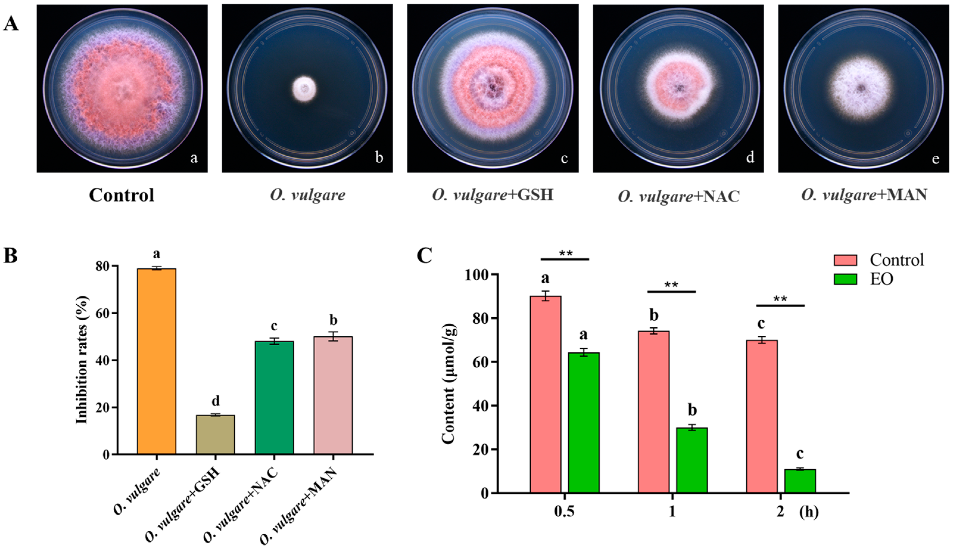
The effect of exogenous ROS scavengers on the fungal inhibitory effect of the O. vulgare EO is shown in Figure 9A. The inhibition rate of the O. vulgare EO against F. oxysporum was 79.05%, while the inhibition rate was only 16.83%, 48.09%, and 50.17% with the addition of GSH, NAC, and MAN, respectively (Figure 9B).

3.10. Intracellular GSH Content

The intracellular GSH content of mycelium was measured (Figure 9C). The results showed that when treated with 1/2 MIC O. vulgare EO, the intracellular GSH of the mycelium was significantly reduced compared to that of the control group. When treated with the O. vulgare EO for 2 h, the intracellular GSH content of the mycelium was only 15.64% of that of the control.

4. Discussion

Atractylodes chinensis is a perennial plant of the genus Atractylodes of the family Asteraceae [42]. In recent years, spurred by the increasing demand for A. chinensis, its artificial cultivation has expanded across many areas. However, its intensive cultivation has led to an increasing incidence of root rot, which seriously impacts its quality and yields [16]. Chemical pesticide usage is a typical strategy utilized for controlling root rot in A. chinensis. Chemical pesticides are effective for the control of pathogenic fungi; however, they also cause serious harm to ecosystems, non-target organisms, and human health [43]. The long-term use of chemical pesticides can also induce the resistance of pathogenic fungi, thus, reducing their effectiveness [44]. Plant EOs have garnered great interest from researchers due to their potent antifungal effects, easy decomposition, and environmental compatibility [45,46]. Labiatae is a large plant family with worldwide distribution, and its members are typically rich in EOs [47,48]. Labiatae EOs contain many aliphatic compounds, aromatic compounds, and terpene derivatives [47]. Previous studies have shown that the EOs of Labiatae plants can inhibit a wide range of bacteria and fungi [30,31,32]. In this study, we evaluated the inhibitory effects of O. vulgare, T. mongolicus, and P. cablin EOs on the root rot pathogen of A. chinensis. Since the O. vulgare EO exhibited the strongest inhibitory effect, the inhibitory mechanism of the O. vulgare EO against the dominant strain of root rot (F. oxysporum) was further investigated. This study provided a theoretical basis for the development of natural and efficient fungicides for the control of root rot in A. chinensis.
The results revealed that the three EOs had inhibitory effects against F. oxysporum, F. solani, and F. redolens. Among them, the O. vulgare and T. mongolicus EOs exhibited significantly high antifungal activities and completely inhibited the mycelial growth of the three pathogenic fungi. EOs possess broad-spectrum antimicrobial activities, which can inhibit the growth of mycelium via diffusion, and plants naturally use these chemicals to resist pests and diseases [26]. The fungicidal effects of volatiles of the O. vulgare and T. mongolicus EOs were further investigated, and results showed that the volatiles of the O. vulgare EO had strong inhibitory effects on the three pathogenic fungi, with an inhibition rate of 100%. Parikh et al. also reported that the volatiles of O. vulgare EO had excellent fungicidal activities and completely inhibited the mycelial growth of pathogenic fungi [26]. However, the inhibitory effects of the volatiles of T. mongolicus EO on pathogenic fungi were lower than that of direct contact. This difference was also reported in a previous study [49], which was related to the concentration, polarity, and volatility of the EO [50].
Determining the composition of an EO is important for exploring its fungicidal mechanism and selective inhibitory properties [40]. This study revealed that the main components of the O. vulgare EO were carvacrol and thymol, and those of the T. mongolicus EO were γ-pinene, o-cymene, and thymol. Wu et al. analyzed the chemical composition of the O. vulgare EO using GC-MS and showed that the contents of carvacrol and thymol in it were 72.69% and 6.02%, respectively [40]. Shin and Kim studied the composition of T. mongolicus EO and showed that the three most abundant components were thymol, γ-pinene, and o-cymene [51]. Our findings were generally consistent with these previous studies, with some variations in content. Variations in the compositions and contents of the EOs of plants may be correlated with climatic conditions, altitude, geographic location, soil nutrients, extraction methods, plant age, harvesting time, and fertilizer and pesticide application [52].
The antifungal activities of EOs may be derived from their principal components [53]. In this study, the antifungal effects of carvacrol, thymol, γ-pinene, and o-cymene were determined with Oxford cups. Carvacrol and thymol exhibited strong inhibitory effects against the three pathogenic fungi under study, while γ-pinene and o-cymene showed weaker inhibitory effects. Notably, the inhibitory effects of carvacrol and thymol against F. solani and F. redolens were stronger than those of chemical pesticides. Carvacrol and thymol are constitutional isomers that belong to the phenolic group; certain phenolic compounds have been shown to have inhibitory activity against some species of fungi and bacteria [54,55,56]. Phenolic compounds contain hydroxyl groups that eliminate pathogenic fungi by binding to and deactivating their enzymatically active centers through the formation of hydrogen bonds [57]. A preceding study indicated that carvacrol and thymol inhibited Xanthomonas campestris pv. campestris (Xcc) to control the black rot of cabbage [58]. Zhao et al. found that carvacrol and thymol inhibited Botrytis cinerea’s mycelial growth [59]. Due to their excellent fungicidal activities and environmental compatibility, carvacrol and thymol can be utilized as novel pesticides for the control of plant diseases in the future. In this study, the O. vulgare EO exhibited very strong direct and indirect inhibitory effects against the three pathogenic fungi, which was likely due to its high level of phenolics [60]. Although less inhibitory than phenolics, o-cymene and γ-pinene may synergistically influence the antimicrobial effect of EOs.
The MIC value refers to the minimum EO concentration required to completely inhibit the growth of pathogenic fungi [61]. Our results indicated that the two EOs and two phenols showed strong inhibitory effects on the root rot pathogens of A. chinensis. Carvacrol and thymol had lower MIC (mg/mL) values compared to the EOs. Furthermore, an earlier study reported that monomers had more potent fungicidal activities than EOs [62]. Carbendazim is a synthetic fungicide that is widely used in agricultural production and has high fungicidal activities. The MICs of carbendazim against F. oxysporum, F. solani, and F. redolens ranged from 0.05–0.07 mg/mL. However, the half-life of carbendazim in nature is very long, and very low doses can produce serious “teratogenic, mutagenic and carcinogenic” effects in mammals [63,64]. Therefore, certain components of plant EOs can serve as new natural fungicides with great potential in the future.
Light microscopy showed that following the O. vulgare EO treatment, cavities appeared on the surface of F. oxysporum mycelium, which became locally deformed or was lysed. Scanning electron microscopy results showed that the O. vulgare EO altered the cell membrane structure of F. oxysporum. The results of transmission electron microscopy (TEM) revealed that the O. vulgare EO could damage the cell wall structures of F. oxysporum, resulting in barrier disruption, which translated to induced abnormalities in the fungal plasma membrane structure and ultimately led to cell death. The fungal cell wall is a cellular structure unique to fungi, which consists of polysaccharides that wrap around the cytoplasm [65]. Fungal cell walls mainly play a protective role in maintaining the inherent cellular morphology and integrity, in addition to sustaining normal cellular metabolism, ion exchange, and osmotic pressure [66,67]. The absence of cell wall structures leads to the rupture of the plasma membrane and cell lysis; thus, the integrity of the cell wall is essential for the survival of the fungus [68]. A previous study showed that the L. verbena EO disrupted the cell wall of Pseudosciaena D4, resulting in increased cell membrane permeability and leakage of intracellular contents, ultimately leading to cell death [69]. A further study disclosed that the M. haplocalyx EO penetrated the cell wall and dissolved the internal organelles to inhibit the growth of F. oxysporum [37]. This study suggests that the cell wall may be a viable target for the O. vulgare EO in F. oxysporum.
Proteins and nucleic acids are critical biomolecules within cytoplasms and nuclei of cells [70,71]. The leakage of DNA and proteins reflects the integrity of the cell membrane [72]. This study showed a significant leakage of DNA and proteins within the mycelium of F. oxysporum after treatment with the O. vulgare EO, which indicated that the plasma membrane was severely damaged. This was consistent with the morphological and ultrastructural results. The hydrophobic characteristics of EOs enable them to enter the cell membranes of pathogenic fungi, alter their structures, and increase their permeability [73].
Reactive oxygen species (ROS) is a general term for a class of oxidatively active molecules that are produced by cellular energy metabolism under aerobic conditions, which play an important role in the physiological and pathological processes of organisms [74]. However, excess ROS induce oxidative stress, which alters the structures of biomolecules (DNA, proteins, and lipids), and ultimately leads to cell death [75]. For this study, DCFH-DA fluorescent probes were used to determine the intracellular ROS content of mycelium [76]. The results revealed that the mycelium of F. oxysporum emitted significant fluorescence following the O. vulgare EO treatment, suggesting that it could exert inhibitory effects by inducing the generation of ROS. Wu et al. reported that the O. vulgare EO inhibited the growth of Rhizoctonia solani by inducing the production of ROS [40]. Reactive oxygen scavenger is a collective term for a class of substances with reactive oxygen scavenging properties (e.g., GSH, NAC, MAN, etc.). In this study, all three ROS scavengers (GSH, NAC, and MAN) alleviated the inhibitory effects of the O. vulgare EO on F. oxysporum, with GSH having the strongest impact. Thus, the inhibitory activities of the O. vulgare EO against F. oxysporum were positively correlated with the accumulation of ROS in mycelial cells.
Glutathione (GSH) is a naturally reactive peptide composed of glutamic acid, cysteine, and glycine and is widely found in living organisms [77,78]. GSH possesses antioxidant and integrative detoxification activities that help cells to maintain normal immune system functions [79,80]. In this study, the content of GSH in the O. vulgare EO-treated mycelium was significantly reduced over a short timeline (0.5–2 h) compared to the control. This result indicated that the O. vulgare EO depleted intracellular GSH, resulting in a large accumulation of ROS in the mycelium.
Plant EOs have potent inhibitory activities against pathogenic fungi and can be used as natural and safe pesticides. However, in recent years, some studies have shown that high concentrations of EOs or extracts can inhibit seed germination [81]. Thus, appropriate concentrations of EOs should be selected to control plant diseases to avoid their negative effects on seeds. In addition, this study was conducted under sealed conditions in the laboratory, and the effect of volatility on the results was not taken into account. EOs and their main ingredients are highly volatile, so their stability is low. Volatility has become a major limitation in the application of EOs, which may cause the actual application effectiveness to be lower than that observed in the experimental results. Currently, researchers have developed methods to improve the stability and persistence of EOs, such as microcapsules, microemulsions, and nanoemulsions [82,83]. In the future, avenues to combine these methods to increase the effectiveness of EOs should be further investigated.

5. Conclusions

In this study, the inhibitory effect of three EOs of Labiatae plants on the root rot pathogens of A. chinensis was investigated. The O. vulgare EO and its main components exhibited excellent inhibitory activities against F. oxysporum, F. solani, and F. redolens. The results of in vivo experiments showed that the O. vulgare EO was effective in reducing the damage caused by F. oxysporum in A. chinensis. The inhibitory mechanism of the O. vulgare EO was summarized as follows: 1. The O. vulgare EO could cause severe damage to cell walls and plasma membranes, increasing the permeability of cell membranes and leading to a massive leakage of intracellular contents. 2. The O. vulgare EO inhibited pathogenic fungi by inducing ROS production and reducing the amount of intracellular GSH, leading to a large accumulation of ROS. Collectively, the O. vulgare EO and its main components can be developed as environmentally friendly fungicides to replace synthetic pesticides for the control of root rot of A. chinensis.

Supplementary Materials

The following supporting information can be downloaded at: https://www.mdpi.com/article/10.3390/horticulturae9101136/s1. Table S1: Chemical composition of the EO from O. vulgare; Table S2: Chemical composition of the EO from T. mongolicus; Figure S1. GC-MS chromatogram of the Origanum vulgare essential oil.; Figure S2. GC-MS chromatogram of the Thymus mongolicus essential oil.

Author Contributions

Conceptualization, methodology, and writing—original draft, S.X.; software, H.S.; formal analysis, S.Z.; investigation, R.Z.; validation, Y.X.; data curation, S.W. (Shijie Wang); resources, S.W. (Shiqiang Wang); supervision, Y.D.; project administration, J.N.; funding acquisition and writing—review and editing, Z.W. All authors have read and agreed to the published version of the manuscript.

Funding

This research was funded by Xi’an Science and Technology Project (20NYYF0057); Fundamental Research Funds for the Central Universities (grant numbers: GK202205002 and GK202205004); Shaanxi Provincial Key R & D Program (2022SF-172, 2021SF-383, and 2020LSFP2-21); and Youth Innovation Team Construction Scientific Research Project of Shaanxi Education Department (21JP027); and Shaanxi Administration of Traditional Chinese Medicine Projects (grant numbers: 2021-QYZL-01 and 2021-QYPT-001).

Data Availability Statement

Date is available upon request from Z.W.

Conflicts of Interest

The authors declare no conflict of interest.

References

  1. Yun, C.; Zhao, Z.; Gu, L.; Zhang, Z.; Wang, S.; Shi, Y.; Miao, N.; Ri, I.; Wang, W.; Wang, H. In vitro production of atractylon and β-eudesmol from Atractylodes chinensis by adventitious root culture. Appl. Microbiol. Biotechnol. 2022, 106, 7027–7037. [Google Scholar] [CrossRef]
  2. Wang, L.; Zhang, H.; Wu, X.; Wang, Z.; Fang, W.; Jiang, M.; Chen, H.; Huang, L.; Liu, C. Phylogenetic relationships of Atractylodes lancea, A. chinensis and A. macrocephala, revealed by complete plastome and nuclear gene sequences. PLoS ONE 2020, 15, e0227610. [Google Scholar] [CrossRef]
  3. Lei, H.; Yue, J.; Yin, X.Y.; Fan, W.; Tan, S.H.; Qin, L.; Zhao, Y.N.; Bai, J.H. HS-SPME coupled with GC-MS for elucidating differences between the volatile components in wild and cultivated Atractylodes chinensis. Phytochem. Anal. 2023, 34, 317–328. [Google Scholar] [CrossRef] [PubMed]
  4. Zhao, J.; Sun, C.; Shi, F.; Ma, S.; Zheng, J.; Du, X.; Zhang, L. Comparative transcriptome analysis reveals sesquiterpenoid biosynthesis among 1-, 2- and 3-year old Atractylodes chinensis. BMC Plant Biol. 2021, 21, 354. [Google Scholar] [CrossRef]
  5. Ma, Z.; Liu, G.; Yang, Z.; Zhang, G.; Sun, L.; Wang, M.; Ren, X. Species Differentiation and Quality Evaluation for Atractylodes Medicinal Plants by GC-MS coupled with Chemometric Analysis. Chem. Biodivers. 2023, 20, e202300793. [Google Scholar] [CrossRef]
  6. Park, Y.J.; Seo, M.G.; Cominguez, D.C.; Han, I.; An, H.J. Atractylodes chinensis Water Extract Ameliorates Obesity via Promotion of the SIRT1/AMPK Expression in High-Fat Diet-Induced Obese Mice. Nutrients 2021, 13, 2992. [Google Scholar] [CrossRef] [PubMed]
  7. Meng, H.; Li, G.Y.; Dai, R.H.; Ma, Y.P.; Zhang, K.; Zhang, C.; Li, X.; Wang, J.H. Two new polyacetylenic compounds from Atractylodes chinensis (DC.) Koidz. J. Asian Nat. Prod. Res. 2011, 13, 346–349. [Google Scholar] [CrossRef] [PubMed]
  8. Chen, L.G.; Jan, Y.S.; Tsai, P.W.; Norimoto, H.; Michihara, S.; Murayama, C.; Wang, C.C. Anti-inflammatory and Antinociceptive Constituents of Atractylodes japonica Koidzumi. J. Agric. Food Chem. 2016, 64, 2254–2262. [Google Scholar] [CrossRef] [PubMed]
  9. Ji, L.; Nasir, F.; Tian, L.; Chang, J.; Sun, Y.; Zhang, J.; Li, X.; Tian, C. Outbreaks of Root Rot Disease in Different Aged American Ginseng Plants Are Associated with Field Microbial Dynamics. Front. Microbiol. 2021, 12, 676880. [Google Scholar] [CrossRef] [PubMed]
  10. La Placa, L.; Giorni, P.; Mondani, L.; Magan, N.; Battilani, P. Comparison of Different Physical Methods and Preservatives for Control of Fusarium proliferatum Rot in Garlic. Horticulturae 2022, 8, 1203. [Google Scholar] [CrossRef]
  11. Haus, M.J.; Wang, W.; Jacobs, J.L.; Peplinski, H.; Chilvers, M.I.; Buell, C.R.; Cichy, K. Root Crown Response to Fungal Root Rot in Phaseolus vulgaris Middle American × Andean Lines. Plant Dis. 2020, 104, 3135–3142. [Google Scholar] [CrossRef] [PubMed]
  12. Zakaria, L. Fusarium Species Associated with Diseases of Major Tropical Fruit Crops. Horticulturae 2023, 9, 322. [Google Scholar] [CrossRef]
  13. Takeda, O.; Miki, E.; Terabayashi, S.; Okada, M.; Lu, Y.; He, H.S.; He, S.A. A comparative study on essential oil components of wild and cultivated Atractylodes lancea and A. chinensis. Planta Med. 1996, 62, 444–449. [Google Scholar] [CrossRef] [PubMed]
  14. Huo, J.H.; Wen, X.L.; Li, S.M.; Feng, L.N.; Lan, S.H.; Dong, L.X.; Guo, S.R.; Li, J.N.; Wang, J.H.; Qi, H.X. ldentification and Biological Characteristics of the Pathogen Causing Root Rot of Atractylodes chinensis. J. Agric. Sci. Technol. 2022, 24, 137–144. [Google Scholar] [CrossRef]
  15. Pietro, A.D.; Madrid, M.P.; Caracuel, Z.; Delgado-Jarana, J.; Roncero, M.I. Fusarium oxysporum: Exploring the molecular arsenal of a vascular wilt fungus. Mol. Plant. Pathol. 2003, 4, 315–325. [Google Scholar] [CrossRef]
  16. Li, C.N.; Li, H.T.; Li, Y.C.; Li, J.H.; Ji, H.; Zhang, L.; Wang, L. Research progress and control of root rot in Atractylodes lancea. J. Agro-Environ. Sci. 2022, 41, 2840–2846. [Google Scholar] [CrossRef]
  17. Huang, X.G.; Li, M.Y.; Yan, X.N.; Yang, J.S.; Rao, M.C.; Yuan, X.F. The potential of Trichoderma brevicompactum for controlling root rot on Atractylodes macrocephala. Can. J. Plant Pathol. 2021, 43, 794–802. [Google Scholar] [CrossRef]
  18. Wang, L.; Tu, H.; Hou, H.; Zhou, Z.; Yuan, H.; Luo, C.; Gu, Q. Occurrence and Detection of Carbendazim Resistance in Botryosphaeria dothidea from Apple Orchards in China. Plant Dis. 2022, 106, 207–214. [Google Scholar] [CrossRef]
  19. Liu, S.; Fu, L.; Wang, S.; Chen, J.; Jiang, J.; Che, Z.; Tian, Y.; Chen, G. Carbendazim Resistance of Fusarium graminearum From Henan Wheat. Plant Dis. 2019, 103, 2536–2540. [Google Scholar] [CrossRef]
  20. Al-Balushi, Z.M.; Agrama, H.; Al-Mahmooli, I.H.; Maharachchikumbura, S.S.N.; Al-Sadi, A.M. Development of Resistance to Hymexazol Among Pythium Species in Cucumber Greenhouses in Oman. Plant Dis. 2018, 102, 202–208. [Google Scholar] [CrossRef]
  21. Lu, Y.; Li, X.; Li, W.; Shen, T.; He, Z.; Zhang, M.; Zhang, H.; Sun, Y.; Liu, F. Detection of chlorpyrifos and carbendazim residues in the cabbage using visible/near-infrared spectroscopy combined with chemometrics. Spectrochim. Acta Part A Mol. Biomol. Spectrosc. 2021, 257, 119759. [Google Scholar] [CrossRef]
  22. Anastassiadou, M.; Brancato, A.; Carrasco Cabrera, L.; Ferreira, L.; Greco, L.; Jarrah, S.; Kazocina, A.; Leuschner, R.; Magrans, J.O.; Miron, I.; et al. Review of the existing maximum residue levels for hymexazol according to Article 12 of Regulation (EC) No 396/2005. EFSA J. 2019, 17, e05895. [Google Scholar] [CrossRef] [PubMed]
  23. Tompros, A.; Wilber, M.Q.; Fenton, A.; Carter, E.D.; Gray, M.J. Efficacy of Plant-Derived Fungicides at Inhibiting Batrachochytrium salamandrivorans Growth. J. Fungi 2022, 8, 1025. [Google Scholar] [CrossRef]
  24. Souto, A.L.; Sylvestre, M.; Tölke, E.D.; Tavares, J.F.; Barbosa-Filho, J.M.; Cebrián-Torrejón, G. Plant-Derived Pesticides as an Alternative to Pest Management and Sustainable Agricultural Production: Prospects, Applications and Challenges. Molecules 2021, 26, 4835. [Google Scholar] [CrossRef] [PubMed]
  25. Aziz, Z.A.A.; Ahmad, A.; Setapar, S.H.M.; Karakucuk, A.; Azim, M.M.; Lokhat, D.; Rafatullah, M.; Ganash, M.; Kamal, M.A.; Ashraf, G.M. Essential Oils: Extraction Techniques, Pharmaceutical and Therapeutic Potential—A Review. Curr. Drug Metab. 2018, 19, 1100–1110. [Google Scholar] [CrossRef]
  26. Parikh, L.; Agindotan, B.O.; Burrows, M.E. Antifungal Activity of Plant-Derived Essential Oils on Pathogens of Pulse Crops. Plant Dis. 2021, 105, 1692–1701. [Google Scholar] [CrossRef]
  27. Ni, Z.-J.; Wang, X.; Shen, Y.; Thakur, K.; Han, J.; Zhang, J.-G.; Hu, F.; Wei, Z.-J. Recent updates on the chemistry, bioactivities, mode of action, and industrial applications of plant essential oils. Trends Food Sci. Technol. 2021, 110, 78–89. [Google Scholar] [CrossRef]
  28. El Khetabi, A.; Lahlali, R.; Ezrari, S.; Radouane, N.; Lyousfi, N.; Banani, H.; Askarne, L.; Tahiri, A.; El Ghadraoui, L.; Belmalha, S.; et al. Role of plant extracts and essential oils in fighting against postharvest fruit pathogens and extending fruit shelf life: A review. Trends Food Sci. Technol. 2022, 120, 402–417. [Google Scholar] [CrossRef]
  29. Raja, R. Medicinally Potential Plants of Labiatae (Lamiaceae) Family: An Overview. Res. J. Med. Plant 2012, 6, 203–213. [Google Scholar] [CrossRef]
  30. Kot, B.; Wierzchowska, K.; Piechota, M.; Czerniewicz, P.; Chrzanowski, G. Antimicrobial activity of five essential oils from lamiaceae against multidrug-resistant Staphylococcus aureus. Nat. Prod. Res. 2019, 33, 3587–3591. [Google Scholar] [CrossRef]
  31. Coimbra, A.; Ferreira, S.; Duarte, A.P. Biological properties of Thymus zygis essential oil with emphasis on antimicrobial activity and food application. Food Chem. 2022, 393, 133370. [Google Scholar] [CrossRef]
  32. Carvalho, F.; Coimbra, A.T.; Silva, L.; Duarte, A.P.; Ferreira, S. Melissa officinalis essential oil as an antimicrobial agent against Listeria monocytogenes in watermelon juice. Food Microbiol. 2023, 109, 104105. [Google Scholar] [CrossRef]
  33. Xiao, S.; Cui, P.; Shi, W.; Zhang, Y. Identification of essential oils with activity against stationary phase Staphylococcus aureus. BMC Complement. Med. Ther. 2020, 20, 99. [Google Scholar] [CrossRef] [PubMed]
  34. Pérez-Vázquez, M.A.K.; Pacheco-Hernández, Y.; Lozoya-Gloria, E.; Mosso-González, C.; Ramírez-García, S.A.; Romero-Arenas, O.; Villa-Ruano, N. Peppermint Essential Oil and Its Major Volatiles as Protective Agents against Soft Rot Caused by Fusarium sambucinum in Cera Pepper (Capsicum pubescens). Chem. Biodivers. 2022, 19, e202100835. [Google Scholar] [CrossRef] [PubMed]
  35. Nafis, A.; Ouedrhiri, W.; Iriti, M.; Mezrioui, N.; Marraiki, N.; Elgorban, A.M.; Syed, A.; Hassani, L. Chemical composition and synergistic effect of three Moroccan lavender EOs with ciprofloxacin against foodborne bacteria: A promising approach to modulate antimicrobial resistance. Lett. Appl. Microbiol. 2021, 72, 698–705. [Google Scholar] [CrossRef]
  36. Aebisher, D.; Cichonski, J.; Szpyrka, E.; Masjonis, S.; Chrzanowski, G. Essential Oils of Seven Lamiaceae Plants and Their Antioxidant Capacity. Molecules 2021, 26, 3793. [Google Scholar] [CrossRef] [PubMed]
  37. Chen, C.-J.; Li, Q.-Q.; Zeng, Z.-Y.; Duan, S.-S.; Wang, W.; Xu, F.-R.; Cheng, Y.-X.; Dong, X. Efficacy and mechanism of Mentha haplocalyx and Schizonepeta tenuifolia essential oils on the inhibition of Panax notoginseng pathogens. Ind. Crops. Prod. 2020, 145, 112073. [Google Scholar] [CrossRef]
  38. Liang, J.-Y.; Xu, J.; Yang, Y.-Y.; Shao, Y.-Z.; Zhou, F.; Wang, J.-L. Toxicity and Synergistic Effect of Elsholtzia ciliata Essential Oil and Its Main Components against the Adult and Larval Stages of Tribolium castaneum. Foods 2020, 9, 345. [Google Scholar] [CrossRef]
  39. Sparkman, O.D. Identification of essential oil components by gas chromatography/quadrupole mass spectroscopy Robert P. Adams. J. Am. Soc. Mass Spectrom. 2005, 16, 1902–1903. [Google Scholar] [CrossRef]
  40. Wu, T.-L.; Zhang, B.-Q.; Luo, X.-F.; Li, A.-P.; Zhang, S.-Y.; An, J.-X.; Zhang, Z.-J.; Liu, Y.-Q. Antifungal efficacy of sixty essential oils and mechanism of oregano essential oil against Rhizoctonia solani. Ind. Crops. Prod. 2023, 191, 115975. [Google Scholar] [CrossRef]
  41. Liu, H.; Wu, J.; Su, Y.; Li, Y.; Zuo, D.; Liu, H.; Liu, Y.; Mei, X.; Huang, H.; Yang, M.; et al. Allyl Isothiocyanate in the Volatiles of Brassica juncea Inhibits the Growth of Root Rot Pathogens of Panax notoginseng by Inducing the Accumulation of ROS. J. Agric. Food Chem. 2021, 69, 13713–13723. [Google Scholar] [CrossRef] [PubMed]
  42. Zhuang, L.X.; Liu, Y.; Wang, S.Y.; Sun, Y.; Pan, J.; Guan, W.; Hao, Z.C.; Kuang, H.X.; Yang, B.Y. Cytotoxic Sesquiterpenoids from Atractylodes chinensis (DC.) Koidz. Chem. Biodivers. 2022, 19, e202200812. [Google Scholar] [CrossRef] [PubMed]
  43. Brugel, M.; Carlier, C.; Reyes-Castellanos, G.; Callon, S.; Carrier, A.; Bouché, O. Pesticides and pancreatic adenocarcinoma: A transversal epidemiological, environmental and mechanistic narrative review. Dig. Liver Dis. 2022, 54, 1605–1613. [Google Scholar] [CrossRef]
  44. Malandrakis, A.A.; Kavroulakis, N.; Chrysikopoulos, C.V. Metal nanoparticles against fungicide resistance: Alternatives or partners? Pest Manag. Sci. 2022, 78, 3953–3956. [Google Scholar] [CrossRef]
  45. Seow, Y.X.; Yeo, C.R.; Chung, H.L.; Yuk, H.G. Plant essential oils as active antimicrobial agents. Crit. Rev. Food Sci. Nutr. 2014, 54, 625–644. [Google Scholar] [CrossRef]
  46. Bolouri, P.; Salami, R.; Kouhi, S.; Kordi, M.; Asgari Lajayer, B.; Hadian, J.; Astatkie, T. Applications of Essential Oils and Plant Extracts in Different Industries. Molecules 2022, 27, 8999. [Google Scholar] [CrossRef]
  47. Karpiński, T.M. Essential Oils of Lamiaceae Family Plants as Antifungals. Biomolecules 2020, 10, 103. [Google Scholar] [CrossRef]
  48. Çalış, İ.; Başer, K.H.C. Review of Studies on Phlomis and Eremostachys Species (Lamiaceae) with Emphasis on Iridoids, Phenylethanoid Glycosides, and Essential Oils. Planta Med. 2021, 87, 1128–1151. [Google Scholar] [CrossRef] [PubMed]
  49. Regnier, T.; Combrinck, S.; Veldman, W.; Du Plooy, W. Application of essential oils as multi-target fungicides for the control of Geotrichum citri-aurantii and other postharvest pathogens of citrus. Ind. Crops. Prod. 2014, 61, 151–159. [Google Scholar] [CrossRef]
  50. Cox, S.D.; Mann, C.M.; Markham, J.L. Interactions between components of the essential oil of Melaleuca alternifolia. J. Appl. Microbiol. 2001, 91, 492–497. [Google Scholar] [CrossRef]
  51. Shin, S.; Kim, J.H. Antifungal activities of essential oils from Thymus quinquecostatus and T. magnus. Planta Med. 2004, 70, 1090–1092. [Google Scholar] [CrossRef] [PubMed]
  52. Ashokkumar, K.; Simal-Gandara, J.; Murugan, M.; Dhanya, M.K.; Pandian, A. Nutmeg (Myristica fragrans Houtt.) essential oil: A review on its composition, biological, and pharmacological activities. Phytother. Res. 2022, 36, 2839–2851. [Google Scholar] [CrossRef] [PubMed]
  53. Dhakad, A.K.; Pandey, V.V.; Beg, S.; Rawat, J.M.; Singh, A. Biological, medicinal and toxicological significance of Eucalyptus leaf essential oil: A review. J. Sci. Food Agric. 2018, 98, 833–848. [Google Scholar] [CrossRef] [PubMed]
  54. Xu, J.; Zhou, F.; Ji, B.P.; Pei, R.S.; Xu, N. The antibacterial mechanism of carvacrol and thymol against Escherichia coli. Lett. Appl. Microbiol. 2008, 47, 174–179. [Google Scholar] [CrossRef]
  55. Netopilova, M.; Houdkova, M.; Rondevaldova, J.; Kmet, V.; Kokoska, L. Evaluation of in vitro growth-inhibitory effect of carvacrol and thymol combination against Staphylococcus aureus in liquid and vapour phase using new broth volatilization chequerboard method. Fitoterapia 2018, 129, 185–190. [Google Scholar] [CrossRef]
  56. Jesus, F.P.; Ferreiro, L.; Bizzi, K.S.; Loreto, É.S.; Pilotto, M.B.; Ludwig, A.; Alves, S.H.; Zanette, R.A.; Santurio, J.M. In vitro activity of carvacrol and thymol combined with antifungals or antibacterials against Pythium insidiosum. J. Mycol. Med. 2015, 25, e89–e93. [Google Scholar] [CrossRef]
  57. Gallucci, M.N.; Carezzano, M.E.; Oliva, M.M.; Demo, M.S.; Pizzolitto, R.P.; Zunino, M.P.; Zygadlo, J.A.; Dambolena, J.S. In vitro activity of natural phenolic compounds against fluconazole-resistant Candida species: A quantitative structure–activity relationship analysis. J. Appl. Microbiol. 2014, 116, 795–804. [Google Scholar] [CrossRef]
  58. Hakalová, E.; Čechová, J.; Tekielska, D.A.; Eichmeier, A.; Pothier, J.F. Combined effect of thyme and clove phenolic compounds on Xanthomonas campestris pv. campestris and biocontrol of black rot disease on cabbage seeds. Front. Microbiol. 2022, 13, 1007988. [Google Scholar] [CrossRef]
  59. Zhao, Y.; Yang, Y.-H.; Ye, M.; Wang, K.-B.; Fan, L.-M.; Su, F.-W. Chemical composition and antifungal activity of essential oil from Origanum vulgare against Botrytis cinerea. Food Chem. 2021, 365, 130506. [Google Scholar] [CrossRef]
  60. Teixeira, B.; Marques, A.; Ramos, C.; Serrano, C.; Matos, O.; Neng, N.R.; Nogueira, J.M.; Saraiva, J.A.; Nunes, M.L. Chemical composition and bioactivity of different oregano (Origanum vulgare) extracts and essential oil. J. Sci. Food Agric. 2013, 93, 2707–2714. [Google Scholar] [CrossRef]
  61. da Silva, R.S.; de Oliveira, M.M.G.; de Melo, J.O.; Blank, A.F.; Corrêa, C.B.; Scher, R.; Fernandes, R.P.M. Antimicrobial activity of Lippia gracilis essential oils on the plant pathogen Xanthomonas campestris pv. campestris and their effect on membrane integrity. Pestic. Biochem. Physiol. 2019, 160, 40–48. [Google Scholar] [CrossRef]
  62. Ma, Y.-N.; Xu, F.-R.; Chen, C.-J.; Li, Q.-Q.; Wang, M.-Z.; Cheng, Y.-X.; Dong, X. The beneficial use of essential oils from buds and fruit of Syzygium aromaticum to combat pathogenic fungi of Panax notoginseng. Ind. Crops Prod. 2019, 133, 185–192. [Google Scholar] [CrossRef]
  63. Zhou, T.; Guo, T.; Wang, Y.; Wang, A.; Zhang, M. Carbendazim: Ecological risks, toxicities, degradation pathways and potential risks to human health. Chemosphere 2023, 314, 137723. [Google Scholar] [CrossRef] [PubMed]
  64. Panda, J.; Kanjilal, T.; Das, S. Optimized biodegradation of carcinogenic fungicide Carbendazim by Bacillus licheniformis JTC-3 from agro-effluent. Biotechnol. Res. Innov. 2018, 2, 45–57. [Google Scholar] [CrossRef]
  65. Gow, N.A.R.; Lenardon, M.D. Architecture of the dynamic fungal cell wall. Nat. Rev. Microbiol. 2023, 21, 248–259. [Google Scholar] [CrossRef] [PubMed]
  66. Liu, T.-T.; Gou, L.-J.; Zeng, H.; Zhou, G.; Dong, W.-R.; Cui, Y.; Cai, Q.; Chen, Y.-X. Inhibitory Effect and Mechanism of Dill Seed Essential Oil on Neofusicoccum parvum in Chinese Chestnut. Separations 2022, 9, 296. [Google Scholar] [CrossRef]
  67. Garcia-Rubio, R.; de Oliveira, H.C.; Rivera, J.; Trevijano-Contador, N. The Fungal Cell Wall: Candida, Cryptococcus, and Aspergillus Species. Front. Microbiol. 2020, 10, 2993. [Google Scholar] [CrossRef]
  68. Wong, F.; Amir, A. Mechanics and Dynamics of Bacterial Cell Lysis. Biophys J. 2019, 116, 2378–2389. [Google Scholar] [CrossRef]
  69. Gao, X.; Liu, J.; Li, B.; Xie, J. Antibacterial Activity and Antibacterial Mechanism of Lemon Verbena Essential Oil. Molecules 2023, 28, 3102. [Google Scholar] [CrossRef]
  70. Saccà, B.; Niemeyer, C.M. Functionalization of DNA nanostructures with proteins. Chem. Soc. Rev. 2011, 40, 5910–5921. [Google Scholar] [CrossRef]
  71. Minchin, S.; Lodge, J. Understanding biochemistry: Structure and function of nucleic acids. Essays Biochem. 2019, 63, 433–456. [Google Scholar] [CrossRef]
  72. Yao, C.; Li, X.; Bi, W.; Jiang, C. Relationship between membrane damage, leakage of intracellular compounds, and inactivation of Escherichia coli treated by pressurized CO2. J. Basic. Microbiol. 2014, 54, 858–865. [Google Scholar] [CrossRef]
  73. Burt, S. Essential oils: Their antibacterial properties and potential applications in foods—A review. Int. J. Food Microbiol. 2004, 94, 223–253. [Google Scholar] [CrossRef] [PubMed]
  74. Tudzynski, P.; Heller, J.; Siegmund, U. Reactive oxygen species generation in fungal development and pathogenesis. Curr. Opin. Microbiol. 2012, 15, 653–659. [Google Scholar] [CrossRef] [PubMed]
  75. Brieger, K.; Schiavone, S.; Miller, F.J., Jr.; Krause, K.H. Reactive oxygen species: From health to disease. Swiss. Med. Wkly. 2012, 142, w13659. [Google Scholar] [CrossRef]
  76. Eruslanov, E.; Kusmartsev, S. Identification of ROS using oxidized DCFDA and flow-cytometry. Methods Mol. Biol. 2010, 594, 57–72. [Google Scholar] [CrossRef]
  77. Schmacht, M.; Lorenz, E.; Senz, M. Microbial production of glutathione. World J. Microbiol. Biotechnol. 2017, 33, 106. [Google Scholar] [CrossRef]
  78. Narayanankutty, A.; Job, J.T.; Narayanankutty, V. Glutathione, an Antioxidant Tripeptide: Dual Roles in Carcinogenesis and Chemoprevention. Curr. Protein. Pept. Sci. 2019, 20, 907–917. [Google Scholar] [CrossRef]
  79. Wangsanut, T.; Pongpom, M. The Role of the Glutathione System in Stress Adaptation, Morphogenesis and Virulence of Pathogenic Fungi. Int. J. Mol. Sci. 2022, 23, 10645. [Google Scholar] [CrossRef] [PubMed]
  80. Forman, H.J.; Zhang, H.; Rinna, A. Glutathione: Overview of its protective roles, measurement, and biosynthesis. Mol. Asp. Med. 2009, 30, 1–12. [Google Scholar] [CrossRef] [PubMed]
  81. Paudel, V.; Gupta, V. Effect of some Essential Oils on Seed Germination and Seedling Length of Parthenium hysterophorous L. Ecoprint Int. J. Ecol. 2009, 15, 1945. [Google Scholar] [CrossRef]
  82. Muhoza, B.; Xia, S.; Wang, X.; Zhang, X.; Li, Y.; Zhang, S. Microencapsulation of essential oils by complex coacervation method: Preparation, thermal stability, release properties and applications. Crit. Rev. Food Sci. Nutr. 2022, 62, 1363–1382. [Google Scholar] [CrossRef] [PubMed]
  83. Barradas, T.N.; de Holanda e Silva, K.G. Nanoemulsions of essential oils to improve solubility, stability and permeability: A review. Environ. Chem. Lett. 2021, 19, 1153–1171. [Google Scholar] [CrossRef]
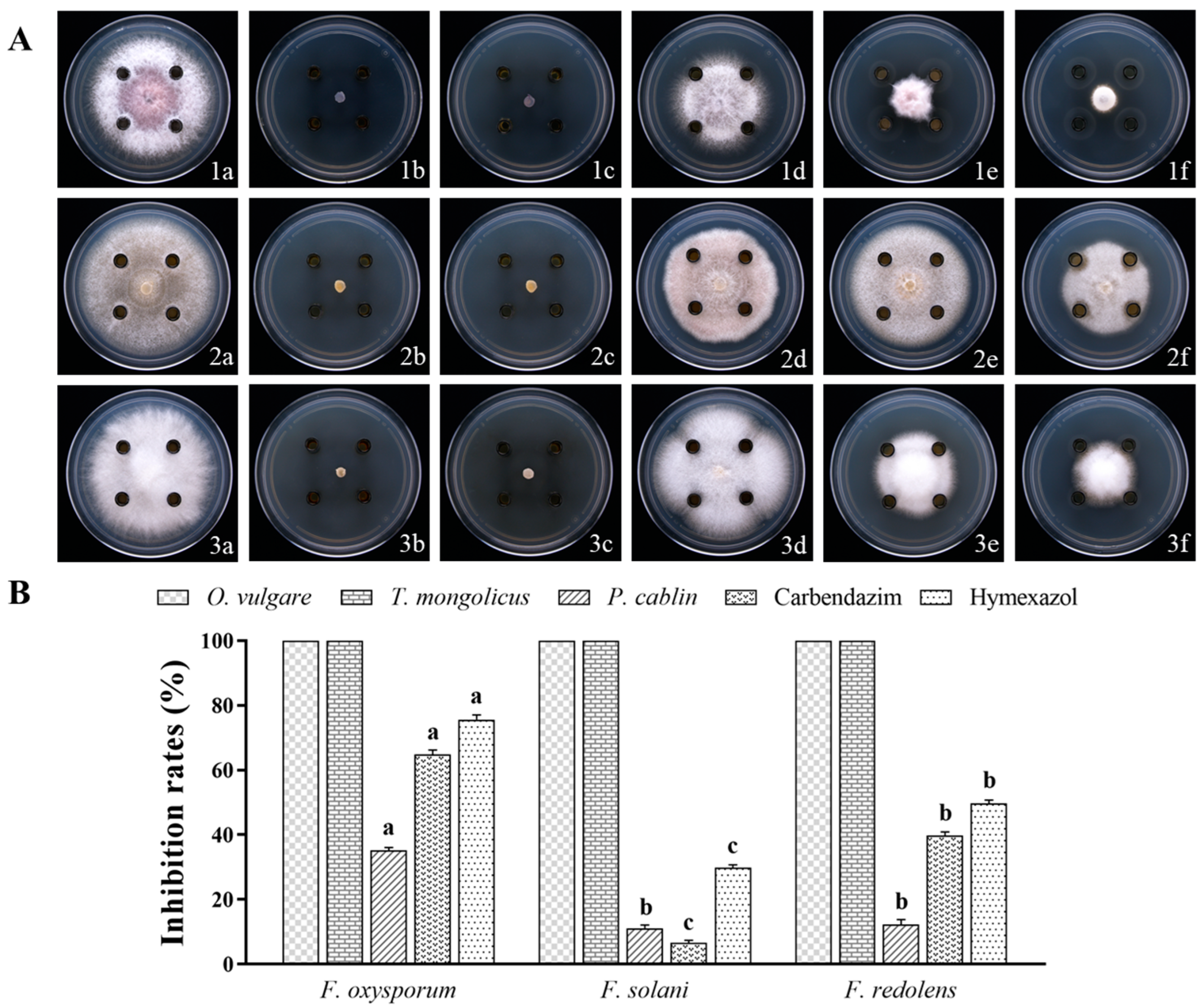
Figure 1. (A) The inhibitory effect of O. vulgare, T. mongolicus, and P. cablin essential oils (EOs) on three pathogenic fungi. The three pathogenic fungi were (1) F. oxysporum, (2) F. solani, and (3) F. redolens. The different treatments were (a) 1-DMSO-T, negative control, (b) O. vulgare, (c) T. mongolicus, (d) P. cablin, (e) carbendazim, positive control, and (f) hymexazol, positive control. (B) The inhibitory rates of O. vulgare, T. mongolicus, and P. cablin EOs in three pathogenic fungi. Different lowercase letters for the same treatment indicate significant differences (p < 0.05) among different fungi.
Horticulturae 09 01136 g001
Figure 2. (A) Antifungal activities of EO volatiles in three pathogenic fungi. The three pathogenic fungi were (1) F. oxysporum, (2) F. solani, and (3) F. redolens. The different treatments were (a) control, (b) O. vulgare, and (c) T. mongolicus. (B) The inhibitory rates of EO volatiles in the three pathogenic fungi. Different lowercase letters for the same treatment indicate significant differences (p < 0.05) among different fungi.
Horticulturae 09 01136 g002
Figure 3. (A) Inhibitory effects of the main components on three pathogenic fungi. The three pathogenic fungi were (1) F. oxysporum, (2) F. solani, and (3) F. redolens. The different treatments were (a) 1-DMSO-T, negative control, (b) carvacrol, (c) thymol, (d) γ-terpinene, (e) o-cymene, (f) carbendazim, positive control, and (g) hymexazol, positive control. (B) The inhibitory rates of the main components on the three pathogenic fungi. Different lowercase letters for the same treatment indicate significant differences (p < 0.05) among different fungi.
Horticulturae 09 01136 g003
Figure 4. Optical microscopy observation of mycelial morphology of F. oxysporum. (A) Control, (B) 1/4 MIC O. vulgare EO treatment, and (C) MIC O. vulgare EO treatment. The red arrows indicate the point of abnormality in the hyphae: (a) hyphae folding and (b) hyphae fracture.
Horticulturae 09 01136 g004
Figure 5. Scanning electron microscopy of mycelial morphology of F. oxysporum. (A) Control, (B) 1/4 MIC O. vulgare EO treatment, and (C) MIC O. vulgare EO treatment. (1) Magnification of 1000× and (2) magnification of 3000×. The red arrows indicate the point of abnormality in the hyphae: (a) hyphae deformation and (b) hyphae rupture.
Horticulturae 09 01136 g005
Figure 6. Ultrastructure of F. oxysporum observed with transmission electron microscope. (AC) represent control group; (DF) represent 1/4 MIC O. vulgare EO treatment group; (GI) represent MIC O. vulgare EO treatment group. CW: cell wall; CM: cell membrane; MIT: mitochondria; V: vacuoles; cy: cytoplasm.
Horticulturae 09 01136 g006
Figure 7. Effects of different concentrations of the O. vulgare EO on the DNA content (A) and the soluble protein content (B) of F. oxysporum. Different lowercase letters indicate statistically significant differences between treatments (p < 0.05).
Horticulturae 09 01136 g007
Figure 8. The effect of the O. vulgare EO on the production of reactive oxygen species. (a) Control and (b) 1/2 MIC O. vulgare EO treatment; (1) bright field, (2) DCFH-DA, and (3) merge.
Horticulturae 09 01136 g008
Figure 9. The effect of exogenous ROS scavengers on the antifungal effect of the O. vulgare EO (A). Inhibition rates for different treatments (B). Intracellular GSH detection (C). Different lowercase letters for the same treatment indicate significant differences across time (p < 0.05). ** represents highly significant differences (p < 0.001).
Horticulturae 09 01136 g009
Table 1. MIC 1 values of the EOs 2 and compounds against fungi (mg/mL).
F. oxysporumF. solaniF. redolens
O. vulgare2.60 ± 0.90 a3.13 ± 0.00 a1.56 ± 0.00 b
T. mongolicus Carvacrol3.13 ± 0.00 a2.60 ± 0.90 a2.60 ± 0.90 a
0.83 ± 0.36 bc1.04 ± 0.36 b0.63 ± 0.00 cd
Thymol1.04 ± 0.36 b2.60 ± 0.90 a1.25 ± 0.00 bc
Carbendazim0.07 ± 0.02 c0.05 ± 0.02 c0.07 ± 0.02 d
1 MIC: minimal inhibitory concentration; 2 EOs: essential oil. Different letters on the same column represent significant differences (p < 0.05).
Disclaimer/Publisher’s Note: The statements, opinions and data contained in all publications are solely those of the individual author(s) and contributor(s) and not of MDPI and/or the editor(s). MDPI and/or the editor(s) disclaim responsibility for any injury to people or property resulting from any ideas, methods, instructions or products referred to in the content.