Article

Acidovorax citrulli Type IV Pili PilR Interacts with PilS and Regulates the Expression of the pilA Gene

1
State Key Laboratory for Biology of Plant Diseases and Insect Pests, Institute of Plant Protection, Chinese Academy of Agricultural Sciences, Beijing 100193, China
2
National Nanfan Research Institute (Sanya), Chinese Academy of Agricultural Sciences, Sanya 572024, China
3
Department of Plant Pathology, College of Plant Protection, Northeast Agricultural University, Harbin 150030, China
4
College of Life Sciences, Jilin Normal University, Siping 136000, China
5
Department of Plant Pathology, Plant Protection College, Shenyang Agricultural University, Shenyang 110866, China
*
Authors to whom correspondence should be addressed.
Horticulturae 2023, 9(12), 1296; https://doi.org/10.3390/horticulturae9121296
Submission received: 25 October 2023 / Revised: 23 November 2023 / Accepted: 29 November 2023 / Published: 30 November 2023
(This article belongs to the Section Genetics, Genomics, Breeding, and Biotechnology (G2B2))

Abstract

:
Acidovorax citrulli can cause bacterial fruit blotch of watermelon, melon, and other cucurbits, and has the potential to cause severe economic losses to growers throughout the world. This article investigated the functions and interactions of the pilR and pilS genes, two important genes in bacterial type IV pili systems, in A. citrulli. For each gene, deletion mutants and complementary strains were constructed via homologous recombination, and their phenotypes were determined. The results showed that the absence of pilR and pilS could significantly reduce the pathogenicity and twitching motility of A. citrulli while increasing the swimming motility, biofilm formation, and in vitro growth. Conversely, complementary strains were no different than the wild-type strain. Using quantitative reverse transcription PCR and promoter activity assays, we confirmed that the deletion of pilR and pilS genes leads to a significant decrease in the transcription level of pilA. Meanwhile, three methods including yeast two-hybrid, glutathione S-transferase pull-down, and luciferase complementation imaging assays were used to verify the direct interaction between the PilR and PilS proteins. These findings revealed the biological function of the pilR and pilS and confirms their regulatory role on pilA.

1. Introduction

The bacterial fruit blotch (BFB) of cucurbits, caused by the pathogen Acidovorax citrulli, is an economically important seed-borne bacterial disease of cucurbit crops such as watermelon (Citrullus lanatus) and melon (Cucumis melo) [1]. BFB was first described in the 1960s [2]. The first major BFB outbreak occurred in the Mariana Islands in 1987 [3]. However, additional outbreaks were soon reported in many other countries, including the United States, China, Israel, and Japan [4,5,6,7,8]. These outbreaks caused economic losses for local farmers.
Type IV pili (T4P) was involved in a variety of plant pathogenic bacterial activities and features, including virulence, colonization, surface adhesion, and biofilm formation [9]. As the gene that encodes for the main structural subunit of T4P, pilA has important biological functions for phytopathogenic bacteria, such as in Ralstonia solanacearum [10], Pseudomonas syringae pv. tabaci [11], Xanthomonas campestris pv. campestris [12], P. syringae pv. tomato [13], and A. citrulli [14,15]. The deletion of the pilA gene results in the loss of or reduction in the ability of pathogenicity, twitching mobility, and biofilm formation.
The two-component system, a response–response regulation mechanism, is widely present in gram-positive bacteria and a small number of gram-negative bacteria [16]. These systems usually contain a conserved histidine kinase and a response regulator. The sensor histidine kinase senses external stimuli and transmits signals to the response regulator via phosphorylation, which then activates downstream effector domains [17]. Two-component systems are known to affect several mechanisms of pathogenicity in phytopathogenic bacteria, including biofilm formation and motility [18,19]. The pilR-pilS (a two-component system) exists in many T4P-expressing bacteria; currently, research on its function is mainly focused on human and animal pathogenic bacteria, for example, Pseudomonas aeruginosa [20,21], Myxococcus xanthus [22,23], and Neisseria gonorrhoeae [24], with little research on plant pathogenic bacteria.
The role of pilR and pilS is currently unknown in A. citrulli. In order to determine the function of pilR and pilS genes of A. citrulli, we constructed deletion mutants using A. citrulli group II strain Aac5 as a model organism. We evaluated the pathogenicity, motility, biofilm formation, and growth rate between the mutants and wild-type (WT) bacteria. At the same time, this study clarified the regulatory relationship between the pilR-pilS system and pilA, using qPCR and pilA promoter activity assay, and elucidated the interactions between the PilR and PilS proteins, using three methods including yeast two-hybrid, glutathione S-transferase (GST) pull-down, and luciferase complementation imaging (LCI) assay.

2. Materials and Methods

2.1. Bacterial Strains, Plasmids, and Growth Conditions

The bacterial strains and plasmids in this study are listed in Table S1. The A. citrulli group II strain Aac5 and its mutant strains and complementary strains were cultured in King’s B (KB) medium at 28 °C for 48 h before use. The yeast strain NMY51 was cultured in yeast extract peptone dextrose (YPD) medium at 30 °C for 3–4 days. Escherichia coli were cultured in Luria–Bertani (LB) medium at 37 °C for 16 h [25]. Antibiotics were added to corresponding media in this study, according to the resistances described in Table S1. The specific antibiotic concentrations are ampicillin (Amp) 100 μg/mL, kanamycin (Kana) 50 μg/mL, chloramphenicol (Cm) 50 μg/mL, and gentamicin (Gm) 50 μg/mL.

2.2. Construction of the pilR and pilS Mutants and their Complementary Strains

The primers pilR-LS/A, pilR-RS/A, pilS-LS/A, pilS-RS/A, and GmS/A were designed using Primer Premier v5.0 (PREMIER Biosoft International, Palo Alto CA, USA) to target 5′ end and 3′ end sequences of the pilR and pilS genes and the gm gene, respectively. The specific primer sequences and characteristics are shown in Table S2. The amplified fragments were recovered using DNA Gel Extraction Kit (Axygen, Suzhou, China) and ligated to pMD19-T vectors (TaKaRa, Dalian, China). After sequencing, 5′ end sequence of the pilR gene, gm gene, and 3′ end sequence of pilR gene were ligated to pK18mobsacB vectors using T4 DNA ligase (TIANGEN, Beijing, China) to construct a suicide plasmid, pK18-pilR-gm. Another suicide plasmid, pK18-pilS-gm, was constructed using the same method. The recombinant plasmids pK18-pilR-gm and pK18-pilS-gm were transferred into the WT Aac5 via triparental hybridization. After homologous recombination exchange, a pure colony was obtained via streak plating on a 10% sucrose KB plate containing Amp and Gm and picking a single colony. This colony was verified for pilR and pilS gene deletion using PCR and sequencing.
The entire gene of pilR and pilS were amplified using KOD high-fidelity enzyme amplification (TOYOBO, Osaka, Japan). The primers were pBBRpilR-S/A and pBBRpilS-S/A, respectively (Table S2). Using ClonExpress® II One Step Cloning Kit (Vazyme Biotech Co., Ltd., Nanjing, China), pilR and pilS were inserted into pBBR1MCS-2 to generate the plasmids pBBRpilR and pBBRpilS, respectively. These plasmids were transferred into ΔpilR and ΔpilS, respectively, by using tripartite mating. PCR and sequencing were then used to confirm that the complementary strains ΔpilRcomp and ΔpilScomp were obtained.

2.3. Pathogenicity Assay

The pathogenicity assays were carried out using all five stains of A. citrulli: the WT strain Aac5, the mutant strains ΔpilR and ΔpilS, and the complementary strains ΔpilRcomp and ΔpilScomp. Each strain was cultured separately in liquid KB media with overnight shaking. The cells were collected via centrifugation, resuspended in sterile water, and diluted to prepare bacterial suspensions with a concentration of 3 × 10 8 CFU/mL. Three-week-old watermelon (Citrullus lanatus cv. “Jingxin#3”) seedlings (Zhongcai Seed Industry Technology Co., Ltd., Beijing, China) were used in the pathogenicity test and were inoculated via spray inoculation. Each treatment used 20 watermelon seedlings, and 200 mL of 3 × 10⁸ CFU/mL bacterial suspension was evenly sprayed on the front and back of watermelon seedling leaves; for the negative control, seedlings were sprayed with sterile water.
After spray inoculation, the watermelon seedlings were covered with a sterile transparent plastic bag and placed in a growth chamber. The growth chamber was set to 12 h light, 25 °C, 12 h dark, 20 °C, and 85% relative humidity. Next day, the plastic bag was removed, and the seedlings were uncovered. After 10 days, the incidence of disease was determined and the disease index (DI) was calculated [26]. In short, the severity was classified at 6 levels, 0, 1, 3, 5, 7, and 9. Level “0” represents no symptoms, while “1” = 25% necrosis of leaves, “3” = 50%, “5” = 75%, “7” = 100%, and “9” = complete death of the seedlings. DI = ∑ (disease scale × number of seedlings in each disease scale) × 100%/∑ (total number of seedlings in each treatment × 9). This experiment was repeated for a total of 3 trials.

2.4. Assays of Twitching and Swimming Motility Assays

A single colony of each of the five experimental strains (Aac5, ΔpilR, ΔpilS, ΔpilRcomp, and ΔpilScomp) was picked and drawn on an antibiotic-amended KB agar plate and incubated at 28 °C for 48 h. The thin, light halo around the single colony was observed [27], using an optical microscope (Olympus IX83, Tokyo, Japan), to assess the twitching mobility of the strains.
Standardized bacterial suspensions were made by diluting suspensions to OD600 = 0.3, using sterilized deionized water. A total of 2 μL of cell suspension was dropped on the center of a 0.3% semi-solid KB medium plate and cultured at 28 °C for 48 h, and the diameter of the halo was measured [28]. For each strain, 9 replicates were assayed. The assay was repeated for a total of 3 trials.

2.5. Biofilm Formation Assay

Biofilm formation assay was performed with a protocol modified from Fei et al. [29], using 24-well microtiter plate. Briefly, the bacterial strains were cultured overnight in antibiotic-amended KB broth and diluted to an OD600 of about 0.3, as described above. Then, 10 μL of a single bacterial suspension was added to a 24-well polypropylene plate containing 1 mL of KB medium in each well. The plates were then incubated at 28 °C for 48 h without agitation. After incubation, bacterial suspensions were discarded, the 24-well plates were gently rinsed with distilled water, and then plates were placed in an oven at 80 °C for 1 h. Each well was stained with 1 mL of 0.1% crystal violet solution for 45 min, then excess stain was discarded and plates were gently triple-rinsed with distilled water. To release the bond crystal violet, 1 mL of 95% ethanol was added to each well and allowed to stand overnight. The OD575 value of each well was measured with a spectrophotometer (Thermo Fisher Scientific Inc., USA). For each strain, 4 replicate wells were used. The entire experiment was repeated for a total of 3 trials.

2.6. In Vitro Growth Assay

The in vitro growth ability of the five experimental strains (Aac5, ΔpilR, ΔpilS, ΔpilRcomp, ΔpilScomp) was measured in KB liquid medium. The bacterial density of each suspension was adjusted to OD600 = 0.3, as described above; the bacterial suspensions were added to fresh KB liquid medium at a v ratio of 1:1000. The bacterially amended KB liquid medium was cultured at 28 °C on a shaker set to 180 rpm. Subsamples were taken every 2 h for 72 h and the OD600 value of each subsample was measured in a Bioscreen C Chamber (FP-1100-C; Oy Growth Curves Ab Ltd., Helsinki, Finland), for a total of 36 subsamples. Each strain was replicated 3 times, and the entire experiment was repeated for a total of 3 trials.

2.7. Quantitative Reverse Transcription PCR (qRT-PCR) Analysis of Gene Expression

Total RNA of the three experimental strains (Aac5, ΔpilR, and ΔpilS) was extracted using Trizol reagent (Invitrogen, Waltham, MA, USA) and reverse transcribed into cDNA using the Fast Quant cDNA First Strand Synthesis Kit (Tiangen, China). The mRNA expression level of the pilA gene of each sample was determined by qPCR using a relative quantitative (2−ΔΔCt) method [30] on an Applied Biosystems 7500 instrument (ABI, Waltham, MA, USA). The primers used (A5pilA-F/R) are shown in Table S2. The mRNA levels were quantified using the SuperReal PreMix Plus (SYBR Green, Tiangen, China) kit. The reaction system and procedures of qPCR are described in the SuperReal PreMix Plus (SYBR Green, Tiangen, China) kit instructions. The gene rpoB was selected as a reference [31]. The experiment was repeated for a total of 3 trials.

2.8. Assay for pilA Promoter Activity

To determine pilA promoter activity, the primer pair GUSpilAF/R (Table S2) was used to amplify the pilA promoter region in A. citrulli Aac5 strain, and pBBRNolacGUS-pilAp was constructed by ligating the pilA promoter region to the pBBRNolacGUS plasmid. The pBBRNolacGUS-pilAp plasmid was validated via sequencing, and then introduced into Aac5, ΔpilR, and ΔpilS strains. Each strain was confirmed to contain the plasmid through PCR validation with the primer 18F/18R (Table S2). These modified strains are referred to as WT-pilAp-GUS, ΔpilR-pilAp-GUS, and ΔpilS-pilAp-GUS, respectively. The pBBRNolacGUS was transferred to the WT strain Aac5, referred to as WT-GUS, which functioned as a negative control. The assay was tested as previously described [32], and this experiment was conducted three times.

2.9. Yeast Two-Hybrid Assay

In order to analyze the interaction relationship between PilS and PilR in yeast, we constructed bait plasmid (pBT3-pilS) and prey plasmid (pPR3-pilR) and transformed them into the TOP10 strains. After confirmation via sequencing (BGI Sequencing, Beijing, China), each plasmid was extracted separately using AxyPrepTM Plasmid Miniprep Kit (Axygen, Suzhou, China). The plasmids pBT3-pilS and pPR3 strains were co-transformed into the prepared yeast competent cells (NMY51) to test for toxicity and self-activation. The pBT3-pilS and pOst1-NubI plasmid were co-transformed into yeast competent cells for functional verification of bait plasmid. Finally, the pBT3-pilS and pPR3-pilR plasmids were co-transformed into NMY51 yeast competent cells. The interaction was determined by looking at whether yeast strains carrying pBT3-pilS and pPR3-pilR plasmids could grow on SD media lacking His.

2.10. Glutathione S-Transferase (GST) Pull-Down Assay

For the GST pull-down assays, 0.5 mg of both GST-tagged PilS fusion protein and His6-tagged PilR fusion protein was combined and incubated on ice for 3 h. Subsequently, the mixture was loaded onto Glutathione Sepharose 4B resin columns (Sangon Biotech, Shanghai, China). After washing five times with wash buffer, proteins were eluted with wash buffer supplemented with 15 mM reduced glutathione. Western blot was used to detect the pull-down effect of GST-tagged PilS fusion protein on His6-tagged PilR fusion protein. Simply put, the 20 μL eluates were separated using SDS-PAGE (4% concentrated glue and 12% separation glue, 80 V 30 min, then 120 V 60 min), transferred to PVDF membranes (Millipore, Billerica, MA, USA), and used anti-His (Sigma-Aldrich, Merck KGaA, Darmstadt, Germany) hybridization. After adding High-sig ECL luminescent liquid (Tanon, Shanghai, China) to the hybrid membranes, the pull-down results were observed by Tanon5200 automatic chemiluminescence image analysis system (Tanon, Shanghai, China). GST and His6 from Wuhan Genecreate (Wuhan, China) were used as negative controls. There were three replications for the pull-down assay.

2.11. Luciferase Complementation Imaging (LCI) Assay

Gene pilS and pilR were inserted into pCAMBIA1300-NLuc (pNL) and pCAMBIA1300-CLuc (pCL) vectors cleaved by restriction enzyme Kpn I and Sal I, respectively, to obtain recombinant plasmids PilS-NLuc and PilR-CLu [33,34]. PilS-NLuc and PilR-CLuc were transformed into Agrobacterium tumefaciens (GV3101), respectively. These two modified Agrobacterium strains were combined at a 1:1 v/v ratio and diluted with deionized water until an OD600 value of 0.5 was obtained. The combined Agrobacterium suspension was then injected into one-month-old tobacco leaves. The bacterial suspensions containing the control vector (NLuc + CLuc, NLuc + PilR − CLuc, and Cluc + PilS − NLuc) and the target vector (PilR-CLuc + PilS − NLuc) were injected into different parts of the same tobacco leaf [35]. This trial was repeated across 3–5 tobacco leaves. After 48 h, tobacco leaves were collected and coated with 1 mM fluorescein solution. Then, the leaves were placed in darkness for 5–7 min and observed using a charge-coupled device imaging apparatus (NightSHADE LB985; Berthold, BadWildbad, Germany). This experiment was repeated for a total of three trials.

2.12. Statistical Analysis

Statistical analysis was performed using SPSS version 22.0 (SPSS, Chicago, IL, USA). Analysis of variance and significant difference of gene expression was performed by independent sample t-test (alpha = 0.05). Drawing was performed using GraphPad Prism 7 (GraphPad, La Jolla, CA, USA).

3. Results

3.1. Successful Construction of Mutants and Complementary Strains

Successful insertion of the plasmids was indicated by antibiotic validation. The mutants ΔpilR and ΔpilS were observed to grow on plates containing Amp and Gm, while the complementary strains ΔpilRcomp and ΔpilScomp were observed to grow on plates containing Amp, Gm, and Kana. Successful insertion of the plasmids was confirmed with PCR amplification and sequencing. Using primer TBpilR-S/A, a 1175 bp amplified fragment was obtained from ΔpilR, while the amplified fragment of wild-type was 2027 bp; using primer TbpilS-S/A, the amplified fragment of ΔpilS was 1743 bp, while the amplified fragment of wild-type was 2379 bp (Figure S1). At the same time, the amplified fragments were recovered and sequenced. The completely correct sequencing results confirmed that ΔpilR and ΔpilS mutants were successfully constructed.

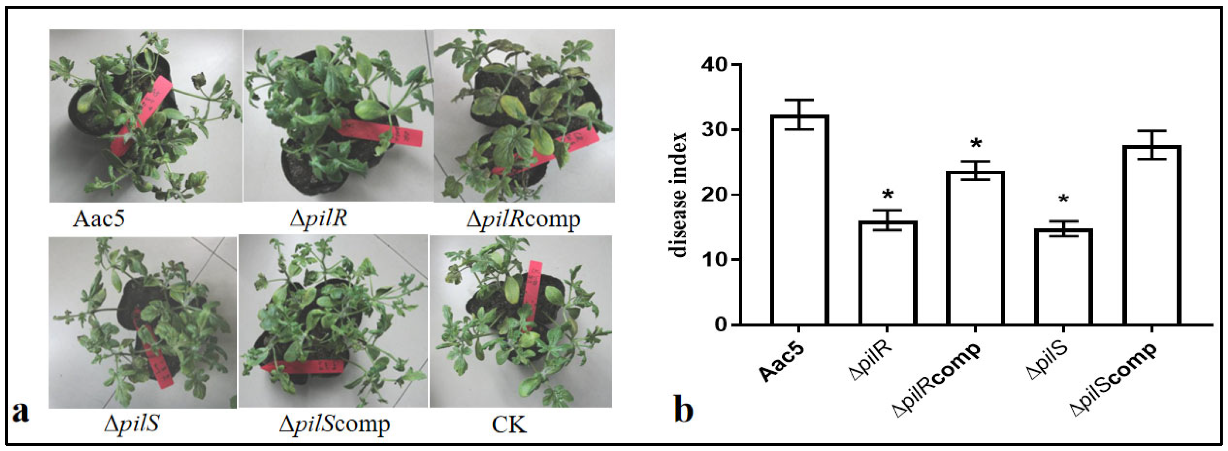
3.2. pilR and pilS Genes Are Required for Virulence in Watermelon

Ten days after inoculation (DAI), typical symptoms of BFB appeared in watermelon seedlings treated with different treatments (Aac5, ΔpilR, ΔpilS, ΔpilRcomp, and ΔpilScomp). The symptoms of watermelon seedlings inoculated with WT strain Aac5 or either of the two complementary strains were more severe than plants inoculated with one of the two mutant strains. No disease symptoms were present on plants inoculated with no-bacteria negative control plants (Figure 1a). The disease index of WT 10 DAI was 32.33; the disease index of ΔpilR and ΔpilS was 16.12 and 14.80, respectively; the disease index of complementary strain ΔpilRcomp and ΔpilScomp was 23.76 and 27.67, respectively (Figure 1b). The WT Aac5 had significantly greater pathogenicity compared to either the ΔpilR and ΔpilS strains (p < 0.05). The pathogenicity of two complementary strains recovered to some extent, and there was no significant difference between ΔpilScomp and WT. Although the halo diameter of ΔpilRcomp was much higher than that of the mutant strain ΔpilR, it was still significantly lower than that of WT.

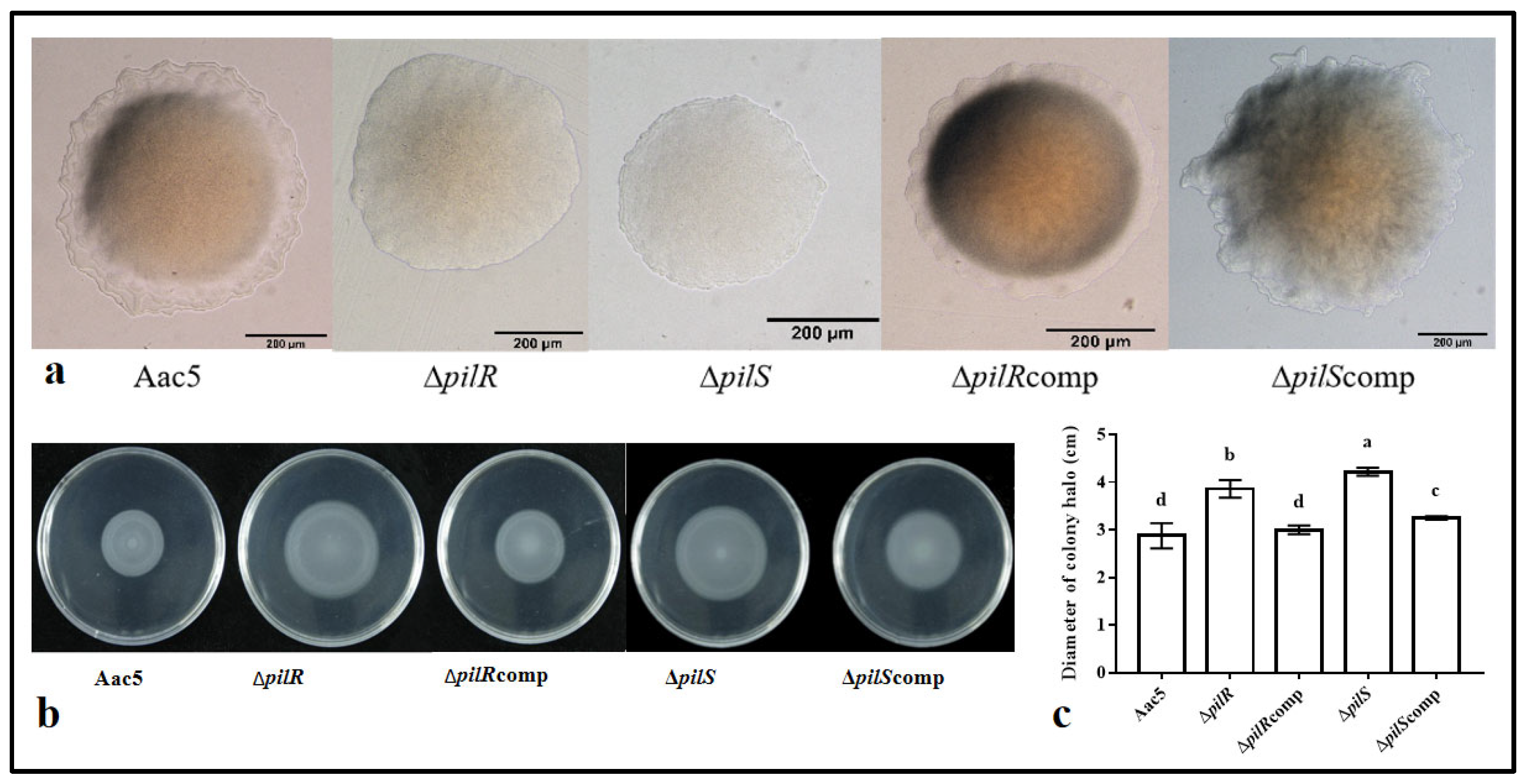
3.3. The Effects of the Deletion of pilR and pilS on the Motility of A. citrulli

The absence of pilR and pilS leads to the loss of the twitching motility of A. citrulli. No twitching-typical halos were observed around the ΔpilR and ΔpilS strain colonies. For the wild-type, ΔpilScomp, and ΔpilRcomp strains, a twitching-typical halo was observed (Figure 2a).
The absence of pilR and pilS leads to an increase in the swimming motility of A. citrulli. The average diameter of the swimming halo of the WT strain Aac5 was 2.7 cm, while the ΔpilR and ΔpilS strains had halo diameters of 3.86 cm and 4.22 cm, respectively, greater than wild-type (p < 0.05). The motility of the complementary strains ΔpilRcomp and ΔpilScomp was intermediate, with mean halo diameters of 3.0 cm and 3.25 cm, respectively (Figure 2b,c). There was no significant difference between ΔpilRcomp and WT. Although the halo diameter of ΔpilScomp was much lower than that of the mutant strain ΔpilS, it was still significantly higher than that of WT.

3.4. Deletion of pilR and pilS Enhanced the Biofilm Formation of A. citrulli

Compared with the WT strain Aac5, the mutant strains ΔpilR and ΔpilS had significantly greater biofilm formation ability (Figure 3a). The OD575 absorbance values were 0.43, 1.45, and 1.38 for the three strains, respectively. Compared to their respective wild-type strains, the biofilm formation ability of ΔpilRcomp and ΔpilScomp significantly decreased. The biofilm formation ability of ΔpilScomp was not significantly different from that of the WT, with absorbance values of OD575 0.44. However, the biofilm formation ability of ΔpilRcomp was significantly higher than that of the WT, with absorbance values of OD575 0.57 (p < 0.05) (Figure 3b).

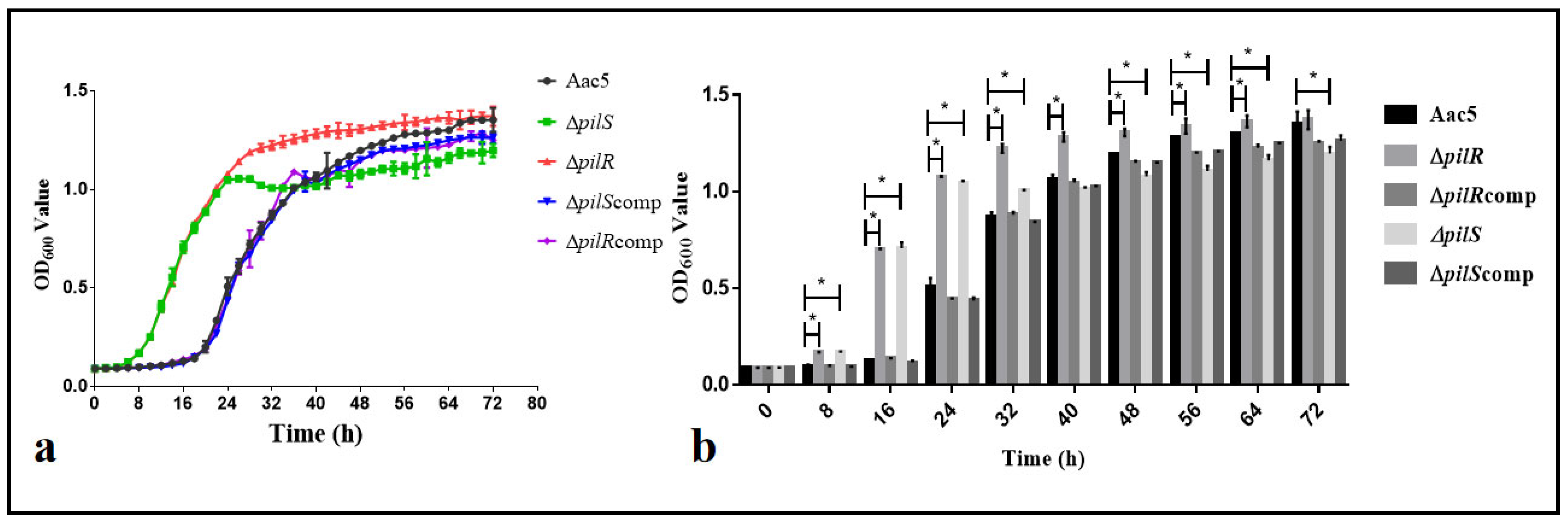
3.5. The Deletion of the pilR or pilS Genes Enhanced the Early Growth Ability of A. citrulli In Vitro

From the growth curve, it can be seen that the ΔpilR and ΔpilS reached the logarithmic and stable stages earlier than the WT and the two complementary strains, and the two complementary strains and WT showed the same curve pattern (Figure 4a). The OD600 values of ΔpilS were significantly higher than that of the WT at each measurement time from 8 to 32 h. After 40 h, the OD600 value of the ΔpilS strain was not significantly different from the WT. For each measurement time from 48 to 72 h, the OD600 value of the ΔpilS strain was significantly less than that of the WT. For each measurement time from 8 to 64 h, the OD600 value of ΔpilR was significantly greater than that of the WT and entered a stable period earlier than the WT. The OD600 values of the two complementary strains were not significantly different from the WT at any measurement time from 8 to 40 h, though they were significantly lower than the WT from 48 to 72 h (p < 0.05) (Figure 4b).

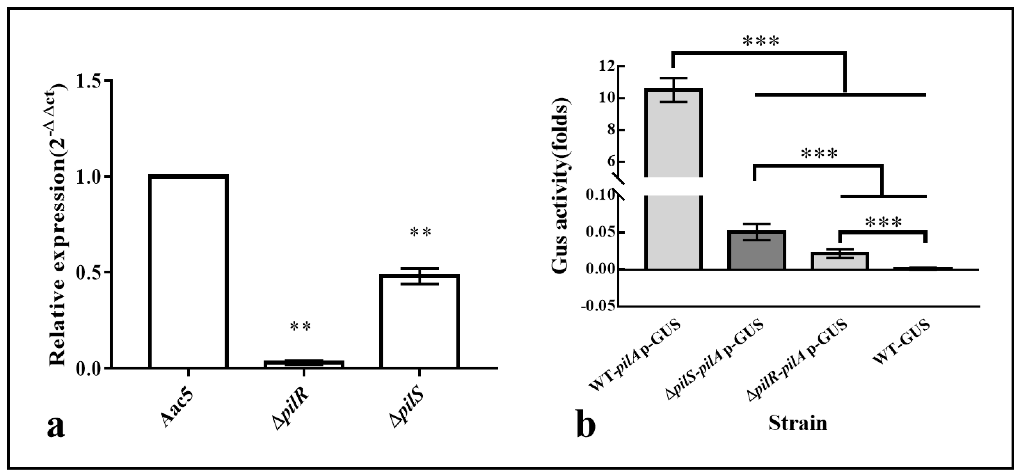
3.6. The Deletion of pilR and pilS Affects the Transcription of pilA

The pilA gene encodes the main structural subunit of T4P and plays an important role in the virulence, twitching motility, and biofilm formation of plant pathogenic bacteria. The qPCR assay showed that pilA had significantly lower transcription levels in pilS and pilR mutants as compared to the WT (p < 0.01). The pilR deletion mutant had the lowest levels of transcription, with nearly zero transcription (Figure 5a). Promoter activity of pilA in the pilR and pilS deletion mutants was measured (Figure 5b). Both pilR and pilS deletion mutants had significantly less pilA promoter activity compared to the WT (p < 0.001).

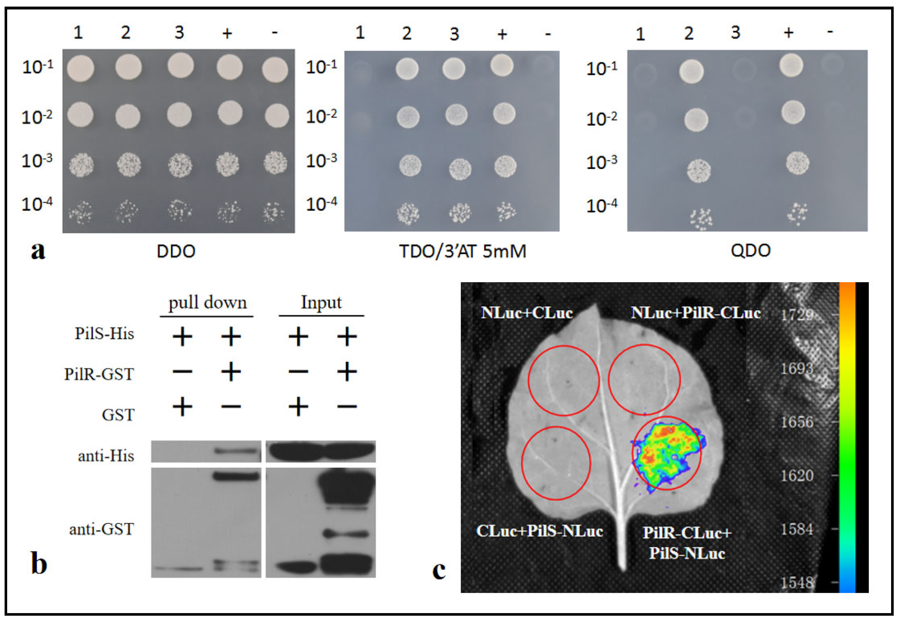
3.7. PilR Interacts with PilS

The ability of the bait protein to self-activate was observed as the yeast diploid containing pilS-pBT3-N and pPR3-N was able to grow on double drop-out (DDO) plate. The activation of the HIS3 reporter gene by a bait protein was inhibited by adding 5 mM 3′AT. The yeast diploid containing pilS-pBT3-N and pilR-pPR3-N was observed to grow on TDO/5 mM 3′AT medium, but did not grow on quadruple drop-out (QDO) plate lacking the amino acids Trp, Leu, His, and Ade (Figure 6a).
The results showed that SUMO-His6-PilS was detected in the GST pull-down assay when assayed in combination with GST-PilR, but was not detected when only the GST tag was assayed (Figure 6b, Figures S2 and S3).
For the luciferase complementation imaging (LCI) assay, chemiluminescence signals were observed in the N. benthamiana leaf area when Agrobacterium co-expressing PilS-Nluc and PilR-Cluc were infiltrated. When negative control Agrobacterium was infiltrated, no chemiluminescence was observed (Figure 6c).

4. Discussion

Although there have been reports of pilA gene transcription being regulated by pilR-pilS (a two-component system) in many T4P expressing bacteria, experimental results vary among different pathogenic bacteria. In Pseudomonas aeruginosa, the deletion of the pilR gene resulted in a significant decrease in the transcriptional level of pilA1, while the deletion of the pilS gene resulted in a decrease in the pilA1 transcription level [20,21]. In Myxococcus xanthus, the expression of pilA may be downregulated by interacting directly with pilS [22,23]. However, the inactivation of the pilRpilS hybrid (Rsp) in Neisseria gonorrhoeae did not affect the formation of pili, suggesting that the pilRpilS system has lost its regulatory function in some taxa. This indicates that the function of pilRpilS has changed during the evolution of bacteria. Among plant pathogenic bacteria, There are few reports on the transcriptional regulation of pilA gene by pilRpilS. In Xylella fastidiosa, the loss of the pilR gene leads to the loss of T4P biosynthetic capacity and twitching mobility [36]; however, the regulatory relationship between pilR and pilA has not been verified. In Xanthomonas oryzae pv. oryzae, the RpoN2-PilRX regulatory system governed T4P gene transcription and influenced bacterial motility and virulence in rice [37].
The studies of Yang et al. [38] have shown that the deletion of the pilA gene leads to a decrease in the pathogenicity of group I strain pslb65 and group II strain Aac5 of A. citrulli, as well as a decrease in twitching motility. In this study, the deletion of pilR and pilS resulted in the significant down-regulation of pilA expression (Figure 5) and confirmed that the pilR and pilS genes directly regulate the expression of the pilA promoter. This suggests that the decreased pathogenicity and loss of twitching motility of the pilR and pilS deletion mutants may be due to lower pilA expression.
σ54 (RpoN) is a global regulatory factor that can regulate multiple biological functions in bacteria [39]. In Xanthomonas oryzae pv. oryzae, the RpoN2-PilRX regulatory system governs type IV pilus (T4P) gene transcription, and affects bacterial motility and virulence [37]. Through sequence alignment, it was found that there are two σ54 (RpoN1 and RpoN2) in the genome of A. citrulli. We will further verify the regulatory relationship between σ54, pilR/pilS, and T4P. The research results will be beneficial for revealing how pilR and pilS genes affect the twitching motility, biofilm formation, and pathogenicity of A. citrulli.
The pilR and pilS genes of A. citrulli have typical domains of two-component system (Figure S4). The deletion mutant of these two genes, ΔpilR and ΔpilS, exhibit similar phenotypes in terms of pathogenicity (Figure 1), motility (Figure 2), biofilm formation ability (Figure 3), and so on. This study successfully validated the direct interaction between PilR and PilS using three methods (GST pull down, Y2H, and LCI assays) (Figure 6). However, the phosphorylation test of PilR and PilS was not successful, and we will continue to conduct the experiment to confirm that pilR/pilS is a response–response regulation mechanism.
The formation of biofilms can enable bacteria to maintain metabolic regulation and reduce the consumption of nutrients and water in adverse environmental conditions, thereby facilitating their survival. The formation of biofilms is usually related to the ability of flagella or pili to adhere to solid surfaces [40]. This study indicated that the biofilm formation ability of group II strain Aac5 is very low compared to group I strain pslb65, which was consistent with previous research results [26,29,38]. This study also indicates that the pilR and pilS deletion mutants had a significant increase in biofilm formation ability. This is in contrast to previous work, which indicates that in many plant pathogenic bacteria, the deletion of T4P genes reduces biofilm formation ability, such as in Pseudomonas syringae pv tabaci 6605 [11] and R. solanacearum [10]. This decrease in biofilm formation after the deletion of a T4P gene was even observed in A. citrulli group I strain M6 [15,27]. These results are seemingly in conflict with the results from the present work with group II strain Aac5. The specific reasons for this phenomenon are currently unclear. In this study, we found that the growth logarithmic phase of pilR and pilS mutants was advanced by about 12 h, and the significant enhancement of early growth ability may be one of the reasons for the enhanced biofilm formation ability.

5. Conclusions

A. citrulli can cause the BFB of watermelon, melon, and other cucurbits, which has caused economic losses to growers around the world. This article examined the functions and interactions of the pilR and pilS genes in A. citrulli. The results indicated that within A. citrulli, the absence of pilR and pilS, deletion mutants, led to significantly less pathogenicity and twitching motility while increasing swimming motility, biofilm formation, and in vitro growth. However, complementary strains were not significantly different than the WT across any measured variable. In addition, the deletion of pilR and pilS genes led to a significant decrease in the transcription levels of pilA. Three methods, including yeast two-hybrid, GST-pull down, and LCI assay were used to verify the direct interaction between the PilR and PilS proteins. These findings revealed the biological function of pilR and pilS, and confirms their regulatory role on pilA. Due to the important biological and pathogenic functions of the pilA gene, future work will continue to explore upstream regulatory mechanisms such as the regulation of T4P by δ54 factor, and will improve our knowledge of the regulatory pathways involved in this system.

Supplementary Materials

The following supporting information can be downloaded at: https://www.mdpi.com/article/10.3390/horticulturae9121296/s1, Table S1: Bacterial strains and plasmids used in this study; Table S2: Sequences and related information of primers used for construction of mutant and complemented strains, qPCR and Yeast two-hybrid; Figure S1: The pilR, pilS gene mutant and complementary strains of group II strain Aac5 were successfully obtained according to PCR verification; Figure S2 Protein validation before GST pull-down; Figure S3 Antibody detection after GST pull-down; Figure S4 The structural domains of PilR and PilS.

Author Contributions

Y.Y. designed the study. Y.Y., W.J., N.F. and P.Q. performed the experiments. L.Y. and W.G. performed data analyses. Y.Y. and W.J. wrote the manuscript. T.Z. critically reviewed the manuscript. All authors have read and agreed to the published version of the manuscript.

Funding

This study was supported by the Hainan Province Science and Technology Special Fund (ZDYF2023XDNY084); Major Science and Technology Special Project of Xinjiang Uygur Autonomous Region “Research and Demonstration of Key Technologies for Green Prevention and Control of Major Harmful Organisms in Xinjiang’s Advanced and Characteristic Crops”; the China Earmarked Fund for Modern Agro-industry Technology Research System (CARS-25); and the Agricultural Science and Technology Innovation Program of the Chinese Academy of Agricultural Sciences (CAAS-ASTIP).

Institutional Review Board Statement

Not applicable.

Informed Consent Statement

Not applicable.

Data Availability Statement

The data presented in this study are available on request from the corresponding author.

Conflicts of Interest

The authors declare no conflict of interest.

References

  1. Schaad, N.W.; Postnikova, E.; Sechler, A.; Claflin, L.E.; Vidaver, A.K.; Jones, J.B.; Agarkova, I.; Ignatov, A.; Dickstein, E.; Ramundo, B.A. Reclassification of subspecies of Acidovorax avenae as A. avenae (Manns 1905) emend., A. cattleyae (Pavarino, 1911) comb. nov., A. citrulli (Schaad et al., 1978) comb. nov., and proposal of A. oryzae sp. nov. Syst. Appl. Microbiol. 2008, 31, 434–446. [Google Scholar] [CrossRef] [PubMed]
  2. Webb, R.E.; Goth, R.W. A seedborne bacterium isolated from watermelon. Plant Dis. Rep. 1965, 49, 818–821. [Google Scholar]
  3. Wall, G.C.; Santos, V.M. A new bacterial disease on watermelon in the Mariana Islands. Phytopathology 1988, 78, 1605. [Google Scholar]
  4. Evans, T.A.; Mulrooney, R.P. First report of watermelon fruit blotch in Delaware. Plant Dis. 1991, 75, 1074. [Google Scholar] [CrossRef]
  5. Black, M.C.; Isakeit, T.; Barnes, L.W. First report of bacterial fruit blotch of watermelon in Texas. Plant Dis. 1994, 78, 831. [Google Scholar] [CrossRef]
  6. O’Brien, R.G.; Martin, H.L. Bacterial blotch of melons caused by strains of Acidovorax avenae subsp. citrulli. Aust. J. Exp. Agric. 1999, 39, 479–485. [Google Scholar] [CrossRef]
  7. Burdman, S.; Kots, N.; Kritzman, G.; Kopelowitz, J. Molecular, physiological, and host-range characterization of Acidovorax avenae subsp. citrulli isolates from watermelon and melon in Israel. Plant Dis. 2005, 89, 1339–1347. [Google Scholar] [CrossRef]
  8. Ren, Y.Z.; Li, H.; Li, G.Y.; Wang, Q.Y.; Li, J.Q. First report of Acidovorax avenae subsp. citrulli infecting edible seed watermelon (Citrullus lanatus var. lanatus) in China. Plant Dis. 2006, 90, 1112. [Google Scholar] [CrossRef]
  9. Burdman, S.; Bahar, O.; Parker, J.K.; Fuente, L.D.L. Involvement of Type IV Pili in Pathogenicity of Plant Pathogenic Bacteria. Genes 2011, 2, 706–735. [Google Scholar] [CrossRef]
  10. Kang, Y.W.; Liu, H.L.; Genin, S.; Schell, M.A.; Denny, T.P. Ralstonia solanacearum requires type 4 pili to adhere to multiple surfaces and for natural transformation and virulence. Mol. Microbiol. 2002, 46, 427–437. [Google Scholar] [CrossRef]
  11. Taguchi, F.; Ichinose, Y. Role of Type IV Pili in Virulence of Pseudomonas syringae pv. tabaci 6605: Correlation of Motility, Multidrug Resistance, and HR-Inducing Activity on a Nonhost Plant. Mol. Plant Microbe Interact. 2011, 24, 1001–1011. [Google Scholar] [CrossRef] [PubMed]
  12. McCarthy, Y.; Ryan, R.P.; O’Donovan, K.; He, Y.Q.; Jiang, B.L.; Feng, J.X.; Tang, J.L.; Dow, J.M. The role of PilZ domain proteins in the virulence of Xanthomonas campestris pv. campestris. Mol. Plant Pathol. 2008, 9, 819–824. [Google Scholar] [CrossRef] [PubMed]
  13. Roine, E.; Raineri, D.M.; Romantschuk, M.; Wilson, M.; Nunn, D.N. Characterization of type IV pilus genes in Pseudomonas syringae pv. tomato DC3000. Mol. Plant Microbe Interact. 1998, 11, 1048–1056. [Google Scholar] [CrossRef] [PubMed]
  14. Bahar, O.; De, L.; Fuente, L.; Burdman, S. Assessing adhesion, biofilm formation and motility of Acidovorax citrulli using microfluidic flow chambers. FEMS Microbiol. Lett. 2010, 312, 33–39. [Google Scholar] [CrossRef]
  15. Tally, R.; Bolaji, B.S.; Saul, B. Association Between Loss of Type IV Pilus Synthesis Ability and Phenotypic Variation in the Cucurbit Pathogenic Bacterium Acidovorax citrulli. Mol. Plant Microbe Interact. 2018, 31, 548–559. [Google Scholar]
  16. Williams, R.H.; Whitworth, D.E. The genetic organisation of prokaryotic two-component system signalling pathways. BMC Genom. 2010, 11, 720. [Google Scholar] [CrossRef] [PubMed]
  17. Stock, A.M.; Robinson, V.L.; Goudreau, P.N. Two-component signal transduction. Annu. Rev. Biochem. 2000, 69, 183–215. [Google Scholar] [CrossRef] [PubMed]
  18. Petrova, Q.E.; Sauer, K. A novel signaling network essential for regulting Pseudomonas aeruginosa biofilm development. PLoS Pathog. 2009, 91, e1000668. [Google Scholar]
  19. Cha, J.Y.; Lee, D.G.; Lee, J.S.; Oh, J.I.; Baik, H.S. GacA directly regulates experssion of several of virulence genes in Pseudomonas syringae pv. tabaci 11528. Biochem. Biophys. Res. Commun. 2012, 417, 665–672. [Google Scholar] [CrossRef]
  20. Hobbs, M.; Collie, E.S.; Free, P.D.; Livingston, S.P.; Mattick, J.S. PilS and PilR, a two-component transcriptional regulatory system controlling expression of type 4 fimbriae in Pseudomonas aeruginosa. Mol. Microbiol. 1993, 7, 669–682. [Google Scholar] [CrossRef]
  21. Boyd, J.M.; Lory, S. Dual function of pilS during transcriptional activation of the Pseudomonas aeruginosa pilin subunit gene. J. Bacteriol. 1996, 178, 831–839. [Google Scholar] [CrossRef] [PubMed]
  22. Wu, S.S.; Kaiser, D. Regulation of expression of the pilA gene in Myxococcus xanthus. J. Bacteriol. 1997, 179, 7748–7758. [Google Scholar] [CrossRef] [PubMed]
  23. Bertrand, J.J.; West, T.; Engel, J.N. Genetic analysis of the regulation of type IV pilus function by the Chp chemosensory system of Pseudomonas aeruginosa. J. Bacteriol. 2010, 192, 994–1010. [Google Scholar] [CrossRef] [PubMed]
  24. Carmen, L.; Giltner, Y.N.; Lori, L.B. Type IV Pilin Proteins: Versatile Molecular Modules. Microbiol. Mol. Biol. Rev. 2012, 76, 740. [Google Scholar] [CrossRef]
  25. Sambrook, J.; Russell, D.W. Molecular Cloning: A Laboratory Manual, 3rd ed.; Cold Spring Harbor Laboratory Press: New York, NY, USA, 2001. [Google Scholar]
  26. Ji, W.; Zhao, M.; Fei, N.; Yang, L.; Qiao, P.; Walcott, R.; Yang, Y.; Zhao, T. Essential Acidovorax citrulli Virulence Gene hrpE Activates Host Immune Response against Pathogen. Int. J. Mol. Sci. 2022, 23, 9144. [Google Scholar] [CrossRef] [PubMed]
  27. Bahar, O.; Goffer, T.; Burdman, S. Type IV Pili are required for virulence, twitching motility, and biofilm formation of Acidovorax avenae subsp. citrulli. Mol. Plant Microbe Interact. 2009, 22, 909–920. [Google Scholar] [CrossRef] [PubMed]
  28. Liu, D.; Zhao, M.; Qiao, P.; Li, Z.; Chen, G.; Guan, W.; Bai, Q.; Walcott, R.; Yang, Y.; Zhao, T. ntrC Contributes to Nitrogen Utilization, Stress Tolerance, and Virulence in Acidovorax citrulli. Microorganisms 2023, 11, 767. [Google Scholar] [CrossRef]
  29. Fei, N.; Ji, W.; Yang, L.; Yu, C.; Qiao, P.; Yan, J.; Guan, W.; Yang, Y.; Zhao, T. Hcp of the Type VI Secretion System (T6SS) in Acidovorax citrulli Group II Strain Aac5 Has a Dual Role as a Core Structural Protein and an Effector Protein in Colonization, Growth Ability, Competition, Biofilm Formation, and Ferric Iron Absorption. Int. J. Mol. Sci. 2022, 23, 9632. [Google Scholar] [CrossRef]
  30. Livak, K.J.; Schmittgen, T.D. Analysis of relative gene expression data using real-time quantitative PCR and the (2−ΔΔCT) Method. Methods 2001, 25, 402–408. [Google Scholar] [CrossRef]
  31. Yang, Y.; Qiao, P.; Wang, T.; Ji, W.; Fei, N.; Zhang, L.; Guan, W.; Zhao, T. Further Characterization of Host Preference of Acidovorax citrulli Based on Growth Competition between Group I and Group II Strains. Horticulturae 2022, 8, 1173. [Google Scholar] [CrossRef]
  32. Zhang, Y.B.; Wei, C.; Jiang, W.D.; Wang, L.; Li, C.R.; Wang, Y.Y. The HD-GYP domain protein RpfG of Xanthomonas oryzae pv. oryzicola regulates synthesis of extracellular polysaccharides that contribute to biofilm formation and virulence on rice. PLoS ONE 2013, 8, e59428. [Google Scholar] [CrossRef] [PubMed]
  33. Chen, H.M.; Zou, Y.; Shang, Y.L.; Lin, H.Q.; Wang, Y.J.; Cai, R.; Tang, X.Y.; Zhou, J.M. Firefly luciferase complementation imaging assay for protein-protein interactions in plants. Plant Physiol. 2008, 146, 368–376. [Google Scholar] [CrossRef] [PubMed]
  34. Zhou, Z.Y.; Bi, G.Z.; Zhou, J.M. Luciferase complementation assay for protein-protein interactions in plants. Curr. Protoc. Plant Biol. 2018, 3, 42–50. [Google Scholar] [CrossRef] [PubMed]
  35. Jiang, J.; Zhao, M.; Zhang, X.; Yang, L.; Fei, N.; Ji, W.; Guan, W.; Walcott, R.; Yang, Y.; Zhao, T. Acidovorax citrulli Effector AopV Suppresses Plant Immunity and Interacts with Aromatic Dehydratase ADT6 in Watermelon. Int. J. Mol. Sci. 2022, 23, 11719. [Google Scholar] [CrossRef] [PubMed]
  36. Li, Y.X.; Hao, G.X.; Galvani, C.D.; Meng, Y.Z.; Dela, F.L.; Hoch, H.C.; Burr, T.J. Type I and type IV pili of Xylella fastidiosa affect twitching motility, biofilm formation and cell-cell aggregation. Microbiology 2007, 153, 719–726. [Google Scholar] [CrossRef]
  37. Yu, C.; Nguyen, D.; Ren, Z.; Liu, J.; Yang, F.; Tian, F.; Fan, S.; Chen, H. The RpoN2-PilRX regulatory system governs type IV pilus gene transcription and is required for bacterial motility and virulence in Xanthomonas oryzae pv. oryzae. Mol. Plant Pathol. 2020, 21, 652–666. [Google Scholar] [CrossRef]
  38. Yang, Y.; Fei, N.; Ji, W.; Qiao, P.; Yang, L.; Liu, D.; Guan, W.; Zhao, T. pilA Gene Contributes to Virulence, Motility, Biofilm Formation, and Interspecific Competition of Bacteria in Acidovorax citrulli. Microorganisms 2023, 11, 1806. [Google Scholar] [CrossRef]
  39. Yang, Y.; Darbari, V.C.; Zhang, N.; Lu, D.; Glyde, R.; Wang, Y.P.; Winkelman, J.T.; Gourse, R.L.; Murakami, K.S.; Buck, M.; et al. Structures of the RNA polymerase-σ54 reveal new and conserved regulatory strategies. Science 2015, 349, 882–885. [Google Scholar] [CrossRef]
  40. Danhorn, T.; Fuqua, C. Biofilm Formation by Plant-Associated Bacteria. Annu. Rev. Microbiol. 2007, 61, 401–422. [Google Scholar] [CrossRef]
Figure 1. Pathogenicity assay of Acidovorax citrulli wild-type strain Aac5, as well as ∆pilR, ∆pilS, and ∆pilRcomp, ∆pilScomp, and a sterile water control (CK) on watermelon seedlings. (a) Symptoms of watermelon seedlings 10 days after inoculation (DAI). Plants were spray inoculated with a single strain with bacterial suspensions of 108 CFU/mL. (b) The disease index of the five treated watermelon seedlings at 10 DAI. Error bars represent standard error of the means of three replicated experiments. Asterisks (*) above a bar indicate strains with significant differences p < 0.05 (Student’s t-test).
Horticulturae 09 01296 g001
Figure 2. Evaluation of swimming and twitching motility of Acidovorax citrulli. (a) The twitching mobility of the A. citrulli wild-type Aac5, pilR and pilS mutants and complementary strains. The strains were cultured on KB medium plate at 28 °C for 48 h, outer halos around the strains were observed using an optical microscope, and the twitching motility were assessed. (b) The swimming mobility of the A. citrulli wild-type Aac5 strain, pilR and pilS mutants, and complementary strains. The wild type strain Aac5, ΔpilR and ΔpilS, and ΔpilRcomp and ΔpilScomp strains were cultured on a 0.3% semi-solid KB medium plate at 28 °C for 36 h, and the swimming halo diameter of each strain was measured. (c) The swimming motility halo average diameter of the five strains. The different letters on the bar chart indicate significant differences in swimming motility between different treated calculated by ANOVA (Duncan, p = 0.05).
Horticulturae 09 01296 g002
Figure 3. Evaluation of biofilm formation of Acidovorax citrulli Aac5, the pilR and pilS mutants, and complementary strains. Bacterial suspensions with OD600 = 0.3 were added to KB medium in a 24-well plate at a v/v ratio of 1:100 and incubated at 28 °C for 48 h. Crystal violet solution (0.1%) was used to stain biofilms for 45 min. The OD575 value was measured after dissolving with 95% alcohol. (a) Typical biofilms on the inner wall of the culture wells. (b) The absorbance value of each treatment at OD575. Error bars represent standard error of the means of three replicated experiments. Treatments with the same letter are not significantly different from each other as calculated by an ANOVA test with an alpha level of 0.05.
Horticulturae 09 01296 g003
Figure 4. Evaluation of the growth of Acidovorax citrulli Aac5 wild-type, the pilR and pilS mutants and complementary strains in KB broth. (a) Growth curve of each strain; (b) Statistical analysis of OD600 values of the 5 strains at different time points. Error bars represent standard error of the means of three replicated experiments. Treatments with an asterisk (*) are significantly different from each other as calculated by an ANOVA test with an alpha level of 0.05.
Horticulturae 09 01296 g004
Figure 5. Evaluation of the relative expression of genes in Acidovorax citrulli Aac5 wild-type, and the pilR and pilS deletion mutants. (a) Relative expression of the pilA gene among the wild-type and deletion mutant strains. Relative expression levels were assessed via qRT PCR, and each deletion mutant was statistically compared to the control wild-type strain. Bars represent mean values calculated from three independent replicates. Error bars represent standard deviation. (b) Relative activity of pilA promotor β-Glucuronidase (GUS) in the wild-type strain carrying pBBR-GUS-pilAp (WT-pilAp-GUS), the pilR and pilS deletion mutants carrying pBBR-GUS-pilAp (ΔpilR-pilAp-GUS, ΔpilS-pilAp-GUS), and the wild-type strain carrying pBBRNolacGUS (WT-GUS), which was used as a negative control. The error bar represents the standard deviation. Two asterisks (**) above a bar indicate strains with significant differences p < 0.01 (Student’s t-test); *** on top of the bar indicates strains with significant differences in the GUS (ANOVA, p < 0.001).
Horticulturae 09 01296 g005
Figure 6. Several assays evaluating the interactions of PilR and PilS proteins. (a) Yeast two-hybrid assay. 1. co-expresstion of pilS-pBT3-N and pPR3-N (self-activation verification); 2. co-expression of pilS-pBT3-N and pOst1-NubI (functional verification); 3. co-expression of pilS-pBT3-N and pilR-pPR3-N (experimental group); +. co-expressed with pTSU2-APP and NubG-Fe65 (positive control group); −. co-expression of pTSU2-APP and PPR3-N (negative control group). On double drop-out (DDO) plates, co-expression of pilS-pBT3-N and pPR3-N can grow, indicating the self-activation phenomenon of PilS; on triple drop-out (TDO) plates containing a concentration of 5 mM of 3AT, co-expression of pilS-pBT3-N and pPR3-N cannot grow, indicating that 5 mM of 3AT can effectively inhibit self activation without affecting its function (co-expression of pilS-pBT3-N and pOst1-NubI can grow), and co-expression of pilS-pBT3-N and pilR-pPR3-N can grow normally, indicating a direct interaction between PilS and PilR; co-expression of pilS-pBT3-N and pilR-pPR3-N cannot grow normally on quadruple drop-out (QDO) plates. (b) Glutathione-S-transferase (GST) pull-down assay. PilR-GST was incubated with equal amounts of purified PilS-His and precipitated by glutathione agarose. The presence of PilS-His in glutathione agarose-bound protein was detected via anti-His immunoblot. The interactions of GST and PilS-His were evaluated as negative controls. The experiment was independently repeated three times. (c) Luciferase complementation imaging (LCI) assay. The GV3101 Agrobacterium carrying PilS-Nluc or PilR-Cluc were co-injected into N. benthamiana leaves. The leaves were collected after 48 h and observed under a live imaging device (NightSHADE LB985; Berthold).
Horticulturae 09 01296 g006
Disclaimer/Publisher’s Note: The statements, opinions and data contained in all publications are solely those of the individual author(s) and contributor(s) and not of MDPI and/or the editor(s). MDPI and/or the editor(s) disclaim responsibility for any injury to people or property resulting from any ideas, methods, instructions or products referred to in the content.

