Article

Influence of Different Types of Carbon Sources on Glucosinolate and Phenolic Compounds in Radish Sprouts

1
Department of Biological Sciences, Keimyung University, 1095 Dalgubeol-daero, Dalseo-gu, Daegu 42601, Republic of Korea
2
Department of Smart Agriculture Systems, Chungnam National University, 99 Daehak-ro, Yuseong-gu, Daejeon 34134, Republic of Korea
3
Department of Crop Science, Chungnam National University, 99 Daehak-ro, Yuseong-gu, Daejeon 34134, Republic of Korea
4
Biological Resource Center, Korea Research Institute of Bioscience and Biotechnology (KRIBB), 181 Ipsin-gil, Jeongeup 56212, Republic of Korea
5
Division of Life Sciences and Convergence Research Center for Insect Vectors, College of Life Sciences and Bioengineering, Incheon National University, Yeonsu-gu, Incheon 22012, Republic of Korea
6
Department of Medicinal and Industrial Crops, Korean National College of Agriculture and Fisheries, Jeonju 54874, Republic of Korea
7
Biological Engineering Discipline, Indian Institute of Technology Gandhinagar, Palaj 382355, Gujarat, India
*
Authors to whom correspondence should be addressed.
Horticulturae 2023, 9(6), 679; https://doi.org/10.3390/horticulturae9060679
Submission received: 1 May 2023 / Revised: 5 June 2023 / Accepted: 6 June 2023 / Published: 8 June 2023
(This article belongs to the Special Issue Vegetable Biofortification: Strategies, Benefits and Challenges)

Abstract

:
Radish sprouts are valued as a key nutrient-dense food in Asian countries. In the current study, we examined how radish sprouts responded to treatments with various carbon sources. The influence of those treatments in parameters such as plant growth and glucosinolate and phenolic compounds levels were analyzed. A positive correlation between plant growth and the accumulation of glucosinolates and phenolics was observed. In this study, except for galactose treatment, all other carbon-exposed radish sprouts showed the highest shoot and root length and fresh weight. Seven different glucosinolate and five phenolic compounds were identified in radish sprouts. The total glucosinolates and phenolic compound content were increased after treatments with different carbon sources. Radish sprouts exposed to sucrose showed the highest total glucosinolates and phenolics content, which was 1.22-fold and 1.45-fold higher than that of untreated sprouts, respectively. Moreover, sucrose exposure led to a higher production of glucoiberin, gluconapoleiferin, 4-hydroxyglucobrassicin, glucoerucin, glucoraphasatin, glucobrassicin, gallic acid, sinapic acid, rutin, and p-coumaric acid compared to that of untreated sprouts. The results obtained in this study will be helpful for researchers around the world to enhance specific glucosinolate and phenolic compounds by treating the radish sprout plants with suitable carbon sources.

1. Introduction

Sugar-based compounds are important energy sources, partly affecting plant growth and morphogenesis. These compounds maintain osmotic potential, influencing cell division or the degree of cell morphogenesis in plant cell and tissue cultures [1,2,3]. Previous studies have reported that both monosaccharides and disaccharides function as signals involved in plant primary and secondary metabolism [1,2,3,4,5]. Previous studies have also reported that different sugars enhance the accumulation of secondary metabolites, and particularly that sucrose enhances anthocyanin and flavonoids in several plants, namely Brassica oleracea, Ipomoea batatas, Morinda citrifolia, Scutellaria baicalensis and Withania somnifera [2,6,7,8,9,10]. In sweet potatoes, the production of phenolic compounds was enhanced by the addition of sorbitol [6], whereas 2-deoxyglucose and mannose increased the accumulation of anthocyanin in grape berries [11]. Wang and Weathers [12] reported that when Artemisia annua L. seedlings were grown under glucose, artemisinin production was enhanced by up to 200% when compared to those grown under sucrose. In contrast, mannitol and mannose did not enhance artemisinin production, and similarly, the combination of sucrose and palatinose also led to a decrease in the yield of artemisinin [12]. In another study, it was reported that the Artemisia annua hairy root was cultured under different sugars (sucrose, glucose, and fructose). The result showed that growth under sucrose and fructose exhibited similar growth patterns; however, it was significantly better than the glucose treatment [13]. Several studies have reported that in plants, several growth regulatory factors are influenced by different carbon sources, whereas shoot and root morphogenesis is significantly affected by the supplementation of carbohydrates [1,3]. A study conducted by Grant and Beevers [14], comparing carrot slices with corn root tips, found that different tissues absorb different monosaccharides. Hence, it is important to consider not only the target tissue, but also the selection of a suitable carbon source.
Cruciferous crops are one of the most commonly cultivated vegetables worldwide, and they include radish, broccoli, Brussels sprouts, cabbage, cauliflower, kale, kohlrabi, and others. These plants, including radishes, have been recognized as functional vegetables due to their health-promoting nutritional compounds, which include minerals, amino acids, vitamins, glucosinolates, phenolics, and flavonoids [15,16,17]. Glucosinolates are rich in plants of the order Brassicales, in the families Cruciferae, Caricaceae, and Capparidaceae, as well as in the genus Drypetes of the Euphorbiaceae family [18,19]. In addition, phenolics are also important compounds in these cruciferous crops, contributing to various health benefits such as anticancer and antioxidant activities [17,20].
Glucosinolates can be hydrolyzed into bioactive molecules by the endogenous β-thioglucosidase glucohydrolase. The hydrolyzed products, namely epithionitriles, nitriles, and oxazolidinethiones, substitute isothiocyanates, and thiocyanates, contribute to plant defense and exhibit diverse biological properties that are possibly beneficial to human health [19,21,22]. Furthermore, glucosinolates from cruciferous crops have been associated with a reduced risk of stomach, lung, breast, rectum, colon, pancreas, and prostate cancer [23,24,25,26,27].
Phenolics are widely distributed secondary metabolites in most plants and are produced from shikimic acid and phenylpropanoid pathways [28]. They possess at least one aromatic ring coupled to one or more hydroxyl substituents and are categorized by their chemical structure; these include flavonoids, lignans, phenolic acids, stilbenes, and tannins [29]. Moreover, phenolics not only exhibit antioxidant activity, which prevents free radicals from causing various diseases in human beings, but also prevent cancer due to inhibiting the binding of carcinogens to DNA [30,31].
Raphanus sativus (radish), belonging to the family Cruciferae, is a juicy, pungent, and sweet edible root vegetable. Radish cultivars vary widely in size, color, shape, and cultivation requirements [32,33]. Several studies have reported that the different varieties of radish cultivars consist of various metabolites, such as amino acids, anthocyanins, carbohydrates, carotenoids, glucosinolates, organic acids, and phenolics [33,34,35,36]. In addition, it contains many important antioxidant compounds (anthocyanin), fiber, vitamins (folic acid), and minerals (calcium, copper, iron, phosphorus, potassium, and zinc) [37]. In particular, the red radish is rich in anthocyanins and its extract is widely used as a natural food coloring agent. The analysis of anthocyanin from the red radish was previously reported by Park et al. [38]. To the best of our knowledge, no previous report has conducted an analysis of the glucosinolates and phenolics in radish in response to different carbon sources. Thus, the current study aims to compare the contents of glucosinolates and phenolics in radish sprouts exposed to different carbon sources.

2. Materials and Methods

2.1. Plant Materials

Raphanus sativus L. (Jukwhan 21 Moo) seeds were purchased from Asia Seed Co., Ltd. (Seoul, Republic of Korea). Seeds were surface sterilized with 70% (v/v) ethanol for 30 s and 4% (v/v) sodium hypochlorite solution for 10 min and then rinsed with distilled water five times. Then, 100 sterilized seeds were used for each treatment and were germinated on ½ Murashige and Skoog (MS) medium [39] at a pH of 5.8 with 0.7% (w/v) phytoagar, supplemented with 100 mM of monosaccharides (galactose, glucose, and fructose) and 100 mM of disaccharides (maltose and sucrose); one set was not treated with a sugar source and was used as a control. For replicates, we used ten plastic boxes for each treatment. The seeds were grown in the growth chamber under a 16 h light/8 h dark cycle (light treatment) at 25 °C and 60% humidity. After 10 days, whole seedlings of radish sprouts (including root) were harvested and dried using a freeze dryer (HyperCool HC3055, Hanil Scientific Inc., Gimpo, Republic of Korea) for at least 3 days to measure the dry weight (DW) of the HRs and to analyze the glucosinolate and phenolic compounds.

2.2. Extraction and Analysis of Desulfo-Glucosinolates

Desulfo-glucosinolates were extracted as previously described [31,40]. Glucosinolates were extracted from 100 mg of freeze-dried radish sprout samples and were mixed with 1.5 mL of 70% methanol (v/v). Then, the mixture was incubated in an ultrasonic cleaning bath (JAC 4020, Ultrasonic, Gyeonggi-do, Republic of Korea) at 70 °C for 5 min to inactivate endo-myrosinases. After centrifugation at 10,000× g and 4 °C for 15 min, the supernatant was transferred into a new tube. The residue was re-extracted twice following the same procedure, and the resulting supernatants were pooled. Crude glucosinolate extracts were transferred to a mini-column, consisting of DEAE-Sephadex A-25 (H+ form by 0.5 M sodium acetate) and washed with ultrapure distilled water. Subsequently, glucosinolates were desulfated via the addition of aryl sulfatase solution (75 μL) to the mini-column. After overnight incubation (16 to 18 h) at room temperature, the elution of desulfo-glucosinolates was performed by adding 0.5 mL (×3) of ultrapure water. The resulting elutes were filtered through a 0.45 µm hydrophilic PTFE syringe filter into amber high-performance liquid chromatography (HPLC) vials. The analysis was conducted using an Agilent Technologies 1200 series (Palo Alto, CA, USA) equipped with a reversed-phase Inertsil ODS-3 column (GL Sciences, Tokyo, Japan) and an E-type cartridge guard column (10 × 2.0 mm i.d., 5 μm) at a flow rate of 0.2 mL/min and an oven temperature of 40 °C; the detection wavelength was 227 nm. The mobile phase was composed of ultrapure water (solvent A) and acetonitrile (solvent B), and the gradient programs were set according to a previous study [31]. Individual glucosinolates were identified based on the retention time and quantitated with 5 mL of sinigrin (0.1 mg/mL) as an external standard, which was prepared following the same extraction protocol and used to create an HPLC area and response factor [31,40]. All the desulfo-glucosinolates were referred to as glucosinolates in the current study.

2.3. Extraction and Analysis of Phenolics

Phenolic compounds were extracted as described previously by Lee et al. [41]. Briefly, 1.2 mL of 80% (v/v) methanol was added to tubes containing 100 mg of freeze-dried fine-powder radish sprout samples and sonicated in a water bath for 60 min. After centrifugation at 16,000× g for 20 min at 4 °C, the supernatant was transferred into a new tube through a 0.45 µm hydrophilic PTFE syringe filter. These filtered extracts were used for HPLC (NS-4000, Futecs Co., Daejeon, Republic of Korea) analysis by using the Optimak column (RStech Co., Daejeon, Republic of Korea). The mobile phase consists of solvent (A) methanol and solvent (B) 0.2% acetic acid–water (acetic acid/water, 2/98, v/v). The conditions of analysis, including the gradient program, were based on a method previously reported by Do et al. [42]; the flow rate was 1 mL/min, the oven temperature was 30 °C, and the detection wavelength was 280 nm. All the standard chemicals (ferulic acid, gallic acid, p-coumaric acid, rutin, and sinapic acid) were purchased from Sigma-Aldrich, St. Louis, MO, USA, the purity of all standards was ≥95%, and they were dissolved in ethanol for HPLC analysis. Phenolic compounds in the samples, were identified from retention times data and standard spiking tests were performed. Chromatograms were evaluated by using Clarity Lite Chromatography software for quantification. The quantification was performed by using the corresponding calibration curves. The linear equations for ferulic acid, gallic acid, p-coumaric acid, rutin, and sinapic acid were y = 39.2744x + 57.5849 (R2 = 0.9999), y = 32.8959x − 26.1737 (R2 = 0.9999), y = 63.4249x + 76.3047 (R2 = 0.9999), y = 8.0971x − 105.5466 (R2 = 0.9995), and y = 16.9702x + 7.3115 (R2 = 0.9999), respectively.

2.4. Statistical Analysis

Statistical analyses were conducted using SPSS Statistics for Windows software version 26.0 (IBM Corp., Armonk, NY, USA). All values are written as means ± standard deviation (SD) of three biological replicates. Means with the same letter are not significantly different at p < 0.05 according to Duncan’s new multiple range test (DMRT). Heat map, principal component analysis (PCA), partial least-squares discriminant analysis (PLS-DA), Pearson correlation analysis, and variable importance in projection (VIP) analysis of all the identified metabolites were performed using MetaboAnalyst 5.0 (http://www.metaboanalyst.ca/, accessed on 17 April 2023) with autoscaling.

3. Results

3.1. Effect of Different Carbon Sources on the Growth of Radish Sprouts

The morphology of radish sprouts varied greatly in response to treatments with different carbon sources when compared to control plants. Except for galactose, all other sugar sources used in this study enhanced the growth of shoots and roots in radish sprouts (Figure 1A and Figure S1) compared with the control. The shoot length of radish sprouts ranged from 4.99 to 7.15 cm in response to different carbon sources and the control (Figure 1A). Shoot growth was 1.23-fold reduced by treatments with galactose when compared with that in control treatments. The root length of radish sprouts ranged between 3.4 and 8.81 cm in response to different treatments. Among the treatments, only sucrose, fructose, and glucose treatment slightly increased the root growth, whereas treatment with galactose did not enhance the root growth when compared with the control. The sucrose, fructose, and glucose treatments induced a 1.28-, 1.21-, and 1.09-fold root growth, respectively, compared with the control conditions (Figure 1B and Figure S1). The pattern of fresh weight in response to different sugar sources nearly overlapped that of the shoot and root growth. Thus, the fresh weight of the radish sprouts treated with sucrose, glucose, fructose, and maltose was higher by 1.05-, 1.14-, and 1.30-fold, respectively, compared with that of the control, whereas the galactose treatment showed the lowest fresh weight (Figure 2A). The highest dry weight was obtained in the radish sprouts treated with glucose, fructose, and sucrose; however, this does not show any significant difference, and the lowest was achieved in the galactose-treated radish sprouts (Figure 2B).

3.2. Accumulation of Glucosinolate in Response to Different Carbon Sources

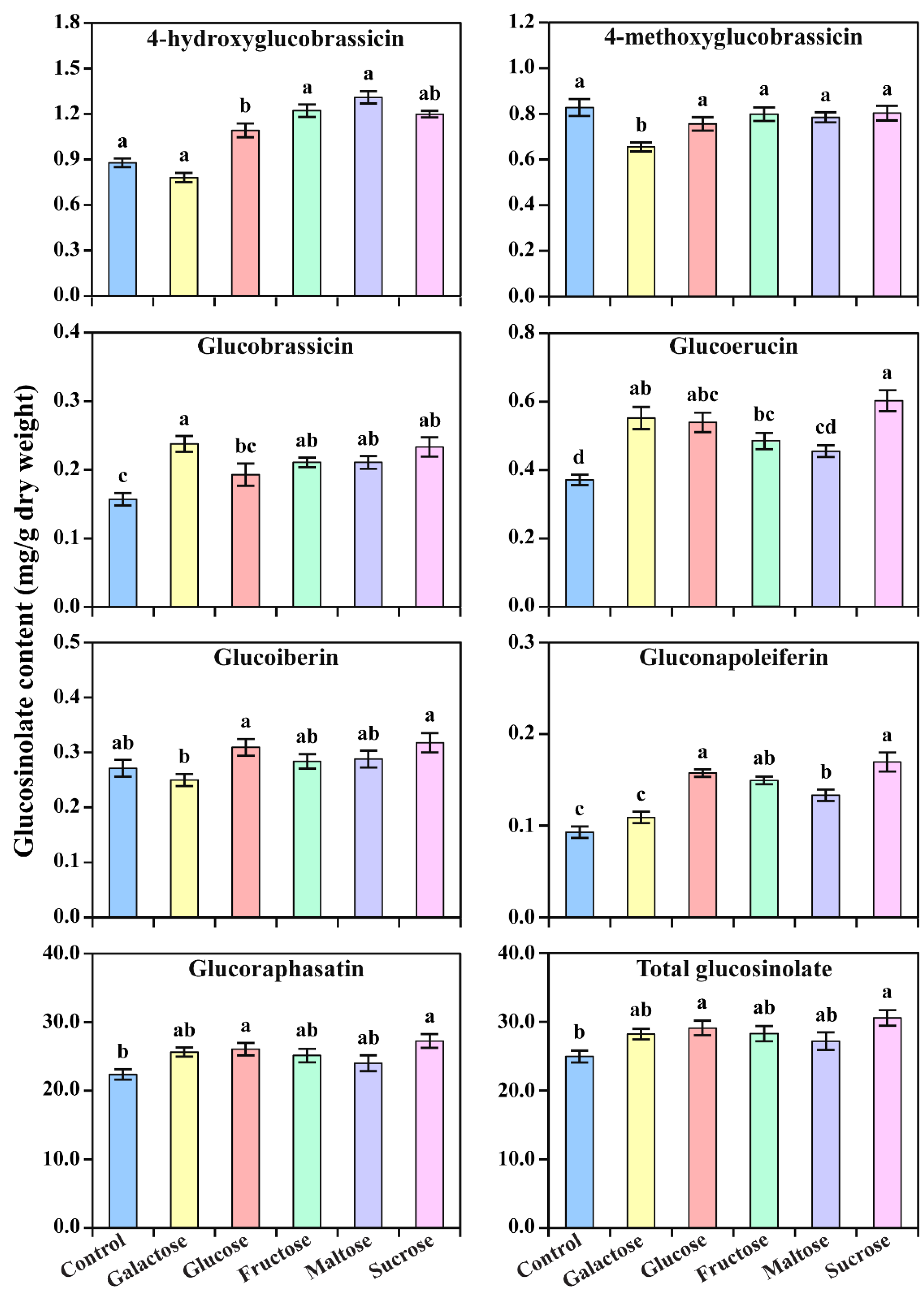
The accumulation of glucosinolate in radish sprouts was analyzed via HPLC. Seven different glucosinolate compounds, i.e., glucoiberin, gluconapoleiferin, glucoerucin, 4-hydroxyglucobrassicin, glucoraphasatin, 4-methoxyglucobrassicin, and glucobrassicin, were detected in the radish sprouts treated with different carbon sources (Figure 3), and their content was highly correlated with the type of carbon source. Except for 4-methoxyglucobrassicin and glucoiberin, the production of glucosinolate compounds was enhanced significantly in treatments with different carbon sources. In particular, the treatments with sucrose and glucose appeared to enhance glucosinolate production in radish sprouts. The total glucosinolate content ranged from 24.97 to 30.58 mg/g dry weight in response to different carbon source treatments. The radish sprouts treated with sucrose had the highest total glucosinolate content, which was 1.22-fold higher than that of the control. The total contents of glucosinolates in the radish sprouts treated with glucose, fructose, galactose, and maltose were 1.17-, 1.13-, 1.13-, and 1.09-fold higher than those of the control, respectively. In addition, sucrose treatment increased the accumulation of gluconapoleiferin, glucoerucin, glucoraphasatin, and glucobrassicin by 1.89-, 1.62-, 1.22-, and 1.44-fold compared with that of the control treatments. The levels of gluconapoleiferin and glucoraphasatin in the radish sprouts treated with glucose were 1.78- and 1.16-fold higher than those in the control, respectively, when radish sprouts were treated with fructose. Similarly, the levels of gluconapoleiferin, 4-hydroxyglucobrassicin, glucoraphasatin, and glucobrassicin were 1.67-, 1.39-, 1.12-, and 1.31-fold higher in the fructose treatment than those of the control, respectively. Galactose treatment similarly enhanced glucosinolate production, particularly the accumulations of glucoerucin, glucoraphasatin, and glucobrassicin, which were increased by 1.49-, 1.15-, and 1.50-fold compared to those of the control. Except for 4-methoxyglucobrassicin, all other glucosinolate compounds were increased when compared with the control. Moreover, the synthesis of 4-methoxyglucobrassicin was not enhanced by the treatment of any carbon sources. The levels of 4-methoxyglucobrassicin were 1.26-fold higher in the control than in the plants treated with galactose.

3.3. Accumulation of Phenolics in Response to Different Carbon Sources

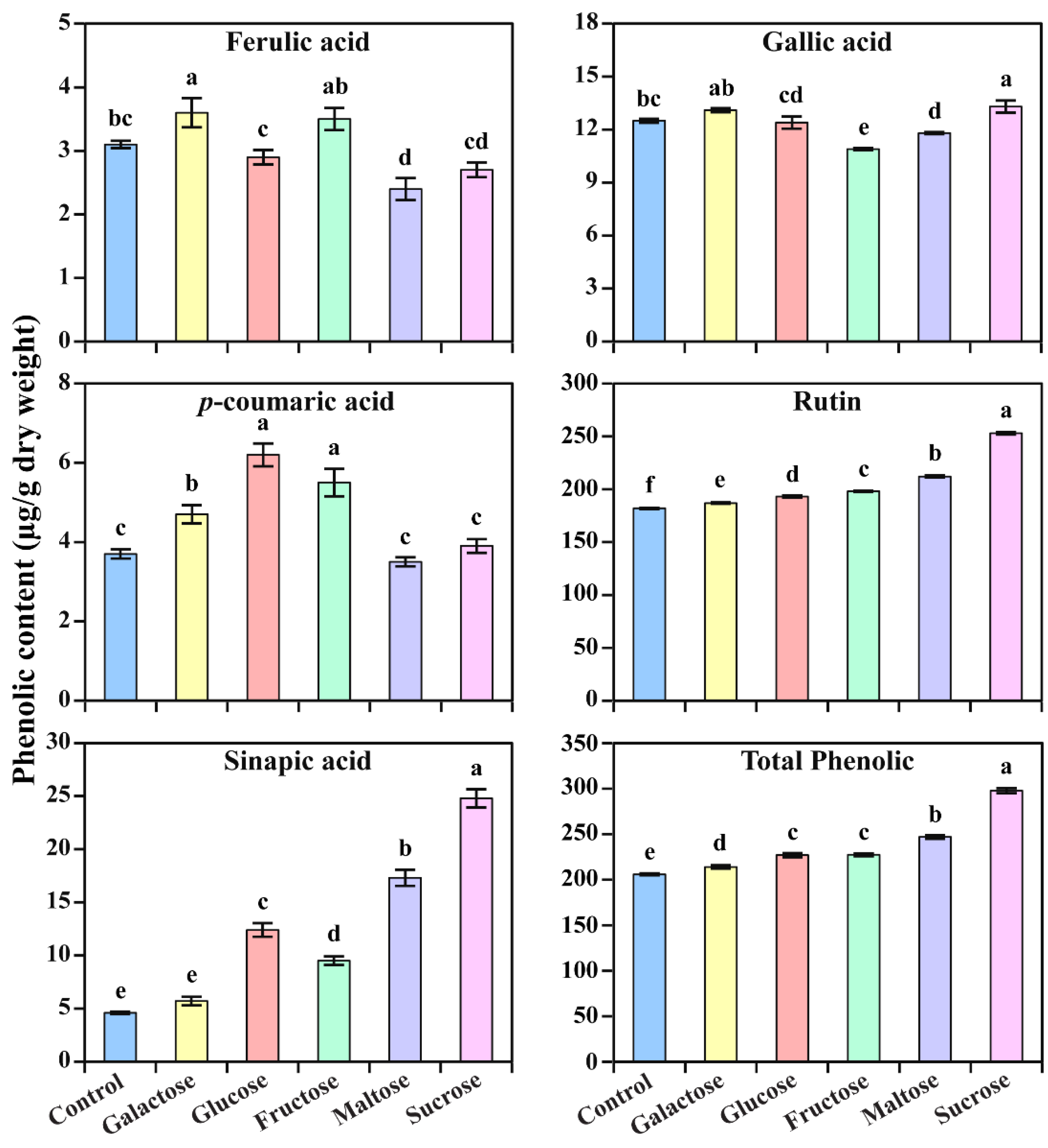
The accumulation of phenolics in radish sprouts was also analyzed via HPLC. Five different phenolic compounds, i.e., gallic acid, ferulic acid, sinapic acid, p-coumaric acid, and rutin, were detected in the radish sprouts grown with different carbon sources (Figure 4 and Figures S2–S4). Their content was significantly different from the various types of carbon sources. The sinapic acid content of radish sprouts is mostly affected by different types of carbon sources when compared to the other five phenolic compounds. Compared with the control, the amounts of sinapic acid were 5.39-, 3.76-, 2.00-, 2.07-, and 1.24-fold higher in the radish sprouts treated with sucrose, maltose, glucose, fructose, and galactose, respectively. The total phenolic content ranged from 20.59 to 29.77 µg/g dry weight in response to different carbon source treatments. In particular, the total phenolic content of radish sprouts grown with sucrose was the highest, which was 1.45-fold higher than that of the control. The total contents of phenolics in maltose, glucose, fructose, and galactose-treated radish sprouts were 1.19-, 1.10-, 1.10-, and 1.04-fold higher than those of the control, respectively. In all types of radish sprouts, rutin was the largest part of the quantified phenolic content. Similar to the sinapic acid content, rutin was more highly accumulated in the sucrose treatment than in the other treatments. Maltose treatment also enhanced the production of rutin. The levels of p-coumaric acid were 1.68-, 1.49-, and 1.27-fold higher in the glucose, fructose, and galactose treatments than that in the control, respectively. However, the levels of ferulic acid were not increased by any treatment except galactose and fructose, which were 1.16- and 1.13-fold higher compared to the control, respectively. In addition, the gallic acid content was enhanced in the galactose and sucrose-treated sprouts.

3.4. Metabolic Profiling of Identified Metabolites in Response to Different Carbon Sources

In total, seven individual glucosinolate and five phenolic compounds were identified and quantified via HPLC from the radish sprouts exposed to different carbon sources. Most of the individual glucosinolate and phenolic contents were significantly higher in the carbon-exposed radish sprouts when compared to the control. The heat map result shows that most of the glucosinolate and phenolic compounds were highest in sucrose (Figure 5). In detail, the individual glucosinolate compounds (glucoiberin, gluconapoleiferin, glucoerucin, 4-hydroxyglucobrassicin, 4-methoxyglucobrassicin, and glucoraphasatin) were slightly higher in the sucrose-exposed sprouts, whereas individual phenolic compounds (sinapic acid and rutin) were significantly higher in the sucrose-exposed sprouts when compared to the other carbon-exposed sprouts. The second-highest individual glucosinolate was obtained in the glucose-exposed sprouts followed by the fructose-exposed plants. In contrast, the second-highest total phenolics content was achieved in the maltose followed by fructose and glucose-exposed plants. In the glucose-exposed sprouts, a total of nine individual glucosinolate and phenolic compounds, including glucoiberin, gluconapoleiferin, 4-hydroxyglucobrassicin, glucoerucin, glucoraphasatin, glucobrassicin, sinapic acid, rutin, and p-coumaric acid, were highest when compared to the control. In the fructose treatment, most of the metabolites, namely glucoiberin, gluconapoleiferin, 4-hydroxyglucobrassicin, glucoerucin, glucoraphasatin, glucobrassicin, 4-methoxyglucobrassicin, ferulic acid, sinapic acid, p-coumaric acid, and rutin, had were higher contents than those in the control. However, in the maltose-exposed sprouts, most of the metabolites, including glucoiberin, gluconapoleiferin, 4-hydroxyglucobrassicin, glucoerucin, glucoraphasatin, glucobrassicin, sinapic acid, and rutin, accumulated at higher levels when compared to the control. In the galactose-exposed sprouts, the contents of glucobrassicin and ferulic acid were higher than those of the control and other carbon-exposed sprouts, whereas the contents of other individual glucosinolates and phenolics were lower. Considering these results, it is observed that the different carbon source enhances the accumulation of different metabolites in radish sprouts.
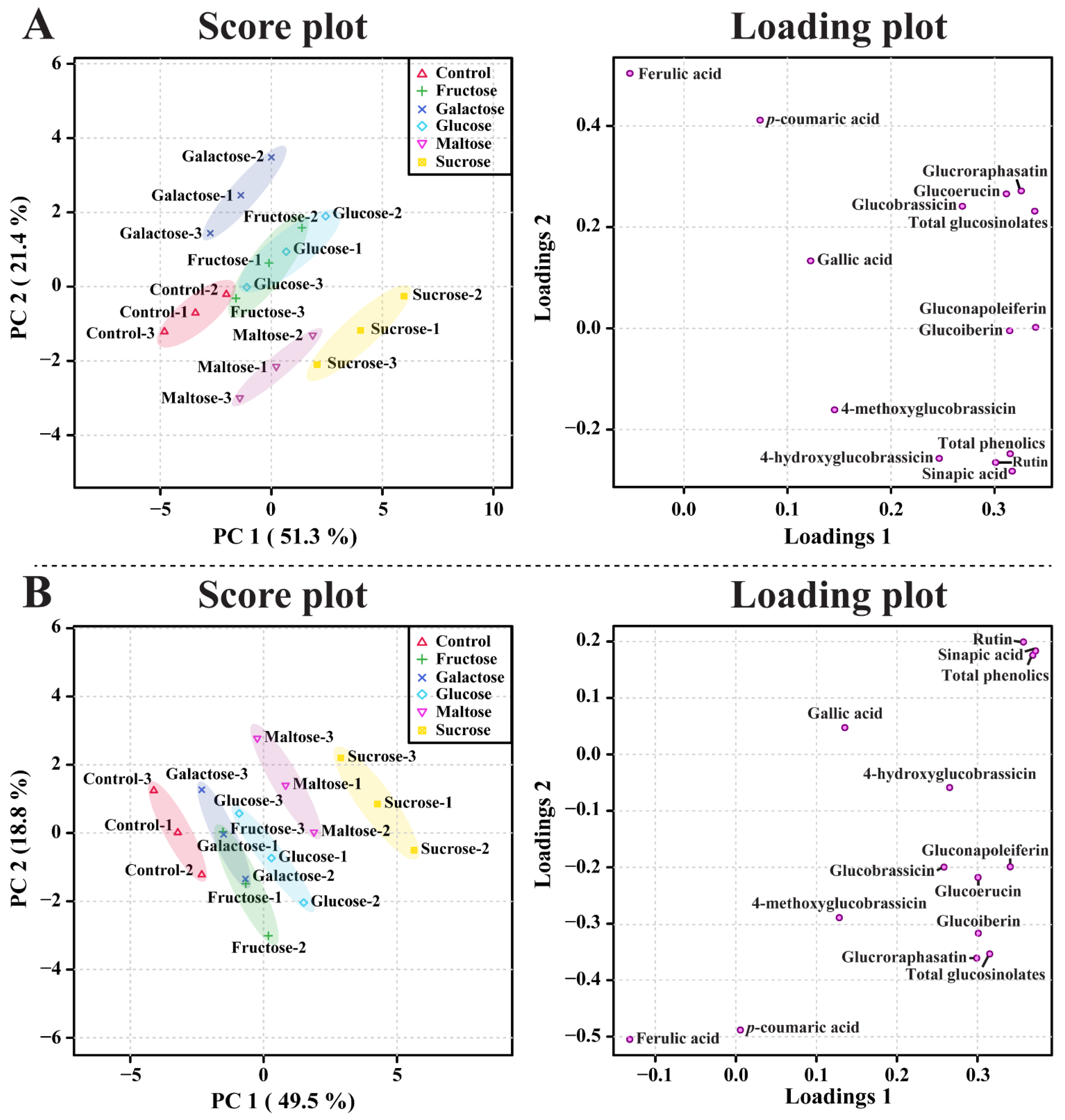
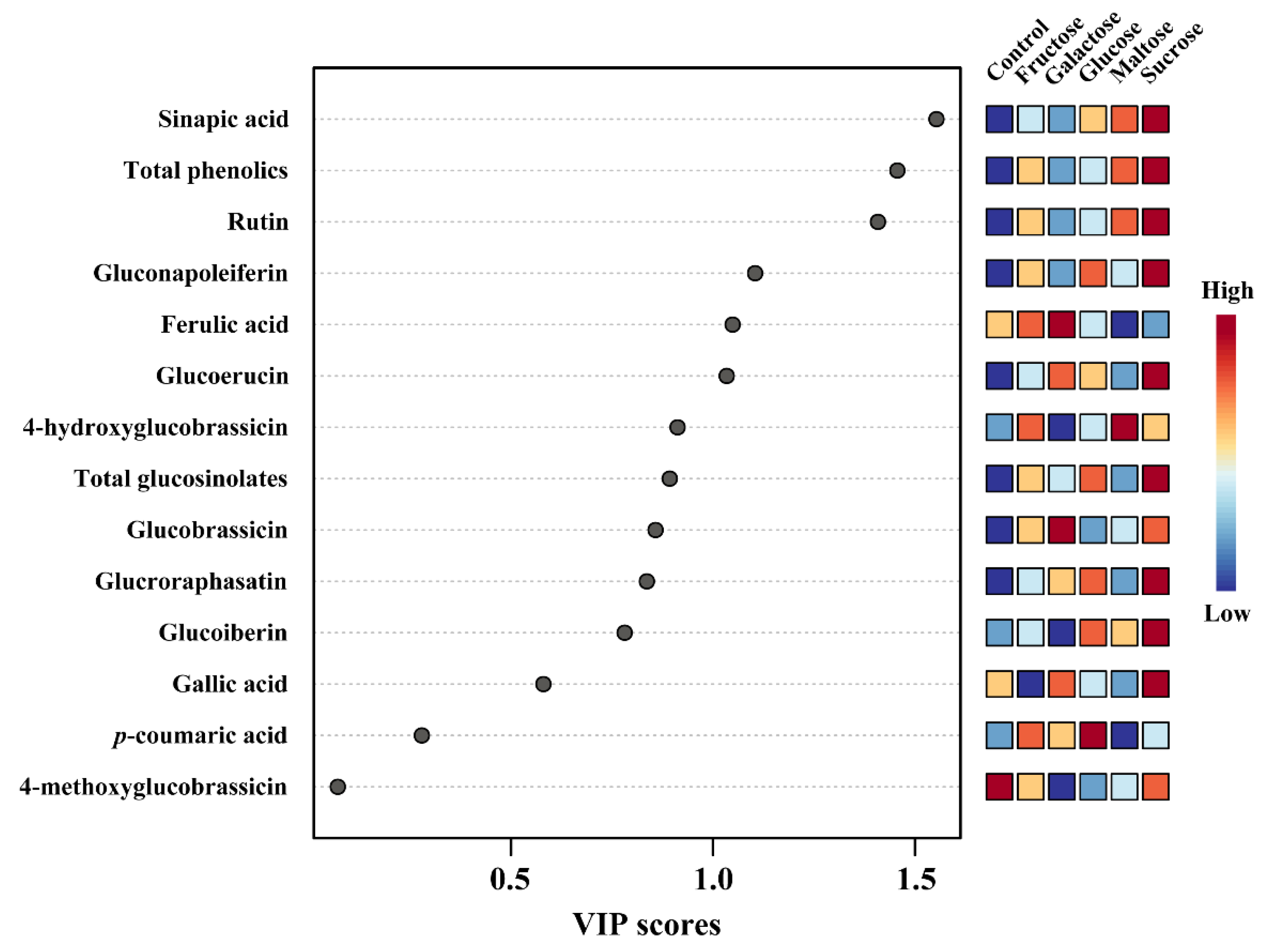
According to the PCA, the PC1 and PC2 showed 51.3% and 21.4% variance, respectively (Figure 6). Among the different carbon-exposed sprouts, the galactose, maltose, and sucrose-exposed sprouts showed clear separation, whereas the other carbon-exposed plants were not separated. This clear separation between the galactose, maltose, sucrose, and other sugar-exposed plants might be due to the ferulic acid, and the associated eigenvector value was −0.052232; the values for gluconapoleiferin, glucoraphasatin, sinapic acid, glucoiberin, and glucoerucin were 0.33968, 0.32573, 0.317, 0.31459, and 0.31139, respectively (Figure 6A). In addition, the PLS-DA was performed to maximize the separation between the different carbon-exposed sprouts. The PLS-DA model showed a clear separation between the different carbon-exposed sprouts, which showed 49.5% (PC1) and 18.8% (PC2), respectively. In the PLS-DA, the separation of the maltose and sucrose-exposed sprouts showed a far group when compared to that of the control and other carbon-exposed sprouts. This clear separation might be due to the ferulic acid, and the associated eigenvector value was −0.13179; the values for sinapic acid, rutin, gluconapoleiferin, glucoiberin, and glucoerucin were 0.37216, 0.35711, 0.3407, 0.30107, and 0.30072, respectively (Figure 6B). In addition, sinapic acid was the most important metabolite for the prediction according to the VIP analysis; its content was highest in the sucrose-exposed sprouts, leading to the far separation (Figure 7). The maltose-exposed sprouts also showed slight separation from the other sugar-exposed sprouts because the second-highest sinapic acid content was achieved in the maltose-exposed sprouts. The other sugar-exposed sprouts were not separated; this is because the total contents of glucosinolates and phenolics were more or less similar to each other. This result supports the heatmap result, which showed that most of the metabolites were highest in the sucrose-exposed sprouts.
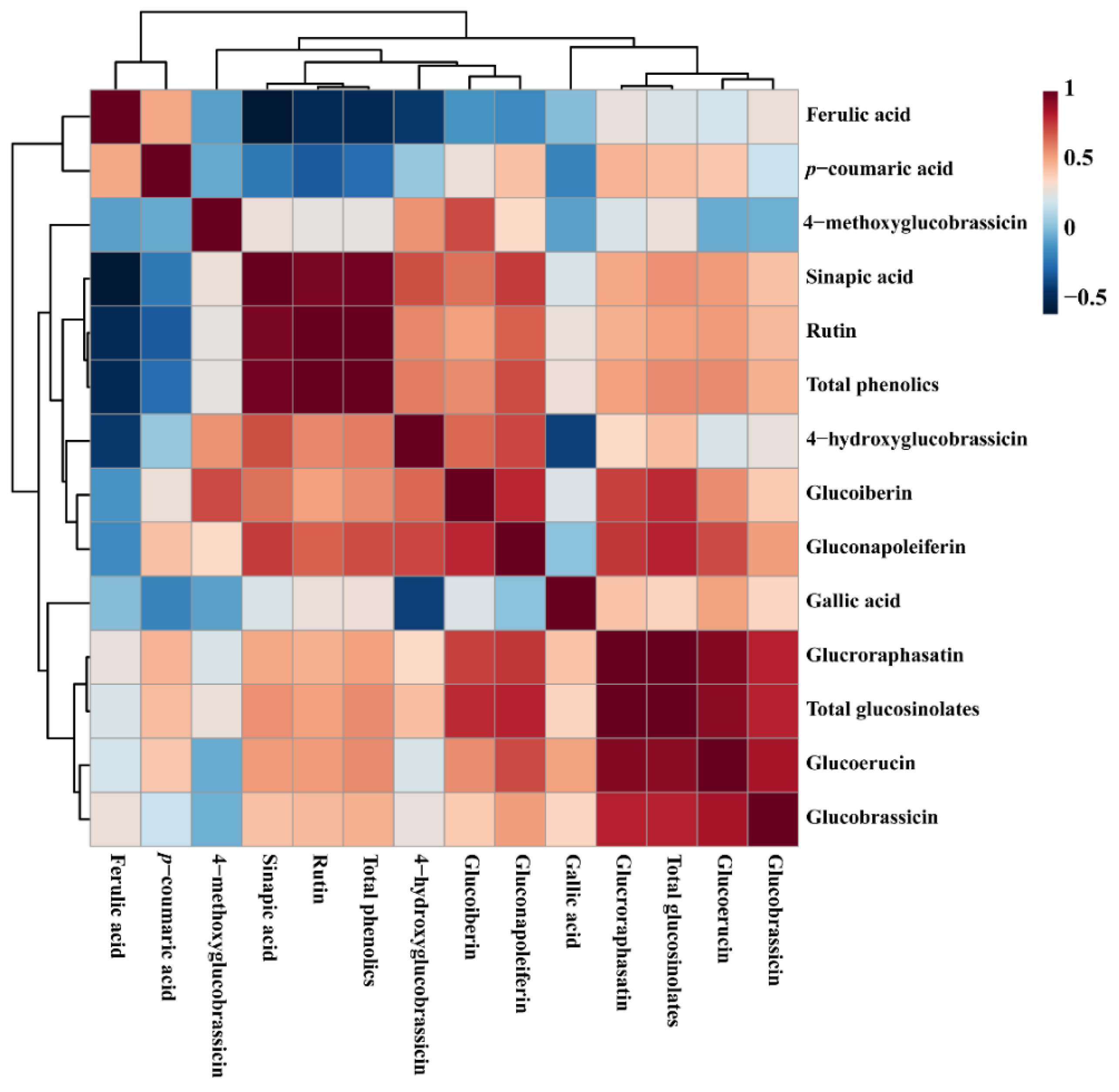
The correlation matrix analysis of the 12 identified metabolites after the sprouts were exposed to different carbon sources (Figure 8) were clustered into two main clusters, which consist of glucosinolate and phenolics based on the positive correlation. The most important metabolite, determined via VIP analysis, was sinapic acid, which showed a positive correlation with most of the metabolites; however, it showed a negative correlation with ferulic acid and p-coumaric acid. Among the identified glucosinolates, glucoraphasatin showed a strong positive correlation with all other identified individual glucosinolate and phenolic compounds. In the phenolic compounds, rutin showed the highest content, which exhibited a positive correlation with most of the metabolites; however, it showed a negative correlation with ferulic acid and p-coumaric acid.

4. Discussion

In this study, the sucrose treatment showed the highest shoot length, followed by glucose, fructose, and maltose. Similar results were reported in Echinacea angustifolia, in which the plants supplemented with sucrose showed the highest shoot length [43]. In addition, the highest shoot regeneration of an Aloe saponaria explant was obtained in the sucrose treatment, followed by glucose, and fructose treatments [44]. In Pharbitis nil, glucose, fructose, and maltose showed very low levels of shoot regeneration and elongation [45]. In another study, in vitro beech cultures showed the highest axillary branching and adventitious shoot regeneration under the glucose treatment [46]. Similarly, in other plants such as Alnus cremastogyne [47], Alnus glutinosa [48], Corylus avellana [49], Prunus mume [50], Rosa chinensis minima [51], and Rosa hybrida [51], the best shoot regeneration was achieved in the glucose and fructose treatments compared to the sucrose treatment. From these results, it is shown that the effect of different carbon sources on shoot regeneration might be species and tissue specific.
Moving on to the root generation, in this study, the highest root length was achieved in the sucrose treatment, followed by the fructose and glucose treatments. A previous study reported that root regeneration and growth are high-energy-demanding processes, and they can occur only when the metabolic substrate is highly available [45]. Sucrose was proven to be a particularly useful sugar, even though glucose gave satisfactory results during in vitro rhizogenesis [52,53]. A previous study reported that in Quber suber, autoclaved fructose had a smaller effect on root induction [52]. A similar effect was observed in P. nil [45]. The reason for this might be a result of the different susceptibility levels of specific species and types of explants to the fructose degradation products produced during autoclaving, such as 5-(hydroxymethyl)-2-furaldehyde. This product might be strongly toxic to plants [54]. From these results, it is shown that sucrose is one of the most prominent carbon sources for the enhancement of root generation in most plant species when compared to other carbon sources.
In the present study, seven glucosinolates and five phenolic compounds were detected in the radish sprouts treated with different carbon sources. In previous studies, 12 glucosinolates, including gluconapoleiferin, glucoerucin, 4-hydroxyglucobrassicin, glucoraphasatin, 4-methoxyglucobrassicin, and glucobrassicin, have been identified in the skin and flesh of radish cultivars [55]; in addition, eight glucosinolates, including glucoiberin, were detected in mature radish taproots [56]. Yuan et al. [57] identified 4-OH-glucobrassicin, glucoraphasatin, 4-methoxyglucobrassicin, and glucobrassicin in radish sprouts under different saline treatments. Furthermore, ten glucosinolates in red radishes [58] and nine glucosinolates in radish seedlings [59], including glucoerucin, 4-hydroxyglucobrassicin, glucoraphasatin, 4-methoxyglucobrassicin, and glucobrassicin, were identified under light conditions and methyl jasmonate treatments. Additionally, analysis of the phenolic contents of four vegetable sprouts showed that the highest contents of phenolic compounds were achieved in the radish sprouts [60]. Similarly, an analysis of the total phenolic content in twelve varieties of vegetable sprouts showed that the highest total phenolic accumulation was achieved in the radish sprouts [61]. This result shows that radish sprouts are rich in glucosinolates and have a high phenolics content.
Sucrose is usually regarded as the most suitable carbon source for plant tissue culture because it is most commonly present in the phloem sap of many plant species and is easily available and economically beneficial [62,63,64]. Several studies have reported that some plant species can be grown on alternative carbon sources, such as sugar alcohols (mannitol and sorbitol), monosaccharide hexoses (fructose, glucose, galactose, mannose, and dextrose), and disaccharides (trehalose, lactose, and maltose) [4,65,66,67,68]. In the present study, treatment with different carbon sources enhanced glucosinolate and phenolic production in radish sprouts. In particular, sucrose, glucose, and fructose contributed to the higher accumulation of glucosinolates; whereas sucrose, maltose, fructose, and glucose showed higher phenolic accumulation. Our results are consistent with previous studies, which reported increased glucosinolate production in broccoli sprouts treated with different sugar sources, including sucrose, glucose, fructose, mannitol, and fructose/glucose (1:1) [8], and an increased production in broccoli sprouts treated with different concentrations of sucrose and mannitol [9]. Similarly, various sugar sources increased the production of flavonoids, baicalin, baicalein, and wogonin, with sucrose, galactose, and fructose promoting the highest accumulation of flavonoids [2]. Sucrose can increase the accumulation of paclitaxel in Taxus chinensis cell suspension culture [69] and enhance the yield of anthraquinone, phenolics, and flavonoids in the adventitious roots of Morinda citrifolia [10]. Similar results were obtained in the hairy root culture of Withania somnifera when exposed to various carbon sources; the highest contents of withaferin A and withanone were achieved in the sucrose-exposed culture [7]. In addition, in another study, the constant maintenance of a sucrose concentration of 2% in the culture media enhanced the withaferin A content in the adventitious root culture of W. somnifera. Similarly, the exposure of ginseng hairy root culture to 2% sucrose positively enhanced the metabolite accumulation [70]. Furthermore, fructose increased catharanthine production in Catharanthus roseus hairy root culture [71] and enhanced the total flavonoid content in Hydrocotyle bonariensis callus [72]. Chattopadhyay et al. [73] reported that glucose is a suitable carbohydrate source for growth and podophyllotoxin production in cell cultures of Podophyllum hexandrum. From the overall results, it is evident that different types of carbon sources enhance the specific metabolites in different plant species.
Previous studies have suggested that active forms of hexokinases are glucose sensors in Arabidopsis and yeast [74,75]. Fructose is the substrate of hexokinase, which enhances the accumulation of flavonoids in Scutellaria baicalensis [2]. In the case of Vitis vinifera cells, mannose (substrate of hexokinase) induces anthocyanin production, whereas 3-O-methyl-D-glucose (cannot be phosphorylated by hexokinase) has no effect [76]. In contrast, in the radish hypocotyl, both mannose and 3-O-methyl-d-glucose do not induce any anthocyanin production [77]. However, in this study, neither fructose nor galactose could significantly accumulate glucosinolate and phenolic content. These results show that glucose itself or glucose metabolites are important for the induction of glucosinolate and phenolic content in radish sprouts. In addition, in this study, disaccharides such as maltose and sucrose showed variation with regard to the accumulation of glucosinolates and phenolics. The reason for this might be that maltose experiences slow hydrolysis, whereas the rapid hydrolysis of sucrose increases the content of hexoses and storage compounds, directing the cells to fast proliferation rates [3,78], which leads to the highest accumulation of glucosinolates and the highest phenolic content.

5. Conclusions

In summary, seven glucosinolates and five phenolics were identified, and the results showed that there was a great difference in the levels of glucosinolates and phenolics after the exposure of radish sprouts to different carbon sources. The production of glucosinolates and phenolics was positively affected by most of the stimuli used in this study. In particular, the radish sprouts treated with sucrose accumulated significantly higher levels of glucosinolates and phenolics. Therefore, this study confirms that different carbon sources play a major role in the production of glucosinolates and phenolics in radish sprouts. This study provides valuable information that can be used to enhance specific secondary metabolites by adding specific carbon sources to red radish sprouts. In addition, this study will be helpful in order to improve the commercial and nutritional quality of red radish sprouts. However, in the future, further studies are needed in order to investigate the glucosinolate and phenylpropanoid biosynthetic pathway gene expression after the exposure of radish sprouts to sucrose. In addition, the analysis of primary metabolites (amino acids, carbohydrates, and organic acids), secondary metabolites (anthocyanin, carotenoid, and triterpenoid), and their gene expression profiles in sucrose-exposed radish sprouts could be an exciting topic for future research.

Supplementary Materials

The following supporting information can be downloaded at: https://www.mdpi.com/article/10.3390/horticulturae9060679/s1, Figure S1: Radish sprouts grown under different carbon sources for ten days; Figure S2: HPLC chromatogram of five standard phenolic compounds; Figure S3: HPLC chromatogram of phenolic compounds from radish sprouts untreated with a carbon source (control); Figure S4: HPLC chromatogram of phenolic compounds from radish sprouts exposed to sucrose.

Author Contributions

R.S. and S.U.P. designed the experiments and analyzed the data. C.H.P., M.C., Y.E.P., H.J.Y., J.K.K. and Y.B.K. performed the experiments and analyzed the data. C.H.P. and R.S. wrote the manuscript. S.S. analyzed the data and edited the manuscript. All authors have read and agreed to the published version of the manuscript.

Funding

This research was supported by the Bisa Research Grant of Keimyung University in 2022.

Institutional Review Board Statement

Not applicable.

Informed Consent Statement

Not applicable.

Data Availability Statement

Data reported are available in the Supplementary Materials.

Conflicts of Interest

The authors declare no conflict of interest.

References

  1. Carvalho, D.C.d.; Silva, A.L.L.d.; Schuck, M.R.; Purcino, M.; Tanno, G.N.; Biasi, L.A. Fox grape cv. Bordô (Vitis labrusca L.) and grapevine cv. Chardonnay (Vitis vinifera L.) cultivated in vitro under different carbohydrates, amino acids and 6-Benzylaminopurine levels. Braz. Arch. Biol. Technol. 2013, 56, 191–201. [Google Scholar] [CrossRef]
  2. Park, C.H.; Kim, Y.S.; Li, X.; Kim, H.H.; Arasu, M.V.; Al-Dhabi, N.A.; Lee, S.Y.; Park, S.U. Influence of different carbohydrates on flavonoid accumulation in hairy root cultures of Scutellaria baicalensis. Nat. Prod. Commun. 2016, 11, 799–802. [Google Scholar] [CrossRef] [PubMed] [Green Version]
  3. Yaseen, M.; Ahmad, T.; Sablok, G.; Standardi, A.; Hafiz, I.A. Role of carbon sources for in vitro plant growth and development. Mol. Biol. Rep. 2013, 40, 2837–2849. [Google Scholar] [CrossRef] [PubMed]
  4. Bhagyalakshmi, N.; Thimmaraju, R.; Narayan, M. Various hexoses and di-hexoses differently influence growth, morphology and pigment synthesis in transformed root cultures of red beet (Beta vulgaris). Plant Cell Tissue Organ Cult. 2004, 78, 183–195. [Google Scholar] [CrossRef]
  5. Rolland, F.; Baena-Gonzalez, E.; Sheen, J. Sugar sensing and signaling in plants: Conserved and novel mechanisms. Annu. Rev. Plant Biol. 2006, 57, 675–709. [Google Scholar] [CrossRef] [PubMed] [Green Version]
  6. El Far, M.M.; Taie, H. Antioxidant activities, total anthocyanins, phenolics and flavonoids contents of some sweetpotato genotypes under stress of different concentrations of sucrose and sorbitol. Aust. J. Basic Appl. Sci. 2009, 3, 3609–3616. [Google Scholar]
  7. Sivanandhan, G.; Rajesh, M.; Arun, M.; Jeyaraj, M.; Dev, G.K.; Manickavasagam, M.; Selvaraj, N.; Ganapathi, A. Optimization of carbon source for hairy root growth and withaferin A and withanone production in Withania somnifera. Nat. Prod. Commun. 2012, 7, 1271–1272. [Google Scholar] [CrossRef] [Green Version]
  8. Guo, R.; Yuan, G.; Wang, Q. Sucrose enhances the accumulation of anthocyanins and glucosinolates in broccoli sprouts. Food Chem. 2011, 129, 1080–1087. [Google Scholar] [CrossRef]
  9. Guo, R.; Yuan, G.; Wang, Q. Effect of sucrose and mannitol on the accumulation of health-promoting compounds and the activity of metabolic enzymes in broccoli sprouts. Sci. Hortic. 2011, 128, 159–165. [Google Scholar] [CrossRef]
  10. Baque, M.; Elgirban, A.; Lee, E.-J.; Paek, K.-Y. Sucrose regulated enhanced induction of anthraquinone, phenolics, flavonoids biosynthesis and activities of antioxidant enzymes in adventitious root suspension cultures of Morinda citrifolia (L.). Acta Physiol. Plant. 2012, 34, 405–415. [Google Scholar] [CrossRef]
  11. Zheng, Y.; Tian, L.; Liu, H.; Pan, Q.; Zhan, J.; Huang, W. Sugars induce anthocyanin accumulation and flavanone 3-hydroxylase expression in grape berries. Plant Growth Regul. 2009, 58, 251–260. [Google Scholar] [CrossRef]
  12. Wang, Y.; Weathers, P. Sugars proportionately affect artemisinin production. Plant Cell Rep. 2007, 26, 1073–1081. [Google Scholar] [CrossRef] [PubMed]
  13. Weathers, P.; DeJesus-Gonzalez, L.; Kim, Y.; Souret, F.; Towler, M. Alteration of biomass and artemisinin production in Artemisia annua hairy roots by media sterilization method and sugars. Plant Cell Rep. 2004, 23, 414–418. [Google Scholar] [CrossRef] [PubMed]
  14. Grant, B.; Beevers, H. Absorption of sugars by plant tissues. Plant Physiol. 1964, 39, 78. [Google Scholar] [CrossRef]
  15. Abellán, Á.; Domínguez-Perles, R.; Moreno, D.A.; García-Viguera, C. Sorting out the value of cruciferous sprouts as sources of bioactive compounds for nutrition and health. Nutrients 2019, 11, 429. [Google Scholar] [CrossRef] [Green Version]
  16. Shree, B.; Kumar, S.; Sharma, S.; Katoch, V. Functional significance of underutilized high value cruciferous vegetables-an exotic gleam in the gloomy guise of their functional importance. S. Afr. J. Bot. 2022, 145, 420–437. [Google Scholar] [CrossRef]
  17. Melim, C.; Lauro, M.R.; Pires, I.M.; Oliveira, P.J.; Cabral, C. The role of glucosinolates from cruciferous vegetables (Brassicaceae) in gastrointestinal cancers: From prevention to therapeutics. Pharmaceutics 2022, 14, 190. [Google Scholar] [CrossRef]
  18. Avato, P.; Argentieri, M. Brassicaceae: A rich source of health improving phytochemicals. Phytochem. Rev. 2015, 14, 1019–1033. [Google Scholar] [CrossRef]
  19. Costa-Pérez, A.; Núñez-Gómez, V.; Baenas, N.; Di Pede, G.; Achour, M.; Manach, C.; Mena, P.; Del Rio, D.; García-Viguera, C.; Moreno, D.A. Systematic review on the metabolic interest of glucosinolates and their bioactive derivatives for human health. Nutrients 2023, 15, 1424. [Google Scholar] [CrossRef]
  20. Li, Z.; Lee, H.W.; Liang, X.; Liang, D.; Wang, Q.; Huang, D.; Ong, C.N. Profiling of phenolic compounds and antioxidant activity of 12 cruciferous vegetables. Molecules 2018, 23, 1139. [Google Scholar] [CrossRef] [Green Version]
  21. Wu, J.; Cui, S.; Liu, J.; Tang, X.; Zhao, J.; Zhang, H.; Mao, B.; Chen, W. The recent advances of glucosinolates and their metabolites: Metabolism, physiological functions and potential application strategies. Crit. Rev. Food Sci. Nutr. 2022, 1–18. [Google Scholar] [CrossRef] [PubMed]
  22. Kamal, R.M.; Abdull Razis, A.F.; Mohd Sukri, N.S.; Perimal, E.K.; Ahmad, H.; Patrick, R.; Djedaini-Pilard, F.; Mazzon, E.; Rigaud, S. Beneficial health effects of glucosinolates-derived isothiocyanates on cardiovascular and neurodegenerative diseases. Molecules 2022, 27, 624. [Google Scholar] [CrossRef] [PubMed]
  23. Morrison, M.E.; Joseph, J.M.; McCann, S.E.; Tang, L.; Almohanna, H.M.; Moysich, K.B. Cruciferous vegetable consumption and stomach cancer: A case-control study. Nutr. Cancer 2020, 72, 52–61. [Google Scholar] [CrossRef] [PubMed]
  24. Soundararajan, P.; Kim, J.S. Anti-carcinogenic glucosinolates in cruciferous vegetables and their antagonistic effects on prevention of cancers. Molecules 2018, 23, 2983. [Google Scholar] [CrossRef] [PubMed] [Green Version]
  25. Nomura, S.J.; Hwang, Y.-T.; Gomez, S.L.; Fung, T.T.; Yeh, S.-L.; Dash, C.; Allen, L.; Philips, S.; Hilakivi-Clarke, L.; Zheng, Y.-L. Dietary intake of soy and cruciferous vegetables and treatment-related symptoms in Chinese-American and non-Hispanic White breast cancer survivors. Breast Cancer Res. Treat. 2018, 168, 467–479. [Google Scholar] [CrossRef]
  26. Ağagündüz, D.; Şahin, T.Ö.; Yılmaz, B.; Ekenci, K.D.; Duyar Özer, Ş.; Capasso, R. Cruciferous vegetables and their bioactive metabolites: From prevention to novel therapies of colorectal cancer. Evid. Based Complement. Altern. Med. 2022, 2022, 1534083. [Google Scholar] [CrossRef]
  27. Connolly, E.L.; Sim, M.; Travica, N.; Marx, W.; Beasy, G.; Lynch, G.S.; Bondonno, C.P.; Lewis, J.R.; Hodgson, J.M.; Blekkenhorst, L.C. Glucosinolates from cruciferous vegetables and their potential role in chronic disease: Investigating the preclinical and clinical evidence. Front. Pharmacol. 2021, 12, 767975. [Google Scholar] [CrossRef]
  28. De la Rosa, L.A.; Moreno-Escamilla, J.O.; Rodrigo-García, J.; Alvarez-Parrilla, E. Phenolic compounds. In Postharvest Physiology and Biochemistry of Fruits and Vegetables; Yahia, E., Carrillo-López, A., Eds.; Elsevier Inc.: Amsterdam, The Netherlands, 2018; pp. 253–271. [Google Scholar]
  29. Choi, M.; Sathasivam, R.; Nguyen, B.V.; Park, N.I.; Woo, S.-H.; Park, S.U. Expression analysis of phenylpropanoid pathway genes and metabolomic analysis of phenylpropanoid compounds in adventitious, hairy, and seedling roots of Tartary buckwheat. Plants 2021, 11, 90. [Google Scholar] [CrossRef]
  30. Favela-González, K.M.; Hernández-Almanza, A.Y.; De la Fuente-Salcido, N.M. The value of bioactive compounds of cruciferous vegetables (Brassica) as antimicrobials and antioxidants: A review. J. Food Biochem. 2020, 44, e13414. [Google Scholar] [CrossRef]
  31. Sathasivam, R.; Park, S.U.; Kim, J.K.; Park, Y.J.; Kim, M.C.; Nguyen, B.V.; Lee, S.Y. Metabolic profiling of primary and secondary metabolites in kohlrabi (Brassica oleracea var. gongylodes) sprouts exposed to different light-emitting diodes. Plants 2023, 12, 1296. [Google Scholar] [CrossRef]
  32. Banihani, S.A. Radish (Raphanus sativus) and diabetes. Nutrients 2017, 9, 1014. [Google Scholar] [CrossRef] [PubMed] [Green Version]
  33. Gamba, M.; Asllanaj, E.; Raguindin, P.F.; Glisic, M.; Franco, O.H.; Minder, B.; Bussler, W.; Metzger, B.; Kern, H.; Muka, T. Nutritional and phytochemical characterization of radish (Raphanus sativus): A systematic review. Trends Food Sci. Technol. 2021, 113, 205–218. [Google Scholar] [CrossRef]
  34. Park, C.H.; Baskar, T.B.; Park, S.-Y.; Kim, S.-J.; Valan Arasu, M.; Al-Dhabi, N.A.; Kim, J.K.; Park, S.U. Metabolic profiling and antioxidant assay of metabolites from three radish cultivars (Raphanus sativus). Molecules 2016, 21, 157. [Google Scholar] [CrossRef] [PubMed] [Green Version]
  35. Park, C.H.; Park, S.-Y.; Park, Y.J.; Kim, J.K.; Park, S.U. Metabolite profiling and comparative analysis of secondary metabolites in Chinese cabbage, radish, and hybrid xBrassicoraphanus. J. Agric. Food Chem. 2020, 68, 13711–13719. [Google Scholar] [CrossRef] [PubMed]
  36. Zhang, J.; Qiu, X.; Tan, Q.; Xiao, Q.; Mei, S. A comparative metabolomics study of flavonoids in radish with different skin and flesh colors (Raphanus sativus L.). J. Agric. Food Chem. 2020, 68, 14463–14470. [Google Scholar] [CrossRef]
  37. El-Beltagi, H.S.; Maraei, R.W.; Shalaby, T.A.; Aly, A.A. Metabolites, nutritional quality and antioxidant activity of red radish roots affected by gamma rays. Agronomy 2022, 12, 1916. [Google Scholar] [CrossRef]
  38. Park, N.I.; Xu, H.; Li, X.; Jang, I.H.; Park, S.; Ahn, G.H.; Lim, Y.P.; Kim, S.J.; Park, S.U. Anthocyanin accumulation and expression of anthocyanin biosynthetic genes in radish (Raphanus sativus). J. Agric. Food Chem. 2011, 59, 6034–6039. [Google Scholar] [CrossRef]
  39. Murashige, T.; Skoog, F. A revised medium for rapid growth and bio assays with tobacco tissue cultures. Physiol. Plant. 1962, 15, 473–497. [Google Scholar] [CrossRef]
  40. International Standards Organisation (ISO). Rapeseed-Determination of Glucosinolates Content—Part 1: Method Using High-Performance Liquid Chromatography; ISO: Geneva, Switzerland, 1992; Volume 9167, pp. 1–9. [Google Scholar]
  41. Lee, S.Y.; Kwon, H.; Kim, J.K.; Park, C.H.; Sathasivam, R.; Park, S.U. Comparative analysis of glucosinolate and phenolic compounds in green and red kimchi cabbage (Brassica rapa L. ssp. pekinensis) hairy roots after exposure to light and dark conditions. Horticulturae 2023, 9, 466. [Google Scholar] [CrossRef]
  42. Do, T.M.H.; Choi, M.; Kim, J.K.; Kim, Y.J.; Park, C.; Park, C.H.; Park, N.I.; Kim, C.; Sathasivam, R.; Park, S.U. Impact of light and dark treatment on phenylpropanoid pathway genes, primary and secondary metabolites in Agastache rugosa transgenic hairy root cultures by overexpressing Arabidopsis transcription factor AtMYB12. Life 2023, 13, 1042. [Google Scholar] [CrossRef]
  43. Chae, S.C. Influence of carbon sources on shoot organogenesis in Echinacea angustifolia DC. Life Sci. 2013, 10, 1300–1303. [Google Scholar]
  44. Kim, J.K.; Baskar, T.B.; Park, S.U. Effect of carbon sources and sucrose concentrations on shoot organogenesis of Aloe saponaria. Biosci. Biotechnol. Res. Asia 2016, 13, 925–930. [Google Scholar] [CrossRef] [Green Version]
  45. Alina, T.; Magdalena, J.; Andrzej, T. The effect of carbon source on callus induction and regeneration ability in Pharbitis nil. Acta Physiol. Plant. 2006, 28, 619–626. [Google Scholar] [CrossRef]
  46. Cuenca, B.; Vieitez, A. Influence of carbon source on shoot multiplication and adventitious bud regeneration in in vitro beech cultures. Plant Growth Regul. 2000, 32, 1–12. [Google Scholar] [CrossRef]
  47. Tang, D.; Ishii, K.; Ohba, K. In vitro regeneration of Alnus cremastogyne Burk from epicotyl explants. Plant Cell Rep. 1996, 15, 658–661. [Google Scholar] [CrossRef]
  48. Welander, M.; Welander, N.; Brackman, A.-S. Regulation of in vitro shoot multiplication in Syringa, Alnus and Malus by different carbon sources. J. Hortic. Sci. 1989, 64, 361–366. [Google Scholar] [CrossRef]
  49. Yu, X.; Reed, B.M. Improved shoot multiplication of mature hazelnut (Corylus avellana L.) in vitro using glucose as a carbon source. Plant Cell Rep. 1993, 12, 256–259. [Google Scholar] [CrossRef]
  50. Harada, H.; Murai, Y. Micropropagation of Prunus mume. Plant Cell Tissue Organ Cult. 1996, 46, 265–267. [Google Scholar] [CrossRef]
  51. Hsia, C.-N.; Korban, S.S. Factors affecting in vitro establishment and shoot proliferation of Rosa hybrida L. and Rosa chinensis minima. Vitr. Cell. Dev. Biol.-Plant 1996, 32, 217–222. [Google Scholar] [CrossRef]
  52. Romano, A.; Noronha, C.; Martins-Loucao, M. Role of carbohydrates in micropropagation of cork oak. Plant Cell Tissue Organ Cult. 1995, 40, 159–167. [Google Scholar] [CrossRef]
  53. Fotopoulos, S.; Sotiropoulos, T. In vitro propagation of the peach rootstock: The effect of different carbon sources and types of sealing material on rooting. Biol. Plant. 2004, 48, 629–631. [Google Scholar] [CrossRef]
  54. Bogunia, H.; Przywara, L. Rola cukrowców w roslinnych kulturach in vitro. (Sugars in plant tissue culture.). Wiad. Bot. 1999, 43, 25–36. [Google Scholar]
  55. Kim, S.-J.; Uddin, M.R.; Park, S.U. Glucosinolate accumulation in three important radish (‘Raphanus sativus’) cultivars. Aust. J. Crop Sci. 2013, 7, 1843–1847. [Google Scholar]
  56. Hanlon, P.R.; Barnes, D.M. Phytochemical composition and biological activity of 8 varieties of radish (Raphanus sativus L.) sprouts and mature taproots. J. Food Sci. 2011, 76, C185–C192. [Google Scholar] [CrossRef] [PubMed]
  57. Yuan, G.; Wang, X.; Guo, R.; Wang, Q. Effect of salt stress on phenolic compounds, glucosinolates, myrosinase and antioxidant activity in radish sprouts. Food Chem. 2010, 121, 1014–1019. [Google Scholar] [CrossRef]
  58. Ahn, M.; Moon, J.; Park, C.; Bang, H.; Kim, G.O.; Kim, S.-J.; Kim, K.-h.; Shin, T. Chungpihongsim radish (Raphanus sativus L. cv. Chungpihongsim) ameliorates ethanol-induced gastric injury in rats. Orient. Pharm. Exp. Med. 2016, 16, 37–43. [Google Scholar] [CrossRef]
  59. Al-Dhabi, N.A.; Arasu, M.V.; Kim, S.J.; RomijUddin, M.; Park, W.T.; Lee, S.Y.; Park, S.U. Methyl jasmonate-and light-induced glucosinolate and anthocyanin biosynthesis in radish seedlings. Nat. Prod. Commun. 2015, 10, 1211–1214. [Google Scholar] [CrossRef] [Green Version]
  60. Pająk, P.; Socha, R.; Gałkowska, D.; Rożnowski, J.; Fortuna, T. Phenolic profile and antioxidant activity in selected seeds and sprouts. Food Chem. 2014, 143, 300–306. [Google Scholar] [CrossRef]
  61. Park, H.; Shin, Y.; Kim, Y.-J. Antioxidant contents and activities of twelve varieties of vegetable sprouts. Korean J. Food Sci. Technol. 2019, 51, 207–213. [Google Scholar]
  62. Ahmad, T.; Abbasi, N.A.; Hafiz, I.A.; Ali, A. Comparison of sucrose and sorbitol as main carbon energy sources in microprogation of peach rootstock GF-677. Pak. J. Bot. 2007, 39, 1269. [Google Scholar]
  63. Fuentes, S.R.; Calheiros, M.B.; Manetti-Filho, J.; Vieira, L.G. The effects of silver nitrate and different carbohydrate sources on somatic embryogenesis in Coffea canephora. Plant Cell Tissue Organ Cult. 2000, 60, 5–13. [Google Scholar] [CrossRef]
  64. Thompson, M.R.; Thorpe, T.A. Metabolic and non-metabolic roles of carbohydrates. In Cell and Tissue Culture in Forestry; Bong, J.M., Durzan, D.J., Eds.; Martinus Nijhoff Publisher: Dardrecht, The Netherlands, 1987; pp. 89–112. [Google Scholar]
  65. Amiri, S.; Kazemitabar, S. Enhancement of callus induction and regeneration efficiency from embryo cultures of Datura stramonium by adjusting carbon sources and concentrations. Afr. J. Biotechnol. 2011, 10, 10101–10107. [Google Scholar]
  66. Jheng, F.-Y.; Do, Y.-Y.; Liauh, Y.-W.; Chung, J.-P.; Huang, P.-L. Enhancement of growth and regeneration efficiency from embryogenic callus cultures of Oncidium ‘Gower Ramsey’ by adjusting carbohydrate sources. Plant Sci. 2006, 170, 1133–1140. [Google Scholar] [CrossRef]
  67. Swedlund, B.; Locy, R.D. Sorbitol as the primary carbon source for the growth of embryogenic callus of maize. Plant Physiol. 1993, 103, 1339–1346. [Google Scholar] [CrossRef] [Green Version]
  68. Thwe, A.A.; Chae, S.C.; Chung, S.-O.; Park, S.U. Enhancement of the in vitro root regeneration efficiency of Rehmannia glutinosa Libosch. stem explants by different carbon sources. Life Sci. 2013, 10, 579–582. [Google Scholar]
  69. Luo, J.; He, G.-Y. Optimization of elicitors and precursors for paclitaxel production in cell suspension culture of Taxus chinensis in the presence of nutrient feeding. Process Biochem. 2004, 39, 1073–1079. [Google Scholar] [CrossRef]
  70. Sivanandhan, G.; Arun, M.; Mayavan, S.; Rajesh, M.; Mariashibu, T.; Manickavasagam, M.; Selvaraj, N.; Ganapathi, A. Chitosan enhances withanolides production in adventitious root cultures of Withania somnifera (L.) Dunal. Ind. Crops Prod. 2012, 37, 124–129. [Google Scholar] [CrossRef]
  71. Jung, K.; Kwak, S.S.; Kim, S.W.; Lee, H.; Choi, C.; Liu, J.R. Improvement of the catharanthine productivity in hairy root cultures ofCatharanthus roseus by using monosaccharides as a carbon source. Biotechnol. Lett. 1992, 14, 695–700. [Google Scholar] [CrossRef]
  72. Masoumian, M.; Arbakariya, A.; Syahida, A.; Maziah, M. Flavonoids production in Hydrocotyle bonariensis callus tissues. J. Med. Plants Res. 2011, 5, 1564–1574. [Google Scholar]
  73. Chattopadhyay, S.; Srivastava, A.K.; Bhojwani, S.S.; Bisaria, V.S. Production of podophyllotoxin by plant cell cultures of Podophyllum hexandrum in bioreactor. J. Biosci. Bioeng. 2002, 93, 215–220. [Google Scholar] [CrossRef]
  74. Gibson, S.I. Plant sugar-response pathways. Part of a complex regulatory web. Plant Physiol. 2000, 124, 1532–1539. [Google Scholar] [CrossRef] [PubMed] [Green Version]
  75. Jang, J.-C.; Sheen, J. Sugar sensing in higher plants. Plant Cell 1994, 6, 1665–1679. [Google Scholar] [PubMed] [Green Version]
  76. Vitrac, X.; Larronde, F.; Krisa, S.; Decendit, A.; Deffieux, G.; Mérillon, J.-M. Sugar sensing and Ca2+–calmodulin requirement in Vitis vinifera cells producing anthocyanins. Phytochemistry 2000, 53, 659–665. [Google Scholar] [CrossRef] [PubMed]
  77. Hara, M.; Oki, K.; Hoshino, K.; Kuboi, T. Enhancement of anthocyanin biosynthesis by sugar in radish (Raphanus sativus) hypocotyl. Plant Sci. 2003, 164, 259–265. [Google Scholar] [CrossRef]
  78. Blanc, G.; Lardet, L.; Martin, A.; Jacob, J.-L.; Carron, M.-P. Differential carbohydrate metabolism conducts morphogenesis in embryogenic callus of Hevea brasiliensis (Mull. Arg.). J. Exp. Bot. 2002, 53, 1453–1462. [Google Scholar] [CrossRef] [Green Version]
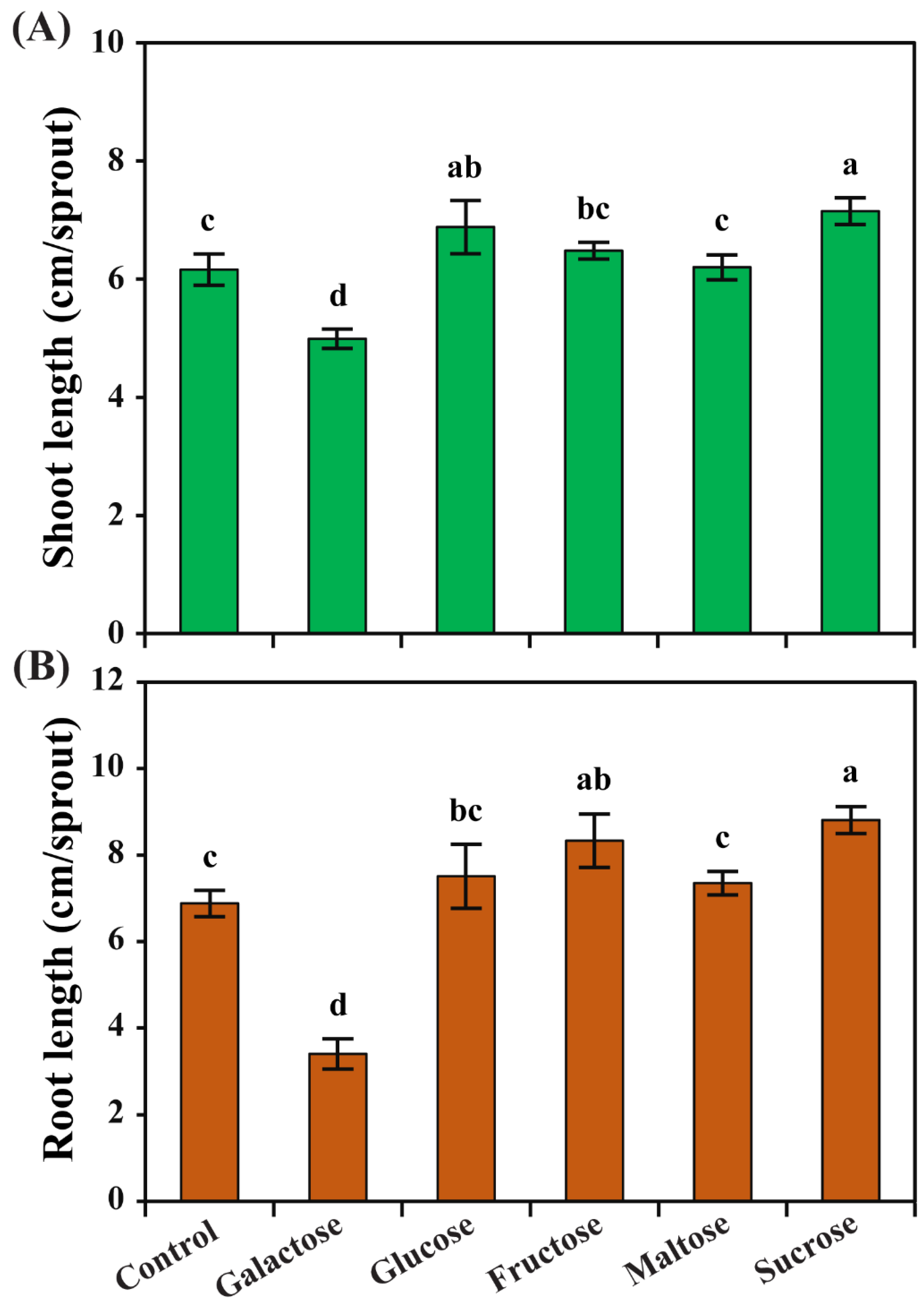
Figure 1. The effects of different carbon sources on (A) shoot and (B) root length of radish sprouts. The height of each error bar and the error bars show the mean and standard error, respectively, from ten replicates. Mean values with different letters (a–d) were significantly different (p < 0.05, ANOVA, DMRT) compared with the control.
Horticulturae 09 00679 g001
Figure 2. The effects of different carbon sources on (A) fresh weight and (B) dry weight of radish sprouts. The height of each error bar and the error bars show the mean and standard error, respectively. Mean values with different letters (a–e) were significantly different (p < 0.05, ANOVA, DMRT) compared with the control.
Horticulturae 09 00679 g002
Figure 3. Effect of different carbon sources on glucosinolate content (mg/g dry weight) in radish sprouts. The height of each error bar and the error bars show the mean and standard error, respectively, from three replicates. Mean values with different letters (a–d) were significantly different (p < 0.05, ANOVA, DMRT) compared with the control.
Horticulturae 09 00679 g003
Figure 4. Effect of different carbon sources on phenolic compound content (µg/g dry weight) in radish sprouts. The height of each error bar and the error bars show the mean and standard error, respectively, from three replicates. Mean values with different letters (a–f) were significantly different (p < 0.05, ANOVA, DMRT) compared with the control.
Horticulturae 09 00679 g004
Figure 5. Heatmap showing the changes in the relative metabolite content in radish sprouts after exposure to different carbon sources. The color scale (2 to −2) at the right denotes the relative metabolite concentrations, with a high concentration in red and a low concentration in blue.
Horticulturae 09 00679 g005
Figure 6. Score and loading plot of the PCA (A) and PLS-DA (B) model of the metabolites identified in radish sprouts after exposure to different carbon sources.
Horticulturae 09 00679 g006
Figure 7. The main components, separating the radish sprouts after exposure to different carbon sources, are based on the VIP scores obtained via the PLS-DA model. The black dot represents the VIP scores reflecting the degree of importance of metabolites, with values >0.07 seen as driving the calculated discrimination.
Horticulturae 09 00679 g007
Figure 8. Correlation matrix of metabolites identified in radish sprouts after exposure to different carbon sources. Each colored box denotes Pearson’s correlation coefficient for a pair of compounds, and the value of the correlation coefficient is shown by the intensity, with a high concentration in red and a low concentration in blue, as displayed on the right-side color scale (1 to −0.5).
Horticulturae 09 00679 g008
Disclaimer/Publisher’s Note: The statements, opinions and data contained in all publications are solely those of the individual author(s) and contributor(s) and not of MDPI and/or the editor(s). MDPI and/or the editor(s) disclaim responsibility for any injury to people or property resulting from any ideas, methods, instructions or products referred to in the content.