Article

Shoot Dieback in Thornless Blackberries in Northern Spain Caused by Diaporthe rudis and Gnomoniopsis idaeicola

Agri-Food Research and Development Regional Service (SERIDA), Ctra AS-267, PK 19, 33300 Villaviciosa, Asturias, Spain
*
Author to whom correspondence should be addressed.
Horticulturae 2023, 9(9), 965; https://doi.org/10.3390/horticulturae9090965
Submission received: 23 June 2023 / Revised: 11 August 2023 / Accepted: 22 August 2023 / Published: 24 August 2023
(This article belongs to the Special Issue The State of The Art of Horticulture Science in Spain)

Abstract

:
A cane disease of a non-commercial thornless blackberry cultivar (genus Rubus, subgenus Rubus Watson) obtained in a breeding program was observed in May 2021 in northern Spain during a field evaluation. Symptoms of the disease appeared in spring and firstly consisted of dark-brown lesions in the petioles, tips, and intermediate zones of the canes, finally causing the leaves, canes, and lateral shoots to die. Two strains were recovered from infected canes and identified by morphological characteristics and multigene analysis as Gnomoniopsis idaeicola (LPPAF-977) and Diaporthe rudis (LPPAF-981). Pathogenicity tests showed that both fungi caused shoot dieback when artificially inoculated, reproducing the symptoms originally observed. Moreover, tissue necrosis was enhanced when Diaporthe rudis and Gnomoniopsis idaeicola were co-inoculated. This is the first report of Diaporthe rudis and Gnomoniopsis idaeicola causing a potentially serious disease to blackberries in Spain.

Graphical Abstract

1. Introduction

Blackberries, overall considered in the genus Rubus L., subgenus Rubus Watson, by most authorities [1], are a taxonomically complex group of perennial and vigorous plants belonging to the family Rosaceae. Blackberries are distributed in all continents, except in Antarctica. Taking advantage of the interfertility, heteroploidy, and high heterogeneity of the wild species that easily forms natural hybrids, domestication from nature’s breeding programs was performed by the seventeenth century in Europe and during the nineteenth century in North America [2]. Mainly, thanks to the superior cultivars obtained by the mid-nineteenth century, interest in blackberry cultivation has increased and encouraged private and public breeding programs, resulting in hundreds of cultivars to date. Many of them have erect growth habit and, since the isolation in the 1980s of ‘Navaho’, the first erect thornless cultivar, this class has become usual due to its high productive and improved fruit-quality characteristics [1]. Most species of blackberries typically bear biennial stems named canes from the perennial root system. During the first year, canes grow and usually do not flower and are named primocanes. After a winter dormant period, primocanes flower and fruit, and then are termed floricanes. In addition, a new type of erect blackberry named primocane-fruiting was achieved for the first time in 2004 [3], being able to produce flowers and fruit on the primocane, in addition to the floricane.
One the one hand, in recent decades, we have seen an increasing interest in the health benefits of fruits and vegetables, as consumers are not only looking for nutritional properties in foods, but also for these to be natural and healthy. Similar to most blue, red, or black vegetables, one of the most outstanding characteristics of blackberries is their high content in antioxidant compounds, such as natural pigments, vitamins, and polyphenols, which are highly valued for their health benefits [4,5,6,7]. This trend contributes to the higher demand for berries, including blackberries, whereby the global, fresh berry market is projected to register a compound annual growth rate (CAGR) of 2.5% during the forecasted period (2021–2026) [8].
On the other hand, crop health is a fundamental aspect from the production point of view and, consequently, from the economic one. Main blackberry cane diseases are caused by fungi [9], among which are those belonging to Diaporthe (anamorph Phomopsis) [10] and Gnomoniopsis genera [11,12,13].
As far as the genus Diaporthe Fuckel 1867 (synonym Diaporthe Nitschke 1870) (Diaporthales, Diaporthaceae) is concerned, it includes both saprophytic and pathogenic species of a large number of plants. Diaporthe represents a highly complex genus containing numerous confusing species, the type species being D. alnea Fuckel 1867. A more in-depth study of the status of the taxonomy in this genus has been conducted by several authors [14,15,16,17]. Symptoms produced are root and fruit rot, dieback, stem cankers, leaf spots, leaf and pod blight, and seed decay [18].
The genus Gnomoniopsis Berl. 1893 (Diaporthales, Gnomoniaceae), whose type species is G. chamaemori (Fr.) Berl. Barr 1978, is considered to be a synonym of Gnomonia (Barr 1978, Monod 1983), and, at present, contains species that include several pathogens that cause diseases in plants of considerable economic importance belonging mainly to the families Rosaceae, Fagaceae, and Tiliaceae [19].
Regarding Diaporthe, several studies have shown that many species are associated with numerous hosts around the world, which highlights its high degree of dissemination and distribution, e.g., D. eres can infect species of economic interest, such as blackberry in Croatia [10], peach trees in Greece [20], and pear and jujube in China [21,22].
In this context, cane wilt was observed for the first time in thornless blackberry plants in a field evaluation in Villaviciosa, Asturias, Spain (43°28′31.58″ N 5°26′34.59″ W, 5 m asl), in May 2021. On the other hand, the plants to be evaluated, obtained from a breeding program, were provided by a private nursery located in Central Spain. Several petioles, shoots, and canes of each plant showed visible necrotic symptoms (Figure 1) and it was observed that shoot injury progressed from the shoot tip to the base, causing dieback. The main objective of this study is to identify the causal agent/s of the said symptoms in blackberry plants.

2. Materials and Methods

2.1. Sample Analysis

Samples recovered from symptomatic blackberry canes were surface sterilized with 96% ethanol for 30 s, dried on sterilized tissue paper, plated on potato dextrose agar (PDA, Difco, France), and incubated at 25 °C for 2–5 days.

2.2. Identification

The identification of the two fungal strains was initially performed by the observation of their morphological characteristics and further confirmed by multigene analysis, including ITS (internal transcribed spacer), tub2 (β-tubulin), and tef-1a (translation elongation factor) genes. In the case of Diaporthe, calmodulin (cal) and histone (his) were also included. The genes were amplified using the TerraTM PCR Direct Polymerase Mix (Takara Bio Company, CA, USA). The primers and PCR conditions were those described in the references included in Table 1. All sequences obtained in this work were deposited in GenBank (Supplementary Tables S1 and S2).
A comparison of the sequences obtained with those deposited in data banks was performed by BLAST.

2.3. Multigen Analysis

Multiple sequence alignments were performed using Clustal W [28]. Phylogenetic trees were constructed using the maximum likelihood method based on the Tamura-Nei model [29], and their topological robustness was evaluated by bootstrap analysis based on 1000 replicates using Mega version 11 software [30].
The sequences available in the databases of the species corresponding to the genera Gnomoniopsis and Diaporthe (Supplementary Tables S1 and S2, respectively) were used to obtain the phylogenetic trees. Sirococcus castaneae and Diaporthella corylina were included as outgroups for Gnomoniopsis and Diaporthe, respectively.
Nucleotide diversity and the number of recombinant sites were calculated using the Tajima test [31].

2.4. Pathogenicity Tests

The tests were conducted on 18-month-old blackberry plants acquired from a commercial nursery in northern Spain specializing in small fruits. Plants were grown in 2.8 L containers in a greenhouse and provided with drip irrigation. Each plant was watered daily with an average of 0.35 L for the duration of the study. An isolate of each of the two fungi derived from the blackberry plants was used in the pathogenicity tests. Each of the two isolates, as well as the combination of both, was inoculated into a lateral shoot on 10 blackberry plants. Half of a 5 mm diameter PDA disc with actively growing fungus was inserted into the end of a shoot by making a longitudinal cut at the end, its tip being previously blunted, and covered with Parafilm© to prevent it from falling off or drying out. Co-inoculation with both fungi was conducted by inserting two quarters of a PDA disc containing each of the two fungi to be inoculated in the artificial cut. Ten blackberry plants were also inoculated with PDA plugs without fungi to use as a negative control.
A randomized block design with two replicates for the inoculations with each of the treatments (LPPAF-977, LPPAF-981, co-inoculation with the two strains, fungus-free control) was established. Co-inoculation was performed by inserting one strain first, followed by the second one in a replicate and in reverse order in the other replicate. Each replicate consisted of 10 plants with 1 inoculated shoot on each of them, making a total of 80 inoculations in the study.
The sampled shoots were cut 25 days after the inoculation and taken to the laboratory. For each treatment, the number of shoots with canker symptoms was registered and the percentage of shoots with damage was calculated; the length of the vertical lesion on symptomatic shoots was also recorded using a 0–150 mm digital vernier electronic caliper.
With the aim of recovering the strains, the shoots were processed in the same way as said for the symptomatic samples collected in the experimental planting.
To ascertain if the fungi were synergistic, an in vitro test was used. Both fungi were cultured at the same time on a PDA plate with only 2 mm spacing between them and incubated for 5 days at 25 °C. Five replicates were created.

2.5. Statistical Analysis

The data were subjected to the relevant statistical analysis using SPSS® WinTM, version 12.0. (SPSS Inc., Chicago, IL, USA). An analysis of variance (ANOVA) was conducted to detect the effects of the pathogen treatments on the vertical lesion, followed by Tukey’s multiple comparison test to check significance at the 5% probability level, and a chi-squared analysis was performed for the qualitative variable presence of canker shoots.

3. Results

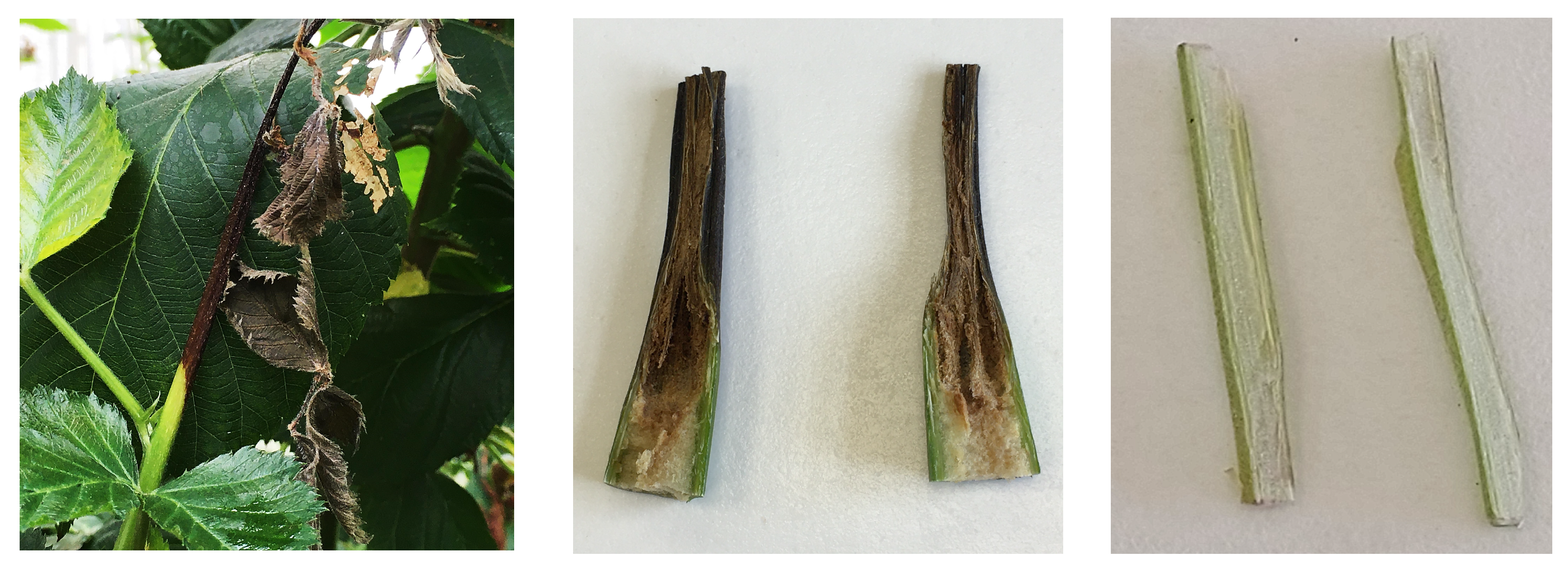
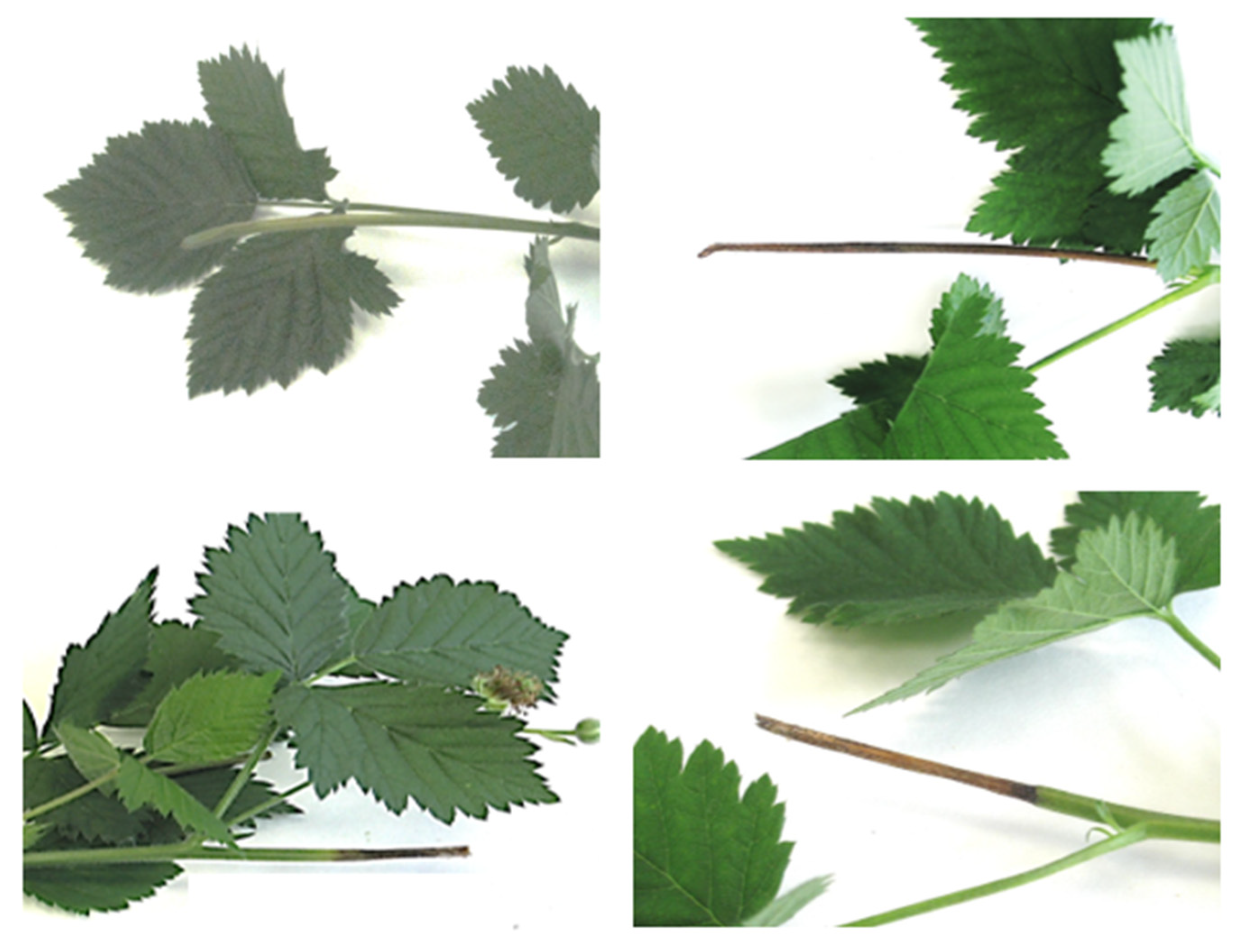
3.1. Pathogen Isolation

Two fungi were consistently isolated from the analyzed tissue samples and the respective strains selected were named LPPAF-977 and LPPAF-981.

3.2. Morphological Characteristics

The strain LPPA-977 grew, developing white circular colonies on the agar surface with sparse to dense aerial hyphae. Conidia were single-celled, hyaline, consistent to Gnomoniopsis, whereas strain LPPA-981 showed a white or gray mycelium with α and β conidia compatible with the genus Diaporthe (Figure 2).

3.3. Identification

Strain LPPAF-977 by BLAST analysis showed that the ITS sequence of 584 bp (accession no. OQ933371) had a 99.48% similarity with the accession no. KC145872 (99% coverage) isolated from Rubus fruticosus in New Zealand. The tub2 sequence of 503 bp (accession no. OQ991913) presented 99.38% identity with accession no. MG860503 (94% coverage) isolated from Rubus fruticosus in Serbia, and the tef-1a sequence of 325 bp (accession no. OQ991914) showed 99.38% identity with accession no. MG773589 (99% coverage) isolated from R. fruticosus in Serbia, all of them corresponding to Gnomoniopsis idaeicola.
The ITS sequence of LPPAF-981 (accession no. OQ933372) of 440 bp exhibited 100% identity (100% coverage) with the Diaporthe rudis isolate CAA832 (accession no. MK792302) and other isolates on Vaccinium corymbosum and several forest species in Portugal. The β-tubulin sequence of 481 bp (accession no. OQ985367) showed 99.79% identity (99% coverage) with D. rudis CBS 143346 (accession no. MG281304) isolated from Vitis vinifera in the Czech Republic. The sequence of tef1a of 457 bp (accession no. OQ991915) presented 99.76% identity (92% coverage) with the D. rudis isolate KP00114 from Pyrus communis in the Netherlands. Calmodulin sequence of 518 bp (accession no. OQ991916) showed 100% identity with a very short coverage (73%) with four strains of D. rudis: CBS 449.82 (accession no. KC343482) isolated from Lupinus sp. in the Netherlands, CBS 266.85 (accession no. KC343479) isolated from Rosa rugosa in the Netherlands, CBS 109768 (accession no. KC343475) isolated from Epilobium angustifolium in Canada, and CBS 100170 (accession no. KC343472) isolated from Fraxinus excelsior in the Netherlands, and also with a strain of D. australafricana CBS 111886 (accession no. KC343280) isolated from Vitis vinifera in Australia. Finally, the sequence of histone of 308 bp (accession no. OR001746) showed 99.68% identity (100% coverage) with strains CAA949 (MT309435) and CAA956 isolated from Pinus pinaster in Portugal, as well as with other D. rudis strains isolated from Vitis vinifera in several countries. The accession codes of all sequences obtained for both fungal species are shown in Supplementary Tables S1 and S2.

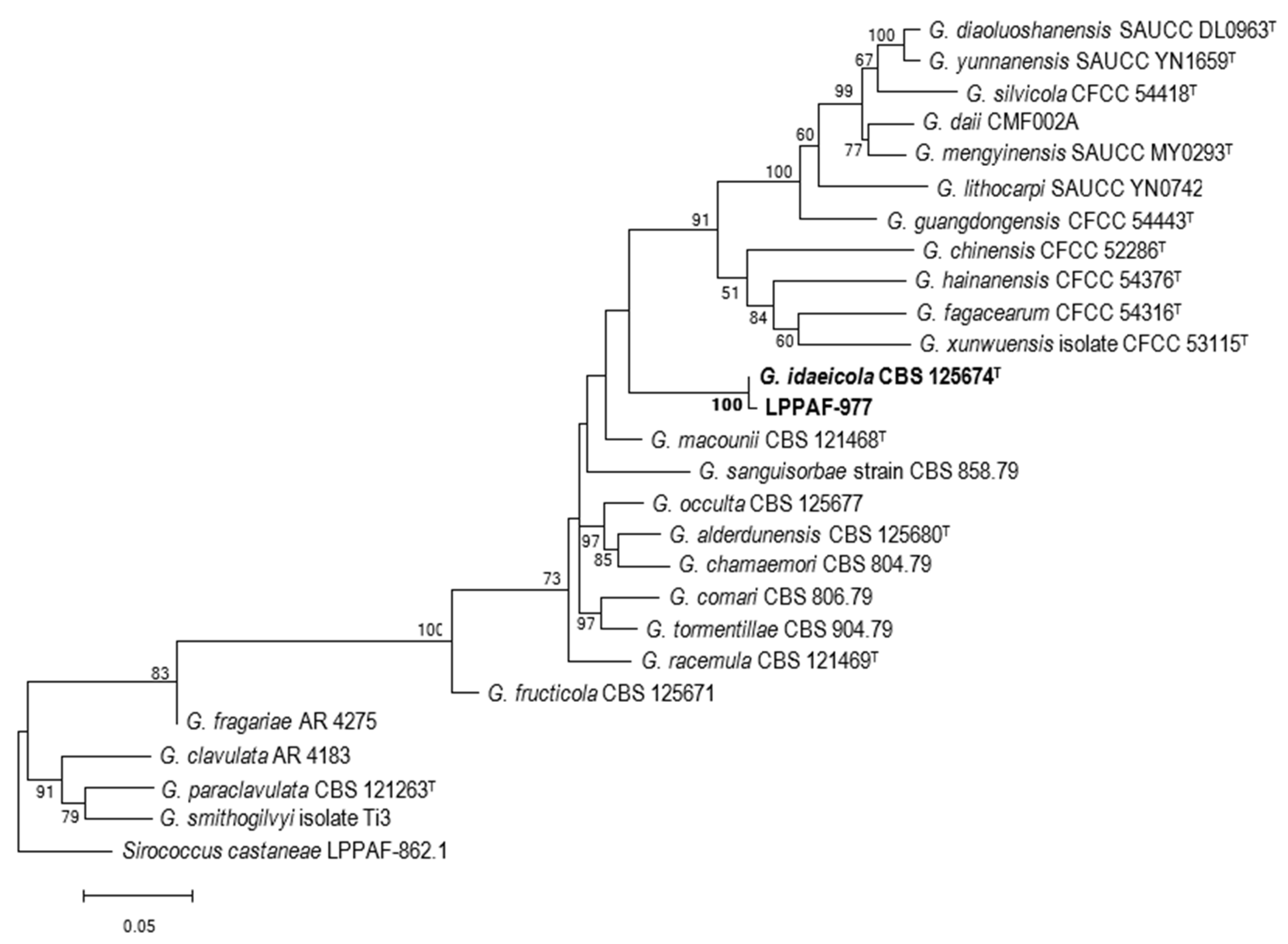
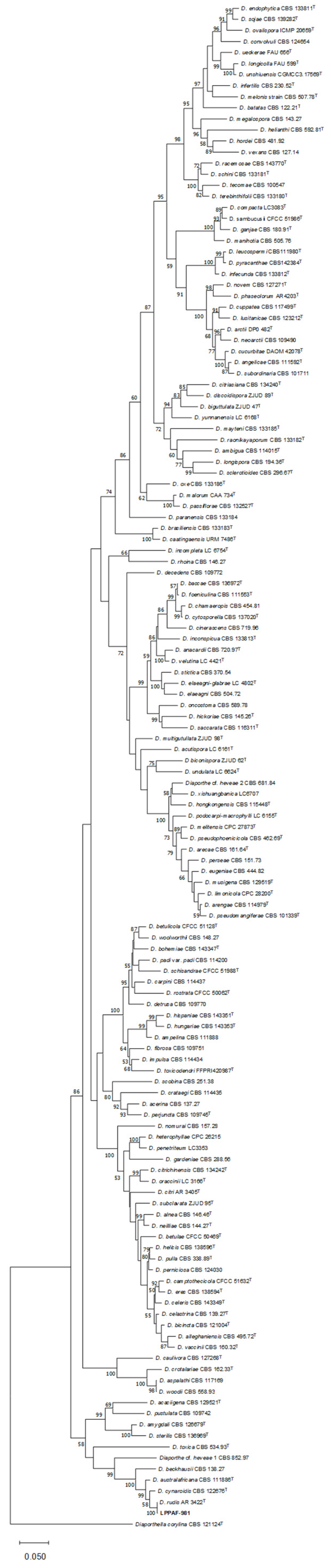
3.4. Multigen Analysis

For Gnomoniopsis, phylogenetic trees were constructed with the single partial sequences of ITS, tub2, and tef-1a genes, and with the three concatenated sequences using those listed in Supplementary Table S1. In the phylogenetic tree obtained with each of the three genes separately, strain LPPAF-977 always clustered with G. idaeicola. The phylogenetic tree with the concatenated sequences of the three loci (total length of 1163 bp (461 bp ITS, 420 bp tub2, and 282 bp tef-1a)) is shown in Figure 3, where we can see that our strain clusters with G. idaeicola with a 100% bootstrap.
With LPPA-981, in the phylogenetic tree created with the single partial sequences of ITS, tub2, tef-1a, and his genes, this strain clustered with D. rudis, while with the cal gene, it clustered with D. canthi (86%) species, far removed from the result obtained for the rest of the genes. Thus, cal was not used in the phylogenetic tree with concatenated sequences. The accession numbers of sequences used are listed in Supplementary Table S2.
Specifically, in the phylogenetic tree with tub2, LPPAF-981 clustered (99%) with D. rudis, D. schoeni, D. pseudotsugae, and D. acericol. With tef-1a, it clustered (87%) with D. rudis, D. asheicola, and D. australafricana, and with his, it clustered (68%) with D. rudis and D. australafricana, which are two closely related species.
The phylogenetic tree with the four concatenated genes showed that LPPAF-981 clustered with D. rudis with a 100% bootstrap (Figure 4).
By means of the Tajima’s test of neutrality, we were able to identify the number of segregating sites and the nucleotide diversity (π) for LPPAF-977 and LPPAF-981 (Table 2).
In both strains, tefl-1a provided the highest nucleotide diversity, whereas, as expected, ITS had the lowest diversity. This may indicate that while ITS is a good primary marker for taxonomy, it may be better not to include it in phylogenetic trees.

3.5. Pathogenicity

Symptoms observed in the field were reproduced on inoculated shoots 25 days after the inoculation (Figure 5). The effects of the fungi treatments in relation to the shoots of blackberries are shown in Figure 6. The results obtained demonstrate significant differences (p < 0.001) between pathogen treatments in terms of the average percentage (%) of canker shoots and vertical lesion (mm).
With regard to the percentage of canker shoots, 100% of the blackberry shoots inoculated with LPPAF-977 as well as those inoculated with the two fungi showed dark-brown lesions, while 10% of the inoculated ones with LPPAF-981 showed no wilt symptoms (Figure 5). Significantly longer tissue necrosis was observed in blackberry shoots co-inoculated with the two pathogens, with a mean lesion length of 40.79 mm. In contrast, shoots inoculated with LPPAF-981 significantly had the lowest values for the lesion lengths: 26.17 mm. No significant differences in the vertical lesion between two pathogens were observed when inoculated independently.
As expected, no symptoms were observed for the two variables analyzed, nor were the fungi under study recovered in any of the control plants, while they were recovered in 100% of the symptomatic tissues of the inoculated blackberry plants, thus fulfilling Koch’s postulates.
When the two strains were sown together in a plate with PDA medium, it was possible to verify that LPPAF-981 grew faster and surrounded LPPAF-977 in the five replicates conducted (Figure 7).

4. Discussion

This study reported for the first time the pathogenicity of two fungi, Gnomoniopsis idaeicola and Diaporthe rudis, causing shoot dieback on blackberries in Spain.
In this work, the identification of the LPPAF-977 strain as G. idaeicola was successfully achieved thanks to the BLAST analysis and individual and concatenated phylogenetic trees. This result is in agreement with other authors using the same three genes (ITS, tub2, and tef-1a) employed in this work [17,32]. It is worth noting that in the Index Fungorum (accessed 8 November 2022) [33], there are 41 species listed in the Gn. Gnomoniopsis; however, nine have been included within the Plagiostoma and Colletotrichum genera. Therefore, these strains were excluded from this study. G. castaneae is synonymous with G. smithogilvyi; therefore, only one of them was included in the phylogenetic tree.
Identification by BLAST of LPPAF-981 as D. rudis was successful with the five genes sequenced (ITS, tub2, tef-1a, his, and cal). In contrast, it was more complex when constructing the phylogenetic trees. Despite the fact that Guarnaccia et al. [34] used only three genes (ITS, tub2, and tef-1a), other authors used up to five loci in previous papers [15,35,36,37,38]. Santos et al. [39] proposed that tef-1a was a superior phylogenetic marker in Diaporthe compared to ITS, while Gomes et al. [15] considered tub2 and his to have higher resolutions than tef-1a and cal. However, Udayanga et al. [27] suggested that the use of cal may lead to errors in the identification of Diaporthe species due to poor sequence quality because of the low specificity of the primers used. In our study, the results support those of Udayanga et al. [27], because the phylogenetic tree obtained with this gene differs completely from the results obtained with the other four genes used. Therefore, it was discarded for inclusion in the final tree created with the concatenated four sequences; therefore, LPPAF-981 clustered with D. rudis with a 100% bootstrap.
The ITS provided the lowest nucleotide diversity in both species; therefore, it was a first genetic marker, but was better for identification to use other genes, such as tef-1a, which was the gene that provided the highest nucleotide diversity that matched the results of Santos et al. [39]. The second gene that provided the highest nucleotide diversity was bet-2 that partially matched with the results of Gomes et al. [15]. Thus, it would not be essential to include the ITS sequence in the phylogenetic tree because it did not provide much differential information. In addition, the use of ITS made it difficult to correctly apply the software used to build phylogenetic trees because it differentiated between the sequences that coded for proteins and those that did not.
G. idaeicola caused noticeable damage as a pathogen in Rubus. Mirhosseini et al. [11] estimated a 30% loss of extremely symptomatic blackberry plants and yield losses of 50–80% in Iran. Stevanović et al. [12] described G. idaeicola as being responsible for the canker, wilt, and death of blackberries in Serbia, reaching an incidence of up to 80%, being responsible for up to 40% plant mortality and estimated yield losses of 50%. These authors also found this species in mixed infections with other fungi, among which was Diaporthe. In Oregon [13], this fungus caused 17% plant mortality. In our study, it was not possible to evaluate the fruit production.
The countries with the highest production of raspberries and blackberries in 2021 were Mexico, Russia, and Serbia, comprising 49% of the production, followed by USA, Poland, Spain, Morocco, Ukraine, Portugal, Bosnia and Herzegovina, the UK, and Chile [8]. Concerning Spain, most blackberry fields are located in southern areas and particularly in the province of Huelva. In northern Spain, where it is also important to consider that the structure of agricultural property is clearly smallholding, blackberry production is on a very small scale. Although local growers are showing great interest in the crop, the production and consumption of blackberries has been gradually increasing, unlike other berries, mainly blueberries and raspberries, which have experienced exponential growth in recent years. Plant breeding programs are aimed at developing crop varieties that are more productive, have a better taste and quality, and are adapted to climate change, pests, and diseases to align with a lower usage of nitrogen fertilizer and pesticides for more sustainable agriculture. The selection of new, high-quality, resistant varieties reduces both the cost of crop production and environmental risks. At present, it is perceived that more consumers are recognizing these aspects as more environmentally friendly. This, coupled with increased health awareness, is contributing to the higher demand for small berries. Our findings suggest the importance of testing new genetics from variety improvement programs for phytosanitary, resilience, and sustainability characteristics well adapted to different climate change scenarios.
In this work, the pathogenic nature of Gnomoniopsis idaeicola and Diaporthe rudis in relation to the shoot dieback of thornless blackberries was confirmed by pathogenicity tests. Nevertheless, both species were co-inoculated twice, each replicate in reverse order, and were also plated together; however, these results do not allow us to establish that there is an antagonism between them. The laboratory results, when both species were sown together, do not correspond with what was observed in the field where G. idaeicola produced a longer vertical lesion than that produced by D. rudis; therefore, this result may indicate that the latter is better adapted to the growth conditions provided by the laboratory. Therefore, further studies must be conducted to clarify this point.
G. idaeicola (P. Karst.) Walker et al. 2010 was described in 1869 as Calosphaeria idaeicola on canes of Rubus idaeus in Finland, and it is present in Finland, France, Bulgaria, Serbia, Spain, Sweden, the USA, Australia, New Zealand, and Iran [40]. This species has been described with a very narrow host range, limited to Rubus spp. [11,12,13,19], but also as endophytes in wheat [41] and Geum peckii [42], and it is present in environmental samples as air from a cave [43] and a sample of human health [44]. There are also records of its presence in Actinidia deliciosa in 1993 in New Zealand (Data of ICMP) and Corylus avellana (data in GenBank), although without information about its pathogenicity.
D. rudis, a synonym for D. viticola [27], has been described as a pathogen in hosts of high agro-economic value, for instance, associated with the stem blight and dieback of blueberries in northern Italy [34], avocados in Chile [45], and Pyrus communis in the USA [46]. In addition, it is also associated with other Diaporthe species causes kiwifruit rot during cold storage in Chile [47] and soybean seed decay in Serbia [48]. It also contributes to grape rot in Italy [49] and was found in Vitis vinifera samples in Portugal, Italy, France, Spain, the UK, and the Czech Republic [36]; however, its pathogenicity has not been tested. Dissanayake et al. [16] described its presence in Cornus sp., Anthoxanthum odoratum, Carlina vulgaris, and Dioscorea communis in Italy. Sequences of strains isolated from many other hosts are available in GenBank, for example, strain ICMO 16419 in Castanea sativa from New Zealand (accession no. KC145904), strain DA244 in Brugmansia sp. from Germany (KC843334), LC6145 in Ilex aquifolium from China, LC6147 Dendrobenthamia japonica from the USA, and CBS 109292 in Laburnum anagyroides from Austria. Thus, since its description, this species has been identified around the world as being associated with numerous hosts [27,50,51], which demonstrates its high degree of dissemination, distribution, and wide host range, similar to that of D. eres.
G. idaeicola has not been previously reported as a pathogen in blackberries in Spain. D. rudis has not been described as a pathogen of Rubus spp. Thus, to the best of our knowledge, this is the first report of G. idaeicola and D. rudis causing dieback in blackberries in Spain.

Supplementary Materials

The following supporting information can be downloaded at: https://www.mdpi.com/article/10.3390/horticulturae9090965/s1, Table S1: Features of the sequences of Gnomoniopsis species used in this work; Table S2: Features of the sequences of Diaporthe species used in this work.

Author Contributions

Conceptualization, A.J.G. and M.C.; methodology, A.J.G.; software, M.C.; investigation, A.J.G.; resources, A.J.G. and M.C.; writing—original draft preparation, A.J.G.; writing—review and editing, A.J.G. and M.C.; funding acquisition, A.J.G. All authors have read and agreed to the published version of the manuscript.

Funding

This research was funded by the Servicio Regional de Investigación y Desarrollo Agroalimentario (SERIDA) and the Consejería de Ciencia, Empresas, Formación y Empleo, Gobierno del Principado de Asturias: Own budgets.

Data Availability Statement

Sequences obtained in this work have been deposited in GenBank under the accession numbers shown in Supplementary Tables S1 and S2.

Acknowledgments

We thank the personnel of SERIDA for field and laboratory support. We also acknowledge J. C. García for providing the non-commercial blackberry samples.

Conflicts of Interest

The authors declare no conflict of interest.

References

  1. Clark, J.R.; Finn, C.E. Blackberry breeding and genetics. In Methods in Temperate Fruit Breeding. Fruit, Vegetable, and Cereal Science and Biotechnology; Flachowsky, H., Hanke, V.M., Eds.; Global Science Books, Ltd.: East Sussex, UK, 2011; pp. 27–43. [Google Scholar]
  2. Jennings, D.L. Raspberry and Blackberries: Their Breeding, Diseases and Growth; Academic Press: San Diego, CA, USA, 1988; 230p. [Google Scholar]
  3. Clark, J.R.; Moore, J.N.; Lopez-Medina, J.; Finn, C.; Perkins-Veazie, P. ‘Prime-Jan’ (‘APF-8’) and ‘Prime-Jim’ (‘APF-12’) primocane-fruiting blackberries. HortScience 2005, 40, 852–855. [Google Scholar] [CrossRef]
  4. Ding, M.; Feng, R.; Wang, S.Y.; Bowman, L.; Lu, Y.; Qian, Y.; Castranova, V.; Jiang, B.-H.; Shi, X. Cyanidin-3-glucoside, a natural product derived from blackberry, exhibits chemopreventive and chemotherapeutic activity. J. Biol. Chem. 2006, 281, 17359–17368. [Google Scholar] [CrossRef]
  5. Kaume, L.; Howard, L.R.; Devareddy, L. The Blackberry Fruit: A Review on Its Composition and Chemistry, Metabolism and Bioavailability, and Health Benefits. J. Agric. Food Chem. 2012, 60, 5716–5727. [Google Scholar] [CrossRef] [PubMed]
  6. Gil-Martínez, L.; Mut-Salud, N.; Ruiz-García, J.A.; Falcón-Piñeiro, A.; Maijó-Ferré, M.; Baños, A.; De la Torre Ramírez, J.M.; Guillamón, E.; Verardo, V.; Gómez-Caravaca, A.M. Phytochemicals determination and antioxidant, antimicrobial, anti-inflamatory and anticancer activities od blackberry fruits. Foods 2023, 12, 1505. [Google Scholar] [CrossRef] [PubMed]
  7. Memete, A.R.; Sarac, I.; Teusdea, A.C.; Budau, R.; Bei, M.; Vicas, S.I. Bioactive compounds and antioxidant capacity of several blackberry (Rubus spp.) fruits cultivars grown in Romania. Horticulturae 2023, 9, 556. [Google Scholar] [CrossRef]
  8. Mercado de Bayas Frescas: Crecimiento, Tendencias, Impacto de COVID-19 y Pronósticos (2023–2028). Available online: https://www.mordorintelligence.com/es/industry-reports/fresh-berries-market (accessed on 3 August 2023).
  9. Ellis, M.A.; Converse, R.H.; Williams, R.N.; Williamson, B. Compendium of Raspberry and Blackberry Diseases and Insects; The American Phytopathological Society: Saint Paul, MN, USA, 1991; 100p. [Google Scholar]
  10. Vrandecic, K.; Jurkovic, D.; Cosic, J.; Postic, J.; Riccioni, L. First Report of Cane Blight on Blackberry Caused by Diaporthe eres in Croatia. Plant Dis. 2011, 95, 612. [Google Scholar] [CrossRef]
  11. Mirhosseini, H.A.; Rahimian, H.; Babaeizad, V.; Hashemi, L. Outbreak of leafspot on blackberry (Rubus fruticosus) caused by Gnomoniopsis sp. in Iran. New Dis. Rep. 2015, 31, 9. [Google Scholar] [CrossRef]
  12. Stevanović, M.; Ristić, D.; Živković, S.; Aleksić, G.; Stanković, I.; Krstić, B.; Bulajić, A. Characterization of Gnomoniopsis idaeicola, the causal agent of canker and wilting of blackberry in Serbia. Plant Dis. 2019, 103, 249–258. [Google Scholar] [CrossRef]
  13. Stockwell, V.O.; Shaffer, B.T.; McGhee, G.C.; Hardigan, M.A. First report of Gnomoniopsis idaeicola causing cane wilt and canker in commercial blackberry fields in Oregon. Plant Dis. 2022, 106, 1980. [Google Scholar] [CrossRef]
  14. Udayanga, D.; Liu, X.; McKenzie, E.H.C.; Chukeatirote, E.; Bahkali, A.H.A.; Hyde, K.D. The genus Phomopsis: Biology, applications, species concepts and names of common phytopathogens. Fungal Divers. 2011, 50, 189–225. [Google Scholar] [CrossRef]
  15. Gomes, R.R.; Glienke, C.; Videira, S.I.R.; Lombard, L.; Groenewald, J.Z.; Crous, P.W. Diaporthe: A genus of endophytic, saprobic and plant pathogenic fungi. Persoonia 2013, 31, 1–41. [Google Scholar] [CrossRef] [PubMed]
  16. Dissanayake, A.J.; Camporesi, E.; Hyde, K.D.; Zhang, W.; Yan, J.Y.; Li, X.H. Molecular phylogenetic analysis reveals seven new Diaporthe species from Italy. Mycosphere 2017, 8, 853–877. [Google Scholar] [CrossRef]
  17. Dissanayake, A.J.; Phillips, A.J.L.; Hyde, K.D.; Yan, J.Y.; Li, X.H. The current status of species in Diaporthe. Mycosphere 2017, 8, 1106–1156. [Google Scholar] [CrossRef]
  18. Marin-Felix, Y.; Hernández-Restrepo, M.; Wingfield, M.J.; Akulov, A.; Carnegie, A.J.; Cheewangkoon, R.; Gramaje, D.; Groenewald, J.Z.; Guarnaccia, V.; Halleen, F.; et al. Genera of phytopathogenic fungi: GOPHY 2. Stud. Mycol. 2019, 92, 47–133. [Google Scholar] [CrossRef]
  19. Walker, D.M.; Castlebury, L.A.; Rossman, A.Y.; Sogonov, M.V.; White, J.F. Systematics of genus Gnomoniopsis (Gnomoniaceae, Diaporthales) based on a three gene phylogeny, host associations and morphology. Mycologia 2010, 102, 1479–1496. [Google Scholar] [CrossRef] [PubMed]
  20. Thomidis, T.; Michailides, T.J. Studies on Diaporthe eres as a new pathogen of peach trees in Greece. Plant Dis. 2009, 93, 1293–1297. [Google Scholar] [CrossRef] [PubMed]
  21. Bai, Q.; Zhai, L.F.; Chen, X.R.; Hong, N.; Xu, W.X.; Wang, G.P. Biological and molecular characterization of five Phomopsis species associated with pear shoot canker in Chine. Plant Dis. 2015, 99, 1704–1712. [Google Scholar] [CrossRef]
  22. Zhang, Q.M.; Yu, C.L.; Li, G.F.; Wang, C.X. First report of Diaporthe eres causing twig canker on Zizyphus jujuba (Jujube) in China. Plant Dis. 2018, 102, 1458. [Google Scholar] [CrossRef]
  23. White, T.J.; Bruns, T.D.; Lee, S.B.; Taylor, J.W. Amplification and direct sequencing of fungal ribosomal RNA Genes for phylogenetics. In PCR—Protocols and Applications—A Laboratory Manual; Innis, M.A., Gelfand, D.H., Sninsky, J.J., White, T.J., Eds.; Academic Press Inc.: New York, NY, USA, 1990; pp. 315–322. [Google Scholar]
  24. Glass, N.L.; Donaldson, G.C. Development of primers sets designed for use with the PCR to amplify conserved genes from filamentous ascomycetes. Appl. Environ. Microbiol. 1995, 61, 1323–1330. [Google Scholar] [CrossRef]
  25. Carbone, I.; Kohn, L.M. A method for designing primer sets for speciation studies in filamentous ascomycetes. Mycologia 1999, 91, 553–556. [Google Scholar] [CrossRef]
  26. Crous, P.W.; Groenewald, J.Z.; Risède, J.-M.; Simoneau, P.; Hywel-Jones, N.L. Calonectria species and their Cylindrocladium anamorphs: Species with sphaeropedunculate vesicles. Stud. Mycol. 2004, 50, 415–430. [Google Scholar]
  27. Udayanga, D.; Castlebury, L.A.; Rossman, A.Y.; Hyde, K.D. Species limits in Diaporthe, molecular re-assessment of D. citri, D. cytosporella, D. foeniculina and D. rudis. Persoonia 2014, 32, 83–101. [Google Scholar] [CrossRef] [PubMed]
  28. Thompson, J.D.; Higgins, D.G.; Gibson, T.J. CLUSTAL W: Improving the sensitivity of progressive multiple sequence alignment through sequence weighting, position-specific gap penalties and weight matrix choice. Nucleic Acids Res. 1994, 22, 4673–4680. [Google Scholar] [CrossRef] [PubMed]
  29. Tamura, K.; Nei, M. Estimation of the number of nucleotide substitutions in the control region of mitochondrial DNA in humans and chimpanzees. Mol. Biol. Evol. 1993, 10, 512–526. [Google Scholar]
  30. Tamura, K.; Stecher, G.; Kumar, S. MEGA 11: Molecular Evolutionary Genetics Analysis Version 11. Mol. Biol. Evol. 2021, 38, 3022–3027. [Google Scholar] [CrossRef]
  31. Tajima, F. Statistical methods to test for nucleotide mutation hypothesis by DNA polymorphism. Genetics 1989, 123, 585–595. [Google Scholar] [CrossRef]
  32. Jiang, N.; Voglmayr, H.; Bian, D.-R.; Piao, C.-G.; Wang, S.-K.; Li, Y. Morphology and Phylogeny of Gnomoniopsis (Gnomoniaceae, Diaporthales) from Fagaceae Leaves in China. J. Fungi 2021, 7, 792. [Google Scholar] [CrossRef]
  33. Index Fungorum. Available online: http://indexfungorum.org/Names/Names.asp (accessed on 8 November 2022).
  34. Guarnaccia, V.; Martino, I.; Tabone, G.; Brondino, L.; Gullino, M.L. Fungal pathogens associated with stem blight and dieback of blueberry in northern Italy. Phytopathol. Mediterr. 2020, 59, 229–245. [Google Scholar] [CrossRef]
  35. Santos, L.; Alves, A.; Alves, R. Evaluating multi-locus phylogenies for species boundaries determination in the genus Diaporthe. PeerJ. 2017, 5, e3120. [Google Scholar] [CrossRef]
  36. Guarnaccia, V.; Groenewald, J.Z.; Woodhall, J.; Armengol, J.; Cinelli, T.; Eichmeier, A.; Ezra, D.; Fontaine, F.; Gramaje, D.; Gutierrez-Aguirregabiria, A.; et al. Diaporthe diversity and pathogenicity revealed from a broad survey of grapevine diseases in Europe. Persoonia 2018, 40, 135–153. [Google Scholar] [CrossRef]
  37. Hilario, S.; Amaral, I.A.; Gonçalves, M.F.M.; Lopes, A.; Santos, L.; Alves, A. Diaporthe species associated with twig blight and dieback of Vaccinium corymbosum in Portugal, with description of four new species. Mycologia 2020, 112, 293–308. [Google Scholar] [CrossRef] [PubMed]
  38. León, M.; Berbegal, M.; Rodríguez-Reina, J.M.; Elena, G.; Abad-Campos, P.; Ramón-Albalat, A.; Olmo, D.; Vicent, A.; Luque, J.; Miarnau, X.; et al. Identification and characterization of Diaporthe spp. associated with twig cankers and shoot blight of almonds in Spain. Agronomy 2020, 10, 1062. [Google Scholar] [CrossRef]
  39. Santos, J.M.; Correia, V.G.; Phillips, A.J.L. Primers for mating-type diagnosis in Diaporthe and Phomopsis: Their use in teleomorph induction in vitro and biological species definition. Fungal Biol. 2010, 114, 255–270. [Google Scholar] [CrossRef] [PubMed]
  40. UK Plant Health Risk Register. Available online: https://planthealthportal.defra.gov.uk/pests-and-diseases/uk-plant-health-risk-register/viewPestRisks.cfm?cslref=30313&riskId=28102 (accessed on 8 February 2023).
  41. Comby, M.; Lacoste, S.; Baillieul, F.; Profizi, C.; Dupont, J. Spatial and temporal variation of cultivable communities of co-occurring endophytes and pathogens in wheat. Front. Microbiol. 2016, 7, 403. [Google Scholar] [CrossRef]
  42. Adams, S.J.; Robicheau, B.M.; LaRue, D.; Browne, R.D.; Walker, A.K. Foliar Endophytic Fungi from the Endangered Eastern Mountain Avens (Geum peckii, Rosaceae) in Canada. Plants 2021, 10, 1026. [Google Scholar] [CrossRef]
  43. Dominguez-Moñino, I.; Jurado, V.; Rogerio-Candelera, M.A.; Hermosin, B.; Saiz-Jimenez, C. Airborne Fungi in Show Caves from Southern Spain. Appl. Sci. 2021, 11, 5027. [Google Scholar] [CrossRef]
  44. Walther, G.; Zimmermann, A.; Theuersbacher, J.; Kaerger, K.; von Lilienfeld-Toal, M.; Roth, M.; Kampik, D.; Geerling, G.; Kurzai, O. Eye Infections Caused by Filamentous Fungi: Spectrum and Antifungal Susceptibility of the Prevailing Agents in Germany. J. Fungi 2021, 7, 511. [Google Scholar] [CrossRef]
  45. Torres, C.; Camps, R.; Aguirre, R.; Besoain, X. First report of Diaporthe rudis in Chile causing stem-end rot on ‘Hass’ avocado fruit imported from California. Plant Dis. 2016, 100, 1951. [Google Scholar] [CrossRef]
  46. KC, A.N.; Rasmussen, A.L. First report of Diaporthe rudis causing fruit rot of European Pears in the United States. Plant Dis. 2019, 103, 2132. [Google Scholar] [CrossRef]
  47. Díaz, G.A.; Latorre, B.A.; Lolas, M.; Ferrada, E.; Naranjo, P.; Zoffoli, J.P. Identification and characterization of Diaporthe ambigua, D. australafricana, D. novem, and D. rudis causing a postharvest fruit rot in kiwifruit. Plant Dis. 2017, 101, 1402–1410. [Google Scholar] [CrossRef]
  48. Petrović, K.; Riccioni, L.; Vidić, M.; Đorđević, V.; Balešević-Tubić, S.; Đukić, V.; Miladinov, Z. First report of Diaporthe novem, D. foeniculina, and D. rudis associated with soybean seed decay in Serbia. Plant Dis. 2016, 100, 2324. [Google Scholar] [CrossRef]
  49. Lorenzini, M.; Zapparoli, G. Diaporthe rudis associated with berry rot of postharvest grapes in Italy. Plant Dis. 2019, 103, 1030. [Google Scholar] [CrossRef]
  50. Huang, F.; Udayanga, D.; Wang, X.; Hou, X.; Mei, X.; Fu, Y.; Hyde, K.D.; Li, H.Y. Endophytic Diaporthe associated with Citrus: A phylogenetic reassessment with seven new species from China. Fungal Biol. 2015, 119, 331–347. [Google Scholar] [CrossRef] [PubMed]
  51. Lombard, L.; van Leeuwen, G.; Guarnaccia, V.; Polizzi, G.; van Rijswick, P.; Rosendah, K.; Crous, P. Diaporthe species associated with Vaccinium in Europe. Phytopathol. Mediterr. 2014, 53, 287–299. [Google Scholar]
Figure 1. (Left): symptoms observed in the field; (center): the inside of affected stem, and (right): healthy stem.
Horticulturae 09 00965 g001
Figure 2. (Left): LPPAF-977. (Right): LPPAF-981.
Horticulturae 09 00965 g002
Figure 3. Phylogenetic tree based on concatenated partial sequences of ITS, tub2, and tef-1a genes (1163 bp), inferred with the maximum likelihood method. The evolutionary distances were computed by the Tamura–Nei model. Bootstrap values ≥50% (based on 1000 replicates) are indicated at branch points. Sirococcus castaneae strain LPPAF-862.1 was used as the outgroup. T, type strain. Bar scale, substitutions per site. Accession numbers of the sequences used to construct the phylogenetic tree are shown in Supplementary Table S1. Evolutionary analyses were performed in MEGA v.11.
Horticulturae 09 00965 g003
Figure 4. Phylogenetic tree based on concatenated partial sequences of ITS, tub2, tef-1a, and his genes, inferred by using the maximum likelihood method and Tamura–Nei model. Bootstrap values ≥50% (based on 1000 replicates) are indicated at branch points. The tree is drawn to scale, with branch lengths measured in the number of substitutions per site (bar scale). There were 138 sequences and a total of 1647 positions involved. Diaporthella corylina strain CBS 121124T was used as the outgroup. T, type strain. Accession numbers of the sequences used to construct the phylogenetic tree are shown in Supplementary Table S2. Evolutionary analyses were performed in MEGA v. 11.
Horticulturae 09 00965 g004
Figure 5. (Above left): control. (Above right): symptoms with LPPA-977. (Below left): symptoms with LPPA-981. (Below right): symptoms with both fungi.
Horticulturae 09 00965 g005
Figure 6. Results of field pathogenicity tests comparing the percentage of canker shoots (A) and the length of the necrotic vertical lesion (mm) (B) between blackberry canes inoculated either with or without pathogens. Boxplots indicate 25–75% quartiles, median (horizontal bars), 5–95% centiles (whiskers), mean values (crosses), and extreme values of the length of the lesion (Horticulturae 09 00965 i001). Chi-squared analysis is shown for percentage data (***: p < 0.001) (A). Different capital letters indicate significant differences (p < 0.001) among pathogens, according to a posteriori Tukey’s test (B).
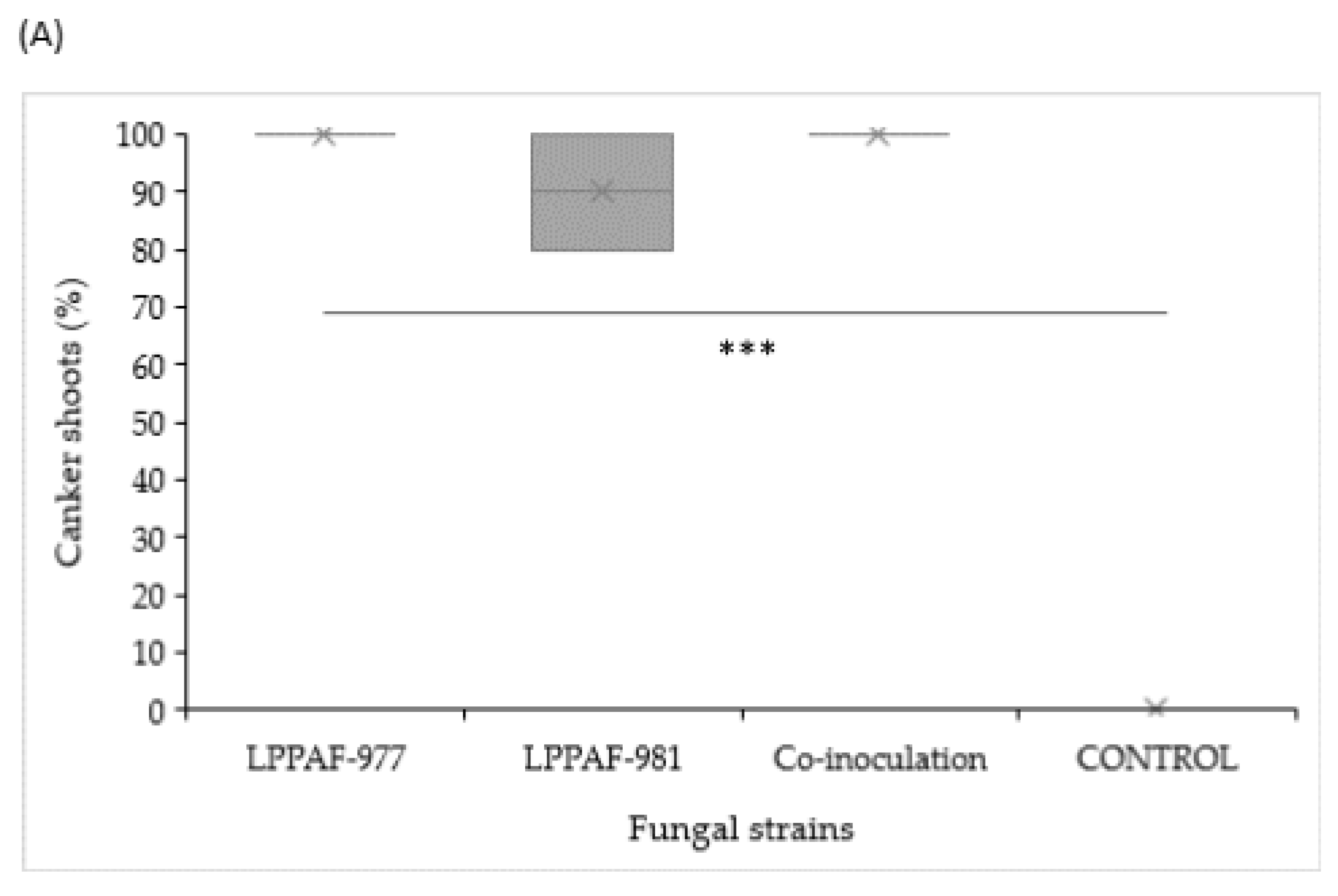
Horticulturae 09 00965 g006aHorticulturae 09 00965 g006b
Figure 7. Growth of LPPAF-977 and LPPAF-981 in Petri dish.
Horticulturae 09 00965 g007
Table 1. Primers used in this work.
GenDefinitionPrimersPrimer SequenceReference
ITSInternal transcribed spacerITS1
ITS4
TCCGTAGGTGAACCTGCGG
TCCTCCGCTTATTGATATGC
[23]
tub2β-tubulinBt2a
Bt2b
GGTAACCAAATCGGTGCTGCTTTC
ACCCTCAGTGTAGTGACCCTTGGC
[24]
tef-1aTranslation elongation factorEF1-728F
EF1-986R
CATCGAGAAGTTCGAGAAGG
TACTTGAAGGAACCCTTACC
[25]
hisHistone H3CYLH3F
H3-1bR
AGGTCCACTGGTGGCAAG
GCGGGCGAGCTGGATGTCCTT
[26]
[24]
calCalmodulinCAL-563F
CL2AR
GAC AAA TCA CCA CCA ARG AG
TTT TTG CAT CAT GAG TTG GA
[27]
Table 2. Results of analyses of G. idaeicola and D. rudis sequences conducted in MEGA v.11.
GenemnSπ
LPPAF-977ITS31407770.046049
tub2273541600.149701
tef-1a292121920.295439
ITS + tub2 + tef-1a279924250.143415
LPPAF-981ITS2115072860.064820
tub22014403500.107078
tef-1a1913112280.158144
his1423451770.093925
ITS + tub2 + tef-1a + his13716478170.097558
m = number of sequences, n = number of positions, S = number of segregating sites, π = nucleotide diversity.
Disclaimer/Publisher’s Note: The statements, opinions and data contained in all publications are solely those of the individual author(s) and contributor(s) and not of MDPI and/or the editor(s). MDPI and/or the editor(s) disclaim responsibility for any injury to people or property resulting from any ideas, methods, instructions or products referred to in the content.

