Repeatability and Comparability of Retinal Blood Vessel Caliber Measurements by OCTA
Abstract
:1. Introduction
2. Materials and Methods
2.1. Subjects
2.2. Imaging
2.3. Analysis
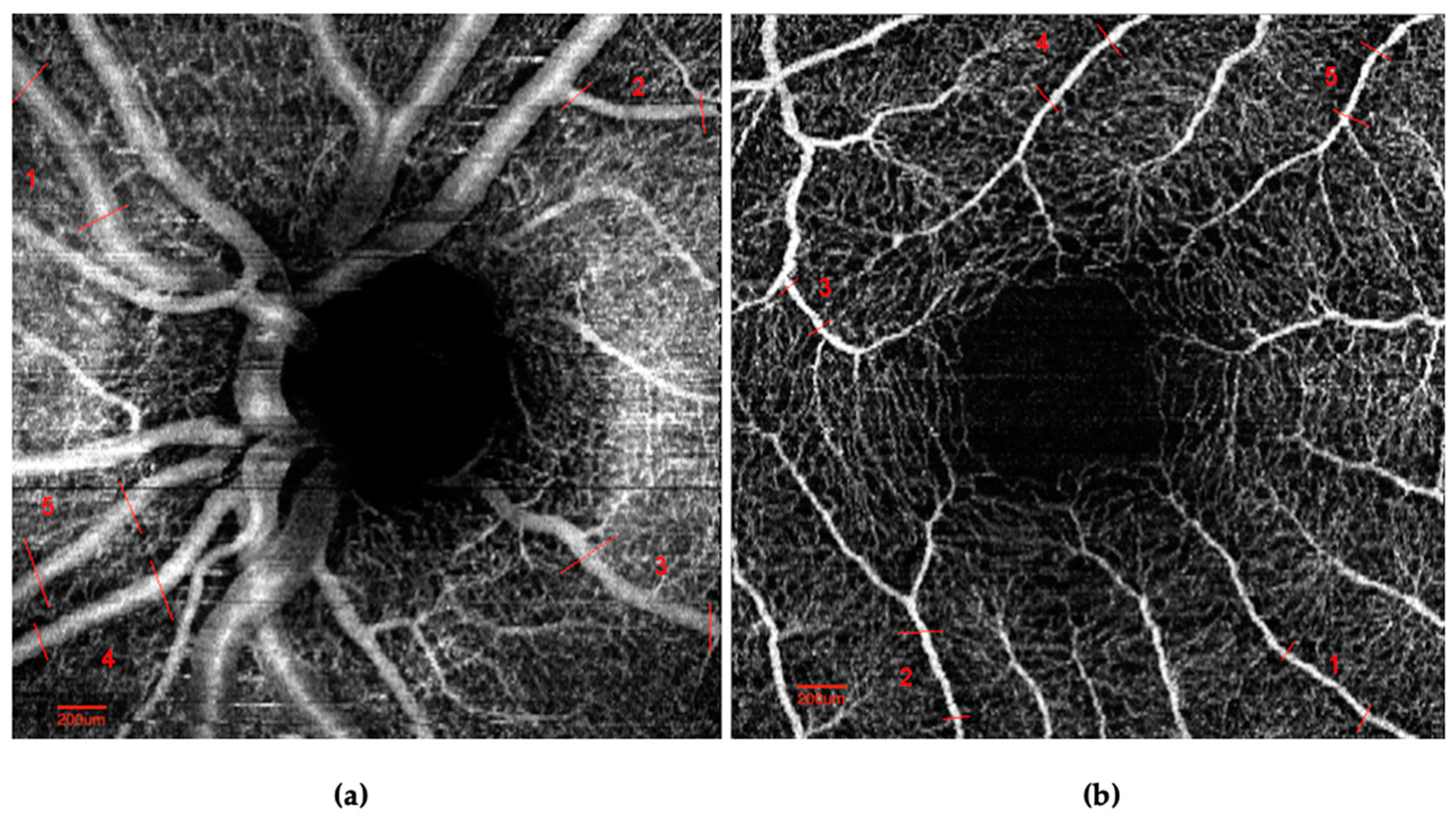
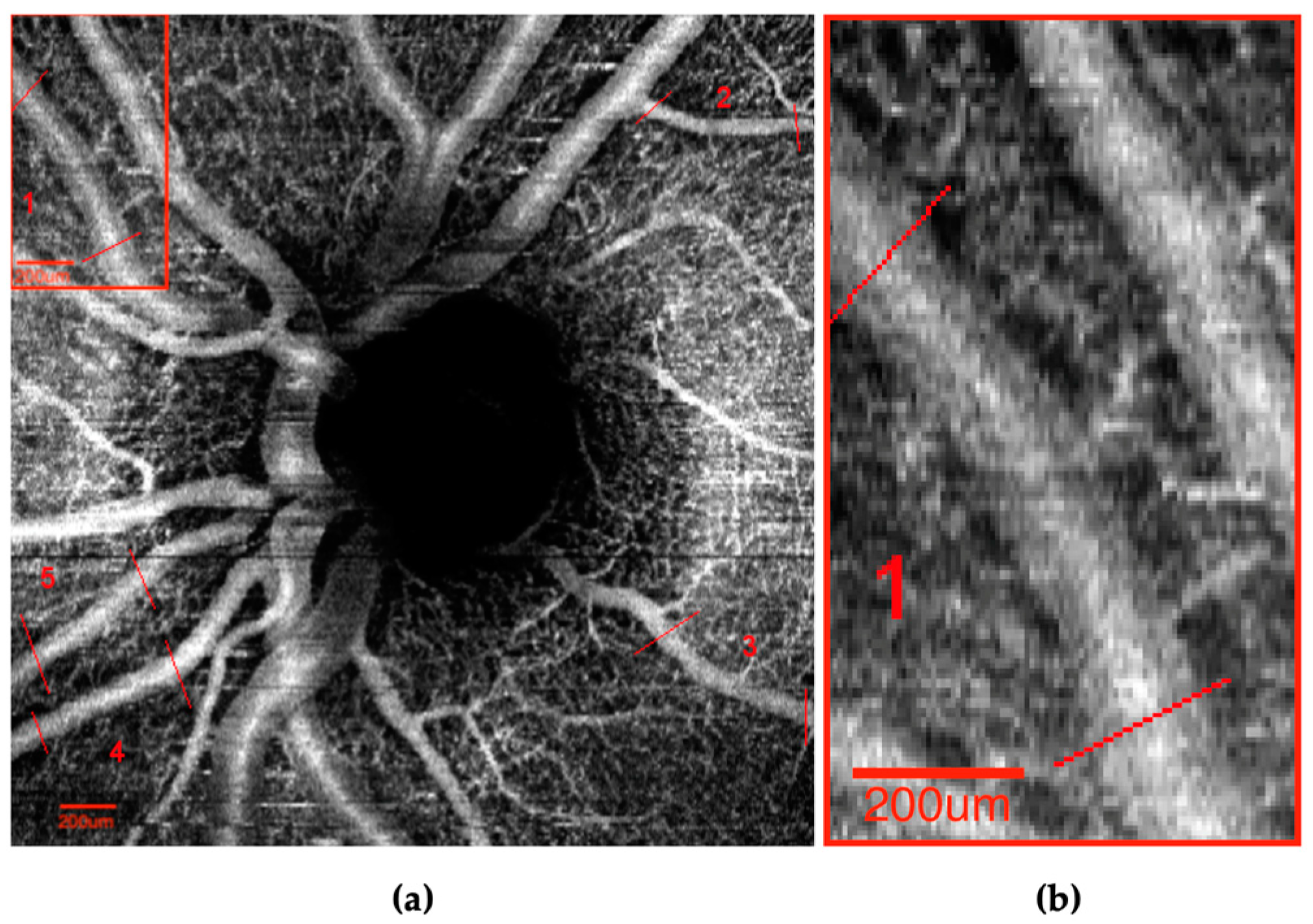
2.3.1. ImageJ and Image Quality—Processing and Vessel Identification
2.3.2. Image Quality Grading
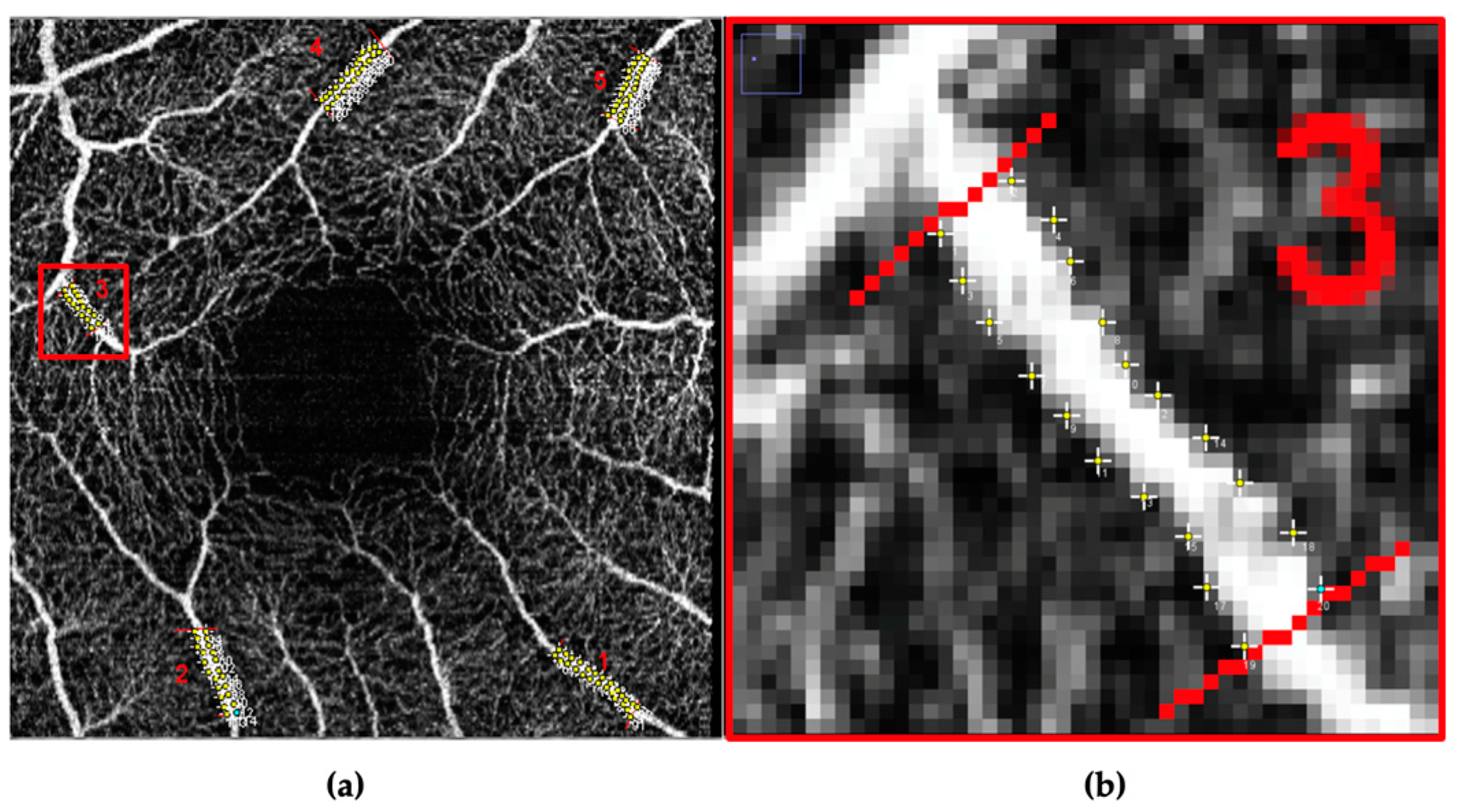
2.3.3. Vessel Caliber Measurements
2.3.4. Vessel Segment Length Measurements
2.4. Statistical Methods
3. Results
3.1. Demographics
3.2. Agreement in Vessel Caliber Measurements of One Image of the Same Vessel by Two Graders (Inter-Grader)
3.3. Agreement in Vessel Caliber Measurements of Two Images of the Same Vessel by the Same Grader (Intra-Grader/Inter-Image)
4. Discussion
Limitations
5. Conclusions
Supplementary Materials
Author Contributions
Funding
Institutional Review Board Statement
Informed Consent Statement
Data Availability Statement
Acknowledgments
Conflicts of Interest
References
- Hirano, T.; Chanwimol, K.; Weichsel, J.; Tepelus, T.; Sadda, S. Distinct Retinal Capillary Plexuses in Normal Eyes as Observed in Optical Coherence Tomography Angiography Axial Profile Analysis. Sci. Rep. 2018, 8, 9380. [Google Scholar] [CrossRef] [PubMed]
- Piltz-seymour, J.R.; Grunwald, J.E.; Hariprasad, S.M.; Dupont, J. Optic nerve blood flow is diminished in eyes of primary open-angle glaucoma suspects. Am. J. Ophthalmol. 2001, 132, 63–69. [Google Scholar] [CrossRef] [PubMed]
- Wong, T.Y. Retinal vessel diameter as a clinical predictor of diabetic retinopathy progression: Time to take out the measuring tape. Arch. Ophthalmol. 2011, 129, 95–96. [Google Scholar] [CrossRef] [PubMed]
- Hwang, T.S.; Gao, S.S.; Liu, L.; Lauer, A.K.; Bailey, S.T.; Flaxel, C.J.; Wilson, D.J.; Huang, D.; Jia, Y. Automated Quantification of Capillary Nonperfusion Using Optical Coherence Tomography Angiography in Diabetic Retinopathy. JAMA Ophthalmol. 2016, 134, 367–373. [Google Scholar] [CrossRef] [PubMed]
- Arrigo, A.; Aragona, E.; Capone, L.; Pierro, L.; Romano, F.; Bandello, F.; Parodi, M.B. Advanced Optical Coherence Tomography Angiography Analysis of Age-related Macular Degeneration Complicated by Onset of Unilateral Choroidal Neovascularization. Am. J. Ophthalmol. 2018, 195, 233–242. [Google Scholar] [CrossRef]
- Al-Sheikh, M.; Akil, H.; Pfau, M.; Sadda, S.R. Swept-Source OCT Angiography Imaging of the Foveal Avascular Zone and Macular Capillary Network Density in Diabetic Retinopathy. Investig. Ophthalmol. Vis. Sci. 2016, 57, 3907–3913. [Google Scholar] [CrossRef] [Green Version]
- Kwon, J.; Choi, J.; Shin, J.W.; Lee, J.; Kook, M.S. An Optical Coherence Tomography Angiography Study of the Relationship between Foveal Avascular Zone Size and Retinal Vessel Density. Investig. Ophthalmol. Vis. Sci. 2018, 59, 4143–4153. [Google Scholar] [CrossRef] [PubMed] [Green Version]
- Asanad, S.; Mohammed, I.; Sadun, A.A.; Saeedi, O.J. OCTA in neurodegenerative optic neuropathies: Emerging biomarkers at the eye-brain interface. Ther. Adv. Ophthalmol. 2020, 12, 2515841420950508. [Google Scholar] [CrossRef]
- Onishi, A.C.; Fawzi, A.A. An overview of optical coherence tomography angiography and the posterior pole. Ther. Adv. Ophthalmol. 2019, 11, 2515841419840249. [Google Scholar] [CrossRef] [Green Version]
- Roy, M.S.; Klein, R.; Janal, M.N. Retinal venular diameter as an early indicator of progression to proliferative diabetic retinopathy with and without high-risk characteristics in African Americans with type 1 diabetes mellitus. Arch. Ophthalmol. 2011, 129, 8–15. [Google Scholar] [CrossRef] [Green Version]
- Agrawal, R.; Joachim, N.; Li, L.J.; Lee, J.; Agarwal, A.; Sim, D.A.; Keane, P.A.; Liew, G.; Pavesio, C.E. Assessment of retinal vascular calibres as a biomarker of disease activity in birdshot chorioretinopathy. Acta Ophthalmol. 2017, 95, e113–e118. [Google Scholar] [CrossRef] [PubMed] [Green Version]
- Couper, D.J.; Klein, R.; Hubbard, L.D.; Wong, T.Y.; Sorlie, P.D.; Cooper, L.S.; Brothers, R.J.; Nieto, F.J. Reliability of retinal photography in the assessment of retinal microvascular characteristics: The Atherosclerosis Risk in Communities Study. Am. J. Ophthalmol. 2002, 133, 78–88. [Google Scholar] [CrossRef]
- Ikram, M.K.; Ong, Y.T.; Cheung, C.Y.; Wong, T.Y. Retinal vascular caliber measurements: Clinical significance, current knowledge and future perspectives. Ophthalmologica 2013, 229, 125–136. [Google Scholar] [CrossRef] [PubMed]
- Dai, C.; Liu, X.; Zhang, H.F.; Puliafito, C.A.; Jiao, S. Absolute retinal blood flow measurement with a dual-beam Doppler optical coherence tomography. Investig. Ophthalmol. Vis. Sci. 2013, 54, 7998–8003. [Google Scholar] [CrossRef] [Green Version]
- Klefter, O.N.; Lauritsen, A.O.; Larsen, M. Retinal hemodynamic oxygen reactivity assessed by perfusion velocity, blood oximetry and vessel diameter measurements. Acta Ophthalmol. 2015, 93, 232–241. [Google Scholar] [CrossRef]
- Jo, Y.H.; Sung, K.R.; Yun, S.C. The Relationship between Peripapillary Vascular Density and Visual Field Sensitivity in Primary Open-Angle and Angle-Closure Glaucoma. Investig. Ophthalmol. Vis. Sci. 2018, 59, 5862–5867. [Google Scholar] [CrossRef] [PubMed] [Green Version]
- Ouyang, Y.; Shao, Q.; Scharf, D.; Joussen, A.M.; Heussen, F.M. Retinal vessel diameter measurements by spectral domain optical coherence tomography. Graefe’s Arch. Clin. Exp. Ophthalmol. 2015, 253, 499–509. [Google Scholar] [CrossRef] [PubMed]
- Kromer, R.; Buhmann, C.; Hidding, U.; Keseru, M.; Keseru, D.; Hassenstein, A.; Stemplewitz, B. Evaluation of Retinal Vessel Morphology in Patients with Parkinson’s Disease Using Optical Coherence Tomography. PLoS ONE 2016, 11, e0161136. [Google Scholar] [CrossRef] [PubMed] [Green Version]
- Moss, H.E.; Treadwell, G.; Wanek, J.; DeLeon, S.; Shahidi, M. Retinal vessel diameter assessment in papilledema by semi-automated analysis of SLO images: Feasibility and reliability. Investig. Ophthalmol. Vis. Sci. 2014, 55, 2049–2054. [Google Scholar] [CrossRef]
- Chen, H.C.; Patel, V.; Wiek, J.; Rassam, S.M.; Kohner, E.M. Vessel diameter changes during the cardiac cycle. Eye 1994, 8 Pt 1, 97–103. [Google Scholar] [CrossRef] [Green Version]
- Dumskyj, M.J.; Aldington, S.J.; Dore, C.J.; Kohner, E.M. The accurate assessment of changes in retinal vessel diameter using multiple frame electrocardiograph synchronised fundus photography. Curr. Eye Res. 1996, 15, 625–632. [Google Scholar] [CrossRef] [PubMed]
- Ghasemi Falavarjani, K.; Al-Sheikh, M.; Darvizeh, F.; Sadun, A.A.; Sadda, S.R. Retinal vessel calibre measurements by optical coherence tomography angiography. Br. J. Ophthalmol. 2017, 101, 989–992. [Google Scholar] [CrossRef] [PubMed]
- Hao, H.; Sasongko, M.B.; Wong, T.Y.; Che Azemin, M.Z.; Aliahmad, B.; Hodgson, L.; Kawasaki, R.; Cheung, C.Y.; Wang, J.J.; Kumar, D.K. Does retinal vascular geometry vary with cardiac cycle? Investig. Ophthalmol. Vis. Sci. 2012, 53, 5799–5805. [Google Scholar] [CrossRef] [PubMed] [Green Version]
- Knudtson, M.D.; Klein, B.E.; Klein, R.; Wong, T.Y.; Hubbard, L.D.; Lee, K.E.; Meuer, S.M.; Bulla, C.P. Variation associated with measurement of retinal vessel diameters at different points in the pulse cycle. Br. J. Ophthalmol. 2004, 88, 57–61. [Google Scholar] [CrossRef] [PubMed] [Green Version]
- Knudtson, M.D.; Lee, K.E.; Hubbard, L.D.; Wong, T.Y.; Klein, R.; Klein, B.E. Revised formulas for summarizing retinal vessel diameters. Curr. Eye Res. 2003, 27, 143–149. [Google Scholar] [CrossRef]
- Papadopoulou, D.N.; Mangioris, G.; Petropoulos, I.K.; Mendrinos, E.; Mavropoulos, A.; Pournaras, C.J. Retinal vessel diameter can reliably be determined in minipigs using Retinal Vessel Analyser with a microscope-mounted fundus camera. Acta Ophthalmol. 2012, 90, e269–e273. [Google Scholar] [CrossRef]
- Sherry, L.M.; Wang, J.J.; Rochtchina, E.; Wong, T.; Klein, R.; Hubbard, L.; Mitchell, P. Reliability of computer-assisted retinal vessel measurementin a population. Clin. Exp. Ophthalmol. 2002, 30, 179–182. [Google Scholar] [CrossRef]
- Sodi, A.; Mucciolo, D.P.; Murro, V.; Zoppetti, C.; Terzuoli, B.; Mecocci, A.; Virgili, G.; Rizzo, S. Computer-Assisted Evaluation of Retinal Vessel Diameter in Retinitis Pigmentosa. Ophthalmic Res. 2016, 56, 139–144. [Google Scholar] [CrossRef]
- Zheng, Q.; Zong, Y.; Li, L.; Huang, X.; Lin, L.; Yang, W.; Yuan, Y.; Li, Y.; He, H.; Gao, Q. Retinal vessel oxygen saturation and vessel diameter in high myopia. Ophthalmic Physiol. Opt. 2015, 35, 562–569. [Google Scholar] [CrossRef]
- Liew, G.; Sim, D.A.; Keane, P.A.; Tan, A.G.; Mitchell, P.; Wang, J.J.; Wong, T.Y.; Fruttiger, M.; Tufail, A.; Egan, C.A. Diabetic macular ischaemia is associated with narrower retinal arterioles in patients with type 2 diabetes. Acta Ophthalmol. 2015, 93, e45–e51. [Google Scholar] [CrossRef]
- Eaton, A.M.; Hatchell, D.L. Measurement of retinal blood vessel width using computerized image analysis. Investig. Ophthalmol. Vis. Sci. 1988, 29, 1258–1264. [Google Scholar]
- Turgut, B. Optical coherence tomography angiography—A general view. J. Opt. Coherence Tomogr. Angiogr.—Gen. View 2016, in press. [Google Scholar] [CrossRef] [Green Version]
- Spaide, R.F.; Fujimoto, J.G.; Waheed, N.K. Image Artifacts in Optical Coherence Tomography Angiography. Retina 2015, 35, 2163–2180. [Google Scholar] [CrossRef] [PubMed]
- Lei, J.; Durbin, M.K.; Shi, Y.; Uji, A.; Balasubramanian, S.; Baghdasaryan, E.; Al-Sheikh, M.; Sadda, S.R. Repeatability and Reproducibility of Superficial Macular Retinal Vessel Density Measurements Using Optical Coherence Tomography Angiography En Face Images. JAMA Ophthalmol. 2017, 135, 1092–1098. [Google Scholar] [CrossRef] [PubMed]
- Chu, Z.; Lin, J.; Gao, C.; Xin, C.; Zhang, Q.; Chen, C.L.; Roisman, L.; Gregori, G.; Rosenfeld, P.J.; Wang, R.K. Quantitative assessment of the retinal microvasculature using optical coherence tomography angiography. J. Biomed. Opt. 2016, 21, 66008. [Google Scholar] [CrossRef] [PubMed] [Green Version]
- Liu, C.H.; Kao, L.Y.; Sun, M.H.; Wu, W.C.; Chen, H.S. Retinal Vessel Density in Optical Coherence Tomography Angiography in Optic Atrophy after Nonarteritic Anterior Ischemic Optic Neuropathy. J. Ophthalmol. 2017, 2017, 9632647. [Google Scholar] [CrossRef] [Green Version]
- Zou, G.Y. Sample size formulas for estimating intraclass correlation coefficients with precision and assurance. Stat. Med. 2012, 31, 3972–3981. [Google Scholar] [CrossRef]
- Kim, A.Y.; Chu, Z.; Shahidzadeh, A.; Wang, R.K.; Puliafito, C.A.; Kashani, A.H. Quantifying Microvascular Density and Morphology in Diabetic Retinopathy Using Spectral-Domain Optical Coherence Tomography Angiography. Investig. Ophthalmol. Vis. Sci. 2016, 57, 362–370. [Google Scholar] [CrossRef] [PubMed]
- Pastore, M.R.; Grotto, A.; Vezzoni, F.; Gaggino, A.; Milan, S.; Gouigoux, S.; Guerin, P.L.; Vinciguerra, A.L.; Cirigliano, G.; Tognetto, D. Reproducibility and Reliability of Spectralis II OCT Angiography Vascular Measurements. Diagnostics 2022, 12, 1908. [Google Scholar] [CrossRef] [PubMed]
- Fondi, K.; Aschinger, G.C.; Bata, A.M.; Wozniak, P.A.; Liao, L.; Seidel, G.; Doblhoff-Dier, V.; Schmidl, D.; Garhöfer, G.; Werkmeister, R.M.; et al. Measurement of Retinal Vascular Caliber from Optical Coherence Tomography Phase Images. Investig. Ophthalmol. Vis. Sci. 2016, 57, 121–129. [Google Scholar] [CrossRef] [Green Version]
- Zhao, Q.; Yang, W.L.; Wang, X.N.; Wang, R.K.; You, Q.S.; Chu, Z.D.; Xin, C.; Zhang, M.Y.; Li, D.J.; Wang, Z.Y.; et al. Repeatability and Reproducibility of Quantitative Assessment of the Retinal Microvasculature Using Optical Coherence Tomography Angiography Based on Optical Microangiography. Biomed. Environ. Sci. 2018, 31, 407–412. [Google Scholar] [CrossRef] [PubMed]
- Eastline, M.; Munk, M.R.; Wolf, S.; Schaal, K.B.; Ebneter, A.; Tian, M.; Giannakaki-Zimmermann, H.; Zinkernagel, M.S. Repeatability of Wide-field Optical Coherence Tomography Angiography in Normal Retina. Transl. Vis. Sci. Technol. 2019, 8, 6. [Google Scholar] [CrossRef] [PubMed] [Green Version]
- Spectralis OCT Angiography Module to Be Presented at AAO. Available online: www.heidelbergengineering.com/int/press-releases/spectralis-oct-angiography-module-to-be-presented-at-aao/ (accessed on 8 May 2022).
- Wang, S.; Xu, L.; Wang, Y.; Wang, Y.; Jonas, J.B. Retinal vessel diameter in normal and glaucomatous eyes: The Beijing eye study. Clin. Exp. Ophthalmol. 2007, 35, 800–807. [Google Scholar] [CrossRef] [PubMed]
- Jonas, J.B.; Nguyen, X.N.; Naumann, G.O. Parapapillary retinal vessel diameter in normal and glaucoma eyes. I. Morphometric data. Investig. Ophthalmol. Vis. Sci. 1989, 30, 1599–1603. [Google Scholar]
- Hall, J.K.; Andrews, A.P.; Walker, R.; Piltz-Seymour, J.R. Association of retinal vessel caliber and visual field defects in glaucoma. Am. J. Ophthalmol. 2001, 132, 855–859. [Google Scholar] [CrossRef]
- Amerasinghe, N.; Aung, T.; Cheung, N.; Fong, C.W.; Wang, J.J.; Mitchell, P.; Saw, S.M.; Wong, T.Y. Evidence of retinal vascular narrowing in glaucomatous eyes in an Asian population. Investig. Ophthalmol. Vis. Sci. 2008, 49, 5397–5402. [Google Scholar] [CrossRef]
- Klein, R.; Klein, B.E.; Moss, S.E.; Wong, T.Y.; Hubbard, L.; Cruickshanks, K.J.; Palta, M. The relation of retinal vessel caliber to the incidence and progression of diabetic retinopathy: XIX: The Wisconsin Epidemiologic Study of Diabetic Retinopathy. Arch. Ophthalmol. 2004, 122, 76–83. [Google Scholar] [CrossRef] [Green Version]
- Benitez-Aguirre, P.; Craig, M.E.; Sasongko, M.B.; Jenkins, A.J.; Wong, T.Y.; Wang, J.J.; Cheung, N.; Donaghue, K.C. Retinal vascular geometry predicts incident retinopathy in young people with type 1 diabetes: A prospective cohort study from adolescence. Diabetes Care 2011, 34, 1622–1627. [Google Scholar] [CrossRef] [Green Version]
- Harrison, W.W.; Chang, A.; Cardenas, M.G.; Bearse, M.A., Jr.; Schneck, M.E.; Barez, S.; Adams, A.J. Blood pressure, vessel caliber, and retinal thickness in diabetes. Optom. Vis. Sci. 2012, 89, 1715–1720. [Google Scholar] [CrossRef] [Green Version]
- Wong, T.Y.; Klein, R.; Klein, B.E.; Meuer, S.M.; Hubbard, L.D. Retinal vessel diameters and their associations with age and blood pressure. Investig. Ophthalmol. Vis. Sci. 2003, 44, 4644–4650. [Google Scholar] [CrossRef]
- Akil, H.; Huang, A.S.; Francis, B.A.; Sadda, S.R.; Chopra, V. Retinal vessel density from optical coherence tomography angiography to differentiate early glaucoma, pre-perimetric glaucoma and normal eyes. PLoS ONE 2017, 12, e0170476. [Google Scholar] [CrossRef]
- Golebiewska, J.; Olechowski, A.; Wysocka-Mincewicz, M.; Odrobina, D.; Baszynska-Wilk, M.; Groszek, A.; Szalecki, M.; Hautz, W. Optical coherence tomography angiography vessel density in children with type 1 diabetes. PLoS ONE 2017, 12, e0186479. [Google Scholar] [CrossRef] [PubMed]
- Shoji, T.; Zangwill, L.M.; Akagi, T.; Saunders, L.J.; Yarmohammadi, A.; Manalastas, P.I.C.; Penteado, R.C.; Weinreb, R.N. Progressive Macula Vessel Density Loss in Primary Open-Angle Glaucoma: A Longitudinal Study. Am. J. Ophthalmol. 2017, 182, 107–117. [Google Scholar] [CrossRef]
- Yarmohammadi, A.; Zangwill, L.M.; Diniz-Filho, A.; Suh, M.H.; Manalastas, P.I.; Fatehee, N.; Yousefi, S.; Belghith, A.; Saunders, L.J.; Medeiros, F.A.; et al. Optical Coherence Tomography Angiography Vessel Density in Healthy, Glaucoma Suspect, and Glaucoma Eyes. Investig. Ophthalmol. Vis. Sci. 2016, 57, 451–459. [Google Scholar] [CrossRef] [Green Version]
- Yarmohammadi, A.; Zangwill, L.M.; Diniz-Filho, A.; Suh, M.H.; Yousefi, S.; Saunders, L.J.; Belghith, A.; Manalastas, P.I.; Medeiros, F.A.; Weinreb, R.N. Relationship between Optical Coherence Tomography Angiography Vessel Density and Severity of Visual Field Loss in Glaucoma. Ophthalmology 2016, 123, 2498–2508. [Google Scholar] [CrossRef] [PubMed] [Green Version]
- Durbin, M.K.; An, L.; Shemonski, N.D.; Soares, M.; Santos, T.; Lopes, M.; Neves, C.; Cunha-Vaz, J. Quantification of Retinal Microvascular Density in Optical Coherence Tomographic Angiography Images in Diabetic Retinopathy. JAMA Ophthalmol. 2017, 135, 370–376. [Google Scholar] [CrossRef]
- Takusagawa, H.L.; Liu, L.; Ma, K.N.; Jia, Y.; Gao, S.S.; Zhang, M.; Edmunds, B.; Parikh, M.; Tehrani, S.; Morrison, J.C.; et al. Projection-Resolved Optical Coherence Tomography Angiography of Macular Retinal Circulation in Glaucoma. Ophthalmology 2017, 124, 1589–1599. [Google Scholar] [CrossRef] [PubMed]
- Triolo, G.; Rabiolo, A.; Shemonski, N.D.; Fard, A.; Di Matteo, F.; Sacconi, R.; Bettin, P.; Magazzeni, S.; Querques, G.; Vazquez, L.E.; et al. Optical Coherence Tomography Angiography Macular and Peripapillary Vessel Perfusion Density in Healthy Subjects, Glaucoma Suspects, and Glaucoma Patients. Investig. Ophthalmol. Vis. Sci. 2017, 58, 5713–5722. [Google Scholar] [CrossRef] [Green Version]
- Jones, A.; Kaplowitz, K.; Saeedi, O. Autoregulation of optic nerve head blood flow and its role in open-angle glaucoma. Expert Rev. Ophthalmol. 2014, 9, 487–501. [Google Scholar] [CrossRef]
- Bedggood, P.; Metha, A. Direct visualization and characterization of erythrocyte flow in human retinal capillaries. Biomed. Opt. Express 2012, 3, 3264–3277. [Google Scholar] [CrossRef]
- Duan, A.; Bedggood, P.A.; Metha, A.B.; Bui, B.V. Reactivity in the human retinal microvasculature measured during acute gas breathing provocations. Sci. Rep. 2017, 7, 2113. [Google Scholar] [CrossRef] [PubMed] [Green Version]
- Zhang, Y.; Roorda, A. Evaluating the lateral resolution of the adaptive optics scanning laser ophthalmoscope. J. Biomed. Opt. 2006, 11, 014002. [Google Scholar] [CrossRef] [PubMed]
- Battu, R.; Dabir, S.; Khanna, A.; Kumar, A.K.; Sinha Roy, A. Adaptive optics imaging of the retina. Indian J. Ophthalmol. 2014, 62, 60–65. [Google Scholar] [CrossRef]
- Cheng, C.S.; Lee, Y.F.; Ong, C.; Yap, Z.L.; Tsai, A.; Mohla, A.; Nongpiur, M.E.; Aung, T.; Perera, S.A. Inter-eye comparison of retinal oximetry and vessel caliber between eyes with asymmetrical glaucoma severity in different glaucoma subtypes. Clin. Ophthalmol. 2016, 10, 1315–1321. [Google Scholar] [CrossRef] [PubMed] [Green Version]
- Wang, J.J.; Shi, Y.; Xie, J.; Tan, A.G.; Hogdson, L.A.; Lee, S.; Wickens, M.; Cosatto, V.F.; Kairaitis, K.; Lindley, R.; et al. Pupil Dilation May Affect Retinal Vessel Caliber Measures. Ophthalmic Epidemiol. 2018, 25, 234–237. [Google Scholar] [CrossRef]


| Demographics | No. of Patients | |
|---|---|---|
| Sex, n (%) | Female | 18 (64%) * |
| Male | 10 (35%) | |
| Race, n (%) | African-American | 19 (68%) * |
| Caucasian | 7 (25%) | |
| Asian | 2 (7%) | |
| Age, n (%) | <50 years | 5 (18%) |
| = or >50 years | 23 (82%) * | |
| Diagnoses | No. of eyes | |
| Glaucoma | 16 | |
| Advanced POAG | 7 | |
| Mild–moderate POAG | 9 | |
| Glaucoma Suspect | 8 | |
| Diabetic Retinopathy | Mild–moderate NPDR | 7 |
| Grade I Hypertensive Retinopathy | 3 | |
| Healthy Controls | 8 | |
| Image Quality | No. of images | |
| Poor | 52 (31.3%) | |
| Fair | 63 (38.0%) * | |
| Good | 61 (36.7%) |
| Mean Vessel Width for Each Subgroup (μm) | Standard Deviation of Two Images 1 (μm) | CoV of Vessel Width | ICC ˆ (95% Confidence Interval) | MDC | |
|---|---|---|---|---|---|
| All Subjects (n = 550) | 54.48 | 5.28 | 0.10 | 0.96 | 14.08 |
| Age * | |||||
| <55 (n = 215) | 59.39 | 6.01 | 0.10 | 0.95 (0.95, 0.96) | 16.04 |
| 55+ (n = 335) | 51.32 | 4.75 | 0.09 | 0.97 (0.96, 0.97) | 12.67 |
| Race | |||||
| White (n = 155) | 60.03 | 3.35 | 0.06 | 0.98 (0.98, 0.98) | 8.89 |
| Asian (n = 40) | 55.31 | 6.62 | 0.12 | 0.89 (0.88, 0.91) | 17.43 |
| Black (n = 355) | 51.96 | 5.77 | 0.11 | 0.96 (0.95, 0.96) | 15.44 |
| Sex | |||||
| Female (n = 335) | 55.75 | 4.54 | 0.08 | 0.97 (0.97, 0.98) | 12.10 |
| Male (n = 215) | 52.49 | 6.26 | 0.12 | 0.94 (0.94, 0.95) | 16.67 |
| Glaucoma * | |||||
| None (n = 295) | 54.14 | 5.63 | 0.10 | 0.95 (0.95, 0.96) | 14.99 |
| Suspect (n = 115) | 61.77 | 4.35 | 0.07 | 0.96 (0.95, 0.97) | 11.50 |
| Mild/mod (n = 85) | 51.16 | 4.91 | 0.10 | 0.97 (0.97, 0.98) | 13.13 |
| Advanced (n = 55) | 46.15 | 5.63 | 0.12 | 0.96 (0.96, 0.97) | 15.05 |
| Diabetes * | |||||
| 0 (n = 340) | 57.44 | 4.93 | 0.10 | 0.96 (0.96, 0.97) | 13.11 |
| 1 (n = 210) | 49.68 | 5.80 | 0.12 | 0.95 (0.94, 0.96) | 15.46 |
| Hypertension | |||||
| 0 (n = 205) | 57.68 | 5.58 | 0.10 | 0.95 (0.94, 0.96) | 14.85 |
| 1 (n = 345) | 52.57 | 5.09 | 0.10 | 0.96 (0.96, 0.97) | 13.55 |
| Pseudophakia | |||||
| 0 (n = 480) | 55.01 | 5.31 | 0.10 | 0.96 (0.95, 0.97) | 14.16 |
| 1 (n = 70) | 50.80 | 5.08 | 0.10 | 0.96 (0.95,0.96) | 13.53 |
| Vessel Width * | |||||
| Low (<33.4) (n = 183) | 28.29 | 3.18 | 0.11 | ||
| Medium (33.4 = 52.7) (n = 184) | 40.97 | 4.21 | 0.10 | ||
| High (52.7+) (n = 183) | 94.23 | 7.47 | 0.08 | ||
| Length † | |||||
| Less than median (n = 281) | 55.24 | 4.88 | 0.09 | 0.97 (0.96, 0.97) | 13.05 |
| More than median(n = 269) | 53.68 | 5.67 | 0.11 | 0.95 (0.94, 0.96) | 15.10 |
| Location * | |||||
| Disc (n = 240) | 80.29 | 5.58 | 0.07 | 0.98 (0.98, 0.98) | 15.07 |
| Macula (n = 210) | 34.49 | 5.03 | 0.15 | 0.72 (0.67, 0.76) | 12.48 |
Disclaimer/Publisher’s Note: The statements, opinions and data contained in all publications are solely those of the individual author(s) and contributor(s) and not of MDPI and/or the editor(s). MDPI and/or the editor(s) disclaim responsibility for any injury to people or property resulting from any ideas, methods, instructions or products referred to in the content. |
© 2023 by the authors. Licensee MDPI, Basel, Switzerland. This article is an open access article distributed under the terms and conditions of the Creative Commons Attribution (CC BY) license (https://creativecommons.org/licenses/by/4.0/).
Share and Cite
Tsai, J.; Asanad, S.; Whiting, M.; Zhang, X.; Magder, L.; Saeedi, O. Repeatability and Comparability of Retinal Blood Vessel Caliber Measurements by OCTA. Vision 2023, 7, 48. https://doi.org/10.3390/vision7030048
Tsai J, Asanad S, Whiting M, Zhang X, Magder L, Saeedi O. Repeatability and Comparability of Retinal Blood Vessel Caliber Measurements by OCTA. Vision. 2023; 7(3):48. https://doi.org/10.3390/vision7030048
Chicago/Turabian StyleTsai, Joby, Samuel Asanad, Martha Whiting, Xuemin Zhang, Laurence Magder, and Osamah Saeedi. 2023. "Repeatability and Comparability of Retinal Blood Vessel Caliber Measurements by OCTA" Vision 7, no. 3: 48. https://doi.org/10.3390/vision7030048
APA StyleTsai, J., Asanad, S., Whiting, M., Zhang, X., Magder, L., & Saeedi, O. (2023). Repeatability and Comparability of Retinal Blood Vessel Caliber Measurements by OCTA. Vision, 7(3), 48. https://doi.org/10.3390/vision7030048


