Abstract
Salmonella enterica serovar Typhi (S. Typhi) that has developed resistance to many antimicrobials poses a serious challenge to public health. Hence, this study aimed to systematically determine the prevalence of antimicrobial resistance (AMR) in S. Typhi isolated from the environment and humans as well as to ascertain the spread of the selected AMR genes in S. Typhi. This systematic review and meta-analysis were performed according to the Preferred Reporting Items for Systematic Review and Meta-Analysis (PRISMA) guidelines, and the study protocol was registered with the International Prospective Register of Systematic Reviews (PROSPERO). A total of 2353 studies were retrieved from three databases, of which 42 studies fulfilled the selection criteria. The pooled prevalence of AMR S. Typhi (using a random-effect model) was estimated at 84.8% (95% CI; 77.3–90.2), with high heterogeneity (I2: 95.35%, p-value < 0.001). The high estimated prevalence indicates that control methods should be improved immediately to prevent the spread of AMR among S. Typhi internationally.
1. Introduction
Salmonella enterica serovar Typhi (S. Typhi), a rod-shaped, gram-negative bacteria from the family Enterobacteriaceae, is responsible for typhoid fever, a serious bloodstream infection. It has been an important cause of morbidity and mortality in the developing world [1]. The incidence of typhoid in low- and middle-income countries in South Asia and Africa is more common than in developed countries [1]. The Global Burden of Disease Study estimated 9.25 million cases of typhoid fever in 2019, resulting in 110,000 deaths, of which the majority occurred in South Asia and sub-Saharan Africa [2]. In nature, S. Typhi can only infect humans and is usually contracted through ingestion of food or water contaminated with the organism’s excrement. It was reported that there were 888 cases and two deaths in Kelantan (Malaysia) during a large outbreak in 2005 [3]. Patients typically present symptoms after a 7-to-14 day asymptomatic period following ingestion of S. Typhi-contaminated food or water. Following the initial asymptomatic period, patients can develop an influenza-like illness with a fever and also nausea, vomiting and diarrhea [4].
It is well-known that antibiotics have reduced the morbidity and mortality of many infectious diseases [5] and have been used as an important weapon to fight typhoid fever. However, the spread and injudicious use of antibiotics in human medicine has led to the rapid emergence and spread of antimicrobial resistance (AMR) in S. Typhi strains [6]. There are various antibiotic-resistance-related genes found in S. Typhi including catA1 (conferring resistance to chloramphenicol), blaTEM-1 (resistant to ampicillin), dhfr7, and sul1 (resistant to co-trimoxazole) [7]. The development of AMR in S. Typhi can occur spontaneously through mutations [8]. In addition, point mutations in the quinolone resistance-determining region (QRDR) harboring the genes for DNA gyrase gyrA and gyrB and topoisomerase IV parC and parE result in quinolone-resistant S. Typhi [7]. However, S. Typhi can also acquire the AMR gene from non-relatives on mobile genetic elements (MGE) such as plasmids and transposon. This horizontal gene transfer (HGT) allows the AMR gene to be transferred among different species of bacteria [9].
To date, the distribution of AMR in S. Typhi data among different countries, regions and environments is necessary for appropriate antimicrobial therapy for patient management and surveillance programs. Hence, in this study, a systematic review and meta-analysis were performed to evaluate the prevalence of AMR in S. Typhi isolated from the environment and humans globally as well as to assess the spread of the selected AMR genes in S. Typhi.
2. Materials and Methods
2.1. Study Design and Protocol
The present systematic review and meta-analysis were conducted following the preferred reporting items for systematic review and meta-analyses (PRISMA 2020) guidelines [10]. In addition, the protocol for this systematic review was registered in the International Prospective Register of Systematic Reviews (PROSPERO), with a registered ID of CRD42022319530.
2.2. Search Strategy
A systematic search was conducted on three databases, namely, PubMed, Scopus and Science Direct using the following keywords: (antimicrobial resistance gene) OR (antibiotic resistance gene) AND (Salmonella Typhi). To ensure comprehensive data collection, filters including publication year, study design and language were not applied in this review.
2.3. Inclusion and Exclusion Criteria
The studies’ titles and abstracts were screened based on the inclusion and exclusion criteria. Those articles that met the inclusion criteria were included and further reviewed. All of the eligible studies included in the review had to meet the following inclusion criteria: (i) Salmonella enterica serotype Typhi isolated from humans and environments, (ii) articles reported on antimicrobial resistance gene of S. Typhi and (iii) published in English. The studies were excluded if they did not involve the isolation of S. Typhi and non-resistant S. Typhi. Books, book chapters, review articles, media reports, case reports and studies written in languages other than English were also excluded from the review.
2.4. Data Extraction and Risk of Bias (RoB) Assessment
Studies that met the criteria for full-text review were selected for further analysis. Subsequently, risk assessment and data extraction were carried out. Selected studies were subjected to the following data extraction: (i) first author; (ii) period of study; (iii) region and country of the conducted studies; (iv) methodology: the total number of samples, type of samples (patient/clinical, environment), and the total number of S. Typhi isolated and; (v) outcome: number of isolates resistance against each antimicrobial, number of bacteria isolates with AMR gene and number of mutated cases. In addition, data on the type of antibiotics resistant among S. Typhi was also extracted. Studies that collected samples from different countries and regions were categorized as worldwide in order to avoid confusion during data extraction and analysis.
The Joanna Briggs Institute (JBI) critical appraisal tool for Studies Reporting Prevalence Data was used to assess the risk of bias (RoB) of the selected studies. Parameters assessed for bias included (i) sample description, i.e., either from an environmental or hospital setting; (ii) study design, sample size and sampling techniques; (iii) use of valid and standard methods in microbiologic techniques such as bacteriologic culture and antimicrobial sensitivity; (iv) confirmation methods for detection of AMR genes in S. Typhi and (v) the statistical analysis used for reporting summary measures. The quality of each study was graded with ‘1’ for an answer to each question of ‘yes’ and ‘0’ for answers of ‘no’ or ‘unclear’. The low-RoB articles (the total quality score was ≥7) were included in the study whereas articles with moderate (the total quality score was between 4 and 6) and high RoB (the total quality score was ≤3) were excluded for further analysis.
2.5. Data Synthesis and Analysis
Data analysis was performed using Comprehensive Meta-Analysis Software (CMA) (Version 3.0). The pooled prevalence of AMR in S. Typhi, and the selected AMR genes (gyrA, gyrB, parC, parE and blaTEM genes) in S. Typhi were measured and the sub-group analysis (to analyze the sources of heterogeneity) was conducted according to the country and region of each study. A random-effect model using the DerSimonian-Laird method of meta-analysis was employed to create the pooled estimates of the reported AMR among S. Typhi and the chosen AMR genes in S. Typhi cases. A forest plot was generated to summarize details of the individual studies alongside the estimated common effect and degree of heterogeneity. The potential for publication bias was examined using funnel plots (visual aid for detecting bias) and the asymmetry of the plot was further assessed using Egger’s regression test. The heterogeneity (i.e., variation in study outcomes between studies) of the study-level estimates was evaluated using Cochran’s Q test and I2 estimates. I2 values of 25%, 50% and 75% were considered low, moderate and high heterogeneity, respectively [11]. For all of the tests, a p-value of <0.05 was considered to be statistically significant.
3. Results
3.1. Overview of the Selected Studies
A total of 2353 studies were retrieved from the targeted online databases, of which 250 duplicates were removed, as presented in the PRISMA flowchart (Figure 1). The titles and abstracts of 2103 studies were screened based on the inclusion and exclusion criteria and 1859 studies were excluded for further analysis. As a result, 244 full texts were assessed for eligibility. A total of 87 studies were removed due to incomplete data on the number of S. Typhi isolates, AMR genes, tests used for the detection of the AMR genes and antimicrobial susceptibility tests for all S. Typhi. A total of 157 studies were included in the final qualitative synthesis before removing 115 studies due to the high and moderate risk of bias based on the risk quality assessment score (≤6 score), a lack of detail on the number of resistant S. Typhi and/or the number of S. Typhi with resistance gene being unspecified. Finally, 42 studies were included in the meta-analysis.
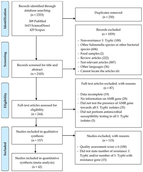
Figure 1.
PRISMA flowchart illustrating the process of identifying, screening and selecting eligible articles used in this study.
3.2. Characteristics of the Eligible Studies
All the eligible studies included in the meta-analyses were of high methodological quality (Appendix A; Table A1). Most of the included studies were conducted in Asia (n = 23) involving nine countries, where India is the main country of origin of the isolates reported in eligible studies (n = 7) (Table 1). Isolates were also collected from Bangladesh and Pakistan; both countries provided four studies. Samples originating from China constituted three studies (n = 3) and the remaining were from Cambodia, Indonesia, Japan, Myanmar and Nepal; each constituted one study from the eligible articles (n = 1).

Table 1.
Major characteristics of the included studies in the systematic review.
A total of 4359 isolates derived from humans (patient/clinical) and environmental sources were tested for phenotypic and genotypic antimicrobial resistance profiles. Of all the selected studies, the highest proportions of S. Typhi isolates were identified from human samples. Of the 42 studies, 39 confirmed that 4242 samples of S. Typhi were isolated from humans (Table 1). Meanwhile, 113 strains were isolated from the environment, as reported by two included studies [16,44]. However, it was unclear from one study how many of the four S. Typhi isolates originated in humans versus the environment [27]. Among the isolates, 3320 were resistant to at least one antimicrobial agent. The majority of the isolates were resistant to ciprofloxacin (58%), chloramphenicol (30%) and nalidixic acid (29%) (Figure 2A). Only one study by Al-Muhanna et al. [21] did not reveal the phenotypic antibiotic resistance profile as the report highlighted only the number of resistant S. Typhi isolates (Table 1). Our analysis showed a total of 42 genes conferring antibiotic resistance. The most reported AMR genes were the gyrA (79%) and parC (45%) genes, both genes conferring resistance quinolones as well as the blaTEM gene (29%), conferring resistance against β-lactam (Figure 2B).

Figure 2.
The frequency of top ten (A) antibiotic resistance in S. Typhi and (B) reported antimicrobial resistance genes. AMC: amoxicillin-clavulanic acid, AMP: ampicillin, AMX: amoxicillin, CHL: chloramphenicol, CIP: Ciprofloxacin, NAL: nalidixic acid, CRO: ceftriaxone, STR: streptomycin, SXT: trimethoprim-sulfamethoxazole and TMP: trimethoprim.
Apart from that, the distribution of mutations in gyrA, gyrB, parC and parE genes was also retrieved from the eligible articles (Appendix B; Table A2). Out of 34 studies that reported the presence of the gyrA gene in resistant S. Typhi, 30 studies showed mutations in gyrA while the remaining studies did not test for the presence of gyrA mutations in the isolates. Mutations at codon 83 (S83F) and 87 (D87N) were the most commonly reported mutations in the gyrA gene (S83F: n = 28 studies, D87N: n = 16 studies). Mutations in the gyrB gene were reported in seven studies in which S464F (n = 4 studies) and S464Y (n = 2 studies) mutations were the most frequently reported. The most commonly reported mutations in topoisomerase IV genes (parC and parE) were S80I (parC) (n = 15 studies) and D420N (parE) (n = 3 studies).
3.3. The Pooled Prevalence of Antimicrobial Resistant Strains in S. Typhi
The pooled prevalence of AMR strains in S. Typhi using a random-effect model was estimated at 84.8% (95% CI; 77.3–90.2), but with high heterogeneity (I2 = 95.35%, p-value < 0.001) (Figure 3). A sub-group analysis based on countries and regions was performed to examine the potential sources of heterogeneity, which were presented in Table 2. The highest pooled prevalence of 98.9% (95% CI; 95.7–99.7) was observed from Iraq (n = 4) and the lowest pooled prevalence was estimated at 18.2% (95% CI, 8.4–35.0) from Peru (n = 1) Heterogeneity was highest among studies conducted in India (I2 = 96.529%) followed by four studies conducted in Pakistan (I2 = 93.686%). Based on regional data, studies from America (n = 2) showed the lowest estimate at 63.7% (95% CI; 2.0–99.3), had I2 of 88.889%, and studies from the Middle East region (n = 6) showed the highest estimate at 97.7% (95% CI; 93.0–99.3) with I2 of 0.000%. In addition, the funnel plot showed evidence of publication bias attributed to the relatively asymmetrical plot (Figure 4A). Moreover, using the trim-and-fill method (under the random effects model), 13 missing studies were imputed to the left side of the mean effect, resulting in the imputed point estimate of 73.080% (Figure 4B). In addition to the funnel plots, Egger’s test was utilized to affirm the extent of bias (t-value = 0.938, p-value = 0.177).
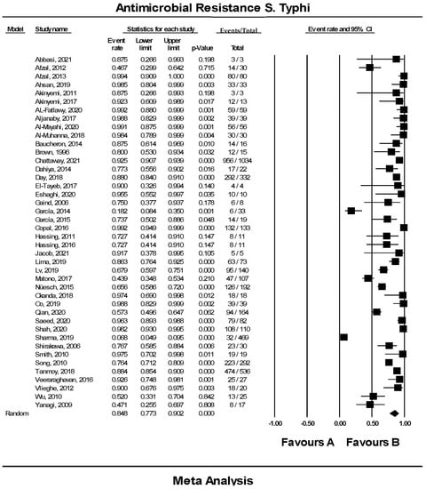
Figure 3.
Forest plot showing the pooled prevalence of resistant S. Typhi isolates estimated by a random effect model of meta-analysis (84.8%, I2: 95.35, 95% CI: 77.3–90.2, p-value < 0.001) [12,13,14,15,16,17,18,19,20,21,22,23,24,25,26,27,28,29,30,31,32,33,34,35,36,37,38,39,40,41,42,43,44,45,46,47,48,49,50,51,52,53].

Table 2.
Sub-group analysis of prevalence of resistant S. Typhi according to countries and regions.

Figure 4.
Funnel plot showing (A) publication bias in studies reporting the prevalence of antimicrobial resistant S. Typhi and (B) result after performing the trim-and-fill method where 13 missing studies (closed circles) were added on the left side of the mean effect.
3.4. The Pooled Prevalence of gyrA Gene in S. Typhi
Out of 42 included studies, only 33 studies reported the presence of the gyrA gene in S. Typhi isolates. The prevalence and heterogeneity of the gyrA gene in resistant S. Typhi are presented in Figure 5. Of the 2922 isolates, the prevalence of gyrA resistance was 91.3% (95% CI; 84.1–95.5), indicating that the majority of the resistant S. Typhi possessed the gyrA gene. The heterogeneity of the included studies was significantly high (I2 = 93.99%, p-value < 0.001). The pooled prevalence based on country revealed Japan with one study as the highest (99.0%, 95% CI; 845.4–99.9) with a heterogeneity of 0.000 (Table 3). The lowest prevalence was observed in Saudi Arabia (n = 1), 25.0% (95% CI; 3.4–76.2) with a heterogeneity of 0.000. Sub-group analysis based on regions showed that America (n = 2) had the highest estimation, 94.3% (I2 = 0.000%, 95% CI; 68.7–99.2), followed by Asia (n = 19), 93.2% (I2 = 91.108%, 95% CI; 85.2–97.0). However, the asymmetrical funnel plot showed the presence of publication bias (Figure 6A). The trim-and-fill method (under the random effects model) showed a point estimate of 88.092% with seven missing studies imputed to the left side of the mean effect (Figure 6B). In addition to the funnel plots, Egger’s test was utilized to affirm the extent of bias (t-value = 1.158, p-value = 0.128).
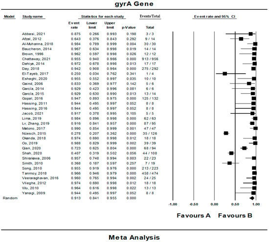
Figure 5.
Forest plot showing the pooled prevalence of gyrA gene in resistant S. Typhi isolates estimated by a random effect model of meta-analysis. (91.3%, I2: 93.99, 95% CI: 84.1–95.5, p-value < 0.001) [12,13,14,15,16,17,18,19,20,21,22,23,24,25,26,27,28,29,30,31,32,33,34,35,36,37,38,39,40,41,42,43,44,45,46,47,48,49,50,51,52,53].

Table 3.
Sub-group analysis of prevalence of gyrA gene in S. Typhi according to countries and regions.

Figure 6.
Funnel plot showing (A) publication bias in studies reporting the prevalence of gyrA gene in antimicrobial resistant S. Typhi and (B) result after performing the trim-and-fill method where seven missing studies (closed circles) were added on the left side of the mean effect.
3.5. The Pooled Prevalence of gyrB Gene in S. Typhi
Eight of the 42 publications showed data for the gyrB gene in resistant S. Typhi. Figure 7 presents a forest plot of the proportions of gyrB gene in resistant isolates. The pooled prevalence was estimated at 8.0% out of 1962 resistant isolates indicating that the gyrB gene was quite rare in these isolates. Overall, data heterogeneity was similar in the gyrA gene (I2 = 93.98%, p-value < 0.001). On the other hand, the pooled prevalence based on country showed Iraq, with one study, as the highest (83.3%, 95% CI; 65.7–92.9) with a heterogeneity of 0.000% (Table 4). The lowest prevalence was observed in the United Kingdom (n = 1), 2.4% (95% CI; 1.1–4.9) with a heterogeneity of 0.000%. By region, the Middle East (n = 1) had the highest estimation, 83.3% (I2 = 0.000%, 95% CI; 65.7–92.9), followed by Europe (n = 3), 6.9% (I2 = 91.477%, 95% CI; 1.9–22.5). In addition, the asymmetrical funnel plot showed the presence of publication bias (Figure 8). However, further tests on this asymmetrical funnel plot could not be performed as the number of studies included for the analysis of the gyrB gene was less than 10 (n = 8) [54].

Figure 7.
Forest plot showing the pooled prevalence of gyrB gene in resistant S. Typhi isolates estimated by a random effect model of meta-analysis. (8.0%, I2: 93.98, 95% CI: 2.8–20.9, p-value < 0.001) [21,24,26,31,36,41,42,49].

Table 4.
Sub-group analysis of prevalence gyrB gene in S. Typhi according to countries and regions.

Figure 8.
Funnel plot showing publication bias in studies reporting the prevalence of gyrB gene in antimicrobial resistant S. Typhi.
3.6. The Pooled Prevalence of parC Gene in S. Typhi
Out of 42 included studies, only 19 studies reported the presence of the parC gene in resistant S. Typhi isolates. The prevalence and heterogeneity of the parC gene in resistant S. Typhi are presented in Figure 9. Of the 2348 isolates, the prevalence of the parC gene resistance was 23.3% (95% CI; 15.0–34.2), indicating that the parC gene was less dominant in the resistant S. Typhi. Heterogeneity analysis showed that data heterogeneity was significantly high (I2 = 91.26%, p-value < 0.001). Sub-group analysis by country revealed that the prevalence of parC gene resistant was highest in Iran (n = 1) at 87.5% (95% CI; 26.6–99.3) with heterogeneity of 0.000% (Table 5). The lowest prevalence was observed in China (n = 1), 1.1% (95% CI; 0.1–7.2) with heterogeneity of 0.000%. Interestingly, France and Switzerland, with the same number of studies (n = 1) showed a similar prevalence of 7.1% (95% CI; 1.0–37.0 (France), 3.8–13.2 (Switzerland)) and heterogeneity of 0.000%. Based on regions, the Middle East (n = 2) had the highest prevalence at 56.6% (95% CI; 6.3–96.2) compared to other regions. Meanwhile, the lowest prevalence was reported in Europe (n = 5), 14.0% (95% CI; 10.8–18.0). Additionally, the asymmetrical funnel plot showed the presence of publication bias (Figure 10A). Moreover, the trim-and-fill method (under the random effects model) showed a point estimate of 21.750% with one missing study imputed to the left side of the mean effect (Figure 10B). In addition to the funnel plots, Egger’s test was utilized to affirm the extent of bias (t-value = 1.085, p-value = 0.147).
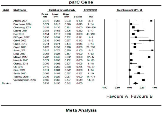
Figure 9.
Forest plot showing the pooled prevalence of parC gene in resistant S. Typhi isolates estimated by a random effect model of meta-analysis. (23.3%, I2: 91.26, 95% CI: 15.0–34.2, p-value < 0.001) [12,22,24,25,27,29,31,32,35,36,38,39,40,41,42,47,49,50].

Table 5.
Sub-group analysis of prevalence parC gene in S. Typhi according to countries and regions.
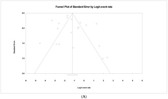
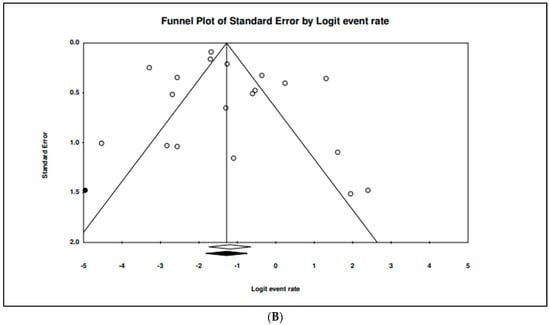
Figure 10.
Funnel plot showing (A) publication bias in studies reporting the prevalence of parC gene in antimicrobial resistant S. Typhi and (B) result after performing trim-and-fill method where one missing study (closed circles) were added on the left side of the mean effect.
3.7. The Pooled Prevalence of parE Gene in S. Typhi
Nine of the 42 publications showed data for the parE gene in resistant S. Typhi. Figure 11 presents a forest plot of the proportions of the parE gene in resistant isolates. The pooled prevalence was estimated at 7.0% out of 2177 resistant isolates, indicating that the parE gene was quite rare in these isolates. Overall, the heterogeneity in data across the eligible studies was significantly high (I2 = 90.47%, p-value < 0.001). On the other hand, the pooled prevalence by country showed Worldwide with one study as the highest (14.8%, 95% CI; 10.7–20.1) with a heterogeneity of 0.000% (Table 6). The lowest prevalence was observed in the United Kingdom (n = 1), 2.1% (95% CI; 0.9–4.5) with a heterogeneity of 0.000%. By region, the highest estimate was also observed as Worldwide (n = 1) 14.8% (95% CI; 10.7–20.1, I2 = 0.000%,), while the lowest prevalence was observed Europe (n = 4) 3.6% (95% CI; 1.7–7.2, I2 = 57.466%,). In addition, the asymmetrical funnel plot showed presence of publication bias (Figure 12). However, further tests on this asymmetrical funnel plot could not be performed as the number of studies included for the analysis of parE gene was less than 10 (n = 9) [54].

Figure 11.
Forest plot showing the pooled prevalence of parE gene in resistant S. Typhi isolates estimated by a random effect model of meta-analysis. (7.0%, I2: 90.47, 95% CI: 3.7–12.7, p-value < 0.001) [22,24,26,31,36,38,42,48,49].

Table 6.
Sub-group analysis of prevalence parE gene in S. Typhi according to regions.

Figure 12.
Funnel plot showing publication bias in studies reporting the prevalence of parE gene in antimicrobial resistance S. Typhi.
3.8. The Pooled Prevalence of blaTEM Gene in S. Typhi
Out of 42 included studies, only 12 studies reported the presence of the blaTEM gene in resistant S. Typhi isolates. The prevalence and heterogeneity of the blaTEM gene in resistant S. Typhi are presented in Figure 13. Of the 2166 isolates, the prevalence of blaTEM in resistant isolates was 55.1% (95% CI; 41.7–67.7), indicating that the majority of the resistant S. Typhi possessed the blaTEM gene. The heterogeneity in the data was high but at an insignificant level (I2 = 95.27, p-value < 0.500). The sub-group analysis based on countries revealed the highest estimate of 95.5% in Canada (n = 1) (95% CI; 55.2–99.7) and had low heterogeneity, 0.000% (Table 7). The lowest prevalence was observed in India (n = 1), 20.0% (95% CI; 2.7–69.1) with a heterogeneity of 0.000%. In terms of regions, America (n = 1) had the highest prevalence at 95.5% (95% CI; 55.2–99.7) compared to other regions. The lowest prevalence was reported in Europe (n = 2), 29.5% (95% CI; 24.6–34.9). Additionally, the asymmetrical funnel plot showed the presence of publication bias (Figure 14A). Moreover, trim-and-fill analysis (under the random effects model) showed a point estimate of 52.870% with one missing study imputed to the left side of the mean effect (Figure 14B). In addition to the funnel plots, Egger’s test was utilized to affirm the extent of bias (t-value = 1.463, p-value = 0.087).

Figure 13.
Forest plot showing the pooled prevalence of blaTEM gene in resistant S. Typhi isolates estimated by a random-effect model of meta-analysis. (55.1%, I2: 95.27, 95% CI: 41.7–67.7, p-value < 0.500) [13,14,19,20,24,26,28,35,36,37,43,49].

Table 7.
Sub-group analysis of prevalence blaTEM gene in S. Typhi according to countries and regions.

Figure 14.
Funnel plot showing (A) publication bias in studies reporting the prevalence of blaTEM gene in antimicrobial resistant S. Typhi and (B) result after performing trim-and-fill method where one missing study (closed circles) was added on the left side of the mean effect.
4. Discussion
S. Typhi infection has recently grown to be a serious burden to healthcare systems, causing higher morbidity and mortality among patients worldwide. Effective choices for the treatment of S. Typhi infections are becoming challenging as resistant strains are increasing globally [5]. Therefore, in the present review, a meta-analysis was performed to estimate the global prevalence of antimicrobial resistance in S. Typhi isolated from the environment and humans. A total of 42 studies that met the inclusion criteria were included for final analysis. Of these, there were 3320 AMR S. Typhi isolates recorded from 23 countries in six regions. A random-effects model was used to analyze the data. As a result, the pooled prevalence of AMR in S. Typhi was estimated to be 84.8% (95% CI; 77.3–90.2).
In general, the prevalence of AMR was high in isolates from the Middle East (97.7%), Africa (93.5%) and Asia (83.5%). These regions are known to be endemic areas for enteric fever where good sanitation and better public health management are not widely practiced [1]. The higher prevalence of AMR was due to the indiscriminate use of antibiotics [52]. Owing to high living expenses, patients in low- and middle-income countries may not have access to or be able to visit health institutions, forcing them to seek care elsewhere, such as in the informal sector or over the counter at local pharmacies [55]. Across our investigation, a significant amount of AMR in Europe (81.4%) was also reported. However, travelers returning from endemic regions and guests visiting family and friends, who are more inclined to be negligent with food and water sources, accounted for the majority of cases [1].
Our analysis found that heterogeneity between studies was high (I2 = 95.35%, p-value < 0.001), contributed by India (I2 = 96.529%) and Pakistan (I2 = 93.686%). The remaining countries were low heterogeneity (I2 = 0.000–22.168%), except for China which had moderate heterogeneity (I2 = 55.694%). In terms of regions, the higher heterogeneity was contributed by Asia (I2 = 96.156%), Europe (I2 = 93.997%) and America (I2 = 88.889%). The other regions, Africa (I2 = 0.000%), Middle East (I2 = 0.000%) and worldwide (I2 = 0.000%), showed a low heterogeneity. The different methods and interpretations of “resistant” S. Typhi may contribute to the high heterogeneity, especially in Asia and Europe. For instance, Afzal et al. [13] used Kirby–Bauer disk diffusion and E-test strips methods and interpreted the results using Clinical and Laboratory Standards Institution (CLSI) 2012 guidelines. In contrast, Lima et al. [36] used disk diffusion and broth-microdilution methods and interpreted the results according to the European Committee on Antimicrobial Susceptibility Testing (EUCAST v8) 2018. Our results show that Asia persists as a major source of antimicrobial resistance in S. Typhi and that clones of the bacteria that develop resistance in Asia are frequently spread throughout the region and beyond. These findings demonstrate the widespread dissemination of antimicrobial-resistant S. Typhi strains internationally, emphasizing the need to consider typhoid management strategies from a global rather than a regional perspective. Antimicrobials have contributed to controlling and eliminating infectious diseases. However, the improper use of these drugs has caused the emergence of resistance strains, and, in turn, very limited drugs are left to treat the disease. Thus, the discovery of new drugs within a particular class of antimicrobials is required to replace existing drugs that have been ineffective. Furthermore, the improvement in antibiotic stewardship plays a significant role in the maintenance of the resistance phenotype and could have a beneficial impact on the proper management of antimicrobial resistant strain outbreaks.
A total of 42 genes conferring antibiotics resistance against S. Typhi isolates were observed. However, only five genes which include gyrA, gyrB, parC, parE and blaTEM were chosen to evaluate their prevalence. These genes were chosen based on the highest frequency of the top five genes that have contributed to the continuous emergence of quinolone- and β-lactam-resistant isolates in S. Typhi [56]. Both quinolones and β-lactam were commonly prescribed to treat S. Typhi-infected individuals [55]. However, the emergence of quinolones- and β-lactam-resistant isolates due to these resistance genes may cause a challenge in the treatment of S. Typhi infection and regulatory safeguards are required for the use of these antibiotic classes. The gyrA, gyrB, parC and parE genes were associated with resistant to quinolones [57]. The gyrA and gyrB genes encode for the DNA gyrase subunit while parC and parE genes encode for the topoisomerase IV subunit [58]. These enzymes are the target sites of quinolones to inhibit DNA replication and cause cell death in the bacteria [57]. However, mutations in these genes, overexpression of the efflux pump system and the innate impermeability of the membrane result in quinolone resistance [59]. On the other hand, the blaTEM gene was associated with resistance to β-lactam [60]. It encodes for β-lactamases that cause enzymatic inactivation (e.g., acetylation of aminoglycosides) and degradation of the β-lactam [61,62].
Based on the result of the analysis, a high prevalence was reported in the gyrA gene, 91.3%, followed by the blaTEM gene, 55.1%, and the parC gene, 23.3%. In contrast, both gyrB and parE genes were relatively rare in the resistant isolates in which the prevalence of these genes was 8.0% and 7.0% respectively. Based on sub-group analysis, America showed high prevalence in gyrA (94.3%, 95% CI; 68.7–99.2) and blaTEM genes (95.5%, 95% CI; 55.2–99.7), while the Middle East showed high prevalence in gyrB (83.3%, 95% CI; 65.7–92.9) and parC genes (56.6%, 95% CI; 6.3–96.2). On the other hand, the parE gene was highly reported worldwide (14.8%, 95% CI; 10.7–20.1). The pooled prevalence of the gyrA gene in Africa reported by Tadesse et al. [63] was 5.7% which was found to be slightly lower compared to our study (Africa = 36.8%, 95%; 18.7–59.7).
Mutations in quinolone-resistance genes were also observed in which mutations in gyrA and parC genes were the most commonly reported. According to Nguyen Van et al. [63] there is a strong association between changes in the gyrA and parC genes and the minimum inhibitory concentrations (MICs) of the antibiotics levofloxacin (LEV) and ciprofloxacin (CIP). The gyrA and parC’s quinolone resistance-determining region (QRDR) is where most mutations that cause fluoroquinolone resistance can be identified [64]. In our study, the most common gyrA mutations occurred in codons Ser-83 (n = 29 studies) and Asp-87 (n = 21 studies). On the other hand, the most common parC mutations occurred in codons Ser-80 (n = 15 studies) and Glu-84 (n = 10 studies). These data were consistent with the findings reported by Fukushima et al. [65] in which most mutations in the QRDR are at gyrA Ser-83 and Asp-87 and at parC Ser-80 and Glu-84.
Despite the comprehensive review, there are few limitations in this study. First, the meta-analysis could not cover all of the countries and regions in order to understand the complete overview of prevalence in AMR S. Typhi and the AMR gene in AMR S. Typhi, which is attributed to the lack of data in some countries. Second, since this review excluded studies written in language other than English, we might have omitted data from some countries, resulting in a publication language bias. In addition, most of the isolates were derived from typhoid cases, and the numbers of isolates from the environmental samples were relatively limited. In summary, more surveillance data are required to adequately reflect the resistant isolates from various countries to overcome the limitations. Moreover, this study was unable to cover all antimicrobial resistance genes to provide a comprehensive view of the prevalence of antimicrobial resistance genes as some of the data on these genes were limited. Other than that, in order to achieve high accuracy of the overall prevalence of antimicrobial resistance genes, studies that did not report the study period are also included in this review as these studies provide data on the prevalence of antimicrobial resistance genes in S. Typhi. Despite this caveat, findings of the present review may still provide useful insights for government decision makers and health practitioners on the urgency to pay attention towards preparing for dealing with the emergence of S. Typhi antibiotic resistance.
5. Conclusions
In conclusion, this systematic review and meta-analysis provided an overview of AMR S. Typhi in humans and the environment. The pooled prevalence of AMR S. Typhi was estimated at 84.8% (95% CI; 77.3–90.2), which was considerably high. Based on the 42 studies, most S. Typhi isolates were resistant to ciprofloxacin, chloramphenicol, nalidixic acid and ampicillin. Mutations in gyrA and parC genes were the most commonly reported whereas mutation in gyrB and parE was rarely reported. These mutations may contribute to the resistance of quinolone in S. Typhi. The establishment of AMR S. Typhi and the propagation of AMR-related genes data must be regularly monitored to provide information needed for management decisions about the use of antibiotics. Preventive actions should be taken immediately to prevent the spread of AMR S. Typhi internationally, and there is a need for a collaborative study on the epidemiology of AMR development and essential intervention across the human health sectors, as well as environmental sector.
We conclude that with the emergence of typhoid infection by resistant S. Typhi, surveillance programs including health awareness should be carried out effectively to reduce cases and control the disease.
Author Contributions
Conceptualization and methodology, N.Y.Y. and N.I.I.N.; data extraction, synthesis, and interpretation, N.I.I.N., N.F.M.Z., M.M.A., M.A.N., B.G., A.H.A.M., M.A.H.A.H., M.N.S.M.S., A.A.Z. and N.Y.Y.; formal analysis, N.I.I.N.; validation, N.Y.Y. and I.A.; writing (original draft preparation), N.I.I.N. and N.Y.Y.; writing (review and editing) M.A.N., I.A. and N.Y.Y.; supervision, N.Y.Y.; funding acquisition, N.Y.Y. and I.A. All authors have read and agreed to the published version of the manuscript.
Funding
This research was funded by USM Short Term grant, 304.CIPPM.6315337 and Higher Institution Centre of Excellence (HICoE) Grant from the Ministry of Higher Education, Malaysia (311/CIPPM/4401005).
Institutional Review Board Statement
Not applicable.
Informed Consent Statement
Not applicable.
Data Availability Statement
The datasets used and/or analyzed during the current study are included in the manuscript.
Acknowledgments
We would like to thank Suhana Ahmad, Rohimah Mohamud and Ahmad Adebayo Irekeola from the Department of Immunology, School of Medical Sciences, Universiti Sains Malaysia, Kubang Kerian 16150, Malaysia, for their technical support and advice on data analysis.
Conflicts of Interest
The authors declare no conflict of interest.
Appendix A

Table A1.
Quality of included studies by JBI critical appraisal checklist for studies reporting prevalence data.
Table A1.
Quality of included studies by JBI critical appraisal checklist for studies reporting prevalence data.
| No. | Author | Checklist | Score | ||||||||
|---|---|---|---|---|---|---|---|---|---|---|---|
| 1 | 2 | 3 | 4 | 5 | 6 | 7 | 8 | 9 | |||
| 1 | Abbasi & Ghaznavi-Rad, 2021 [12] | YES | YES | YES | YES | YES | YES | NO | NO | YES | 7 |
| 2 | Afzal et al., 2012 [13] | YES | YES | YES | YES | NO | YES | YES | YES | NO | 7 |
| 3 | Afzal et al., 2013 [14] | YES | YES | YES | YES | NO | YES | YES | NO | YES | 7 |
| 4 | Ahsan & Rahman, 2019 [15] | YES | YES | YES | YES | NO | YES | YES | NO | YES | 7 |
| 5 | Akinyemi et al., 2011 [16] | YES | YES | YES | YES | YES | YES | YES | YES | YES | 9 |
| 6 | Akinyemi et al., 2017 [17] | YES | YES | YES | YES | YES | YES | YES | YES | YES | 9 |
| 7 | Al-Fatlawy & Al-Hadrawi, 2020 [18] | YES | YES | YES | NO | NO | YES | YES | YES | YES | 7 |
| 8 | Aljanaby & Medhat, 2017 [19] | YES | YES | YES | YES | YES | YES | YES | YES | YES | 9 |
| 9 | Al-Mayahi & Jaber, 2020 [20] | YES | YES | YES | YES | YES | YES | YES | YES | YES | 9 |
| 10 | Al-Muhanna et al., 2018 [21] | YES | YES | YES | YES | NO | YES | YES | NO | YES | 7 |
| 11 | Baucheron et al., 2014 [22] | YES | NO | YES | YES | YES | YES | YES | NO | YES | 7 |
| 12 | Brown et al., 1996 [23] | NO | NO | YES | YES | YES | YES | YES | YES | YES | 7 |
| 13 | Chattaway et al., 2021 [24] | YES | YES | YES | YES | NO | YES | YES | YES | YES | 8 |
| 14 | Dahiya et al., 2014 [25] | YES | YES | YES | NO | YES | YES | YES | YES | YES | 8 |
| 15 | Day et al., 2018 [26] | YES | YES | YES | YES | YES | YES | YES | YES | NO | 8 |
| 16 | El-Tayeb et al., 2017 [27] | YES | YES | YES | YES | YES | YES | YES | YES | YES | 9 |
| 17 | Eshaghi et al., 2020 [28] | YES | YES | YES | YES | NO | YES | YES | NO | YES | 7 |
| 18 | Gaind et al., 2006 [29] | YES | YES | YES | NO | YES | YES | YES | NO | YES | 7 |
| 19 | García et al., 2014 [30] | YES | NO | YES | YES | YES | YES | YES | NO | YES | 7 |
| 20 | García-Fernández et al., 2015 [31] | YES | YES | YES | YES | NO | YES | YES | YES | YES | 8 |
| 21 | Gopal et al., 2016 [32] | YES | YES | YES | NO | NO | YES | YES | YES | YES | 7 |
| 22 | Hassing et al., 2011 [33] | YES | YES | YES | YES | NO | YES | YES | YES | YES | 8 |
| 23 | Hassing et al., 2016 [34] | YES | YES | YES | YES | NO | YES | YES | YES | NO | 7 |
| 24 | Jacob et al., 2021 [35] | YES | YES | YES | YES | YES | YES | NO | YES | YES | 8 |
| 25 | Lima et al., 2019 [36] | YES | YES | YES | YES | YES | YES | YES | YES | YES | 9 |
| 26 | Lv, Zhang & Song, 2019 [37] | YES | YES | YES | YES | NO | YES | YES | YES | YES | 8 |
| 27 | Matono et al., 2017 [38] | YES | YES | YES | YES | YES | YES | YES | YES | YES | 9 |
| 28 | Nüesch-Inderbinen et al., 2015 [39] | YES | YES | YES | YES | NO | YES | YES | NO | YES | 7 |
| 29 | Okanda et al., 2018 [40] | YES | YES | YES | YES | NO | YES | YES | YES | YES | 8 |
| 30 | Oo et al., 2019 [41] | YES | YES | YES | YES | NO | YES | YES | NO | YES | 7 |
| 31 | Qian et al., 2020 [42] | YES | YES | YES | YES | NO | YES | YES | YES | YES | 8 |
| 32 | Saeed et al., 2020 [43] | YES | YES | YES | YES | NO | YES | YES | NO | YES | 7 |
| 33 | Shah et al., 2020 [44] | YES | YES | YES | YES | NO | YES | YES | NO | YES | 7 |
| 34 | Sharma et al., 2019 [45] | YES | YES | YES | YES | NO | YES | YES | YES | NO | 7 |
| 35 | Shirakawa et al., 2006 [46] | YES | YES | YES | YES | NO | YES | YES | YES | YES | 8 |
| 36 | Smith, Govender & Keddy, 2010 [47] | YES | YES | YES | YES | NO | YES | YES | NO | YES | 7 |
| 37 | Song et al., 2010 [48] | YES | YES | YES | YES | YES | YES | YES | YES | YES | 9 |
| 38 | Tanmoy et al., 2018 [49] | YES | YES | YES | YES | YES | YES | YES | YES | YES | 9 |
| 39 | Veeraraghavan et al., 2016 [50] | YES | YES | YES | YES | NO | YES | YES | YES | YES | 8 |
| 40 | Vlieghe et al., 2012 [51] | YES | YES | YES | YES | YES | YES | YES | YES | YES | 9 |
| 41 | Wu et al., 2010 [52] | YES | YES | YES | NO | NO | YES | YES | YES | YES | 7 |
| 42 | Yanagi et al., 2009 [53] | YES | YES | YES | YES | YES | YES | YES | NO | YES | 8 |
-Checklist questions: (1). Was the sample frame appropriate to address the target population?; (2). Were study participants sampled in appropriate way?; (3). Was the sample size adequate?; (4). Were the study subjects and the setting described in?; (5). Was a sample size justification, power description, or variance and effect estimates provided?; (6). Were valid methods used for the identification of the condition?; (7). Was the condition measured in a standard, reliable way for all participants?; (8). Was there appropriate statistical analysis?; (9). Was the response rate adequate, and if not, was the low response rate managed appropriately? -Score: ‘1’ for ‘yes’, ‘0’ for ‘no’; score ‘7’ to ‘9’ were of sufficient quality.
Appendix B

Table A2.
Distribution of mutations in gyrA, gyrB, parC and parE genes.
Table A2.
Distribution of mutations in gyrA, gyrB, parC and parE genes.
| No. | Study ID (Ref.) | Mutations (n) | |||
|---|---|---|---|---|---|
| gyrA Gene | gyrB Gene | parC Gene | parE Gene | ||
| 1 | Abbasi & Ghaznavi-Rad, 2021 [12] | S83L (3) | NA | S80I (3) | NA |
| 2 | Afzal et al., 2012 [13] | S83F (9) | NA | NA | NA |
| 3 | Baucheron et al., 2014 [22] | S83F (10), S83Y (2), D87G (1), D87N (2) | NA | S80I (1) | D420N (2) |
| 4 | Brown et al., 1996 [23] | S83F (10), D87Y (2) | NA | NA | NA |
| 5 | Chattaway et al., 2021 [24] | S83F (768), D87V (4), S83Y (119), D87G (8), D87N (131), E133G (6), A119E (1), V85A (1), D87X (1) | S464Y (9), S464F (22) | S80I (127), E84G (11), E84K (6), G78D (1), D79G (4), Y74X (1), P98X (1) | E460D (1), S458A (23) |
| 6 | Dahiya et al., 2014 [25] | S83F (13), S83Y (3), D87G (1), D87N (6) | NA | S80I (6) | NA |
| 7 | Day et al., 2018 [26] | S83F (205), S83Y (50), D87Y (4), D87G (5), D87N (42) | S464F (7) | S80I (33), E84G (2), E84K (3), G78D (2), D79G (5) | E460K (3), S458A (2), L502F (1) |
| 8 | Eshaghi et al., 2020 [28] | S83F (10) | NA | NA | NA |
| 9 | Gaind et al., 2006 [29] | S83F (5), D87N (3) | NA | S80I (4), D69E (1) | NA |
| 10 | García et al., 2014 [30] | S83F (3), D87N (3) | NA | NA | NA |
| 11 | García-Fernández et al., 2015 [31] | S83F (10), S83Y (1), D87N (3), D82N (1) | G435A (1), G435E (3), G435V (1) | S80I (2), T57S (1) | S493F (1) |
| 12 | Gopal et al., 2016 [32] | S83F (125) | NA | W106G (29) | NA |
| 13 | Hassing et al., 2011 [33] | S83F (6), S83Y (2) | NA | NA | NA |
| 14 | Hassing et al., 2016 [34] | S83F (6), S83Y (2) | NA | NA | NA |
| 15 | Jacob et al., 2021 [35] | S83F (5), D87N (4) | NA | S80I (4), E84K (1) | NA |
| 16 | Lima et al., 2019 [36] | S83F (27), S83Y (29), D87G (2), D87N (3), D538N (51), N529S (3) | S464F (4) | S80I (1), S80E (1), E84K (2) | A364V (5), L416F (1), S339L (1) |
| 17 | Lv, Zhang & Song, 2019 [37] | S83F (62), D87Y (25) | NA | NA | NA |
| 18 | Matono et al., 2017 [38] | S83F (47), D87N (33) | NA | S80I (33), E84G (4) | D420N (3) |
| 19 | Nüesch-Inderbinen et al., 2015 [39] | S83F (27), S83Y (8), D87N (7) | NA | S80I (7), E84G (1), E84K (1) | NA |
| 20 | Okanda et al., 2018 [40] | S83F (12), S83Y (6) | NA | D69A (1) | NA |
| 21 | Oo et al., 2019 [41] | S83F (39), D87N (16) | G342E (1) | S80I (16) | NA |
| 22 | Qian et al., 2020 [42] | S83F (39), S83Y (5), D87G (3), D87N (18), E133G (59), S87G (1), D79G (3) | S426G (1) | E84K (1) | I444S (5), Y434S (1) |
| 23 | Shirakawa et al., 2006 [46] | S83F (22) | NA | NA | NA |
| 24 | Smith, Govender & Keddy, 2010 [47] | S83F (1), D82G (3), A119S (1), S83A (2), S83M (1), D87C (1), A119G (1), G81S (1) | NA | S80I (1), S80F (1), S80K (1), T57A (1), T57G (1), T57S (1), S80R (3) | NA |
| 25 | Song et al., 2010 [48] | S83F (176), S83Y (27), D87G (10) | NA | NA | D420N (33) |
| 26 | Tanmoy et al., 2018 [49] | S83F (290), D87Y (2), S83Y (124), D87G (10), D87N (29), A119E (1), D538N (346), N529S (6) | S464Y (7), S464F (7) | S80I (1), E84G (1), E84K (10), D69A (2), S80R (2), T620M (1) | E460K (1), L502F (1), A364V (53), L416F (2), T447A (9), S339L (2), A365S (2) |
| 27 | Veeraraghavan et al., 2016 [50] | S83F (17), S83Y (5), D87Y (2), D87N (8) | NA | S80I (8), E84G (2), E84K (2), G72S (2) | NA |
| 28 | Vlieghe et al., 2012 [51] | S83F (18), E133G (18) | NA | NA | NA |
| 29 | Wu et al., 2010 [52] | S83F (9), D87G (1), D87N (3) | NA | NA | NA |
| 30 | Yanagi et al., 2009 [53] | D87Y (8) | NA | NA | NA |
Numbers in parentheses, ( ), indicate the number of isolates. A: Alanine, C: cysteine, D: aspartic acid, E: glutamic acid, F: phenylalanine, G: glycine, I: isoleucine, K: lysine, L: leucine, M: methionine, N: asparagine, NA: not applicable, P: proline, R: arginine, S: serine, T: threonine, V: valine, W: tryptophan, X: any amino acid, Y: tyrosine.
References
- Bhandari, J.; Thada, P.K.; DeVos, E. Typhoid Fever. National Library of Medicine, 2022. Available online: https://www.ncbi.nlm.nih.gov/books/NBK557513/ (accessed on 28 May 2022).
- The Lancet Global Health. A bright future in typhoid vaccines. Lancet Glob. Health 2021, 9, e1623. [Google Scholar] [CrossRef]
- Faris, A.N.A.; Najib, M.A.; Nazri, M.N.M.; Hamzah, A.S.A.; Aziah, I.; Yusof, N.Y.; Mohamud, R.; Ismail, I.; Mustafa, F.H. Colorimetric Approach for Nucleic Acid Salmonella spp. Detection: A systematic review. Int. J. Environ. Res. Public Health 2022, 19, 10570. [Google Scholar] [CrossRef] [PubMed]
- Crump, J.A.; Sjölund-Karlsson, M.; Gordon, M.; Parry, C.M. Epidemiology, Clinical Presentation, Laboratory Diagnosis, Antimicrobial Resistance, and Antimicrobial Management of Invasive Salmonella Infections. Clin. Microbiol. Rev. 2015, 28, 901–937. [Google Scholar] [CrossRef] [PubMed]
- Asri, N.A.M.; Ahmad, S.; Mohamud, R.; Hanafi, N.M.; Zaidi, N.F.M.; Irekeola, A.A.; Shueb, R.H.; Yee, L.C.; Noor, N.M.; Mustafa, F.H.; et al. Global Prevalence of Nosocomial Multidrug-Resistant Klebsiella pneumoniae: A Systematic Review and Meta-Analysis. Antibiotics 2021, 10, 1508. [Google Scholar] [CrossRef] [PubMed]
- Ramatla, T.; Tawana, M.; Onyiche, T.E.; Lekota, K.E.; Thekisoe, O. Prevalence of antibiotic resistance in salmonella serotypes concurrently isolated from the environment, animals, and humans in South Africa: A systematic review and meta-analysis. Antibiotics 2021, 10, 1435. [Google Scholar] [CrossRef]
- Makhtar, W.R.W.W.; Bharudin, I.; Samsulrizal, N.H.; Yusof, N.Y. Whole Genome Sequencing Analysis of Salmonella enterica Serovar Typhi: History and Current Approaches. Microorganisms 2021, 9, 2155. [Google Scholar] [CrossRef]
- Ventola, C.L. The antibiotic resistance crisis: Part 1: Causes and threats. Pharm. Ther. 2015, 40, 277–283. [Google Scholar]
- Munita, J.M.; Arias, C.A. Mechanisms of Antibiotic Resistance. Microbiol. Spectrum. 2016, 4, 119–127. [Google Scholar] [CrossRef]
- Page, M.J.; Moher, D.; Bossuyt, P.M.; Boutron, I.; Hoffmann, T.C.; Mulrow, C.D.; Shamseer, L.; Tetzlaff, J.M.; Akl, E.A.; Brennan, S.E.; et al. PRISMA 2020 explanation and elaboration: Updated guidance and exemplars for reporting systematic reviews. BMJ 2021, 372, n160. [Google Scholar] [CrossRef]
- Higgins, J.P.T.; Thompson, S.G. Quantifying heterogeneity in a meta-analysis. Stat. Med. 2002, 21, 1539–1558. [Google Scholar] [CrossRef]
- Abbasi, E.; Ghaznavi-Rad, E. Quinolone resistant Salmonella species isolated from pediatric patients with diarrhea in central Iran. BMC Gastroenterol. 2021, 21, 140. [Google Scholar] [CrossRef] [PubMed]
- Afzal, A.; Sarwar, Y.; Ali, A.; Haque, A. Current status of fluoroquinolone and cephalosporin resistance in salmonella enterica serovar Typhi isolates from Faisalabad, Pakistan. Pak. J. Med. Sci. 2012, 28, 602–607. [Google Scholar]
- Afzal, A.; Sarwar, Y.; Ali, A.; Maqbool, A.; Salman, M.; Habeeb, M.A.; Haque, A. Molecular evaluation of drug resistance in clinical isolates of Salmonella enterica serovar Typhi from Pakistan. J. Infect. Dev. Ctries. 2013, 7, 929–940. [Google Scholar] [CrossRef] [PubMed][Green Version]
- Ahsan, S.; Rahman, S. Azithromycin Resistance in Clinical Isolates of Salmonella enterica Serovars Typhi and Paratyphi in Bangladesh. Microb. Drug Resist. 2019, 25, 8–13. [Google Scholar] [CrossRef]
- Akinyemi, K.; Iwalokun, B.; Foli, F.; Oshodi, K.; Coker, A. Prevalence of multiple drug resistance and screening of enterotoxin (stn) gene in Salmonella enterica serovars from water sources in Lagos, Nigeria. Public Health 2011, 125, 65–71. [Google Scholar] [CrossRef]
- Akinyemi, K.O.; Iwalokun, B.A.; Oyefolu, A.O.B.; Fakorede, C. Occurrence of extended-spectrum and AmpC β-lactamases in multiple drug resistant Salmonella isolates from clinical samples in Lagos, Nigeria. Infect. Drug Resist. 2017, 10, 19–25. [Google Scholar] [CrossRef]
- AL-Fatlawy, H.N.K.; AL-Hadrawi, H.A.N. Molecular profiling of class I integron gene in MDR Salmonella Typhi isolates. J. Pure Appl. Microbiol. 2020, 14, 1825–1833. [Google Scholar] [CrossRef]
- Aljanaby, A.A.J.; Medhat, A.R. Prevalence of Some Antimicrobials Resistance Associated-genes in Salmonella Typhi Isolated from Patients Infected with Typhoid Fever. J. Biol. Sci. 2017, 17, 171–184. [Google Scholar]
- Al-Mayahi, F.S.A.; Jaber, S.M. A preliminary study of multiple antibiotic resistance (Mar) and extensively drug-resistant (xdr) of bacterial causing typhoid fever isolated from stool specimens in al-diwaniya, iraq. EurAsian J. BioSci. 2020, 14, 2369–2378. [Google Scholar]
- Al-Muhanna, S.G.; Al-Kraety, I.A.A.; Tikki, K.A.; Zand, B. Molecular characterization of chromosomal and extra chromosomal elements related to antibiotics resistance genes in Salmonella typhi. Plant Arch. 2018, 18, 655–660. [Google Scholar]
- Baucheron, S.; Monchaux, I.; Le Hello, S.; Weill, F.-X.; Cloeckaert, A. Lack of efflux mediated quinolone resistance in Salmonella enterica serovars Typhi and Paratyphi A. Front. Microbiol. 2014, 5, 12. [Google Scholar] [CrossRef] [PubMed]
- Brown, J.C.; Shanahan, P.M.A.; Jesudason, M.V.; Thomson, C.J.; Amyes, S.G.B. Mutations responsible for reduced susceptibility to 4-quinolones in clinical isolates of multi-resistant Salmonella Typhi in India. J. Antimicrob. Chemother. 1996, 37, 891–900. [Google Scholar] [CrossRef] [PubMed]
- Chattaway, M.A.; Gentle, A.; Nair, S.; Tingley, L.; Day, M.; Mohamed, I.; Jenkins, C.; Godbole, G. Phylogenomics and antimicrobial resistance of Salmonella Typhi and Paratyphi A, B and C in England, 2016–2019. Microb. Genom. 2021, 7, 633. [Google Scholar] [CrossRef] [PubMed]
- Dahiya, S.; Kapil, A.; Lodha, R.; Kumar, R.; Das, B.K.; Sood, S.; Kabra, S. Induction of resistant mutants of Salmonella enterica serotype Typhi under ciprofloxacin selective pressure. Indian J. Med. Res. 2014, 139, 746–753. [Google Scholar]
- Day, M.R.; Doumith, M.; Nascimento, V.D.; Nair, S.; Ashton, P.M.; Jenkins, C.; Dallman, T.J.; Stevens, F.J.; Freedman, J.; Hopkins, K.; et al. Comparison of phenotypic and WGS-derived antimicrobial resistance profiles of Salmonella enterica serovars Typhi and Paratyphi. J. Antimicrob. Chemother. 2017, 73, 365–372. [Google Scholar] [CrossRef]
- El-Tayeb, M.A.; Ibrahim, A.S.S.; Al-Salamah, A.A.; Almaary, K.S.; Elbadawi, Y.B. Prevalence, serotyping and antimicrobials resistance mechanism of Salmonella enterica isolated from clinical and environmental samples in Saudi Arabia. Braz. J. Microbiol. 2017, 48, 499–508. [Google Scholar] [CrossRef]
- Eshaghi, A.; Zittermann, S.; Bharat, A.; Mulvey, M.R.; Allen, V.G.; Patel, S.N. Importation of extensively drug-resistant Salmonella enterica serovar typhi casess in Ontario, Canada. Antimicrob. Agents Chemother. 2020, 64, 2581–2619. [Google Scholar] [CrossRef]
- Gaind, R.; Paglietti, B.; Murgia, M.; Dawar, R.; Uzzau, S.; Cappuccinelli, P.; Deb, M.; Aggarwal, P.; Rubino, S. Molecular characterization of ciprofloxacin-resistant Salmonella enterica serovar Typhi and Paratyphi A causing enteric fever in India. J. Antimicrob. Chemother. 2006, 58, 1139–1144. [Google Scholar] [CrossRef]
- García, C.; Lejon, V.; Horna, G.; Astocondor, L.; Vanhoof, R.; Bertrand, S.; Jacobs, J. Intermediate Susceptibility to Ciprofloxacin among Salmonella enterica Serovar Typhi Isolates in Lima, Peru. J. Clin. Microbiol. 2014, 52, 968–970. [Google Scholar] [CrossRef]
- García-Fernández, A.; Gallina, S.; Owczarek, S.; Dionisi, A.M.; Benedetti, I.; Decastelli, L.; Luzzi, I. Emergence of Ciprofloxacin-Resistant Salmonella enterica Serovar Typhi in Italy. PLoS ONE 2015, 10, e0132065. [Google Scholar] [CrossRef]
- Gopal, M.; Elumalai, S.; Arumugam, S.; Durairajpandian, V.; Kannan, M.A.; Selvam, E.; Seetharaman, S. GyrA ser83 and ParC trp106 Mutations in Salmonella enterica Serovar Typhi Isolated from Typhoid Fever Patients in Tertiary Care Hospital. J. Clin. Diagn. Res. 2016, 10, DC14–DC18. [Google Scholar] [CrossRef] [PubMed]
- Hassing, R.-J.; Menezes, G.A.; van Pelt, W.; Petit, P.L.; van Genderen, P.J.; Goessens, W.H. Analysis of mechanisms involved in reduced susceptibility to ciprofloxacin in Salmonella enterica serotypes Typhi and Paratyphi A isolates from travellers to Southeast Asia. Int. J. Antimicrob. Agents 2011, 37, 240–243. [Google Scholar] [CrossRef] [PubMed][Green Version]
- Hassing, R.-J.; Goessens, W.H.; Zeneyedpour, L.; Sultan, S.; van Kampen, J.J.; Verbon, A.; van Genderen, P.J.; Hays, J.P.; Luider, T.M.; Dekker, L.J. Detection of amino acid substitutions in the GyrA protein of fluoroquinolone-resistant typhoidal Salmonella isolates using high-resolution mass spectrometry. Int. J. Antimicrob. Agents 2016, 47, 351–356. [Google Scholar] [CrossRef]
- Jacob, J.J.; Pragasam, A.K.; Vasudevan, K.; Veeraraghavan, B.; Kang, G.; John, J.; Nagvekar, V.; Mutreja, A. Salmonella Typhi acquires diverse plasmids from other Enterobacteriaceae to develop cephalosporin resistance. Genomics 2021, 113, 2171–2176. [Google Scholar] [CrossRef]
- Lima, N.C.B.; Tanmoy, A.M.; Westeel, E.; de Almeida, L.G.P.; Rajoharison, A.; Islam, M.; Endtz, H.P.; Saha, S.K.; de Vasconcelos, A.T.R.; Komurian-Pradel, F. Analysis of isolates from Bangladesh highlights multiple ways to carry resistance genes in Salmonella Typhi. BMC Genom. 2019, 20, 530. [Google Scholar] [CrossRef] [PubMed]
- Lv, D.; Zhang, D.; Song, Q. Expansion of Salmonella Typhi clonal lineages with ampicillin resistance and reduced ciprofloxacin susceptibility in Eastern China. Infect. Drug Resist. 2019, 12, 2215–2221. [Google Scholar] [CrossRef] [PubMed]
- Matono, T.; Morita, M.; Yahara, K.; Lee, K.-I.; Izumiya, H.; Kaku, M.; Ohnishi, M. Emergence of resistance mutations in salmonella enterica serovar typhi against fluoroquinolones. Open Forum Infect. Dis. 2017, 4, ofx230. [Google Scholar] [CrossRef]
- Nüesch-Inderbinen, M.; Abgottspon, H.; Sägesser, G.; Cernela, N.; Stephan, R. Antimicrobial susceptibility of travel-related Salmonella enterica serovar Typhi isolates detected in Switzerland (2002–2013) and molecular characterization of quinolone resistant isolates. BMC Infect. Dis. 2015, 15, 212. [Google Scholar] [CrossRef]
- Okanda, T.; Haque, A.; Ehara, T.; Huda, Q.; Ohkusu, K.; Miah, R.A.; Matsumoto, T. Characteristics of Resistance Mechanisms and Molecular Epidemiology of Fluoroquinolone-Nonsusceptible Salmonella enterica Serovar Typhi and Paratyphi A Isolates from a Tertiary Hospital in Dhaka, Bangladesh. Microb. Drug Resist. 2018, 24, 1460–1465. [Google Scholar] [CrossRef]
- Oo, K.M.; Myat, T.O.; Htike, W.W.; Biswas, A.; Hannaway, R.F.; Murdoch, D.R.; A Crump, J.; E Ussher, J. Molecular mechanisms of antimicrobial resistance and phylogenetic relationships of Salmonella enterica isolates from febrile patients in Yangon, Myanmar. Trans. R. Soc. Trop. Med. Hyg. 2019, 113, 641–648. [Google Scholar] [CrossRef]
- Qian, H.; Cheng, S.; Liu, G.; Tan, Z.; Dong, C.; Bao, J.; Hong, J.; Jin, D.; Bao, C.; Gu, B. Discovery of seven novel mutations of gyrB, parC and parE in Salmonella Typhi and Paratyphi strains from Jiangsu Province of China. Sci. Rep. 2020, 10, 7359. [Google Scholar] [CrossRef] [PubMed]
- Saeed, M.; Rasool, M.H.; Rasheed, F.; Saqalein, M.; Nisar, M.A.; Imran, A.A.; Tariq, S.; Amir, A.; Ikram, A.; Khurshid, M. Extended-spectrum beta-lactamases producing extensively drug-resistant Salmonella Typhi in Punjab, Pakistan. J. Infect. Dev. Ctries 2020, 14, 169–176. [Google Scholar] [CrossRef] [PubMed]
- Shah, A.; Anjum, A.; Yousaf, A.; Kabir, M.; Alam, S.; Siddique, S.; Hussain, A. Risk assessment of multidrug resistance in salmonella enterica serovar typhi associated with avian environment. Fresenius Environ. Bull. 2020, 29, 5540–5552. [Google Scholar]
- Sharma, P.; Kumari, B.; Dahiya, S.; Kulsum, U.; Kumar, S.; Manral, N.; Pandey, S.; Kaur, P.; Sood, S.; Das, B.K.; et al. Azithromycin resistance mechanisms in typhoidal salmonellae in India: A 25 years analysis. Indian J. Med. Res. 2019, 149, 404–411. [Google Scholar] [CrossRef] [PubMed]
- Shirakawa, T.; Acharya, B.; Kinoshita, S.; Kumagai, S.; Gotoh, A.; Kawabata, M. Decreased susceptibility to fluoroquinolones and gyrA gene mutation in the Salmonella enterica serovar Typhi and Paratyphi A isolated in Katmandu, Nepal, in 2003. Diagn. Microbiol. Infect. Dis. 2006, 54, 299–303. [Google Scholar] [CrossRef] [PubMed]
- Smith, A.M.; Govender, N.; Keddy, K.H. Quinolone-resistant Salmonella Typhi in South Africa, 2003–2007. Epidemiol. Infect. 2010, 138, 86–90. [Google Scholar] [CrossRef]
- Song, Y.; Roumagnac, P.; Weill, F.-X.; Wain, J.; Dolecek, C.; Mazzoni, C.J.; Holt, K.E.; Achtman, M. A multiplex single nucleotide polymorphism typing assay for detecting mutations that result in decreased fluoroquinolone susceptibility in Salmonella enterica serovars Typhi and Paratyphi A. J. Antimicrob. Chemother. 2010, 65, 1631–1641. [Google Scholar] [CrossRef]
- Tanmoy, A.M.; Westeel, E.; De Bruyne, K.; Goris, J.; Rajoharison, A.; Sajib, M.S.I.; van Belkum, A.; Saha, S.K.; Komurian-Pradel, F.; Endtz, H.P. Salmonella enterica Serovar Typhi in Bangladesh: Exploration of Genomic Diversity and Antimicrobial Resistance. mBio 2018, 9, e02112-18. [Google Scholar] [CrossRef]
- Veeraraghavan, B.; Anandan, S.; Muthuirulandi Sethuvel, D.P.; Puratchiveeran, N.; Walia, K.; Devanga Ragupathi, N.K. Molecular Characterization of Intermediate Susceptible Typhoidal Salmonella to Ciprofloxacin, and its Impact. Mol. Diagn. Ther. 2016, 20, 213–219. [Google Scholar] [CrossRef]
- Vlieghe, E.R.; Phe, T.; De Smet, B.; Veng, C.H.; Kham, C.; Bertrand, S.; Vanhoof, R.; Lynen, L.; Peetermans, W.E.; Jacobs, J.A. Azithromycin and Ciprofloxacin Resistance in Salmonella Bloodstream Infections in Cambodian Adults. PLoS Negl. Trop. Dis. 2012, 6, e1933. [Google Scholar] [CrossRef]
- Wu, W.; Wang, H.; Lu, J.; Wu, J.; Chen, M.; Xu, Y.; Lu, Y. Genetic Diversity of Salmonella enteric serovar Typhi and Paratyphi in Shenzhen, China from 2002 through 2007. BMC Microbiol. 2010, 10, 32. [Google Scholar] [CrossRef] [PubMed]
- Yanagi, D.; de Vries, G.C.; Rahardjo, D.; Alimsardjono, L.; Wasito, E.B.; De, I.; Kinoshita, S.; Hayashi, Y.; Hotta, H.; Osawa, R.; et al. Emergence of fluoroquinolone-resistant strains of Salmonella enterica in Surabaya, Indonesia. Diagn. Microbiol. Infect. Dis. 2009, 64, 422–426. [Google Scholar] [CrossRef] [PubMed]
- Page, M.J.; Higgins, J.P.; Sterne, J.A. Assessing risk of bias due to missing results in a synthesis. In Cochrane Handbook for Systematic Reviews of Interventions Version 6.3, 2nd ed.; Higgins, J., Thomas, J., Chandler, J., Cumpston, M., Li, T., Page, M., Welch, V., Eds.; John Wiley & Sons: Chichester, UK, 2020; Chapter 13; Available online: www.training.cochrane.org/handbook (accessed on 30 August 2022).
- Browne, A.J.; Kashef Hamadani, B.H.; Kumaran, E.A.P.; Rao, P.; Longbottom, J.; Harriss, E.; Moore, C.E.; Dunachie, S.; Basnyat, B.; Baker, S.; et al. Drug-resistant enteric fever worldwide, 1990 to 2018: A systematic review and meta-analysis. BMC Med. 2020, 18, 1. [Google Scholar] [CrossRef]
- Katiyar, A.; Sharma, P.; Dahiya, S.; Singh, H.; Kapil, A.; Kaur, P. Genomic profiling of antimicrobial resistance genes in clin-ical isolates of Salmonella Typhi from patients infected with Typhoid fever in India. Sci. Rep. 2020, 10, 8299. [Google Scholar] [CrossRef]
- Redgrave, L.S.; Sutton, S.B.; Webber, M.A.; Piddock, L.J.V. Fluoroquinolone resistance: Mechanisms, impact on bacteria, and role in evolutionary success. Trends Microbiol. 2014, 22, 438–445. [Google Scholar] [CrossRef] [PubMed]
- Aldred, K.J.; Kerns, R.J.; Osheroff, N. Mechanism of Quinolone Action and Resistance. Biochemistry 2014, 53, 1565–1574. [Google Scholar] [CrossRef]
- Nouri, R.; Rezaee, M.A.; Hasani, A.; Aghazadeh, M. The role of gyrA and parC mutations in fluoroquinolones-resistant Pseudomonas aeruginosa isolates from Iran. Braz. J. Microbiol. 2016, 47, 925–930. [Google Scholar] [CrossRef] [PubMed]
- Ghazaei, C. Phenotypic and Molecular Detection of β-lactamase Genes blaTEM, blaCTX, and blaSHV Produced by Salmonella spp. Isolated from Poultry Meat. Gene Cell Tissue 2018, 5, e84367. [Google Scholar] [CrossRef]
- Rice, L.B. Mechanisms of Resistance and Clinical Relevance of Resistance to β-Lactams, Glycopeptides, and Fluoroquinolones. Mayo Clin. Proc. 2012, 87, 198–208. [Google Scholar] [CrossRef]
- Tang, S.S.; Apisarnthanarak, A.; Hsu, L.Y. Mechanisms of β-lactam antimicrobial resistance and epidemiology of major community- and healthcare-associated multidrug-resistant bacteria. Adv. Drug Deliv. Rev. 2014, 78, 3–13. [Google Scholar] [CrossRef]
- Tadesse, G.; Tessema, T.S.; Beyene, G.; Aseffa, A. Molecular epidemiology of fluoroquinolone resistant Salmonella in Africa: A systematic review and meta-analysis. PLoS ONE 2018, 13, e0192575. [Google Scholar] [CrossRef]
- Nguyen, K.V.; Nguyen, T.V.; Nguyen, H.T.T.; Le, D.V. Mutations in the gyrA, parC, and mexR genes provide functional insights into the fluoroquinolone-resistant Pseudomonas aeruginosa isolated in Vietnam. Infect. Drug Resist. 2018, 11, 275–282. [Google Scholar] [CrossRef] [PubMed]
- Fukushima, K.; Saito, T.; Kohyama, A.; Watanabe, K. Increased Quinolone-Resistant Mutations of gyrA and parC Genes after Pouchitis Treatment with Ciprofloxacin. Dig. Surg. 2020, 37, 321–330. [Google Scholar] [CrossRef] [PubMed]
Publisher’s Note: MDPI stays neutral with regard to jurisdictional claims in published maps and institutional affiliations. |
© 2022 by the authors. Licensee MDPI, Basel, Switzerland. This article is an open access article distributed under the terms and conditions of the Creative Commons Attribution (CC BY) license (https://creativecommons.org/licenses/by/4.0/).