Article

Genetic Diversity, Haplotype Relationships, and kdr Mutation of Malaria Anopheles Vectors in the Most Plasmodium knowlesi-Endemic Area of Thailand

by
Tanawat Chaiphongpachara
1,*,
Sedthapong Laojun
1,
Tanasak Changbunjong
2,3,
Suchada Sumruayphol
4,
Nantana Suwandittakul
1,
Sakultip Chookaew
5 and
Yuppayong Atta
5
1
Department of Public Health and Health Promotion, College of Allied Health Sciences, Suan Sunandha Rajabhat University, Samut Songkhram 75000, Thailand
2
Department of Pre-Clinic and Applied Animal Science, Faculty of Veterinary Science, Mahidol University, Nakhon Pathom 73170, Thailand
3
The Monitoring and Surveillance Center for Zoonotic Diseases in Wildlife and Exotic Animals (MoZWE), Faculty of Veterinary Science, Mahidol University, Nakhon Pathom 73170, Thailand
4
Department of Medical Entomology, Faculty of Tropical Medicine, Mahidol University, Bangkok 10400, Thailand
5
Vector Borne Disease Control Center 11.5, Ranong 85000, Thailand
*
Author to whom correspondence should be addressed.
Trop. Med. Infect. Dis. 2022, 7(12), 412; https://doi.org/10.3390/tropicalmed7120412
Submission received: 9 November 2022 / Revised: 25 November 2022 / Accepted: 26 November 2022 / Published: 1 December 2022
(This article belongs to the Special Issue Emerging Vector-Borne Diseases and Public Health Challenges)

Abstract

:
Plasmodium knowlesi, a malaria parasite that occurs naturally in long-tailed macaques, pig-tailed macaques, and banded leaf monkeys, is currently regarded as the fifth of the human malaria parasites. We aimed to investigate genetic diversity based on the cytochrome c oxidase subunit I (COI) gene, detect Plasmodium parasites, and screen for the voltage-gated sodium channel (VGSC)-mutation-mediated knockdown resistance (kdr) of Anopheles mosquitoes in Ranong province, which is the most P. knowlesi-endemic area in Thailand. One hundred and fourteen Anopheles females belonging to eight species, including An. baimaii (21.05%), An. minimus s.s. (20.17%), An. epiroticus (19.30%), An. jamesii (19.30%), An. maculatus s.s. (13.16%), An. barbirostris A3 (5.26%), An. sawadwongporni (0.88%), and An. aconitus (0.88%), were caught in three geographical regions of Ranong province. None of the Anopheles mosquitoes sampled in this study were infected with Plasmodium parasites. Based on the sequence analysis of COI sequences, An. epiroticus had the highest level of nucleotide diversity (0.012), followed by An. minimus (0.011). In contrast, An. maculatus (0.002) had the lowest level of nucleotide diversity. The Fu’s Fs and Tajima’s D values of the Anopheles species in Ranong were all negative, except the Tajima’s D values of An. minimus (0.077). Screening of VGSC sequences showed no presence of the kdr mutation of Anopheles mosquitoes. Our results could be used to further select effective techniques for controlling Anopheles populations in Thailand’s most P. knowlesi-endemic area.

1. Introduction

Four species of malaria parasite have long been known to cause human health issues, including Plasmodium vivax, P. ovale, P. malariae, and P. falciparum [1]. Plasmodium knowlesi, a malaria parasite that occurs naturally in long-tailed macaques (Macaca fascicularis), pig-tailed macaques (Ma. nemestrina), and banded leaf monkeys (Presbytis melalophos), is now regarded as the fifth human malaria parasite [2,3]. The first naturally acquired human infection was documented in 1965 when a traveler acquired P. knowlesi after a brief stay in peninsular Malaysia [4]. Human P. knowlesi infections are prevalent in Southeast Asian countries such as Thailand, Cambodia, Myanmar, the Philippines, Singapore, Vietnam, Malaysia, and Indonesia. [3]. In addition, this zoonotic malaria parasite has also been reported in other regions after being carried by travelers who visited Southeast Asian countries such as Malaysia [5,6] and Thailand [7,8].
Mosquitoes of the genus Anopheles are responsible for spreading malaria to humans. Although wild Plasmodium-infected Anopheles mosquitoes have been extensively surveyed to validate their role as malaria vectors, few studies have been able to confirm vectors of P. knowlesi due to a lack of appropriate molecular tools [9]. In 1961, Anopheles hackeri was identified as the natural vector of simian malaria P. knowlesi in peninsular Malaysia, based on sporozoites inoculated into a rhesus monkey [10]. However, this Anopheles species cannot transmit P. knowlesi to humans because it feeds mainly on monkeys and does not attack humans. The confirmation of P. knowlesi vectors using molecular techniques was begun in 2006 by Vythilingam et al. [11], who discovered that An. latens is a vector of P. knowlesi in Sarawak, Malaysia, using a nested polymerase chain reaction (PCR) assay. In 2011, Marchand et al. [12] reported P. knowlesi infections in An. dirus sensu stricto (s.s.) in Southern Vietnam. Jiram et al. [13] confirmed that An. cracens is a vector of P. knowlesi in Kuala Lipis in peninsular Malaysia. In 2009, P. knowlesi infections were also found in An. sundaicus sensu lato (s.l.) in Katchal Island, India [14]. Recently, An. balabacensis and An. donaldi were identified as vectors of P. knowlesi in Lawas, Northern Sarawak, Malaysian Borneo, based on the detection of Plasmodium DNA in the salivary glands of wild Anopheles mosquitoes using a nested PCR assay [9]. As noted earlier, Anopheles mosquitoes, confirmed to be P. knowlesi vectors in the past, are only found in Malaysia, with single reports from Vietnam and India. Therefore, other countries should continue to investigate Anopheles mosquito vectors to control knowlesi malaria effectively. Since Anopheles vectors behave differently in different regions, a malaria vector in one region may not be a malaria vector in another [15].
Thailand is a malaria-epidemic country, especially in border areas, caused by P. vivax and P. falciparum [16]. Nevertheless, the trend of P. vivax and P. falciparum malaria cases is one of annual decrease. Thus, Thailand’s Ministry of Public Health set a goal of eliminating malaria by 2024. However, the surge of knowlesi malaria cases may hinder Thailand’s efforts to eliminate malaria. In 2004, the first case of a human P. knowlesi infection was reported in Thailand. The patient had a travel history that included a few weeks in the forest in Prachuap Khiri Khan province [17]. After the first case was reported, there continued to be a few humans infected per year (<10 cases) until 31 cases were reported in 2018. The following year, P. knowlesi infection rates remained high (19 cases in 2019), and they began to increase in 2020 (22 cases) and 2021 (72 cases). In January–October 2022, 140 P. knowlesi infected patients were reported [18]. Although the number of P. knowlesi infected patients is currently on the rise, there is no information available on the natural vectors of P. knowlesi in Thailand, which makes controlling the disease difficult.
Ranong is one of Thailand’s southern provinces, near the Myanmar border, and is the most P. knowlesi-endemic area in Thailand, with 53 infected patients in 2022 (accounting for 96.36% of total cases during January–October 2022). In contrast, other malaria infections are rare (one case of P. vivax and another of P. falciparum in 2022). A substantial portion of Ranong is forested area, a vital habitat for the primary malaria vectors in Thailand, including An. dirus, An. minimus, and An. maculatus [15,16]. Meanwhile, a portion of Ranong is coastal area, which is the habitat of An. epiroticus, a secondary malaria vector in Thailand [19]. For malaria control to be successful, comprehensive knowledge of Anopheles vectors is necessary [15]. However, in-depth information on Anopheles mosquitoes in Thailand’s most P. knowlesi-endemic area is still lacking.
The genetic diversity of insect vectors in endemic areas is critical, providing useful information about the taxonomic status of species and the spatial limits of natural populations [20]. This knowledge permits researchers to understand and predict the epidemiology, distribution, and transmission dynamics of vector-borne diseases based on the basic biology of the vectors [20]. The cytochrome c oxidase subunit I (COI) gene is a frequently utilized marker in molecular studies on the genetic diversity of insects, including Anopheles mosquitoes, due to its high accuracy [21,22,23].
In addition, genetic monitoring of Anopheles mosquitoes also allows for more effective vector control strategies. Malaria vector control via the use of insecticide-treated nets (ITNs), long-lasting insecticide nets (LLINs), and indoor residual spraying (IRS) of insecticides is the primary technique for reducing malaria transmission [24]. However, insecticide-resistant Anopheles mosquitoes have been reported in many countries [25]. The voltage-gated sodium channel (VGSC) is the main target for both pyrethroid and dichlorodiphenyltrichloroethane (DDT) insecticides [25]. Molecular studies can help to examine the polymorphisms associated with the resistance of several insects, including Anopheles mosquitoes, against pyrethroids and DDT, also called knockdown resistance (kdr), based on genetic mutations of codon 1014 in the VGSC gene [26,27,28].
To optimize entomological information for vector control strategies in Thailand’s most P. knowlesi-endemic area, in-depth molecular information on Anopheles mosquitoes is required. The present study aimed to investigate genetic diversity based on COI, detect Plasmodium parasites, and screen for VGSC-mutation-mediated knockdown resistance of Anopheles mosquitoes in Ranong province, which is Thailand’s most P. knowlesi-endemic area.

2. Materials and Methods

2.1. Ethics Statement

The current investigation was conducted in compliance with the conditions outlined in the guidelines for animal care and usage in research developed by the Suan Sunandha Rajabhat University in Thailand. The Institutional Animal Care and Use Committee of the Suan Sunandha Rajabhat University in Bangkok, Thailand, reviewed and approved all experimental procedures and fieldwork beforehand (Animal Ethics Permission number: IACUC 64-010/2021).

2.2. Study Sites and Sample Collection

We conducted our study in Ranong province, Thailand’s most P. knowlesi-endemic area [18]. Ranong is the northernmost province on Thailand’s Andaman coast and shares a border with Myanmar. It is located around 580 km from Bangkok, Thailand. Three different locations in Ranong province were selected for Anopheles collection, including northern (10°45′48.3″ N, 98°53′31.4″ E), central (9°57′20.1″ N, 98°42′05.3″ E), and southern (9°22′06.8″ N, 98°27′52.1″ E) areas. The northern part of Ranong province includes the northernmost Kraburi and La-un districts and is covered by mountains and forests. The central part includes high forested hills on the right bank of a large reservoir (Hat Som Paen reservoir), the left bank of which is adjacent to the Andaman Sea. In addition, the central area includes large islands such as Koh Chang and Koh Phayam. The southern area includes the southernmost districts of the province, Kapur and Suk Samran, boundaried on the left side by the Andaman coast and on the right side by high mountain and forest areas. Many natural water sources on the left bank of the central and southern sampling areas are brackish water sources. All three areas of Ranong are knowlesi malaria outbreak zones (a total of 8 cases in 2020–2022 for the northern area or Kraburi and La-un districts; 24 cases for the central area or Mueang Ranong district; and 46 cases for the southern area or Kapur and Suk Samran districts), according to a malaria report from Thailand’s Ministry of Public Health [18].
We conducted adult Anopheles collections once every two months between January and June 2022 in accordance with the survey plan of the Ranong Vector Borne Disease Control Center. Anopheles mosquitoes from three different locations in Ranong province (Figure 1) were collected throughout the night between 18:00 and 6:00 over five nights, using 12 BG-Pro CDC-style traps (BioGents, Regensburg, Germany) with BG-lure cartridges (BioGents, Regensburg, Germany) and solid carbon dioxide (dry ice). The mosquito bags were removed from the traps in the morning (6:00 a.m.) and kept in the freezer at −20 °C until the mosquitoes died. Then, the gathered mosquito samples were brought to the College of Allied Health Sciences laboratory at Suan Sunandha Rajabhat University in Thailand and stored in the freezer at −20 °C until further use.

2.3. Morphological and Molecular Species Identification

The initial identification of wild-caught Anopheles mosquitoes at the species/group level was performed via morphological examination under a stereo microscope (Nikon Corp., Tokyo, Japan), using an illustrated key of adult Anopheles from Thailand [29]. Each morphologically identified Anopheles specimen was kept individually in a 1.5 mL micro-centrifuge tube with silica gel (one specimen/tube) and stored at −20 °C until required. Next, all Anopheles specimens were reconfirmed using molecular methods to distinguish sibling species and prevent operator mistakes. Genomic DNA was extracted from the legs of individual Anopheles mosquitoes using the FavorPrep™ mini kit (Favorgen Biotech, Ping-Tung, Taiwan) according to the manufacturer’s protocol. Multiplex allele-specific PCR (MAS-PCR) assays based on the internal transcribed spacer 2 (ITS2) region of DNA were used to identify the following: (1) five sibling species of the Dirus complex, including An. dirus s.s., An. baimaii, An. cracens, An. nemophilous, and An. scanloni; (2) five species of the Maculatus group, including An. maculatus s.s., An. dravidicus, An. pseudowillmori, An. rampae, and An. sawadwongporni; and (3) five species of the Funestus group, including An. minimus s.s., An. harrisoni, An. aconitus, An. pampanai, and An. varuna, according to the previous protocols of Walton et al. [30], Walton et al. [31], and Garros et al. [32], respectively. For the molecular identification of other Anopheles species, we compared COI Anopheles sequences to the barcode reference library.

2.4. Detection of Malaria-Infected Anopheles Mosquitoes

For screening of Plasmodium sporozoites in Anopheles mosquitoes, the fast COX-I PCR method was used, as described previously by Echeverry et al. [33]. We extracted Plasmodium DNA from the head and thorax of each female Anopheles mosquitoes. An approximately 520 bp segment of the Plasmodium DNA COI region was amplified using the primer pair COX-IF (5′ AGA ACG AAC GCT TTT AAC GCC TG 3′) and COX-IR (3′ ACT TAA TGG TGG ATA TAA AGT CCA TCC wGT 5′). The PCR amplifications were conducted using a thermal cycler (Biometra TOne Series, Germany) in a total volume of 25 μL, containing 4 μL of DNA template, a 1 µM concentration of each primer, 1x blood Phusion PCR Master Mix (Thermo Scientific, Waltham, MA, USA), and distilled water up to 25 μL. The PCR reaction conditions were as follows: initial steps at 98 °C at 4 min followed by 70 cycles of 98 °C at 1 s, 69 °C at 5 s, and 72 °C at 35 s, with a final extension at 72 °C at 10 min. Each PCR contained negative (water without DNA) and positive (DNA of P. falciparum from culture) controls. PCR products were spread by electrophoresis on 1% agarose gels stained with Midori Green DNA stain (Nippon Gene, Tokyo, Japan), under an ImageQuant LAS 500 imager (GE Healthcare Japan Corp., Tokyo, Japan). A specimen showing a clear DNA band size of 540 bp on agarose gel was considered infectious (Plasmodium-genus-positive). If a positive sample had been found, PCR products would have been sent to a service company for DNA sequencing, and then those sequences would have been used for species assessments of Plasmodium parasites by comparing them to reference sequences in the Barcode of Life Data System database (BOLD).

2.5. Polymerase Chain Reaction (PCR) and Sequencing of COI and VGSC Genes

Genomic DNA derived from the legs of each Anopheles specimen was used to amplify COI and VGSC gene fragments. The PCR amplification of approximately 709 bp of the COI gene was performed using two primers, including forward primer COI_F (5′-GGA TTT GGA AAT TGA TTA GTT CCT T-3′) and reverse primer COI_R (5′-AAA AAT TTT AAT TCC AGT TGG AAC AGC-3′) [34]. PCR was conducted according to the previously reported procedure of Chaiphongpachara et al. [35].
The PCR amplification of an approximately 300 bp fragment flanking codon 1014 of the VGSC gene was performed using two primers, including forward primer AgF_kdr (5′-GAC CAT GAT CTG CCA AGA TGG AAT-3′) and reverse primer An_kdr_R2 (5′-GAG GAT GAA CCG AAA TTG GAC-3′) [26]. The 25 µL PCR reaction consisted of 1U Platinum Taq DNA polymerase (Invitrogen, Carlsbad, CA), a 0.4 µM concentration of each primer, 1× reaction buffer, 1.5 mM MgCl2, 0.2 mM dNTPs, 4 μL of DNA template, and distilled water up to 25 μL. PCR amplifications were performed in a thermal cycler with the following temperature cycles: 94 °C at 5 min, 45 °C at 30 s, and 72 °C at 30 s, followed by 36 cycles of 94 °C at 30 s, 50 °C at 45 s, and 72 °C at 1 min; 35 cycles of 94 °C at 40 s, 54 °C at 60 s, and 72 °C at 1 min.
PCR amplification products of COI and VGSC were visualized on 1% agarose gels stained with Midori Green DNA stain (Nippon Gene, Tokyo, Japan) under an ImageQuant LAS 500 imager (GE Healthcare Japan Corp., Tokyo, Japan) for quality evaluation, before being sent to Solgent Company in Daejeon, South Korea, for the purification of PCR products and DNA sequencing.

2.6. Molecular Analyses

The trace files of COI and VGSC sequences for Anopheles specimens were manually aligned, checked, and edited, and consensus sequences were created from forward and reverse sequences using the BioEdit version 7.2 [36]. Afterward, COI and VGSC consensus sequences were aligned and manually edited using Clustal X [37] in the MEGA X (Molecular Evolutionary Genetics Analysis) software [38].
We compared the COI sequences of our Anopheles specimens to those available in GenBank to confirm species identification using the Basic Local Alignment Search Tool (BLAST, available online http://blast.ncbi.nlm.nih.gov/Blast.cgi (accessed on 10 October 2022) and the National Center for Biotechnology Information (NCBI) and BOLD (https://www.boldsystems.org/ (accessed on 10 October 2022) databases. Acceptance of Anopheles specimens required ≥98% nucleotide sequence identity for the available species sequences in the databases [39]. In addition, the intraspecific and interspecific genetic distances of all Anopheles species were calculated using the Kimura two-parameter distance algorithm (K2P) in MEGA X. We constructed a phylogenetic tree based on maximum likelihood (ML) with Tamura three-parameter plus gamma distribution plus invariable site model (best-fit substitution model) for COI sequences, using bootstrapping values defined for 1000 repetitions in MEGA X, in order to examine the evolutionary relationships among Anopheles species.
We used DNA Sequences Polymorphism (DnaSp) 6 software [40] to calculate the number of polymorphic (segregating) sites (s), nucleotide diversity (π), number of haplotypes (h), haplotype diversity (Hd), the average number of nucleotide differences (k), and statistical tests of neutrality, namely Tajima’s D test [41] and Fu’s Fs test [42], based on the mitochondrial COI gene, to investigate the genetic diversity of Anopheles mosquitoes in each species. In addition, haplotype networks of each Anopheles species were created using the median-joining network method in PopArt 1.7 to visualize the relationships among Anopheles individuals. For screening of kdr mutations in the VGSC gene, we investigated the VGSC sequences of all the specimens to find known resistant mutations (L1014C, L1014F, and L1014S).

3. Results

3.1. Anopheles Mosquitoes

In this study, 114 Anopheles females were caught in three geographical regions of Ranong province. Molecular identification revealed that the Anopheles specimens represented eight species, including An. baimaii (21.05%), An. minimus s.s. (20.17%), An. epiroticus (19.30%), An. jamesii (19.30%), An. maculatus s.s. (13.16%), An. barbirostris A3 (5.26%), An. sawadwongporni (0.88%), and An. aconitus (0.88%) (Table 1). Their distributions in Ranong province, as obtained in this study, are presented in Figure 1.
The southern part of Ranong had the highest number of Anopheles mosquito species (n = 7), followed by the northern (n = 4) and central (n = 2) parts, respectively. Anopheles baimaii was the only Anopheles species found across all three parts of Ranong province. In contrast, An. aconitus and An. sawadwongporni were extremely rare, with just a single specimen found in the southern and northern parts of the province, respectively. Thus, An. aconitus and An. sawadwongporni, represented by only one specimen each, were excluded from genetic diversity analyses.

3.2. Malaria Parasite Detection

According to the fast COX-I PCR method results, none of the 114 Anopheles mosquitoes examined were infected with Plasmodium parasites.

3.3. Nucleotide Sequences

One hundred and fourteen COI sequences of Anopheles mosquitoes were submitted to the GenBank database under accession numbers OP253978–OP254091 (Table 2) and were used for COI sequence analyses.
Intraspecific genetic divergences of Anopheles mosquitoes ranged from 0.2 to 1.3%, with an average value of 0.7% (Table 3). The highest intraspecific divergence was observed in An. epiroticus (1.3%), followed by An. minimus (1.1%), An. baimaii (0.5%), and An. jamesii (0.5%). In contrast, interspecific genetic divergences of Anopheles mosquitoes varied from 6.5 to 14.6%, with an average value of 10.7% (Table 3). The highest interspecific divergence was observed between An. jamesii and An. barbirostris A3 (13.6%), followed by those between An. epiroticus and An. baimaii (12%) and between An. jamesii and An. epiroticus (11.8%). Based on the genetic distances of all individuals, the maximum intra- and minimum interspecific genetic values were nonoverlapping; thus, all Anopheles species in this study could be correctly differentiated by COI sequence analysis.

3.4. Phylogenetic Analysis

The ML phylogenetic tree showed that the species of Anopheles mosquitoes identified in this study was clearly separated into clades, supported by perfect bootstrap values (100%) (Figure 2). The An. minimus clade was sister to the An. aconitus clade, with the An. jamesii clade and the An. epiroticus clade far away, respectively. The An. barbirostris A3 and An. baimaii clades, which were sister clades, had the most distant relationships with other species. Sister clades of the An. maculatus group were positioned between sister clades of An. barbirostris A3 and An. baimaii, and the An. epiroticus clade. In addition, the phylogenetic analysis based on COI sequences indicated that the An. minimus and An. epiroticus clades were split into two distinct subclades and the An. jamesii clade was split into three distinct subclades.

3.5. Genetic Diversity

One hundred and twelve sequences were used to estimate the genetic diversity (the single sequences of An. aconitus and An. sawadwongporni were excluded). The nucleotide and haplotype diversity values of six Anopheles species are depicted in Table 4.
The genetic diversity index assessment showed that An. epiroticus had the highest level of nucleotide diversity (0.012 ± 0.001 SD), followed by An. minimus (0.011 ± 0.002 SD), An. baimaii (0.005 ± 0.001 SD), An. jamesii (0.005 ± 0.001 SD), An. barbirostris A3 (0.003 ± 0.001 SD), and An. maculatus (0.002 ± 0.000 SD) (Table 4). The highest level of haplotype diversity was observed in An. epiroticus (0.974 ± 0.022 SD/h = 17), followed by An. baimaii (0.938 ± 0.039 SD/h = 17), An. minimus (0.925 ± 0.032 SD/h = 12), An. jamesii (0.900 ± 0.041 SD/h = 11), An. barbirostris A3 (0.800 ± 0.172 SD/h = 4), and An. maculatus (0.743 ± 0.090 SD/h = 5).
The Fu’s Fs and Tajima’s D values of six Anopheles species in Ranong were almost all negative, except Tajima’s D values of An. minimus (0.077, Table 4). These results suggested a high number of low-frequency mutations and that Anopheles populations in Ranong are undergoing demographic expansion. Significantly negative Fu’s FS values support this finding; however, Fu’s FS was significant in An. baimaii, An. epiroticus, and An. jamesii, whereas Tajima’s D was not significant in all species.

3.6. Haplotype Relationships

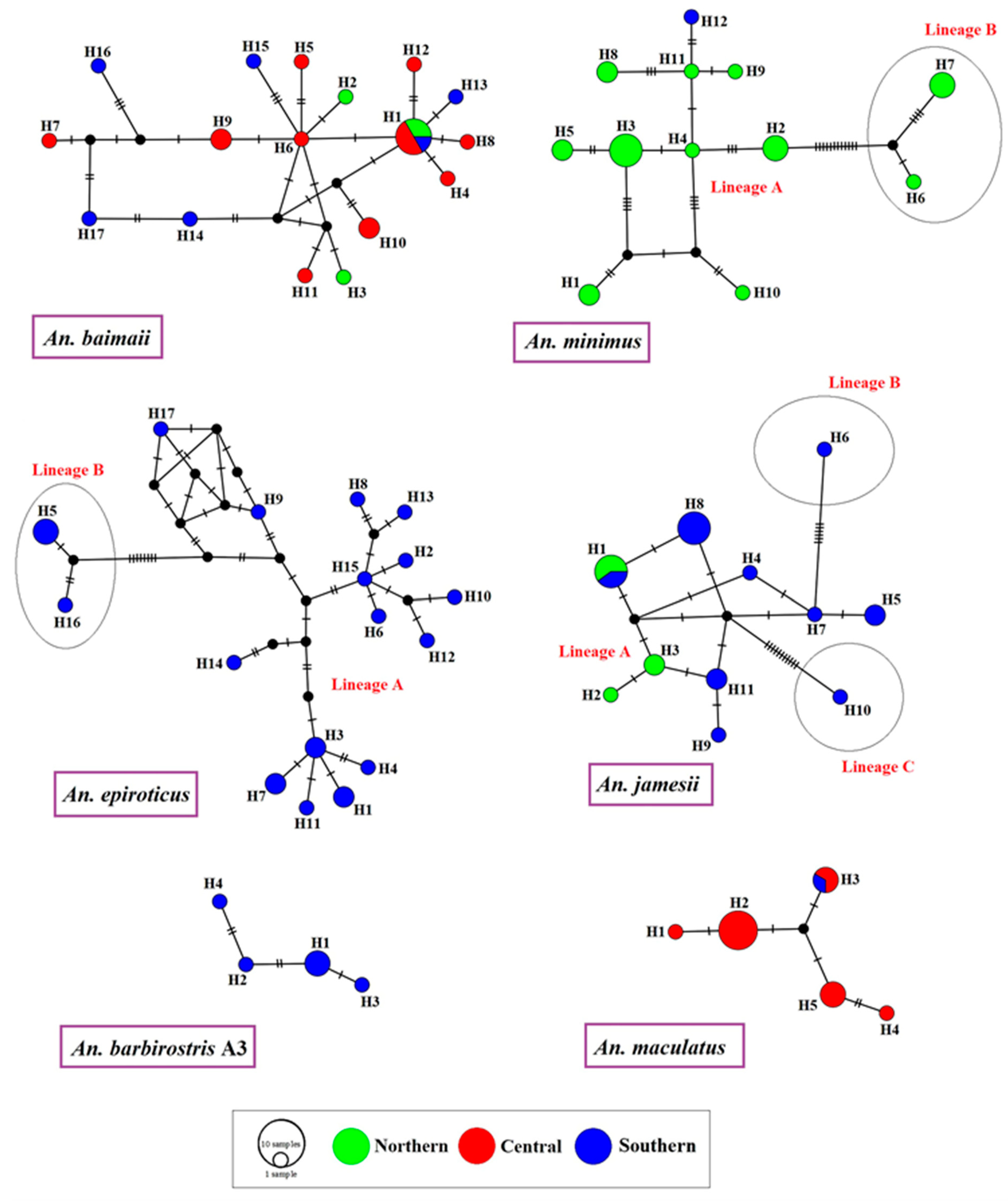
The frequencies of and relationships between 112 haplotypes of An. baimaii, An. barbirostris A3, An. epiroticus, An. jamesii, An. maculatus, and An. minimus identified in Ranong based on COI sequences are shown in median-joining haplotype networks (Figure 3).
The network analysis of An. baimaii revealed that H1 was the central haplotype that was highly connected to haplotype lines, and was the only one found in all localities of Ranong (northern, central, and southern parts). The haplotype network of An. minimus showed two distinct genetic lineages, A and B, based on mutation steps on the haplotype lines, similar to the An. epiroticus network, which showed two lineages.
The central haplotype of An. minimus and An. epiroticus could not be identified because their frequencies were not clearly different. The haplotype network of An. jamesii showed that H8 was the most common haplotype and H1 was a shared haplotype between the northern and southern populations. Based on mutation steps, three genetic lineages of An. jamesii were identified. The haplotype networks of An. barbirostris A3 showed that H1 was the most frequent haplotype, and all haplotypes were connected in a straight line, whereas the An. maculatus network showed that H2 was the most frequent haplotype and H3 was the shared haplotype, including samples from the central and southern parts.

3.7. Screening VGSC-Mutation-Mediated Knockdown Resistance

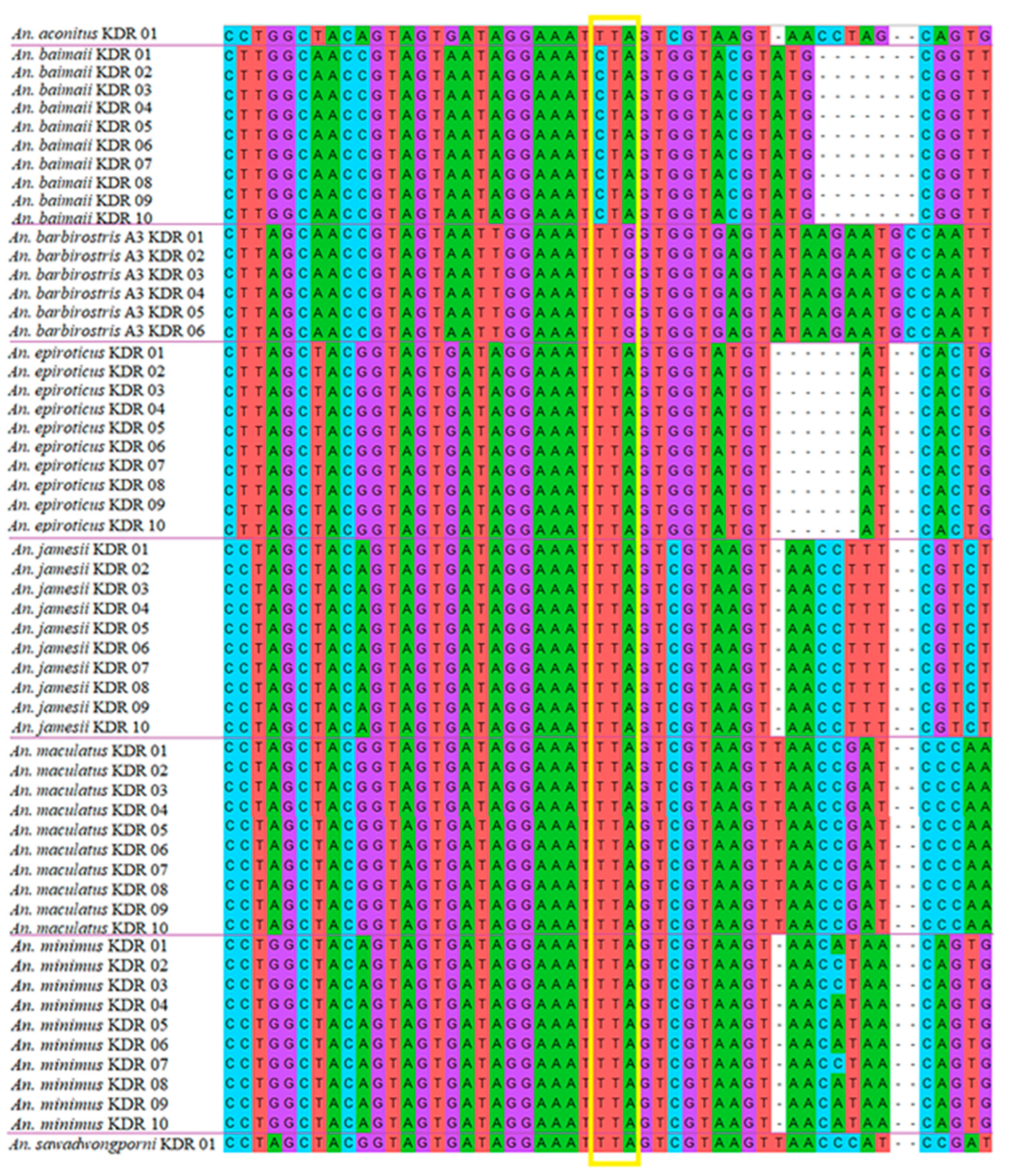
One hundred and fourteen DNA sequences of the VGSC gene fragment from the Anopheles specimens were checked for screening of knockdown resistance mutations. All the sequenced specimens presented only the L1014 wild-type allele in the VGSC gene (Table 5). No kdr-resistant alleles (L1014C, L1014F, or L1014S) were found in any of the 114 Anopheles specimens screened (Figure 4, Table 5).

4. Discussion

In this study, specimens of eight species of Anopheles mosquitoes collected from Ranong province, which is Thailand’s most P. knowlesi-endemic area, were subjected to species confirmation using molecular methods; the species included An. aconitus, An. baimaii, An. barbirostris A3, An. epiroticus, An. jamesii, An. maculatus s.s., An. minimus s.s., and An. sawadwongporni. In the northern part of Ranong, a total of four Anopheles species were found: An. baimaii, An. jamesii, An. sawadwongporni, and An. minimus s.s. as the dominant species. In the central area, An. baimaii and An. maculatus s.s. were found to be the dominant species. Most An. minimus mosquitoes live in forest edge areas, so they are common in the northern part of Ranong where there are many forest edge areas. Meanwhile, the Hat Som Paen reservoir area, which consists of densely forested high mountains in the central part of Ranong, is a suitable habitat for An. baimaii. We also found An. maculatus in the reservoir area, which is likely their habitat, although previous reports indicated that they were predominantly distributed along the edge of the forest [15,16]. In the southern part of Ranong, An. epiroticus is the dominant species because their habitat is coastal areas.
Unfortunately, no Anopheles mosquitoes in this survey were infected with Plasmodium parasites. However, some species of Anopheles mosquitoes require special entomological surveillance, based on previous reports of P. knowlesi infections in other countries. Several previous studies in Malaysia have reported that Anopheles mosquitoes in the Leucosphyrus group are important vectors of P. knowlesi [43,44,45]. Anopheles baimaii (previously known as An. dirus species D) is a species member in the Dirus complex and belongs to the Leucosphyrus group [46]. This Anopheles species is considered the primary vector of human malaria in Thailand [47]. Our study results indicated that they are distributed in forested areas throughout Ranong. In addition, An. nemophilous, belonging to the Leucosphyrus group, has also been reported in Ranong province [48]. A previous study reported P. knowlesi infections in An. sundaicus s.l. in Katchal Island, India [14]. Anopheles epiroticus (previously known as An. sundaicus species A) is a common Anopheles species found near coastal areas in Ranong and other provinces of Thailand [49,50]. This Anopheles species belongs to the Sundaicus complex and is considered Thailand’s secondary vector of malaria [48,51]. Anopheles barbirostris species A3 is a cryptic species in the Barbirostris complex belonging to the Barbirostris subgroup [52]. In Thailand, this mosquito species has previously only been reported in Kanchanaburi province [52]. Our study is the first to demonstrate the additional distribution of An. barbirostris A3 in Thailand. However, An. barbirostris A3 is another species that should not be overlooked because An. donaldi, a member species in the Barbirostris subgroup, has been reported to carry P. knowlesi infections in Lawas, Northern Sarawak, Malaysian Borneo [9]. In addition, investigations of blood meal sources of malaria vector mosquitoes using specific PCR assays should be continued in the future to determine their anopheline anthropophilic, zoophilic, or zoo-anthropophilic origin. The host preferences of Anopheles species are very important pieces of information in evaluating their ability to transmit simian malaria to humans [53].
The genetic distances between the eight Anopheles taxa based on 114 COI sequences showed that the maximum intra- and minimum interspecific genetic values were nonoverlapping, indicating the existence of a distinct barcode gap. The presence of a barcoding gap confirms the success of DNA barcoding for species identification [54]. Recently, Chaiphongpachara et al. [35] succeeded in identifying several mosquito species in Thailand based on nucleotide differences in the COI gene, except for An. dirus and An. baimaii. Our study results provide supporting evidence that DNA barcoding based on COI can be used to identify mosquito species. However, other DNA markers, such as ITS2, must be used for species identification in cases where Anopheles mosquitoes in the Dirus complex are found [55].
The nucleotide diversity (π) and nucleotide diversity (Hd) are important genetic indicators used to measure genetic diversity among populations. The nucleotide diversity values of An. baimaii, An. barbirostris A3, An. epiroticus, An. jamesii, An. maculatus, and An. minimus in Ranong were lower than the haplotype diversity values, indicating a recent Anopheles population expansion to a small effective population size after a bottleneck [56]. This demographic event occurred long enough ago for the haplotypes to increase through mutation, but not enough for the accumulation of large sequence differences [57]. Furthermore, a high level of haplotype diversity results from a large population and different environments and living habits suitable for their rapid development in nature [58].
Our results showed that genetic diversity values were similar in some species and different in others, for multifactorial reasons. Mosquito genetic diversity has both internal and external causes [59]. Internal causes of genetic diversity are genetic mutations or changes, whereas external factors are strongly related to the ecological environment of the mosquitoes [59]. Genetic diversity is an important factor that allows natural populations to adapt to and survive long-term changes or adverse environmental conditions [60]. Ranong is one of the provinces in southern Thailand with unique ecological features. The area is covered by mountains and fertile forests, and is adjacent to the Andaman Sea. It is also the wettest province in Thailand. It has been previously reported that Anopheles mosquito populations can swiftly adapt to alterations in environmental conditions, which may impact the genetic diversity within species at the population level and their gene flow [56,61]. However, a limitation of our study was the assessment of Anopheles vectors in only one endemic area, which provided insufficient data for this answer.
The Fu’s Fs and Tajima’s D values of An. baimaii, An. barbirostris A3, An. epiroticus, An. jamesii, and An. maculatus in Ranong all showed negative values, supporting population size expansion in Ranong. If a population is selectively neutral and at equilibrium between genetic drift and selectively neutral mutation, the Tajima’s D value is expected to be zero. Positive Tajima’s D values indicate a sudden decrease in population size and/or balancing selection, whereas negative Tajima’s D values indicate population size expansion after a recent bottleneck or mutational selection [41]. Our results were consistent with the population structure of An. baimaii in Thailand, indicating that the population is expanding [56].
The ML phylogenetic tree and haplotype network results revealed two distinct genetic lineages, A and B, of An. minimus and An. epiroticus in Ranong. Recently, Bunmee et al. [56] reported the existence of A and B lineages for An. minimus s.s. in Thailand, which agrees with our research findings. In many of Thailand’s malaria transmission areas, such as Tak, Surat Thani, Yala, Chanthaburi, and Trat, there are two lineages of An. minimus, which are often found together [56]. However, it is unclear whether the two lineages have the potential to transmit malaria or other different behaviors. In addition, our study is the first to reveal two distinct genetic lineages of An. epiroticus based on the COI gene, which shows genetic variation and local adaptation. Syafruddinid et al. [62] explained that the COI gene is suitable for assessing genetic variation within populations of An. epiroticus because mtDNA has a high mutation rate. However, this gene cannot be used as a molecular marker to differentiate between An. epiroticus and its other sibling members [62]. Although three genetic lineages of An. jamesii were identified, only one group had many samples, whereas the other two groups had only one member each. Consequently, future genetic studies on this species should be conducted.
The early detection and surveillance of VGSC-mutation-mediated knockdown resistance (kdr) in Anopheles populations can provide entomological data on the causes of pyrethroid resistance in insects to inform the development of strategies to control malaria vectors [63]. The present study showed no presence of kdr mutation in the VGSC gene among Anopheles mosquitoes from Thailand’s most P. knowlesi-endemic area. This result is similar to those of previous Anopheles investigations in Ubon Ratchathani province, northeastern Thailand [64]. However, this study is limited by a lack of information on the susceptibility of the mosquito samples tested. Therefore, we do not know the true state of insecticide resistance in these Anopheles populations. Further entomological investigations into the susceptibility of adult mosquito vectors to insecticides are required.

5. Conclusions

Our study demonstrated the genetic diversity of Anopheles mosquitoes in Thailand’s most P. knowlesi-endemic area. Our genetic diversity analysis will contribute to a more comprehensive genetic profile of Anopheles vectors in Thailand. In addition, our attempts to detect P. knowlesi infection in Anopheles mosquitoes did not reveal infected specimens. However, three Anopheles species, including An. baimaii, An. barbirostris A3, and An. epiroticus, should be kept under special surveillance as P. knowlesi infections have been found in these species in other countries. This entomological information could lead to the selection of appropriate methods for controlling these Anopheles populations, such as insecticide-treated nets (ITNs) and indoor residual spraying (IRS), in order to control the spread of monkey malaria to humans in Thailand’s most P. knowlesi-endemic area. In addition, educating the population about vector breeding sites and strategies for protection against Anopheles mosquitoes, such as applying mosquito repellent or wearing protective clothes when entering forests where monkeys reside, are crucial ways to help reduce the incidence of knowlesi malaria.

Author Contributions

T.C. (Tanawat Chaiphongpachara): conceptualization, methodology, resources, formal analysis, investigation, writing—original draft, writing—review and editing, visualization, supervision. S.L.: conceptualization, methodology, resources, formal analysis, investigation, writing—original draft. T.C. (Tanasak Changbunjong): methodology, resources, formal analysis, investigation, writing—original draft, writing—review and editing, visualization. S.S.: methodology, resources, formal analysis, investigation, writing—original draft, writing—review and editing, visualization. N.S.: conceptualization, investigation, resources, writing—original draft, visualization. S.C.: investigation, resources. Y.A.: investigation, resources. All authors have read and agreed to the published version of the manuscript.

Funding

This research was funded by the Suan Sunandha Rajabhat University.

Institutional Review Board Statement

The animal study protocol was approved by the Institutional Animal Care and Use Committee of the Suan Sunandha Rajabhat University, Thailand (Animal Ethics Permission number: IACUC 64-010/2021).

Informed Consent Statement

Not applicable.

Data Availability Statement

Not applicable.

Acknowledgments

The authors like to express their gratitude to the Suan Sunandha Rajabhat University in Thailand and Vector Borne Disease Control Center 11.5, Ranong, Thailand for their support of this study.

Conflicts of Interest

The authors declare no conflict of interest.

References

  1. Cox, F.E. History of the discovery of the malaria parasites and their vectors. Parasit. Vectors 2010, 3, 5. [Google Scholar] [CrossRef] [PubMed] [Green Version]
  2. Li, M.I.; Mailepessov, D.; Vythilingam, I.; Lee, V.; Lam, P.; Ng, L.C.; Tan, C.H. Prevalence of simian malaria parasites in macaques of Singapore. PLoS Negl. Trop. Dis. 2021, 15, e0009110. [Google Scholar] [CrossRef] [PubMed]
  3. Müller, M.; Schlagenhauf, P. Plasmodium Knowlesi in travellers, update 2014. Int. J. Infect. Dis. 2014, 22, 55–64. [Google Scholar] [CrossRef] [PubMed] [Green Version]
  4. Chin, W.; Contacos, P.G.; Coatney, G.R.; Kimball, H.R. A naturally acquired quotidian-type malaria in man transferable to monkeys. Science 1965, 149, 865. [Google Scholar] [CrossRef]
  5. Kantele, A.; Marti, H.; Felger, I.; Müller, D.; Jokiranta, T.S. Monkey malaria in a European traveler returning from Malaysia. Emerg. Infect. Dis. 2008, 14, 1434–1436. [Google Scholar] [CrossRef] [PubMed]
  6. Bronner, U.; Divis, P.C.S.; Färnert, A.; Singh, B. Swedish traveller with Plasmodium knowlesi malaria after visiting Malaysian Borneo. Malar. J. 2009, 8, 15. [Google Scholar] [CrossRef] [Green Version]
  7. Ehrhardt, J.; Trein, A.; Kremsner, P.G.; Frank, M. Plasmodium knowlesi and HIV co-infection in a German traveller to Thailand. Malar. J. 2013, 12, 283. [Google Scholar] [CrossRef] [Green Version]
  8. Froeschl, G.; Beissner, M.; Huber, K.; Bretzel, G.; Hoelscher, M.; Rothe, C. Plasmodium knowlesi infection in a returning German traveller from Thailand: A Case report on an emerging malaria pathogen in a popular low-risk travel destination. BMC Infect. Dis. 2018, 18, 4–9. [Google Scholar] [CrossRef] [Green Version]
  9. Ang, J.X.D.; Kadir, K.A.; Mohamad, D.S.A.; Matusop, A.; Divis, P.C.S.; Yaman, K.; Singh, B. New vectors in northern Sarawak, Malaysian Borneo, for the zoonotic malaria parasite, Plasmodium knowlesi. Parasit. Vectors 2020, 13, 472. [Google Scholar] [CrossRef]
  10. Wharton, R.H.; Eyles, D.E. Anopheles hackeri, a vector of Plasmodium knowlesi in Malaya. Science 1961, 134, 279–280. [Google Scholar] [CrossRef]
  11. Vythilingam, I.; Tan, C.H.; Asmad, M.; Chan, S.T.; Lee, K.S.; Singh, B. Natural transmission of Plasmodium knowlesi to humans by Anopheles latens in Sarawak, Malaysia. Trans. R. Soc. Trop. Med. Hyg. 2006, 100, 1087–1088. [Google Scholar] [CrossRef] [PubMed]
  12. Marchand, R.P.; Culleton, R.; Maeno, Y.; Quang, N.T.; Nakazawa, S. Co-infections of Plasmodium knowlesi, P. falciparum, and P. vivax among humans and Anopheles dirus mosquitoes, southern Vietnam. Emerg. Infect. Dis. 2011, 17, 1232–1239. [Google Scholar] [CrossRef] [Green Version]
  13. Jiram, A.I.; Vythilingam, I.; Noorazian, Y.M.; Yusof, Y.M.; Azahari, A.H.; Fong, M.Y. Entomologic investigation of Plasmodium knowlesi vectors in Kuala Lipis, Pahang, Malaysia. Malar. J. 2012, 11, 213. [Google Scholar] [CrossRef] [PubMed] [Green Version]
  14. Vidhya, P.T.; Sunish, I.P.; Maile, A.; Zahid, A.K. Anopheles sundaicus mosquitoes as vector for Plasmodium knowlesi, andaman and Nicobar Islands, India. Emerg. Infect. Dis. 2019, 25, 817–820. [Google Scholar] [CrossRef] [PubMed] [Green Version]
  15. Sumruayphol, S.; Chaiphongpachara, T.; Samung, Y.; Ruangsittichai, J.; Cui, L.; Zhong, D.; Sattabongkot, J.; Sriwichai, P. Seasonal dynamics and molecular differentiation of three natural Anopheles species (Diptera: Culicidae) of the Maculatus group (Neocellia series) in malaria hotspot villages of Thailand. Parasit. Vectors 2020, 13, 574. [Google Scholar] [CrossRef] [PubMed]
  16. Parker, D.M.; Carrara, V.I.; Pukrittayakamee, S.; McGready, R.; Nosten, F.H. Malaria ecology along the Thailand-Myanmar border. Malar. J. 2015, 14, 388. [Google Scholar] [CrossRef] [PubMed] [Green Version]
  17. Jongwutiwes, S.; Putaporntip, C.; Iwasaki, T.; Sata, T.; Kanbara, H. Naturally acquired Plasmodium knowlesi malaria in human, Thailand. Emerg. Infect. Dis. 2004, 10, 2211–2213. [Google Scholar] [CrossRef] [PubMed]
  18. Ministry of Public Health, Thailand. Malaria Report. Available online: http://malaria.ddc.moph.go.th/malariaR10/index_v2.php (accessed on 1 August 2022).
  19. Ritthison, W.; Tainchum, K.; Manguin, S.; Bangs, M.J.; Chareonviriyaphap, T. Biting Patterns and Host Preference of Anopheles epiroticus in Chang Island, Trat Province, Eastern Thailand. J. Vector Ecol. 2014, 39, 361–371. [Google Scholar] [CrossRef]
  20. McCoy, K.D. The population genetic structure of vectors and our understanding of disease epidemiology. Parasite 2008, 15, 444–448. [Google Scholar] [CrossRef] [Green Version]
  21. Chen, B.; Pedro, P.M.; Harbach, R.E.; Somboon, P.; Walton, C.; Butlin, R.K. Mitochondrial DNA variation in the malaria vector Anopheles minimus across China, Thailand and Vietnam: Evolutionary hypothesis, population structure and population history. Heredity 2011, 106, 241–252. [Google Scholar] [CrossRef]
  22. Weeraratne, T.C.; Surendran, S.N.; Walton, C.; Karunaratne, S.H.P.P. Genetic diversity and population structure of malaria vector mosquitoes Anopheles subpictus, Anopheles peditaeniatus, and Anopheles vagus in five districts of Sri Lanka. Malar. J. 2018, 17, 271. [Google Scholar] [CrossRef] [PubMed] [Green Version]
  23. Li, S.; Jiang, F.; Lu, H.; Kang, X.; Wang, Y.; Zou, Z.; Wen, D.; Zheng, A.; Liu, C.; Liu, Q.; et al. Mosquito diversity and population genetic structure of six mosquito species from Hainan island. Front. Genet. 2020, 11, 602863. [Google Scholar] [CrossRef] [PubMed]
  24. Wangdi, K.; Furuya-Kanamori, L.; Clark, J.; Barendregt, J.J.; Gatton, M.L.; Banwell, C.; Kelly, G.C.; Doi, S.A.R.; Clements, A.C.A. Comparative effectiveness of malaria prevention measures: A systematic review and network meta-analysis. Parasit. Vectors 2018, 11, 210. [Google Scholar] [CrossRef] [PubMed] [Green Version]
  25. Silva, A.P.B.; Santos, J.M.M.; Martins, A.J. Mutations in the voltage-gated sodium channel gene of anophelines and their association with resistance to pyrethroids—A review. Parasit. Vectors 2014, 7, 450. [Google Scholar] [CrossRef] [Green Version]
  26. Syafruddin, D.; Hidayati, A.P.; Asih, P.B.; Hawley, W.A.; Sukowati, S.; Lobo, N.F. Detection of 1014F kdr mutation in four major anopheline malaria vectors in Indonesia. Malar. J. 2010, 9, 315. [Google Scholar] [CrossRef] [PubMed] [Green Version]
  27. Fang, Y.; Shi, W.Q.; Wu, J.T.; Li, Y.Y.; Xue, J.B.; Zhang, Y. Resistance to pyrethroid and organophosphate insecticides, and the geographical distribution and polymorphisms of target-site mutations in voltage-gated sodium channel and acetylcholinesterase 1 genes in Anopheles sinensis populations in Shanghai, China. Parasit. Vectors 2019, 12, 396. [Google Scholar] [CrossRef] [PubMed]
  28. Yang, C.; Feng, X.; Liu, N.; Li, M.; Qiu, X. Target-site mutations (AChE-G119S and kdr) in Guangxi Anopheles sinensis populations along the China-Vietnam border. Parasit. Vectors 2019, 12, 77. [Google Scholar] [CrossRef] [PubMed]
  29. Rattanarithikul, R.; Harrison, B.A.; Harbach, R.E.; Panthusiri, P.; Coleman, R.E. Illustrated keys to the mosquitoes of Thailand IV. Anopheles. Southeast Asian J. Trop. Med. Public Health 2006, 37 (Suppl. S2), 1–128. [Google Scholar]
  30. Walton, C.; Handley, J.M.; Kuvangkadilok, C.; Collins, F.H.; Harbach, R.E.; Baimai, V.; Butlin, R.K. Identification of five species of the Anopheles dirus complex from Thailand, using allele-specific polymerase chain reaction. Med. Vet. Entomol. 1999, 13, 24–32. [Google Scholar] [CrossRef]
  31. Walton, C.; Somboon, P.; O’Loughlin, S.M.; Zhang, S.; Harbach, R.E.; Linton, Y.M.; Chen, B.; Nolan, K.; Duong, S.; Fong, M.Y.; et al. Genetic diversity and molecular identification of mosquito species in the Anopheles Maculatus group using the ITS2 region of rDNA. Infect. Genet. Evol. 2007, 7, 93–102. [Google Scholar] [CrossRef]
  32. Garros, C.; Koekemoer, L.L.; Coetzee, M.; Coosemans, M.; Manguin, S. A single multiplex assay to identify major malaria vectors within the African Anopheles funestus and the Oriental An. minimus groups. Am. J. Trop. Med. Hyg. 2004, 70, 583–590. [Google Scholar] [CrossRef] [PubMed]
  33. Echeverry, D.F.; Deason, N.A.; Makuru, V.; Davidson, J.; Xiao, H.; Niedbalski, J.; Yu, X.; Stevenson, J.C.; Bugoro, H.; Aparaimo, A.; et al. Fast and robust single PCR for Plasmodium sporozoite detection in mosquitoes using the cytochrome oxidase I gene. Malar. J. 2017, 16, 230. [Google Scholar] [CrossRef] [PubMed]
  34. Kumar, N.P.; Rajavel, A.R.; Natarajan, R.; Jambulingam, P. DNA barcodes can distinguish species of Indian mosquitoes (Diptera: Culicidae). J. Med. Entomol. 2007, 44, 1–7. [Google Scholar] [CrossRef] [PubMed]
  35. Chaiphongpachara, T.; Changbunjong, T.; Laojun, S.; Nutepsu, T.; Suwandittakul, N.; Kuntawong, K.; Sumruayphol, S.; Ruangsittichai, J. Mitochondrial DNA barcoding of mosquito species (Diptera: Culicidae) in Thailand. PLoS ONE 2022, 17, e0275090. [Google Scholar] [CrossRef] [PubMed]
  36. Hall, T.A. Bioedit: A user-friendly biological sequence alignment editor and analysis program for Windows 95/98/NT. Nucleic Acids Symp. Ser. 1999, 41, 95–98. [Google Scholar]
  37. Thompson, J.D.; Gibson, T.J.; Plewniak, F.; Jeanmougin, F.; Higgins, D.G. The Clustal X windows interface: Flexible strategies for multiple sequence alignment aided by quality analysis tools. Nucleic Acids Res. 1997, 25, 4876–4882. [Google Scholar] [CrossRef] [Green Version]
  38. Kumar, S.; Stecher, G.; Li, M.; Knyaz, C.; Tamura, K. MEGA X: Molecular evolutionary genetics analysis across computing platforms. Mol. Biol. Evol. 2018, 35, 1547–1549. [Google Scholar] [CrossRef]
  39. Davidson, J.R.; Wahid, I.; Sudirman, R.; Small, S.T.; Hendershot, A.L.; Baskin, R.N.; Burton, T.A.; Makuru, V.; Xiao, H.; Yu, X.; et al. Molecular analysis reveals a high diversity of Anopheles species in Karama, West Sulawesi, Indonesia. Parasit. Vectors 2020, 13, 379. [Google Scholar] [CrossRef]
  40. Rozas, J.; Ferrer-Mata, A.; Sanchez-DelBarrio, J.C.; Guirao-Rico, S.; Librado, P.; Ramos-Onsins, S.E.; Sanchez-Gracia, A. DnaSP 6: DNA sequence polymorphism analysis of large data sets. Mol. Biol. Evol. 2017, 34, 3299–3302. [Google Scholar] [CrossRef]
  41. Tajima, F. Statistical method for testing the neutral mutation hypothesis by DNA polymorphism. Genetics 1989, 123, 585–595. [Google Scholar] [CrossRef]
  42. Fu, Y.X.; Li, W.H. Statistical tests of neutrality of mutations. Genetics 1993, 133, 693–709. [Google Scholar] [CrossRef] [PubMed]
  43. Moyes, C.L.; Shearer, F.M.; Huang, Z.; Wiebe, A.; Gibson, H.S.; Nijman, V.; Mohd-Azlan, J.; Brodie, J.F.; Malaivijitnond, S.; Linkie, M.; et al. Predicting the geographical distributions of the macaque hosts and mosquito vectors of Plasmodium knowlesi malaria in forested and non-forested areas. Parasit. Vectors 2016, 9, 242. [Google Scholar] [CrossRef] [PubMed]
  44. Pramasivan, S.; Ngui, R.; Jeyaprakasam, N.K.; Liew, J.W.K.; Low, V.L.; Mohamed Hassan, N.; Wan Sulaiman, W.Y.; Jaraee, R.; Abdul Rahman, R.; Jelip, J.; et al. Spatial distribution of Plasmodium knowlesi cases and their vectors in Johor, Malaysia: In light of human malaria elimination. Malar. J. 2021, 20, 426. [Google Scholar] [CrossRef] [PubMed]
  45. Van de straat, B.; Sebayang, B.; Grigg, M.J.; Staunton, K.; Garjito, T.A.; Vythilingam, I.; Russell, T.L.; Burkot, T.R. Zoonotic malaria transmission and land use change in Southeast Asia: What is known about the vectors. Malar. J. 2022, 21, 109. [Google Scholar] [CrossRef]
  46. Harbach, R.E. Anopheles Classification. Available online: https://mosquito-taxonomic-inventory.myspecies.info/node/11358 (accessed on 1 August 2022).
  47. Tananchai, C.; Tisgratog, R.; Juntarajumnong, W.; Grieco, J.P.; Manguin, S.; Prabaripai, A.; Chareonviriyaphap, T. Species diversity and biting activity of Anopheles dirus and Anopheles baimaii (Diptera: Culicidae) in a malaria prone area of western Thailand. Parasit. Vectors 2012, 5, 211. [Google Scholar] [CrossRef] [Green Version]
  48. Tainchum, K.; Kongmee, M.; Manguin, S.; Bangs, M.J.; Chareonviriyaphap, T. Anopheles species diversity and distribution of the malaria vectors of Thailand. Trends Parasitol. 2015, 31, 109–119. [Google Scholar] [CrossRef]
  49. Chaiphongpachara, T.; Laojun, S. Variation over time in wing size and shape of the coastal malaria vector Anopheles (Cellia) epiroticus Linton and Harbach (Diptera: Culicidae) in Samut Songkhram, Thailand. J. Adv. Vet. Anim. Res. 2019, 6, 208–214. [Google Scholar] [CrossRef]
  50. Chaiphongpachara, T.; Laojun, S. Wing morphometric variability of the malaria vector Anopheles (Cellia) epiroticus Linton et Harbach (Diptera: Culicidae) for the duration of the rainy season in coastal areas of Samut Songkhram, Thailand. Folia Parasitol. 2020, 67, 1–7. [Google Scholar] [CrossRef] [Green Version]
  51. Sumruayphol, S.; Apiwathnasorn, C.; Komalamisra, N.; Ruangsittichai, J.; Samung, Y.; Chavalitshewinkoon-Petmitr, P. Bionomic status of Anopheles epiroticus Linton & Harbach, a coastal malaria vector, in Rayong province, Thailand. Southeast Asian J. Trop. Med. Public Health 2010, 41, 541–547. [Google Scholar]
  52. Wilai, P.; Namgay, R.; Ali, R.S.M.; Saingamsook, J.; Saeung, A.; Junkum, A.; Walton, C.; Harbach, R.E.; Somboon, P. A multiplex PCR based on mitochondrial COI sequences for identification of members of the Anopheles barbirostris complex (Diptera: Culicidae) in Thailand and other countries in the region. Insects 2020, 11, 409. [Google Scholar] [CrossRef]
  53. Jeyaprakasam, N.K.; Low, V.L.; Liew, J.W.K.; Pramasivan, S.; Wan-Sulaiman, W.Y.; Saeung, A.; Vythilingam, I. Blood meal analysis of Anopheles vectors of simian malaria based on laboratory and field. Sci. Sci. Rep. 2022, 12, 354. [Google Scholar] [CrossRef] [PubMed]
  54. Beebe, N.W. DNA barcoding mosquitoes: Advice for potential prospectors. Parasitology 2008, 145, 622–633. [Google Scholar] [CrossRef] [Green Version]
  55. Chaiphongpachara, T.; Changbunjong, T.; Sumruayphol, S.; Laojun, S.; Suwandittakul, N.; Kuntawong, K. Geometric morphometrics versus DNA barcoding for the identification of malaria vectors Anopheles dirus and An. baimaii in the Thai—Cambodia border. Sci. Rep. 2022, 12, 13236. [Google Scholar] [CrossRef] [PubMed]
  56. Bunmee, K.; Thaenkham, U.; Saralamba, N.; Ponlawat, A.; Zhong, D.; Cui, L.; Sattabongkot, J.; Sriwichai, P. Population genetic structure of the malaria vector Anopheles minimus in Thailand based on mitochondrial DNA markers. Parasit. Vectors 2021, 14, 496. [Google Scholar] [CrossRef] [PubMed]
  57. Chanthran, S.S.D.; Lim, P.E.; Li, Y.; Liao, T.Y.; Poong, S.W.; Du, J.; Hussein, M.A.S.; Sade, A.; Rumpet, R.; Loh, K.H. Genetic diversity and population structure of Terapon jarbua (Forskål, 1775) (Teleostei, Terapontidae) in malaysian waters. Zookeys 2020, 911, 139–160. [Google Scholar] [CrossRef] [PubMed] [Green Version]
  58. Fang, D.A.; Luo, H.; He, M.; Mao, C.; Kuang, Z.; Qi, H.; Xu, D.; Tan, L.; Li, Y. Genetic diversity and population differentiation of naked carp (Gymnocypris przewalskii) Revealed by cytochrome oxidase subunit I and d-loop. Front. Ecol. Evol. 2022, 10, 827654. [Google Scholar] [CrossRef]
  59. Fang, Y.; Zhang, J.; Wu, R.; Xue, B.; Qian, Q.; Gao, B. Genetic polymorphism study on Aedes albopictus of different geographical regions based on dna barcoding. BioMed Res. Int. 2018, 2018, 1501430. [Google Scholar] [CrossRef] [Green Version]
  60. Makhawi, A.M.; Liu, X.B.; Yang, S.R.; Liu, Q.Y. Genetic variations of ND5 gene of mtDNA in populations of Anopheles sinensis (Diptera: Culicidae) malaria vector in China. Parasit. Vectors 2013, 6, 290. [Google Scholar] [CrossRef] [Green Version]
  61. Yawson, A.E.; Weetman, D.; Wilson, M.D.; Donnelly, M.J. Ecological zones rather than molecular forms predict genetic differentiation in the malaria vector Anopheles gambiae s.s. in Ghana. Genetics 2007, 175, 751–761. [Google Scholar] [CrossRef] [Green Version]
  62. Syafruddinid, D.; Lestari, Y.E.; Permana, D.H.; Asih, P.B.S.; Laurent, B.S.; Zubaidah, S.; Rozi, I.E.; Kosasih, S.; Shinta; Sukowati, S.; et al. Anopheles sundaicus complex and the presence of Anopheles epiroticus in Indonesia. PLoS Negl. Trop. Dis. 2020, 14, e0008385. [Google Scholar]
  63. Dong, K.; Du, Y.; Rinkevich, F.; Nomura, Y.; Xu, P.; Wang, L.; Silver, K.; Zhorov, B.S. Molecular biology of insect sodium channels and pyrethroid resistance. Insect Biochem. Mol. Biol. 2014, 50, 1–17. [Google Scholar] [CrossRef] [PubMed] [Green Version]
  64. Sumarnrote, A.; Overgaard, H.J.; Marasri, N.; Fustec, B.; Thanispong, K.; Chareonviriyaphap, T.; Corbel, V. Status of insecticide resistance in Anopheles mosquitoes in Ubon Ratchathani province, northeastern Thailand. Malar. J. 2017, 16, 299. [Google Scholar] [CrossRef] [PubMed]
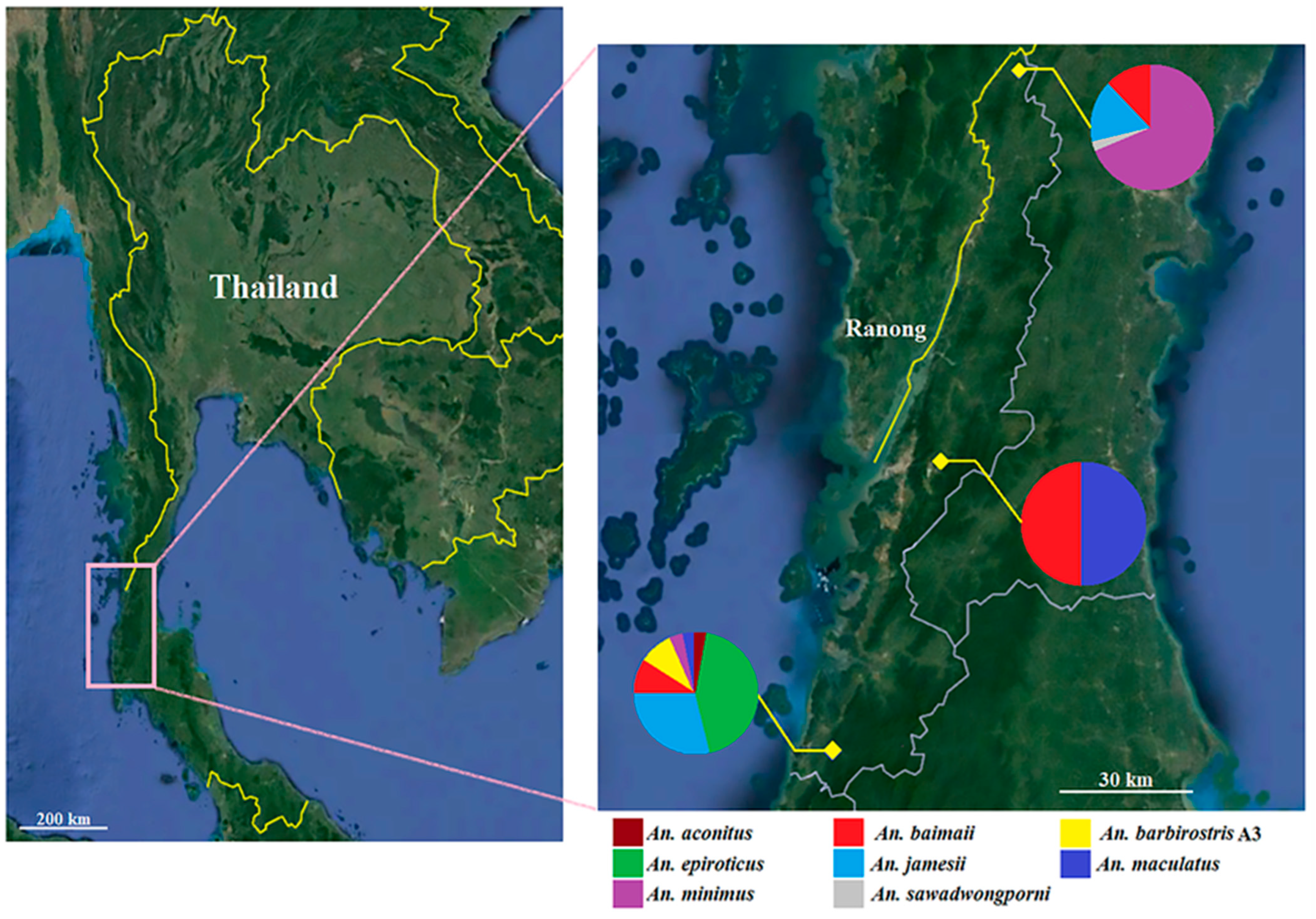
Figure 1. Map of study sites and diversity of Anopheles species in Ranong province, southern Thailand. Pie charts in this map show the frequency of each species in each location. This map was obtained from Google Earth Pro v 7.1.8 (https://earth.google.com (accessed on 10 October 2022)).
Tropicalmed 07 00412 g001
Figure 2. Maximum likelihood tree of eight Anopheles species identified in this study. Color bands indicate each species of Anopheles mosquito. The small colored circles represent the areas in which the samples were collected. Bootstrap values (1000 replicates) >90% are shown at the nodes. Eight sequences obtained from GenBank were used as species references, including An. baimaii (GenBank accession number: OL742839), An. minimus s.s. (OL742874), An. epiroticus (OL742858), An. jamesii (OL742865), An. maculatus s.s. (OL742869), An. barbirostris A3 (MT394436), An. sawadwongporni (OL742914), and An. aconitus (OL742831). Furthermore, Aedes aegypti (OL743100) was used as an outgroup in this analysis.
Tropicalmed 07 00412 g002
Figure 3. COI haplotype networks of Anopheles specimens collected from three locations in Ranong province, Southern Thailand. Anopheles species represented by only one specimen, including An. aconitus and An. sawadwongporni, were excluded from the analyses. A colored circle represents each haplotype, and the circle’s size is proportional to the total sequence of each haplotype. The number of mutations is shown by the dashes along the haplotype lines; the different colored circles represent different locations in Thailand.
Tropicalmed 07 00412 g003
Figure 4. DNA sequences of a voltage-gated sodium channel gene fragment from specimens of the eight Anopheles species sampled in Ranong. The yellow rectangle shows the codon at the 1014 position, for which all samples presented an L1014 wild-type allele (TTA, TTG, or CTA).
Tropicalmed 07 00412 g004
Table 1. Species and numbers of Anopheles sampled in the geographical region of Ranong province, Thailand (January–June 2022), identified using molecular methods.
Anopheles SpeciesRanong ProvinceTotal (%)
Northern PartCentral PartSouthern Part
An. aconitus0011 (0.88)
An. baimaii414624 (21.05)
An. barbirostris A30066 (5.26)
An. epiroticus002222 (19.30)
An. jamesii601622 (19.30)
An. maculatus s.s.014115 (13.16)
An. minimus s.s.220123 (20.17)
An. sawadwongporni1001 (0.88)
Total (%)33 (28.95)28 (24.56)53 (46.49)114 (100)
Table 2. GenBank accession numbers of COI sequences obtained in this study.
Anopheles SpeciesnLocationGenBank Accession Numbers
An. Aconitus1Southern part of RanongOP253978
An. Baimaii4Northern part of RanongOP253979–OP253982
14Central part of RanongOP253983–OP253996
6Southern part of RanongOP253997–OP254002
An. barbirostris A36Southern part of RanongOP254003–OP254008
An. epiroticus22Southern part of RanongOP254009–OP254030
An. jamesii6Northern part of RanongOP254031–OP254036
16Southern part of RanongOP254037–OP254052
An. maculatus14Central part of RanongOP254053–OP254066
1Southern part of RanongOP254067
An. minimus22Northern part of RanongOP254068–OP254089
1Southern part of RanongOP254090
An. sawadwongporni1Northern part of RanongOP254091
Table 3. Interspecific and intraspecific K2P genetic distances of Anopheles species were collected based on COI sequences in this study.
Anopheles Species% Mean Sequence Divergence (Min–Max)
12345678
1. An. aconitusNA
2. An. baimaii11.5%
(11.1–11.7)
0.5%
(0.0–1.3)
3. An. barbirostris A311.1%
(10.9–11.2)
9.6%
(9.3–10.1)
0.3%
(0.0–0.7)
4. An. epiroticus10.7%
(9.9–11.2)
12%
(10.9–12.9)
11.6%
(10.9–12.4)
1.3%
(0.0–2.7)
5. An. jamesii10%
(9.8–10.4)
11.3%
(10.7–12.4)
13.6%
(13.1–14.6)
11.8%
(11.1–12.6)
0.5%
(0.0–1.9)
6. An. maculatus10.2%
(10.1–10.4)
8.9%
(8.3–9.8)
10.2%
(9.9–10.4)
11.6%
(10.8–12.4)
9.5%
(9.3–9.8)
0.2%
(0.0–0.7)
7. An. minimus8.2%
(7.9–8.4)
10.9%
(10.4–11.2)
11.2%
(10.6–11.6)
11.5%
(10.6–12.7)
11%
(10.6–12.1)
10%
(9.5–10.4)
1.1%
(0.0–2.9)
8. An. sawadwongporni10.6%
(10.6–10.6)
11.5%
(11.2–11.9)
10.6%
(10.4–10.7)
11.5%
(10.9–11.9)
11.7%
(11.6–12.1)
6.6%
(6.5–6.6)
9.9%
(9.5–10.3)
NA
Table 4. Genetic diversity indices and neutrality tests of Anopheles species in Ranong.
Anopheles Speciesnsπ (±SD)hHd (±SD)kNeutrality Tests
Fu’s FsTajima’s D
An. aconitus11
An. baimaii24220.005 ± 0.001170.938 ± 0.0393.616−10.476 *−1.621
An. barbirostris A3650.003 ± 0.00140.800 ± 0.1722.067−0.439−0.315
An. epiroticus22330.012 ± 0.001170.974 ± 0.0228.797−4.804 *−0.220
An. jamesii22200.005 ± 0.001110.900 ± 0.0413.667−2.544 *−1.233
An. maculatus1560.002 ± 0.00050.743 ± 0.0901.695−0.214−0.285
An. minimus23290.011 ± 0.002120.925 ± 0.0328.016−0.2110.077
An. sawadwongporni11
Anopheles aconitus and An. sawadwongporni, represented by only one specimen each, were excluded from the analyses. An asterisk (*) after Fu’s Fs and Tajima’s D values represents the statistical difference at p < 0.05. Abbreviations: n = number of sequences; s = number of polymorphic (segregating) sites; π = nucleotide diversity; h = number of haplotypes; Hd = haplotype diversity; k = average number of nucleotide differences.
Table 5. Screening for kdr mutations in the voltage-gated sodium channel (VGSC) gene in specimens of eight Anopheles species obtained in this study.
SpeciesAllelic Frequency
L1014 WildL1014C (TGT)L1014F (TTT)L1014S (TCA)
TTATTGCTA
An. Aconitus100000
An. baimaii0024000
An. barbirostris A3060000
An. epiroticus2200000
An. jamesii2200000
An. maculatus1500000
An. minimus2300000
An. sawadwongporni100000
Total84624000
Publisher’s Note: MDPI stays neutral with regard to jurisdictional claims in published maps and institutional affiliations.