Article

Prevalence of Intestinal Parasitic Infections, Genotypes, and Drug Susceptibility of Giardia lamblia among Preschool and School-Aged Children: A Cross-Sectional Study in Thailand

by
Boonchai Wongstitwilairoong
,
Thunyarat Anothaisintawee
*,
Nattaya Ruamsap
*,
Paphavee Lertsethtakarn
,
Paksathorn Kietsiri
,
Wirote Oransathid
,
Wilawan Oransathid
,
Siriphan Gonwong
,
Sasikorn Silapong
,
Umaporn Suksawad
,
Siriporn Sornsakrin
,
Ladaporn Bodhidatta
,
Daniel M. Boudreaux
and
Jeffrey R. Livezey
Department of Bacterial and Parasitic Diseases, US Army Medical Directorate of the Armed Forces Research Institute of Medical Sciences, Bangkok 10120, Thailand
*
Authors to whom correspondence should be addressed.
Trop. Med. Infect. Dis. 2023, 8(8), 394; https://doi.org/10.3390/tropicalmed8080394
Submission received: 1 May 2023 / Revised: 18 July 2023 / Accepted: 21 July 2023 / Published: 1 August 2023
(This article belongs to the Section Neglected and Emerging Tropical Diseases)

Abstract

:
This study aimed to estimate the prevalence of intestinal parasitic infections in children and assess the drug susceptibility and genotypes/assemblages of Giardia lamblia in Thailand. This cross-sectional study was conducted among children aged 3–12 years in Sangkhlaburi District, Kanchanaburi Province, Thailand, between 25 September 2017 and 12 January 2018. Parasites were identified by stool microscopic examination, cultivation of intestinal parasitic protozoa, and enzyme-linked immunosorbent assay (ELISA). Drug susceptibility and genotype of G. lamblia were performed, respectively, by a resazurin assay and Triosephosphate Isomerase A and B genes using modified primers and probes. Among the 661 participants, 445 had an intestinal parasitic infection, resulting in a prevalence of 67.32% (95% CI: 63.60–70.89%). Blastocystis hominis was the most prevalent protozoa infection (49.32%; 95% CI: 45.44–53.22%), while Ascaris lumbricoides was the most prevalent helminth infection (0.91%; 95% CI: 0.33–1.97%). The prevalence of G. lamblia was 17.40%, with genotype B being the most common. According to our study, intestinal parasitic infections were commonly found in Thai children. G. lamblia was the most common pathogenic protozoa infection identified and exhibited less susceptibility to metronidazole compared to furazolidone and mebendazole.

1. Introduction

Intestinal parasitic infections are a major public health problem, leading to substantial morbidity and mortality among preschool and school-age children in developing countries [1,2]. Tropical and subtropical regions have the highest prevalence of intestinal parasitic infections, primarily due to deprived socioeconomic status and poor personal and environmental hygiene among the majority of the population [3,4]. Despite having a low mortality rate, intestinal parasitic infections have long-lasting impacts on the host’s health and nutritional state [5]. These parasites induce intestinal damage, leading to a reduction in the absorption of nutrients [3]. Furthermore, symptoms caused by the parasites, such as diarrhea, vomiting, and loss of appetite, decrease food consumption and hinder the absorption of fats and proteins from the gastrointestinal tract [6]. These consequences result in malnutrition and nutrient insufficiency in infected individuals.
Despite Thailand’s improvements in infrastructure and public utilities, the water supply system continues to be insufficient in many regions. People in remote areas still use groundwater or other untreated natural water sources [7], exposing them to a heightened risk of intestinal parasitic infections, especially in children. Based on findings from previous studies [8,9,10,11], the prevalence of intestinal parasitic infections in school-aged children in Thailand ranged from 12.6% to 68.0%, depending on the study region. Ascaris lumbricoides, Trichuris trichiura, and hookworms were the most prevalent helminths, while Giardia lamblia and Entamoeba coli were the most prevalent protozoa. Despite the implementation of treatment and mass drug therapy targeting school children in Thailand to reduce the prevalence of parasitic infection, several regions continue to report a persistently high prevalence of intestinal parasites in schoolchildren [10,12]. This is believed to be due to medication noncompliance in infected patients and drug resistance [13].
Currently, eight assemblages (A–H) of G. lamblia have been identified, with assemblages A and B being associated with human infections with potential zoonotic infections. In contrast, other assemblages are associated with different hosts [14]. There are few data available on the current state of G. lamblia in Thailand [15,16]. The inclusion of G. lamblia detection and genotyping can provide valuable epidemiological information and potentially enhance our understanding of the G. lamblia transmission route. For Giardia therapy, several classes of antimicrobial drugs are available for treating giardiasis, however, treatment failure is increasingly reported, especially for the most commonly used drugs, such as metronidazole and tinidazole, which are members of the nitroimidazole family [17]. Potential factors such as inappropriate dosing and repeated courses of treatment over long periods may also contribute to the development of drug resistance in Giardia [18]. Numerous reports have demonstrated an increasing resistance or tolerance to anti-parasitic agents that are commonly used, especially for 5-nitroimidazoles (e.g., metronidazole and tinidazole) which are the drug of choice for Giardia therapy [19]. Particularly, therapeutic failure after treatment with 5-nitroimidazole compounds (e.g., metronidazole, secnidazole, and tinidazole) has been frequently reported [20,21,22]. Moreover, up to 50% of nitroimidazole treatment failure in giardiasis has been identified among travelers returning from South Asia [23].
Therefore, this cross-sectional study aimed to estimate the prevalence of intestinal parasitic infections and assess the association between intestinal parasitic infections and nutritional status in children living in the Sangkhlaburi district, Kanchanaburi province Thailand. Moreover, genotypes and in-vitro drug susceptibility of G. lamblia were also evaluated to monitor genetic diversity and drug susceptibility profiles of Giardia circulating in the endemic settings of Thailand.

2. Materials and Methods

2.1. Study Sites and Study Population

This cross-sectional study was conducted in Sangkhlaburi District, Kanchanaburi Province, Thailand, between 25 September 2017 and 12 January 2018. Children aged 3–12 years old who attended Bann Huay Malai, Border Patrol Police United Bank Bangkok, and United Christian schools located in Sangkhlaburi District were enrolled in this study. Figure 1 shows a map with the geographic location of the study sites and the province of the study.
Potential participants were recruited by invitation. Parents/guardians of the potential subjects were informed verbally about the study procedures by research nurses. The parents/guardians were requested to sign an informed consent form to allow the children to participate in the study. A signed assent form was also obtained from children aged 7–12 years old who were willing and had consent from their parents/guardians to participate in the study. Children taking anti-parasitic drugs in the past 2 weeks prior to enrollment were excluded from the study.
This study was approved by the Ethical Review Committee of the Ministry of Public Health, Nonthaburi, Thailand, and the Walter Reed Army Institute of Research, Silver Spring, Maryland.

2.2. Data Collection

After obtaining signed assent/consent forms, demographic (i.e., age, sex) and clinical data, including stool grade and symptoms (e.g., abdominal cramps, nausea/vomiting, fever) were collected by interviewing the study’s subjects for children aged 7–12 years and by interviewing their parents/guardians for children aged less than 7 years. Stool grade was classified into normal stool (fully formed or soft stool) and diarrheal stool (thick liquid, opaque watery, or rice watery stool).

2.3. Nutritional Assessment

The body weight of the study’s participants was measured to the nearest 0.1 kg using an electronic digital scale. Their height was determined to the nearest 0.1 cm. Their Body Mass Index (BMI) was calculated by dividing body weight (in kg) by height (in meter2). The participant’s nutritional statuses were defined according to the World Health Organization (WHO) criteria [24]. The participants were classified as wasting, stunting, and underweight if their weight for height (WFH), height for age (HFA), and weight for age (WFA) were lower than −2 standard deviation (SD), respectively.

2.4. Stool Collection

Single stool samples were collected by self-collection or with assistance from study personnel or parents/guardians. Parasitic identification was performed using 3 methods: (1) stool microscopic examination, (2) cultivation of intestinal parasitic protozoa, and (3) enzyme-linked, immunosorbent assay (ELISA) [25]. Any stool specimens that were Giardia positive by microscopic examination and/or ELISA were selected for cyst purification and excystation. Trophozoite isolates were used for drug susceptibility testing.

2.5. Laboratory Evaluations

2.5.1. Microscopic Examination

Fresh stool samples were examined microscopically by trained technicians at study sites on a direct smear for fecal white blood cells (WBC), fecal red blood cells (RBC), intestinal protozoa, and helminths [26].

2.5.2. Cultivation of Intestinal Parasites

Approximately 50 mg of the stool sample was cultivated in a screw-capped tube containing Boeck and Drbohlav Locke egg medium (LE media) [27] and incubated at 37 °C for at least 72 h prior to examination. A drop of sediment was extracted from the tube and examined on a microscope slide. If no parasite was identified from the first examination, the sediment was re-suspended and transferred to a fresh LE culture tube for an additional 72 h of the incubation period for a second examination. If no parasite was seen from the second examination, sub-culturing was performed as described for the second examination with an additional 72 h of incubation. The culture was classified as negative if no parasite was identified from the third examination.

2.5.3. Identification of Entamoeba histolytica, G. lamblia, and Cryptosporidium by ELISA

An aliquot of each stool specimen (approximately 0.5–2 mL) was qualitatively detected E. histolytica, G. lamblia, and Cryptosporidium by a sandwich ELISA using commercial diagnostic test kits (TechLab, Inc., Blacksburg, VA, USA) [25]. The procedure was performed according to the manufacturer’s instructions.

2.5.4. Purification and Excystation of G. lamblia Cysts

The stool mixture (5 g in 30 mL of cold deionized water (CDW)) was filtered through a sterile gauze. The filtrate was layered on 0.85 M sucrose and centrifuged at 600× g, 10 min, 4 °C. Interface was collected and then layered on a discontinuous sucrose gradient (0.85 M and 0.4 M). After harvesting the interface, cysts were washed and suspended in CDW with antibiotics (250 units/mL of penicillin G, 250 µg/mL of streptomycin, 100 µg/mL of gentamicin, and 0.25 µg/mL of amphotericin B; all reagents from Invitrogen, Carlsbad, CA, USA). Two steps of excystation were followed according to the established procedures [28]. Details of the procedure are provided in Appendix A.

2.5.5. Drug Susceptibility Testing of G. lamblia

Stocks of metronidazole, furazolidone, and mebendazole (Sigma-Aldrich, Burlington, MA, USA) were prepared by dissolving them in 100% Dimethyl sulfoxide (DMSO; Sigma Aldrich, Burlington, MA, USA). The maximal dilution of compounds did not exceed 0.1% (v/v) DMSO, which had no effect on G. lamblia growth. A resazurin assay [29] was performed to determine the drug’s susceptibility against metronidazole, furazolidone, and mebendazole. G. lamblia WB-C6 (ATCC #50803; genotype AI) was used as a reference isolate to identify the optimal growth conditions. Prior to testing drug susceptibility, growth conditions of G. lamblia WB-C6 were determined by varying cell seeding (500 to 2 × 10⁴ trophozoites/well) in 96-well plates and varying incubation times (24, 48, and 72 h). Optimal conditions were determined to be 1 × 10⁴ trophozoites/well and incubation of 48 h (see Figure S1).
To test drug susceptibility, 100 µL of the serially diluted drugs in TYI-S-33 medium were incubated with 100 µL of 1 × 10⁴ trophozoites in 96-well microplates (Corning Costar# 3603, Glendale, AZ, USA) at 37 °C under oxygen-deprived conditions for 48 h. After washing wells with 200 µL of phosphate-buffered-saline (PBS), 220 µL of 20 µM resazurin in PBS was added and then incubated for 4 h. Fluorescence intensity was measured at 595 nm using a 550 nm excitation wavelength (SpectraMax M2e microplate reader; Molecular Devices, Silicon Valley, CA, USA). In each plate, background (medium only), vehicle (0.1% DMSO), and no vehicle (Giardia and media) were included as control wells. Assays were run in triplicates with three independent experiments. The percentage of growth inhibition was calculated [30]. The half-maximal inhibitory concentrations (IC50) were determined from the generated dose-response curve (GraphPad Prism 7.05; GraphPad Software Inc., San Diego, CA, USA).

2.5.6. G. lamblia Detection and Genotyping

Nucleic acids were extracted using the QIAamp Fast DNA stool kit as described previously [31] with an added step of using a bead beater. Approximately 106 copies of phocine herpesvirus (PhHV) were spiked into the lysis buffer to serve as a DNA extrinsic control to monitor the efficiency of the extraction and inhibition in real-time PCR reactions [32].
Samples that were positive by ELISA and/or microscopy were subjected to Giardia screening with real-time PCR using modified primers and probes [31,33] (see Table S1). The reaction was set up in 20 µL of IQ power mix (Bio-Rad, Hercules, CA, USA), 4 µL of DNA template, 0.2 µM of each primer, and 0.1µM of the probe. The thermal cycling condition was as follows: 95 °C for 5 min, followed by 45 cycles of 95 °C for 15 s, and then 60 °C for 1 min in RotorgeneQ (Qiagen, Germantown, MD, USA).
All positive samples were further genotyped based on Triosephosphate Isomerase A and B (TPI A and TPI B) genes using modified primers and probes [34] (see Table S1). The reaction and thermal cycling conditions were similar to the screening assay described above, with the exception that the primer concentration was 0.25 µM.

2.6. Statistical Analysis

Characteristics of the study participants (i.e., demographic and clinical data), genotypes, and drug susceptibility of G. lamblia were described by frequency and percentage for categorical data. Continuous data were presented by mean and standard deviation (SD) if the data had a normal distribution; otherwise, the median and range were applied. The prevalence of intestinal parasitic infections in participants was estimated by dividing the number of children who had intestinal parasitic infections by the total number of participants. The characteristics of subjects that had and did not have intestinal parasitic infections were compared using the t-test for continuous data and the chi-square test for categorical data. The association between intestinal parasitic infections and nutritional status was assessed using logistic regression analysis. One-way ANOVA was used to compare the mean IC50 among the 10 G. lamblia isolates. One-way ANOVA with Tukey’s multiple comparisons test was used for the comparison of the mean IC50 among the three drugs in each isolate. A p-value less than 0.05 with a two-sided test was considered statistical significance for all tests. All statistical analyses were performed using STATA program version 17 (College Station, TX, USA).

3. Results

A total of 661 participants were included in this study. Around half of the study’s participants (49.17%) were recruited from the United Christian School, followed by Bann Huay Malai (34.19%) and the Border Patrol Police United Bank Bangkok School (16.64%). The characteristics of the study’s participants are presented in Table 1. The mean age, weight, and height of the participants were 8.29 years (SD: 2.14), 26.72 kg (9.56), and 126.54 cm (13.01), respectively. A total of 357 participants were female (54%). Only 27 (4%) and 36 (5%) participants had diarrhea symptoms and fever at the time of enrollment. None of the students had red blood cells in their stool, and only 9 (1%) had white blood cells in their stool. Comparisons of demographic data, stool characteristics, and clinical signs and symptoms between students with and without intestinal parasitic infections did not show any significant difference, except that the participants from the Border Patrol Police United Bank Bangkok School had a higher rate of intestinal parasitic infections (80%) than the participants from Bann Huay Malai (67.26%) and United Christian School (63.08%). Additionally, there were no significant differences observed in symptoms and signs between children infected with pathogenic intestinal parasites (i.e., G. lamblia, E. histolytica, and intestinal helminths) and children who were not infected with these parasites (see Table S2).

3.1. Prevalence of Intestinal Parasitic Infection

The prevalence of intestinal parasitic infections among study participants is presented in Table 2. A total of 445 students had intestinal parasitic infections, with a prevalence of 67.32% (95% CI: 63.60–70.89%). The prevalence of intestinal parasitic infections was highest in the Border Patrol Police United Bank Bangkok School (80%), followed by Bann Huay Malai School (67.26%) and United Christian School (63.08%). Protozoa, helminth, and both protozoa and helminth infections were present in 430, 2, and 13 of the students, respectively, indicating a prevalence of 65.05, 0.3%, and 1.97%, respectively. The prevalence of protozoa and helminth infections was highest in the Border Patrol Police United Bank Bangkok School (80% for protozoa infections, 2.73% for helminth infections), followed by the Bann Huay Malai School (67.02% for protozoa infections, and 2.65% for helminth infections), and the United Christian School (62.77% for parasitic infections and 1.85% for helminth infections). When conducting a stratified analysis based on age groups (preschool and school-aged), it was observed that helminthic infections were more prevalent among preschool children, whereas protozoa infections were commonly found in school-aged children (see Table 2).
Seven intestinal protozoa were identified, B. hominis being the most prevalent, followed by Endolimax nana, G. lamblia, E. coli, Iodamoeba buetschlii, Pentatrichomonas hominis, and E. histolytica. A total of 20 (3.03%) of the study’s participants were infected with unspeciated Entamoeba (Entamoeba spp.). None of the study’s participants were infected with Cryptosporidium. Pathogenic protozoa like G. lamblia were found to be more prevalent in preschool children compared to schoolchildren (see Table 2).
The prevalence of all helminths detected in the study’s subjects was lower than 1%. A. lumbricoides was the most prevalent, while S. stercoralis and T. trichiura had the lowest prevalence. The prevalence of A. lumbricoides and Enterobius vermicularis was higher in preschool children compared to schoolchildren.

3.2. Association between Intestinal Parasitic Infection and Nutritional Status

Eighty-seven, 89, and 50 students had WFA, HFA, and WFH under 2 SD, repsectively. Thus, the prevalence of underweight, stunting, and wasting in participants was 13.16% (95% CI: 10.68–15.98%), 13.46% (95% CI: 10.95–16.31%), and 7.56% (95% CI: 5.67–9.85%), respectively. Students who were infected with protozoa and/or helminths were more likely to be underweight, stunting, and wasting. These associations, however, failed to reach statistical significance. However, when assessed, the association between infections from the most prevalent protozoa (i.e., G. lamblia, B. hominis, and E. nana) and nutritional status, infections with these protozoa were significantly associated with children being underweight (see Table 3).
While comparing individual parasites, B. hominis was significantly associated with being underweight, while E. nana and I. buetschlii infections were significantly associated with a student being stunted (see Table 3). Infections of A. lumbricoides and/or E. vermicularis were highly associated with being underweight, stunted, and wasted in the students, but these associations were not statistically significant.

3.3. Excystation of G. lamblia Cysts

A total of 115 stools were positive for G. lamblia by either microscopy, ELISA, or both. An amount of 85 stools that were positive by microscopy and/or strongly positive by ELISA (optical density of >1.5) were selected for cyst purification.
The cyst purification yielded 61 specimens that contained a significant number of purified cysts (at least 2 × 10⁴ cysts). Of the 61 stool specimens, G. lamblia trophozoites with axenic cultures were successfully established in 10 (16%) specimens (see Figure S2). Surprisingly, a significant number of purified cysts (51/61 stools; 83.6%) were not successfully established as cultures. The purified cysts from 5 stool specimens (5/61; 8.2%) were heavily contaminated with bacteria during the excystation process, while cysts from 46 stool specimens (46/61; 75.4%) were capable to excyst into motile trophozoites but failed to attach to the tube wall. We observed that a high number of purified cysts in stool samples is one of the critical factors for successful excystation and axenization. Therefore, only 10 G. lamblia isolates were tested for drug susceptibility.

3.4. Drug Susceptibility Test of G. lamblia Isolates

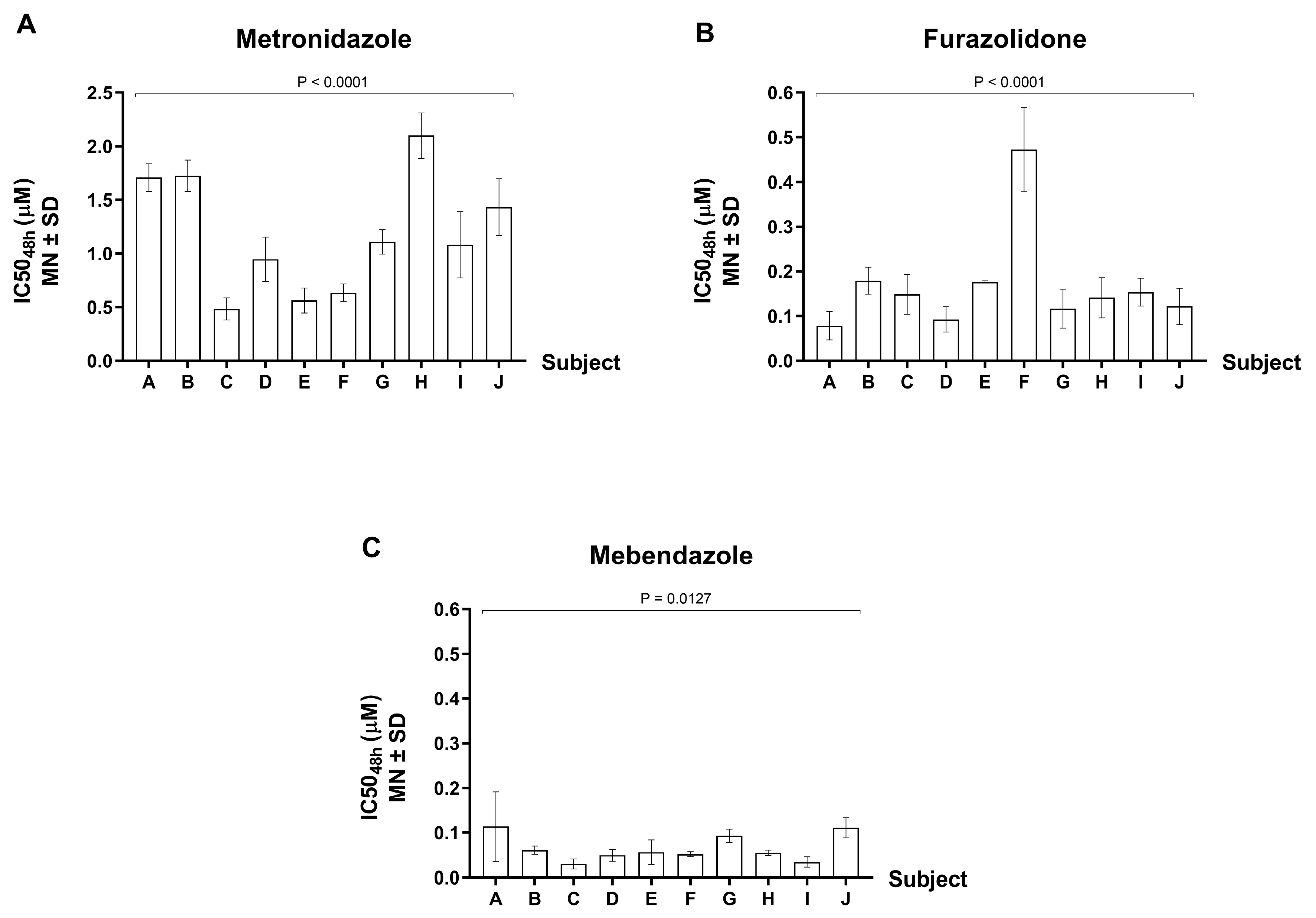
The IC50 value for each G. lamblia isolate was assessed after 48 h of drug exposure (Figure 2). For metronidazole, the IC50 varied from 0.484 to 2.1 μM, representing a 4.3-fold variation of susceptibility. For furazolidone, the IC50 varied from 0.079 to 0.473 μM, representing a 6-fold variation of susceptibility. For mebendazole, the IC50 varied from 0.030 to 0.114 μM, representing a 3.8-fold variation of susceptibility. By statistical analysis, the mean IC50 values showed significant difference across the 10 G. lamblia isolates for mebendazole (p-value = 0.0127), and a strongly significant difference was detected for metronidazole and furazolidone (p-value < 0.0001).
The mean IC50 (±SD) of all 10 isolates were 1.18 ± 0.55 μM for metronidazole, 0.17 ± 0.11 μM for furazolidone, and 0.07 ± 0.03 μM for mebendazole. In these in vitro tests, our G. lamblia isolates had the least susceptibility to metronidazole among the three drugs tested. Further, the ratio of drug concentration for metronidazole vs. mebendazole was 17-fold (1.18/0.07), while metronidazole vs. furazolidone was 7-fold (1.18/0.17). We also found that IC50 of the majority of G. lamblia isolates tested against metronidazole were significantly higher than that tested against mebendazole (10/10 isolates; p-values ranging from <0.0001 to 0.0009) and tested against furazolidone (9/10 isolates; p-values ranging from <0.0001 to 0.0018). Apparently, the mean IC50 of one sample against furazolidone was significantly higher than mebendazole (p-value = 0.0009). However, the remaining 9 samples did not show a significant difference in IC50 between these two drugs. Collectively, our in vitro findings suggest that metronidazole was considered less effective as a giardiasis treatment than mebendazole and furazolidone.

3.5. G. lamblia Genotypes

Among 115 stool samples that were positive for G. lamblia, eight stool samples were positive from microscopy alone, 55 samples were positive from ELISA alone, and 52 samples were positive from both ELISA and microscopy. Of these 115 stool samples, 104 samples were positive by real-time PCR and were further genotyped. However, only 73 samples (70.19%) were typeable as Giardia TPI A of 24.66% (18/73), Giardia TPI B of 71.23% (52/73), and Giardia TPI A/B of 4.11% (3/73). Among non-typeable samples, 19 were negative by microscopy but positive by ELISA, while 12 samples were positive by microscopy and ELISA.

4. Discussion

Intestinal parasitic infections are a significant public health burden, especially in tropical and subtropical areas with poor sanitation and high levels of poverty [3]. Thailand is located in a tropical region, which is a highly prevalent area for parasitic infections. Although public utilities have improved in many areas of Thailand, especially in rural and remote areas, they still have problems with sanitation. Our study found that the prevalence of intestinal parasitic infections was high in school-aged and preschool children, with more than half of these children infected with at least one type of intestinal parasite. Another study conducted in the Karen hill tribe in the Northern part of Thailand also found a high prevalence of intestinal parasitic infections in about half of the school children (47.7%) [10]. The high prevalence of intestinal parasitic infections found in our and other studies might be due to the study sites being located in remote areas where the water supply and sanitation are not well developed. This hypothesis is supported by the findings of a study conducted in the suburban areas of Saraburi province, located in the Central region of Thailand. The prevalence of intestinal parasitic infections in children identified in that study was 22.1% [35], which is lower than the prevalence found in our study. Another study conducted in the rural area of Nopphitam District in Nakhon Si Thammarat province also found a low prevalence of intestinal parasitic infections in children (16%) [12]. Although the difference in prevalence between our study and this study may be due to the use of different methods to identify parasitic infections. In our study, we employed direct smear cultivation using LE media and ELISA methods, whereas the study conducted in Nakhon Si Thammarat utilized formalin-ethyl acetate sedimentation concentration that might have a lower sensitivity than our methods for detecting intestinal parasitic infections.
Our participants were school and preschool children. Children in this age range have a high risk of intestinal parasitic infections due to their increased behavioral risk, frequent outdoor exposure, and poor personal cleanliness [36]. Although the prevalence of intestinal parasitic infections was high in children, most of them were caused by non-pathogenic protozoa, while a minority of them were infected with intestinal helminths. Only 2.3% of children in our study were infected with intestinal helminths, while the study conducted among school children in the southern part of Thailand reported a slightly higher prevalence of intestinal helminth infections (6.3%) than our study [11]. These discrepancies may be attributable to variances in host genetics as well as the level of personal and community hygiene and sanitation [37,38].
The most serious effect of intestinal parasitic infections in children is the increased risk of malnutrition. Malnutrition is a significant public health problem and is the leading cause of death in children under five years of age [39]. Intestinal parasitic infections increase the risk of malnutrition in children because these infections can damage a child’s internal mucosa, leading to impaired digestion and poor absorption of nutrients [3]. A deficiency in some nutrients can affect the host’s immunological function, making the body more susceptible to infectious diseases and intestinal parasites. As part of a vicious cycle, parasitic infections can exacerbate nutrient loss, resulting in stunted growth and low nutritional status. Our study did not identify a significant association between overall intestinal parasitic infections and being underweight or stunting or wasting in children. This aligns with the majority of previous studies, which also did not reveal a significant association between intestinal parasitic infections and being underweight in children [40]. However, when each parasite was considered separately, several previous studies showed a strong correlation between A. lumbricoides infection and malnutrition in children [41,42,43]. In our study, the presence of A. lumbricoides or E. vermicularis was associated with being underweight, stunted, or wasted. However, these relationships did not achieve statistical significance, possibly due to the low prevalence of these helminth infections among our participants.
Our study found a significant association between B. hominis and being underweight in children, while E. nana and I. buetschlii were significantly associated with stunted growth. These three intestinal protozoa are non-pathogenic protozoa and rarely cause symptoms in immunocompetent persons. Thus, it is doubtful that these protozoa can induce malnutrition in children, or these associations might be influenced by other unmeasured factors, such as inadequate food intake with an undernourished diet. B. hominis and E. nana can be transmitted from human to human via the fecal-oral route, from animal to human via the water supply (eating uncooked vegetables and drinking unboiled water) and soil [44]. Thus, infected individuals often have a low socioeconomic status and inhabit unhygienic areas. Low socioeconomic status and living in unhygienic areas are significant risk factors for malnutrition in children. Thus, the relationship between non-pathogenic protozoa and malnutrition found in our study might be confounded by these factors. However, some previous evidence suggests that B. hominis infection could induce both acute and chronic diarrhea, which are the causes of malnutrition in immunocompetent children and adults [45,46,47]. Therefore, the pathogenicity of B. hominis remains inconclusive, and additional research is required on this topic. In addition, diagnosis of non-pathogenic protozoa may be important to prevent malnutrition in children. Identification of non-pathogenic parasites is also important because it highlights fecal contamination.
For the excystation of Giardia cysts, our rate of 16% for in vitro establishment of Giardia from cysts to trophozoites is slightly lower than the rates indicated in the literature, ranging from 21% to 44% [48,49,50]. In our case, the major cause of failure to produce a high rate of excystation is the inability of trophozoites to attach to the tube surface. We speculated that genetic variations could be a significant factor that contributes to the adaptability of trophozoites to survive in the culture media, as reported by Meloni and Thompson [50].
G. lamblia isolates from this study exhibited variation in drug susceptibility, with the greatest variability displayed in response to furazolidone, followed by metronidazole and mebendazole. Based on the in vitro drug susceptibility data, our G. lamblia isolates showed the least susceptibility to metronidazole among the three drugs tested. In other words, metronidazole is considered the least effective for giardiasis treatment in this study. Our finding was concordant with a previous report in Thailand that illustrated that metronidazole was less effective in giardiasis treatment in comparison to tinidazole and ornidazole [51]. To conclude that our G. lamblia isolates are resistant to metronidazole, mebendazole, and furazolidone, further investigations of drug susceptibility and molecular studies are needed to include G. lamblia susceptible strain to these three drugs as a control.
Metronidazole is the first-line treatment option for giardiasis. It is low cost, available over-the-counter, and a broad-spectrum therapeutic. Importantly, these factors contribute to its overuse which potentially drives the increasing emergence of metronidazole-resistant Giardia [52,53]. In most countries, the efficacy rate of metronidazole has been reported in ranges of 60–100% [54]. However, metronidazole treatment failure has been identified in a wide range between 15 and 70% after the 5–7 days course of standard treatment [13,23,55]. Furthermore, inappropriate dosing, e.g., metronidazole of <500 mg/day, could contribute to resistance in Giardia [18]. In recent years, numerous reports have addressed the increasing Giardia treatment failure of metronidazole [13,20]. A recent study demonstrated that approximately 20% of patients with chronic giardiasis had refractory to tinidazole or metronidazole, and as high as 70% of the refractory infections were originally from Asia [55]. Combination therapy of metronidazole with another drug was also recommended to improve treatment efficacy against resistant Giardia infections [17,56]. Further, genetic variation of key enzymes or proteins involved in the metronidazole metabolizing pathway was found to be highly associated with developing metronidazole resistance among Giardia clinical isolates [57,58,59]. Of note, G. lamblia trophozoites isolated in this study presented variable susceptibility to common antigiardial agents. It is imperative to further investigate the metabolomic profiling of these G. lamblia isolates. This significant data could provide a better understanding of gene variation associated with drug resistance. It is also interesting to explore the drug susceptibility of these G. lamblia isolates against other drugs that are frequently used for giardiasis treatment, such as tinidazole, albendazole, and nitazoxanide. Knowing the drug susceptibility pattern of G. lamblia isolates circulating within this high prevalence area could assist researchers in this field in extending knowledge on drug susceptibility distributions.
To the best of our knowledge, there is no report indicating metronidazole resistance in G. lamblia isolates in Thailand. A possible reason could be that the use of G. lamblia trophozoites isolated from human stool for studying drug susceptibility is challenging, e.g., bacterial contamination, low numbers of Giardia cysts, and no development of cysts into trophozoites. Based on our direct experience, these factors contribute to the failure to achieve a high success rate of excystation from cysts to trophozoites. Nevertheless, in vitro susceptibility data is highly valuable to guide physician decisions for the treatment of giardiasis, particularly with resistant parasites that are more difficult or impossible to treat with currently available regimens. Collectively, our drug susceptibility data highlights the importance of consistent monitoring of drug-resistant patterns of Giardia, especially for the communities in endemic areas. The data could inform public health settings and hygiene control measures to improve health management programs for effective treatment of Giardia infection in the area with a high prevalence of Giardia infection. Ultimately, this will reduce the transmission of resistant Giardia as an infectious intestinal parasite to the environment.
In this study, several methods were performed to detect G. lamblia to ensure that all of the G. lamblia-positive samples were included in the genotyping identification. G. lamblia assemblage B, based on TPI, was the most common assemblage identified in our study, which is similar to a report from Cambodia [60]. However, there is a considerable geographical variation of the common assemblage reported in Thailand, where assemblage A (subtype AII) was common in a province in central Thailand [61], but assemblage B was common among hill tribe children in northern Thailand [62]. The discordance between detection methods was not unprecedented, as each method detected different components of the pathogen. The samples that were not typeable could potentially be of other assemblages that were not tested for, specifically assemblage E [63].

Strengths and Limitations

Our study has several strengths. We measured the prevalence of intestinal parasitic infections and assessed the association between these infections and chronic malnutrition, which is a significant health problem in school-aged children in an endemic area. Cultivation of intestinal parasites using LE media was applied in our study to increase the chance of parasitic detection. Moreover, drug susceptibility testing was performed for G. lamblia, one of the most prevalent pathogenic protozoa in Thailand [15]. Furthermore, our findings provide significant data on the variable susceptibility of G. lamblia to common drugs used for the treatment of giardiasis.
However, our study has some limitations. Firstly, our study design is a cross-sectional study. Therefore, no causal relationship between any association between parasitic infections and chronic malnutrition found in our study can be assumed. In addition, some factors related to both parasitic infections and malnutrition were not measured in our study. Therefore, our findings may be prone to confounding bias. Moreover, recruiting participants by invitation might affect the validity of the findings from the study because the characteristics of people who were willing or not willing to participate in the study might have different traits, such as socioeconomic status. The differences in these characteristics might be associated with the prevalence of intestinal parasitic infections and might deviate the results from the truth. Additionally, only microscopic examination was applied to detect helminthic infections. Thus, the prevalence of helminthic infections found in our study might be underestimated. In addition, there are limitations on the interpretation of drug susceptibility because the control strain(s) susceptible to the three drugs tested were not available. In this regard, we can only compare variable susceptibility among the G. lamblia isolates. The genotyping was limited to the identification of two assemblages of one gene based on real-time PCR due to an already wide breadth of the surveillance scope. However, the genotyping results show that assemblages A and B are the predominant assemblages, while untypeable samples can be subject to further characterization.

5. Conclusions

Our findings highlight the ongoing burden of intestinal parasite infections in Thai children living in remote places, as well as the potential emergence of anti-parasitic resistance in G. lamblia. In addition, infections with G. lamblia, B. hominis, and E. nana were associated with children being underweight. As a result, early screening and diagnosis of intestinal parasitic infections are important for eliminating these intestinal parasite diseases and preventing malnutrition in children.

Supplementary Materials

The following supporting information can be downloaded at: https://www.mdpi.com/article/10.3390/tropicalmed8080394/s1, Table S1: Probes and primers used for the screening and genotyping of Giardia from stool samples. Table S2: Frequency of symptoms in study participants. Figure S1: Growth phase of G. lamblia WB-C6 strain. Figure S2: Photomicrographs of excystation and axenization of G. lamblia isolate in TYI-S-33 medium.

Author Contributions

Conceptualization, B.W., N.R., P.L. and L.B.; Data curation, P.K., W.O. (Wirote Oransathid), W.O. (Wilawan Oransathid), S.G., S.S. (Sasikorn Silapong), U.S. and S.S. (Siriporn Sornsakrin); Formal analysis, T.A.; Investigation, P.K., W.O. (Wirote Oransathid), W.O. (Wilawan Oransathid), S.G. and S.S. (Sasikorn Silapong); Methodology, B.W., N.R., P.L. and L.B.; Project administration, U.S. and S.S. (Siriporn Sornsakrin); Software, T.A.; Supervision, D.M.B. and J.R.L.; Writing—original draft, B.W. and T.A.; Writing—review & editing, N.R., P.L., P.K., W.O. (Wirote Oransathid), W.O. (Wilawan Oransathid), S.G., S.S. (Sasikorn Silapong), U.S., S.S. (Siriporn Sornsakrin), L.B., D.M.B. and J.R.L. All authors have read and agreed to the published version of the manuscript.

Funding

This research was funded by the Armed Forces Health Surveillance Center-Global Emerging Infections Surveillance and Response System (AFHSC-GEIS), grant number P0050_17_AF.

Institutional Review Board Statement

The study was conducted in accordance with the Declaration of Helsinki and approved by the Institutional Review Board (or Ethics Committee) of the Ethical Review Committee of the Ministry of Public Health, Nonthaburi, Thailand, and the Walter Reed Army Institute of Research, Silver Spring, Maryland (WR#2374, date of approval 17 August 2017). Material has been reviewed by the Walter Reed Army Institute of Research. There is no objection to its presentation and/or publication. The opinions or assertions contained herein are the private views of the author and are not to be construed as official or as reflecting true views of the Department of the Army or the Department of Defense. The investigators have adhered to the policies for the protection of human subjects as prescribed in AR 70–25.

Informed Consent Statement

Informed consent was obtained from all subjects involved in the study.

Data Availability Statement

Data is available upon request after approval by the research committee.

Acknowledgments

We would like to thank Kanthinich Thima, a staff of the Department of Protozoology, Faculty of Tropical Medicine, Mahidol University, for her generous support in training the cultivation of Giardia.

Conflicts of Interest

The authors declare no conflict of interest. The funders had no role in the design of the study; in the collection, analyses, or interpretation of data; in the writing of the manuscript; or in the decision to publish the results.

Appendix A

Briefly, approximately 2 × 10⁴ cysts suspended in 1.5 mL of the stage 1 excystation pH 2.0 were incubated for 30 min in a 37 °C water bath. Then cell pellet was resuspended in 1.5 mL of the stage 2 excystation solution pH 8.0 and incubated for 60 min in a 37 °C water bath. In a final step, the pellet was resuspended in 12.5 mL of the complete modified TYI-S-33 medium (supplemented with 10% adult bovine serum (Sigma Aldrich, USA) and antibiotics) in a tight secure-sealed screw cap tube and incubated at 37 °C with a slanted position. The division of the trophozoites was observed daily under an inverted microscope. The complete modified TYI-S-33 medium was replaced every 2–4 days. G. lamblia trophozoites were cryopreserved until used.

References

  1. Pullan, R.L.; Smith, J.L.; Jasrasaria, R.; Brooker, S.J. Global numbers of infection and disease burden of soil transmitted helminth infections in 2010. Parasites Vectors 2014, 7, 37. [Google Scholar] [CrossRef] [Green Version]
  2. World Health Organization. Soil-Transmitted Helminth Infections. Fact Sheet, Updated January 2017. 2017. Available online: https://www.who.int/news-room/fact-sheets/detail/soil-transmitted-helminth-infections (accessed on 5 July 2023).
  3. Hesham, M.S.; Edariah, A.B.; Norhayati, M. Intestinal parasitic infections and micronutrient deficiency: A review. Med. J. Malays. 2004, 59, 284–293. [Google Scholar]
  4. Cappello, M. Global health impact of soil-transmitted nematodes. Pediatr. Infect. Dis. J. 2004, 23, 663–664. [Google Scholar] [CrossRef] [PubMed]
  5. Stephenson, L.S.; Latham, M.C.; Ottesen, E.A. Malnutrition and parasitic helminth infections. Parasitology 2000, 121, S23–S38. [Google Scholar] [CrossRef] [PubMed] [Green Version]
  6. Lunn, P.G.; Northrop-Clewes, C.A. The impact of gastrointestinal parasites on protein-energy malnutrition in man. Proc. Nutr. Soc. 1993, 52, 101–111. [Google Scholar] [CrossRef] [PubMed] [Green Version]
  7. OpenDevelopment Thailand. SDG 6 Clean Water and Sanitation. Available online: https://thailand.opendevelopmentmekong.net/topics/sdg-6-clean-water-and-sanitation/ (accessed on 26 June 2023).
  8. Waikagul, J.; Krudsood, S.; Radomyos, P.; Radomyos, B.; Chalemrut, K.; Jonsuksuntigul, P.; Kojima, S.; Looareesuwan, S.; Thaineau, W. A cross-sectional study of intestinal parasitic infections among schoolchildren in Nan Province, Northern Thailand. Southeast Asian J. Trop. Med. Public Health 2002, 33, 218–223. [Google Scholar]
  9. Warunee, N.; Choomanee, L.; Sataporn, P.; Rapeeporn, Y.; Nuttapong, W.; Sompong, S.; Thongdee, S.; Bang-On, S.; Rachada, K. Intestinal parasitic infections among school children in Thailand. Trop. Biomed. 2007, 24, 83–88. [Google Scholar]
  10. Yanola, J.; Nachaiwieng, W.; Duangmano, S.; Prasannarong, M.; Somboon, P.; Pornprasert, S. Current prevalence of intestinal parasitic infections and their impact on hematological and nutritional status among Karen hill tribe children in Omkoi District, Chiang Mai Province, Thailand. Acta Trop. 2018, 180, 1–6. [Google Scholar] [CrossRef]
  11. Sarasombath, P.T. Prevalence and Health Effects of Intestinal Parasitic Infection in School Children in Satun Province, Thailand: A Cross-Sectional Study. Siriraj Med. J. 2017, 69, 167–174. [Google Scholar]
  12. Punsawad, C.; Phasuk, N.; Bunratsami, S.; Thongtup, K.; Viriyavejakul, P.; Palipoch, S.; Koomhin, P.; Nongnaul, S. Prevalence of intestinal parasitic infections and associated risk factors for hookworm infections among primary schoolchildren in rural areas of Nakhon Si Thammarat, southern Thailand. BMC Public Health 2018, 18, 1118. [Google Scholar] [CrossRef]
  13. Mørch, K.; Hanevik, K. Giardiasis treatment: An update with a focus on refractory disease. Curr. Opin. Infect. Dis. 2020, 33, 355–364. [Google Scholar] [CrossRef]
  14. Heyworth, M.F. Giardia duodenalis genetic assemblages and hosts. Parasite 2016, 23, 13. [Google Scholar] [CrossRef] [Green Version]
  15. Tungtrongchitr, A.; Sookrung, N.; Indrawattana, N.; Kwangsi, S.; Ongrotchanakun, J.; Chaicumpa, W. Giardia intestinalis in Thailand: Identification of genotypes. J. Health Popul. Nutr. 2010, 28, 42–52. [Google Scholar] [CrossRef] [PubMed] [Green Version]
  16. Wongjindanon, N.; Suksrichavalit, T.; Subsutti, W.; Sarachart, T.; Worapisuttiwong, U.; Norramatha, P. Current infection rate of Giardia lamblia in two provinces of Thailand. Southeast Asian J. Trop. Med. Public Health 2005, 36 (Suppl. S4), 21–25. [Google Scholar]
  17. Watkins, R.R.; Eckmann, L. Treatment of giardiasis: Current status and future directions. Curr. Infect. Dis. Rep. 2014, 16, 396. [Google Scholar] [CrossRef]
  18. Yereli, K.; Balcioglu, I.C.; Ertan, P.; Limoncu, E.; Onag, A. Albendazole as an alternative therapeutic agent for childhood giardiasis in Turkey. Clin. Microbiol. Infect. 2004, 10, 527–529. [Google Scholar] [CrossRef] [PubMed] [Green Version]
  19. Loderstadt, U.; Frickmann, H. Antimicrobial resistance of the enteric protozoon Giardia duodenalis—A narrative review. Eur. J. Microbiol. Immunol. 2021, 11, 29–43. [Google Scholar] [CrossRef]
  20. Lalle, M.; Hanevik, K. Treatment-refractory giardiasis: Challenges and solutions. Infect. Drug Resist. 2018, 11, 1921–1933. [Google Scholar] [CrossRef] [Green Version]
  21. Ansell, B.R.; McConville, M.J.; Ma’ayeh, S.Y.; Dagley, M.J.; Gasser, R.B.; Svard, S.G.; Jex, A.R. Drug resistance in Giardia duodenalis. Biotechnol. Adv. 2015, 33, 888–901. [Google Scholar] [CrossRef] [PubMed]
  22. Nabarro, L.E.; Lever, R.A.; Armstrong, M.; Chiodini, P.L. Increased incidence of nitroimidazole-refractory giardiasis at the Hospital for Tropical Diseases, London: 2008–2013. Clin. Microbiol. Infect. 2015, 21, 791–796. [Google Scholar] [CrossRef] [Green Version]
  23. Peters, T.E.; Kreuels, B.; Addo, M.M.; Tannich, E.; Rothe, C. Risk factors for and management of metronidazole-refractory giardiasis in international travellers: A retrospective analysis. Travel. Med. Infect. Dis. 2021, 43, 102090. [Google Scholar] [CrossRef] [PubMed]
  24. World Health Organization. Child Growth Standards. Available online: https://www.who.int/tools/child-growth-standards (accessed on 20 July 2022).
  25. Lurchachaiwong, W.; Serichantalergs, O.; Lertsethtakarn, P.; Ruamsap, N.; Srijan, A.; Oransathid, W.; Khemnu, N.; Vesely, B.A.; Demons, S.T.; Waters, N.C.; et al. Enteric etiological surveillance in acute diarrhea stool of United States Military Personnel on deployment in Thailand, 2013–2017. Gut Pathog. 2020, 12, 17. [Google Scholar] [CrossRef] [PubMed]
  26. Center for Disease Control and Prevention. DPDx—Laboratory Identification of Parasites of Public Health Concern. Available online: https://www.cdc.gov/dpdx/diagnosticprocedures/stool/microexam.html (accessed on 29 June 2023).
  27. Saksirisampant, W.; Nuchprayoon, S.; Pradniwat, P.; Lamchuan, D. Boeck and Drbohlav Locke egg serum medium fordetection of Blastocystis hominisBlastocystis homin. Chulalongkorn Med. J. 2010, 54, 528–536. [Google Scholar]
  28. Davids, B.J.; Gillin, F.D. Methods for Giardia Culture, Cryopreservation, Encystation, and Excystation In Vitro. In Giardia: A Model Organism, 1st ed.; Lujan, H.D., Svärd, S., Eds.; Springer: Vienna, Austria, 2011. [Google Scholar] [CrossRef]
  29. Hahn, J.; Seeber, F.; Kolodziej, H.; Ignatius, R.; Laue, M.; Aebischer, T.; Klotz, C. High Sensitivity of Giardia duodenalis to Tetrahydrolipstatin (Orlistat) In Vitro. PLoS ONE 2013, 8, e71597. [Google Scholar] [CrossRef] [PubMed] [Green Version]
  30. Popruk, S.; Thima, K.; Udonsom, R.; Chiabchalard, R.; Mahittikorn, A.; Palukul, K.; Thepouypom, A. Activity of Plant Essential Oils against Giardia duodenalis. Southeast Asian J. Trop. Med. Public Health 2017, 48, 756–761. [Google Scholar]
  31. Liu, J.; Gratz, J.; Amour, C.; Kibiki, G.; Becker, S.; Janaki, L.; Verweij, J.J.; Taniuchi, M.; Sobuz, S.U.; Haque, R.; et al. A laboratory-developed TaqMan Array Card for simultaneous detection of 19 enteropathogens. J. Clin. Microbiol. 2013, 51, 472–480. [Google Scholar] [CrossRef] [PubMed] [Green Version]
  32. Liu, J.; Gratz, J.; Maro, A.; Kumburu, H.; Kibiki, G.; Taniuchi, M.; Howlader, A.M.; Sobuz, S.U.; Haque, R.; Talukder, K.A.; et al. Simultaneous detection of six diarrhea-causing bacterial pathogens with an in-house PCR-luminex assay. J. Clin. Microbiol. 2012, 50, 98–103. [Google Scholar] [CrossRef] [Green Version]
  33. Llewellyn, S.; Inpankaew, T.; Nery, S.V.; Gray, D.J.; Verweij, J.J.; Clements, A.C.A.; Gomes, S.J.; Traub, R.; McCarthy, J.S. Application of a Multiplex Quantitative PCR to Assess Prevalence and Intensity Of Intestinal Parasite Infections in a Controlled Clinical Trial. PLoS Neglected Trop. Dis. 2016, 10, e0004380. [Google Scholar] [CrossRef]
  34. Almeida, A.; Pozio, E.; Cacciò, S.M. Genotyping of Giardia duodenalis cysts by new real-time PCR assays for detection of mixed infections in human samples. Appl. Environ. Microbiol. 2010, 76, 1895–1901. [Google Scholar] [CrossRef] [Green Version]
  35. Assavapongpaiboon, B.; Bunkasem, U.; Sanprasert, V.; Nuchprayoon, S. A Cross-Sectional Study on Intestinal Parasitic Infections in Children in Suburban Public Primary Schools, Saraburi, the Central Region of Thailand. Am. J. Trop. Med. Hyg. 2018, 98, 763–767. [Google Scholar] [CrossRef] [Green Version]
  36. Belizario, V.Y., Jr.; Totañes, F.I.; de Leon, W.U.; Lumampao, Y.F.; Ciro, R.N. Soil-transmitted helminth and other intestinal parasitic infections among school children in indigenous people communities in Davao del Norte, Philippines. Acta Trop. 2011, 120 (Suppl. S1), S12–S18. [Google Scholar] [CrossRef]
  37. Quinnell, R.J.; Pullan, R.L.; Breitling, L.P.; Geiger, S.M.; Cundill, B.; Correa-Oliveira, R.; Brooker, S.; Bethony, J.M. Genetic and Household Determinants of Predisposition to Human Hookworm Infection in a Brazilian Community. J. Infect. Dis. 2010, 202, 954–961. [Google Scholar] [CrossRef] [Green Version]
  38. Pullan, R.L.; Kabatereine, N.B.; Quinnell, R.J.; Brooker, S. Spatial and Genetic Epidemiology of Hookworm in a Rural Community in Uganda. PLoS Neglected Trop. Dis. 2010, 4, e713. [Google Scholar] [CrossRef] [Green Version]
  39. World Health Organization. Malnutrition. Available online: https://www.who.int/news-room/fact-sheets/detail/malnutrition (accessed on 29 December 2022).
  40. Cossa-Moiane, I.; Roucher, C.; Mac Quene, T.; Campos-Ponce, M.; de Deus, N.; Polman, K.; Doak, C. Association between Intestinal Parasite Infections and Proxies for Body Composition: A Scoping Review. Nutrients 2022, 14, 2229. [Google Scholar] [CrossRef] [PubMed]
  41. Zhou, H.; Watanabe, C.; Ohtsuka, R. Impacts of dietary intake and helminth infection on diversity in growth among schoolchildren in rural south China: A four-year longitudinal study. Am. J. Hum. Biol. 2007, 19, 96–106. [Google Scholar] [CrossRef] [PubMed]
  42. Verhagen, L.M.; Incani, R.N.; Franco, C.R.; Ugarte, A.; Cadenas, Y.; Sierra Ruiz, C.I.; Hermans, P.W.M.; Hoek, D.; Campos Ponce, M.; de Waard, J.H.; et al. High Malnutrition Rate in Venezuelan Yanomami Compared to Warao Amerindians and Creoles: Significant Associations with Intestinal Parasites and Anemia. PLoS ONE 2013, 8, e77581. [Google Scholar] [CrossRef] [PubMed] [Green Version]
  43. Jardim-Botelho, A.; Brooker, S.; Geiger, S.M.; Fleming, F.; Souza Lopes, A.C.; Diemert, D.J.; Corrêa-Oliveira, R.; Bethony, J.M. Age patterns in undernutrition and helminth infection in a rural area of Brazil: Associations with ascariasis and hookworm. Trop. Med. Int. Health 2008, 13, 458–467. [Google Scholar] [CrossRef] [Green Version]
  44. Jinatham, V.; Maxamhud, S.; Popluechai, S.; Tsaousis, A.D.; Gentekaki, E. Blastocystis One Health Approach in a Rural Community of Northern Thailand: Prevalence, Subtypes and Novel Transmission Routes. Front. Microbiol. 2021, 12, 746340. [Google Scholar] [CrossRef]
  45. Oyofo, B.A.; Subekti, D.; Tjaniadi, P.; Machpud, N.; Komalarini, S.; Setiawan, B.; Simanjuntak, C.; Punjabi, N.; Corwin, A.L.; Wasfy, M.; et al. Enteropathogens associated with acute diarrhea in community and hospital patients in Jakarta, Indonesia. FEMS Immunol. Med. Microbiol. 2002, 34, 139–146. [Google Scholar] [CrossRef]
  46. Graczyk, T.K.; Shiff, C.K.; Tamang, L.; Munsaka, F.; Beitin, A.M.; Moss, W.J. The association of Blastocystis hominis and Endolimax nana with diarrheal stools in Zambian school-age children. Parasitol. Res. 2005, 98, 38–43. [Google Scholar] [CrossRef]
  47. Shah, M.; Tan, C.B.; Rajan, D.; Ahmed, S.; Subramani, K.; Rizvon, K.; Mustacchia, P. Blastocystis hominis and Endolimax nana Co-Infection Resulting in Chronic Diarrhea in an Immunocompetent Male. Case Rep. Gastroenterol. 2012, 6, 358–364. [Google Scholar] [CrossRef] [PubMed]
  48. Cruz, A.; Sousa, M.I.; Azeredo, Z.; Leite, E.; Figueiredo de Sousa, J.C.; Cabral, M. Isolation, excystation and axenization of Giardia lamblia isolates: In vitro susceptibility to metronidazole and albendazole. J. Antimicrob. Chemother. 2003, 51, 1017–1020. [Google Scholar] [CrossRef] [PubMed] [Green Version]
  49. Hautus, M.A.; Kortbeek, L.M.; Vetter, J.C.; Laarman, J.J. In vitro excystation and subsequent axenic growth of Giardia lamblia. Trans. R. Soc. Trop. Med. Hyg. 1988, 82, 858–861. [Google Scholar] [CrossRef]
  50. Meloni, B.P.; Thompson, R.C. Comparative studies on the axenic in vitro cultivation of Giardia of human and canine origin: Evidence for intraspecific variation. Trans. R. Soc. Trop. Med. Hyg. 1987, 81, 637–640. [Google Scholar] [CrossRef]
  51. Charoenlarp, P.; Bunnag, D. Treatment of parasitic infections in Thailand. Southeast Asian J. Trop. Med. Public Health 1986, 17, 620–626. [Google Scholar] [PubMed]
  52. Krakovka, S.; Ribacke, U.; Miyamoto, Y.; Eckmann, L.; Svard, S. Characterization of Metronidazole-Resistant Giardia intestinalis Lines by Comparative Transcriptomics and Proteomics. Front. Microbiol. 2022, 13, 834008. [Google Scholar] [CrossRef]
  53. Carter, E.R.; Nabarro, L.E.; Hedley, L.; Chiodini, P.L. Nitroimidazole-refractory giardiasis: A growing problem requiring rational solutions. Clin. Microbiol. Infect. 2018, 24, 37–42. [Google Scholar] [CrossRef] [Green Version]
  54. Arguello-Garcia, R.; Leitsch, D.; Skinner-Adams, T.; Ortega-Pierres, M.G. Drug resistance in Giardia: Mechanisms and alternative treatments for Giardiasis. Adv. Parasitol. 2020, 107, 201–282. [Google Scholar] [CrossRef]
  55. Requena-Mendez, A.; Goni, P.; Rubio, E.; Pou, D.; Fumado, V.; Lobez, S.; Aldasoro, E.; Cabezos, J.; Valls, M.E.; Trevino, B.; et al. The Use of Quinacrine in Nitroimidazole-resistant Giardia duodenalis: An Old Drug for an Emerging Problem. J. Infect. Dis. 2017, 215, 946–953. [Google Scholar] [CrossRef]
  56. Canete, R.; Noda, A.L.; Rodriguez, M.; Brito, K.; Herrera, E.; Kofoed, P.E.; Ursing, J. 5-Nitroimidazole refractory giardiasis is common in Matanzas, Cuba and effectively treated by secnidazole plus high-dose mebendazole or quinacrine: A prospective observational cohort study. Clin. Microbiol. Infect. 2020, 26, 1092.e1–1092.e6. [Google Scholar] [CrossRef]
  57. Galeh, T.M.; Kazemi, A.; Mahami-Oskouei, M.; Baradaran, B.; Spotin, A.; Sarafraz, S.; Karamat, M. Introducing nitazoxanide as a promising alternative treatment for symptomatic to metronidazole-resistant giardiasis in clinical isolates. Asian Pac. J. Trop. Med. 2016, 9, 887–892. [Google Scholar] [CrossRef] [PubMed] [Green Version]
  58. Saghaug, C.S.; Klotz, C.; Kallio, J.P.; Brattbakk, H.R.; Stokowy, T.; Aebischer, T.; Kursula, I.; Langeland, N.; Hanevik, K. Genetic variation in metronidazole metabolism and oxidative stress pathways in clinical Giardia lamblia assemblage A and B isolates. Infect. Drug Resist. 2019, 12, 1221–1235. [Google Scholar] [CrossRef] [Green Version]
  59. Muller, J.; Hemphill, A.; Muller, N. Physiological aspects of nitro drug resistance in Giardia lamblia. Int. J. Parasitol. Drugs Drug Resist. 2018, 8, 271–277. [Google Scholar] [CrossRef] [PubMed]
  60. Moore, C.E.; Elwin, K.; Phot, N.; Seng, C.; Mao, S.; Suy, K.; Kumar, V.; Nader, J.; Bousfield, R.; Perera, S.; et al. Molecular Characterization of Cryptosporidium Species and Giardia duodenalis from Symptomatic Cambodian Children. PLoS Negl. Trop. Dis. 2016, 10, e0004822. [Google Scholar] [CrossRef] [PubMed] [Green Version]
  61. Boontanom, P.; Mungthin, M.; Tan-Ariya, P.; Naaglor, T.; Leelayoova, S. Epidemiology of giardiasis and genotypic characterization of Giardia duodenalis in preschool children of a rural community, central Thailand. Trop. Biomed. 2011, 28, 32–39. [Google Scholar]
  62. Saksirisampant, W.; Boontanom, P.; Mungthin, M.; Tan-Ariya, P.; Lamchuan, D.; Siripattanapipong, S.; Leelayoova, S. Prevalence of giardiasis and genotypic characterization of Giardia duodenalis in hilltribe children, Northern Thailand. Trop. Biomed. 2012, 29, 331–338. [Google Scholar]
  63. Fantinatti, M.; Bello, A.R.; Fernandes, O.; Da-Cruz, A.M. Identification of Giardia lamblia Assemblage E in Humans Points to a New Anthropozoonotic Cycle. J. Infect. Dis. 2016, 214, 1256–1259. [Google Scholar] [CrossRef] [Green Version]
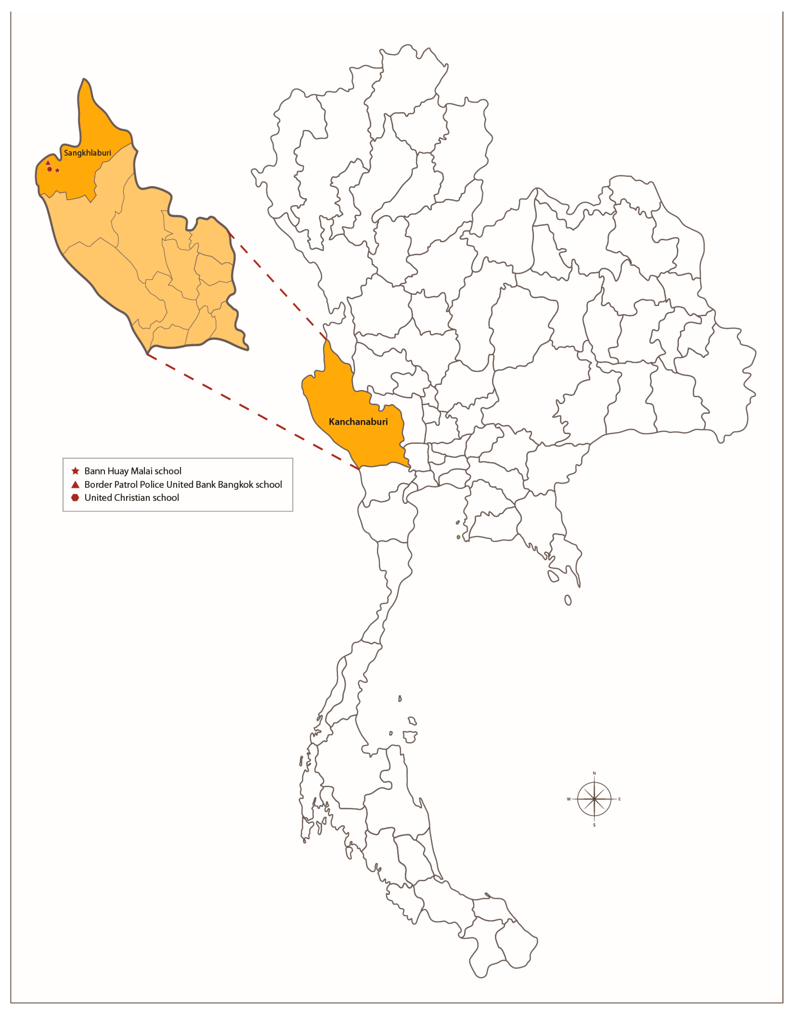
Figure 1. Location of the study sites at Bann Huay Malai, Border Patrol Police United Bank Bangkok, and United Christian schools, Sangkhlaburi District, Kanchanaburi Province, Thailand.
Tropicalmed 08 00394 g001
Figure 2. Drug susceptibility test of G. lamblia isolates from 10 volunteers. The data are expressed as IC50 (µM) ± SD of three independent experiments. X-axis describes individual subjects: (A) Metronidazole; (B) Furazolidone; (C) Mebendazole. The subjects designated as A–J in the X-axis were de-identified from specimen ID without linking to the identifying subject information.
Tropicalmed 08 00394 g002
Table 1. Characteristics of study participants.
CharacteristicTotal Participant
N = 661
Parasitic Infection
N = 445 (67.32%)
Non-Parasitic Infection
N = 216 (32.68%)
Chi2 Value, p-Value
Age; mean (SD); year8.29 (2.14)8.36 (2.11)8.12 (2.22)0.174
Sex
-
male
304 (45.99)198 (44.49)106 (49.07)1.228, 0.268
-
female
357 (54.01)247 (55.51)110 (50.93)
Height; mean (SD); cm126.54 (13.01)126.29 (12.84)127.07 (13.35)0.469
Weight; mean (SD); kg26.72 (9.56)26.35 (9.34)27.47 (9.97)0.160
Stool characteristics
-
hard
216 (32.68)137 (30.79)79 (36.57)2.214, 0.137
-
soft
392 (59.30)273 (61.35)119 (59.05)2.358, 0.125
-
loose
49 (7.41)32 (7.19)17 (7.87)0.098, 0.755
-
watery
4 (0.61)3 (0.67)1 (0.46)0.108, 1.00
-
rice watery
0 (0)0 (0)0 (0)1.00
Symptoms & signs
-
Abdominal cramping
64 (9.68)49 (11.01)15 (6.94)2.750, 0.097
-
Nausea
10 (1.51)8 (1.80)2 (0.93)0.742, 0.511
-
Vomiting
30 (4.54)22 (4.94)8 (3.70)0.516, 0.472
-
Diarrhea
27 (4.08)22 (4.94)5 (2.31)2.565, 0.109
-
Fever
36 (5.45)27 (6.07)9 (4.17)1.020, 0.312
-
RBCs in stool
0 (0)0 (0)0 (0)1.00
-
WBCs in stool
9 (1.36)8 (1.79)1 (0.46)1.929, 0.367
Study’s site
-
Bann Huay Malai School
226 (34.19)152 (67.26)74 (32.74)10.70, 0.005
-
Border Patrol Police United Bank Bangkok School
110 (16.64)88 (80.0)22 (20.0)
-
United Christian School
325 (49.17)205 (63.08)120 (36.92)
Table 2. Prevalence of intestinal parasitic infections.
OrganismsPrevalence (%) (95% Confidence Interval)
TotalPreschool Age *School Age *
Overall infection
-
Helminths
0.30 (0.04–1.09)1.10 (0.03–5.97)0.18 (0.004–0.97)
-
Protozoa
65.05 (61.28–68.69)56.04 (45.25–66.44)66.49 (62.45–70.36)
-
Both helminth and protozoa
1.97 (1.05–3.34)3.30 (0.69–9.33)1.75 (0.84–3.20)
Protozoa infection
Non-pathogenic
-
Blastocystis hominis
49.32 (45.44–53.22)36.26 (26.44–47.01)51.40 (47.21–55.58)
-
Endolimax nana
26.93 (23.58–30.48)24.18 (15.81–34.28)27.37 (23.75–31.23)
-
Entamoeba coli
9.53 (7.40–12.03)3.30 (0.69–9.33)10.53 (8.13–13.34)
-
Iodamoeba buetschlii
5.45 (3.84–7.46)3.30 (0.69–9.33)5.79 (4.02–8.03)
-
Entamoeba spp.
3.03 (1.86–4.63)1.10 (0.03–5.97)3.33 (2.02–5.16)
-
Pentatrichomonas hominis
1.97 (1.05–3.34)1.10 (0.03–5.97)2.11 (1.09–3.65)
Pathogenic
-
Entamoeba histolytica
0.76 (0.25–1.76)0 (0–3.97)0.88 (0.29–2.04)
-
Giardia lamblia
17.40 (14.58–20.51)28.57 (19.59–39.0)15.61 (12.73–18.86)
-
Cryptosporidium
0 (0–0.56)0 (0–3.97)0 (0–0.65)
Helminth infection
-
Ascaris lumbricoides
0.91 (0.33–1.97)2.20 (0.27–7.71)0.70 (0.19–1.79)
-
Hookworm
0.45 (0.09–1.32)0 (0–3.97)0.53 (0.11–1.53)
-
Enterobius vermicularis
0.61 (0.17–1.54)2.20 (0.27–7.71)0.35 (0.04–1.26)
-
Strongyloides stercoralis
0.15 (0.004–0.84)0 (0–3.97)0.18 (0.004–0.97)
-
Trichuris trichiura
0.15 (0.004–0.84)0 (0–3.97)0.18 (0.004–0.97)
* Preschool age is defined as age 3 to 5 years, and school-age is defined as age 6 to 12 years.
Table 3. Associations between intestinal parasitic infections and nutritional statuses.
OrganismWFA HFA WFH
YesNoOR (95% CI)p-ValueYesNoOR (95% CI)p-ValueYesNoOR (95% CI)p-Value
Overall infection
-
Absence
21
(9.72)
195 (90.28)10.07023
(10.65)
193
(89.35)
10.14116
(7.41)
200
(92.59)
10.915
-
Presence
66
(14.83)
379
(85.17)
1.62
(0.96–2.72)
66
(14.83)
379
(85.17)
1.46
(0.88–2.42)
34
(7.64)
411
(92.36)
1.03
(0.56–1.92)
Helminth infection
-
Absence
84
(13.0)
562
(87.0)
10.43386
(13.31)
560
(86.69)
10.45748
(7.43)
598
(92.57)
10.401
-
Presence
3
(20.0)
12
(80.0)
1.67
(0.46–6.05)
3
(20.0)
12
(80.0)
1.63
(0.45–5.89)
2
(13.33)
13
(86.67)
1.92
(0.42–8.74)
Protozoa infection
-
Absence
21
(9.63)
197
(90.37)
10.06223
(10.55)
195
(89.45)
10.12516
(7.34)
202
(92.66)
10.878
-
Presence
66
(14.90)
377
(85.10)
1.64
(0.98–2.76)
66
(14.90)
377
(85.10)
1.48
(0.90–2.46)
34
(7.67)
409
(92.33)
1.05
(0.57–1.95)
Protozoa infection
B. hominis
-
Absence
34
(10.15)
301
(89.85)
10.02137
(11.04)
298
(88.96)
10.06625
(7.46)
310
(92.54)
10.920
-
Presence
53
(16.26)
273
(83.74)
1.72
(1.08–2.72)
52
(15.95)
274
(84.05)
1.53
(0.97–2.40)
25
(7.67)
301
(92.33)
1.03
(0.58–1.83)
E. coli
-
Absence
81
(13.55)
517
(86.45)
10.37283
(13.88)
515
(86.12)
10.33946
(7.69)
552
(92.31)
10.702
-
Presence
6
(9.52)
57
(90.48)
0.67
(0.28–1.61)
6
(9.52)
57
(90.48)
0.65
(0.27–1.56)
4
(6.35)
59
(93.65)
0.81
(0.28–2.34)
E. nana
-
Absence
57
(11.80)
428
(88.2)
10.09057
(11.80)
426
(88.20)
10.04037
(7.66)
446
(92.34)
10.878
-
Presence
30
(16.85)
148
(83.15)
1.51
(0.94–2.45)
32
(17.98)
146
(82.02)
1.64
(1.02–2.63)
13
(7.30)
165
(92.70)
0.95
(0.49–1.83)
G. lamblia
-
Absence
73
(13.37)
473
(86.63)
10.73077
(14.10)
469
(85.90)
10.29744
(8.06)
502
(91.94)
10.299
-
Presence
14
(12.17)
101
(87.83)
0.90
(0.49–1.65)
12
(10.43)
103
(89.57)
0.71
(0.37–1.35)
6
(5.22)
109
(94.78)
0.63
(0.26–1.51)
P. hominis
-
Absence
85
(13.12)
563
(86.88)
10.81186
(13.27)
562
(86.73)
10.31449
(7.56)
599
(92.44)
10.986
-
Presence
2
(15.38)
11
(84.62)
1.20
(0.26–5.53)
3
(23.08)
10
(76.92)
1.96
(0.53–7.27)
1
(7.69)
12
(92.31)
1.02
(0.13–8.00)
I. buetschlii
-
Absence
79
(12.64)
546
(87.36)
10.10480
(12.80)
545
(87.2)
10.04247
(7.52)
578
(92.48)
10.858
-
Presence
8
(22.22)
28
(27.78)
1.97
(0.87–4.49)
9
(25.0)
27
(75.0)
2.27
(1.03–5.00)
3
(8.33)
33
(91.67)
1.12
(0.33–3.78)
Most prevalent protozoa (G. lamblia, B. Hominis, and E. nana)
-
Absence
22
(8.94)
224
(91.06)
10.01525
(10.16)
221
(89.84)
10.05718
(7.32)
228
(92.68)
10.853
-
Presence
350
(84.34)
65
(15.66)
1.89
(1.13–3.15)
64
(15.42)
351
(84.58)
1.61
(0.99–2.64)
32
(7.71)
383
(92.29)
1.06
(0.58–1.93)
Helminth infection
A. lumbricoides
-
Absence
86
(13.13)
569
(86.87)
10.79987
(13.28)
568
(86.72)
10.17649
(7.48)
606
(92.52)
10.413
-
Presence
1
(16.67)
5
(83.33)
1.32
(0.15–11.46)
2
(33.33)
4
(66.67)
3.26
(0.59–18.09)
1
(16.67)
5
(83.33)
2.47
(0.28–21.59)
Hookworm
-
Absence
87
(13.22)
571
(86.78)
--89
(13.53)
569
(86.47)
--50
(7.6)
608
(92.4)
--
-
Presence
0
(0.0)
3
(100)
- 0
(0.0)
3
(100)
- 0
(0.0)
3
(100)
-
E. vermicularis
-
Absence
85
(12.94)
572
(87.06)
10.05888
(13.39)
569
(86.61)
10.50849
(7.46)
608
(92.54)
10.223
-
Presence
2
(50.0)
2
(50.0)
6.73
(0.94–48.41)
1
(25.0)
3
(75.0)
2.16
(0.22–20.95)
1
(25.0)
3
(75.0)
4.14
(0.42–40.51)
S. stercoralis
-
Absence
87
(13.18)
573
(86.82)
--89
(13.48)
571
(86.52)
--50
(7.58)
610
(92.42)
--
-
Presence
0
(0.0)
1
(100)
- 0
(0.0)
1
(100)
- 0
(0.0)
1
(100)
-
T. trichiura
-
Absence
87
(13.18)
573
(86.82)
--89
(13.48)
571
(86.52)
--50
(7.58)
610
(92.42)
--
-
Presence
0
(0.0)
1
(100)
- 0
(0.0)
1
(100)
- 0
(0.0)
1
(100)
-
CI, confidence interval; OR, odds ratio; WFA, weight for Age; HFA, height for age; WFH, weight for height.
Disclaimer/Publisher’s Note: The statements, opinions and data contained in all publications are solely those of the individual author(s) and contributor(s) and not of MDPI and/or the editor(s). MDPI and/or the editor(s) disclaim responsibility for any injury to people or property resulting from any ideas, methods, instructions or products referred to in the content.