Article

Are Pathogenic Leptospira Species Ubiquitous in Urban Recreational Parks in Sydney, Australia?

1
Sydney School of Veterinary Science, The University of Sydney, Sydney, NSW 2006, Australia
2
Elizabeth Macarthur Agricultural Institute (EMAI), Woodbridge Road, Menangle, NSW 2568, Australia
*
Author to whom correspondence should be addressed.
Trop. Med. Infect. Dis. 2024, 9(6), 128; https://doi.org/10.3390/tropicalmed9060128
Submission received: 20 April 2024 / Revised: 28 May 2024 / Accepted: 3 June 2024 / Published: 6 June 2024

Abstract

:
Leptospirosis is a zoonotic disease caused by the spirochete bacteria Leptospira spp. From December 2017 to December 2023, a total of 34 canine leptospirosis cases were reported in urban Sydney, Australia. During the same spatio-temporal frame, one locally acquired human case was also reported. As it was hypothesised that human residents and companion dogs might both be exposed to pathogenic Leptospira in community green spaces in Sydney, an environmental survey was conducted from December 2023 to January 2024 to detect the presence of pathogenic Leptospira DNA in multipurpose, recreational public parks in the council areas of the Inner West and City of Sydney, Australia. A total of 75 environmental samples were collected from 20 public parks that were easily accessible by human and canine visitors. Quantitative PCR (qPCR) testing targeting pathogenic and intermediate Leptospira spp. was performed, and differences in detection of Leptospira spp. between dog-allowed and dog-prohibited areas were statistically examined. The global Moran’s Index was calculated to identify any spatial autocorrelation in the qPCR results. Pathogenic leptospires were detected in all 20 parks, either in water or soil samples (35/75 samples). Cycle threshold (Ct) values were slightly lower for water samples (Ct 28.52–39.10) compared to soil samples (Ct 33.78–39.77). The chi-squared test and Fisher’s exact test results were statistically non-significant (p > 0.05 for both water and soil samples), and there was no spatial autocorrelation detected in the qPCR results (p > 0.05 for both sample types). Although further research is now required, our preliminary results indicate the presence of pathogenic Leptospira DNA and its potential ubiquity in recreational parks in Sydney.

1. Introduction

Leptospirosis is a zoonotic disease caused by spirochete bacteria of the genus Leptospira [1]. The genus consists of two clades and four subclades, with at least 64 genomically distinct species [2]. The pathogenic Leptospira organisms (clade P, including two subclades of pathogenic and intermediate pathogenic Leptospira) could potentially infect many mammalian species, including dogs and humans. In reservoir hosts, Leptospira causes asymptomatic, chronic infection, replicates in the host’s proximal renal tubules, and is shed in the urine [3]. Theoretically, all mammalian species can act as reservoirs for Leptospira, but the most important reservoir species in human environments are rodents [1]. While chronic infection and shedding occur in humans and dogs [4,5], leptospirosis in these two species is more likely to be acute and life-threatening (incidental hosts) [1,3,6]. Infection occurs through direct contact with infected kidney tissues or urine or indirectly from contaminated environmental sources (most commonly water and soil) [7]. Leptospira spp. are known for their resilience in the environment post-excretion, with reported survival times in open natural and sterile experimental environments of several months [8,9]. The multiplication of Leptospira spp. in poorly drained, moist soil has been observed [10].
In humans, leptospirosis is a disease highly associated with extreme meteorological events and occupational exposure. In both developing and developed countries, outbreaks of human leptospirosis are frequently observed following heavy rainfall and flooding [11,12,13,14,15,16], with the hypothesis that pathogenic Leptospira organisms inhabit soil and water sediments at various depths prior to being released by excessive erosion [17]. Occupations that are directly exposed to mammalian reservoirs (e.g., rodents and cattle) or indirectly exposed to contaminated water and soil are at a higher risk of leptospirosis (e.g., farmers, abattoir workers, construction workers, and veterinarians) [18,19,20]. Some outdoor recreational activities (e.g., camping, whitewater rafting, swimming, and caving) have also been identified as risk factors for leptospirosis [21,22,23,24]. Leptospirosis in dogs involves the same direct and indirect transmission pathways, and thus infection is also associated with flooding events [25], reservoir hosts, and environmental exposure related to the purpose of the animal (e.g., hunting or herding dogs in rural areas) [26,27]. Although leptospirosis was traditionally considered a rural disease, recent studies have described a shift in both human and canine patient demographics to “less-exposed” groups living in urban areas [23]. However, little research has been performed into the epidemiology of Leptospira in highly human-modified, metropolitan areas with high income and population density.
In Sydney, New South Wales (NSW), Australia, both locally acquired canine and human leptospirosis cases were not reported for decades [28,29,30]. Unlike human leptospirosis, canine leptospirosis is not a notifiable disease in the state of NSW [31]. Since 2017, canine leptospirosis has apparently re-emerged with veterinarians voluntarily sharing information about confirmed canine leptospirosis cases in urban Sydney via personal communication [28]. As of January 2024, the number of veterinarian-reported canine leptospirosis cases have risen to 34. These cases have not shown significant seasonality, and no association with local climate (temperature and rainfall) has been found [32]. The clinical cases were correlated with canopy coverage in the neighbourhood, but not with the presence of recreational areas in the vicinity [32]. In 2021, a locally acquired case of human leptospirosis was reported. This patient had a history of working at a recreational golf course in eastern urban Sydney [30]. This human case, together with a previously identified correlation between landscape factors and cases of canine leptospirosis, indicates possible environmental exposure of humans and dogs to pathogenic Leptospira in urban green spaces in Sydney and the potential ubiquity of the pathogen across the city. To test these two hypotheses, a focused environmental survey of pathogenic Leptospira in urban green spaces (e.g., recreational parks) is required.
The current study aimed to describe the environmental presence of pathogenic Leptospira spp. in Sydney urban recreational parks in the Australian summer (December 2023–January 2024), test any differences in pathogenic Leptospira observations between dog-allowed and dog-prohibited areas, and identify any spatial autocorrelation of the test results.

2. Materials and Methods

2.1. Sampling Locations

The study followed a cross-sectional, observational design.
From 13 December 2023 to 17 January 2024, environmental samples were collected from recreational parks managed by local councils in the Inner West and the City of Sydney, Australia [33,34]. The City of Sydney council area is a heavily populated urban area (26.68 km2 with 218,096 residents in 2022 [35]), which contains the central business district of Sydney. The Inner West council area is the area immediately west of the City of Sydney, which housed 183,105 persons across 35.22 km2 in 2022 [36]. The mean monthly rainfall during the study period was 14 mm, and the mean monthly maximum and minimum temperature were 28.2–28.3 °C and 19.4–20.7 °C, respectively [37].
The list of parks sampled was manually compiled, and only parks with both dog-allowed and dog-prohibited (e.g., playgrounds, food-processing areas, including BBQ grounds) areas were shortlisted. The coordinates (decimal degrees) of the parks were manually extracted from Google Earth (Google, Mountain View, CA, USA) [38]. A shapefile of the study area (council areas of the Inner West and City of Sydney) was accessed from the 2021 Australian Census geopackage (Australian Bureau of Statistics, Canberra, Australia) [39], then processed as a single polygon and visualised using ArcGIS Pro 2.5.0 (ESRI, Redlands, CA, USA) [40]. The polygon was then subdivided into four quadrants of equal area (approximately 15.50 km2 per quadrant). Within each spatial quadrant, five parks were tentatively, non-randomly selected from the candidate park list. Substitution of manually selected parks was also conducted upon field observation: only parks where at least one water sample could be collected within either a dog-allowed or dog-prohibited area were included, and parks without open or stagnant water resources were substituted with other parks in the same quadrant. The spatial relationship of parks was also subjectively considered in addition to the inclusion criteria, to ensure the selected parks were relatively homogenously distributed across the study area.

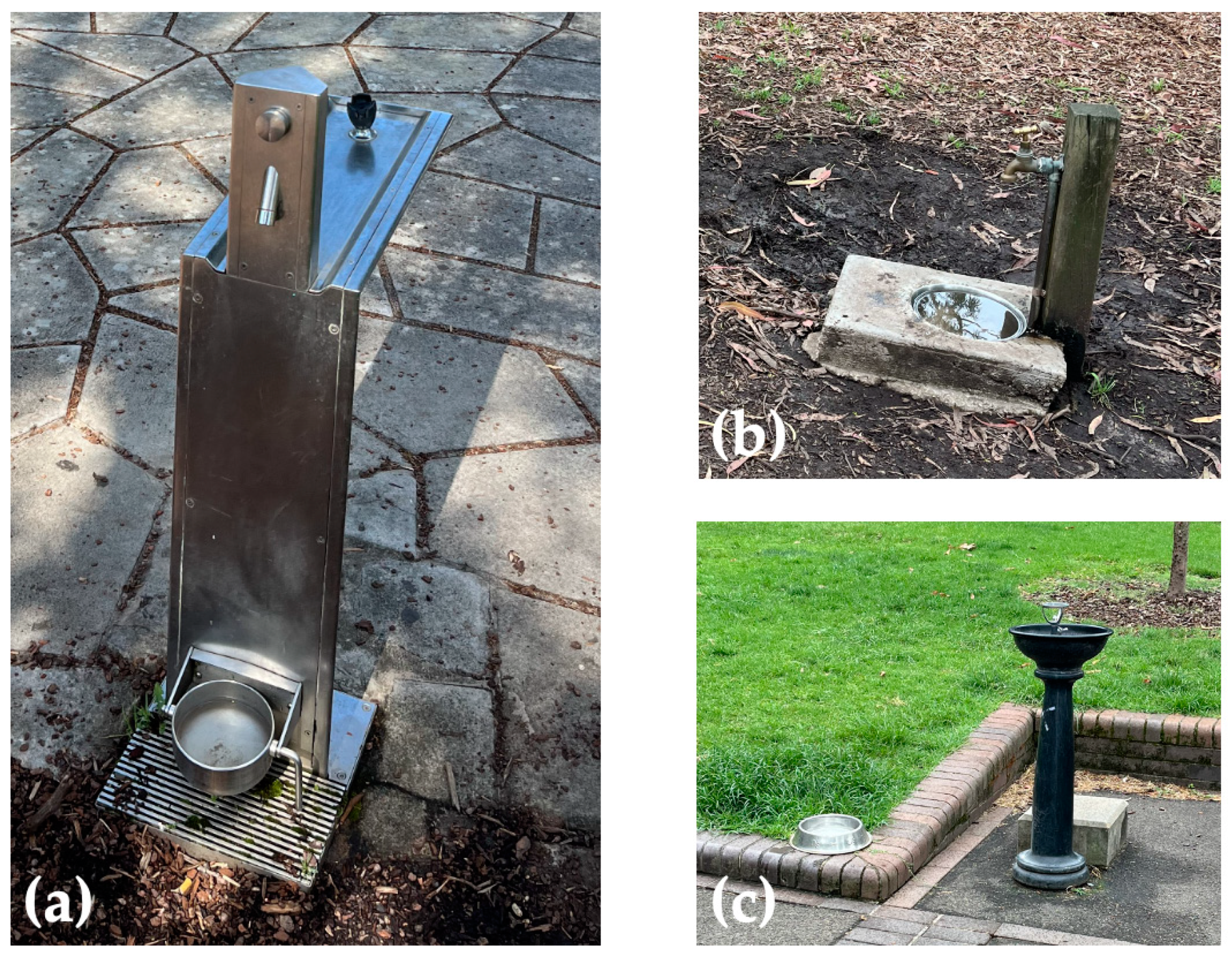
2.2. Water and Soil Sampling

For environmental sampling at each park, ideally, one soil sample and one water sample were collected from both dog-allowed and dog-prohibited areas (fenced or open food preparation areas and playgrounds), respectively. If water was not available in both areas, only one water sample was collected. Due to the descriptive nature of the current study, sample collection was conducted without restricting sampling time and under various weather conditions. All environmental samples were stored in sterile specimen containers at ambient temperature (18–30 °C) and transported to the University of Sydney, Camperdown, within four hours post-collection. Prior to DNA extraction and PCR testing, the samples were kept refrigerated at 4 °C.
The targeted water sources in the current study were primarily stagnant water (public dog water bowls, decorative fountains, puddles, water play apparatus, and a pond) to which human and canine visitors have access in the dog-allowed areas, and stagnant water to which human visitors (e.g., children) have access in the dog-prohibited areas. Water samples were also collected from the seashore and human drinking fountains when stagnant water bodies were not observed in the area. From each water body, at least 150 mL of water was collected using sterile pipettes and a glass measuring cup disinfected with 1:100 F10 veterinary disinfectant solution (quaternary ammonium compounds 54.0 g/L) (F10 Products, Loughborough, UK) [41] between sample collections and soaked in the 1:100 F10 solution for at least 20 min on the morning of each collection date. The sea-water samples were collected 10 cm from the seashore of beaches where off-leash dogs or dog footprints were observed on the collection date. The pond water sample was collected 10 cm from the pond edge. All sea and pond water samples were collected from the surface of water bodies (at approximately 5 cm depth).
At least 50 g of soil samples were collected at a depth of approximately 10 cm from open space (e.g., grass lawn, beach) and ground underneath canopy cover or playground mulch. All sampling sites were easily accessible by humans in the dog-prohibited areas, or humans and dogs in the dog-allowed areas. The soil samples were collected using metal trowels disinfected with the 1:100 F10 solution in between sampling sessions and soaked in the same solution for at least 20 min on the morning of each collection date.

2.3. DNA Extraction and Quantitative PCR Testing

DNA extraction and quantitative PCR (qPCR) testing of samples were both performed at the Elizabeth Macarthur Agricultural Institute (EMAI), the state veterinary laboratory for NSW. The Qiagen DNeasy Blood and Tissue Kit [42] was used to extract DNA from water samples as performed by others [43,44], while the Qiagen DNeasy PowerSoil Kit [45] was used to extract DNA from water sediment and soil samples, according to the manufacturer’s instructions (Qiagen Inc., Toronto, ON, Canada). Water samples were centrifuged at 4500× g for 10 min, and three samples with a pellet greater than 0.5 cm3 underwent the soil extraction procedure using the sediment instead of the water extraction procedure [46]. Of the 75 samples, 37 were spiked with 1 μL of in-house synthetic DNA (10,000 copies) to serve as an internal process control (see below). All DNA extracts were stored at 4 °C prior to qPCR testing, for a maximum of 24 h.
The qPCR assay used is a published protocol that detects an 87-base pair (bp) target within the rrs (16S) gene present in pathogenic and intermediate Leptospira spp. (subclade P1 and P2) [47,48,49]. TaqManTM Environmental Master Mix 2.0 (Thermo Fisher Scientific, Waltham, MA, USA), a primer concentration of 10 μM each of forward and reverse primers (F—5′ CCCGCGTCCGATTAG 3′; R—5′ TCCATTGTGGCCGRA/GACAC 3′), a 5 μM probe concentration (5′ CTCACCAAGGCGACGATCGGTAGC 3′), and 2 μL of DNA extract, were included in each reaction. Cycling conditions were a 95 °C denaturation step for 10 min, followed by 45 cycles of 95 °C for 15 s and 60 °C for one minute in a QuantStudio 5 Real-Time PCR machine (Thermo Fisher Scientific).
An in-house internal process control was used to determine if inhibition occurred in any of the qPCR reactions. This required the spiking of samples with a known target DNA sequence during the DNA extraction process. Identical primer concentrations as above and a 10 μM probe concentration were used in a multiplex reaction with the rrs target. Inhibition was considered to have occurred in the samples if the internal control had a cycle threshold (Ct) greater than 35. A no-template control (NTC) reaction that contained PCR reagents, but no DNA sample was used to detect the presence of DNA contamination in the reagents.
A sample was considered qPCR positive for Leptospira DNA if amplification occurred before 40 cycles. A sample was considered not detected (negative qPCR result) if no amplification occurred before 45 cycles. Samples for which a Ct value between 40 and 45 was recorded were considered inconclusive results. For statistical analyses, inconclusive results were regarded as qPCR negative for Leptospira DNA.
A subset of qPCR positive samples was submitted for sequencing (Australian Genome Research Facility, Sydney, Australia) and the Basic Local Alignment Search Tool (BLAST) used in Genbank® to confirm amplicons contained Leptospira DNA.

2.4. Statistical Tests and Spatial Autocorrelation

As the two extraction methods (water vs. soil/water sediment) resulted in different amounts of DNA extracted, statistical tests were only conducted among the same sample group (i.e., soil vs. soil, water vs. water), using the positive or negative detection of pathogenic Leptospira DNA in the sample as a dichotomous variable. Insufficient water sediment samples were tested to perform this comparison with this sample subtype, and therefore all three sediment samples were included in the water sample group. A right-tailed chi-squared test and a one-tailed Fisher’s exact test were performed using Microsoft Excel Version 2403 (Microsoft, Redmond, WA, USA) [50] to detect statistically significant differences in the number of positives between water and soil samples, as well as samples collected from dog-allowed and dog-prohibited areas.
For preliminary spatial analysis, the continuous results were joined to the collection sites (parks) and mapped using the previously extracted park coordinates in ArcGIS Pro 2.5.0. The Global Moran’s Index was calculated for water and soil samples separately.

3. Results

From a shortlist of 55 multipurpose, recreational public parks in the council areas of the Inner West and City of Sydney, Australia, 20 (36%) parks were sampled (Figure 1). Of the 75 environmental samples tested, 35 tested qPCR positive with a Ct ≤ 40, and 22 samples returned inconclusive results (Ct 40–45). The qPCR reaction was negative in 18 samples (no amplification occurred before 45 cycles). Positive qPCR results were observed in either water or soil samples in all 20 parks (Table 1). All qPCR reactions demonstrated an internal process control Ct < 35, indicating there was no PCR inhibition in any of the tested samples.
In the 33 water samples, 12 samples (12/33, 36.4%) tested qPCR positive with a Ct range of 28.52–39.10. Thirteen samples were negative, and eight samples returned inconclusive Ct values ranging from 40.19 to 43.64. Among the 12 positive samples, the DNA in 10 samples was extracted using the Qiagen DNeasy Blood and Tissue Kits (Ct 28.52–39.10); 2 samples were prepared from water sediment using the Qiagen DNeasy PowerSoil Kit (Ct 35.20 and 38.51). Table 2 summarises the qPCR results for water samples collected from different locations. Of the 33 water samples tested, 15 were collected from three types of public dog water bowls (Figure 2a–c). None of the samples from the dog water bowls embedded in a drinking station (Figure 2a, n = 11) tested qPCR positive, the one water sample from the water bowl illustrated in Figure 2b tested qPCR positive (Ct 38.35), and two of three water samples from dog water bowls that were voluntarily placed by residents (Figure 2c) were qPCR positive (Ct 38.47, 39.10).
In the 42 soil samples, 23 (23/42, 54.8%) tested qPCR positive with a Ct range of 33.78–39.77. Fourteen samples were inconclusive (Ct 40.22–44.02) and five samples yielded no amplification (negative results). It was notable that three soil samples were sand samples, two collected from beaches with off-leash dog areas, and one from a playground (sand pit). One of the beach samples (Ct 37.82) and the sand pit sample (Ct 35.67) tested qPCR positive.
Purified qPCR amplicons from 14 samples yielding the lowest Ct values (Ct < 36) were submitted for sequencing to confirm the presence of Leptospira DNA. Of these 14 samples eight were derived from soil samples and six from water samples. Four soil samples produced results consistent with Leptospira DNA (90.2–100% identity on BLAST alignment). The remaining ten samples did not have discernible sequences, probably due to the low level of target amplicon in the samples. Of the samples that yielded Leptospira sequences, one sample had 100% identity to the species L. interrogans and L. borgpetersonii, confirming the presence of pathogenic leptospires, two samples yielded sequences with 90.2% and 96.55% identity to L. wolffii, which is of intermediate pathogenicity [51,52], and one sample contained sequences with 97.4% identity to Leptospira strain WFW58, which was isolated from a waterfall in Thailand and is also considered of intermediate pathogenicity [53]. Sequences were not obtained from any saprophytic leptospires.
Between the two sample groups (water and soil), there was no difference in the overall frequency of positive results (chi-squared test p = 0.11 and Fisher’s exact p = 0.97). Between the dog-allowed and dog-prohibited areas, chi-squared test (water p = 0.2; soil p = 0.51) and Fisher’s Exact test (water p = 0.95; soil p = 0.83) results on the presence of positive qPCR results were both statistically non-significant. For both sample groups, Global Moran’s Index was also non-significant (water Moran’s Index = 0.04, p = 0.636; soil Moran’s Index = −0.45, p = 0.054).

4. Discussion

Despite not being reported in the peer-reviewed literature for over 40 years (1976) in urban Sydney, canine leptospirosis has been considered endemic since December 2017 [32,54]. The current study confirmed the presence of DNA from pathogenic and intermediate pathogenic Leptospira spp. in urban Sydney and suggests the ubiquity of pathogenic Leptospira in recreational parks across the Inner West and City of Sydney council areas. Our results indicate potential exposure to pathogenic Leptospira from drinking water, natural water bodies, and soil in urban recreational areas, although the suspected ubiquity in various environmental media needs to be further confirmed. Thus, while we report the presence of pathogenic Leptospira in the parks, the magnitude and virulence of Leptospira across the locations require further investigation.
The detection of DNA from pathogenic and intermediate Leptospira organisms (P1 and P2) using an rrs gene TaqMan assay [47] is consistent with previous environmental surveys where the assay was applied to soil and water samples [48,49]. In Iran [52], Thailand [55], and Peru [5], clinical human leptospirosis and multi-species asymptomatic infections associated with intermediate pathogenic Leptospira (e.g., L. wolffii) were observed. However, conclusions beyond this observation remain speculative to date, and concerns over the limitations of interpreting the presence of intermediate Leptospira in environmental samples have been raised [49]. For this reason, qPCR assays based on the lipL32 gene of pathogenic Leptospira (P1) [56] for environmental leptospiral detection are preferred by some researchers [9,49,57]. Although rrs and lipL32 assays were both originally designed for human clinical samples, there has been no report of false positive detection of saprophytic leptospires using either approach, and the application of both assays in non-cultured environmental samples has been validated [46,49,58]. It is also worth noting that the concentration of Leptospira spp. in the samples was relatively low (14/35 positive samples returned Ct < 36). The low leptospiral level likely contributed to the unsuccessful sequencing of ten of the fourteen amplicons, and thus the sequencing outcomes demonstrate the characteristics of environment pathogenic Leptospira in Sydney urban parks. We cannot conclude that there was no saprophytic leptospire amplified in the PCR reactions, and the complexity and diversity of bacteria in the environment could have caused an unidentified effect on the results. As amplification inhibition was absent and none of the four sequences acquired from the amplicons were saprophytic Leptospira, we are optimistic about the amplification efficiency and sensitivity of the qPCR testing performed. To further confirm the distribution of the two subclades of Leptospira and assess the potential spatial heterogeneity, a subsequential, P1-specific assay of the lipL32 gene should be conducted. Alternatively, a lipL32 nested-PCR assay to address the low leptospiral concentration [59] or a rrs and lipL32 multiplex assay for higher specificity [60] might be considered.
The four positive sequencing results were sampled from soil, which might suggest that soil is the major environmental reservoir of pathogenic Leptospira compared to water in urban Sydney. From water on the metallic surface of a human drinking fountain positioned approximately one metre above the ground, a positive PCR reaction was also detected. This result triggers several questions: is it associated with wildlife contamination, or could it indicate transmission of Lepstopira via aerosolized soil in a natural environment? In rats trapped in the City of Sydney council area, 8.1% of the sampled individuals were infected with leptospires and carried the bacteria in their kidneys, which could result in the shedding of the bacteria if individuals are chronically infected [61]. Other common Australian urban wildlife, including flying foxes [62], bandicoots [63], and possums [64], are all potential reservoirs of pathogenic Leptospira organisms. It is acknowledged that soil leptospires can inhabit soil for extended periods and contaminate neighbouring water sources [65,66]. Recently, the proliferation of pathogenic Leptospira was also observed in waterlogged soil [10]. Considering the unknown interactions between wildlife and environmental reservoirs, together with the long survival of Leptospira in soil, understanding the transmission pathways and risk mapping in Sydney can be difficult. It is unrealistic to fully explain the mechanisms underlying the current findings, but investigations into the local wildlife population dynamics should be of high priority to build an overview of the topic.
Regarding the potential role of companion dogs in the transmission of human leptospirosis, we did not find any difference in positive PCR results between dog-allowed and dog-prohibited areas, but we are still far from rejecting the potential canine contribution to the contaminated environment. As legally prescribed by the Companion Animals Act 1998 [67], companion dogs are prohibited within 10 metres of a children’s playground or of a food preparation area. However, the legislation is not properly followed by the public, based on our observations. During sample collection, dogs were observed in a decorative fountain, in BBQ areas, and in an unfenced children’s playground. The urinary shedding of Leptospira in Sydney dogs was described in previous clinical canine patients [28], but evidence of asymptomatic shedding in dogs is absent.
Prior to further confirmation, the current results should not be interpreted as indicating the prevalence of pathogenic Leptospira in Sydney urban recreational parks. However, the ubiquity of pathogenic Leptospira is highly suspected. The high positive rate in urban environments leads to the assumption that human and canine residents are exposed to certain levels of pathogenic Leptospira in their daily lives. Chronic, low-dose exposure (e.g., occupational exposure) is often considered a risk factor leading to clinical infection [68,69,70]. Clinical canine and human leptospirosis is not frequently reported in the study areas (eight canine cases reported within or near the Inner West and City of Sydney council areas in 2023). The gap between the suspected ubiquitous exposure and the infection rate further supports the low concentrations of pathogenic Leptospira in the environment. Furthermore, one or more of the following factors could be contributing concurrently: (i) low virulence of the Leptospira strains in the urban parks; (ii) the unconfirmed spatial or space-time heterogeneity in Leptospira distribution [71]; (iii) protective effect provided by the non-mandatory canine vaccination (Protech® C2i containing L. interrogans serovar Copenhageni [72]) (a vaccine against Leptospira spp. for people is not available in Australia [73]); and (iv) the association between pre-existing conditions or concurrent injuries (e.g., impaired skin barriers [74,75], contacting spiky raspberry plants and soil without skin protection in a recent human leptospirosis outbreak on a farm in rural NSW [76]) and clinical leptospirosis. This study serves as a preliminary attempt to understand and monitor pathogenic Leptospira in urban Sydney, and thus it does not reach any level of public health alert. Risks secondary to the environmental Leptospira exposure in the study area cannot be evaluated solely from it, but care should be recommended in or near the study area due to the leptospiral presence at the parks, and both medical and veterinary professionals should be informed of the potential risks of contracting leptospirosis from environmental spillover.
As discussed above, the most prominent limitation of this study was the single qPCR assay performed on the samples, which should be strengthened by follow-up testing. Aside from the need for implementing assays for other genes (e.g., lipL32), improvement in the DNA extraction methods employed should also be considered. Two commercial DNA extraction kits (DNeasy PowerSoil Kit and DNeasy Tissue Kit) were used in the present study for extraction from soil (and highly sedimented water) and less sedimented water samples, respectively. In contrast to the DNeasy PowerSoil Kit being acknowledged as one of the optimal approaches for DNA extraction from soil samples [46,49,65] and turbid water samples [46], a variety of extraction methods for water samples have been described in the literature. Commercial kits and protocols designed for extracting DNA from blood and tissue (e.g., QIAmp DNA Mini Kit) were used for DNA extraction from environmental surface water samples in Peru [58], and the DNeasy Tissue Kit is a relatively common tool utilised in aquatic biology research [43,44]. In a study in which more turbid, stagnant water samples (e.g., water from animal troughs) were analysed, the QIAmp DNA Stool Mini Kit was used [77]. The DNeasy PowerWater Kit has also been frequently applied to less sedimented water samples (e.g., coastal water, river water, or drinking water) [46,48,78]. Although the DNeasy PowerWater Kit retrieved more DNA from ultrapure water in an experiment in which the performance of several commercial extraction kits on different types of environmental samples was compared, the DNeasy PowerSoil Kit demonstrated excellent efficiency in DNA extraction from sedimented water samples (e.g., pond water, sewage) [46]. The use of the DNeasy Tissue Kit and Dneasy PowerSoil Kit on subtypes of environmental water requires further validation, but trialling the DNeasy PowerSoil Kit on relatively less sedimented water samples (e.g., pond water) is worth considering.

5. Conclusions

By analysing 75 environmental samples collected from recreational parks in the council areas of Inner West and City of Sydney, Australia, we conclude that pathogenic Leptospira spp. are present and potentially ubiquitous in park environments in urban Sydney. Subsequential PCR assays should be considered to confirm the ubiquity of Leptospira in Sydney urban parks. Our initial detection of pathogenic Leptospira demonstrates the potential for environmental surveys to be part of longitudinal pathogenic Leptospira surveillance strategies and the possible development of a geographically specific risk model.

Author Contributions

Conceptualization, X.L., M.E.W. and M.P.W.; methodology, X.L., M.E.W., R.M., C.J. and M.P.W.; formal analysis, X.L.; investigation, X.L., R.M. and M.E.W.; resources, R.M., M.E.W. and C.J.; writing—original draft preparation, X.L. and R.M.; writing—review and editing, M.E.W., C.G., J.M.N., C.J. and M.P.W.; visualization, X.L.; supervision, C.G. and J.M.N.; funding acquisition, M.P.W. and M.E.W. All authors have read and agreed to the published version of the manuscript.

Funding

This research was funded by Bequest of Estate of the Late Bruce Hynes (N2151 B1369 110418), Sydney School of Veterinary Science, University of Sydney.

Institutional Review Board Statement

Not applicable.

Informed Consent Statement

Not applicable.

Data Availability Statement

Data are available upon request.

Conflicts of Interest

The authors declare no conflicts of interest.

References

  1. Ellis, W.A. Animal leptospirosis. In Leptospira and Leptospirosis; Adler, B., Ed.; Current topics in microbiology and immunology; Springer: Berlin, Germany, 2015; Volume 387, pp. 99–137. [Google Scholar]
  2. Vincent, A.T.; Schiettekatte, O.; Goarant, C.; Neela, V.K.; Bernet, E.; Thibeaux, R.; Ismail, R.; Mohd Khalid, M.K.N.; Amran, F.; Masuzawa, T.; et al. Revisiting the taxonomy and evolution of pathogenicity of the genus Leptospira through the prism of genomics. PLoS Neglected Trop. Dis. 2019, 13, e0007270. [Google Scholar] [CrossRef]
  3. Picardeau, M. Diagnosis and epidemiology of leptospirosis. Med. Mal. Infect. 2013, 43, 1–9. [Google Scholar] [CrossRef] [PubMed]
  4. Miotto, B.A.; Guilloux, A.G.A.; Tozzi, B.F.; Moreno, L.Z.; da Hora, A.S.; Dias, R.A.; Heinemann, M.B.; Moreno, A.M.; de Souza Filho, A.F.; Lilenbaum, W.; et al. Prospective study of canine leptospirosis in shelter and stray dog populations: Identification of chronic carriers and different Leptospira species infecting dogs. PLoS ONE 2018, 13, e0200384. [Google Scholar] [CrossRef] [PubMed]
  5. Ganoza, C.A.; Matthias, M.A.; Saito, M.; Cespedes, M.; Gotuzzo, E.; Vinetz, J.M. Asymptomatic renal colonization of humans in the Peruvian Amazon by Leptospira. PLoS Neglected Trop. Dis. 2010, 4, e612. [Google Scholar] [CrossRef]
  6. Haake, D.A.; Levett, P.N. Leptospirosis in humans. In Leptospira and Leptospirosis; Adler, B., Ed.; Current Topics in Microbiology and Immunology; Springer: Berlin, Germany, 2015; Volume 387, pp. 65–97. [Google Scholar]
  7. Bierque, E.; Thibeaux, R.; Girault, D.; Soupé-Gilbert, M.-E.; Goarant, C.; Dellagostin, O.A. A systematic review of Leptospira in water and soil environments. PLoS ONE 2020, 15, e0227055. [Google Scholar] [CrossRef]
  8. Bierque, E.; Soupé-Gilbert, M.-E.; Thibeaux, R.; Girault, D.; Guentas, L.; Goarant, C. Leptospira interrogans retains direct virulence after long starvation in water. Curr. Microbiol. 2020, 77, 3035–3043. [Google Scholar] [CrossRef] [PubMed]
  9. Thibeaux, R.; Geroult, S.; Benezech, C.; Chabaud, S.; Soupé-Gilbert, M.-E.; Girault, D.; Bierque, E.; Goarant, C.; Munoz-Zanzi, C. Seeking the environmental source of Leptospirosis reveals durable bacterial viability in river soils. PLoS Neglected Trop. Dis. 2017, 11, e0005414. [Google Scholar] [CrossRef] [PubMed]
  10. Yanagihara, Y.; Villanueva, S.Y.A.M.; Nomura, N.; Ohno, M.; Sekiya, T.; Handabile, C.; Shingai, M.; Higashi, H.; Yoshida, S.I.; Masuzawa, T.; et al. Leptospira is an environmental bacterium that grows in waterlogged soil. Microbiol. Spectr. 2022, 10, e0215721. [Google Scholar] [CrossRef]
  11. Chusri, S.; McNeil, E.B.; Hortiwakul, T.; Charernmak, B.; Sritrairatchai, S.; Santimaleeworagun, W.; Pattharachayakul, S.; Suksanan, P.; Thaisomboonsuk, B.; Jarman, R.G. Single dosage of doxycycline for prophylaxis against leptospiral infection and leptospirosis during urban flooding in southern Thailand: A non-randomized controlled trial. J. Infect. Chemother. 2014, 20, 709–715. [Google Scholar] [CrossRef]
  12. Dechet, A.M.; Parsons, M.; Rambaran, M.; Mohamed-Rambaran, P.; Cumbermack, A.F.; Persaud, S.; Baboolal, S.; Ari, M.D.; Shadomy, S.V.; Zaki, S.R.; et al. Leptospirosis outbreak following severe flooding: A rapid assessment and mass prophylaxis campaign; Guyana, January–February 2005. PLoS ONE 2012, 7, e39672. [Google Scholar] [CrossRef]
  13. Londe, L.D.; da Conceiçao, R.S.; Bernardes, T.; Dias, M.C.D. Flood-related leptospirosis outbreaks in Brazil: Perspectives for a joint monitoring by health services and disaster monitoring centers. Nat. Hazards 2016, 84, 1419–1435. [Google Scholar] [CrossRef]
  14. Naing, C.; Reid, S.A.; Aye, S.; Htet, N.H.; Ambu, S. Risk factors for human leptospirosis following flooding: A meta-analysis of observational studies. PLoS ONE 2019, 14, e0217643. [Google Scholar] [CrossRef] [PubMed]
  15. Smith, J.K.G.; Young, M.M.; Wilson, K.L.; Craig, S.B. Leptospirosis following a major flood in Central Queensland, Australia. Epidemiol. Infect. 2013, 141, 585–590. [Google Scholar] [CrossRef] [PubMed]
  16. Socolovschi, C.; Angelakis, E.; Renvoisé, A.; Fournier, P.E.; Marie, J.L.; Davoust, B.; Stein, A.; Raoult, D. Strikes, flooding, rats, and leptospirosis in Marseille, France. Int. J. Infect. Dis. 2011, 15, E710–E715. [Google Scholar] [CrossRef] [PubMed]
  17. Thibeaux, R.; Genthon, P.; Govan, R.; Selmaoui-Folcher, N.; Tramier, C.; Kainiu, M.; Soupé-Gilbert, M.-E.; Wijesuriya, K.; Goarant, C. Rainfall-driven resuspension of pathogenic Leptospira in a leptospirosis hotspot. Sci. Total Environ. 2024, 911, 168700. [Google Scholar] [CrossRef] [PubMed]
  18. Atil, A.; Jeffree, M.S.; Rahim, S.S.S.A.; Hassan, M.R.; Lukman, K.A.; Ahmed, K. Occupational deterinants of Leptospirosis among urban service workers. Int. J. Environ. Res. Public Health 2020, 17, 427. [Google Scholar] [CrossRef] [PubMed]
  19. Schonning, M.H.; Phelps, M.D.; Warnasuriya, J.; Agampodi, S.B.; Furu, P. A case-control study of environmental and occupational risks of leptospirosis in Sri Lanka. EcoHealth 2019, 16, 534–543. [Google Scholar] [CrossRef] [PubMed]
  20. Baharom, M.; Ahmad, N.; Hod, R.; Ja’afar, M.H.; Arsad, F.S.; Tangang, F.; Ismail, R.; Mohamed, N.; Radi, M.F.M.; Osman, Y. Environmental and occupational factors associated with leptospirosis: A systemic review. Heliyon 2024, 10, e23473. [Google Scholar] [CrossRef] [PubMed]
  21. Agampodi, S.B.; Karunarathna, D.; Jayathilala, N.; Rathnayaka, H.; Agampodi, T.C.; Karunanayaka, L. Outbreak of leptospirosis after white-water rafting: Sign of a shift from rural to recreational leptospirosis in Sri Lanka? Epidemiol. Infect. 2014, 142, 843–846. [Google Scholar] [CrossRef]
  22. Monahan, A.M.; Miller, I.S.; Nally, J.E. Leptospirosis: Risks during recreational activities. J. Appl. Microbiol. 2009, 107, 707–716. [Google Scholar] [CrossRef]
  23. Lee, H.S.; Guptill, L.; Johnson, A.J.; Moore, G.E. Signalment changes in canine leptospirosis between 1970 and 2009. J. Vet. Intern. Med. 2014, 28, 294–299. [Google Scholar] [CrossRef] [PubMed]
  24. Harland, A.L.; Cave, N.J.; Jones, B.R.; Benschop, J.; Donald, J.J.; Midwinter, A.C.; Squires, R.A.; Collins-Emerson, J.M. A serological survey of leptospiral antibodies in dogs in New Zealand. N. Z. Vet. J. 2013, 61, 98–106. [Google Scholar] [CrossRef] [PubMed]
  25. Raghavan, R.K.; Brenner, K.M.; Higgins, J.J.; Shawn Hutchinson, J.M.; Harkin, K.R. Evaluations of hydrologic risk factors for canine leptospirosis: 94 cases (2002–2009). Prev. Vet. Med. 2012, 107, 105–109. [Google Scholar] [CrossRef] [PubMed]
  26. Goh, S.H.; Ismail, R.; Lau, S.F.; Megat Abdul Rani, P.A.; Mohd Mohidin, T.B.; Daud, F.; Bahaman, A.R.; Khairani-Bejo, S.; Radzi, R.; Khor, K.H. Risk factors and prediction of leptospiral seropositivity among dogs and dog handlers in Malaysia. Int. J. Environ. Res. Public Health 2019, 16, 1499. [Google Scholar] [CrossRef] [PubMed]
  27. Orr, B.; Westman, M.E.; Malik, R.; Purdie, A.; Craig, S.B.; Norris, J.M. Leptospirosis is an emerging infectious disease of pig-hunting dogs and humans in North Queensland. PLoS Neglected Trop. Dis. 2022, 16, e0010100. [Google Scholar] [CrossRef] [PubMed]
  28. Griebsch, C.; Kirkwood, N.; Ward, M.P.; So, W.; Weerakoon, L.; Donahoe, S.; Norris, J.M. Emerging leptospirosis in urban Sydney dogs: A case series (2017–2020). Aust. Vet. J. 2022, 100, 190–200. [Google Scholar] [CrossRef] [PubMed]
  29. Griebsch, C.; Kirkwood, N.; Ward, M.P.; Norris, J.M. Serological evidence of exposure of healthy dogs to Leptospira in Sydney, New South Wales, Australia. Aust. Vet. J. 2024, 102, 215–221. [Google Scholar] [CrossRef] [PubMed]
  30. New South Wales Department of Health. Communicable Diseases Weekly Report: Week 4, 24 January to 30 January 2021. Available online: https://www.health.nsw.gov.au/Infectious/Reports/Publications/cdwr/2021/cdwr-week-4-2021.pdf (accessed on 17 November 2023).
  31. NSW Government. Leptospirosis Fact Sheet. Available online: https://www.health.nsw.gov.au/Infectious/factsheets/Pages/Leptospirosis.aspx (accessed on 13 March 2024).
  32. Lu, X.; Griebsch, C.; Norris, J.M.; Ward, M.P. Landscape, socioeconomic, and meteorological risk factors for canine letopirosis in urban Sydney (2017–2023): A spatial and temporal study. Vet. Sci. 2023, 10, 697. [Google Scholar] [CrossRef] [PubMed]
  33. Inner West Council. Parks by Suburb. Available online: https://www.innerwest.nsw.gov.au/explore/parks-sport-and-recreation/parks-and-playgrounds/parks-by-suburb-parks-by-suburb (accessed on 5 December 2023).
  34. City of Sydney Council. Parks. Available online: https://www.cityofsydney.nsw.gov.au/parks (accessed on 5 December 2023).
  35. City of Sydney Council. City of Sydney: Community Profile. Available online: https://profile.id.com.au/sydney (accessed on 26 March 2024).
  36. Inner West Council. Inner West Council: Community Profile. Available online: https://profile.id.com.au/inner-west/ (accessed on 26 March 2024).
  37. Australian Bureau of Meteorology. Climate Data Online. Available online: http://www.bom.gov.au/climate/data/ (accessed on 6 April 2024).
  38. Google. Google Earth. Available online: https://earth.google.com/web/ (accessed on 1 April 2023).
  39. Australian Bureau of Statistics. Census GeoPackages. Available online: https://www.abs.gov.au/census/find-census-data/geopackages?release=2021&geography=NSW&table=G01&gda=GDA2020 (accessed on 2 April 2023).
  40. ESRI. ArcGIS Pro 2.5.0. Available online: https://www.esri.com/en-us/arcgis/products/arcgis-pro/overview (accessed on 1 March 2023).
  41. Health and Hygiene. F10 Products. Available online: https://www.f10products.co.uk/ (accessed on 2 February 2024).
  42. Qiagen Inc. DNeasy Blood & Tissue Handbook. Available online: https://www.qiagen.com/jp/resources/download.aspx?id=68f29296-5a9f-40fa-8b3d-1c148d0b3030&lang=en (accessed on 14 May 2024).
  43. Djurhuus, A.; Port, J.; Closek, C.J.; Yamahara, K.M.; Romero-Maraccini, O.; Walz, K.R.; Goldsmith, D.B.; Michisaki, R.; Breitbart, M.; Boehm, A.B.; et al. Evaluation of filtration and DNA extraction methods for environmental DNA biodiversity assessments across multiple trophic levels. Front. Mar. Sci. 2017, 4, 314. [Google Scholar] [CrossRef]
  44. Lear, G.; Dickle, I.; Banks, J.; Boyer, S.; Buckley, H.L.; Buckley, T.R.; Cruickshank, R.; Dopheide, A.; Handley, K.M.; Hermans, S.; et al. Methods for the extraction, storage, amplification and sequencing of DNA from samples. N. Z. J. Ecol. 2018, 42, 10–50A. [Google Scholar] [CrossRef]
  45. Qiagen Inc. DNeasy PowerSoil Pro Kit Handbook. Available online: https://www.qiagen.com/jp/resources/download.aspx?id=9bb59b74-e493-4aeb-b6c1-f660852e8d97&lang=en (accessed on 4 February 2024).
  46. Riediger, I.N.; Hoffmaster, A.R.; Casanovas-Massana, A.; Biondo, A.W.; Ko, A.I.; Stoddard, R.A.; Stevenson, B. An optimized method for quantification of pathogenic Leptospira in environmental water samples. PLoS ONE 2016, 11, e0160523. [Google Scholar] [CrossRef] [PubMed]
  47. Smythe, L.D.; Smith, I.L.; Smith, G.A.; Dohnt, M.F.; Symonds, M.L.; Barnett, L.J.; McKay, D.B. A quantitative PCR (TaqMan) assay for pathogenic Leptospira spp. BMC Infect. Dis. 2002, 2, 13. [Google Scholar] [CrossRef] [PubMed]
  48. Viau, E.J.; Boehm, A.B. Quantitative PCR—based detection of pathogenic Leptospira in Hawai’ian coastal streams. J. Water Health 2011, 9, 637–646. [Google Scholar] [CrossRef] [PubMed]
  49. Schneider, A.G.; Casanovas-Massana, A.; Hacker, K.P.; Wunder, E.A.; Begon, M.; Reis, M.G.; Childs, J.E.; Costa, F.; Lindow, J.C.; Ko, A.I.; et al. Quantification of pathogenic Leptospira in the soils of a Brazilian urban slum. PLoS Neglected Trop. Dis. 2018, 12, e0006415. [Google Scholar] [CrossRef] [PubMed]
  50. Microsoft. Microsoft Excel. Available online: https://www.microsoft.com/en-au/microsoft-365/excel (accessed on 5 September 2023).
  51. Lehmann, J.S.; Matthias, M.A.; Vinetz, J.M.; Fouts, D.E. Leptospiral pathogenomics. Pathogens 2014, 3, 280–308. [Google Scholar] [CrossRef] [PubMed]
  52. Zakeri, S.; Khorami, N.; Ganji, Z.F.; Sepahian, N.; Malmasi, A.-A.; Gouya, M.M.; Djadid, N.D. Leptospira wolffii, a potential new pathogenic Leptospira species detected in human, sheep and dog. Infect. Genet. Evol. 2010, 10, 273–277. [Google Scholar] [CrossRef]
  53. Chaiwattanarungruengpaisan, S.; Suwanpakdee, S.; Sangkachai, N.; Chamsei, T.; Taruyanon, K.; Thongdee, M. Potentially pathogenic Leptospira species isolated from a waterfall in Thailand. Jpn. J. Infect. Dis. 2018, 71, 65–67. [Google Scholar] [CrossRef]
  54. Watson, A.; Davis, P.; Johnson, J. Suspected leptospirosis outbreak in kennelled greyhounds. Aust. Vet. Pract. 1976, 6, 84–88. [Google Scholar]
  55. Van, C.D.; Doungchawee, G.; Suttiprapa, S.; Arimatsu, Y.; Kaewkes, S.; Sripa, B. Association between Opisthorchis viverrini and Leptospira spp. infection in endemic Northeast Thailand. Parasitol. Int. 2017, 66, 503–509. [Google Scholar] [CrossRef]
  56. Stoddard, R.A.; Gee, J.E.; Wilkins, P.P.; McCaustland, K.; Hoffmaster, A.R. Detection of pathogenic Leptospira spp. through TaqMan polymerase chain reaction targeting the lipL32 gene. Diagn. Microbiol. Infect. Dis. 2009, 64, 247–255. [Google Scholar] [CrossRef]
  57. Casanovas-Massana, A.; Costa, F.; Riediger, I.N.; Cunha, M.; de Oliveira, D.; Mota, D.C.; Sousa, E.; Querino, V.A.; Nery, N.; Reis, M.G.; et al. Spatial and temporal dynamics of pathogenic Leptospira in surface waters from the urban slum environment. Water Res. 2018, 130, 176–184. [Google Scholar] [CrossRef] [PubMed]
  58. Ganoza, C.A.; Matthias, M.A.; Collins-Richards, D.; Brouwer, K.C.; Cunningham, C.B.; Segura, E.R.; Gilman, R.H.; Gotuzzo, E.; Vinetz, J.M. Determining risk for severe leptospirosis by molecular analysis of environmental surface waters for pathogenic Leptospira. PLoS Med. 2006, 3, e308. [Google Scholar] [CrossRef] [PubMed]
  59. Wójcik-Fatla, A.; Zając, V.; Wasiński, B.; Sroka, J.; Cisak, E.; Sawczyn, A.; Dutkiewicz, J. Occurrence of Leptospira DNA in water and soil samples collected in eastern Poland. Anim. Agric. Environ. Med. 2014, 21, 730–732. [Google Scholar] [CrossRef] [PubMed]
  60. Vital-Brazil, J.M.; Balassiano, I.T.; de Oliveira, F.S.; Costa, A.D.D.; Hillen, L.; Pereira, M.M. Multiplex PCR-based detection of Leptospira in environmental water samples obtained from a slum settlement. Mem. Do Inst. Oswaldo Cruz 2010, 105, 353–355. [Google Scholar] [CrossRef] [PubMed]
  61. Bedoya-Pérez, M.A.; Westman, M.E.; Loomes, M.; Chung, N.Y.N.; Knobel, B.; Ward, M.P. Pathogenic Leptospira species are present in urban rats in Sydney, Australia. Microorganisms 2023, 11, 1731. [Google Scholar] [CrossRef]
  62. Cox, T.E.; Smythe, L.D.; Leung, L.K.-P. Flying foxes as carriers of pathogenic Leptospira species. J. Wildl. Dis. 2005, 41, 753–757. [Google Scholar] [CrossRef] [PubMed]
  63. Slack, A.T.; Symonds, M.L.; Dohnt, M.F.; Corney, B.G.; Smythe, L.D. Epidemiology of Leptospira weilii serovar Topaz infections in Australia. Commuicable Dis. Intell. 2007, 31, 216–222. [Google Scholar]
  64. Eymann, J.; Smythe, L.D.; Symonds, M.L.; Dohnt, M.F.; Barnett, J.L.; Cooper, D.W.; Herbert, C.A. Leptospirosis serology in the common brushtail possum (Trichosurus vulpecula) from urban Sydney, Australia. J. Wildl. Dis. 2007, 43, 492–497. [Google Scholar] [CrossRef]
  65. Casanovas-Massana, A.; Pedra, G.G.; Wunder, E.A.; Diggle, P.J.; Begon, M.; Ko, A.I. Quantification of Leptospira interrogans survival in soil and water microcosms. Appl. Environ. Microbiol. 2018, 84, e00507–e00518. [Google Scholar] [CrossRef]
  66. Barragan, V.A.; Mejia, M.E.; Trávez, A.; Zapata, S.; Hartskeerl, R.A.; Haake, D.A.; Trueba, G.A. Interactions of Leptospira with environmental bacteria from surface water. Curr. Microbiol. 2011, 62, 1802–1806. [Google Scholar] [CrossRef]
  67. NSW Government. Companion Animals Act 1998 No 87; NSW Government: Sydney, Australia, 2023.
  68. Victoriano, A.F.B.; Smythe, L.D.; Gloriani-Barzaga, N.; Cavinta, L.L.; Kasai, T.; Limpakarnjanarat, K.; Ong, B.L.; Gongal, G.; Hall, J.; Coulombe, C.A.; et al. Leptospirosis in the Asia Pacific region. BMC Infect. Dis. 2009, 9, 147. [Google Scholar] [CrossRef] [PubMed]
  69. Jansen, A.; Schöneberg, I.; Frank, C.; Alpers, K.; Schneider, T.; Stark, K. Leptospirosis in Germany, 1962–2003. Emerg. Infect. Dis. 2005, 11, 1048–1054. [Google Scholar] [CrossRef] [PubMed]
  70. Slack, A.T.; Symonds, M.L.; Dohnt, M.F.; Smythe, L.D. The epidemiology of leptospirosis and the emergence of Leptospira borgpetersenii serovar Arborea in Queensland, Australia, 1998–2004. Epidemiol. Infect. 2006, 134, 1217–1225. [Google Scholar] [CrossRef] [PubMed]
  71. Miller, E.; Barragan, V.; Chiriboga, J.; Weddell, C.; Luna, L.; Jimenez, D.J.; Aleman, J.; Mihaljevic, J.R.; Olivas, S.; Marks, J.; et al. Leptospira in river and soil in a highly endemic area of Ecuador. BMC Microbiol. 2021, 21, 17. [Google Scholar] [CrossRef] [PubMed]
  72. Boehringer Ingelheim. Animal Health Products. Available online: https://www.boehringer-ingelheim.com/au/products/animal-health-products-australia (accessed on 13 February 2024).
  73. Safe Work NSW. Leptospirosis. Available online: https://www.safework.nsw.gov.au/hazards-a-z/diseases/leptospirosis (accessed on 5 March 2024).
  74. Asoh, T.; Miyahara, S.; Villanueva, S.Y.A.M.; Kanemaru, T.; Takigawa, T.; Mori, H.; Gloriani, N.G.; Yoshida, S.; Saito, M. Protective role of stratum corneum in percutaneous Leptospira infection in a hamster model. Microb. Pathog. 2023, 182, 106243. [Google Scholar] [CrossRef] [PubMed]
  75. Gostic, K.M.; Wunder, E.A.; Bisht, V.; Hamond, C.; Julian, T.R.; Ko, A.I.; Lloyd-Smith, J.O. Mechanistic dose–response modelling of animal challenge data shows that intact skin is a crucial barrier to leptospiral infection. Philos. Trans. R. Soc. B Biol. Sci. 2019, 374, 20190367. [Google Scholar] [CrossRef] [PubMed]
  76. Katelaris, A.L.; Glasgow, K.; Lawrence, K.; Corben, P.; Zheng, A.; Sumithra, S.; Turahui, J.; Terry, J.; van den Berg, D.; Hennessy, D.; et al. Investigation and response to an outbreak of leptospirosis among raspberry workers in Australia, 2018. Zoonoses Public Health 2020, 67, 35–43. [Google Scholar] [CrossRef] [PubMed]
  77. Muñoz-Zanzi, C.; Mason, M.R.; Encina, C.; Astroza, A.; Romero, A. Leptospira contamination in household and environmental water in rural communities in southern Chile. Int. J. Environ. Res. Public Health 2014, 11, 6666–6680. [Google Scholar] [CrossRef]
  78. Servillano, M.; Vosloo, S.; Cotto, I.; Dai, Z.H.; Jiang, T.; Santana, J.M.S.; Padilla, I.Y.; Rosario-Pabon, Z.; Vega, C.V.; Cordero, J.F.; et al. Spatial-temporal targeted and non-targeted surveys to assess microbiological composition of drinking water in Puerto Rico following Hurricane Maria. Water Res. X 2021, 13, 100123. [Google Scholar] [CrossRef]
Figure 1. The sampled recreational parks (20 from 55 parks shortlisted) in the council areas of Inner West and City of Sydney (labelled as Sydney in the figure), Australia. Ten parks from each council area were sampled. The green dots indicate sampled parks, and the red dots indicate unsampled parks.
Tropicalmed 09 00128 g001
Figure 2. (ac). Three types of public dog water bowls were sampled in Sydney urban recreational parks. (a) Water bowl as part of the drinking station (n = 11); (b) water bowl receiving water from a garden tap (n = 1); (c) water bowl placed and refilled by residents (in this image on the edge of the brick ledge; n = 3).
Tropicalmed 09 00128 g002
Table 1. The quantitative PCR (qPCR), sequencing and Basic Local Alignment Search Tool (BLAST) results from 75 environmental samples collected from 20 parks in the council areas of Inner West and City of Sydney, Australia (2023–2024 summer). Two parks without adequate water samples available for testing (Park #3 and Park #4) had multiple soil samples collected for testing instead and therefore have more than one result displayed for the same sample type. Samples with a cycle threshold (Ct) value lower than 40 were considered qPCR positive. Samples with Ct values of 40–45 were inconclusive and considered negative for pathogenic Leptospira DNA in the statistical analyses. Samples without amplification occurring prior to 45 cycles were considered qPCR negative (i.e., no Ct was recorded). Ct values are recorded in brackets.
No. of Parks (n = 20)Water Results (n = 33)Soil Results (n = 42)Positive Sequencing and BLAST Output (n = 4/14)
Dog-Allowed Area (ct) (Positive n = 9/20)Dog-Prohibited Area (Ct) (Positive n = 3/13)Dog-Allowed Area (Ct) (Positive n = 12/20)Dog-Prohibited Area (Ct) (Positive n = 11/22)
1Positive (36.25)NegativeNegativePositive (39.72)
2Positive (39.01)Inconclusive (43.64)NegativeInconclusive (40.58)
3NegativeN/APositive (38.81)Positive (37.79)
Positive (35.77) 1
4NegativeN/AInconclusive (42.20)Positive (35.05) 1,*
Negative
100% identity to Leptospira spp.
5Positive (31.91) 1N/AInconclusive (41.90)Inconclusive (44.01)
6Positive (38.47)Positive (38.90)Positive (39.51)Positive (36.26)
7Inconclusive (40.56)NegativePositive (35.95) 1Inconclusive (42.26)
8Positive (32.76) 1NegativePositive (37.82)Positive (37.31)
9Positive (33.35) 1N/AInconclusive (40.45)Negative
10Inconclusive (43.23)Inconclusive (43.11)Positive (38.49)Positive (38.56)
11NegativeNegativePositive (38.12)Inconclusive (40.22)
12NegativeInconclusive (40.41)Positive (33.95) 1Inconclusive (42.06)
13Positive (38.51)NegativePositive (39.77)Inconclusive (40.43)
14NgeativeN/APositive (39.52)Positive (37.91)
15Inconclusive (40.41)NegativePositive (36.37)Inconclusive (43.44)
16Inconclusive (43.59)N/AInconclusive (41.32)Positive (33.78) 1,*97.44% identity to Leptospira spp.
17NegativePositive (35.20) 1NegativeInconclusive (41.47)
18Positive (39.10)NegativePositive (34.87) 1Inconclusive (44.02)
19Positive (38.35)Positive (28.52) 1Inconclusive (43.02)Positive (35.67) 1,*96.55% identity to Leptospira spp.
20Inconclusive (40.19)N/APositive (35.14) 1Positive (35.57) 1,*90.2% identity to Leptospira spp.
N/A: N/A in Table 1 indicates the unavailability of the specific sample type in the park due to varied park design or climate conditions. Cells in bold represents positive qPCR, sequencing and BLAST results. 1 Samples submitted to sequencing. * Sequences returned with positive sequence outcomes.
Table 2. The quantitative PCR (qPCR) results from 33 water samples collected from various locations in 20 recreational parks in Sydney (2023–2024 Australian summer). In total, 12/33 (36.4%) water samples tested qPCR positive for pathogenic Leptospira DNA. Ct = cycle threshold.
Collection SitesNumber of Positive Samples (%)Ct Values
Pond1/1 (100%)36.25
Seashore2/2 (100%)31.91, 32.76
Puddle3/7 (43.86%)33.35–38.51
Decorative fountain1/2 (50%)39.01
Water play apparatus1/2 (50%)28.52
Dog water bowl3/15 (20%)38.35–39.10
Human drinking fountain1/4 (25%)38.90
Disclaimer/Publisher’s Note: The statements, opinions and data contained in all publications are solely those of the individual author(s) and contributor(s) and not of MDPI and/or the editor(s). MDPI and/or the editor(s) disclaim responsibility for any injury to people or property resulting from any ideas, methods, instructions or products referred to in the content.

