Article

Human Defensin 5 Inhibits Plasmodium yoelii Development in Anopheles stephensi by Promoting Innate Immune Response

1
Department of Tropical Medicine, College of Military Preventive Medicine, Army Medical University, Chongqing 400038, China
2
School of Public Health, the Key Laboratory of Environmental Pollution Monitoring and Disease Control, Ministry of Education, Guizhou Medical University, Guiyang 550025, China
*
Author to whom correspondence should be addressed.
Trop. Med. Infect. Dis. 2024, 9(8), 169; https://doi.org/10.3390/tropicalmed9080169
Submission received: 3 June 2024 / Revised: 23 July 2024 / Accepted: 23 July 2024 / Published: 25 July 2024
(This article belongs to the Special Issue Epidemiology, Detection and Treatment of Malaria)

Abstract

:
Malaria poses a serious threat to human health. Existing vector-based interventions have shortcomings, such as environmental pollution, strong resistance to chemical insecticides, and the slow effects of biological insecticides. Therefore, the need to develop novel strategies for controlling malaria, such as reducing mosquito vector competence, is escalating. Human defensin 5 (HD5) has broad-spectrum antimicrobial activity. To determine its effect on Plasmodium development in mosquitoes, HD5 was injected into Anopheles stephensi at various time points. The infection density of Plasmodium yoelii in An. stephensi was substantially reduced by HD5 treatment administered 24 h prior to infection or 6, 12, or 24 h post-infection (hpi). We found that HD5 treatment upregulated the expression of the innate immune effectors TEP1, MyD88, and Rel1 at 24 and 72 hpi. Furthermore, the RNA interference of MyD88, a key upstream molecule in the Toll signaling pathway, decreased the HD5-induced resistance of mosquitoes against Plasmodium infection. These results suggest that HD5 microinjection inhibits the development of malaria parasites in An. stephensi by activating the Toll signaling pathway.

1. Introduction

Malaria, transmitted by the bite of female Anopheles mosquitoes, is a parasitic disease that caused an estimated 249 million malaria cases and 608,000 deaths in 2022 [1]. Vector-based interventions are the principal currently available methods for reducing malaria. However, the long and widespread use of chemical insecticides not only causes environmental pollution but also promotes resistance in mosquitoes. Biological insecticides show relatively slow effects and short killing efficiencies, in addition to the problem of resistance [2]. Therefore, there is an urgent need to identify novel strategies for combating malaria and reducing mosquito vector competence [3]. In addition to the extermination of mosquitoes, malaria transmission can be blocked by interfering with the development of Plasmodium, thereby reducing the vector capacity of Anopheles mosquitoes.
A female mosquito ingests a blood meal infected with Plasmodium gametocytes. The fertilization of the gametes results in the formation of zygotes in the midgut. Then, they develop into motile ookinetes and invade the midgut epithelium 18–26 h post-infection [4]. As ookinetes migrate through the midgut epithelium and oocysts develop, mosquitoes defend against Plasmodium by the Toll and Imd NF-κB immune signaling pathways, which contain some key immune molecules, such as TEP1, MyD88, Rel1, and Rel2 [5]. Insect antimicrobial peptides (AMPs) defend against the infection of microorganisms by activating different signaling pathways, such as Toll-like receptors (TLRs), NF-κB, MAPK, and Janus kinase–signal transducer and activators of transcription (JAK-STAT) pathways [6]. Defensins are cys-rich AMPs with three pairs of intramolecular disulfide bonds [7]. They are an important component of innate immunity in mammals, insects, and plants. Defensin is involved in various biological reactions, including host cellular immunity responses against a variety of pathogenic microorganisms [8]. Defensins reportedly promote the innate and adaptive immune regulation of hosts [9]. Insect defensins are synthesized mainly in the fat body and then spread by the hemolymph throughout the entire body to fight infection [10]. The injection of two insect defensins, Aeschna cyanea (dragon fly) and Phormia terranovae (flesh fly), exhibited a profound toxic effect on the oocysts of Plasmodium gallinaceum in Aedes aegypti and on isolated sporozoites [11]. Although mosquito defensins are required for the antimicrobial defense against Gram-positive bacteria, they have no significant effect on the development of the parasite midgut stages of Plasmodium berghei in Anopheles gambiae [12].
Mammalian defensins have more broad-spectrum and highly effective antimicrobial activity. Between insects and mammals, defensins are differentiated by a conserved cysteine motif [13]. Human defensin 5 (HD5) is a member of the alpha family of short antimicrobial peptides secreted by Paneth cells located at the base of intestinal crypts [14,15]. Research showed that HD5 displays a parasiticidal role against Toxoplasma gondii [16]. Human primary endocervical epithelial cells (HPECs) participate in mucosal immune defense by upregulating the secretion of HD5 by activating Toll-like receptor 4 (TLR4) [17].
Anopheles stephensi is an endemic vector of malaria in Southeast Asia [18]. The animal models of An. stephensi and Plasmodium yoelii, which infect rodents, are often used to study the relationship between the vector and disease [19]. Hence, we investigated whether HD5 can regulate innate immunity in An. stephensi and affect the development of P. yoelii. In the present study, we showed that HD5 effectively inhibited P. yoelii infection in An. stephensi by activating the Toll signaling pathway. This is the first study to decipher the effect of exogenous HD5 on Plasmodium development in mosquitoes and its innate immunity-related mechanisms. It may direct us to understand the underlying antimalarial mechanisms of exogenous defensins into mosquitoes to investigate the possibility of genetically manipulating the insect’s immune system.

2. Materials and Methods

2.1. Mosquito Rearing and Infection

The An. stephensi Hor strain was maintained at 28 °C and 70–80% relative humidity with a 12 h light/dark photocycle, according to the standard rearing procedures in the laboratory. The red fluorescent protein transgenic P. yoelii BY265 strain was kindly donated by Professor Wenyue Xu from the Army Medical University and passed through the mice. Female An. stephensi mosquitoes (3–5 days old) were allowed to feed on P. yoelii-infected 4–6-week-old Kunming mice with 5–10% parasitemia at 24 °C, as described previously [20]. The mosquitoes were anesthetized with CO2 and dissected at day 9 post-infection. Plasmodium oocysts were counted under a fluorescence microscope. The infection rate and density were determined.

2.2. HD5 Treatment

The HD5 polypeptide was directly synthesized using chemical methods with 95% purity and was kindly donated by Professor Cheng Wang from the Institute of Combined Injury of the Army Medical University. The dry powder of HD5 was dissolved into the stock solution (1 mg/mL) and then diluted with ddH2O to a final working concentration (200 μg/mL) before injection. Female An. stephensi (3–5 days old) were intrathoracically injected with 69 nL HD5. Age-matched mosquitoes injected with 69 nL ddH2O served as controls. The injection was conducted 24 h prior to infection with P. yoelii or 6, 12, 24, or 72 h post-infection (hpi).

2.3. RNA Isolation, cDNA Synthesis, and Quantitative PCR

RNA expression was tested using pooled RNA from 10 mosquitoes. Ten female An. stephensi mosquitoes from each group were anesthetized with CO2 and used for gene transcript analysis 24 and 72 h post-infection (hpi). Total RNA was extracted in accordance with the manufacturer’s instructions for the HiPure Universal RNA Mini Kit (Magen, Guangzhou, China) and reverse-transcribed into cDNA using the Reverse Transcription Kit (Takara, Dalian, China). Thereafter, real-time quantitative PCR was performed using the KAPA SYBR® FAST qPCR Kit (KAPA Biosystems, Wilmington, MA, USA) with a Bio-Rad CFX96 Touch™ real-time PCR instrument (Bio-Rad, Hercules, CA, USA) to determine the transcriptional levels of immune molecules, such as thioester-containing protein 1 (TEP1), MyD88, Rel1, Imd, Rel2, and Caspar, using the conserved S7 as the internal reference gene [21]. The primers used are listed in Table 1. The expression of each gene relative to the ribosomal S7 RNA was determined using the 2−ΔΔCT method [22].

2.4. Western Blot

Fifteen An. stephensi mosquitoes from each group were homogenized in RIPA lysis buffer (50 mM Tris, pH 7.4; 150 mM NaCl; 1% sodium deoxycholate; 1% Triton X-100; 1 mM EDTA; 0.1% SDS; 1× protease inhibitor; and 1× phosphatase inhibitor) 24 and 72 h after blood meal. Total protein was extracted, denatured at 95 °C for 5 min. Then, protein samples were separated using 10% sodium dodecyl sulfate–polyacrylamide gel electrophoresis and transferred onto a PVDF membrane for 1 h at 100 V. The membrane was blocked in 5% skimmed milk for 1 h at room temperature, then incubated overnight with a 1:2000 TEP1 antibody (designed by our laboratory, synthesized by Genecreate, Wuhan, China) and reference protein β-actin antibody at 4 °C. The membranes were washed three times with 1× TBST for 5 min and then incubated with an anti-rabbit secondary antibody for 1 h at room temperature. After washing as described above, membrane signals were acquired using the Chemi DOCTM MP Imaging System (Bio-Rad).

2.5. RNA Interference

RNA interference was conducted to further confirm the role of the Toll signaling pathway in the effect of HD5 treatment on the susceptibility of An. stephensi to P. yoelii. An. stephensi cDNA was subjected to PCR using TEP1 and MyD88 gene-specific primers with a 5′ extension of T7 promoter tags (5′-TAATACGACTCACTATAGGG-3′) as described previously [23]. A GFP fragment was used to construct control double-stranded RNA (dsRNA) (Table 2). The dsRNA was transcribed in vitro using PCR products as templates with the MEGAscript T7 Kit (Ambion Life Technologies, Austin, TX, USA) following the manufacturer’s instructions. Female mosquitoes (3–5 days old) were intrathoracically injected with 69 nL dsTEP1 or dsMyD88 using a Nanoject II microinjector (Drummond Scientific Co., Bromall, PA, USA). Equivalent amounts of dsGFP were used as a control. Gene silencing efficiency was detected three days after dsRNA injection by qRT-PCR, as described above. The mosquitoes were challenged with P. yoelii BY265RFP infection via blood-feeding. Plasmodium oocysts in mosquitoes were microscopically counted eight days after infection. The infection rates and densities were determined.

2.6. Statistical Analysis

All statistical analyses were performed using GraphPad Prism (version 8.0) and SigmaStat (version 3.5) software. The Chi-square test was used to analyze the infection rate. Student’s t-test was used to compare normally distributed data with equal variance. The Mann–Whitney U-test was used for non-normally distributed data to compare oocyst counts between the HD5 and control groups. Statistical significance was set at p < 0.05. Bonferroni corrections were conducted for multiple comparisons.

3. Results

3.1. HD5 Injection Prior to Infection Reduced P. yoelii Infection Density in An. stephensi

To explore whether HD5 injection pre-treatment affects the development of Plasmodium oocysts in An. stephensi, the mosquitoes were intrathoracically injected with HD5 24 h prior to infection. Oocysts from the control and HD5 treatment groups were counted at day 9 post-infection (Figure 1a). Infection rates and densities were compared between the two groups. And the effects of different HD5 concentrations on the parasite development in An. stephensi were explored. As a result, the low concentration of 50 μg/mL had minimal effect on parasite infection. The administration of HD5 at concentrations of 100 and 200 μg/mL led to substantially lower infection densities of Plasmodium than that in the control group (Figure 1b). To further confirm the inhibitory effect of 200 μg/mL HD5 on oocyst development, an independent experiment with this concentration was conducted. It was shown that 200 μg/mL HD5 treatment decreased oocyst count by 61% compared to the control group (Figure 1c,d). Thus, all the following experiments were conducted at 200 μg/mL. No significant difference in infection prevalence was noted between the control and HD5 treatment groups (Figure 1e). These data suggest that HD5 treatment before infection influenced oocyst development in An. stephensi and reduced the infection density.

3.2. HD5 Treatment Post-Infection Inhibited P. yoelii Infection in An. stephensi

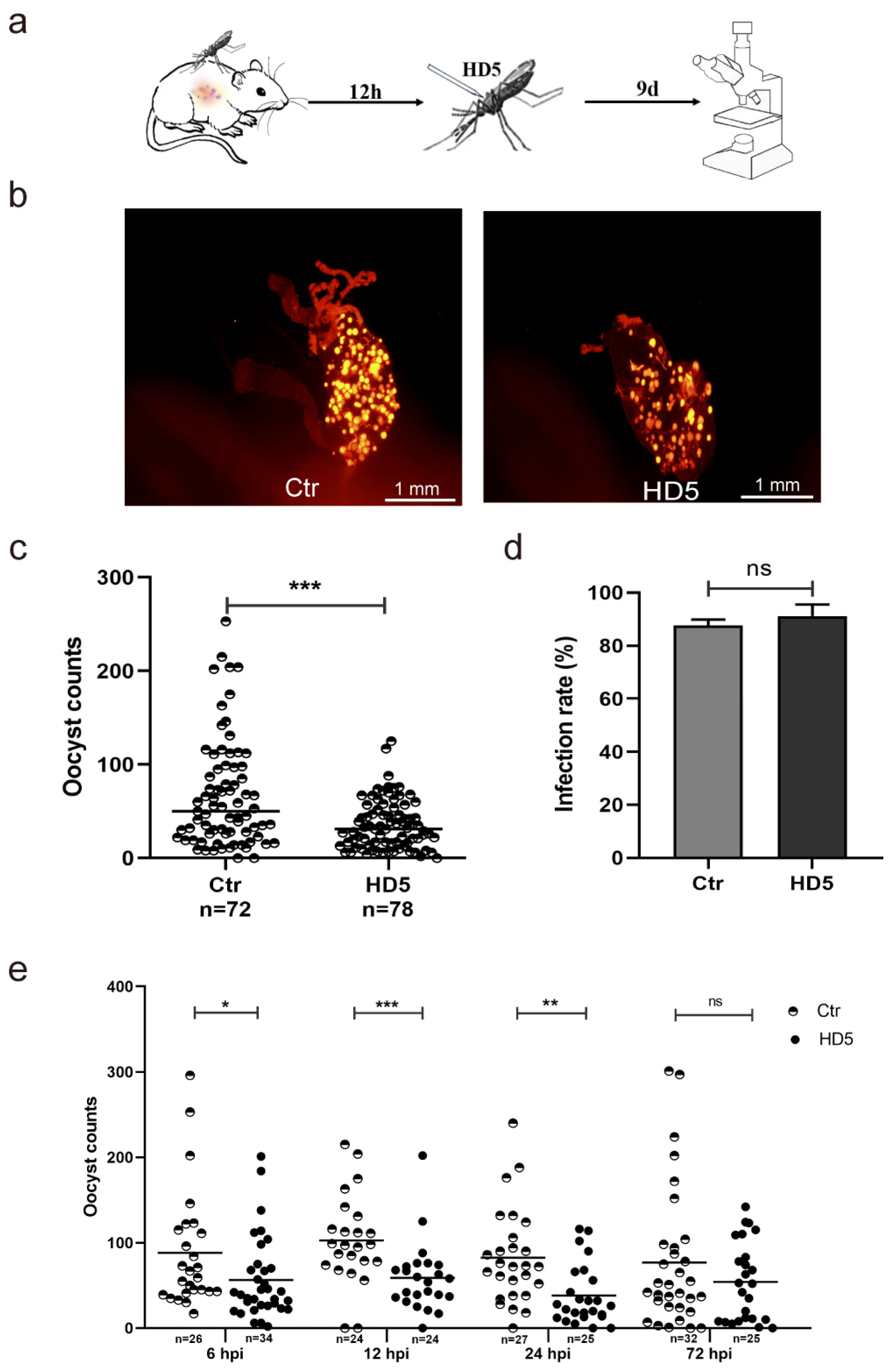
To explore whether injecting HD5 into mosquitoes after blood-feeding affects parasitic infection, HD5 was intrathoracically injected into An. stephensi post-infection. The mosquitoes were dissected, and the oocysts were counted at day 9 post-infection (Figure 2a). We first determined the effect of HD5 injection at 12 hpi on the development of Plasmodium in mosquitoes. The results showed that the HD5 injection substantially reduced oocyst count compared to the control group without affecting infection rates (Figure 2b–d). The inhibitory effect of HD5 on Plasmodium development in mosquitoes was observed when injection was conducted at 6, 12, 24, or 72 hpi. The results showed that the injection of HD5 led to a 32% decrease at 6 hpi, a 44% decrease at 12 hpi, a 44% decrease at 24 hpi, and a 23% decrease at 72 hpi compared to the control groups (Figure 2e). These results indicated that HD5 could intervene in the development of the early stage of the oocyst. In summary, HD5 effectively inhibited P. yoelii development in An. stephensi if HD5 was injected prior to infection or administered early after infection.

3.3. HD5 Treatment Upregulated TEP1 Expression in An. stephensi

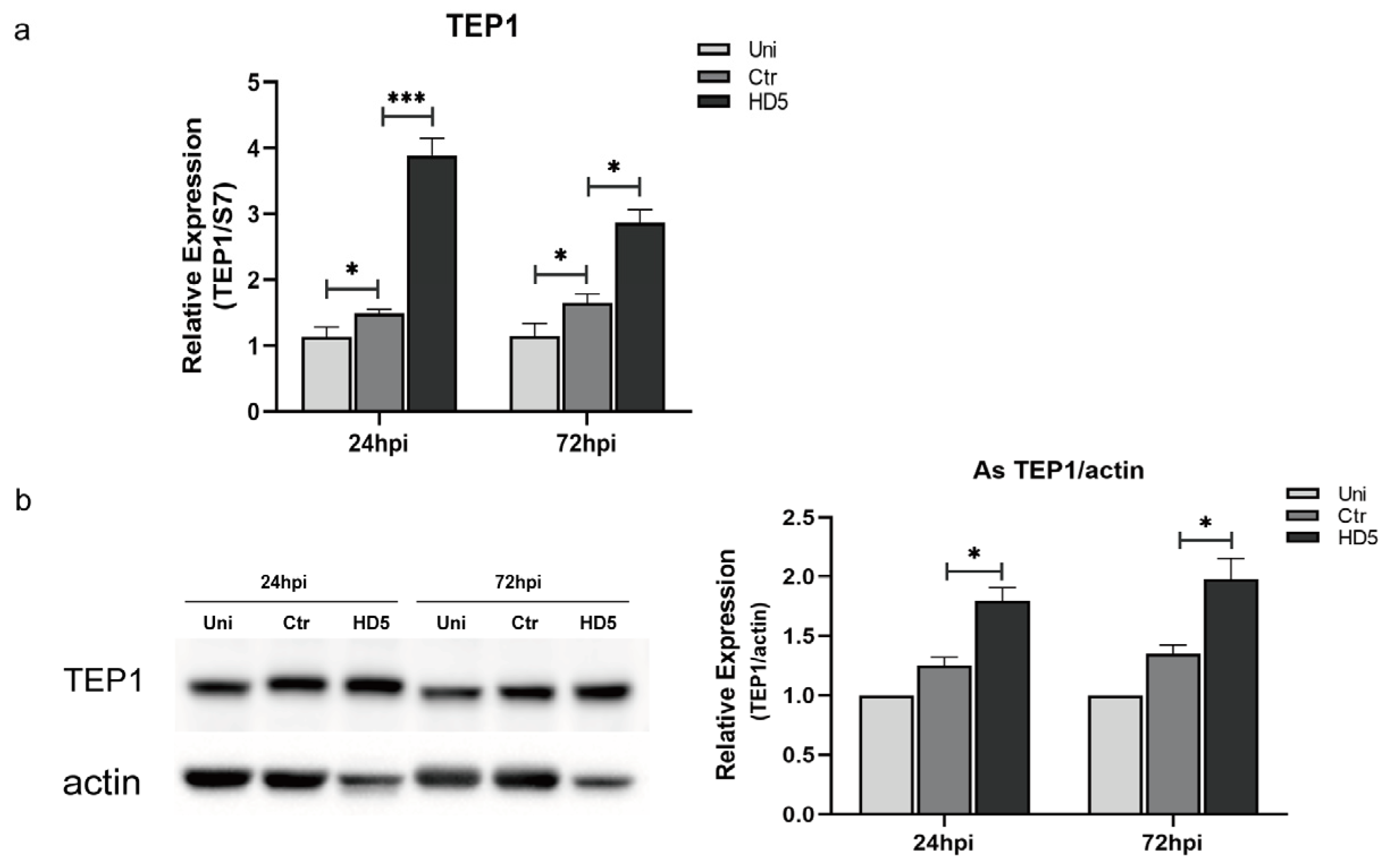
Studies have indicated that TEP1 plays a crucial role in mosquito resistance against malaria parasites [24]. The midgut epithelium is invaded and crossed by Plasmodium ookinetes, triggering a robust TEP1-involved, complement-like immune response [25,26]. As the main protein responsible for mediating Plasmodium lysis, TEP1 binds to the surface of ookinetes and mediates Plasmodium lysis [27]. To investigate whether HD5 treatment affected the expression of TEP1, real-time quantitative PCR and Western blotting were performed to detect changes in the transcription and protein levels of TEP1 by HD5 injection at 12 hpi. The results showed that HD5 treatment upregulated both the transcriptional and protein levels of TEP1 in mosquitoes at 24 and 72 hpi, compared to those in the control groups (Figure 3a,b). Additionally, the damage of injection upregulated the transcription levels of TEP1 but had minimal effect on protein levels comparing the un-injected and injected with ddH2O groups. These results suggest that HD5 treatment could increase the expression of TEP1 and improve the immune response of mosquitoes against malaria parasites.

3.4. Toll Signaling Pathway Was Upregulated by Injection with HD5

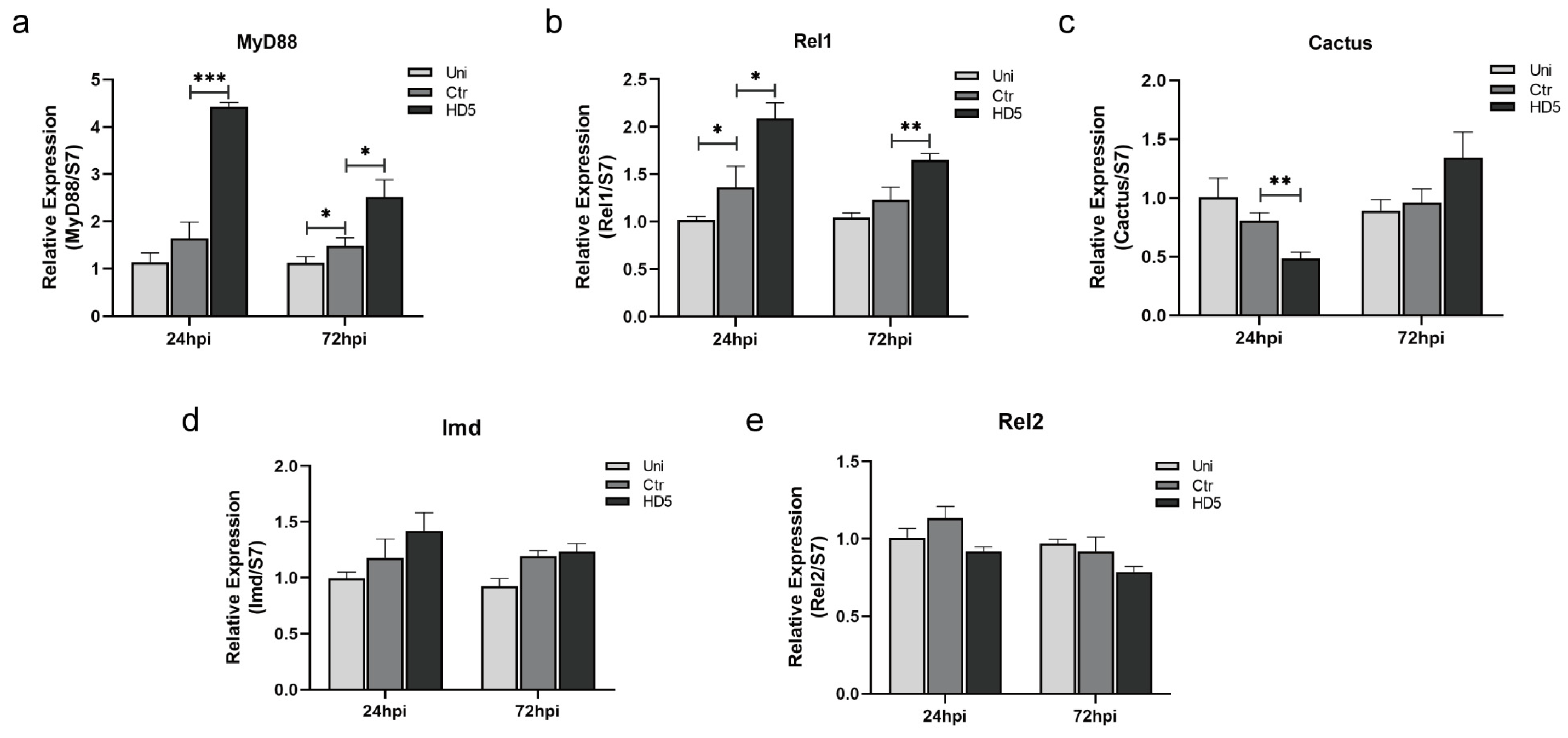
Parasites are killed by the innate immune system in mosquitoes [28]. Four major signaling pathways have been demonstrated to defend against pathogens, among which the Toll and Imd signaling pathways are considered to be mainly involved in anti-Plasmodium defense in mosquitoes [5,29]. To explore whether HD5 treatment activates the Toll or Imd signaling pathways and further influence oocyst development, the key effector molecules in the signaling pathways were evaluated using qRT-PCR. We detected the expression of MyD88, Rel1, and Cactus of the Toll signaling pathway. The expression of both MyD88 and Rel1 was upregulated at 24 hpi and 72 hpi, following HD5 treatment at 12 hpi (Figure 4a,b). The expression of the negative regulatory factor, Cactus, was downregulated at 24 hpi (Figure 4c). This indicates that the Toll signaling pathway was activated. In addition, we detected the expression of Imd and Rel2, which are two key molecules in the Imd signaling pathway. However, there was no significant change in the expression of these genes between the HD5 and control groups (Figure 4d,e), indicating that the Imd signaling pathway was not activated by HD5. These results suggest that the Toll signaling pathway may be activated by HD5 treatment, resulting in the upregulation of TEP1 expression which then results in a decline in oocyst count.

3.5. Inhibition of Oocyst Development in An. stephensi by HD5 Could Be Reversed by Toll Signaling Pathway Interference

To confirm the role of Toll signaling pathway upregulation by HD5 treatment in the inhibition of malaria parasite development in mosquitoes, the gene expression of MyD88 and TEP1 was silenced using RNA interference (RNAi), followed by the observation of oocyst development. The gene expression of TEP1 and MyD88 was efficiently silenced, according to the qRT-PCR results (Figure 5a). As shown in Figure 5b, the inhibitory effect of HD5 on oocyst development decreased slightly when MyD88 expression was silenced by RNAi, indicating that the Toll signaling pathway plays a role in the HD5-mediated transmission blocking of malaria. There were more oocysts in the dsTEP1 group than in the dsGFP group without HD5 treatment, which indicated the role of TEP1 in the killing of Plasmodium. In addition, there were fewer oocysts in the HD5 group than in the control group after the injection of dsGFP, which further confirmed the inhibitory effect of HD5 treatment on oocyst development. Nonetheless, HD5 reduced oocyst counts when TEP1 expression was silenced by RNAi, indicating that HD5 treatment may also affect other immune effectors, in addition to TEP1, in the Toll signaling pathway to inhibit oocyst development. In summary, these results suggest that the activation of the Toll signaling pathway by HD5 reduces the susceptibility of Anopheles mosquitoes to Plasmodium.

4. Discussion

New strategies are urgently needed to block malaria transmission. It is worth investigating whether HD5 has a parasiticidal effect on Plasmodium and influence on the innate immunity of mosquitoes. We found that the number of oocysts was substantially reduced by the microinjection of HD5 into Anopheles mosquitoes, indicating that HD5 can reduce the susceptibility of Anopheles mosquitoes to Plasmodium. Oocyst counts varied when HD5 was injected intrathoracically into An. stephensi at different time points before and after Plasmodium infection, which may be related to the development of Plasmodium in mosquitoes. Female mosquitoes were infected by ingesting a blood meal containing Plasmodium gametocytes. Oocyst development was substantially inhibited when HD5 was administered prior to infection or injected at 6, 12, or 24 hpi. This suggests that HD5 mainly acts on the early stage of sporogony. Each oocyst contains thousands of haploid sporozoites, which then migrate to the salivary glands through the mosquito hemolymph. Thus, the status of oocysts determine following the development of sporozoites and can further affect malaria transmission. However, without an effect on infection rates, HD5 treatment reduced oocysts in mosquitoes, which may result in fewer sporozoites and decrease the vector competence of Anopheles. However, there is a limitation of this study, such as the possibility that the rodent malaria parasite may differentiate with human Plasmodium. It is necessary to investigate whether HD5 has the same effect on human Plasmodium species such as P. falciparum in the future.
Our study found that the expression of TEP1 was upregulated by HD5 treatment, indicating that TEP1 is probably involved in the effect of HD5 on the restriction of Plasmodium. The silencing of TEP1 expression via RNAi increased the oocyst count, which was consistent with previous reports [30]. However, it did not reverse the effect of HD5 on Plasmodium development, indicating the involvement of other effector molecules or the insufficient silencing of TEP1 expression by dsRNA. In Drosophila, the inactivation of the Toll and Imd pathways has been used to turn off many immune genes, including antimicrobial peptide genes [31]. The detection and comparison of the expression of key molecules in these two signaling pathways showed that the Toll signaling pathway was upregulated by HD5 injection, whereas the Imd pathway was not affected. The role of the Toll signaling pathway in the inhibitory effect of HD5 on parasite development in Anopheles was further confirmed by silencing MyD88, a key upstream molecule in the Toll pathway. This is consistent with the finding that the activation of the Toll signaling pathway in Anopheles gambiae by silencing Cactus promotes the activation of the complement-like system in mosquitoes, leading to the elimination of the oocyte cell malaria parasite [32].
Additionally, both the inhibitory effect on malaria parasites and effect on the immune responses of mosquitoes are not HD5-specific. Actually, various physical, chemical, and biological factors have been shown to influence the immune response of mosquitoes. In the present study, ddH2O was used as the negative control to maintain the consistency of HD5 solvent and exclude the effect of injection with ddH2O. The results of this study using ddH2O as the control can reflect the effect of HD5 on the development of malaria parasites in mosquitoes, as well as on the expression of the key molecules of innate immune signaling pathways such as TEP1 and Toll, etc. It does not rule out the possibility that other adventitious factors such as a random sequence polypeptide have the same effect. It is necessary to use a polypeptide without an effect on the immune response of mosquitoes as a negative control to further explore the effect-related sequence characteristics of HD5 in the future.
Many cys-rich peptides, such as phormicins, royalisin, tenecin-1, heliomicin, spodoptericin, gallerimycin, coprisin, and lucifensin, are classified as the insect defensin family [33,34]. They are widely distributed in Diptera, Hemiptera, Hymenoptera, Coleoptera, and Lepidoptera [35], even in the ancient insect order of Odonata [36]. However, it has not been reported that mosquitoes have the ortholog of HD5 with similar inhibitory effect. Researchers have identified various defensin gene isoforms of insects including mosquitoes, which have been sequenced and used as molecular markers for phylogenetic analysis [37]. According to the multiple alignment of human and mosquito defensins and the constructed phylogeny tree, HD5 was most closely related to DEF4 in An. stephensi with only 21% homology (Figure S1). So, it is hard to regard DEF4 as the ortholog of HD5 in An. stephensi. The differences in the sequences of nucleotides and amino acids between HD5 and mosquito defensins may cause various antimalarial effects. If the ortholog of HD5 in mosquitoes can be found in the future, malaria transmission can be blocked via upregulating its expression. It is also possible to make genetic modifications to mosquito defensin to improve the antimalarial effect in the future.
In addition, a type 1 recombinant defensin (rDef1.3) produced from Triatoma pallidipennis showed in vitro lytic activity on Trypanosoma and Leishmania parasites. The Leishmania major parasite was the most affected with 50% of dead cells or with damaged membranes [38]. The silencing of Phlebotomus papatasi defensin (PpDef) expression using the RNAi method caused increased parasite levels in the sand fly, indicating that defensin can resist Leishmania major infection [39]. The expression of AMPs in mosquitoes is a new approach for killing parasites. The defensin A (DefA) gene has been engineered in Aedes aegypti to construct genetically stable transgenic mosquitoes. The expression of DefA can be activated by blood meal and shows antibacterial activity [40]. The expression of cecropin A in transgenic Anopheles gambiae can reduce the number of P. berghei oocysts [41]. However, overexpressing a single AMP in transgenic mosquitoes is not efficient to interrupt pathogen transmission. The coexpression of cecropin A and defensin A in transgenic Aedes aegypti mosquitoes results in a cooperative antibacterial and anti-Plasmodium action. The number of Plasmodium gallinaceum oocysts was dramatically reduced in midguts, and no sporozoites were found in the salivary glands of the transgenic mosquitoes [42]. As an effector molecule, human defensin 130 (DEFB130) played a possible role in parasite elimination in macrophages [43]. The coexpression of two or more AMPs in transgenic mosquitoes cooperatively affects Plasmodium development. Therefore, HD5 and insect defensins can be engineered in mosquitoes to reduce parasite infection and interrupt parasite transmission. It will provide the possibility of using the cooperation of defensins to enhance the activity of Anopheles to defend against Plasmodium.

5. Conclusions

In conclusion, the present study focused on the inhibitory effect of HD5 on Plasmodium development in mosquitoes and its innate immunity-related mechanisms. The results indicated that the intrathoracic injection of HD5 into An. stephensi decreased P. yoelii infection density by upregulating the Toll signaling pathway. This study can help us not only understand the effect and mechanism of HD5 on the susceptibility of Anopheles mosquitoes to Plasmodium but also develop new strategies to block malarial transmission.

Supplementary Materials

The following supporting information can be downloaded at https://www.mdpi.com/article/10.3390/tropicalmed9080169/s1, The multiple alignment of the defensins derived from the retrieved sequences from various nucleotide and protein databases is shown in Figure S1.

Author Contributions

Conceptualization, T.L. and Y.W.; Data curation, T.L. and S.Y.; Formal analysis, T.L.; Funding acquisition, Y.W.; Methodology, T.L. and J.W.; Project administration, Y.W.; Resources, D.Z.; Validation, X.L.; Visualization, Z.L.; Writing—original draft, T.L.; Writing—review and editing, T.L., X.Y. and Y.W. All authors have read and agreed to the published version of the manuscript.

Funding

This research was funded by Scientific and Technological Innovation Capacity Enhancement Special Project of the Army Medical University, grant number 2021XJS05, National Natural Science Foundation of China, grant number 81971971.

Institutional Review Board Statement

All procedures were carried out according to the guidelines for animal care and safe use of the Army Medical University and were approved by the Animal Institute of Army Medical University (Approval number: AMUWEC20230233, dated 28 February 2023).

Informed Consent Statement

Not applicable.

Data Availability Statement

All data generated or analyzed during this study are included in this published article and its Supplementary Materials. The original study data were presented to the authors.

Acknowledgments

We would like to thank Cheng Wang for supplying HD5 and the staff at our laboratory for their contribution in maintaining the mosquitoes.

Conflicts of Interest

The authors declare no conflicts of interest.

References

  1. WHO. World Malaria Report 2023; WHO: Geneva, Switzerland, 2023. [Google Scholar]
  2. Biabi, M.; Fogang, B.; Essangui, E.; Maloba, F.; Donkeu, C.; Keumoe, R.; Cheteug, G.; Magoudjou, N.; Slam, C.; Kemleu, S.; et al. High Prevalence of Polyclonal Plasmodium falciparum Infections and Association with Poor IgG Antibody Responses in a Hyper-Endemic Area in Cameroon. Trop. Med. Infect. Dis. 2023, 8, 390. [Google Scholar] [CrossRef]
  3. Hemingway, J. The role of vector control in stopping the transmission of malaria: Threats and opportunities. Philos. Trans. R. Soc. Lond. B Biol. Sci. 2014, 369, 20130431. [Google Scholar] [CrossRef] [PubMed]
  4. Volohonsky, G.; Paul-Gilloteaux, P.; Stafkova, J.; Soichot, J.; Salamero, J.; Levashina, E.A. Kinetics of Plasmodium midgut invasion in Anopheles mosquitoes. PLoS Pathog. 2020, 16, e1008739. [Google Scholar] [CrossRef] [PubMed]
  5. Clayton, A.M.; Dong, Y.; Dimopoulos, G. The Anopheles innate immune system in the defense against malaria infection. J. Innate Immun. 2014, 6, 169–181. [Google Scholar] [CrossRef] [PubMed]
  6. Cui, H.; Xia, Q. Research progress in anti-inflammatory activity of insect antimicrobial peptides and anti-inflammatory mechanism based on signal pathway. Tianjin Med. J. 2022, 50, 1002–1008. (In Chinese) [Google Scholar]
  7. Gao, X.; Ding, J.; Liao, C.; Xu, J.; Liu, X.; Lu, W. Defensins: The natural peptide antibiotic. Adv. Drug Deliv. Rev. 2021, 179, 114008. [Google Scholar] [CrossRef] [PubMed]
  8. Hazlett, L.; Wu, M. Defensins in innate immunity. Cell Tissue Res. 2011, 343, 175–188. [Google Scholar] [CrossRef] [PubMed]
  9. Oppenheim, J.J.; Biragyn, A.; Kwak, L.W.; Yang, D. Roles of antimicrobial peptides such as defensins in innate and adaptive immunity. Ann. Rheum. Dis. 2003, 62 (Suppl. 2), ii17–ii21. [Google Scholar] [CrossRef] [PubMed]
  10. Yakovlev, A.Y.; Nesin, A.P.; Simonenko, N.P.; Gordya, N.A.; Tulin, D.V.; Kruglikova, A.A.; Chernysh, S.I. Fat body and hemocyte contribution to the antimicrobial peptide synthesis in Calliphora vicina R.-D. (Diptera: Calliphoridae) larvae. In Vitro Cell Dev. Biol. Anim. 2017, 53, 33–42. [Google Scholar] [CrossRef]
  11. Shahabuddin, M.; Fields, I.; Bulet, P.; Hoffmann, J.A.; Miller, L.H. Plasmodium gallinaceum: Differential killing of some mosquito stages of the parasite by insect defensin. Exp. Parasitol. 1998, 89, 103–112. [Google Scholar] [CrossRef]
  12. Blandin, S.; Moita, L.F.; Kocher, T.; Wilm, M.; Kafatos, F.C.; Levashina, E.A. Reverse genetics in the mosquito Anopheles gambiae: Targeted disruption of the Defensin gene. EMBO Rep. 2002, 3, 852–856. [Google Scholar] [CrossRef] [PubMed]
  13. Zhao, L.; Alto, B.W.; Smartt, C.T.; Shin, D. Transcription Profiling for Defensins of Aedes aegypti (Diptera: Culicidae) during Development and in Response to Infection with Chikungunya and Zika Viruses. J. Med. Entomol. 2018, 55, 78–89. [Google Scholar] [PubMed]
  14. Cunliffe, R.N.; Rose, F.R.; Keyte, J.; Abberley, L.; Chan, W.C.; Mahida, Y.R. Human defensin 5 is stored in precursor form in normal Paneth cells and is expressed by some villous epithelial cells and by metaplastic Paneth cells in the colon in inflammatory bowel disease. Gut 2001, 48, 176–185. [Google Scholar] [CrossRef]
  15. Elphick, D.; Liddell, S.; Mahida, Y.R. Impaired luminal processing of human defensin-5 in Crohn’s disease: Persistence in a complex with chymotrypsinogen and trypsin. Am. J. Pathol. 2008, 172, 702–713. [Google Scholar] [CrossRef]
  16. Tanaka, T.; Rahman, M.M.; Battur, B.; Boldbaatar, D.; Liao, M.; Umemiya-Shirafuji, R.; Xuan, X.; Fujisaki, K. Parasiticidal activity of human alpha-defensin-5 against Toxoplasma gondii. In Vitro Cell Dev. Biol. Anim. 2010, 46, 560–565. [Google Scholar] [CrossRef] [PubMed]
  17. Ma, J.M.; Yang, H.X. Role of Toll-like receptor 4 and human defensin 5 in primary endocervical epithelial cells. Chin. Med. J. 2010, 123, 1762–1767. [Google Scholar] [CrossRef]
  18. Ishtiaq, F.; Swain, S.; Kumar, S.S. Anopheles stephensi (Asian Malaria Mosquito). Trends Parasitol. 2021, 37, 571–572. [Google Scholar] [CrossRef]
  19. Simwela, N.V.; Waters, A.P. Current status of experimental models for the study of malaria. Parasitology 2022, 149, 729–750. [Google Scholar] [CrossRef]
  20. Conteh, S.; Chattopadhyay, R.; Anderson, C.; Hoffman, S.L. Plasmodium yoelii-infected A. stephensi inefficiently transmit malaria compared to intravenous route. PLoS ONE 2010, 5, e8947. [Google Scholar]
  21. Liew, J.; Fong, M.Y.; Lau, Y.L. Quantitative real-time PCR analysis of Anopheles dirus TEP1 and NOS during Plasmodium berghei infection, using three reference genes. PeerJ 2017, 5, e3577. [Google Scholar] [CrossRef]
  22. Maren, N.A.; Duduit, J.R.; Huang, D.; Zhao, F.; Ranney, T.G.; Liu, W. Stepwise Optimization of Real-Time RT-PCR Analysis. Methods Mol. Biol. 2023, 2653, 317–332. [Google Scholar] [PubMed]
  23. Yu, S.; Wang, P.; Qin, J.; Zheng, H.; Wang, J.; Liu, T.; Yang, X.; Wang, Y. Bacillus sphaericus exposure reduced vector competence of Anopheles dirus to Plasmodium yoelii by upregulating the Imd signaling pathway. Parasit. Vectors 2020, 13, 446. [Google Scholar] [CrossRef] [PubMed]
  24. Volohonsky, G.; Hopp, A.K.; Saenger, M.; Soichot, J.; Scholze, H.; Boch, J.; Blandin, S.A.; Marois, E. Transgenic Expression of the Anti-parasitic Factor TEP1 in the Malaria Mosquito Anopheles gambiae. PLoS Pathog. 2017, 13, e1006113. [Google Scholar] [CrossRef] [PubMed]
  25. Reyes, R.V.; Sousa, G.L.; Sneed, S.D.; Farrant, K.V.; Christophides, G.K.; Povelones, M. Stimulation of a protease targeting the LRIM1/APL1C complex reveals specificity in complement-like pathway activation in Anopheles gambiae. PLoS ONE 2019, 14, e0214753. [Google Scholar] [CrossRef] [PubMed]
  26. Williams, M.; Contet, A.; Hou, C.D.; Levashina, E.A.; Baxter, R. Anopheles gambiae TEP1 forms a complex with the coiled-coil domain of LRIM1/APL1C following a conformational change in the thioester domain. PLoS ONE 2019, 14, e0218203. [Google Scholar] [CrossRef] [PubMed]
  27. Kwon, H.; Arends, B.R.; Smith, R.C. Late-phase immune responses limiting oocyst survival are independent of TEP1 function yet display strain specific differences in Anopheles gambiae. Parasit. Vectors 2017, 10, 369. [Google Scholar] [CrossRef] [PubMed]
  28. Cirimotich, C.M.; Dong, Y.; Garver, L.S.; Sim, S.; Dimopoulos, G. Mosquito immune defenses against Plasmodium infection. Dev. Comp. Immunol. 2010, 34, 387–395. [Google Scholar] [CrossRef]
  29. Souvannaseng, L.; Hun, L.V.; Baker, H.; Klyver, J.M.; Wang, B.; Pakpour, N.; Bridgewater, J.M.; Napoli, E.; Giulivi, C.; Riehle, M.A.; et al. Inhibition of JNK signaling in the Asian malaria vector Anopheles stephensi extends mosquito longevity and improves resistance to Plasmodium falciparum infection. PLoS Pathog. 2018, 14, e1007418. [Google Scholar] [CrossRef] [PubMed]
  30. Blandin, S.; Shiao, S.H.; Moita, L.F.; Janse, C.J.; Waters, A.P.; Kafatos, F.C.; Levashina, E.A. Complement-like protein TEP1 is a determinant of vectorial capacity in the malaria vector Anopheles gambiae. Cell 2004, 116, 661–670. [Google Scholar] [CrossRef]
  31. Lemaitre, B.; Nicolas, E.; Michaut, L.; Reichhart, J.M.; Hoffmann, J.A. Pillars article: The dorsoventral regulatory gene cassette spatzle/Toll/cactus controls the potent antifungal response in Drosophila adults. Cell. 1996. 86: 973-983. J. Immunol. 2012, 188, 5210–5220. [Google Scholar]
  32. Barletta, A.; Saha, B.; Trisnadi, N.; Talyuli, O.; Raddi, G.; Barillas-Mury, C. Hemocyte differentiation to the megacyte lineage enhances mosquito immunity against Plasmodium. eLife 2022, 11, e81116. [Google Scholar] [CrossRef] [PubMed]
  33. Dassanayake, R.S.; Silva, G.Y.; Tobe, S.S. Evolutionary selective trends of insect/mosquito antimicrobial defensin peptides containing cysteine-stabilized alpha/beta motifs. Peptides 2007, 28, 62–75. [Google Scholar] [CrossRef] [PubMed]
  34. Yi, H.Y.; Chowdhury, M.; Huang, Y.D.; Yu, X.Q. Insect antimicrobial peptides and their applications. Appl. Microbiol. Biotechnol. 2014, 98, 5807–5822. [Google Scholar] [CrossRef]
  35. Volkoff, A.N.; Rocher, J.; D’Alencon, E.; Bouton, M.; Landais, I.; Quesada-Moraga, E.; Vey, A.; Fournier, P.; Mita, K.; Devauchelle, G. Characterization and transcriptional profiles of three Spodoptera frugiperda genes encoding cysteine-rich peptides. A new class of defensin-like genes from lepidopteran insects? Gene 2003, 319, 43–53. [Google Scholar] [CrossRef]
  36. Bulet, P.; Cociancich, S.; Reuland, M.; Sauber, F.; Bischoff, R.; Hegy, G.; Van Dorsselaer, A.; Hetru, C.; Hoffmann, J.A. A novel insect defensin mediates the inducible antibacterial activity in larvae of the dragonfly Aeschna cyanea (Paleoptera, Odonata). Eur. J. Biochem. 1992, 209, 977–984. [Google Scholar] [CrossRef]
  37. de Araujo, C.A.; Lima, A.C.; Jansen, A.M.; Galvao, C.; Jurberg, J.; Costa, J.; Azambuja, P.; Waniek, P.J. Genes encoding defensins of important Chagas disease vectors used for phylogenetic studies. Parasitol. Res. 2015, 114, 4503–4511. [Google Scholar] [CrossRef] [PubMed]
  38. Diaz-Garrido, P.; Cardenas-Guerra, R.E.; Martinez, I.; Poggio, S.; Rodriguez-Hernandez, K.; Rivera-Santiago, L.; Ortega-Lopez, J.; Sanchez-Esquivel, S.; Espinoza, B. Differential activity on trypanosomatid parasites of a novel recombinant defensin type 1 from the insect Triatoma (Meccus) pallidipennis. Insect Biochem. Mol. Biol. 2021, 139, 103673. [Google Scholar] [CrossRef] [PubMed]
  39. Vomackova, K.B.; Sassu, F.; Volf, P.; Telleria, E.L. RNAi-mediated gene silencing of Phlebotomus papatasi defensins favors Leishmania major infection. Front. Physiol. 2023, 14, 1182141. [Google Scholar]
  40. Kokoza, V.; Ahmed, A.; Cho, W.L.; Jasinskiene, N.; James, A.A.; Raikhel, A. Engineering blood meal-activated systemic immunity in the yellow fever mosquito, Aedes aegypti. Proc. Natl. Acad. Sci. USA 2000, 97, 9144–9149. [Google Scholar] [CrossRef]
  41. Kim, W.; Koo, H.; Richman, A.M.; Seeley, D.; Vizioli, J.; Klocko, A.D.; O’Brochta, D.A. Ectopic expression of a cecropin transgene in the human malaria vector mosquito Anopheles gambiae (Diptera: Culicidae): Effects on susceptibility to Plasmodium. J. Med. Entomol. 2004, 41, 447–455. [Google Scholar] [CrossRef]
  42. Kokoza, V.; Ahmed, A.; Woon, S.S.; Okafor, N.; Zou, Z.; Raikhel, A.S. Blocking of Plasmodium transmission by cooperative action of Cecropin A and Defensin A in transgenic Aedes aegypti mosquitoes. Proc. Natl. Acad. Sci. USA 2010, 107, 8111–8116. [Google Scholar] [CrossRef] [PubMed]
  43. Terkawi, M.A.; Takano, R.; Furukawa, A.; Murakoshi, F.; Kato, K. Involvement of beta-defensin 130 (DEFB130) in the macrophage microbicidal mechanisms for killing Plasmodium falciparum. Sci. Rep. 2017, 7, 41772. [Google Scholar] [CrossRef] [PubMed]
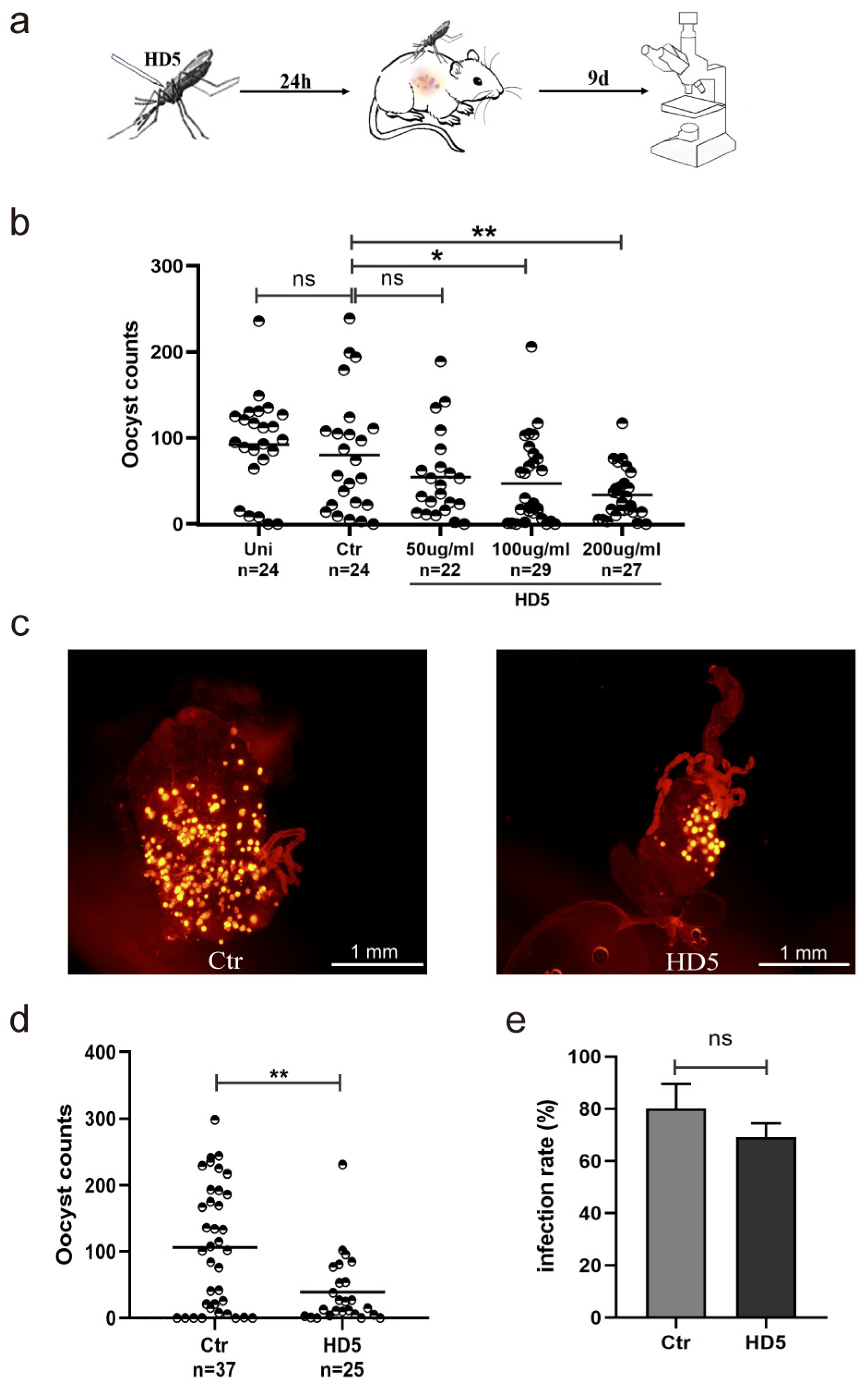
Figure 1. HD5 injection prior to infection decreased the density of oocysts in An. stephensi. (a) A schematic overview of HD5 treatment in An. stephensi. (b) The effects of different concentrations of HD5 on Plasmodium development in mosquitoes (c) The infection of P. yoelii in the midguts of An. stephensi (yellow dots represent oocysts). (d) The infection density of Plasmodium in mosquitoes between the HD5 (200 μg/mL) and control groups. (e) The infection rates of the HD5 (200 μg/mL) and control groups. Each dot represents the oocyst of an individual mosquito (a,d). Horizontal black bars indicate the median values. Significance was determined using Mann–Whitney tests in (a,d), and using the Chi-square test in (e). *, p < 0.05; **, p < 0.01; ns, no significant difference. Uni, un-injection; Ctr, control, injection with ddH2O; HD5, injection with HD5.
Tropicalmed 09 00169 g001
Figure 2. Post-infection HD5 injection inhibits P. yoelii infection in An. stephensi. (a) A schematic overview of HD5 microinjection into An. stephensi. (b) The infection of P. yoelii in the midguts of An. stephensi injected with ddH2O or HD5 at 12 hpi (yellow dots represent oocysts). (c) The infection density of Plasmodium in mosquitoes injected with ddH2O or HD5 at 12 hpi. (d) Plasmodium infection rates in An. stephensi injected with ddH2O or HD5 at 12 hpi. (e) The oocyst counts of Plasmodium in An. stephensi injected with ddH2O or HD5 at different time points. Each dot represents the oocyst of an individual mosquito. Horizontal black bars indicate the median values (c,e). Significance was determined using Mann–Whitney tests in (c,e) and using the Chi-square test in (d). *, p < 0.05; **, p < 0.01; ***, p < 0.001; ns, no significant difference. Ctr, control, injection with ddH2O; HD5, injection with HD5.
Tropicalmed 09 00169 g002
Figure 3. HD5 affects TEP1 expression in An. stephensi infected with P. yoelii. TEP1 transcriptional levels detected by real-time quantitative PCR (a) and protein levels determined by Western blot analysis (b). The bar chart represents the quantification of the signal intensities of TEP1 relative to actin from three independent replicates determined by ImageJ software 5.0 (b). The relative expression was set as 1 for the Uni group at each time point. Error bars indicate standard errors. Significance was determined using t-tests. *, p < 0.05; ***, p < 0.001. Uni, un-injection; Ctr, control, injection with ddH2O; HD5, injection with HD5.
Tropicalmed 09 00169 g003
Figure 4. The impact of HD5 treatment on the activation of the Toll and Imd signaling pathways in An. stephensi. A gene expression analysis of MyD88 (a), Rel1 (b), Cactus (c), Imd (d), and Rel2 (e). The expression levels of targeted genes were normalized to S7. The relative expression was set as 1 for the Uni group at each time point. Error bars indicate standard errors. Significance was determined using t-tests. *, p < 0.05; **, p < 0.01; ***, p < 0.001. Uni, un-injection; Ctr, control, injection with ddH2O; HD5, injection with HD5.
Tropicalmed 09 00169 g004
Figure 5. RNAi of TEP1 and MyD88 altered P. yoelii oocyst density in HD5-treated and untreated mosquitoes. (a) Silencing efficiency of TEP1 and MyD88 expression in mosquitoes by RNAi method. (b) Oocyst counts when TEP1 and MyD88 were silenced by RNAi or not in HD5-treated and untreated mosquitoes. Each dot represents oocyst of individual mosquito. Horizontal black bars indicate median values. Significance was determined using t-test in (a) and by Mann–Whitney tests in (b). *, p < 0.05; **, p < 0.01; ***, p < 0.001; ns, no significant difference. Ctr, control, injection with ddH2O; HD5, injection with HD5.
Tropicalmed 09 00169 g005
Table 1. Gene primers for quantitative PCR.
Target GenePrimer Sequence (5′ to 3′)
As-TEP 1F: ACCGATTGTCCAAGTTCTCG
R: AGCGCATCTGGTTCTGGTAG
As-MyD88F: TCGGCGGACAGTGACATTATTACG
R: TCACGATCCTTCAGACACAGTTGC
As-Rel 1F: GAACTGGATTCGGTCACGCTAAGG
R: CGGCAGATAATCAGGTCGGACATG
As-CactusF: CGCTTGCAGATGCTAGTGGTCAG
R: CCGCTGTTCGCTGGCTGTTC
As-ImdF: CGACCGGAATGCAGGTGTATCAG
R: CCGCAGAGCCACTCGTTGAAG
As-Rel 2F: AATTACCCGCATTCTGATCG
R: CTCCAGCACGTAGTTCACGA
As-S7F: CTAACGACACGAAGACCACAAGA
R: CAACCTGCAACGACAGCAAAA
Table 2. Specific gene primers for RNAi.
Target GenePrimer Sequence (5′ to 3′)
T7-dsGFPF: TAATACGACTCACTATAGGGAGTCAAGTTCAACGTGTCCGGCG
R: TAATACGACTCACTATAGGGAGAGGACCATTTGATCGCGCTT
T7-dsTEP1F: TAATACGACTCACTATAGGGAGTCGGGCTGAAGGCGTTGACC
R: TAATACGACTCACTATAGGGAGTGCCACCTTGAATCGTCTGA
T7-dsMyD88F: TAATACGACTCACTATAGGGAGAGGTGAGCGTCAAAGAGACG
R: TAATACGACTCACTATAGGGAGTTTTACTGGCTTGTCCGGCT
Disclaimer/Publisher’s Note: The statements, opinions and data contained in all publications are solely those of the individual author(s) and contributor(s) and not of MDPI and/or the editor(s). MDPI and/or the editor(s) disclaim responsibility for any injury to people or property resulting from any ideas, methods, instructions or products referred to in the content.