Article

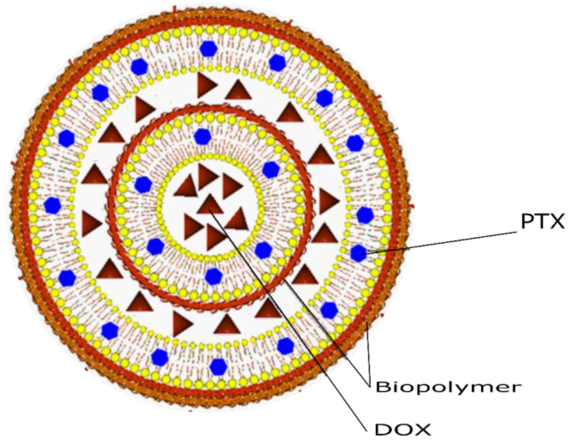
Co-Encapsulation of Paclitaxel and Doxorubicin in Liposomes Layer by Layer

by
Isaac Izcoatl Mota Díaz
1,*,
Janna Douda
2,3,
Patricia García López
4,
Sandra Edith Cabrera Becerra
5,
Miguel Ángel Gómez Álvarez
6,
Rebeca Jiménez Rodríguez
2,
Rafael Jurado León
4 and
Pedro López Sánchez
5
1
Unidad Profesional Interdisciplinaria de Ingeniería Campus Palenque, Instituto Politécnico Nacional, Carretera Palenque-Pakal-Na S/N, Palenque 29960, Chis, Mexico
2
Unidad Profesional Interdisciplinaria en Ingeniería y Tecnologías Avanzadas, Instituto Politécnico Nacional, Othón Mendizábal S/N, La Escalera 07320, CDMX, Mexico
3
Escuela Nacional de Ciencias Biológicas, Instituto Politécnico Nacional, Zacatenco 07738, CDMX, Mexico
4
Instituto Nacional de Cancerología (INCan), Tlalpan 4080, CDMX, Mexico
5
Escuela Superior de Medicina, Instituto Politécnico Nacional, Salvador Díaz Mirón esq. Plan de San Luis S/N, Miguel Hidalgo, Casco de Santo Tomas 11340, CDMX, Mexico
6
Centro de Investigación y de Estudios Avanzados del Instituto Politécnico Nacional, Libramiento Norponiente No. 2000 Real de Juriquilla, Santiago de Querétaro 76230, QRO, Mexico
*
Author to whom correspondence should be addressed.
Colloids Interfaces 2024, 8(4), 42; https://doi.org/10.3390/colloids8040042
Submission received: 20 April 2024 / Revised: 22 June 2024 / Accepted: 28 June 2024 / Published: 2 July 2024

Abstract

:
The synergistic effect of antineoplastic drug co-encapsulation systems has made them highly regarded due to their improved pharmacological efficacy. Biopolymer-coated liposomes were evaluated for paclitaxel and doxorubicin co-encapsulation in MCF-7 and MDA-MB-231 breast cancer cell lines. These nanosystems are characterized by dynamic light scattering, transmission electron microscopy, and UV–VIS spectroscopy. The conventional and hybrid liposomal systems presented sizes of 150 to 230 nm and %EE greater than 80% for the encapsulated active ingredients. These drug-laden liposomal systems significantly decreased cell viability in both breast cancer cell lines compared with liposome-free drugs. The delivery of antineoplastic drugs in breast cancer therapy could potentially benefit from new hybrids for drug co-encapsulation.

1. Introduction

Liposomes are spherical structures consisting mainly of phospholipids that form spontaneously when dispersed in aqueous media [1,2], and according to the methodology used, they can be unilaminar (a hydrophilic compartment and another hydrophobic compartment) or multilamellar (multiple hydrophilic and hydrophobic compartments) [3,4,5,6]. These structures have been extensively studied, both for their similarity to cell membranes and for their application as non-viral vectors for drug delivery and controlled release [2,7,8,9].
On the other hand, the methodologies used in the elaboration of liposomes are the method of rehydration of bicapalipidica, sonication, extrusion, and reverse phase, to mention a few [10,11]. The selection of the elaboration methodology or the combination of methodologies depends on the physicochemical properties of the compounds to be encapsulated, the phospholipids to be used in the formulation, the pH and surface modifications to be applied to the liposomes to provide specificity to the organ or tissue of interest [10].
Based on the amphiphilic properties of phospholipids, in a liposome, active ingredients with different properties can be encapsulated because they have a hydrophilic and hydrophobic compartment [5,12,13], which provides them with the possibility of co-encapsulation (encapsulation of more than one active substance in a single nanostructure) [14,15] and takes advantage of the synergy effect that can be obtained by combining drugs with different mechanisms of action in target cells [16,17,18], such as doxorubicin (DOX), which prevents DNA replication in high-replication cells [19,20,21], or paclitaxel (PTX), which stabilizes and prevents the depolymerization of microtubules [22,23,24].
In addition, it has been shown that the use of biopolymers adds new features and possibilities to the design of nanodrugs, allowing us to use liposomes as templates and allowing for their administration in different forms, such as cutaneous or oral [25,26,27]. Polymers confer resistance to external environments and also selectivity properties, as is the case of hyaluronic acid with CD44 receptors overexpressed in tumor cells [28,29].
Currently, several nanoformulations have been developed for the treatment of different types of chronic degenerative diseases, among which are Alzheimer’s, metabolic syndrome, diabetes, and cancer [30,31,32]. As examples of treatments for cancer, there are Doxopeg® and Caelyx®, which encapsulate DOX, and Ambraxane® for PTX because they are potent chemotherapy drugs [16,33,34,35]. However, these are monopharmaceuticals because they only encapsulate some of these active ingredients [10,36].
In addition, surface loading and nanoparticle morphology have been found to play an important role in interacting with biological systems. It has been observed that the interactions of encapsulated drugs modify the morphology of liposomal systems, allowing the immune system to degrade them and lose their effectiveness [37,38,39]. On the other hand, to prevent cytotoxic effects generated by antineoplastic agents and amplify their effect, they can be co-encapsulated with glycoprotein P (P-pg) inhibitors such as disulfiram or Elacridar [40,41].
The aim of this study was to compare the in vitro response, in breast cancer cell lines, of a conventional liposomal system of co-encapsulation of antineoplastic drugs (paclitaxel and doxorubicin) against a liposomal system coated with biopolymers that encapsulate the same drugs, and we conclude that this hybrid system could be a good candidate and present a non-Fick behavior.

2. Materials and Methods

2.1. Materials

The phospholipids DMPC (1,2-dimiristoyl-sn-glycerol-3-phosphatocolin), DSPE (1,2-diesteal-sn-glycerol-3-phosphoethanolamine), DOPE (1,2-dioleoyl-sn-glycerol-3-phosphoethanolamine), and DOTAP (1,2-dioleoyl-methylammonium-propane (chlorine salt) were acquired from Avanti Polar Lipids (Alabaster, AL, USA); paclitaxel (PTX), doxorubicin (DOX), Chitosan (CS), and poly lactic-co-glycolic acid (PLGA) was acquired from Sigma-Aldrich (St Louis, MO, USA); and CyQUANT™ was acquired from Thermo Scientific (Waltham, MA, USA). All growing media and supplements were purchased from Sigma-Aldrich. The other reagents used were analytical grade and used without further purification.

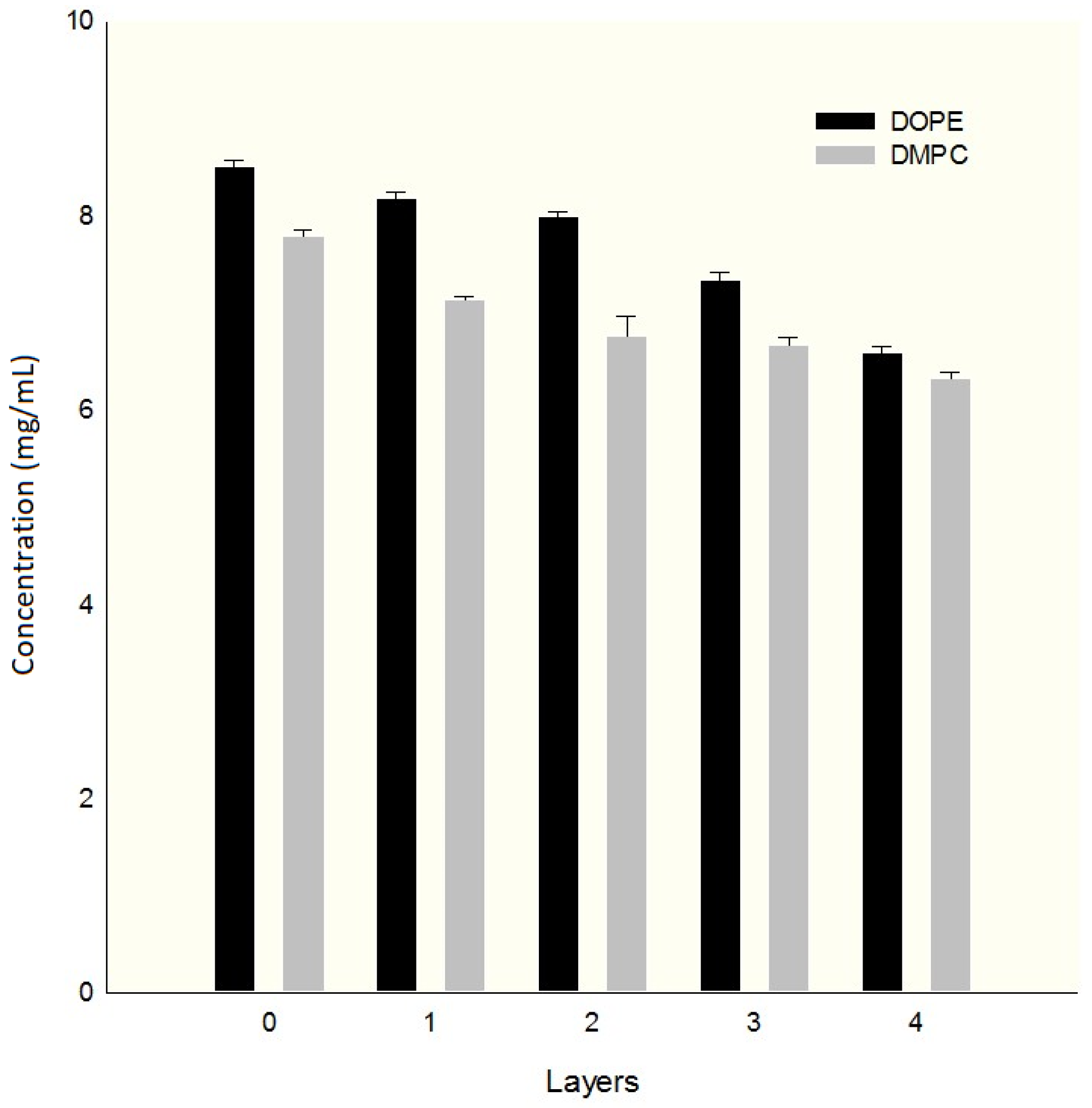
2.2. Preparation of Liposomes

Liposomes were prepared by the rehydration method of the lipid bilayer. A DOPE:DMPC:DSPE:DOTAP phospholipid composition molar ratio of 60:30:10:10 was dissolved in chloroform–methanol 6:1 (v/v); for liposomes with the drug, at this point, 1 mg of PTX was added. The mixture was heated in a water bath at 40 °C. The solvent was removed using a vacuum overnight.
The lipid bilayer was rehydrated with 2 mL of ammonium sulfate at 250 mM and sonicated with a 1/8” tip probe (VCX–130, Sonics and Materials, Newtown, CT, USA) for 30 min at 60 Watts. After completing this process, the liposomes were frozen and thawed 7 times and then extruded into a 100 nm polycarbonate membrane in a mini-extruder (Avanti Polar Lipids, Alabaster, AL, USA) 11 times.
The samples obtained were left in a water bath for 30 min at 45 °C and allowed to cool to room temperature to be stored at 4 °C. The unencapsulated drug and ammonium sulfate were removed by dialysis with a 50 KDa molecular weight Flot-a-Lizer (Thermo Fisher Scientific, Inc., Waltham, MA, USA) for 4 h at 4 °C in a solution of PBS at physiological pH. The excess solution was removed with Spectragel (Thermo Fisher Scientific, Inc.) for 1 h; the resulting solution was resuspended in 2 mL in phosphate-buffered saline (PBS) solution.
DOX was added and left in a water bath at 45 °C for 30 min with moderate agitation; after that time, the solution was cooled to room temperature and stored for 1 h at 4 °C. For the conventional method of liposome preparation, the dispersion medium was changed by PBS, and for hybrid liposomes, physiological solution was used at pH 6; this was conducted with Amicon Ultra 30 KDa (Merck, Darmstadt, Germany) centrifugal filters.
The hybrid liposomes were covered using the Layer by Layer (LbL) methodology: 50 µL of each of the biopolymer solutions in the physiological solution were added. They were stirred in a vortex for 10 min and centrifuged at 1000 RPM to remove excess polyelectrolytes generated; the process was repeated 4 times with the following deposition order: PLGA-CS-PLGA-CS. The solvent was changed by centrifugation with Amicon Ultra filters.
The process was first performed for 30 nm liposomes, as described above. They were then centrifuged in a Beckman Coulter 90L (Beckman Coulter, Brea, CA, USA) at 85 K RPM ultracentrifuge for 45 min with uncoated liposomes of 100 nm in size and suspended in ammonium sulfate, after which time, the sediment was resuspended and re-centrifuged as indicated above; this supernatant was removed, and the sediment was resuspended in PBS to proceed to charge the liposomes with a DOX concentration of 600 µg/mL.
The exempted drug was removed by centrifugation with Amicon Ultra 30 KDa filters at 15,000 RPM until the solution was removed, and the sample was resuspended in physiological solution to deposit the polymers as indicated above.

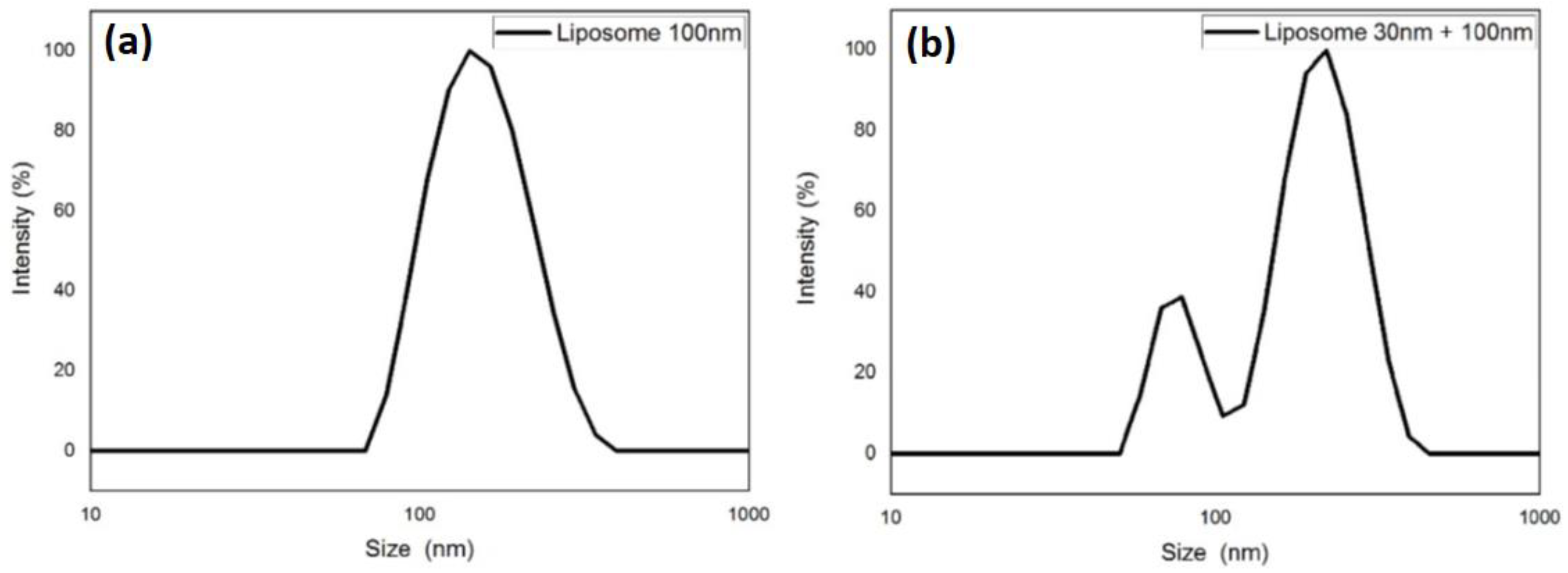
2.3. Size Determination by DLS and Potential ζ

Particle size, polydispersion index, and ζ potential were determined with the Malvern Zetasizer nano ZS90 (Malvern Instruments, Worcestershire, UK). The measurements were carried out in triplicate.

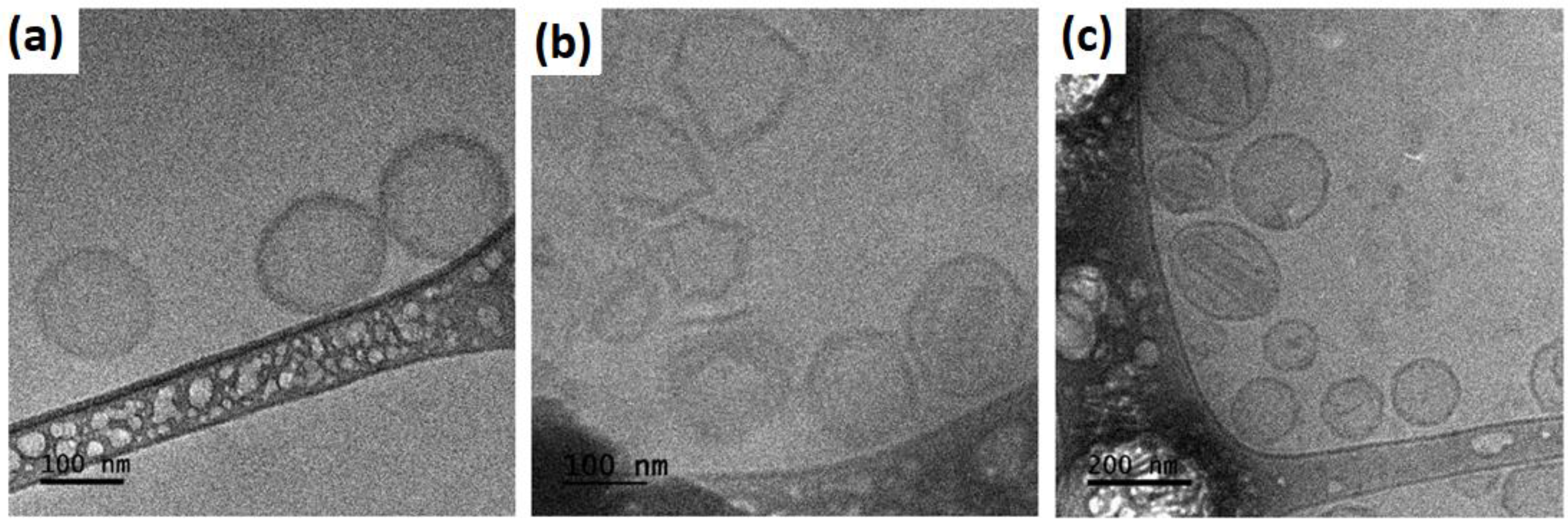
2.4. Morphology by Cryo-TEM

The morphology of the liposomes was observed via transmission electron Cryo-Microscopy (Cryo-TEM, JEM-2100, JEOL, Japan, Tokyo); a 2 µL sample was deposited on a carbon-coated copper network; and the excess sample was removed and stored under cryogenic conditions with butane in a liquid nitrogen bath.

2.5. Determination of Encapsulation Efficiency (%EE)

UV–VIS was used to determine the encapsulation of the drug; measurements were made in a NanoVue plus spectrophotometer (GE Healthcare Bio-Sciences Corp., Piscataway, NJ, USA) with an optical pass of 0.5 nm. The determination was made using the methodology used by Lee et al. [42] with some modifications, the most important of which was the use of chloroform to solubilize encapsulated PTX instead of acetonitrile, and the second, the addition of 2% SDS to achieve liposome degradation.
Absorbance 480 was measured for DOX and PTX at 227 nm. The percentage of %EE was determined with the formula
% E E = D R U G M E D D R U G T O T × 100
where
  • DRUGMED—the concentration obtained after the degradation of the nanosystem (final amount).
  • DRUGTOT—the concentration of drug used in the elaboration (starting amount).

2.6. Cell Viability Assay

The cells were routinely maintained as a monolayer in RPMI-1640 medium supplemented with 10% fetal bovine serum and incubated at 37 °C in a 5% CO2 atmosphere and high humidity. Cells were harvested with 0.025% trypsin and 1 mM EDTA.
For the viability assay, 18,000 MCF-7 cells or 12,000 MDA-MB-231 cells were seeded in a 96-well assay plate in triplicate and allowed to attach for 24 h. Then, the cells were exposed to conventional doxorubicin formulations of PTX-DOX conventional liposomes (M1), without the addition of biopolymers, or PTX-DOX hybrid liposomes coated with biopolymers (M2) at concentrations of 0; 0.1; 0.5; and 1 μM of doxorubicin for 24 h.
The viability was then measured with the fluorescent probe CyQUANT Cell Proliferation Assay Kit (Invitrogen; Thermo Fisher Scientific, Inc.). At the final exposition of the free doxorubicin or liposomal formulations, the medium was removed from the plate, and the wells were washed carefully with PBS. Then, the plate was stored at −70 °C for 24 h, and 200 μL of the CyQUANT® GR dye/cell-lysis buffer was added to each sample well.
The sample was mixed and incubated for 2–5 min at room temperature, protected from light. Then, the sample fluorescence was measured using a Varioskan® LUX Microplate Photometer (Thermo Fisher Scientific, Inc.) with ~480 nm excitation and ~520 nm emission. The fluorescence measure was normalized to 0 μM of doxorubicin.

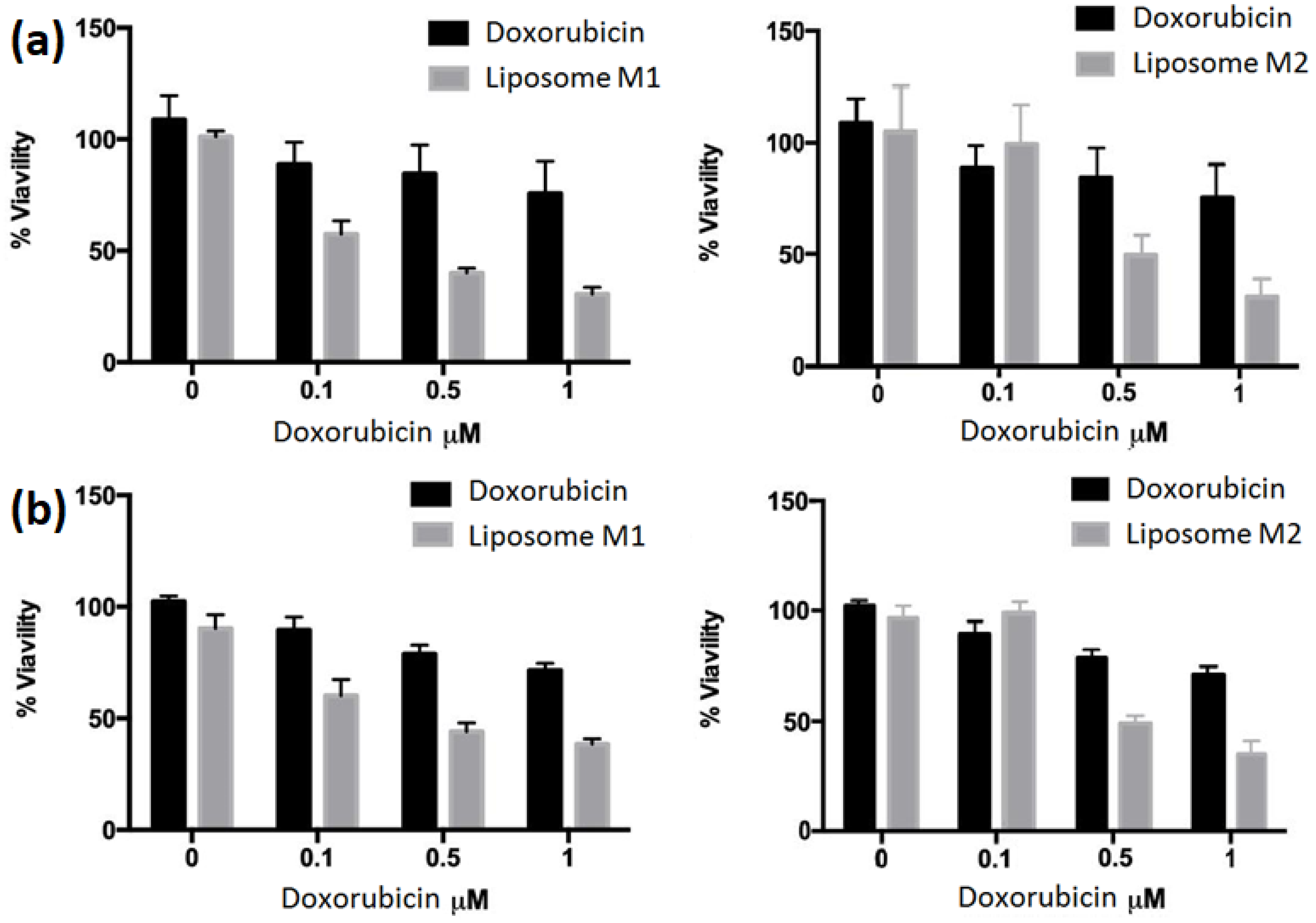
3. Results and Discussion

The representation of the hybrid system encapsulating DOX and PTX forms a core-shell structure with a liposome of 30 nm as a nucleus and a liposome of 100 nm as a cuirass, each of which is covered by four layers of polymers, as shown in Figure 1; biopolymer layers (core-shell) are generated by the interaction of the biopolymers chitosan (CS) and poly lactic-co-glycolic acid (PLGA).
PTX is a drug whose solubility is limited, which reduces the percentage of encapsulation for this antineoplastic agent between 4 and 5% [43,44,45], so one strategy to overcome this drawback is the generation of multilamellar structures; the purpose of this strategy is to generate a great number of hydrophobic compartments, allowing for a great amount of encapsulated PTX, as shown by Liu and collaborators [46]. For this reason, a hybrid system was proposed using the LbL methodology, which consists of the sequence deposition of polymer layers in aqueous solutions on spherical or flat substrates; thus, the thickness of the coating can be controlled depending on the number of deposits and the characteristics of biopolymers (molecular weight) [28,47,48,49].
This methodology alters the permeability of encapsulated substances by alternating the deposition of polymer layers with opposite charges on a substrate, which changes the diffusion speed. Fick’s law is a well-known model for transport phenomena [50,51]. According to our results, the conventional liposomal system presented Fick-type behavior, while the hybrid system presented non-Fick behavior; our data are congruent with other studies that have found that when performing coatings, the release rate of compounds encapsulated in liposomes is modified [52,53,54].
On the other hand, the systems developed had a drug ratio of 1:1 for in vitro tests; both M1 and M2 had a stability greater than 30 days stored at 4 °C and a leak of 4% measured in relation to DOX. This is because we do not have an HPLC team to measure the leakage of PTX by the amount of drug being encapsulated or its low solubility in aqueous media. In the literature, we can find works where they employ another strategy to overcome the low encapsulation of PTX, which consists of the conjugation of PTX and DOX loaded by an active gradient based on Cu+; this prodrug allows for the co-encapsulation of the two active ingredients [55,56].
DLS graphs are provided for M1 with a polydispersity index of 0.232 and for M2 after the four CS and PLGA polymer deposits in Figure 2. These data coincide with other studies in which DLS analysis was applied to measure particle and protein size to predict thermal stability [57,58] and are consistent with research working on the co-encapsulation of antineoplastic active ingredients. On the other hand, the hybrid liposomes presented two peaks of populations, which we attribute to the fact that not all liposomes of 30 nm could form the nucleus of the nucleus–shell structure with liposomes of 100 nm; in addition to this, the use of successive deposits generated a polyelectrolytic complex.
The M2 system used CS, which is one of the most abundant biopolymers in the world; it has low toxicity and solubility in aqueous solutions, antimicrobial properties, regenerative effects, and the ability to generate films. For this last reason, it has been used to coat flat or spherical substrates, such as liposomes, for the design of nanopharmaceuticals [49,59,60]. PLGA was also used, which is a biopolymer with degradation properties and is biocompatible; in addition to this, it has mechanical properties that are of great interest for the development of drug delivery applications [61]. With these two polymers, alternating deposits were made to generate the coatings on the hybrid liposomes.
The encapsulation efficiency percentages (%EE) were slightly lower than those reported by Yuan [62] and were within the average of the investigations that have co-encapsulated DOX and PTX [29,46,55,63,64]. This parameter depends very much on the methodology used and the steps used for the development of systems. In sample M2, the %EE decreased slightly for the two active ingredients, which we attribute to the centrifugation processes and the generation of successive deposits on the surface of the liposomes, as mentioned above.
With respect to the particle size and its potential ζ for the M2 sample, two populations can be seen, and the polydispersity index increases with respect to the M1 sample. According to the change in the potential ζ used to measure the electrokinetic potential of the particles [65], it was observed that, in liposomes, when centrifugation occurs, there is an increase in the rate of polydispersity, showing that the population is heterogeneous [66,67], and for M2, a change in the potential ζ can observed in the function of the charge of the polymer with respect to the deposit on the liposome [68].
Table 1 shows the growth of liposomes according to the number of deposits made for the coating of spherical substrates and the change in the potential ζ, and it can be seen that there is a change from a negative initial value of −26 to a negative value of −9 after four deposits; this for a sample of liposomes of 100 nm. The data obtained are consistent with those reported in the literature: as the biopolymer deposits alternate, the potential ζ decreases. [69,70].
In Figure 3, phospholipid loss is shown as a function of the number of polymer layers that are deposited on the liposome as substrates, both for liposomal formulations with most phospholipids, such as DMPC, and for DOPE, quantification was carried out using the Stewart methodology [71], through which it was found that there was a greater loss of phospholipids in samples consisting mainly of DMPC compared with samples consisting mainly of DOPE; this is due to the difference in the transition temperatures of these phospholipids, the deposits, and the centrifugation periods to which the samples were treated to eliminate the polyelectrolytic complex generated by the interaction of biopolymers of different loads [72].
Table 2 shows the characteristics of the two formulations developed, M1 and M2, indicating particle sizes, potential ζ, and %EE for each system; we can observe the hydrodynamic radius and PDI of the conventional liposomes and hybrid liposomes before and after drug encapsulation, and it can be observed that, in M2, there is an increase in the hydrodynamic radius and PDI, which indicates that we have a heterogeneous population compared with M1, where we find a homogeneous population. In addition, it can be seen that the potential ζ in M1 is greater than in M2. This is important because the more negative this parameter is, the greater the liposomal solution’s stability [73]. As for the %EE, there is also a reduction in the percentage of encapsulation in M2; the above is due to the successive centrifugation steps that generate the loss of liposomes and the aggregation of these with the generated polyelectrolytic complex [69,74].
Figure 4 shows the morphology of M1, of which Figure 4a shows the white liposomes, which do not encapsulate any of the active ingredients; Figure 4b shows the formation of flat faces in the liposomes generated by the encapsulation of PTX in the lipid bilayer, and this formation is due to the encapsulation of this hydrophobic compound between the chains of phospholipid tails and depends on the concentration of the drug, the phospholipid type, unsaturations, and the chain length, among others [44,75,76,77]. Figure 4c shows the liposomal system encapsulating the two active ingredients, which show flat faces in liposomes and the form of a “coffee bean” in the center of liposomes. This is due to the encapsulation of DOX in the hydrophilic liposome compartment and the active loading of this antineoplastic by the active load gradient [78,79,80].
As for the morphology generated for M2, it is presented in Figure 5, where it can be observed that, when the relationship between DOX and PTX drugs varies, flat faces are present due to the encapsulation of PTX in the hydrophobic compartments of the liposomes and the formation of the nucleus–cuirass structure, as are the liposomes of the initial populations at 30 and 100 nm. In addition, by increasing the concentration of DOX, the formation of these flat faces is reduced. We believe that this is because the presence of PTX in the membrane of liposomes limits the exchange necessary for active loading, modifying the characteristic shape of each drug in the liposomes.
Regarding the cell viability results for the MCF-7 and MDA-MB-231 lines (Figure 6), it was found that the response of M1 and M2, compared with free DOX, for a concentration of 0.1 μM, M2 had no effect compared with M1 for line MCF-7 (Figure 6a) or MDA-MB-231 (Figure 6b); for M2, it can be observed that, in the system, this concentration did not affect cell growth. In contrast, the concentrations of 0.5 and 1 μM show a decrease in cell viability for the two samples compared with the free drug and a downward trend that describes M1 with a concentration of 0.1 μM. We believe that this is due to the influence of biopolymers since these are covering liposomes that modify the type of transport, generating a non-Fick-type diffusion for M2, whereas M1 exhibits Fick-type behavior [52].

4. Conclusions

Hybrid liposomes were assembled using the LbL technique, while conventional liposomes without polymer coating were assembled by the thin-film layer rehydration method. The two systems were used to encapsulate PTX and DOX. Cryo-TEM was used to observe the morphological change generated by each of the active ingredients, and cell viability tests were performed for two lines of MCF-7 breast cancer and MDA-MB-231 and compared with free DOX at 24 h. It was found that the behavior of the systems was similar in the cell lines used; however, the effect of hybrid liposomes showed a slow release, and this is considered to be due to a modification of the rate of release of encapsulated drugs due to the presence of biopolymers.
Based on our findings, hybrid co-encapsulation systems could be suitable candidates for extending the release of active ingredients for breast cancer treatment. However, a detailed analysis of the release profiles and the specificity of the systems is necessary.

Author Contributions

Writing—original draft, J.D. and I.I.M.D.; conceptualization, J.D. and I.I.M.D.; writing—review and editing, I.I.M.D., J.D., S.E.C.B., R.J.R., P.G.L., M.Á.G.Á., R.J.L., and P.L.S.; investigation, I.I.M.D., S.E.C.B., R.J.R., P.G.L., M.Á.G.Á., R.J.L., and J.D. All authors contributed equally to the proposal and development of this research. The manuscript was written through the contribution of all authors. All authors have read and agreed to the published version of the manuscript.

Funding

The research was largely supported by SIP-IPN Projects 20200698 and 20200945; Consejo Nacional de Humanidades, Ciencias y Tecnologías: PhD scholarship 645451.

Data Availability Statement

The data and materials are available upon reasonable request to C. Janna Douda ([email protected]) and Isaac I. Mota Díaz ([email protected]).

Acknowledgments

The authors kindly acknowledge the Instituto Politécnico Nacional and the Centro de Nanociencias y Micro y Nanotecnología del Instituto Politécnico Nacional. The first author greatly appreciates the support of the Consejo Nacional de Humanidades, Ciencias y Tecnologias (CONAHCyT).

Conflicts of Interest

The authors declare no conflicts of interest.

References

  1. Patil, Y.P.; Jadhav, S. Novel methods for liposome preparation. Chem. Phys. Lipids 2014, 177, 8–18. [Google Scholar] [CrossRef]
  2. Filipczak, N.; Pan, J.; Yalamarty, S.S.K.; Torchilin, V.P. Recent advancements in liposome technology. Adv. Drug Deliv. Rev. 2020, 156, 4–22. [Google Scholar] [CrossRef]
  3. Lichtenberg, D.; Barenholz, Y. Liposomes: Preparation, characterization, and preservation. Methods Biochem. Anal. 1988, 33, 337–462. [Google Scholar] [CrossRef]
  4. Daraee, H.; Etemadi, A.; Kouhi, M.; Alimirzalu, S.; Akbarzadeh, A. Application of liposomes in medicine and drug delivery. Artif. Cells Nanomed. Biotechnol. 2016, 44, 381–391. [Google Scholar] [CrossRef]
  5. Jash, A.; Krueger, A.; Rizvi, S.S. Venturi-based rapid expansion of supercritical solution (Vent-RESS): Synthesis of liposomes for pH-triggered delivery of hydrophilic and lipophilic bioactives. Green Chem. 2022, 24, 5326–5337. [Google Scholar] [CrossRef]
  6. Akbarzadeh, A.; Rezaei-Sadabady, R.; Davaran, S.; Joo, S.W.; Zarghami, N.; Hanifehpour, Y.; Samiei, M.; Kouhi, M.; Nejati-Koshki, K. Liposome: Classification, preparation, and applications. Nanoscale Res. Lett. 2013, 8, 102. [Google Scholar] [CrossRef]
  7. Wahane, A.; Waghmode, A.; Kapphahn, A.; Dhuri, K.; Gupta, A.; Bahal, R. Role of lipid-based and polymer-based non-viral vectors in nucleic acid delivery for next-generation gene therapy. Molecules 2020, 25, 2866. [Google Scholar] [CrossRef]
  8. Karmali, P.P.; Chaudhuri, A. Cationic liposomes as non-viral carriers of gene medicines: Resolved issues, open questions, and future promises. Med. Res. Rev. 2007, 27, 696–722. [Google Scholar] [CrossRef]
  9. Rajappan, K.; Tanis, S.P.; Mukthavaram, R.; Roberts, S.; Nguyen, M.; Tachikawa, K.; Sagi, A.; Sablad, M.; Limphong, P.; Leu, A. Property-driven design and development of lipids for efficient delivery of siRNA. J. Med. Chem. 2020, 63, 12992–13012. [Google Scholar] [CrossRef]
  10. Olusanya, T.O.; Haj Ahmad, R.R.; Ibegbu, D.M.; Smith, J.R.; Elkordy, A.A. Liposomal drug delivery systems and anticancer drugs. Molecules 2018, 23, 907. [Google Scholar] [CrossRef]
  11. Lombardo, D.; Kiselev, M.A. Methods of liposomes preparation: Formation and control factors of versatile nanocarriers for biomedical and nanomedicine application. Pharmaceutics 2022, 14, 543. [Google Scholar] [CrossRef]
  12. Immordino, M.L.; Brusa, P.; Stella, B.; Arpicco, S.M.; Dosio, F.; Cattel, L. Preparazione e caratterizzazione di liposomi convenzionali ed a lunga circolazione contenenti taxani. Acta Technol. Legis Medicam. 2001, 12, 101–107. [Google Scholar]
  13. Laura Immordino, M.; Dosio, F.; Cattel, L. Stealth liposomes: Review of the basic science, rationale, and clinical applications, existing and potential. Int. J. Nanomedicine 2006, 1, 297–315. [Google Scholar]
  14. Gürsoy, A.; Kut, E.; Özkırımlı, S. Co-encapsulation of isoniazid and rifampicin in liposomes and characterization of liposomes by derivative spectroscopy. Int. J. Pharm. 2004, 271, 115–123. [Google Scholar] [CrossRef]
  15. Huang, M.; Liang, C.; Tan, C.; Huang, S.; Ying, R.; Wang, Y.; Wang, Z.; Zhang, Y. Liposome co-encapsulation as a strategy for the delivery of curcumin and resveratrol. Food Funct. 2019, 10, 6447–6458. [Google Scholar] [CrossRef]
  16. Hu, C.-M.J.; Aryal, S.; Zhang, L. Nanoparticle-assisted combination therapies for effective cancer treatment. Ther. Deliv. 2010, 1, 323–334. [Google Scholar] [CrossRef]
  17. Franco, M.S.; Oliveira, M.C. Liposomes co-encapsulating anticancer drugs in synergistic ratios as an approach to promote increased efficacy and greater safety. Anti-Cancer Agents Med. Chem. (Former. Curr. Med. Chem.-Anti-Cancer Agents) 2019, 19, 17–28. [Google Scholar] [CrossRef]
  18. Gabizon, A.; Ohana, P.; Amitay, Y.; Gorin, J.; Tzemach, D.; Mak, L.; Shmeeda, H. Liposome co-encapsulation of anti-cancer agents for pharmacological optimization of nanomedicine-based combination chemotherapy. Cancer Drug Resist. 2021, 4, 463. [Google Scholar] [CrossRef]
  19. Yang, F.; Teves, S.S.; Kemp, C.J.; Henikoff, S. Doxorubicin, DNA torsion, and chromatin dynamics. Biochim. Biophys. Acta (BBA)-Rev. Cancer 2014, 1845, 84–89. [Google Scholar] [CrossRef]
  20. Thorn, C.F.; Oshiro, C.; Marsh, S.; Hernandez-Boussard, T.; McLeod, H.; Klein, T.E.; Altman, R.B. Doxorubicin pathways: Pharmacodynamics and adverse effects. Pharmacogenetics Genom. 2011, 21, 440. [Google Scholar] [CrossRef]
  21. Carvalho, C.; Santos, R.X.; Cardoso, S.; Correia, S.; Oliveira, P.J.; Santos, M.S.; Moreira, P.I. Doxorubicin: The good, the bad and the ugly effect. Curr. Med. Chem. 2009, 16, 3267–3285. [Google Scholar] [CrossRef]
  22. Weaver, B.A. How Taxol/paclitaxel kills cancer cells. Mol. Biol. Cell 2014, 25, 2677–2681. [Google Scholar] [CrossRef]
  23. Yusuf, R.; Duan, Z.; Lamendola, D.; Penson, R.; Seiden, M. Paclitaxel resistance: Molecular mechanisms and pharmacologic manipulation. Curr. Cancer Drug Targets 2003, 3, 1–19. [Google Scholar] [CrossRef]
  24. Lim, P.T.; Goh, B.H.; Lee, W.-L. Taxol: Mechanisms of action against cancer, an update with current research. In Paclitaxel; Elsevier: Amsterdam, The Netherlands, 2022; pp. 47–71. [Google Scholar]
  25. Jain, S.; Kumar, D.; Swarnakar, N.K.; Thanki, K. Polyelectrolyte stabilized multilayered liposomes for oral delivery of paclitaxel. Biomaterials 2012, 33, 6758–6768. [Google Scholar] [CrossRef]
  26. Tan, X.; Fang, Y.; Ren, Y.; Li, Y.; Wu, P.; Yang, X.; Liu, W. D-α-tocopherol polyethylene glycol 1000 succinate-modified liposomes with an siRNA corona confer enhanced cellular uptake and targeted delivery of doxorubicin via tumor priming. Int. J. Nanomed. 2019, 14, 1255–1268. [Google Scholar] [CrossRef]
  27. Alavi, S.; Haeri, A.; Dadashzadeh, S. Utilization of chitosan-caged liposomes to push the boundaries of therapeutic delivery. Carbohydr. Polym. 2017, 157, 991–1012. [Google Scholar] [CrossRef]
  28. Jeon, S.; Yoo, C.Y.; Park, S.N. Improved stability and skin permeability of sodium hyaluronate-chitosan multilayered liposomes by Layer-by-Layer electrostatic deposition for quercetin delivery. Colloids Surf. B Biointerfaces 2015, 129, 7–14. [Google Scholar] [CrossRef]
  29. Song, M.; Liang, Y.; Li, K.; Zhang, J.; Zhang, N.; Tian, B.; Han, J. Hyaluronic acid modified liposomes for targeted delivery of doxorubicin and paclitaxel to CD44 overexpressing tumor cells with improved dual-drugs synergistic effect. J. Drug Deliv. Sci. Technol. 2019, 53, 101179. [Google Scholar] [CrossRef]
  30. Jash, A.; Ubeyitogullari, A.; Rizvi, S.S. Liposomes for oral delivery of protein and peptide-based therapeutics: Challenges, formulation strategies, and advances. J. Mater. Chem. B 2021, 9, 4773–4792. [Google Scholar] [CrossRef]
  31. Hu, M.; Gou, T.; Chen, Y.; Xu, M.; Chen, R.; Zhou, T.; Liu, J.; Peng, C.; Ye, Q. A Novel Drug Delivery System: Hyodeoxycholic Acid-Modified Metformin Liposomes for Type 2 Diabetes Treatment. Molecules 2023, 28, 2471. [Google Scholar] [CrossRef]
  32. Ross, C.; Taylor, M.; Fullwood, N.; Allsop, D. Liposome delivery systems for the treatment of Alzheimer’s disease. Int. J. Nanomed. 2018, 13, 8507–8522. [Google Scholar] [CrossRef]
  33. Bulbake, U.; Doppalapudi, S.; Kommineni, N.; Khan, W. Liposomal formulations in clinical use: An updated review. Pharmaceutics 2017, 9, 12. [Google Scholar] [CrossRef]
  34. Wu, D.; Si, M.; Xue, H.-Y.; Wong, H.-L. Nanomedicine applications in the treatment of breast cancer: Current state of the art. Int. J. Nanomed. 2017, 12, 5879–5892. [Google Scholar] [CrossRef]
  35. Fulton, M.D.; Najahi-Missaoui, W. Liposomes in cancer therapy: How did we start and where are we now. Int. J. Mol. Sci. 2023, 24, 6615. [Google Scholar] [CrossRef]
  36. Rommasi, F.; Esfandiari, N. Liposomal nanomedicine: Applications for drug delivery in cancer therapy. Nanoscale Res. Lett. 2021, 16, 95. [Google Scholar] [CrossRef]
  37. Albanese, A.; Tang, P.S.; Chan, W.C. The effect of nanoparticle size, shape, and surface chemistry on biological systems. Annu. Rev. Biomed. Eng. 2012, 14, 1–16. [Google Scholar] [CrossRef]
  38. Felice, B.; Prabhakaran, M.P.; Rodríguez, A.P.; Ramakrishna, S. Drug delivery vehicles on a nano-engineering perspective. Mater. Sci. Eng. C 2014, 41, 178–195. [Google Scholar] [CrossRef]
  39. Mu, Q.; Jiang, G.; Chen, L.; Zhou, H.; Fourches, D.; Tropsha, A.; Yan, B. Chemical basis of interactions between engineered nanoparticles and biological systems. Chem. Rev. 2014, 114, 7740–7781. [Google Scholar] [CrossRef]
  40. Wong, H.L.; Bendayan, R.; Rauth, A.M.; Wu, X.Y. Simultaneous delivery of doxorubicin and GG918 (Elacridar) by new polymer-lipid hybrid nanoparticles (PLN) for enhanced treatment of multidrug-resistant breast cancer. J. Control. Release 2006, 116, 275–284. [Google Scholar] [CrossRef]
  41. Rolle, F.; Bincoletto, V.; Gazzano, E.; Rolando, B.; Lollo, G.; Stella, B.; Riganti, C.; Arpicco, S. Coencapsulation of disulfiram and doxorubicin in liposomes strongly reverses multidrug resistance in breast cancer cells. Int. J. Pharm. 2020, 580, 119191. [Google Scholar] [CrossRef]
  42. Lee, W.L.; Guo, W.M.; Ho, V.H.; Saha, A.; Chong, H.C.; Tan, N.S.; Tan, E.Y.; Loo, S.C.J. Delivery of doxorubicin and paclitaxel from double-layered microparticles: The effects of layer thickness and dual-drug vs. single-drug loading. Acta Biomaterialia 2015, 27, 53–65. [Google Scholar] [CrossRef] [PubMed]
  43. Needham, D.; Sarpal, R.S. Binding of paclitaxel to lipid interfaces: Correlations with interface compliance. J. Liposome Res. 1998, 8, 147–163. [Google Scholar] [CrossRef]
  44. Steffes, V.M.; Zhang, Z.; MacDonald, S.; Crowe, J.; Ewert, K.K.; Carragher, B.; Potter, C.S.; Safinya, C.R. PEGylation of paclitaxel-loaded cationic liposomes drives steric stabilization of bicelles and vesicles thereby enhancing delivery and cytotoxicity to human cancer cells. ACS Appl. Mater. Interfaces 2019, 12, 151–162. [Google Scholar] [CrossRef] [PubMed]
  45. Koudelka, Š.; Turánek, J. Liposomal paclitaxel formulations. J. Control. Release 2012, 163, 322–334. [Google Scholar] [CrossRef] [PubMed]
  46. Liu, Y.; Fang, J.; Kim, Y.-J.; Wong, M.K.; Wang, P. Codelivery of doxorubicin and paclitaxel by cross-linked multilamellar liposome enables synergistic antitumor activity. Mol. Pharm. 2014, 11, 1651–1661. [Google Scholar] [CrossRef] [PubMed]
  47. De Cock, L.J.; De Koker, S.; De Geest, B.G.; Grooten, J.; Vervaet, C.; Remon, J.P.; Sukhorukov, G.B.; Antipina, M.N. Polymeric multilayer capsules in drug delivery. Angew. Chem. Int. Ed. 2010, 49, 6954–6973. [Google Scholar] [CrossRef] [PubMed]
  48. Antipov, A.A.; Sukhorukov, G.B. Polyelectrolyte multilayer capsules as vehicles with tunable permeability. Adv. Colloid Interface Sci. 2004, 111, 49–61. [Google Scholar] [CrossRef] [PubMed]
  49. Liu, X.Q.; Picart, C. Layer-by-Layer assemblies for cancer treatment and diagnosis. Adv. Mater. 2016, 28, 1295–1301. [Google Scholar] [CrossRef]
  50. Guenneau, S.; Puvirajesinghe, T. Fick’s second law transformed: One path to cloaking in mass diffusion. J. R. Soc. Interface 2013, 10, 20130106. [Google Scholar] [CrossRef]
  51. Paradisi, P.; Cesari, R.; Mainardi, F.; Tampieri, F. The fractional Fick’s law for non-local transport processes. Phys. A Stat. Mech. Its Appl. 2001, 293, 130–142. [Google Scholar] [CrossRef]
  52. Cuomo, F.; Lopez, F.; Piludu, M.; Miguel, M.G.; Lindman, B.; Ceglie, A. Release of small hydrophilic molecules from polyelectrolyte capsules: Effect of the wall thickness. J. Colloid Interface Sci. 2015, 447, 211–216. [Google Scholar] [CrossRef] [PubMed]
  53. Ramos, P.E.; Cerqueira, M.A.; Cook, M.T.; Bourbon, A.I.; Khutoryanskiy, V.V.; Charalampoulos, D.; Teixeira, J.A.; Vicente, A.A. Development of an immobilization system for in situ micronutrients release. Food Res. Int. 2016, 90, 121–132. [Google Scholar] [CrossRef] [PubMed]
  54. Ansarifar, E.; Mohebbi, M.; Shahidi, F.; Koocheki, A.; Ramezanian, N. Novel multilayer microcapsules based on soy protein isolate fibrils and high methoxyl pectin: Production, characterization and release modeling. Int. J. Biol. Macromol. 2017, 97, 761–769. [Google Scholar] [CrossRef] [PubMed]
  55. Wang, Y.; Wang, J.; Yang, L.; Wei, W.; Sun, B.; Na, K.; Song, Y.; Zhang, H.; He, Z.; Sun, J. Redox dual-responsive paclitaxel-doxorubicin heterodimeric prodrug self-delivery nanoaggregates for more effective breast cancer synergistic combination chemotherapy. Nanomed. Nanotechnol. Biol. Med. 2019, 21, 102066. [Google Scholar] [CrossRef] [PubMed]
  56. Yu, J.; Wang, Y.; Zhou, S.; Li, J.; Wang, J.; Chi, D.; Wang, X.; Lin, G.; He, Z.; Wang, Y. Remote loading paclitaxel–doxorubicin prodrug into liposomes for cancer combination therapy. Acta Pharm. Sin. B 2020, 10, 1730–1740. [Google Scholar] [CrossRef] [PubMed]
  57. Shiba, K.; Niidome, T.; Katoh, E.; Xiang, H.; Han, L.; Mori, T.; Katayama, Y. Polydispersity as a parameter for indicating the thermal stability of proteins by dynamic light scattering. Anal. Sci. 2010, 26, 659–663. [Google Scholar] [CrossRef]
  58. Yeap, S.P.; Lim, J.; Ngang, H.P.; Ooi, B.S.; Ahmad, A.L. Role of particle–particle interaction towards effective interpretation of Z-average and particle size distributions from dynamic light scattering (DLS) analysis. J. Nanosci. Nanotechnol. 2018, 18, 6957–6964. [Google Scholar] [CrossRef]
  59. Shukla, S.K.; Mishra, A.K.; Arotiba, O.A.; Mamba, B.B. Chitosan-based nanomaterials: A state-of-the-art review. Int. J. Biol. Macromol. 2013, 59, 46–58. [Google Scholar] [CrossRef] [PubMed]
  60. Vahed, S.Z.; Salehi, R.; Davaran, S.; Sharifi, S. Liposome-based drug co-delivery systems in cancer cells. Mater. Sci. Eng. C 2017, 71, 1327–1341. [Google Scholar] [CrossRef]
  61. Makadia, H.K.; Siegel, S.J. Poly lactic-co-glycolic acid (PLGA) as biodegradable controlled drug delivery carrier. Polymers 2011, 3, 1377–1397. [Google Scholar] [CrossRef]
  62. Yuan, M.; Qiu, Y.; Zhang, L.; Gao, H.; He, Q. Targeted delivery of transferrin and TAT co-modified liposomes encapsulating both paclitaxel and doxorubicin for melanoma. Drug Deliv. 2016, 23, 1171–1183. [Google Scholar] [CrossRef] [PubMed]
  63. Roque, M.; Geraldes, D.; da Silva, C.; Oliveira, M.; Nascimento, L. Long-Circulating and Fusogenic Liposomes Loaded with Paclitaxel and Doxorubicin: Effect of Excipient, Freezing, and Freeze-Drying on Quality Attributes. Pharmaceutics 2022, 15, 86. [Google Scholar] [CrossRef] [PubMed]
  64. Zhu, D.; Wu, S.; Hu, C.; Chen, Z.; Wang, H.; Fan, F.; Qin, Y.; Wang, C.; Sun, H.; Leng, X. Folate-targeted polymersomes loaded with both paclitaxel and doxorubicin for the combination chemotherapy of hepatocellular carcinoma. Acta Biomater. 2017, 58, 399–412. [Google Scholar] [CrossRef] [PubMed]
  65. Guzelsu, N.; Wienstien, C.; Kotha, S. A new streaming potential chamber for zeta potential measurements of particulates. Rev. Sci. Instrum. 2010, 81, 015106. [Google Scholar] [CrossRef] [PubMed]
  66. Mehn, D.; Iavicoli, P.; Cabaleiro, N.; Borgos, S.E.; Caputo, F.; Geiss, O.; Calzolai, L.; Rossi, F.; Gilliland, D. Analytical ultracentrifugation for analysis of doxorubicin loaded liposomes. Int. J. Pharm. 2017, 523, 320–326. [Google Scholar] [CrossRef] [PubMed]
  67. Bakonyi, M.; Berkó, S.; Budai-Szűcs, M.; Kovács, A.; Csányi, E. DSC for evaluating the encapsulation efficiency of lidocaine-loaded liposomes compared to the ultracentrifugation method. J. Therm. Anal. Calorim. 2017, 130, 1619–1625. [Google Scholar] [CrossRef]
  68. Pinheiro, A.C.; Bourbon, A.I.; Cerqueira, M.A.; Maricato, É.; Nunes, C.; Coimbra, M.A.; Vicente, A.A. Chitosan/fucoidan multilayer nanocapsules as a vehicle for controlled release of bioactive compounds. Carbohydr. Polym. 2015, 115, 1–9. [Google Scholar] [CrossRef] [PubMed]
  69. Guzmán, E.; Cavallo, J.A.; Chuliá-Jordán, R.; Gómez, C.; Strumia, M.C.; Ortega, F.; Rubio, R.G. pH-induced changes in the fabrication of multilayers of poly (acrylic acid) and chitosan: Fabrication, properties, and tests as a drug storage and delivery system. Langmuir 2011, 27, 6836–6845. [Google Scholar] [CrossRef]
  70. Yan, S.; Zhu, J.; Wang, Z.; Yin, J.; Zheng, Y.; Chen, X. Layer-by-layer assembly of poly (L-glutamic acid)/chitosan microcapsules for high loading and sustained release of 5-fluorouracil. Eur. J. Pharm. Biopharm. 2011, 78, 336–345. [Google Scholar] [CrossRef]
  71. Stewart, J.C.M. Colorimetric determination of phospholipids with ammonium ferrothiocyanate. Anal. Biochem. 1980, 104, 10–14. [Google Scholar] [CrossRef]
  72. Guzmán, E.; Mateos-Maroto, A.; Ruano, M.; Ortega, F.; Rubio, R.G. Layer-by-Layer polyelectrolyte assemblies for encapsulation and release of active compounds. Adv. Colloid Interface Sci. 2017, 249, 290–307. [Google Scholar] [CrossRef] [PubMed]
  73. Guimarães, D.; Cavaco-Paulo, A.; Nogueira, E. Design of liposomes as drug delivery system for therapeutic applications. Int. J. Pharm. 2021, 601, 120571. [Google Scholar] [CrossRef] [PubMed]
  74. Cuomo, F.; Lopez, F.; Miguel, M.G.; Lindman, B.R. Vesicle-templated layer-by-layer assembly for the production of nanocapsules. Langmuir 2010, 26, 10555–10560. [Google Scholar] [CrossRef] [PubMed]
  75. Kuntsche, J.; Horst, J.C.; Bunjes, H. Cryogenic transmission electron microscopy (cryo-TEM) for studying the morphology of colloidal drug delivery systems. Int. J. Pharm. 2011, 417, 120–137. [Google Scholar] [CrossRef]
  76. Kursch, B.; Lüllmann, H.; Mohr, K. Influence of various cationic amphiphilic drugs on the phase-transition temperature of phosphatidylcholine liposomes. Biochem. Pharmacol. 1983, 32, 2589–2594. [Google Scholar] [CrossRef] [PubMed]
  77. Panwar, P.; Pandey, B.; Lakhera, P.; Singh, K. Preparation, characterization, and in vitro release study of albendazole-encapsulated nanosize liposomes. Int. J. Nanomed. 2010, 5, 101–108. [Google Scholar] [CrossRef]
  78. Gubernator, J.; Chwastek, G.; Korycińska, M.; Stasiuk, M.; Grynkiewicz, G.; Lewrick, F.; Süss, R.; Kozubek, A. The encapsulation of idarubicin within liposomes using the novel EDTA ion gradient method ensures improved drug retention in vitro and in vivo. J. Control. Release 2010, 146, 68–75. [Google Scholar] [CrossRef]
  79. Gubernator, J. Active methods of drug loading into liposomes: Recent strategies for stable drug entrapment and increased in vivo activity. Expert Opin. Drug Deliv. 2011, 8, 565–580. [Google Scholar] [CrossRef]
  80. Li, T.; Nowell, C.J.; Cipolla, D.; Rades, T.; Boyd, B.J. Direct comparison of standard transmission electron microscopy and cryogenic-TEM in imaging nanocrystals inside liposomes. Mol. Pharm. 2019, 16, 1775–1781. [Google Scholar] [CrossRef]
Figure 1. Representation of the hybrid DOX and PTX co-encapsulation system.
Colloids 08 00042 g001
Figure 2. DLS graphs showing (a) conventional liposomes co-encapsulating DOX and PTX and (b) hybrid liposomes co-encapsulating DOX and PTX after being coated with CS and PLGA.
Colloids 08 00042 g002
Figure 3. Quantification of the loss of phospholipids for DOPE and DMPC when they are in greater proportions.
Colloids 08 00042 g003
Figure 4. Cryo-TEM of conventional liposome images: (a) 100 nm blank liposomes, (b) liposomes encapsulated PTX, and (c) co-encapsulated DOX and PTX.
Colloids 08 00042 g004
Figure 5. Cryo-TEM images of the hybrid liposome systems: (a) liposomes co-encapsulating DOX and PTX in a ratio of 1:1 and (b) in a 2:1 ratio.
Colloids 08 00042 g005
Figure 6. Cell viability for samples of conventional liposomes (M1) and a hybrid liposome system (M2): (a) MCF-7 and (b) MDA-MB-231 cell lines.
Colloids 08 00042 g006
Table 1. Changes in potential ζ and polydispersity index (PDI).
RH (nm)ζ (mV)PDI
blank94.0850 ± 0.600−26.610 ± 0.8500.00500 ± 0.0004
Layer 1101.190 ± 0.247+18.705 ± 0.4410.0130 ± 0.001
Layer 2131.283 ± 0.344−14.193 ± 0.2140.209 ± 0.006
Layer 3164.518 ± 0.331+12.276 ± 0.2170.258 ± 0.008
Layer 4204.287 ± 0.513−8.9980 ± 0.3430.448 ± 0.007
RH: hydrodynamic radius; ζ: potential ζ; PDI: polydispersion index. Values are expressed as mean ± standard error of the mean (SEM).
Table 2. Characteristic values of the formulations.
RH initial (nm)PDIinitialRH final (nm)PDIfinalζ (mV)%EE DOX-PTX
M1122.613 ± 0.7560.137 ± 0.001144.188 ± 0.4730.232± 0.007−26.842 ± 0.27884.4 ± 2.10–73.6 ± 6.23
M2103.073 ± 0.128
63.4820 ± 0.605
0.0430 ± 0.005
0.312 ± 0.005
193.007 ± 0.533
86.6810 ± 0.279
0.795 ± 0.011−9.9820 ± 0.039080.2 ± 1.65–60.4 ± 5.14
RH: hydrodynamic radius; ζ: potential ζ; PDI: polydispersion index. Values are expressed as mean ± standard error of the mean (SEM). M1, conventional liposomes; M2, hybrid liposomes.
Disclaimer/Publisher’s Note: The statements, opinions and data contained in all publications are solely those of the individual author(s) and contributor(s) and not of MDPI and/or the editor(s). MDPI and/or the editor(s) disclaim responsibility for any injury to people or property resulting from any ideas, methods, instructions or products referred to in the content.

