Next Article in Journal
2022 WUOF/SIU International Consultation on Urological Diseases: Neoadjuvant and Adjuvant Therapy for Renal Cell Carcinoma
Previous Article in Journal
2022 WUOF/SIU International Consultation on Urological Diseases: Ablative Therapies for Localized Primary Renal Cell Carcinoma
 
 
Société Internationale d’Urologie Journal is published by MDPI from Volume 5 Issue 1 (2024). Previous articles were published by another publisher in Open Access under a CC-BY (or CC-BY-NC-ND) licence, and they are hosted by MDPI on mdpi.com as a courtesy and upon agreement with Société Internationale d’Urologie.
Review

2022 WUOF/SIU International Consultation on Urological Diseases: Management of Locally Advanced Renal Cell Carcinoma

by
Vsevolod B. Matveev
1,*,
Sarah P. Psutka
2,
Grant D. Stewart
3,
Gennady Bratslavsky
4 and
E. Jason Abel
5
1
Department of Urology, N. N. Blokhin National Research Institute of Oncology, Moscow 115478, Russia
2
University of Washington Medical Center, Seattle Cancer Care Alliance, Seattle, WA 98195, USA
3
Department of Surgery, University of Cambridge, Cambridge Biomedical Campus, Cambridge CB2 0QH, UK
4
Department of Urology, SUNY Upstate Medical University, Syracuse, NY 13210, USA
5
Departments of Urology and Radiology, University of Wisconsin School of Medicine and Public Health, Madison, WI 53726, USA
*
Author to whom correspondence should be addressed.
Soc. Int. Urol. J. 2022, 3(6), 451-463; https://doi.org/10.48083/EGWH6536
Submission received: 14 July 2022 / Revised: 1 September 2022 / Accepted: 14 September 2022 / Published: 16 November 2022

Abstract

:
Renal cell carcinoma (RCC) has a natural tendency to invade the venous system with formation of a venous tumor thrombus in the renal vein, which can extend proximally into the inferior vena cava (IVC) and in some cases into the right atrium. The presence of venous involvement significantly worsens prognosis. Despite recent advances in systemic therapies, surgery remains the most effective method of treatment and in the case of complete removal of all tumor, it provides satisfactory long-term survival and must be attempted whenever possible. Several surgical techniques have been proposed, but all are associated with a high rate of perioperative complications and mortality. Minimally invasive approaches are mainly applicable for less extended IVC thrombi, while open surgery remains the gold standard for this category of patients. Most IVC thrombi can be managed without use of circulatory support by using different methods of IVC control depending on the thrombus level. However, use of cardiac bypass with or without deep hypothermic cardiac arrest is indicated in some patients with bulky intraatrial tumor thombi. In select patients presenting with IVC tumor thrombus and synchronous distant metastases, cytoreductive nephrectomy with IVC tumor thrombectomy may be considered with or without neoadjuvant systemic therapy. Surgery for RCC with venous thrombus is complex and requires experienced multidisciplinary surgical, anesthesia, and critical care teams at high-volume centers to achieve the best outcomes.

Introduction

Venous tumor thrombus (VTT) involvement is identified in up to 10% of patients with RCC[1]. Currently, surgery remains the only curative approach for M0 RCC with IVC invasion. While an open approach is most commonly utilized, there is growing interest regarding the role for minimally invasive approaches in appropriately selected patients; however, there is limited data regarding long-term outcomes regarding these approaches. In select patients presenting with IVC tumor thrombus and synchronous distant metastases, cytoreductive nephrectomy with IVC tumor thrombectomy may be considered with or without neoadjuvant systemic therapy.

Classification of VTT in Renal Cell Carcinoma

An unusual hallmark of RCC is its predilection for vascular invasion. The VTT can then form a cast of the main renal vein (pT3a) and extend proximally into the inferior vena cava (IVC; pT3b), in some cases extending into the right atrium of the heart (pT3c) or can invade directly from the venous lumen into the endothelium (pT3c)[2].
Recent estimates suggest that VTT is identified in approximately 1 in 10 patients with newly diagnosed RCC[3,4,5,6,7,8,9,10,11,12]. Involvement of the right cardiac chambers is encountered in 1% of cases[13].
VTT is classically categorized according to the height or extent of the VTT, according to the Mayo Classification system[14] (Table 1). The level of tumor thrombus is relevant not only from the perspective of oncologic prognosis but also in terms of anticipating surgical complexity and surgical planning[12].
Other features of the VTT such as direct invasion of the tumor thrombus into the endothelium and presence of bland thrombus are associated with both increased surgical complexity[15,16] and inferior cancer-specific survival[17,18,19].
Macroscopically, venous tumor thrombi can also be categorized according to their consistency, and may be described as either solid or friable, where friability has been associated with worse pathologic features and prognosis[20,21].

Pathophysiology of Tumor Venous Thrombosis and Clinical Manifestations of RCC with IVC Tumor Thrombus

Complete occlusion of the IVC by VTT or by proximal bland thrombus propagation has multiple potential physiologic consequences. IVC obstruction may result in lower-extremity edema, development of varicoceles, and pelvic pain from gonadal vein obstruction.
Reduced venous return to the right atrium (reduction in preload) may lead to hemodynamic instability, and altered mental status and death. Other commonly reported symptoms may include fatigue, dizziness, weight loss, night sweats, anorexia, palpitations, diaphoresis, dizziness, and shortness of breath on exertion[22].
Failure of the kidneys to have adequate venous drainage can result in progression of renal insufficiency. Similarly, obstruction of the hepatic veins can lead to hepatic congestion, which presents with transaminitis, hepatic insufficiency, ascites, and Budd-Chiari syndrome[23,24,25,26].
Up to 5% of patients with RCC and IVC tumor thrombus may present with pulmonary embolism[27,28,29]. Finally, level IV IVC tumor thrombus that grows into the right atrium can cause atrioventricular blockage with acute heart failure and death.
Collateral venous return in the setting of IVC obstruction may include drainage via the azygos-hemiazygos circulation, vertebral pathways via prominent lumbar veins, collateralization of the portal-venous system, as well as via aberrant parasitic vessels or superficial/subcutaneous veins, as is observed in patients presenting with a “Caput medusa”[30,31]. The degree of collateralization depends on the duration and extent of the IVC obstruction.

Diagnostic Imaging and Staging

Staging evaluation consists of cross-sectional imaging of the chest, abdomen, and pelvis to characterize the size of the primary tumor and assess for potential involvement of adjacent structures or distant metastases. Crosssectional imaging provides critical details regarding the level of the thrombus, the presence or absence of IVC occlusions, the degree of venous collateralization, and the volume and location of association bland thrombus[22,32,33]. Multiphase computed tomography (CT) and magnetic resonance imaging (MRI) are acceptable staging imaging modalities. In patients with lower-extremity edema, a Doppler ultrasound (US) is indicated to assess the venous patency of the lower extremities. Also, in patients with neurological symptoms, head imaging (CT or MRI) should be obtained to rule out brain metastases. Venography is not currently utilized to evaluate for VTT due to its invasiveness and moderate risk for associated complications[34].
At the time of surgery, transesophageal echocardiography can provide real-time, additional information regarding the upper extent of the thrombus and involvement of the intra- and supra-hepatic IVC, hepatic veins, and right atrium. It can also provide helpful information regarding the mobility of the thrombus[35].

Neoadjuvant Therapy Before Radical Nephrectomy and Thrombectomy

There is no level I or II evidence of presurgical systemic therapy in either nonmetastatic or metastatic RCC VTT. Retrospective studies have been undertaken and these have focused on mixed groups of targeted therapies[36,37,38,39]: sunitinib[40,41], axitinib[42], and pazopanib[43]. NAXIVA is a phase 2 feasibility clinical trial (NCT03494816) that was undertaken to determine the safety, efficacy, and effect of neoadjuvant axitinib on VTT[44].
Drug and surgery-related adverse events were as expected, with Clavien-Dindo grade ≥3 complications observed in 11.8% (2 of 17) of patients. The trial provided initial evidence that vascular endothelial growth factor receptor tyrosine kinase inhibitors (VEGFR TKIs) can successfully downstage VTT (75% [15 of 20] with reduction in VTT length, 35.3% [7 of 20] with reduction in Mayo level), leading to a reduction in the extent of surgery in 41.2% (7 of 17) of patients[44]. However, before this strategy can be considered for routine clinical practice, future randomized studies evaluating contemporary standard of care treatment combinations (immuno-oncology therapy [IO]/IO or IO/TKI) are needed.

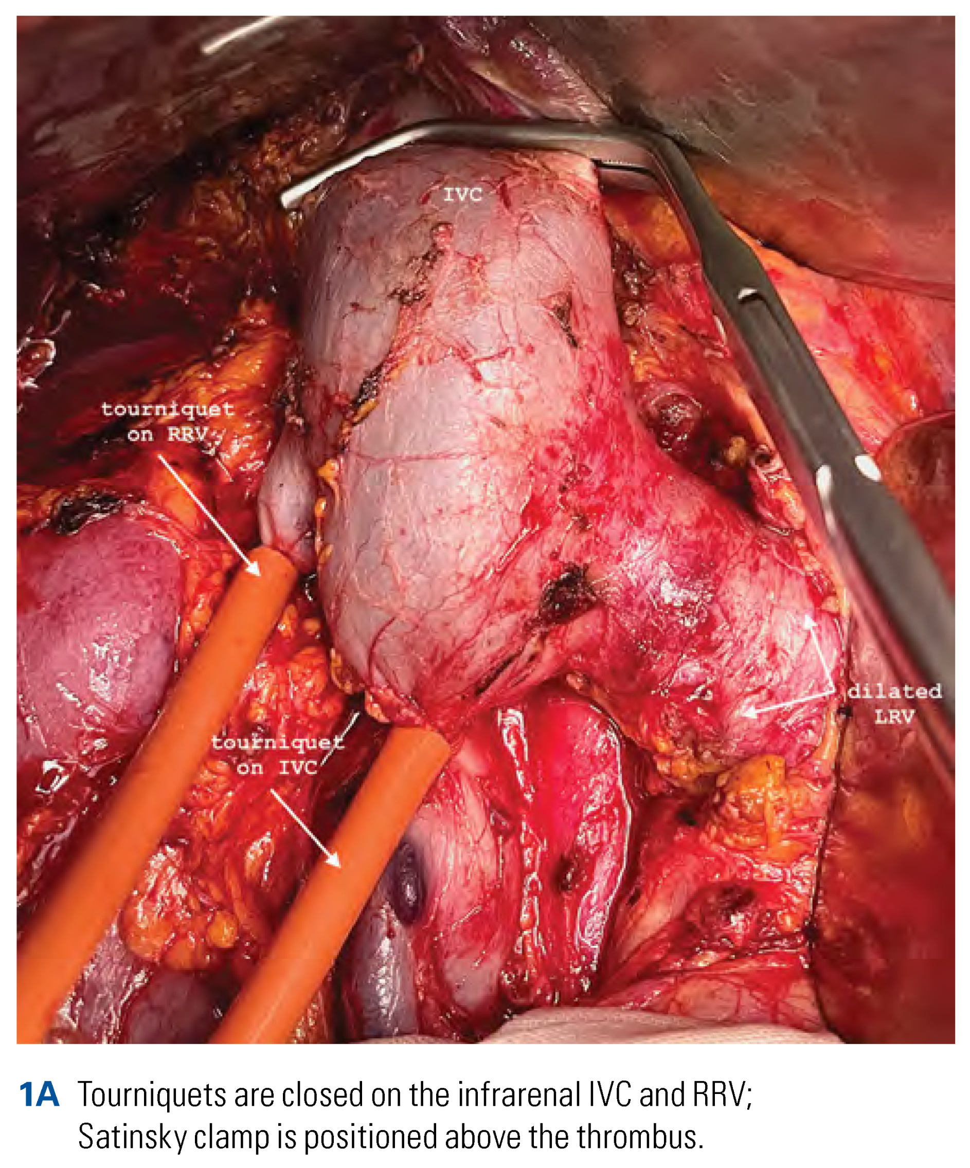
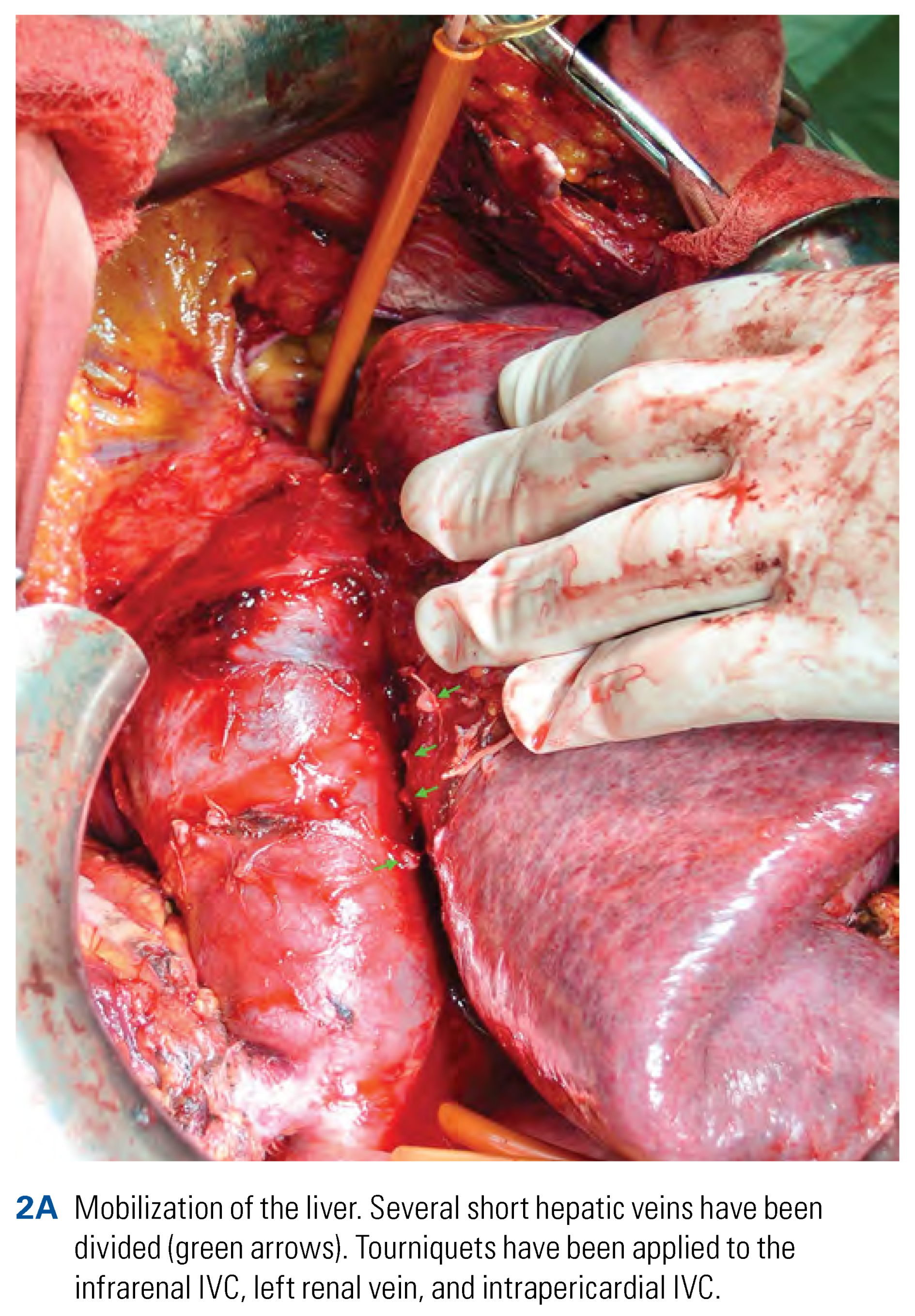
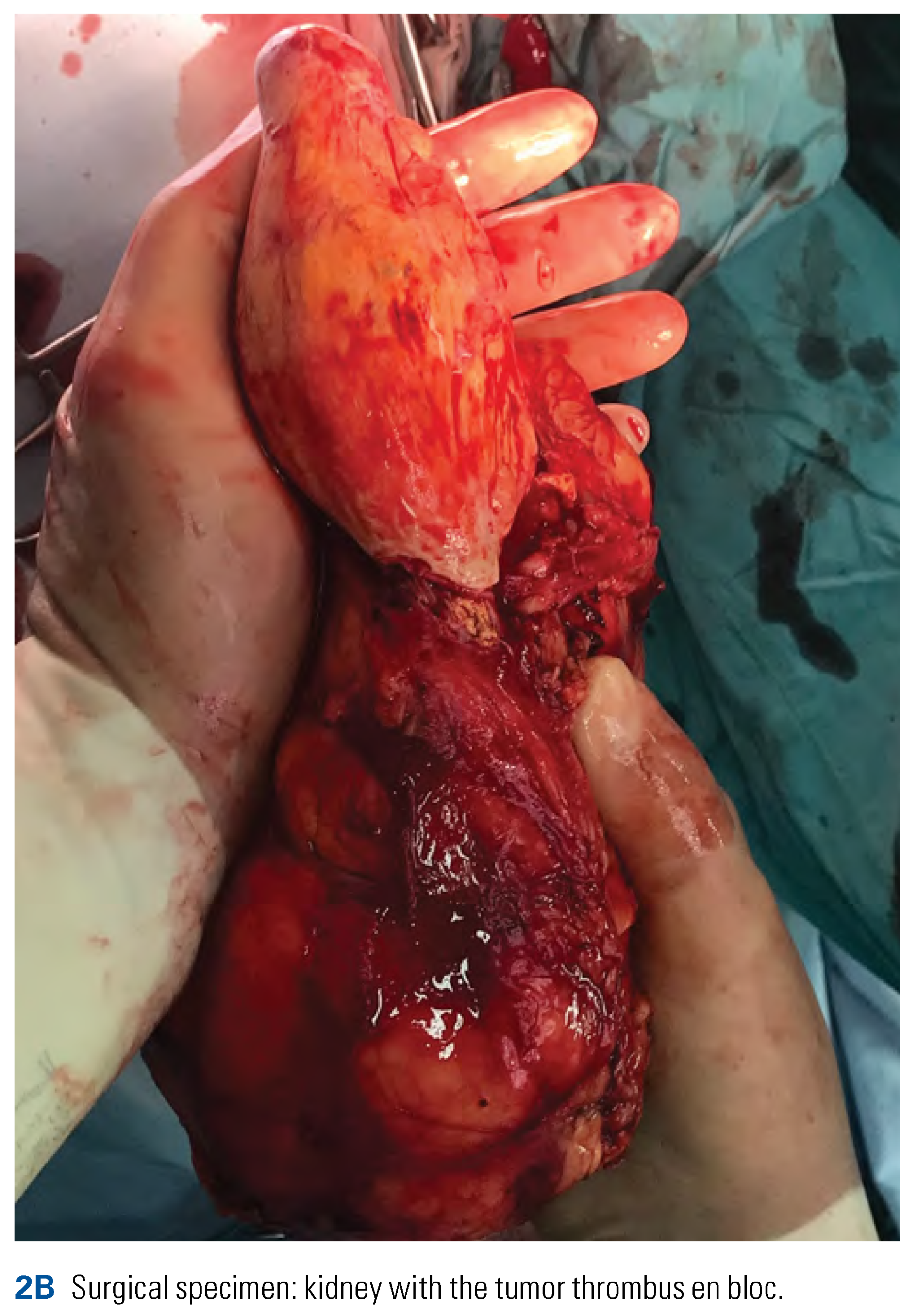
Surgical Technique of Radical Nephrectomy with IVC Thrombectomy

Surgery in patients with level III and IV VTT is generally performed through midline, chevron, or thoracoabdominal incision.
To minimize the morbidity associated with a thoracoabdominal approach, radical nephrectomy and thrombectomy may be performed via midline abdominal approach, which can be used for all levels of VTT.
Regardless of the primary tumor laterality, the access to the retroperitoneum is achieved by incising the posterior peritoneum lateral to the ascending colon. The right colon is reflected medially, and the duodenum is kocherized to expose the anterior surface of the IVC and aorta. Early ligation of the renal artery decreases the bleeding from venous collaterals and may permit for retraction of the cephalad most extent of the VTT. The mobilization of the kidney should be deferred until after complete vascular control is achieved.

Thrombectomy in Patients with Level I-III Tumor Thrombus

After ligation of the renal artery, the vena cava is completely dissected from the surrounding structures above and below the renal vein. Rummel tourniquets or vascular clamps are placed around the infrarenal IVC, contralateral renal vein, and suprarenal IVC cephalad to the superior extent of the VTT.
In patients with a subhepatic level II VTT, several (2–4) accessory hepatic veins from the caudate lobe of the liver are ligated and divided. As a result of this maneuver, 3 to 5 cm of additional IVC is exposed.
In the case of the right-sided primary, the right kidney is then completely mobilized outside Gerota's fascia, leaving the kidney attached only by the renal vein. In the case of a left-sided tumor, the kidney is mobilized only after completion of thrombectomy.
The tourniquets on infrarenal IVC, contralateral renal vein, and IVC above the upper extent of the thrombus are sequentially closed (Figure 1A). The antero-lateral wall of the subhepatic vena cava is longitudinally incised, and the thrombus is extracted with blunt and sharp dissection from the vessel wall, followed by a circumferential excision of the ostium of the renal vein (Figure 1B). After evaluating for remnant thrombus fragments adhering to the IVC intima, the defect of the IVC is closed with a 4–0 running vascular suture, the cavorraphy is backbled to avoid an air embolism, and the clamps are removed in the following order: (1) suprarenal IVC, (2) contralateral renal vein, (3) infrarenal IVC.
The management of patients with level III VTT requires mobilization of the liver, which allows for exposure of the intrahepatic and subdiaphragmatic segments of the IVC (Figure 2A). Resection of a level III VTT can be safely performed in most of the cases without circulatory support[45,46]. IVC above the upper extent of the thrombus is controlled at the intrapericardial level by using the transdiaphragmatic approach. Absence of tributaries makes circular mobilization of the intrapericardial part of the IVC significantly easier to perform compared with subdiaphragmatic circular mobilization. To accomplish a transdiaphragmatic IVC clamp, the diaphragm and underlying pericardium are incised above the IVC. Incision of the pericardium on both sides of the intrapericardial IVC allows for passing a tourniquet around the intrapericardial IVC[47] (Figure 2A).
Alternatively, the pericardium can be separated from the incised diaphragm and left intact. The advantages of approaching the intrapericardial IVC through a diaphragmatic incision include simplicity, decreased surgical trauma, and the ability to perform the procedure by one surgical team using a strictly abdominal approach[46,47].
The hepatoduodenal ligament is isolated to control the hepatic circulation with a Pringle maneuver. After closure of all tourniquets with a Pringle maneuver, the IVC is incised at the retrohepatic level. Widely opening the IVC at this level allows the surgeon to perform visual inspection of the ostium of the major and minor hepatic veins.
Following thrombectomy, the closure of the upper part of the cavotomy is started. As soon as the suture line reaches the subhepatic portion of the IVC, a Satinsky clamp is placed above the last stitch, the tourniquet from the intrapericardial IVC and the Satinsky clamp from the hepatoduodenal ligament are released, thereby restoring hepatic circulation and venous blood return to the right atrium. The rest of the procedure is similar to cases with level II VTT. Ideally, the thrombus is removed en bloc with the kidney (Figure 2B).
In patients with level III VTT and left-sided primary, the tourniquets are placed on the right renal vein (or right renal artery), while the left renal vein is sutured and divided with a TA stapler prior to thrombectomy. Contraindication for this approach includes partial left renal vein and IVC occlusion by a thin tumor thrombus, which may dislodge and cause pulmonary embolization. Transection of the left renal vein in patients with fixed IVC thrombi is safe and allows for greater IVC mobility, provides good access to the left renal artery and aorta, and facilitates en bloc removal of the thrombus with the ostium and VTT-containing stump of the left renal vein.
In cases where a level III thrombus is free floating, the thrombus can be extracted via an infrahepatic-only cavotomy similar to patients with level II VTT.

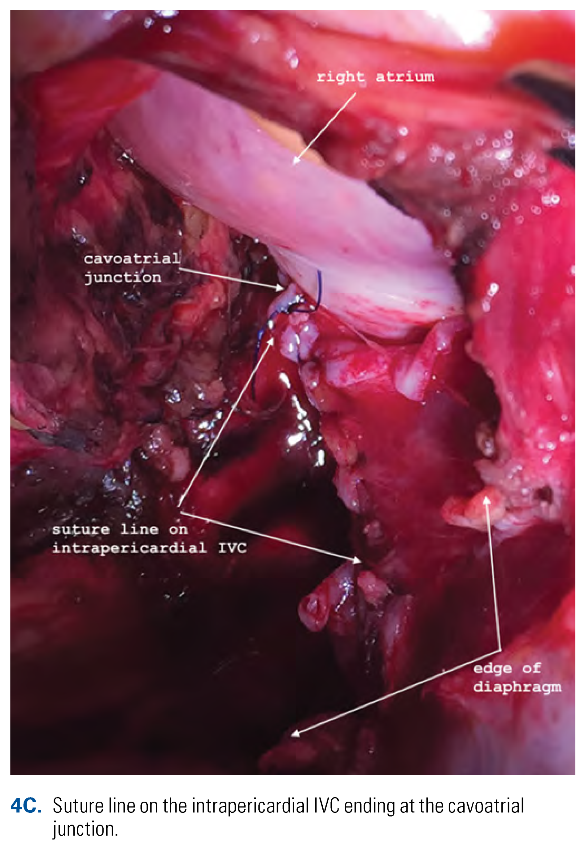
Thrombectomy in Patients with Supradiaphragmatic (Level IV) Tumor Thrombus

In general, the approaches used for supradiaphragmatic thrombectomy can be divided into those that use any type of circulatory support, and those that avoid it. No surgical method was shown to be superior for the excision of VTT. Most patients with nonadherent intraatrial thrombus can be managed without circulatory support[45,46,47].
From a practical point of view, level IV thrombi can be subdivided into intrapericardial and intraatrial VTT. The first steps of the procedures are the same for either case, and follow the techniques described above for patients with level III tumor thrombi. In patients with intrapericardial thrombus only, there is no need for complete mobilization of the intrapericardial IVC. Instead, only incisions of the pericardium on both sides of the intrapericardial IVC are performed for tourniquet placement.
On the contrary, in the case of an intraatrial thrombus, the intrapericardial IVC needs to be completely mobilized so that it can be encircled at the cavoatrial junction. The diaphragm at the IVC hiatus must be completely mobilized from the IVC to widen the natural narrowing for safer extraction of the thrombus from the right atrium (Figure 3A) The upper tourniquet on the intrapericardial IVC is left unclamped until the apex of the thrombus is removed from the heart. After all other tourniquets are sequentially clamped with a Pringle maneuver, the incision of the retrohepatic IVC is continued up to the cavoatrial junction. The thrombus is gently mobilized from the retrohepatic IVC (Figure 3B). If the head of the thrombus is mobile, the retrograde blood flow from the right atrium will evacuate the thrombus. If the thrombus is adherent to the vessel wall, the incision is extended on the intrapericardial IVC and the thrombus is resected from the intima of the intrapericardial IVC under direct visualization. In the case of a large head of the tumor thrombus, the incision may even be extended 5 to 10 mm on the right atrium wall (Figure 4). As soon as the thrombus is evacuated from the right atrium and the intrapericardial IVC, the upper tourniquet is closed. The rest of the procedure progresses as described above for patients with level III thrombus.
The majority of cases with level IV thrombus can be managed without circulatory support unless the head of the thrombus is too bulky for removal without performing wide atriotomy. Thrombi that have a long history of surveillance and those that shrank after preoperative systemic therapy may be adherent to the vessel wall. Removal of the thrombus in these patients may be associated with excessive blood loss. Circulatory support in these circumstances helps to maintain hemodynamic stability.

Thrombectomy in Patients with Tumor IVC Wall Invasion and/or Descending Bland Thrombosis

Vena cava resection is required in cases where the wall of the IVC is directly invaded by tumor. Repairs of the IVC include primary repair by venorrhaphy, prosthetic and autologous patch repair, and circumferential complete IVC replacement. In the majority of cases, resection of the dilated part of IVC does not result in clinically significant IVC narrowing, and primary cavorraphy is preferred. Indications for prosthetic repair include narrowing of IVC of > 50%. However, grafts expose patients to an increased risk for infection and thrombosis. Therefore, the optimal management of the IVC after resection is controversial[47].
Descending thrombosis in patients with RCC and VTT is quite frequent and occurs in 9.8% of patients[48]. Bland infrarenal IVC thrombus carries the risk for embolization, and recurrence from residual tumor cells, which can be found in 16% at the margin of tumor and bland thrombus. IVC interruption or segmental resection is required in most of the cases. The infrarenal IVC can be safely resected without reconstruction below the level of the proximal bland thrombus or just above the IVC bifurcation without any consequences[46,48,49,50].
Most authors indicate that lower-extremity edema after ligation of the IVC occurs rarely, is well tolerated, and resolves spontaneously[49,50].
The suprarenal IVC can be safely resected with ligation of the left renal vein in patients with right-sided RCC with VTT. The collateral venous return from the left kidney is sufficient to preserve normal renal function. Thus, resection of the IVC en bloc with rightsided RCC and tumor thrombus from the level of the major hepatic veins to the level of IVC bifurcation (en bloc cavectomy) can be performed without reconstruction[20,48] (Figure 5).
In patients with left-sided RCC, the right renal vein must be spared to preserve adequate venous drainage from the kidney.

Minimally Invasive Radical Nephrectomy and IVC Thrombectomy

Since the first report of laparoscopic IVC thrombectomy in 2000, numerous studies have highlighted the feasibility of minimally invasive techniques[51,52,53,54,55,56,57,58,59,60,61,62].
Most minimally invasive IVC thrombectomies have been performed on right-sided renal tumors[52]. Some have advocated that in individuals with left-sided renal tumors, preoperative arterial embolization may be beneficial due to the limited access to the left renal artery while the patient is in the flank position with the right side up[57]. Regardless of the tumor laterality and the decision regarding embolization, the “thrombus first” approach is necessary: the IVC thrombectomy should be completed prior to nephrectomy.
Most surgeons performing minimally invasive IVC thrombectomies prefer a transperitoneal approach. Applied in both pure laparoscopic and robot-assisted techniques, a transperitoneal approach may be used for thrombi associated with both right- and left-sided renal tumors[57,58]. Regardless of the laterality of the renal mass, the patient is placed in the left lateral decubitus position with the right side up. A12-mm camera port is inserted with additional three or four ports placed in a paramedian or mid-clavicular vertical line. Typically, one small port is placed at the sub-xiphoid region for liver retraction as well as an additional 12-mm assistant port[52,57,62,63]. The dissection is begun with an incision to the posterior peritoneum, followed by mobilization of the colon medially, duodenal kocherization, and exposure of the IVC and the renal vessels[51,52,61,62,63,64,65,66].

Minimally Invasive Level 0-I-II Thrombectomy

Early arterial ligation may help to slightly reduce the size of the thrombus. Ligation of the renal artery (with either an endo-GIA stapler or Hem-o-lok clips) may be performed at the hilum or in the interaortocaval space.
For thrombi limited to the renal vein, the latter may become flattened and allow for the thrombus to be “milked” away from the IVC. An interoperative US is helpful to identify of the extent of the thrombus[58,67]. For left-sided level I tumor thrombi, one should consider doing the surgery in the right lateral decubitus (if th e thrombus appears that it can be milked away from the IVC).
For IVC thrombi projecting less than 2 cm into the IVC, dissection of the contralateral renal vein and preplacement of a vessel loop with a secured Hem-o-lock clip (Rummel tourniquet) for later identification and cinching is often performed[68]. Simple use of laparoscopic bulldogs can be safely performed to cross-clamp the cava and contralateral vein[66].
Similar to level 0 thrombi, VTT exclusion with excision of a small IVC cuff surrounding the renal vein ostium may be employed via the laparoscopic Satinsky clamp and vascular stapler[52,69]. For larger level I and level II thrombi, the IVC must be cross-clamped and the right adrenal vein may require ligation. For right-sided thrombi, Rummel tourniquets or the vascular bulldogs should be cinched in the following order: (1) infrarenal IVC, (2) left renal vein, and (3) suprarenal IVC. For left-sided thrombi, the right renal artery should also be controlled with a vascular bulldog to prevent right renal engorgement[62]. Incision of the IVC is performed until the cephalad extent of the thrombus is delivered. The renal vein is circumferentially excised at the level of the ostium of the IVC. Upon thrombus removal, irrigation of the IVC lumen with heparinized saline is performed[52,55,60,69]. Closure of the IVC with 4–0 prolene sutures in a single layer allows for excellent hemostasis.

Minimally Invasive Level III Thrombectomy

Initially performed by Bratslavsky and Cheng in March of 2013 for an 11-cm IVC thrombus, this surgery remains technically demanding and dangerous[51].
Incision of the right triangular ligament and cephalad retraction of the liver will expose the retrohepatic IVC. Short hepatic veins from the caudate lobe can be ligated using laparoscopic clips[51,56]. Rummel tourniquets are cinched in the same order as in cases of level II thrombi.
For left-sided thrombi, transecting the ipsilateral renal vein with an endo-GIA vascular stapler is often employed, as the rest of the left renal vein will be removed with the specimen[57]. Upon en bloc thrombus removal with the renal vein stump closure of the cavatomy is performed with restoration of blood flow via the IVC. The patient is then repositioned and the left nephrectomy is completed.

Results of Surgical Management of RCC with VTT

Invasion of the venous system by RCC has long been associated with a poor prognosis[70]. However, the ability to provide a durable cure for some RCC patients provided a rationale for aggressive surgical management[71].
A systematic review of surgery for RCC with IVC thrombus cited major perioperative complication rates as high as 70% and perioperative mortality rates ranging from 3% to 16%[72].
Most data demonstrate increased mortality as thrombus height increases, with higher mortality observed in patients with IVC thrombus that extends above the diaphragm or above the hepatic confluence of veins (Neves/Mayo level III and IV)[14]. Early mortality rates from notable studies range from 8% to 22%[8,11,12,73,74,75,76].
No difference in mortality was observed between centers or among patients treated with cardiopulmonary bypass, similar to later reports[73]. Hospital volume and surgeon volumes are associated with early mortality[77,78,79]. Patients with metastatic RCC have a significantly worse prognosis compared to locally advanced RCC, with a cumulative mortality of 32% in the 9 months following surgery for metastatic patients[80].

Perioperative Complications

In population-level data, Toren et al. estimated the overall complication rate to be 78% in 633 patients treated with nephrectomy and IVC thrombectomy, including a 37% rate of surgical complications[79]. Similar overall rates of complications have been reported at large centers[72,81], with Blute et al. demonstrating that 30-day complication rates varied from 9% to 30% stratifying from thrombus level 0 to IV[28].
Commonly reported intraoperative complications include hemorrhage, injury of adjacent structures, and cardiac events[28]. Tumor thrombus embolization is a feared complication that occurs in 1.5% of patients and is associated with a reported 75% risk for mortality[82]. Early postoperative complications include hemorrhage requiring transfusion, venous thromboembolic events, cardiac events, pulmonary events, renal failure, and infectious complications. Late complications may include lower-extremity edema, chronic renal insufficiency, and incisional hernia[28]. Independent predictors of major complications include preoperative systemic symptoms (weight loss or fatigue) and thrombus level[83]. Use of cardiac bypass or deep hypothermic cardiac arrest was not identified as a predictor of major complications in this study[83] or subsequent analyses[73]. Complication rates are lower at higher-volume centers[77] and with more experienced surgeons[79].

Oncologic Outcomes

Managed expectantly, RCC with VTT is associated with a median survival of 5 months with increased risk for cancer-specific mortality observed with pT3b/c and metastatic disease[84]. In a population-based study of patients treated with nephrectomy and venous tumor thrombectomy, 1-year overall survival in patients with localized disease was 90%[85]. A 5-year, cancer-specific mortality in patients with RCC and VTT ranges from 40% to 60%[28,86,87,88]. Prognostic factors associated with cancer-specific mortality include increasing level of VTT, nodal and systemic metastases, advanced Fuhrman grade, non-clear cell histology, and increasing tumor size[10,81,82,86,89,90,91].

Conclusion

Renal cell carcinoma with venous thrombus provides a fascinating example of seemingly implausible tumor biology. Although these aggressive tumors have acquired the ability to invade and shed tumor cells into the largest blood vessel in the human body, some patients do not develop metastatic disease and may be cured when treated with definitive, aggressive surgery resulting in complete extirpation.
Surgery for RCC with venous thrombus is complex and requires experienced multidisciplinary surgical, anesthesia, and critical care teams at high-volume centers to achieve ideal outcomes. The use of presurgical and postsurgical systemic therapies is likely to increase with advances in systemic therapy in the future and represents a high-priority area for contemporary investigation.

Competing Interests

None declared.

Abbreviations

IOImmune-oncology therapy
RCCrenal cell carcinoma
VTTvenous tumor thrombus

References

  1. Ali, A.S.; Vasdev, N.; Shanmuganathan, S.; Paez, E.; Dark, J.H.; Manas, D.; et al. The surgical management and prognosis of renal cell cancer with IVC tumour thrombus: 15-years of experience using a multi-specialty approach at a single UK referral center. Urol. Oncol. 2013, 31, 1298–1304. [Google Scholar] [CrossRef] [PubMed]
  2. Amin, M.B.; Greene, F.; Edge, S.; Compton, C.C.; Gershenwald, J.E.; Brookland, R.K.; et al. AJCC Cancer Staging Manual, 8th ed.; Springer International Publishing: Berlin/Heidelberg, Germany, 2017. [Google Scholar]
  3. Marshall, F.F. Surgery of renal cell carcinoma with inferior vena caval involvement. Semin. Urol. 1989, 7, 186–190. [Google Scholar]
  4. Slaton, J.W.; Balbay, M.D.; Levy, D.A.; Pisters, L.L.; Nesbitt, J.C.; Swanson, D.A.; et al. Nephrectomy and vena caval thrombectomy in patients with metastatic renal cell carcinoma. Urology. 1997, 50, 673–677. [Google Scholar] [CrossRef]
  5. Kim, H.L.; Zisman, A.; Han, K.R.; Figlin, R.A.; Belldegrun, A.S. Prognostic significance of venous thrombus in renal cell carcinoma. Are renal vein and inferior vena cava involvement different? J. Urol. 2024, 171 Pt 1, 588–591. [Google Scholar] [CrossRef]
  6. Moinzadeh, A.; Libertino, J.A. Prognostic significance of tumour thrombus level in patients with renal cell carcinoma and VTTombus extension. Is all T3b the same? J. Urol. 2004, 171 Pt 1, 598–601. [Google Scholar] [CrossRef]
  7. Sosa, R.E.; Muecke, E.C.; Vaughan, E.D., Jr.; McCarron, J.P., Jr. Renal cell carcinoma extending into the inferior vena cava: The prognostic significance of the level of vena caval involvement. J. Urol. 1984, 132, 1097–1100. [Google Scholar] [CrossRef]
  8. Wagner, B.; Patard, J.J.; Méjean, A.; Bensalah, K.; Verhoest, G.; Zigeuner, V.; et al. Prognostic value of renal vein and inferior vena cava involvement in renal cell carcinoma. Eur. Urol. 2009, 55, 452–459. [Google Scholar] [CrossRef]
  9. Boorjian, S.A.; Blute, M.L. Surgery for vena caval tumour extension in renal cancer. Curr. Opin. Urol. 2009, 19, 473–477. [Google Scholar] [CrossRef]
  10. Boorjian, S.A.; Sengupta, S.; Blute, M.L. Renal cell carcinoma: Vena caval involvement. BJU Int. 2007, 99 Pt B, 1239–1244. [Google Scholar] [CrossRef]
  11. Granberg, C.F.; Boorjian, S.A.; Schaff, H.V.; Orszulak, T.A.; Leibovich, B.C.; Lohse, C.M.; et al. Surgical management, complications, and outcome of radical nephrectomy with inferior vena cava tumour thrombectomy facilitated by vascular bypass. Urology 2008, 72, 148–152. [Google Scholar] [CrossRef]
  12. Blute, M.L.; Boorjian, S.A.; Leibovich, B.C.; Lohse, C.M.; Frank, I.; Karnes, R.J. Results of inferior vena caval interruption by greenfield filter, ligation or resection during radical nephrectomy and tumour thrombectomy. J. Urol. 2007, 178, 440–445; discussion 444. [Google Scholar] [CrossRef]
  13. Chiappini, B.; Savini, C.; Marinelli, G.; Martin Suarez, S.; Di Eusanio, M.; Fiorani, V.; et al. Cavoatrial tumour thrombus: Single-stage surgical approach with profound hypothermia and circulatory arrest, including a review of the literature. J. Thorac. Cardiovasc. Surg. 2002, 124, 684–688. [Google Scholar] [CrossRef]
  14. Neves, R.J.; Zincke, H. Surgical treatment of renal cancer with vena cava extension. BJU Int. 1987, 59, 390–395. [Google Scholar] [CrossRef]
  15. Gohji, K.; Yamashita, C.; Ueno, K.; Shimogaki, H.; Kamidono, S. Preoperative computerized tomography detection of extensive invasion of the inferior vena cava by renal cell carcinoma: Possible indication for resection with partial cardiopulmonary bypass and patch grafting. J. Urol. 1994, 152 Pt 1, 1993–1996; discussion 1997. [Google Scholar] [CrossRef]
  16. Psutka, S.P.; Boorjian, S.A.; Thompson, R.H.; Schmit, G.D.; Schmitz, J.J.; Bowere, T.C.; et al. Clinical and radiographic predictors of the need for inferior vena cava resection during nephrectomy for patients with renal cell carcinoma and caval tumour thrombus. BJU Int. 2015, 116, 388–396. [Google Scholar] [CrossRef]
  17. Hutchinson, R.; Rew, C.; Chen, G.; Woldu, S.; Krabbe, L.-M.; Mieissner, M.; et al. The adverse survival implications of bland thrombus in renal cell carcinoma with venous tumor thrombus. Urology 2018, 115, 119–124. [Google Scholar] [CrossRef]
  18. Wang, H.; Li, X.; Huang, Q.; Panic, A.; Shen, D.; Jia, W.; et al. Prognostic role of bland thrombus in patients treated with resection of renal cell carcinoma with inferior vena cava tumour thrombus. Urol. Oncol. 2021, 39, 302–e301. [Google Scholar] [CrossRef]
  19. Liu, Z.; Li, L.; Hong, P.; Zhu, G.; Tang, S.; Zhao, X.; et al. A predictive model for tumour invasion of the inferior vena cava wall using multimodal imaging in patients with renal cell carcinoma and inferior vena cava tumour thrombus. Biomed. Res. Int. 2020, 2020, 9530618. [Google Scholar]
  20. Weiss, V.L.; Braun, M.; Perner, S.; Harz, A.; Vorreuther, R.; Kristiansen, G.; et al. Prognostic significance of VTT consistency in patients with renal cell carcinoma (RCC). BJU Int. 2014, 113, 209–217. [Google Scholar] [CrossRef]
  21. Bertini, R.; Roscigno, M.; Freschi, M.; Strada, E.; Angiolilli, D.; Petralia, G.; et al. Impact of VTT consistency (solid vs friable) on cancer-specific survival in patients with renal cell carcinoma. Eur. Urol. 2011, 60, 358–365. [Google Scholar] [CrossRef]
  22. Psutka, S.P.; Leibovich, B.C. Management of inferior vena cava tumour thrombus in locally advanced renal cell carcinoma. Ther. Adv. Urol. 2015, 7, 216–229. [Google Scholar] [CrossRef] [PubMed]
  23. Ciancio, G.; Soloway, M. Renal cell carcinoma invading the hepatic veins. Cancer 2001, 92, 1836–1842. [Google Scholar] [CrossRef]
  24. Ciancio, G.; Vaidya, A.; Savoie, M.; Soloway, M. Management of renal cell carcinoma with level III thrombus in the inferior vena cava. J. Urol. 2002, 168 Pt 1, 1374–1377. [Google Scholar] [CrossRef]
  25. Kume, H.; Kameyama, S.; Kasuya, Y.; Tajima, A.; Kawabe, K. Surgical treatment of renal cell carcinoma associated with Budd-Chiari syndrome: Report of four cases and review of the literature. Eur. J. Surg. Oncol. 1999, 25, 71–75. [Google Scholar] [CrossRef]
  26. Lessard, L.; Bach, D.; Wall, W.; Luke, P.P. Intrahepatic extension of renal cell carcinoma tumour thrombus causing Budd-Chiari syndrome. Can. Urol. Assoc. J. 2011, 5, 383–384. [Google Scholar] [CrossRef] [PubMed]
  27. Abel, E.J.; Thompson, R.H.; Margulis, V.; Heckman, J.E.; Merril, M.M.; Darwish, O.M.; et al. Perioperative outcomes following surgical resection of renal cell carcinoma with inferior vena cava thrombus extending above the hepatic veins: A contemporary multicenter experience. Eur. Urol. 2014, 66, 584–592. [Google Scholar] [CrossRef]
  28. Blute, M.L.; Leibovich, B.C.; Lohse, C.M.; Cheville, J.C.; Zincke, H. The Mayo Clinic experience with surgical management, complications and outcome for patients with renal cell carcinoma and VTT. BJU Int. 2004, 94, 33–41. [Google Scholar] [CrossRef]
  29. Parekh, D.J.; Cookson, M.S.; Chapman, W.; Harrell FJr Wells, N.; Chang, S.S.; et al. Renal cell carcinoma with renal vein and inferior vena caval involvement: Clinicopathological features, surgical techniques and outcomes. J. Urol. 2005, 173, 1897–1902. [Google Scholar] [CrossRef] [PubMed]
  30. Kapur, S.; Paik, E.; Rezaei, A.; Vu, D.N. Where there is blood, there is a way: Unusual collateral vessels in superior and inferior vena cava obstruction. Radiographics 2010, 30, 67–78. [Google Scholar] [CrossRef]
  31. Wotkowicz, C.; Wszolek, M.F.; Libertino, J.A. Resection of renal tumours invading the vena cava. Urol. Clin. N. Am. 2008, 35, 657–671. [Google Scholar] [CrossRef]
  32. Lawindy, S.M.; Kurian, T.; Kim, T.; Mangar, D.; Armstrong, P.A.; Alsina, A.E.; et al. Important surgical considerations in the management of renal cell carcinoma (RCC) with inferior vena cava (IVC) tumour thrombus. BJU Int. 2012, 110, 926–939. [Google Scholar] [CrossRef] [PubMed]
  33. Woodruff, D.Y.; Van Veldhuizen, P.; Muehlebach, G.; Johnson, P.; Williamson, T.; Holzbeierlein, J.M. The perioperative management of an inferior vena caval tumour thrombus in patients with renal cell carcinoma. Urol. Oncol. 2013, 31, 517–521. [Google Scholar] [CrossRef] [PubMed]
  34. Guo, H.F.; Song, Y.; Na, Y.Q. Value of abdominal ultrasound scan, CT and MRI for diagnosing inferior vena cava tumour thrombus in renal cell carcinoma. Chin. Med. J. 2009, 122, 2299–2302. [Google Scholar] [PubMed]
  35. Calderone, C.E.; Tuck, B.C.; Gray, S.H.; Porter, K.K.; Rais-Bahrami, S. The role of transesophageal echocardiography in the management of renal cell carcinoma with VTTombus. Echocardiography 2018, 35, 2047–2055. [Google Scholar] [CrossRef] [PubMed]
  36. Cost, N.G.; Delacroix SEJr Sleeper, J.P.; Smith, P.J.; Youssef, R.F.; Chapin, B.F.; et al. The impact of targeted molecular therapies on the level of renal cell carcinoma vena caval tumour thrombus. Eur. Urol. 2011, 59, 912–918. [Google Scholar] [CrossRef]
  37. Bigot, P.; Fardoun, T.; Bernhard, J.C.; Xylinas, E.; Berger, J.; Rouprêt, M.; et al. Neoadjuvant targeted molecular therapies in patients undergoing nephrectomy and inferior vena cava thrombectomy: Is it useful? World J. Urol. 2014, 32, 109–114. [Google Scholar] [CrossRef] [PubMed]
  38. Peng, C.; Gu, L.; Wang, L.; Huang, Q.; Wang, B.; Guo, G.; et al. Role of presurgical targeted molecular therapy in renal cell carcinoma with an inferior vena cava tumour thrombus. OncoTargets Ther. 2018, 11, 1997–2005. [Google Scholar] [CrossRef] [PubMed]
  39. Fukuda, H.; Kondo, T.; Takagi, T.; Iizuka, J.; Nagashima, Y.; Tanabe, K. Limited benefit of targeted molecular therapy for inferior vena cava thrombus associated with renal cell carcinoma. Int. J. Clin. Oncol. 2017, 22, 767–773. [Google Scholar] [CrossRef]
  40. Ujike, T.; Uemura, M.; Kawashima, A.; Nagahara, A.; Fujita, K.; Miyagawa, Y.; et al. Clinical and histopathological effects of presurgical treatment with sunitinib for renal cell carcinoma with inferior vena cava tumour thrombus at a single institution. Anticancer. Drugs 2016, 27, 1038–1043. [Google Scholar] [CrossRef]
  41. Horn, T.; Thalgott, M.K.; Maurer, T.; Hauner, K.; Schulz, S.; Fingerle, A.; et al. Presurgical treatment with sunitinib for renal cell carcinoma with a level III/IV vena cava tumour thrombus. Anticancer. Res. 2012, 32, 1729–1735. [Google Scholar]
  42. Tanaka, Y.; Hatakeyama, S.; Hosogoe, S.; Tanaka, T.; Hamano, I.; Kusaka, A.; et al. Presurgical axitinib therapy increases fibrotic reactions within tumour thrombus in renal cell carcinoma with thrombus extending to the inferior vena cava. Int. J. Clin. Oncol. 2018, 23, 134–141. [Google Scholar] [CrossRef]
  43. Terakawa, T.; Hussein, A.A.; Bando, Y.; Guru, K.A.; Furukawa, J.; Shigemura, K.; et al. Presurgical pazopanib for renal cell carcinoma with inferior vena caval thrombus: A single-institution study. Anticancer. Drugs 2018, 29, 565–571. [Google Scholar] [CrossRef] [PubMed]
  44. Stewart, G.D.; Welsh, S.J.; Ursprung, S.; Gallagher, F.A.; Mendichovszky, I.; Riddick, A.; Eisen, T.; et al. NAXIVA: A phase II neoadjuvant study of axitinib for reducing extent of VTTombus in clear cell renal cell cancer (RCC) with venous invasion. J. Clin. Oncol. 2021, 39 (Suppl. 6), 275. [Google Scholar] [CrossRef]
  45. Skinner, D.G.; Pritchett, T.R.; Lieskovsky, G.; Boyd, S.D.; Stiles, Q.R. Vena caval involvement by renal cell carcinoma. Surgical resection provides meaningful long-term survival. Ann. Surg. 1989, 210, 387–392; discussion 392–394. [Google Scholar] [CrossRef] [PubMed] [PubMed Central]
  46. Ciancio, G.; Livingstone, A.S.; Soloway, M. Surgical management of renal cell carcinoma with tumour thrombus in the renal and inferior vena cava: The University of Miami experience in using liver transplantation techniques. Eur. Urol. 2007, 51, 988–994; discussion 994–995. [Google Scholar] [CrossRef] [PubMed]
  47. Davydov, M.I.; Matveev, V.B. Surgical management of renal cell carcinoma with tumour thrombosis of renal vein and inferior vena cava. Cancer Urol. 2005, 1, 8–15. [Google Scholar]
  48. Davydov, M.I.; Matveev, V.B.; Volkova, M.I.; et al. Predictors of the immediate results of thrombectomy in kidney cancer patients with venous tumour thrombosis. Cancer Urol. 2014, 10, 31–39. [Google Scholar]
  49. Daylami, R.; Amiri, A.; Goldsmith, B.; Troppmann, C.; Schneider, P.D.; Khatri, V.P. Inferior vena cava leiomyosarcoma: Is reconstruction necessary after resection? J. Am. Coll. Surg. 2010, 210, 185–190. [Google Scholar] [CrossRef] [PubMed]
  50. Ayyathurai, R.; Garcia-Roig, M.; Gorin, M.A.; González, J.; Manoharan, M.; Kava, B.R.; et al. Bland thrombus association with tumour thrombus in renal cell carcinoma: Analysis of surgical significance and role of inferior vena caval interruption. BJU Int. 2012, 110 Pt B, E449–E455. [Google Scholar] [CrossRef] [PubMed]
  51. Bratslavsky, G.; Cheng, J.S. Robotic-assisted radical nephrectomy with retrohepatic vena caval tumour thrombectomy (Level III) combined with extended retroperitoneal lymph node dissection. Urology 2015, 86, 1235–1240. [Google Scholar] [CrossRef] [PubMed]
  52. Sun, Y.; de Castro Abreu, A.L.; Gill, I.S. Robotic inferior vena cava thrombus surgery: Novel strategies. Curr. Opin. Urol. 2014, 24, 140–147. [Google Scholar] [CrossRef]
  53. Varkarakis, I.M.; Bhayani, S.B.; Allaf, M.E.; Inagaki, T.; Gonzalgo, M.L.; Jarrett, T.W. Laparoscopic-assisted nephrectomy with inferior vena cava tumor thrombectomy: Preliminary results. Urology 2004, 64, 925–929. [Google Scholar] [CrossRef]
  54. Kovac, J.R.; Luke, P.P. Hand-assisted laparoscopic radical nephrectomy in the treatment of a renal cell carcinoma with a level II vena cava thrombus. Int. Braz. J. Urol. 2010, 36, 327–331. [Google Scholar] [CrossRef]
  55. Shao, P.; Li, J.; Qin, C.; Lv, Q.; Ju, X.; Li, P.; et al. Laparoscopic radical nephrectomy and inferior vena cava thrombectomy in the treatment of renal cell carcinoma. Eur. Urol. 2015, 68, 115–122. [Google Scholar] [CrossRef]
  56. Ramirez, D.; Maurice, M.J.; Cohen, B.; Krishnamurthi, V.; Haber, G.-P. Robotic level III IVC tumor thrombectomy: Duplicating the open approach. Urology 2016, 90, 204–247. [Google Scholar] [CrossRef]
  57. Gill, I.S.; Metcalfe, C.; Abreu, A.; Duddalwar, V.; Chopra, S.; Cunningham, M.; et al. Robotic level III inferior vena cava tumor thrombectomy: Initial series. J. Urol. 2015, 194, 929–938. [Google Scholar] [CrossRef]
  58. Savage, S.J.; Gill, I.S. Laparoscopic radical nephrectomy for renal cell carcinoma in a patient with level I renal vein tumor thrombus. J. Urol. 2000, 163, 1243–1244. [Google Scholar] [CrossRef]
  59. Fergany, A.F.; Gill, I.S.; Schweizer, D.K.; Kaouk, J.H.; ElFettouh, H.A.; Cherullo, E.E.; et al. Laparoscopic radical nephrectomy with level II vena caval thrombectomy: Survival porcine study. J. Urol. 2002, 168, 2629–2631. [Google Scholar] [CrossRef]
  60. Abaza, R.; Shabsigh, A.; Castle, E.; Allaf, M.; Hu, J.C.; Rogers, C.; Memon, M.; et al. Multi-institutional experience with robotic nephrectomy with inferior vena cava tumor thrombectomy. J. Urol. 2016, 195 Pt 1, 865–871. [Google Scholar] [CrossRef]
  61. Abaza, R. Initial series of robotic radical nephrectomy with vena caval tumor thrombectomy. Eur. Urol. 2011, 59, 652–656. [Google Scholar] [CrossRef] [PubMed]
  62. Wang, B.; Li, H.; Ma, X.; Zhang, X.; Gu, L.; Li, X.; et al. Robot-assisted laparoscopic inferior vena cava thrombectomy: Different sides require different techniques. Eur. Urol. 2016, 69, 1112–1119. [Google Scholar] [CrossRef] [PubMed]
  63. Romero, F.R.; Muntener, M.; Bagga, H.S.; Brito, F.A.R.; Sulman, A.; Jarrett, T.W. Pure laparoscopic radical nephrectomy with level II vena caval thrombectomy. Urology 2006, 68, 1112–1114. [Google Scholar] [CrossRef] [PubMed]
  64. Ball, M.W.; Gorin, M.A.; Jayram, G.; Pierorazio, P.M.; Allaf, M.E. Robot-assisted radical nephrectomy with inferior vena cava tumor thrombectomy: Technique and initial outcomes. Can. J. Urol. 2015, 22, 7666–7670. [Google Scholar]
  65. Gupta, N.P.; Ansari, M.S.; Khaitan, A.; Sivaramakrishna, M.S.; Hemal, A.K.; Dogra, P.N.; et al. Impact of imaging and thrombus level in management of renal cell carcinoma extending to veins. Urol. Int. 2004, 72, 129–134. [Google Scholar] [CrossRef]
  66. Scott, K.; Manwaring, J.; Amankwah, K.; Bratslavsky, G. Robotic assisted caval replacement for recurrent renal cell carcinoma invading the wall of the inferior vena cava. Urology 2022, 161, 131–134. [Google Scholar] [CrossRef]
  67. Desai, M.M.; Gill, I.S.; Ramani, A.P.; Matin, S.F.; Kaouk, J.H.; Campero, J.M.; et al. Laparoscopic radical nephrectomy for cancer with level I renal vein involvement. J. Urol. 2003, 169, 487–491. [Google Scholar] [CrossRef]
  68. Ball, M.W.; Gorin, M.A.; Jayram, G.; Pieroazio, P.M.; Allaf, M. Robot-assisted radical nephrectomy with inferior vena cava tumor thrombectomy: Technique and initial outcomes. Can. J. Urol. 2015, 22, 7666–7670. [Google Scholar] [PubMed]
  69. Lee, J.Y.; Mucksavage, P. Robotic radical nephrectomy with vena caval tumor thrombectomy: Experience of novice robotic surgeons. Korean J. Urol. 2012, 53, 879–882. [Google Scholar] [CrossRef]
  70. Riches, E.W.; Griffiths, I.H.; Thackray, A.C. New growths of the kidney and ureter. Br. J. Urol. 1951, 23, 297–356. [Google Scholar] [CrossRef]
  71. Skinner, D.G.; Pfister, R.F.; Colvin, R. Extension of renal cell carcinoma into the vena cava: The rationale for aggressive surgical management. J. Urol. 1972, 107, 711–716. [Google Scholar] [CrossRef]
  72. Kirkali, Z.; Van Poppel, H. A critical analysis of surgery for kidney cancer with vena cava invasion. Eur. Urol. 2007, 52, 658–662. [Google Scholar] [CrossRef] [PubMed]
  73. Nguyen, H.G.; Tilki, D.; Dall’Era, M.A.; Durbin-Johnson, B.; Carballido, J.A.; Chandrasekar, T.; et al. Cardiopulmonary bypass has no significant impact on survival in patients undergoing nephrectomy and level III-IV inferior vena cava thrombectomy: Multi-institutional analysis. J. Urol. 2015, 194, 304–308. [Google Scholar] [CrossRef] [PubMed]
  74. Shuch, B.; Crispen, P.L.; Leibovich, B.C.; LaRochelle, J.C.; Pouliot, F.; Pantuck, A.J.; et al. Cardiopulmonary bypass and renal cell carcinoma with level IV tumour thrombus: Can deep hypothermic circulatory arrest limit perioperative mortality? BJU Int. 2011, 107, 724–728. [Google Scholar] [CrossRef] [PubMed]
  75. Ciancio, G.; Gonzalez, J.; Shirodkar, S.P.; Angulo, J.C.; Soloway, M.S. Liver transplantation techniques for the surgical management of renal cell carcinoma with tumor thrombus in the inferior vena cava: Stepby-step description. Eur. Urol. 2011, 59, 401–406. [Google Scholar] [CrossRef] [PubMed]
  76. Casey, R.G.; Raheem, O.A.; Elmusharaf, E.; Madhavan, P.; Tolan, M.; Lynch, T.H. Renal cell carcinoma with IVC and atrial thrombus: A single centre’s 10 year surgical experience. Surgeon 2013, 11, 295–299. [Google Scholar] [CrossRef] [PubMed]
  77. Hsu, R.C.J.; Salika, T.; Maw, J.; Lyratzopoulos, G.; Gnanapragasam, V.J.; Armitage, J.N. Influence of hospital volume on nephrectomy mortality and complications: A systematic review and meta-analysis stratified by surgical type. BMJ Open 2017, 7, e016833. [Google Scholar] [CrossRef]
  78. Yap, S.A.; Horovitz, D.; Alibhai, S.M.; Abouassaly, R.; Timilshina, N.; Finelli, A. Predictors of early mortality after radical nephrectomy with renal vein or inferior vena cava thrombectomyA population-based study. BJU Int. 2012, 110, 1283–1288. [Google Scholar] [CrossRef]
  79. Toren, P.; Abouassaly, R.; Timilshina, N.; Kulkarni, G.; Alibhai, S.; Finelli, A. Results of a national population-based study of outcomes of surgery for renal tumors associated with inferior vena cava thrombus. Urology 2013, 82, 572–577. [Google Scholar] [CrossRef] [PubMed]
  80. Abel, E.J.; Spiess, P.E.; Margulis, V.; Master, V.A.; Mann, M.; Zargar-Shostari, K.; et al. Cytoreductive nephrectomy for renal cell carcinoma with venous tumor thrombus. J. Urol. 2017, 198, 281–288. [Google Scholar] [CrossRef]
  81. Martinez-Salamanca, J.I.; Linares, E.; González, J.; Bertini, R.; Carballido, J.A.; Chromecki, T.; et al. Lessons learned from the International Renal Cell Carcinoma-Venous Thrombus Consortium (IRCC-VTC). Curr. Urol. Rep. 2014, 15, 404. [Google Scholar] [CrossRef]
  82. Shuch, B.; Larochelle, J.C.; Onyia, T.; Vallera, C.; Margulis, D.; Pantuck, A.J.; et al. Intraoperative thrombus embolization during nephrectomy and tumor thrombectomy: Critical analysis of the University of CaliforniaLos Angeles experience. J. Urol. 2009, 181, 492–498; discussion 498–499. [Google Scholar] [CrossRef]
  83. Abel, E.J.; Thompson, R.H.; Margulis, V.; et al. Perioperative outcomes following surgical resection of renal cell carcinoma with inferior vena cava thrombus extending above the hepatic veins: A contemporary multicenter experience. Eur. Urol 2014, 66, 584–592. [Google Scholar] [CrossRef]
  84. Reese, A.C.; Whitson, J.M.; Meng, M.V. Natural history of untreated renal cell carcinoma with venous tumor thrombus. Urol. Oncol. 2013, 31, 1305–1309. [Google Scholar] [CrossRef] [PubMed]
  85. Whitson, J.M.; Reese, A.C.; Meng, M.V. Population based analysis of survival in patients with renal cell carcinoma and venous tumor thrombus. Urol. Oncol. 2013, 31, 259–263. [Google Scholar] [CrossRef]
  86. Haddad, A.Q.; Wood, C.G.; Abel, E.J.; Krabbe, L.-M.; Darwish, O.M.; Thompson, R.H.; et al. Oncologic outcomes following surgical resection of renal cell carcinoma with inferior vena caval thrombus extending above the hepatic veins: A contemporary multicenter cohort. J. Urol. 2014, 192, 1050–1056. [Google Scholar] [CrossRef] [PubMed]
  87. Hirono, M.; Kobayashi, M.; Tsushima, T.; Obara, W.; Shinohara, N.; Ito, K.; et al. Members of the Japanese Society of Renal Cancer. Impacts of clinicopathologic and operative factors on short-term and longterm survival in renal cell carcinoma with venous tumor thrombus extension: A multi-institutional retrospective study in Japan. BMC Cancer 2013, 13, 447. [Google Scholar] [CrossRef]
  88. Pouliot, F.; Shuch, B.; Larochelle, J.C.; Panticuk, A.; Belledegrun, A.S. Contemporary management of renal tumors with venous tumor thrombus. J. Urol. 2010, 184, 833–841. [Google Scholar] [CrossRef] [PubMed]
  89. Martinez-Salamanca, J.I.; Huang, W.C.; Millan, I.; Bertini, R.; Bianco, F.J.; Carballido, J.A.; et al. International Renal Cell Carcinoma-Venous Thrombus Consortium. Prognostic impact of the 2009 UICC/AJCC TNM staging system for renal cell carcinoma with venous extension. Eur Urol. 2011, 59, 120–127. [Google Scholar] [CrossRef]
  90. Kaushik, D.; Linder, B.J.; Thompson, R.H.; Eisenberg, M.S.; Lohse, C.M.; Cheville, J.C.; et al. The impact of histology on clinicopathologic outcomes for patients with renal cell carcinoma and venous tumor thrombus: A matched cohort analysis. Urology 2013, 82, 136–141. [Google Scholar] [CrossRef]
  91. Tilki, D.; Hu, B.; Nguyen, H.G.; Dall’Era, M.A.; Bertini, R.; Carballido, J.A.; et al. Impact of synchronous metastasis distribution on cancer specific survival in renal cell carcinoma after radical nephrectomy with tumor thrombectomy. J. Urol. 2015, 193, 436–442. [Google Scholar] [CrossRef]
Figure 1. Radical nephrectomy with thrombectomy in cT3bN0M0 RCC of the right kidney with level IV tumor thrombus.
Siuj 03 00451 g001aSiuj 03 00451 g001b
Figure 2. Radical nephrectomy with thrombectomy in cT3bN0M0 RCC of the right kidney with level III tumor thrombus.
Siuj 03 00451 g002aSiuj 03 00451 g002b
Figure 3. Radical nephrectomy with thrombectomy in cT3bN0M0 RCC of the left kidney with level II tumor thrombus.
Siuj 03 00451 g003aSiuj 03 00451 g003b
Figure 4. Radical nephrectomy with thrombectomy in cT3bN0M0 RCC of the right kidney with large intraatrial level IV tumor thrombus.
Siuj 03 00451 g004aSiuj 03 00451 g004b
Figure 5. En bloc resection of the IVC with RCC and associated caval thrombus.
Siuj 03 00451 g005aSiuj 03 00451 g005b
Table 1. Classification of VTT level in RCC: The Mayo Classification system14.
Siuj 03 00451 i001

