Article

Role of Multiparametric Magnetic Resonance Imaging and Targeted Biopsy in the Detection of Clinically Significant Prostate Cancer in Patients with Suspicious Digital Rectal Examination

1
Department of Clinical and Experimental Medicine, University of Messina, 98125 Messina, Italy
2
Gaetano Barresi Department of Human and Paediatric Pathology, Urology Section, University of Messina, 98125 Messina, Italy
3
Department of Economics, Division of Statistical and Mathematical Sciences, University of Messina, 98125 Messina, Italy
4
Department of Biomedical Sciences and Morphological and Functional Imaging, Diagnostic Imaging Section, University of Messina, 98125 Messina, Italy
5
Gaetano Barresi Department of Human and Paediatric Pathology, Pathology Section, University of Messina, 98125 Messina, Italy
6
Urology Unit, Santa Maria della Misericordia University Hospital, 33100 Udine, Italy
*
Author to whom correspondence should be addressed.
Soc. Int. Urol. J. 2024, 5(2), 122-132; https://doi.org/10.3390/siuj5020021
Submission received: 18 April 2023 / Revised: 27 August 2023 / Accepted: 3 September 2023 / Published: 16 April 2024

Abstract

:
Objectives: Few studies have examined the role of prostate MRI in patients with suspicious digital rectal examination (DRE) and/or PSA > 10 ng/mL. In a cohort of non-screened patients with suspicious DRE, we investigated the rate of avoidable prostate biopsies and potentially missed clinically significant prostate cancer (csPCa) with negative MRI, and the concordance between targeted biopsy (TBx) and systematic biopsy (SBx) in detecting csPCa with positive MRI. Methods: We retrospectively examined 199 biopsy-naïve patients with suspicious DRE who underwent prostate MRI before transperineal biopsy. Prostate Imaging—Reporting and Data System (PI-RADS) v2.1 ≥ 3 category of the index lesion defined a positive MRI. Combined TBx/SBx and SBx alone were performed for positive and negative MRI, respectively. An International Society of Urogenital Pathology Grade Group ≥ 2 defined csPCa. We calculated the csPCa detection rate of TBx, SBx, and combined TBx/SBx. The Cohen kappa statistic was used to measure the concordance between TBx and SBx. Results: Ninety-one (45.7%) csPCa cases were detected. MRI was positive in 153 (76.9%) patients. In the 46 patients with negative MRI, SBx detected 5 (10.9%) csPCa cases. Prostate biopsy could, thus, be avoided in 41/199 (20.6%) patients at the cost of missing 5/91 (5.5%) csPCa cases. The concordance between TBx and SBx in detecting csPCa with positive mpMRI was substantial (k = 0.70). Specifically, 6/86 (6.9%) csPCa cases were detected with TBx, and 17/86 (19.7%) with SBx alone. Concordance was almost perfect (k = 0.82) in patients with PSA > 10 ng/mL. Only 4/38 (10.5%) csPCa cases were missed by TBx, and only 1 (2.6%) csPCa case was identified by TBx alone. Conclusions: MRI in patients with suspicious DRE could avoid roughly 21% of unnecessary biopsies at the cost of missing approximately 6% of csPCa cases. Moreover, MRI and TBx complemented SBx in detecting csPCa in the subgroup with PSA > 10 ng/mL.

1. Introduction

International guidelines on prostate cancer (PCa) early detection recommend the use of multiparametric magnetic resonance imaging (mpMRI) in biopsy-naïve men with PSA ≥ 3 ng/mL and/or suspicious digital rectal examination (DRE) [1,2]. The application of the Prostate Imaging-Reporting and Data System (PI-RADS) criteria and mpMRI-informed targeted biopsy (TBx) has resulted in a decreased number of unnecessary prostate biopsies and detection of clinically insignificant PCa. A recent meta-analysis reported an overall predictive positive value (PPV) for positive mpMRI (PI-RADS ≥ 3) of 40% (95% confidence interval [CI] 36–43%) with an incremental value based on PCa suspicion. Specifically, PPV was 13%, 40%, and 69% for PI-RADS 3, 4, and 5 lesions, respectively. The authors concluded that PPV of even highly suspicious lesions is not high enough to omit systematic biopsy (SBx) [3].
Few studies have investigated the role of mpMRI in the setting of patients with DRE suspicious for cancer and/or PSA > 10 or 20 ng/mL. Approximately 16% of patients included in studies reporting on PPV of mpMRI had a suspicious DRE [3], and most studies investigating the role of mpMRI did not stratify outcomes according to DRE status [3,4]. Moreover, DRE suspicious for cancer and/or PSA > 10 or 20 ng/mL represented exclusion criteria in other studies evaluating mpMRI and TBx [5,6]. In 2019, Morote et al. showed that mpMRI was not beneficial to men with PSA ≥ 10 ng/mL and positive DRE both in the biopsy-naïve and repeat biopsy setting. In this study, no men with PSA ≥ 10 ng/mL and positive DRE had negative mpMRI, and TBx did not increase the detection of clinically significant PCa (csPCa) compared with SBx [7]. Conversely, in 2021, Omri et al., comparing 47 patients with negative versus 37 with positive DRE, concluded that the advantages of using mpMRI and TBx were more pronounced in patients with normal compared with suspicious DRE. Moreover, they concluded that TBx should also be combined with SBx in patients with positive DRE [8]. In 2021, Morote et al. analyzed the role of mpMRI and TBx in 34 patients with PSA > 20 ng/mL and normal DRE, showing that TBx may complement SBx in detecting the highest number of csPCa in this clinical setting [4].
With the objective of evaluating the utility of mpMRI in the setting of patients with suspicious DRE, we evaluated (1) the rate of avoidable prostate biopsies and potentially missed csPCa when mpMRI was negative and, (2) the concordance between TBx and SBx in the detection of csPCa when mpMRI was positive, in a cohort of non-screened patients with suspicious DRE.

2. Methods

2.1. Patients

Clinical records of consecutive patients who underwent prostate biopsy for suspected PCa at our academic centre between March 2019 and December 2021 were prospectively collected. For the present study, we selected biopsy-naïve patients with DRE suspicious for cancer who underwent mpMRI before TBx and/or SBx. To reduce bias due to the clinical expertise, only patients receiving DRE by the senior author (V.F.) were included. Suspicious DRE was defined as any nodularity or induration of the prostate with clinical suspicion of PCa.
Patients who underwent previous prostate biopsy as well as those who were on 5-alfa reductase inhibitors at the time of prostate mpMRI were excluded from the analysis. The Institutional Review Board approved this retrospective analysis of data. All patients agreed to participate and authorized data collection for scientific purposes.
For each patient, the following clinical parameters were extracted from the database: age at prostate biopsy, family history of PCa, PSA, and DRE status. Moreover, PSA density (PSAD) was calculated as PSA (ng/mL) divided by the prostate volume (cc) estimated on mpMRI. PSAD was further categorized in two categories according to the cut-off value of 0.15 ng/mL/cc.

2.2. Prostate MRI

Prostate mpMRI was performed on a 1.5 T magnet (Ingenia, Philips Healthcare, Best, The Netherlands) with a 32-channel pelvic phased-array coil. The mpMRI protocol consisted of T2-weighted Turbo spin echo (TSE) imaging on sagittal, axial, coronal plan with a voxel size of 0.435 mm × 0.435 mm × 3 mm, axial diffusion-weighted imaging (SS-EPI DWI) with a b value of 0, 50, 100, 1000, 1500, and 2000; voxel size of 1.02 mm × 1.02 mm × 6 mm; and dynamic contrast enhanced imaging (DCE 3D T1 GE, THRIVE). The apparent diffusion coefficient (ADC) map was built using the vendor’s software to fit the signal intensity versus the b-values of the diffusion-weighted (DWI) sequence up to the b-value of 1000 s/mm2. Dynamic contrast-enhanced (DCE) imaging was acquired after administering 0.2 mL/Kg of a gadolinium-based contrast agent (Dotarem, Guerbet, Villepinte, France) intravenously. A power injector (MEDRAD® Spectris Solaris EP MR, Warrendale, PA, USA) remotely controlled contrast administration at an injection rate of 3 mL/s. No subtraction images were obtained.
Images analysis was performed by a single experienced radiologist (experience of >1500 examinations, roughly 400 cases/year) (AB). The PI-RADS v2.1 criteria were applied to categorize the image findings and calculate the prostate volume [9]. The index lesion was defined as the one showing the highest PI-RADS v.2.1 category. Lesions were assumed to be suspicious for PCa when categorized as PI-RADS v.2.1 ≥ 3. The radiologist was not blinded to indications for mpMRI and clinical information including PSA.

2.3. Prostate Biopsy

All patients underwent transperineal TRUS-guided prostate biopsy under local anaesthesia and without antibiotic prophylaxis by experienced urologists (>1500 procedures). Briefly, prostate biopsy was carried out with the patient placed in the lithotomy position through a single median transperineal access 1.5 cm above the anal sphincter. A 16-gauge coaxial needle (BPB Medica, Mirandola, Italy, 5 cm long) was inserted up to the prostate apex through the anesthetized perineal path under TRUS guidance (Hitachi Arietta V70a Diagnostic Ultrasound System, with a 7.75-MHz linear probe, Hitachi Ltd., Tokyo, Japan). Upon removal of the blunt tip stylet, the guiding cannula of the coaxial needle was used as transperineal metallic path for the repeated atraumatic passage of the biopsy needle (Tru-Cut 18 G, cutting length of 23 mm) [10]. Cognitive TBx of PI-RADS v2.1 ≥ 3 was performed first, deploying 1–3 cores on the target. Then, SBx including the traditional 12-core template (traditional sextant biopsy followed by additional lateral sextant peripheral cores) was performed as previously reported [10]. If large suspicious lesions were located within the sextant areas, they were included in the systematic sampling.
Biopsy specimens were assessed by two experienced uropathologists (A.I. and M.M.) who assigned PCa grading according to the International Society of Urological Pathology (ISUP) criteria [11]. Clinically insignificant tumours were defined as ISUP Grade Group 1, csPCa was defined as ISUP Grade Group ≥ 2, and aggressive tumours were classified as ISUP Grade Group > 3. Moreover, the number of positive cores was reported.

2.4. Statistical Analyses
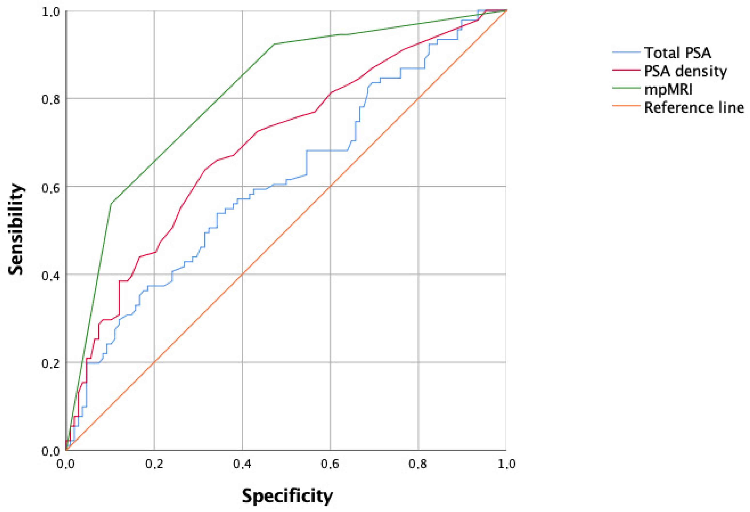
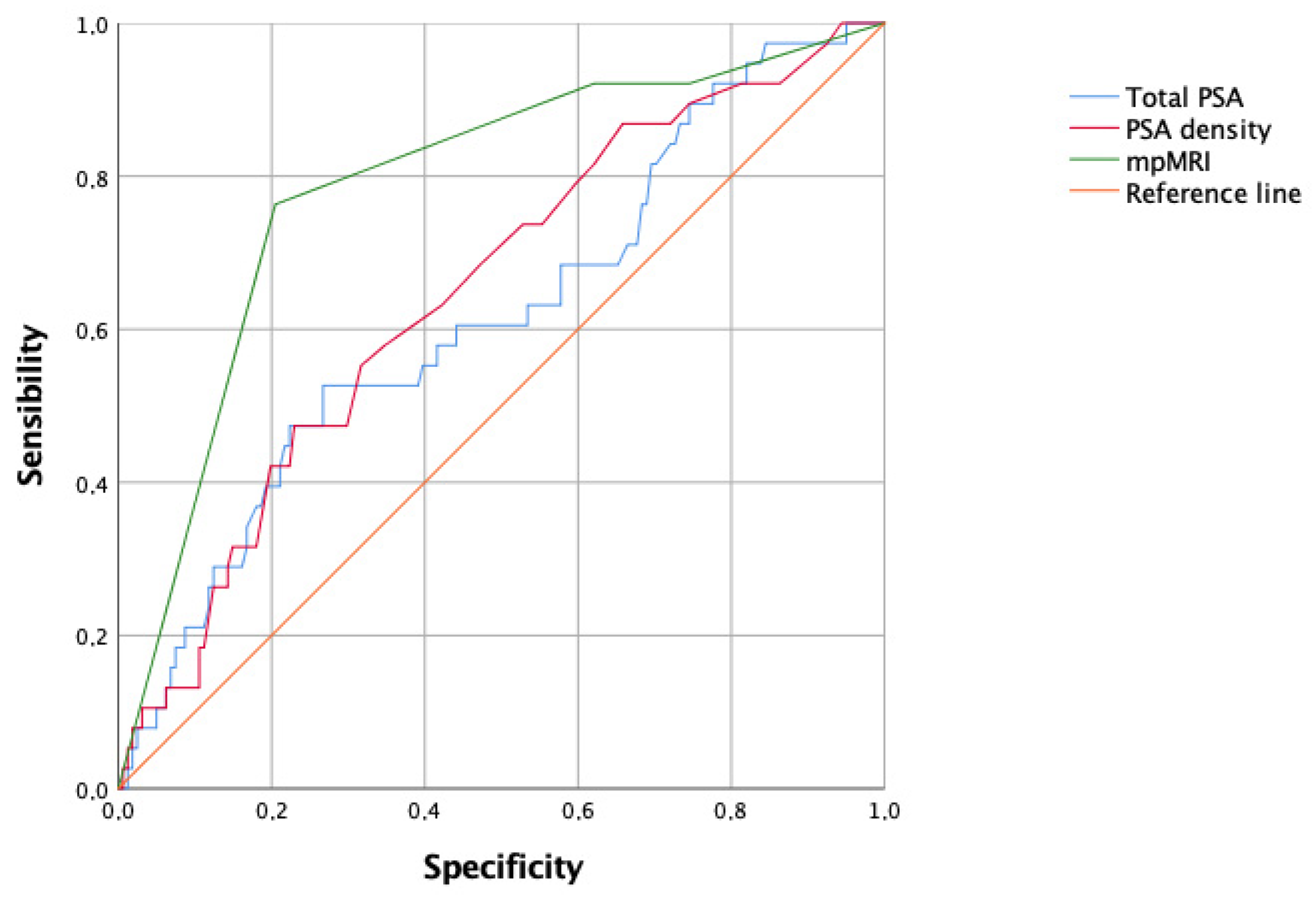
Frequencies and proportions were reported for categorical variables, whereas median and interquartile range (IQR) were reported for non-parametric continuous variables. We calculated the PCa detection rate of TBx, defined as the per-patient prevalence of csPCa and aggressive tumours in PI-RADS v2.1 ≥ 3 index lesions, of SBx and of combined TBx/SBx. The Mann–Whitney U-test and Pearson’s chi-square test were used to compare the continuous and categorical variables, respectively. An analysis of the receiver operating characteristic curve was performed to test the accuracy of mpMRI, PSA, and PSAD to detect csPCa and aggressive tumours. The corresponding areas under the curve (AUC) were compared using the DeLong method. Moreover, the sensitivity, specificity, negative predictive value (NPV), and PPV of mpMRI were calculated with 95%CI. The Cohen kappa statistic was used to measure the concordance between TBx and SBx. Specifically, values ≤ 0 indicated no agreement, 0.01–0.20 none to slight, 0.21–0.40 fair, 0.41–0.60 moderate, 0.61–0.80 substantial, and 0.81–1.00 almost perfect agreement. Moreover, in order to identify independent predictors of csPCa, we performed a binary logistic analysis including age, PSA, PSAD (cut-off 0.15 ng/mL/cc), and PI-RADS v2.1 categories in the multivariable model.
All clinical records were prospectively collected in a dedicated database by a dedicated data manager. Statistical analyses were performed using the SPSS version 23.0 (IBM Corp., Armonk, NY, USA) software. All of the reported p values were two-sided and statistical significance was set at p < 0.05.

3. Results

A total of 199 patients with DRE suspicious for PCa were included in the present analysis. Only 2 (1%) patients had a family history for PCa. The clinical characteristics are shown in Table 1. The median age was 68 (IQR 63–74) years and median PSA was 7.9 ng/mL (IQR 5.3–13.5). Seventy-two (36.2%) and 26 (13.1%) patients had a PSA > 10 ng/mL and >20 ng/mL, respectively. The median prostate volume was 50 (IQR 40–67) cc and the median PSAD was 0.16 (IQR 0.08–0.28) ng/mL/cc. Notably, 103 (51.8%) patients had a PSAD > 0.15 ng/mL/cc (Table 1). The median size of the index lesion suspicious for cancer was 14 mm (IQR 12–18 mm). PI-RADS v2.1 categories significantly correlated with PSA (p < 0.001), PSAD at the cut-off of 0.15 ng/mL/cc (p < 0.001) and the median size of the index lesion (p < 0.0001) (Table 2).
Combined TBx/SBx was performed in 153 (76.9%) patients with a PI-RADS v2.1 ≥ 3 index lesion. Conversely, SBx alone was performed in the remaining 46 (23.1%) patients with a negative mpMRI (PI-RADS v2.1 category 1 or 2).
Combined TBx/SBx detected 120 (60.3%) tumours overall. Median number of positive cores was 6 (IQR 4–9). According to ISUP Grade Group, 29 (24.2%) tumours were category 1, 18 (15%) category 2, 35 (29.2%) category 3, 29 (24.2%) category 4, and 9 (7.5%) category 5. Therefore, 29 (24.2%) tumours were clinically insignificant, 91 (75.8%) were csPCa, and 38 (31.7%) tumours were aggressive.
Table 3 summarizes the correlation between mpMRI findings and prostate biopsy outcomes. In detail, 36 (78.2%) patients with PI-RADS v2.1 categories 1 or 2, and 14 (77.8%) of those with PI-RADS v2.1 category 3 showed a negative SBx. Conversely, only 5 (10.9%) and 2 (11.1%) csPCa cases were detected in the subgroup of PI-RADS v2.1 1 or 2, and 3, respectively. Prostate biopsy could, thus, be theoretically avoided in 41/199 (20.6%) patients with PI-RADS v2.1 1 or 2 categories at the cost of missing 5/91 (5.5%) csPCa cases.
According to PSA cut-off of 10 ng/mL, 33 (71.7%) patients with PI-RADS categories 1 or 2 had a PSA ≤ 10 ng/mL and 13 (28.3%) had a PSA > 10 ng/mL. Missed csPCa cases were 3 (9%) in the former subgroup and 2 (15.4%) in the latter one, respectively (p = 0.53).
The accuracy of mpMRI to detect csPCa was 0.811 (95%CI 0.751–0.871), which was significantly higher compared with PSAD (AUC 0.690, 95%CI 0.620–0.767) (p = 0.01) and PSA (AUC 0.613, 95%CI 0.534–0.692) (p < 0.001) (Figure 1). The sensitivity, specificity, NPV and PPV of mpMRI were 94.5% (95%CI 87.6–98.1%), 37.9% (95%CI 28.8–47.8%), 89.1% (95% CI 77.1–95.2%), and 56.2% (95% 52.3–60%), respectively. Similarly, the accuracy of mpMRI to detect aggressive prostate cancers was 0.788 (95%CI 0.705–0.871), which was significantly higher than PSAD (AUC 0.648, 95%CI 0.552–0.740) (p = 0.03) and PSA (AUC 0.617, 95%CI 0.516–0.718) (p = 0.01) (Figure 2).
Multivariable analysis to predict csPCa showed that PI-RADS categories 4 (OR 6.288, 95%CI 2.154–18.354) and 5 (OR 29.515, 95%CI 9.158–95.128), and a PSAD cut-off of 0.15 ng/mL/cc (OR 2.173, 95%CI 1.068–4.423) were independent predictors (Table 4). According to the multivariable model, the combination of PSAD > 0.15 ng/mL/cc and PI-RADS categories 4 or 5 showed an accuracy of 0.815 (95%CI 0.756–0.87) in detecting csPCa.
Table 5 shows the detection rate of csPCa with TBx and SBx in the 153 patients with PI-RADS v2.1 ≥ 3 categories. The overall concordance between TBx and SBx for csPCa detection was substantial (k = 0.70). In detail, 63 (78.8%) patients with positive TBx also had positive SBx. Conversely, TBx identified 6 (8.2%) csPCa cases missed at SBx, and failed to detect 17 (21.3%) csPCa cases diagnosed with SBx alone. Notably, the concordance between TBx and SBx was moderate (k = 0.60) in the subgroup of patients with PSA ≤ 10 ng/mL, and almost perfect (k = 0.82) in the subgroup of patients with PSA > 10 ng/mL.
The sensibility, specificity, NPV, and PPV of TBx for csPCa were 78.7% (95%CI 68–87%), 91.7% (95%CI 82.9–96.9%), 79.7% (95%CI 71.9–85.8%), and 91.3% (95%CI 82.8–95.8%), respectively. The overall concordance between TBx and SBx for the detection of aggressive PCa was almost perfect (k = 0.86). Two (1.7%) aggressive PCa cases were detected with TBx alone, and 5 (15.2%) aggressive tumours were missed by TBx and detected by SBx alone (Table 6).

4. Discussion

The present study showed that 23% of mpMRI performed in patients with DRE suspicious for PCa were negative. Notably, in this subgroup of patients, SBx identified only 10.9% of csPCa, while 89.1% biopsies were negative or detected insignificant PCa. Although prostate biopsy in naïve patients with suspicious DRE should always be recommended, our data could be useful for patient counselling and could justify postponing biopsy in selected cases, provided careful monitoring is followed. Theoretically, the use of mpMRI in patients with DRE suspicious for cancer could avoid roughly 21% of unnecessary biopsies at the cost of missing approximately 6% of csPCa cases.
When considering patients with PI-RADS v2.1 ≥ 3 categories on mpMRI, the concordance between TBx and SBx in detecting csPCa was substantial, and it was almost perfect in the subgroup of patients with PSA > 10 ng/mL. Our study showed that TBx should be considered complementary to SBx in patients with suspicious DRE.
While in the pre-PSA era, DRE was the most widely used diagnostic test to detect PCa, the introduction of PSA testing significantly decreased the number of PCa diagnosed due to suspicious DRE alone [12]. In detail, in patients with PSA < 2 ng/mL, suspicious DRE showed a PPV of 5–30% [13]. However, in patients with elevated PSA, the PPV of positive DRE reached a value of 48.6% compared with 22.4% in those with negative DRE [14,15]. Moreover, suspicious DRE was shown to be an independent predictor of high-grade PCa [16,17]. Conversely, in 2018, a meta-analysis including 7 studies and 9241 patients reported a pooled PPV and NPV of 41% and 64%, respectively, thereby supporting no routine use of DRE for PCa detection [18].
DRE is still recommended by international guidelines, and palpable suspicious lesions represent a strong indication to perform prostate biopsy with or without previous mpMRI [2]. A recent population-based PCa screening study reported a 10.9% of men with suspicious DRE estimating a sensitivity of 28% and specificity of 93% for predicting csPCa among men with PSA > 3 ng/mL [19].
Only a few studies investigated the utility of mpMRI and TBx in the setting of biopsy-naïve patients with suspicious DRE. In a recent systematic review investigating the PPV of PI-RADS criteria to detect csPCa, Mazzone et al. reported that only 50% of selected studies reported DRE status, and palpable lesions were reported in approximately 16% of cases. However, the authors did not report subgroup analysis according to DRE status [3].
In 2019, Morote et al. analyzed a series of 768 patients who underwent TBx for suspicious lesions PI-RADS > 1 combined with 12-core SBx. The authors concluded that mpMRI was not beneficial to men with PSA ≥ 10 ng/mL and suspicious DRE. In this subgroup of 51 patients, mpMRI could avoid only 2.5% of unnecessary initial biopsies and no repeat biopsy. Moreover, TBx did not increase the number of csPCa already detected by SBx [7]. In our study including naïve-biopsy patients, the use of mpMRI could avoid 20% of unnecessary biopsies. However, our data confirmed that in patients with PSA > 10 ng/mL and suspicious DRE, the use of mpMRI and TBx did not offer any advantage in detecting csPCa or aggressive tumours compared with SBx.
In 2021, Omri et al., analyzing a cohort of 91 patients, showed a significantly higher detection rate of csPCa with TBx compared with SBx in patients with both normal and suspicious DRE. The authors highlighted that the contribution of mpMRI and TBx was more pronounced in patients with negative DRE compared with those with suspicious DRE. However, they supported the combined use of TBx and SBx in patients with suspicious DRE [8]. Our data in patients with suspicious DRE apparently support this strategy, especially if PSA is ≤10 ng/mL.
Several diagnostic studies reporting on DRE status did not clarify the definition of suspicious DRE. Moreover, the accuracy of DRE might be strongly influenced by the operator’s experience. Therefore, the strict definition of suspicious DRE and the inclusion of only cases evaluated by a single expert urologist should be considered as a strength of our study. Moreover, mpMRI image analysis was performed by a single expert radiologist. Notably, in order to reflect the routine clinical practice and to increase the generalizability of the findings of the present study, mpMRI images and biopsy specimens were not re-reviewed. Moreover, the use of transperineal prostate biopsy may represent a further strength of the present study considering literature data and international guidelines supporting the use of transperineal over transrectal route [2].
A potential limitation of the present study could be the use of cognitive instead of fusion TBx. However, in a systematic review, Wegelin et al. showed no difference between fusion and cognitive TBx in PCa detection rate [20]. In 2018, Monda et al., analyzing a series of 510 patients, showed no significant difference between the diagnostic ability of cognitive and fusion TBx for detecting csPCa [21]. Similar results were reported by Turkay et al. in 2020 in a study comparing men receiving fusion or cognitive TBx [22]. Moreover, Hayes et al. observed similar PCa detection rates between fusion and cognitive TBx, except for anteriorly located tumours [23]. Conversely, Yamada et al. reported the detection superiority of software-assisted MRI/ultrasound fusion TBx compared with cognitive TBx, above all in smaller lesions [24]. However, software-assisted TBx is likely to be more expensive compared with cognitive sampling [25]. Additionally, in our study, the number of targeted cores was not standardized and ranged between 1–3 according to the lesion size and operator’s choice. Other potential limitations include the retrospective and single-centre design and the relatively small sample size potentially influencing the comparison between different subgroups.

5. Conclusions

Negative mpMRI (PI-RADS v2.1 categories 1 or 2) in patients with DRE suspicious for cancer could be associated with a high percentage of negative prostate biopsy or detection of insignificant PCa. Theoretically, performing mpMRI in patients with suspicious DRE could avoid roughly 21% of unnecessary biopsies at the cost of missing approximately 6% of csPCa cases.
TBx for PI-RADS v2.1 ≥ 3 categories had the same yield as SBx. Moreover, the concordance between TBx and SBx was almost perfect in the subgroups of patients with PSA > 10 ng/mL. Therefore, based on our findings, mpMRI and TBx complement SBx in detecting csPCa in patients with suspicious DRE and PSA > 10 ng/mL.

Author Contributions

Conceptualization, V.F.; methodology, V.F.; software, V.F. and A.A.; formal analysis, V.F. and A.A.; investigation, A.B. (Alessandro Buttitta), M.R., G.A., A.B. (Alfredo Blandino), A.I. and M.M.; data curation, A.B. (Alessandro Buttitta), M.R., G.A., A.B. (Alfredo Blandino), A.I. and M.M.; writing—original draft preparation, V.F.; writing—review and editing, G.G.; visualization, G.G.; supervision, V.F. All authors have read and agreed to the published version of the manuscript.

Funding

This research received no external funding.

Institutional Review Board Statement

The study was conducted in accordance with the Declaration of Helsinki, and approved by the Institutional Review Board of the University Hospital of Messina.

Informed Consent Statement

Informed consent was obtained from all subjects involved in the study.

Data Availability Statement

Data is publicly unavailable due to privacy restrictions.

Conflicts of Interest

The authors declare no competing interests.

References

  1. Van Poppel, H.; Hogenhout, R.; Albers, P.; van den Bergh, R.C.; Barentsz, J.O.; Roobol, M.J. A European Model for an Organised Risk-stratified Early Detection Programme for Prostate Cancer. Eur. Urol. Oncol. 2021, 4, 731–739. [Google Scholar] [CrossRef]
  2. Mottet, N.; Cornford, P.; van den Bergh, R.C.N.; Briers, E.; Eberli, D.; De Meerleer, G.; De Santis, M.; Gillessen, S.; Grummet, J.; Henry, A.M.; et al. EAU Guidelines on Prostate Cancer 2023. Available online: www.uroweb.org (accessed on 27 August 2023).
  3. Mazzone, E.; Stabile, A.; Pellegrino, F.; Basile, G.; Cignoli, D.; Cirulli, G.O.; Sorce, G.; Barletta, F.; Scuderi, S.; Bravi, C.A.; et al. Positive predictive value of prostate imaging reporting and data system version 2 for the detection of clinically significant prostate cancer: A systematic review and meta-analysis. Eur. Urol. Oncol. 2021, 4, 697–713. [Google Scholar] [CrossRef]
  4. Morote, J.; Borque-Fernando, A.; Triquell, M.; Campistol, M.; Celma, A.; Regis, L.; Abascal, J.M.; Servian, P.; Planas, J.; Mendez, O.; et al. A clinically significant prostate cancer predictive model using digital rectal examination prostate volume category to stratify initial prostate cancer suspicious and reduce magnetic resonance imaging demand. Cancers 2022, 14, 5100. [Google Scholar] [CrossRef]
  5. Ahmed, H.U.; El-Shater Bosaily, A.; Brown, L.C.; Gabe, R.; Kaplan, R.; Parmar, M.K.; Collaco-Moraes, Y.; Ward, K.; Hindley, R.G.; Freeman, A.; et al. Diagnostic accuracy of multi-parametric MRI and TRUS biopsy in prostate cancer (PROMIS): A paired validating confirmatory study. Lancet 2017, 389, 815–822. [Google Scholar] [CrossRef]
  6. Porpiglia, F.; Manfredi, M.; Mele, F.; Cossu, M.; Bollito, E.; Veltri, A.; Cirillo, S.; Regge, D.; Faletti, R.; Passera, R.; et al. Diagnostic pathway with multiparametric magnetic resonance imaging versus standard pathway: Results from a randomized prospective study in biopsy-naïve patients with suspected prostate cancer. Eur. Urol. 2017, 72, 282–288. [Google Scholar] [CrossRef]
  7. Morote, J.; Celma, A.; Roche, S.; de Torres, I.M.; Mast, R.; Semedey, M.E.; Regis, L.; Planas, J. Who benefits from multiparametric magnetic resonance imaging after suspicion of prostate cancer? Eur. Urol. Oncol. 2019, 2, 664–669. [Google Scholar] [CrossRef]
  8. Omri, N.; Alex, S.; Jacob, B.; Ofer, N. The additive value of mpMRI on prostate cancer detection: Comparison between patients with and without a suspicious digital rectal examination. Urol. Oncol. 2021, 39, 728.e7–728.e11. [Google Scholar] [CrossRef]
  9. Turkbey, B.; Brown, A.M.; Sankineni, S.; Wood, B.J.; Pinto, P.A.; Choyke, P.L. Multiparametric prostate magnetic resonance imaging in the evaluation of prostate cancer. CA Cancer J. Clin. 2016, 66, 326–636. [Google Scholar] [CrossRef]
  10. Ficarra, V.; Novella, G.; Novara, G.; Galfano, A.; Pea, M.; Martignoni, G.; Artibani, W. The potential impact of prostate volume in the planning of optimal number of cores in the systematic transperineal prostate biopsy. Eur. Urol. 2005, 48, 932–937. [Google Scholar] [CrossRef]
  11. Epstein, J.I.; Allsbrook, W.C., Jr.; Amin, M.B.; Egevad, L.L.; ISUP Grading Committee. The 2014 International Society of Urological Pathology (ISUP) Consensus Conference on Gleason Grading of Prostate Cancrinoma: Definition of grading patients and proposal for a new grading system. Am. J. Surg. Pathol. 2016, 40, 244–252. [Google Scholar] [CrossRef]
  12. Carroll, P.; Coley, C.; McLeod, D.; Schellhammer, P.; Sweat, G.; Wasson, J.; Zietman, A.; Thompson, I. Prostate-specific antigen best practice policy-part 1: Early detection and diagnosis of prostate cancer. Urology 2001, 57, 217–224. [Google Scholar] [CrossRef]
  13. Carvalhal, G.F.; Smith, D.S.; Mager, D.E.; Ramos, C.; Catalona, W.J. Digital rectal examination for detecting prostate cancer at prostate specific antigen levels of 4 ng/ml or less. J. Urol. 1999, 161, 835–839. [Google Scholar] [CrossRef]
  14. Okotie, O.T.; Roehl, K.A.; Han, M.; Loeb, S.; Gashti, S.N.; Catalona, W.J. Characteristics of prostate cancer detected by digital rectal examination only. Urology 2007, 70, 1117–1120. [Google Scholar] [CrossRef]
  15. Gosselaar, C.; Roobol, M.J.; Roemeling, S.; Schröder, F.H. The role of digital rectal examination in subsequent visits in the European randomized study of screening for prostate cancer (ERSPC). Eur. Urol. 2008, 54, 581–588. [Google Scholar] [CrossRef]
  16. Borden, L.S.; Wright, J.L.; Kim, J.; Latchamsetty, K.; Porter, C.R. An abnormal digital rectal examination is an independent predictor of Gleason 7 prostate cancer in men undergoing initial prostate biopsy: A prospective study of 790 men. BJU Int. 2007, 99, 559–563. [Google Scholar] [CrossRef]
  17. Thompson, I.M.; Ankerst, D.P.; Chi, C.; Goodman, P.J.; Tangen, C.M.; Lucia, M.S.; Feng, Z.; Parnes, H.L.; Coltman, C.A., Jr. Assessing prostate cancer risk: Results from the Prostate Cancer Prevention Trial. J. Natl. Cancer Inst. 2006, 98, 529–534. [Google Scholar] [CrossRef]
  18. Naji, L.; Randhawa, H.; Sohani, Z.; Dennis, B.; Lautenbach, D.; Kavanagh, O.; Bawor, M.; Banfield, L.; Profetto, J. Digital rectal examination for prostate cancer screening in primary care: A systematic review and meta-analysis. Ann. Fam. Med. 2018, 16, 149–154. [Google Scholar] [CrossRef]
  19. Andersson, J.; Palsdottir, T.; Lantz, A.; Aly, M.; Grönberg, H.; Egevad, L.; Eklund, M.; Nordström, T. Digital rectal examination in Stockholm3 biomarker-based prostate cancer screening. Eur. Urol. Open Sci. 2022, 44, 69–75. [Google Scholar] [CrossRef]
  20. Wegelin, O.; van Melick, H.H.E.; Hooft, L.; Bosch, J.R.; Reitsma, H.B.; Barentsz, J.O.; Somford, D.M. Comparing Three Different Techniques for Magnetic Resonance Imaging-targeted Prostate Biopsies: A Systematic Review of In-bore versus Magnetic Resonance Imaging-transrectal Ultrasound fusion versus Cognitive Registration. Is There a Preferred Technique? Eur. Urol. 2017, 71, 517–531. [Google Scholar] [CrossRef]
  21. Monda, S.M.; Vetter, J.M.; Andriole, G.L.; Fowler, K.J.; Shetty, A.S.; Weese, J.R.; Kim, E.H. Cognitive Versus Software Fusion for MRI-targeted Biopsy: Experience Before and After Implementation of Fusion. Urology 2018, 119, 115–120. [Google Scholar] [CrossRef]
  22. Turkay, R.; Inci, E.; Yildiz, O.; Ozgur, E.; Taşci, A. Cognitive Versus Magnetic Resonance-Ultrasound Fusion Prostate Biopsy: Which One Is Worthier to Perform? Ultrasound Q 2020, 36, 345–349. [Google Scholar] [CrossRef]
  23. Hayes, M.; Bassale, S.; Chakiryan, N.H.; Boileau, L.; Grassauer, J.; Wagner, M.; Foster, B.; Coakley, F.; Isharwal, S.; Amling, C.L.; et al. Selecting patients for magnetic resonance imaging cognitive versus ultrasound fusion biopsy of the prostate: A within-patient comparison. BJU Compass 2022, 3, 443–449. [Google Scholar] [CrossRef]
  24. Yamada, Y.; Shiraishi, T.; Ueno, A.; Ueda, T.; Fujihara, A.; Naitoh, Y.; Hongo, F.; Ukimura, O. Magnetic resonance imaging-guided targeted prostate biopsy: Comparison between computer-software-based fusion versus cognitive fusion technique in biopsy-naïve patients. Int. J. Urol. 2020, 27, 67–71. [Google Scholar] [CrossRef]
  25. Marra, G.; Ploussard, G.; Futterer, J.; Valerio, M.; EAU-YAU Prostate Cancer Working Party. Controversies in MR targeted biopsy: Alone or combined, cognitive versus software-based fusion, transrectal versus transperineal approach? World J. Urol. 2019, 37, 277–287. [Google Scholar] [CrossRef]
Figure 1. Accuracy of PSA, PSA density and mpMRI to predict clinically significant prostate cancer (overall cohort). mpMRI = multiparametric MRI.
Siuj 05 00021 g001
Figure 2. Accuracy of PSA, PSA density and mpMRI to predict aggressive prostate cancer (overall cohort). mpMRI = multiparametric MRI.
Siuj 05 00021 g002
Table 1. Clinical characteristics of the 199 patients with digital rectal examination suspicious for cancer included in the present analysis.
VariableValue
Median (IQR) age (years)68 (63–74)
Median (IQR) serum PSA level (ng/mL)7.9 (5.3–13.5)
PSA categories, n (%)
-
≤10 ng/mL
-
10.1–20 ng/mL
-
>20 ng/mL

127 (63.8%)
46 (23.1%)
26 (13.1%)
Median (IQR) prostate volume (cc)50 (40–67)
Median (IQR) PSAD (ng/mL/cc)0.16 (0.08–0.28)
PSAD categories, n (%)
-
≤0.15 ng/mL/cc
-
>0.15 ng/mL/cc

96 (48.2%)
103 (51.8%)
PI-RADS v2.1 categories, n (%)
-
1–2
-
3
-
4
-
5

46 (23.1%)
18 (9%)
73 (36.7%)
62 (31.2%)
IQR = interquartile range; PI-RADS = Prostate Imaging—Reporting and Data System; PSAD = PSA density.
Table 2. PI-RADS v2.1 categories stratified according to PSA, PSAD with a cut-off of 0.15 ng/mL/cc, and the median size of the index lesion.
PSA (ng/mL) PSAD (ng/mL/cc) Size of the Index Lesion (mm)
PI-RADS v2.1 categories≤1010.1–20>20p value≤0.15>0.15p valueMedian (IQR)p value
1–2
(n = 46)
33
(71.7%)
9
(19.6%)
4
(8.7%)
<0.00129
(63%)
17
(37%)
0.002N.A.<0.001
3
(n = 18)
11
(61.1%)
6
(33.3%)
1
(5.6%)
11
(61.1%)
7
(38.9%)
8 (7–12)
4
(n = 73)
55
(75.3%)
14
(19.2%)
4
(5.5%)
38
(52.1%)
35
(47.9%)
12 (10–15)
5
(n = 62)
28
(45.2%)
17
(27.4%)
17
(27.4%)
18
(29%)
44
(71%)
17 (14–24)
Total
(n = 199)
12746269610314 (12–18)
IQR = interquartile range; PI-RADS = Prostate Imaging—Reporting and Data System; PSAD = PSA density.
Table 3. Correlation between PI-RADS v2.1 categories and prostate biopsy outcomes.
PI-RADS v2.1 CategoriesProstate Biopsy Outcomes
Negative (%)Positive (Any Cancer) (%)csPCa (%)Aggressive PCa (%)
1–2 (n = 46)36 (78.2%)10 (21.7%)5 (10.9%)3 (6.5%)
3 (n = 18)14 (77.8%)4 (22.2%)2 (11.1%)0
4 (n = 73)25 (34.2%)48 (65.8%)33 (45.2%)6 (8.2%)
5 (n = 62)4 (6%)58 (93.5%)51 (82.3%)29 (46.8%)
Total (n = 199)79 (39.7%)120 (60.3%)91 (45.7%)38 (19.1%)
p value <0.001<0.001<0.001
csPCa = clinically significant prostate cancer; PI-RADS = Prostate Imaging—Reporting and Data System; PCa = prostate cancer.
Table 4. Multivariable analysis predicting csPCa in the 199 patients with suspicious digital rectal examination.
VariablesCategoriesOR95%CIp Value
Agecontinuous1.0160.975–1.0590.45
PSAcontinuous0.9990.992–1.0070.87
PSAD<0.15 ng/mL/cc
≥0.15 ng/mL/cc
Referent
2.173

1.068–4.423
0.03
PI-RADS v2.1 categories1–2
3
4
5
Referent
0.861
6.288
29.515

0.148–5.009
2.154–18.354
9.158–95.128
<0.001
CI = confidence interval; csPCa = clinically significant prostate cancer; OR = odds ratio; PI-RADS = Prostate Imaging–Reporting and Data System; PSAD = PSA density.
Table 5. Clinically significant PCa detected in patients with PI-RADS v2.1 ≥ 3 categories who underwent both TBx and SBx.
SBxTotal
All cases
(n = 153)
TBxNegative or insignificant PCaClinically significant PCaTotal
Negative or insignificant PCa67
(91.8%)
17
(21.3%)
84
(54.9%)
Clinically significant PCa6
(8.2%)
63
(78.7%)
69
(45.1%)
Total73
(100%)
80
(100%)
153
SBxTotal
Patients with PSA ≤ 10 ng/mL
(n = 94)
TBxNegative or insignificant PCaClinically significant PCaTotal
Negative or insignificant PCa46
(90.2%)
13
(30.2%)
59
(62.8%)
Clinically significant PCa5
(9.8%)
30
(69.8%)
35
(37.2%)
Total25
(100%)
43
(100%)
94
SBxTotal
Patients with PSA > 10 ng/mL
(n = 59)
TBxNegative or insignificant PCaClinically significant PCaTotal
Negative or insignificant PCa21
(95.5%)
4
(10.8%)
25
(42.4%)
Clinically significant PCa1
(4.5%)
33
(91.2%)
34
(57.4%)
Total22
(100%)
37
(100%)
59
PCa = prostate cancer; PI-RADS = Prostate Imaging—Reporting and Data System; SBx = systematic biopsy; TBx = targeted biopsy.
Table 6. Aggressive PCa detected in the 153 patients with PI-RADS v2.1 ≥ 3 categories who underwent both TBx and SBx.
SBxTotal
All cases (n = 153)TBxNegative or non-aggressive PCaAggressive PCaTotal
Negative or non-aggressive PCa118
(98.3%)
5
(15.2%)
123
(80.4%)
Aggressive PCa2
(1.7%)
28
(84.8%)
30
(19.6%)
Total120
(100%)
33
(100%)
153
PCa = prostate cancer; PI-RADS = Prostate Imaging—Reporting and Data System; SBx = systematic biopsy; TBx = targeted biopsy.
Disclaimer/Publisher’s Note: The statements, opinions and data contained in all publications are solely those of the individual author(s) and contributor(s) and not of MDPI and/or the editor(s). MDPI and/or the editor(s) disclaim responsibility for any injury to people or property resulting from any ideas, methods, instructions or products referred to in the content.

