Review

The Promising Potential of Cold Atmospheric Plasma Therapies

Department of Materials Chemistry, Adsorption and Catalysis, Faculty of Chemistry, Nicolaus Copernicus University in Toruń, Gagarina 7, 87-100 Toruń, Poland
*
Author to whom correspondence should be addressed.
Plasma 2024, 7(2), 465-497; https://doi.org/10.3390/plasma7020025
Submission received: 1 March 2024 / Revised: 29 April 2024 / Accepted: 8 June 2024 / Published: 12 June 2024

Abstract

:
The outstanding properties and chemistry of cold atmospheric plasma (CAP) are not sufficiently understood due to their relatively complex systems and transient properties. In this paper, we tried to present a detailed review of the applications of CAP in modern medicine, highlighting the biochemistry of this phenomenon. Due to its unique characteristics, CAP has emerged as a promising tool in various medical applications. CAP, as a partially—or fully ionized—gas-retaining state of quasi-neutrality, contains many particles, such as electrons, charged atoms, and molecules displaying collective behaviour caused by Coulomb interactions. CAP can be generated at atmospheric pressure, making it suitable for medical settings. Cold plasma’s anti-microbial properties create an alternative method to antibiotics when treating infections. It also enhances cell proliferation, migration, and differentiation, leading to accelerated tissue regeneration. CAP can also be a powerful tool in anti-tumour therapies, stem cell proliferation, dental applications, and disease treatment, e.g., neurology. It is our belief that this article contributes to the deeper understanding of cold plasma therapy and its potential in medicine. The objective of this study is to demonstrate the potential of this relatively novel approach as a promising treatment modality. By covering a range of various biomedical fields, we hope to provide a comprehensive overview of CAP applications for multiple medical conditions. In order to gain further insight into the subject, we attempted to gather crucial research and evidence from various studies, hopefully creating a compelling argument in favour of CAP therapy. Our aim is to highlight the innovative aspects of CAP therapy where traditional methods may have limitations. Through this article, we intend to provide a convenient reference source for readers engaged in the examination of CAP’s potential in medicine.

1. Introduction

1.1. Establishment of the Subject Matter

Plasma, often referred to as the fourth as well as the most common state of matter in the Universe, is an ionized gas with the ability to conduct electrical charges [1] A distinction can be made between high-temperature plasma and low-temperature plasma, known as ‘cold’ plasma or non-thermal plasma (NTP), or cold atmospheric plasma (CAP). The first type is one component of stars, and it is produced when hydrogen bombs explode. The second type can be produced even at room temperature.
Cold plasma is a mixture of both ionized and non-ionized molecules or atoms in the ground and excited state, electrons, as well as free radicals, including reactive oxygen species (ROS) and reactive nitrogen species (RNS), ozone, and UV radiation [2]. Despite the presence of particles with different charges, the plasma retains quasi-neutrality, i.e., the sum of the positive and negative charges of these particles is zero. There may be local areas with a predominance of one type of charge, but such collective plasma behaviour occurs with long-range interactions between its different areas [3].
On Earth, under normal conditions, plasma is considered a rather rare phenomenon. On the contrary, most of the matter in the Universe is ionized and therefore occurs in a plasma state (about 99.9% of the matter) [4]. Such a state is produced by various mechanisms. In stars, neutral atoms are ionized due to high temperatures. Interstellar gases are ionized by UV radiation [5]. Plasmas that can be produced in nature as well as in the laboratory can be characterized by a wide range of temperatures and pressures. They can be classified using Debye lengths and frequencies, which depend purely on the number of electrons, their density, and temperature [6].
As mentioned, plasma is broadly divided, depending on its thermal properties, into two categories. When the generated constituents appear to be in the state of thermal equilibrium, i.e., electron temperature (Te) is equal to ion temperature (Ti), it is said to be a hot or thermal plasma (created at higher temperature and under higher gas pressure conditions [7]). In plasma considered to be cold, a significant disparity between the temperature of the electrons and that of other species occurs [7]. This type of plasma can be created easily even when temperature and pressure are at normal levels—as the name suggests, close to room temperature. The phenomena of cold plasmas stem from their unique chemical and physical properties [1]. Over the years, a wide range of uses for cold plasmas has been reported as follows: in industry, where they can be used for cleaning, surface modification, polymer material preparation and material processing; in medicine, where they are successful in sterilizing equipment and/or surfaces, healing wounds, promoting stem cell proliferation, tissue or cell removal with surgical precision, and treating cancer cells [8].
Numerous discoveries and inventions have led to the creation of a new field of science referred to as plasma medicine, which is considered a relatively new and rapidly growing area combining plasma physics, engineering and bioengineering as well as medicine [2]. The remarkable nature of CAP offers a minimally invasive surgery, allowing for targeting specific cells, i.e., cancer cells. The interaction of CAP with the treated tissue thus allows specific removal of cells without the risk of inducing necrosis [9]. In general, effects obtained by using CAP methods include cell detachment without affecting cell viability, modifying the processes of controlled cell death or migration, etc. [10]. Low-temperature plasma can initiate chemical reactions that will produce the desired effect mostly through free radicals’ synthesis. The overall process can be modified and controlled by adjusting used pressure, temperature, gas composition, gas flow rate, and power applied to create a discharge [11].
Cold plasmas have proved to be a worthy candidate for innovative cancer treatment methods [12]. Therefore, a proper understanding of the interaction between CAP and cells or tissues is necessary. Knowledge of chemicals that are created during plasma treatment and their impact on living organisms has become obligatory. Formed thus, these compounds may induce various effects depending on their concentration or the environment they are exposed to [13]. For instance, reactive oxygen species (ROS) may cause a ‘plasma killing effect’, meaning they are lethal for cells, creating cytotoxicity [14]. In contrast, other types of free radicals, such as reactive nitrogen species (RNS), may induce a more positive or even desired ‘healing plasma effect’ [15].
To better grasp the complex of plasma chemistry to advance and expand cold plasma uses in oncology, it is mandatory to get to the bottom of the science behind CAP cancer treatment and its potential for future clinical application. When focusing on CAP anti-cancer efficacy, it is best to explore the recent developments as well as difficulties encountered when applying this technique. Plasmas used in oncology are usually created with the help of devices such as dielectric barrier discharges, plasma jets, corona discharges, glow discharges, etc.; sometimes, a mix of the tools mentioned is used [4].
Over the preceding few decades, countless investigations into the many forms and possibilities of CAP applications have proven to have a rapid evolutionary potential and have shown promise for new and improved transdisciplinary industries [16]. Such an advanced step forward might completely restructure the health industry as we know it [4]. Unfortunately, the unusual chemical and physical properties of cold plasmas, along with the complexities of their interactions when exposed to living tissue, sadly remain shrouded in mystery. Both directing and enhancing the generation and formation of many chemical reactions present during plasma treatment are challenges to consider for the developments in CAP chemistry and their applications for biochemistry and medicine [17,18].
Having various types and combinations of formed reactive species is fundamental when proving their vast potential for activating specific cell signalling pathways. CAP treatment can be distinguished by its powerful cytotoxic properties towards tumour cells, proven both in vitro and in vivo [19,20,21,22]. It has been shown in the literature that normally developing and healthy cells are much less sensitive to the same cold plasma treatment compared to the cells displaying tumorigenic potential [9,21,23,24]. In other words, CAP treatment offers a great possibility of specifically targeting cancer cells without the risk of damaging neighbouring healthy cells.
The aforementioned effects are considered to be deeply dependent on cold plasma chemistry [25]. The presence of various free radicals, their concentration, and their type (long-lived or short-lived species) play crucial roles in CAP anti-cancer properties. Other plasma elements, such as ions, electrons, all kinds of radiation, and electric fields, are relegated to a secondary role.
What is also worth mentioning is an additional unique feature of the fourth state of matter; specifically, it is its ability to self-organize which causes it to form coherent structures [26]. Said structures possess the unique ability to instantaneously modulate the charged particles, electric field, and free radicals such as ROS/RNS. As a result of self-induced organization, cold plasma may adapt to specific cells by simply adjusting to their in situ composition. This remarkable feature may be the reason for highly selective internal interactions between plasma and cells [15,27].
Among the strategies mentioned that are used for producing cold plasma under atmospheric pressure, it can be easily noted that two primary aspects of CAP devices dominate clinical as well as preclinical exploration in cold plasma therapies (Figure 1). Those aspects are covered by plasma jets and dielectric barrier discharges (DBDs [28]). DBDs can be distinguished by igniting plasma in a void between an insulated HV (high-voltage) electrode and the subject; this type is called a volume DBD. The other contains an individualized electrode structure approach (e.g., grid-like or circular) that is insulated from a counter electrode, called surface DBDs [29]. The operating gas used for cold plasma generation in DBDs varies from ambient air and its singular components to noble gases or mixtures [30]. Used gas has a great impact on the plasma’s properties and abilities shown during treatment. Even though cold plasmas created under atmospheric conditions have been useful in lowering a microbiological load, they lack the powerful sterilization and disinfecting characteristics that can be obtained when using low-pressure plasmas [31]. Fortunately, milder effects displayed by CAP allow for safer immediate administration to cells and tissues. When it comes to lower microbial strain, the abilities of atmospheric cold plasma make it a plausible candidate for replacing antibiotics or even combating antibiotic-resistant strains of bacteria [32].
Furthermore, CAPs are known to preserve their virucidal, antifungal, antibacterial, and even sporicidal activity [33,34]. Another benefit of CAP-generating systems worth mentioning is their inexpensive production rates. Affordable and effective CAP devices will certainly lessen the financial burden on the healthcare budget caused by conventional cancer treatment methods [17,35].

1.2. Limitations and Current Problems in Plasma Medicine

Although CAP emerges as promising asset of modern medicine, this method is still subjected to certain limitations. In the table below, there are some current issues revolving around the use of cold plasma medicine in oncology and idealized solutions, which may inspire the reader to search for new ways of method improvement (Table 1).

2. CAP Generation Techniques

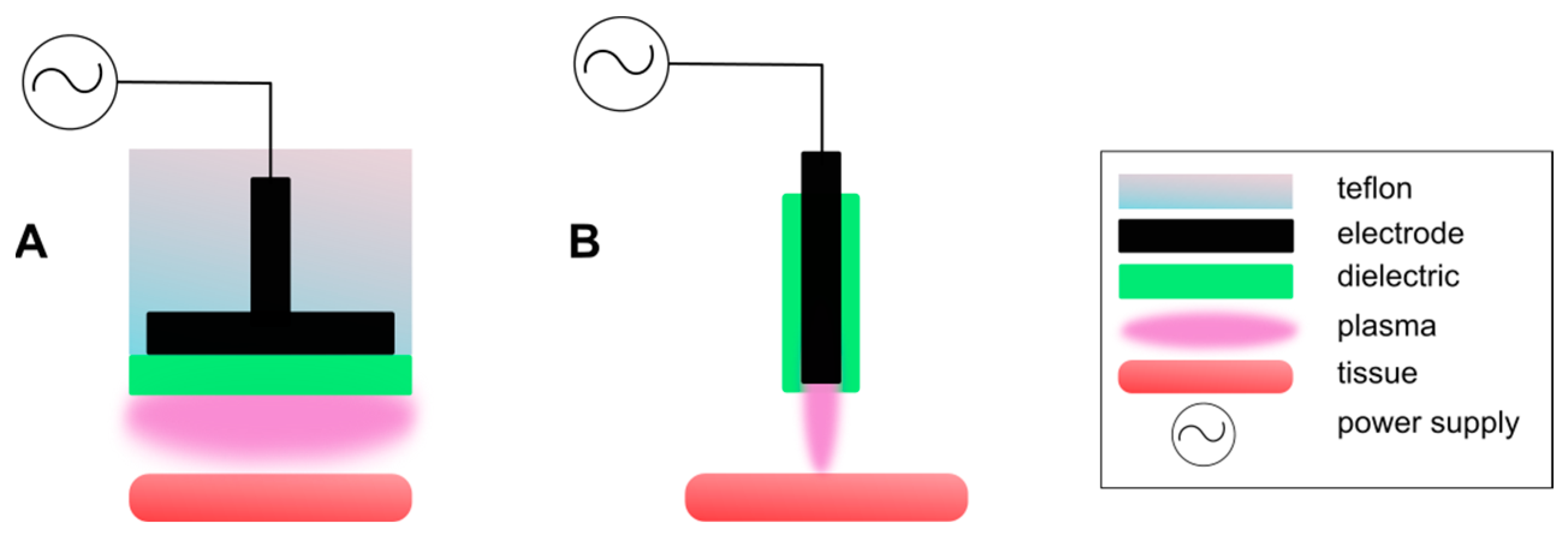
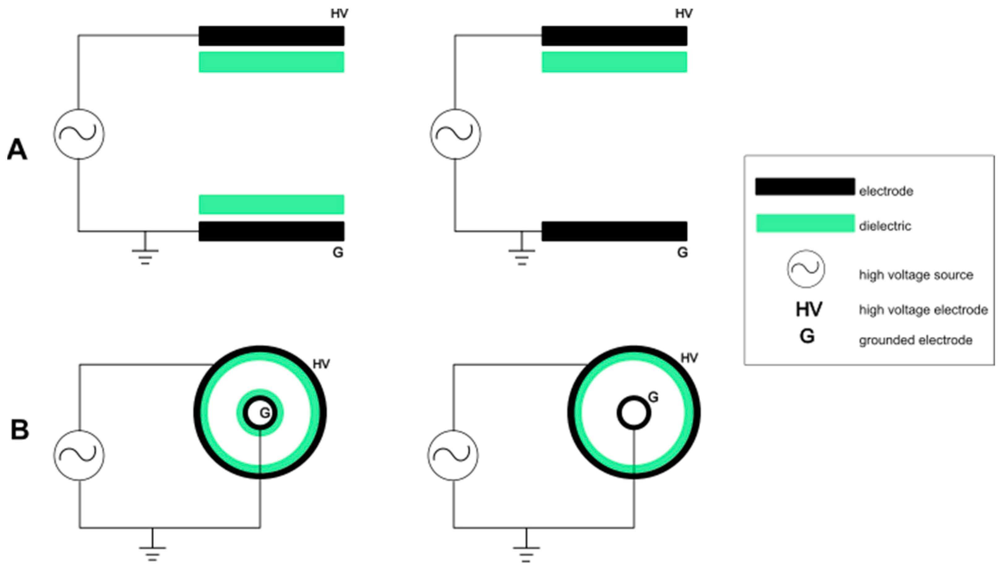
2.1. Dielectric Barrier Discharge

Dielectric Barrier Discharge (DBD), known for its ability to create an alternating or pulsing electric field, is a popular choice when looking for a CAP generation method (Figure 1 and Figure 2 [44,45,46,47]). One of the two electrodes must be covered by a material displaying dielectric properties in order to create any discharge [48]. The main role of the mentioned dielectric layer is reducing current as well as preventing the spark or arc transition. The DBD method is known for generating a “silent” discharge because no noise can be heard during the process [49].
Usually, the electrode gap oscillates between 0.1 mm and several centimetres long [48]. Various materials can be used to create a dielectric layer, i.e., polymers, glass, ceramics, quartz, etc. To prevent spark or arc transitions, proper discharge current insulation must be ensured [48,50]. The material used when creating a dielectric layer must feature a high enough breakdown strength. It can be achieved simply by enhancing the layer thickness. Unfortunately, a thicker layer requires a larger voltage input. In most cases, a simple solution is to build a chamber around the electrode setup. This creates an opportunity to introduce various operating gas mixtures in the working space between the electrodes and thus manipulate the properties of the created cold plasma. The DBD method can and often is used when operating at kHz in industrial settings, driven by high-voltage sources [51]. This can also be used when driven by lower voltage sources, enabling safe application for tissues or cells. To put it concisely, DBD devices may be modified as pleased and set up in various ways (flat, parallel, or coaxial plates), but the fundamental idea remains unchanged.
An interesting modification of classic DBDs was made by Elaissi et al. [52], called DBD with floating electrodes or FE-DBD for short. Just like with the classic DBD, floating DBD also has two electrodes. The first one is an active as well as a high-voltage insulated electrode. The second one, being the main difference between typical DBDs and FE-DBD, is not grounded at all (Figure 2); it is floating, just as the name suggests. Similar to the classic DBD reactors, the anticancer effect generated by the FE-DBD plasma is mediated by reactive species generated by the cold atmospheric plasma but can be improved by higher electron density and lower energy consumption. The results show that pulse width has a profound effect on plasma during cancer treatment, and the optimal pulse width used for power is approximately 1 µs. Moreover, the authors recommend a small distance between the electrodes, a higher dielectric constant, and a small dielectric thickness to obtain a higher value of electron density responsible for generating reactive species initiating apoptosis of cancer cells.
When working at ambient pressure, created discharge often turns out to be inconsistent, which may unfortunately lead to uneven sample treatment. The discharge’s visual appearance is heavily correlated with dynamic dispersion. Depending on the given circumstances, DBDs may generate various types of plasma when speaking about their visual appearance. However diffused or homogenous plasma is far more possible to obtain using this method than filamentary plasma [53]. Many research groups over the years have successfully generated diffused, homogeneous plasmas when using atmospheric pressure [28,29,30,51,53]. Instead of a streamer breakdown, a Townsend breakdown is started to create a glow DBD. When creating an avalanche, even in weaker electric fields as well as to prevent the proliferation of positive space charges, certain conditions must occur. One of the mandatory parameters is the presence of so-called initial seed electrons. Those electrons must gather in the gap, firstly in a defined quantity and secondly before the breakdown [51]. During the first bisection of the applied voltage phase in DBDs, the remaining species allow for subsequent discharge processes by initiating the creation of seeding electrons and elevating the initial field. This phenomenon is often called the “memory effect” [53].

2.2. Plasma Jet

One type of cold plasma jet worth mentioning is APPJ (Atmospheric Plasma Pressured Jet). This is one of the most elastic and adaptable methods for CAP generation used in medical fields [16]. The question of what makes APPJ so appealing naturally arises. The nature of used electrodes does not limit or restrict created plasmas in any way. Thus, this makes them a perfect candidate when it comes to direct therapy on various objects of any desired forms, shapes, and sizes. Another useful asset of cold plasma jets is that they enable quick transportation of all the necessary active species (especially the short-lived ones) to a very specific area in the treated tissue. Just like with DBD, there are countless possibilities when it comes to modifying and assembling the plasma jet set. Over the years, various jet configurations have been compiled and documented as methods for CAP generation [9,54,55,56]. The vast number of different devices as well as the versatility of modifications used for plasma jets makes it incredibly difficult to categorize them.
As it can be noticed in Figure 3A, the DFE jets are composed of one internal active electrode and one outer electrode that is grounded. It is usually powered by a radiofrequency (RF) energy source. Such configuration displays a relatively high gas temperature, raising a need for a cooling system addition to ensure its continued functioning. There is a risk of arcing in case the normal jet operating requirements are not fulfilled. This unfortunately disqualifies DFE jets from direct medical applications [16].
When it comes to DBD jets, created plasma does not touch any of the electrodes on account of the presence of a dielectric material layer in between them. The created jet requires a mild energy input of a mere few watts. To power such devices, alternating kHz currents and pulsed direct currents may be applied. The phenomenon of plasma arcing does occur alongside this method, yet it is not as dangerous as in the previously mentioned DFE method [57]. That feature gives them (DBD plasma jets) a perfect opportunity for use in biomedical settings. The most popular electrode arrangements for DBD jets can be seen in Figure 3B. The discharge of DBD-like plasma jets looks quite similar to normal DBD jets because the created plasma is excluded from touching the electrodes. Similarly to DFE jets, there is no insulating medium placed to separate the active electrode from the treated sample. The plasma created in these jets may gain a high level of reactivity and can therefore receive more power than conventional plasma accelerators. They are usually powered by kHz alternating currents, RF, or pulsed direct current energy sources. If such jets are designated for medical uses, a special caution is required, as well as including the prevention of plasma arcing. In Figure 3C, a standard setup of a DBD-like plasma jet is composed.
The last one is different from the other electrode configurations (Figure 3D). As the name suggests, the SE jet consists of only one electrode. This kind of jet can be powered by direct or alternating current kHz, RF, and pulsed direct current power. Unfortunately, the possible risk of creating an electric spark makes them unsuitable for applications in the medical field. The fundamental electrode setup can be seen in Figure 3D.

3. Delivery Methods

Cold plasma delivery methods can be categorized into direct and indirect approaches (Figure 4 left side). These methods are utilized in various applications, especially in cancer treatment. The direct method involves delivering CAP straightly to human tissues, in vitro cells, and in vivo models. The second one, the indirect method, includes preparing plasma-activated medium (PAM) and then delivering said medium to cell cultures or tumours. Immediate treatment instantly produces reactive species on the cell sample, while in the case of indirect treatment, the reactive species generation strongly depends on the treatment time. Direct treatment shows quicker results when compared to the indirect way, though it may be difficult to use it deep inside the subject. Moreover, one of the greatest benefits of PAM is creating the possibility of storing, so that plasma-cured medium containing a wide variety of short- and long-lived reactive species [29]. Both techniques have their advantages and disadvantages; thus, choosing an adequate type of delivery depends strongly on the kind of treatment. Some experimental results have suggested that direct CAP treatment displays slightly more significant cytotoxic impact than indirect CAP treatment, despite the presence of an equivalent number of reactive species. This may be caused by decreasing the activity of short-lived reactive species when they are suspended in a medium instead of reaching the treated tissue directly after generation [58]. To establish the realistic impact of cold plasma on cancer cells, at least three criteria must be taken into account:
  • Long-lived reactive species;
  • Short-lived reactive species;
  • Physical considerations.
In recent years, there has been a significant advancement in the field of cold atmospheric plasma (CAP) as a promising avenue for therapeutic advancement. Cold plasma discharge results in production of RONS. That feature presents a unique opportunity wherein these species can be transferred to liquids, rendering them potent agents in combating various diseases, notably cancer. The biological effectiveness of RONS-activated liquids mirrors that of CAP themselves, heralding a new era of less invasive treatments. However, the challenge lies in the rapid dispersion of these activated liquids upon injection into the body, necessitating the development of efficient carriers for localized delivery of RONS. This critical need underscores the ongoing efforts to devise vehicles that can effectively contain and deliver RONS to targeted areas, thereby maximizing therapeutic outcomes.
For example, in the context of pancreatic cancer, where survival rates have remained stagnant compared to other malignancies, the urgency for effective therapies is particularly pronounced. Gas plasma–oxidized liquid treatment has emerged as a promising approach, demonstrating encouraging preclinical results by targeting the tumour microenvironment’s redox state. In the study conducted by Miebach et al. [59], carrier solutions were enriched with reactive oxygen (ROS) and nitrogen (RNS) species capable of inducing oxidative stress in tumour cells, leading to a spectrum of anti-tumour effects. However, the clinical significance of these findings has often been limited by the absence of medical-grade solutions in many studies. Here, we investigated the efficacy of gas plasma–oxidized Ringer’s lactate (oxRilac), a physiological solution commonly used in clinical settings, on two pancreatic cancer cell lines to induce tumour toxicity and stimulate immunogenicity. The tumour toxicity of oxRilac solutions was validated in three-dimensional tumour spheroids monitored over 72 h and in vivo using stereomicroscope imaging of excised GFP-expressing tumours. We observed dose-dependent induction of cell death signalling in both cell lines, accompanied by the increased surface expression of key markers of immunogenic cell death (ICD). Nuclear magnetic resonance (NMR) spectroscopy analysis suggested potential reaction pathways contributing to non-ROS-related effects.
Similarly, in osteosarcoma (OS), a primary bone cancer characterized by a daunting prognosis, cold plasmas offer a ray of hope through their ability to generate RONS in liquid environments. Despite the focus of in vitro models on plasma-treated culture media, the clinical potential of plasma-activated saline solutions in osteosarcoma treatment remains largely untapped. Research carried out by Mateu-Sanz et al. [60] aimed to elucidate the underlying mechanisms of action of plasma-activated Ringer’s saline (PAR) in OS therapy using both cell and organotypic cultures. Cold atmospheric plasma jets were employed to produce PAR, which induced cytotoxic effects in human OS cells (SaOS-2, MG-63, and U2-OS), correlating with the concentration of generated reactive oxygen and nitrogen species. Notably, the viability of human-bone-marrow-derived mesenchymal stem cells (hBM-MSCs) remained sustained under the same treatments, providing evidence of selectivity. Organotypic cultures of murine OS further corroborated the time-dependent cytotoxicity observed in 2D cultures. Histological analysis revealed a reduction in proliferating cells, as evidenced by decreased Ki-67 expression. Importantly, the selectivity of PAR was shown to be closely linked to the concentrations of reactive species, with differences in intracellular reactive oxygen species levels and DNA damage between OS cells and hBM-MSCs emerging as key factors influencing cell apoptosis.
While recent advancements have underscored the selectivity of plasma-treated liquids in targeting and eliminating cancer cells, the challenge of rapid dispersion within bodily fluids persists, emphasizing the necessity to encapsulate RONS within biocompatible delivery systems for optimal therapeutic efficacy. For this reason, gelatin solutions have been investigated as potential carriers for storing RONS generated by atmospheric pressure plasma jets, with a focus on evaluating their release characteristics. The concentration of RONS was examined by Labay et al. [61] in 2% gelatin under varying plasma parameters (treatment duration, distance from the nozzle, and gas flow) using two distinct plasma jets. Significantly higher levels of reactive species (such as H2O2 and NO2) were found in the polymer solution compared to water following plasma treatment. The amount of RONS generated in gelatin showed marked enhancement relative to water, with concentrations of H2O2 and NO2 ranging from 2 to 12 times higher for longer plasma treatment durations. Plasma-treated gelatin exhibited the release of these RONS into a liquid medium, resulting in the effective killing of bone cancer cells. In vitro studies conducted on the sarcoma osteogenic (SaOS-2) cell line exposed to plasma-treated gelatin demonstrated time-dependent cytotoxicity, with prolonged plasma treatment durations of gelatin leading to increased efficacy. In another case, Labay et al. [62] explored the generation of RONS (including H2O2, NO2, and short-lived RONS) in alginate hydrogels by comparing two types of atmospheric pressure plasma jets—kINPen and a helium needle—under various plasma treatment conditions (duration, gas flow rate, and distance from the sample). The physicochemical properties of the hydrogels remain unaffected by the plasma treatment, while the hydrogel demonstrates a significantly higher capacity for RONS generation compared to a typical isotonic saline solution. However, a portion of the RONS is rapidly released into the surrounding medium, emphasizing the need for hydrogel designs with in situ crosslinking to retain them. Notably, the plasma-treated hydrogels exhibit sustained release of RONS. Importantly, these hydrogels remain fully biocompatible, as any potentially cytotoxic substances generated by the plasma are effectively removed, indicating no detrimental alterations to the polymer.
Additionally, research proves to be sympathetic to plasma jets over DBD plasma. It has been proven that despite the device type, it is the treatment duration that often plays the main role in cell apoptosis processes. The variety and diversity displayed by cancer cell lines allow the CAP methods to shine in terms of resourcefulness, potential new possibilities, and uncovered potential applications in medicine.
Despite many components occurring during the cold plasma discharge, some studies suggest that direct and indirect treatment exerts similar effects if used on the same cellular medium. This indicates the importance of RONS influence over other CAP components in cancer treatment [63].
The choice between CAP delivery methods depends purely on the desired outcomes, specific application requirements, and conditions of the treatment. Both proved to be effective tools in various medical applications.

4. CAP in Medicine

Over the years, cold plasma has proven its worth in many industry domains, namely cosmetology, food processing, material modifications, nanochemistry, veterinary, and, most importantly, medicine [64]. In the latter field, despite many successes, CAP is still developing. This brings hope for better prognosis in anti-tumour therapy, stem cell proliferation, dental applications, and disease and infection treatment [65]. CAP has shown great potential in the following areas:
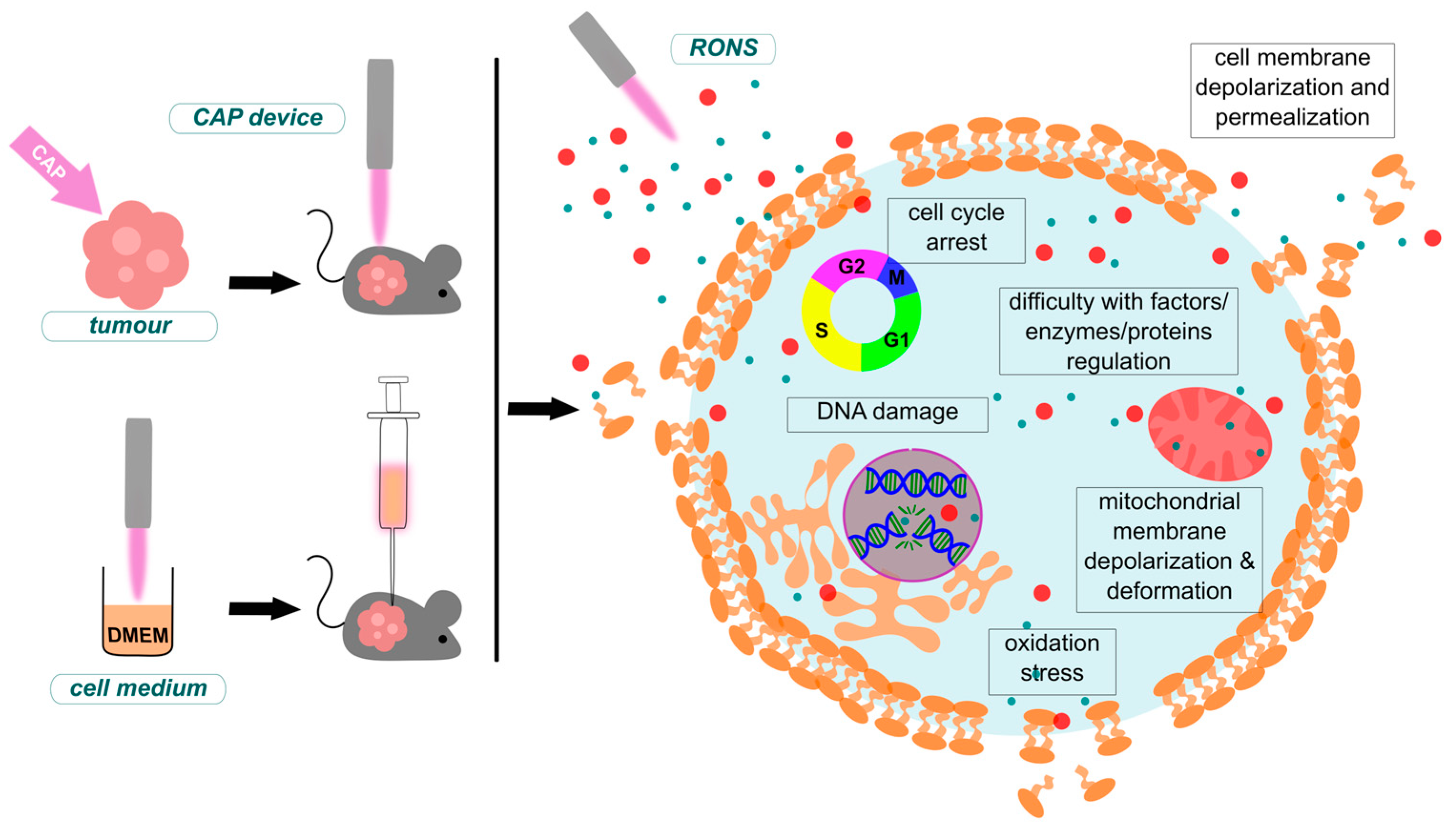
  • Cancer treatment—cold plasma has successfully demonstrated anti-cancer properties and is currently being explored as a potential therapy for certain types of cancer. It can induce apoptosis in cancer cells, slow down tumour growth or decrease its size, and sensitize cancer cells to other treatments like chemotherapy and radiation therapy [66].
  • Immunology—CAP therapy has proved to modulate immune responses by influencing actions of innate and adaptive immune cells, potentially enhancing the body’s defence mechanisms against pathogens and tumour cells [2].
  • Viral infections—cold plasma is being investigated for various applications in virology due to its ability to efficiently kill pathogens while sparing human cells. CAP-generated RONS have shown promise in the inactivation of viruses on surfaces and in the air, offering a novel approach to disinfection in healthcare and public spaces, as well as a modality for the treatment of infections [67].
  • Neurology—CAP has shown potential in tumour treatment, including brain tumours like glioblastoma multiforme (GBM). Furthermore, it can serve as a beneficial treatment for neurodegenerative diseases like Alzheimer’s and Parkinson’s [68].
  • Promoting wound healing—when applied to wounds, cold plasma works in two ways. First of all, it creates a cytotoxic environment for pathogens and even their spores by delivering reactive species like RNS and ROS. Secondarily it stimulates the process of wound regeneration by promoting tissue growth factors synthesis. It has successfully been used to treat chronic wounds, diabetic ulcers, and infected wounds, showing positive results in both accelerating healing and reducing infection rates [69,70].
  • Stem cell—CAP displays the ability to influence the differentiation of stem cells and progenitor cells. Enhancing the growth rate while influencing cell differentiation processes is crucial for regenerative medicine applications [71].
  • Dental applications—it is used for disinfection, treatment of gum disease (periodontitis), and promoting oral tissue regeneration. Plasma’s ability to kill bacteria results in the disinfection of dental implants and root canals. It also found an application in improving the bonding of dental materials to teeth [47].

4.1. Cancer Treatment

Cold plasma is a mixture of various factors that can individually influence apoptosis in tumour cells. The fact that they are combined and interacting with each other in the plasma beam, and later in the treated sample, makes it far more difficult to fully comprehend the complexity of reactions and processes occurring in treated cells or tissues. It is safe to say that reactive species such as RNS and ROS (or simply RONS) play a major role in the complex reactions. Therefore, knowledge of a reactive species’ life span, reactivity, mechanics, and concentration is crucial to understanding the bigger picture. Since the beginning, it has been one of the greatest challenges in plasma application in medicine. Therefore, a constant pursuit of understanding the reasoning of chemical, biological, and physical effects induced by plasma is necessary. This indicates the need to understand the production mechanisms of specific reactive species, the medium’s composition and volume, characteristics of carrier gases, the surface of contact, and the distance between plasma and sample, as well as the treatment duration [72].
Furthermore, a proper adjustment of these elements may be crucial when defining a suitable dose of different treatments. It has been reported by many researchers that dose-dependent therapy with increased exposure duration results in promising improvements and achievements when it comes to the treatment effects on several cancer cell lines [73]. Multiple studies show that atmospheric plasma, when directed at the damaged tissue location, decreases progress in the spread of metastases [74,75]. Various in vitro and in vivo experiments with different cancerous cell lines are reinforcing the conviction of remarkable anticancer plasma properties against liver cancer, myeloma, lung cancer, glioma cells, skin cancer, bone cancer, melanoma cells, the central nervous system, and cervical cancer [20,36,76,77,78,79]. It has been noted that ROS and RNS are successful in inducing an apoptotic reaction in tumour cells, enhancing cell immune comeback, and stimulating angiogenesis [75].

4.1.1. Mechanisms of CAP Anti-Cancer Effects and Selectivity

There are serval differences in structure as well as functionality when comparing a cancerous cell to a healthy cell. For example, the former displays an increased number of aquaporins or a reduced level of cholesterol in the cell membrane, leading to an escalated vulnerability of cancer cells to CAP-generated RONS exposure. Cell death pathways vary due to cancer type, applied plasma conditions, and type of generated RONS. Therefore, this makes it challenging to pinpoint a singular scheme of action that would apply to every situation. However, there are few repetitive mechanisms leading to a CAP-induced cancer cell death pathway. In this chapter we have put together an overview of the current understanding of molecular mechanisms involved in the efficacy and selectivity of CAP in cancer treatment.
Cancer cells are known to be more vulnerable when exposed to oxidative stress than normal cells [25]. One of the main reasons for this is the increased expression of aquaporins (AQs) leading to larger amounts of them in the membranes of cancer cells [80]. AQs were firstly identified as transmembrane water [81]. Nowadays, it has been confirmed that AQs are also able to facilitate transport of small molecules including glycerol, ammoniac, urea, and carbon dioxide [82,83], as well as free radicals. There are multiple types of AQs varying in diameter. For example, the diameter of AQ1 is 2.8 Å. Even though it is too small to efficiently transport hydrogen peroxide into the cell [83], this free radical still penetrates the cell membrane faster through AQ1 than through the lipid double layer. Another example would be AQ8, with a diameter of 3.2 Å, which is a pore size large enough to sufficiently to transport hydrogen peroxide [84]. Therefore, the increased amount of AQs in cancer cells, compared to normal cells, enhances the transition of plasma-generated RONS.
CAP-generated RONS cause the peroxidation of membrane lipids, eventually leading to the generation of pores large enough to facilitate RONS transition into the cells. While every cell membrane is composed of lipids, the effect of permeabilization (the act, process, or result of making something, such as a membrane or cell wall, permeable) may be enhanced in cancer cells [85]. The reason behind this phenomenon revolves around reduced levels of cholesterol, a lipid responsible for membrane stability and fluidity [86]. High-cholesterol normal cells create a barrier against RONS through an appropriate level of membrane lipids condensation. Cancer cells with decreased levels of cholesterol, therefore displaying less condensate lipids in membrane, are more sensitive to oxidative stress triggered by plasma-generated RONS [87].
One of the main cell death pathways is endoplasmic reticulum (ER) stress-induced apoptosiss [88]. ER is a key organelle responsible for proper cellular functioning and metabolic control. Any functional imbalance in ER, referred to as “ER stress”, leads to multiple disorders, such as organelle dysfunction, aberrant metabolism, inflammation, and insulin resistance [89]. Furthermore, RONS-induced ER stress is involved in triggering calcium influx into the mitochondria, through interaction with the inositol trisphosphate receptor [IP3-RR] and the ryanoid receptor [RR]. This leads to Ca2+ concentration imbalance, eventually inducing the unfolded protein response (UPR) [90]. This specific mechanism is a self-defence system against ER stress. It is a collaborated action of three main signalling proteins, namely inositol-requiring protein-1 (IRE1), protein kinase RNA (PKR)-like ER kinase (PERK), and activating transcription factor 6 (ATF6). The UPR system is able to maintain homeostasis and protect cells from mildly damaging oxidative stress. However excessive RONS invasion via CAP exposure results in ER stress far beyond UPR control. In this particular situation, the three signalling pathways are triggered, namely C/EBP homologous protein (CHOP), Caspase, and JNK, subsequently initiating an apoptosis response in the mitochondria [91].
Increased RONS concentration is also responsible for the induction of DNA lesions, oxidative damage, and DNA single-strand as well as double-strand breaks (DSB). The last one, DSB, usually occurs during replication stress or in the case of CAP exposure, i.e., when introduced to ionizing radiation [92]. In mammal cells, a complex mechanism evolved in order to protect the genome from DSB. This aforementioned mechanism is a set of actions collectively referred to as the DNA damage response (DDR). It is composed of cell cycle checkpoints, DNA break detection, and DNA repair pathways. The mechanism begins by acknowledging the DNA breakage. Then, the activation of ATM, Ataxia telangiectasia, and Rad3-related (ATR) kinases follows [93]. This set of kinases begins the phosphorylation processes. For example, ATM phosphorylates histone H2AX to γH2AX, which serves as an indirect marker for DSB [94]. Subsequently, it also initiates various signalling cascades in order to regulate actions such as replication, translation, progression of the cell cycle, and senescence. If the RONS-induced damage is far beyond repair, the apoptosis is evoked [95].
In conclusion, CAP-generated RONS are able to overwhelm the antioxidant system, limiting its protective properties against oxidative stress in cancer cells while sparing the normal cells which are capable of managing excessive RONS.

4.1.2. Differentiation of CAP-Induced Apoptosis and Autophagy Mechanisms

Apoptosis is a process of programmed cell death. This mechanism is employed when there is a need to eliminate cells that have been irreversibly damaged. It also plays a role in preventing cancer. If, for any reason, apoptosis would be prevented, it can result in uncontrolled cell division and the subsequent development of a tumour [96].
CAP-induced apoptosis is firstly stimulated by the generation of primary free radicals (1O2), which inactivate catalase molecules, decreasing the defence potential of cancer cells against free radicals. This can trigger a specific chain reaction resulting in the increased production of even more RONS, both 1O2 and H2O2 or ONOO-, which consequently block downstream catalase molecules, GSH, and trigger the HOCl-dependent apoptosis pathway, resulting in membrane lipid peroxidation [97,98].
Mitochondrial apoptotic pathway can be activated by the action of CAP. Jawaid et al. [99] indicated that He-based CAP contributes to the activation of the FAS receptor, leading to caspase-8 activation. Additionally, studies conducted on Jurkat (immortalized T cells, leukemia model), HT29 (colorectal cancer), A549 (lung cancer), MCF-7 (breast adenocarcinoma cells), and DU145 (prostate cancer) cells show that CAP-induced upregulation of cleaved caspase-8 results in the upregulation of BAX gene expression (both at the mRNA and protein level), the release of cytochrome c from the mitochondrion, and further upregulation of cleaved caspase-9 and caspase-3, along with the downregulation of anti-apoptotic Bcl-2 protein expression [100,101,102,103].
Autophagy is a highly conserved cellular degradation process in which parts of the cytosol and organelles are sequestered in a double-membrane vesicle, the autophagosome, and delivered to a degradative organelle, the vacuole/lysosome, for degradation and eventual recycling of the resulting macromolecules.
Similarly to apoptosis, CAP-induced autophagy is dependent upon RONS generation. Although the scheme of autophagy is a diverse action, it is possible to identify two main mechanisms of activation for this process [104].
The first mechanism concerns the interaction of CAP with the membrane receptor EGF. Studies suggest that by downregulating acetylation and phosphorylation, this receptor is inactivated, resulting in the activation of two intracellular pathways. The first path involves blocking the phosphorylation of RAS and MEK proteins, resulting in the expression of transcription factors, such as ZKSCAN3, TFEB, FOXO1, CRTC2, and CREBBP, that stimulate autophagy [104]. The second pathway involves the upregulation of phosphorylation in the intracellular JNK protein. Hence, p-JNK binds to the BIRC6 protein, which, under natural conditions, binds to LC3B, leading to its ubiquitination and degradation. When BIRC6 binds to p-JNK, the free LC3B protein can stimulate the initiation of autophagy [105].
The second mechanism is related to blocking the membrane receptor GFR; it also revolves around two intracellular signal transduction pathways [104]. The first pathway relies on silencing the GFR, which, in turn, silences the PI3K/AKT/mTOR/p70S6K pathway, resulting in the upregulation of beclin-1 expression, the conversion of LC3-II to LC3-I, and the downregulation of p62 expression, altogether causing phagophore formation and the initiation of autophagy [106]. In parallel, it is indicated that the formation of LC3 and beclin-1 may occur due to the activation of ERK1/2 and JNK, respectively, triggered by CAP-generated RONS. At the same time, JNK activation may lead to c-jun phosphorylation and further to AP-1 activation, which stimulates beclin-1 and ATG5/7 formation [107].

4.1.3. Selected CAP Anti-Cancer Studies

Attri et al. [108] investigated the CAP influence of RONS on tumour cell fatality using various cell lines: A549 (lung carcinoma epithelial cells), HEK293 (Human Embryonic Kidney), MRC5 (Medical Research Council cell strain 5—a diploid cell culture line composed of fibroblasts), and T98G (fibroblast-like cell, isolated from glioblastoma multiforme). Cell lines MRC5 and HEK293 did not display any significant changes during the experiment. This was probably caused by cellular interactions of plasma-generated RONS along with H2O2, resulting in the modification of the mitochondrial matrix of the mentioned cells. Further actions can be described as inducing the intrinsic apoptotic cascade, elevating the expression level of pro-apoptotic genes, and reducing the expression level of anti-apoptotic genes. Additionally, a shift in the expression level of cellular signalling protein MAPK/ERK1/2 was noticed. Normal cells and cancerous cells differ in metabolic properties and therefore respond to the treatment differently [109].
Abnormal cancerous cell metabolic processes after CAP treatment were also investigated by Xu et al. [110] The results of performed investigations showed that leukaemia cells displayed distinct alanine, aspartate, and glutamate metabolisms. Moreover, decreased glutaminase function in tumour cells has been reported, resulting in less glutamine being metabolized to glutamic acid. Glutamine aggregation processes closely related to glutamic acid deficit may interfere with the multiplication of leukaemia cells, resulting in apoptotic processes.
Various studies on experimental animals and cell lines prove the anti-tumour effectiveness of CAP devices (Table 2). For example, glioblastoma cell lines displayed reduced survivability after plasma treatment [111,112]. Several publications have shown that CAP applied to different cancerous brain cell lines induced the loss of their survivability, which resulted in cell death [72,113]. Plasma release followed by apoptotic cell death was also observed in primary lung carcinoma TC-1 cell lines; to a much smaller degree, this has been also noticed in fibroblast cell lines [114]. The effectiveness of the in vivo plasma action was shown after the subcutaneous introduction of the 4T1 breast cell line into test subjects. Mice injected with the mentioned cell line were treated using CAP. The tool diameter was approximately 250 µm. Cancer growth was reduced during the 3 min CAP treatment. Interestingly, the proportion of pro- to anti-apoptotic gene expression at the cellular stage changed significantly [115].
The in vitro and in vivo influence of the 3 min CAP treatment on melanoma B16/F10 cell lines in mice was investigated by Mashayekh et al. [58] The treatment proved to be fatal, with most cells eventually compressing the malignancies. Plasma is most conveniently delivered with access to the cutaneous structure; thus, multiple evaluations of the impact of CAP on skin carcinoma have been performed. For instance, G361 malignant melanoma cells reduced vitality and separated from the interface after CAP treatment [116]. This was caused by events such as integrin activation, reducing the level of focal adhesion kinase (FAK) as well as modifying the architecture of actin filaments [23].
Table 2. Plasma treatment in various cancer types.
Cancer TypeCell LineCAP Treatment Modality/
Device/Feed Gas
Main Observations
Lung(SW900) [11]Direct:
* Plasma jet device [11]
O2/N2
High selectivity, death of 60–70% SW900 cancer cells [11].
Morphological changes at the cellular and subcellular levels, suppressing cancer cell growth [101].
(A549) [117]Indirect:
Piezobrush PZ2 (Relyon Plasma GmbH, Regensburg, Germany) [117]
Air
Disruption of the mitochondrial-nuclear network in cancer cells treated with PAM [117].
Colorectal(HT29) [101] Direct:
Piezobrush PZ2 (Relyon Plasma GmbH, Regensburg, Germany) [101]
Air
Morphological changes at the cellular and subcellular levels suppress cancer cell growth [101].
Melanoma(B16-F10) [115]Direct:
* Plasma jet device
He
Indirect:
* Plasma jet device
-
Viability reduced to 0% after 48 h of treatment [115].
Significant cell death and substantial reduction in tumour growth.
(DSMZ: ACC74) [118]Indirect:
miniFlatPlaSter (Terraplasma GmbH, Garching, Germany) [118]
Air
CAP-treated solutions under acidic conditions caused protein nitration in cells [118].
LeukaemiaMOLM13 [110]Direct:
* Plasma jet device [110]
He
Glutaminase activity of He plasma jet group was decreased [110].
Breast(MDA-MB-231) [119]Indirect:
* Plasma jet device [119]
He
Direct:
kINPen IND plasma jet (Neoplas Tools GmbH, Greifswald, Germany) [120]
Air
CAP-treated media displays anti-cancer capabilities [119].
Reduction in viability of cells and increase in apoptosis rate [120].
(MCF-7) [119]
(HCC1806) [120]Direct:
kINPen IND plasma jet (Neoplas Tools GmbH, Greifswald, Germany) [120]
Air
Reduction in viability of cells and increase in apoptosis rate [120].
Bladder(SCaBer) [121]Indirect:
* Plasma jet device [121]
Air
PAM, in a dose-dependent way, was considered to be an effective apoptotic agent lasting for several hours [121].
(HT-1376) [122]Direct:
kINPen IND plasma jet (Neoplas Tools GmbH, Greifswald, Germany) [122]
Air
Reduction in metabolic activity and protein content followed by a decrease in cell viability [122].
(TCCSUP) [122]
Cervical(HeLa) [123]Indirect:
* Plasma jet device [123]
-
CAP-Jet (PlasmaMed Inc., New York, USA) [124]
Ar
Inactivation of cancer cells [123].
Elevated ROS generation and induced substantial apoptosis in the cancer cells [124].
(CaSki) [124]Direct:
CAP-Jet (PlasmaMed Inc., New York, USA) [124]
Ar
Elevated ROS generation and induced substantial apoptosis in the cancer cells [124].
(HCT116) [124]
SIHA [97] Indirect:
* Portable plasma ‘corona pen’ [97]
Air
Efficient apoptosis induction through the HOCl signalling pathway, finalized by lipid peroxidation [97].
GastricMKN-45 [97]
SarcomaSKN-MC [97]
* custom-built device.

4.1.4. Comparison of CAP Anti-Cancer Therapy and Conventional Methods

Conventional methods in oncology often refer to the established approaches used to diagnose, treat, and manage cancer. They are typically composed of surgery, chemotherapy, radiotherapy, targeted therapy, or a combination of said treatments [125]. Surgery generally involves the removal of tumours and surrounding tissue, aiming to eliminate as much of the cancer-occupied area as possible. Chemotherapy revolves around drug use in order to destroy cancer cells or at least inhibit their growth. Chemical substances are often given intravenously or orally. Radiotherapy uses high-energy radiation to induce cancer cell death or shrink the size of the tumours. Targeted therapy is focused on blocking the action or signalling pathways of specific molecules involved in cancer growth. These conventional methods can be used alone or in combination, depending on the type and stage of cancer, with the aim of achieving remission or prolonging survival while minimizing side effects.
The development of CAP in medicine, especially in cancer treatment, holds great promise for revolutionizing healthcare (Table 3). This technology offers both a non-invasive and versatile approach to several medical applications. CAP’s ability to generate reactive oxygen and nitrogen species (RONS) offers unique opportunities for targeted therapy, enhances cancer cell death processes while sparing the normal cells, and enables antimicrobial treatment and modulation of biological processes [104]. By harnessing the power of CAP, researchers have an opportunity to develop safer, more effective, and minimally invasive treatments. Hence, this significantly improves treatment outcomes and healthcare delivery.
In summary, CAP offers several advantages over conventional methods, such as selectivity in targeting cancer cells, the potential to prevent metastasis, and preservation of healthy tissues. However, further research and more clinical trials are needed to fully establish its efficacy and safety compared to traditional cancer treatments. Furthermore, CAP may be used in combination with conventional therapies to enhance treatment outcomes and reduce side effects.

4.2. CAP in Immunology

CAP-generated RONS are the main cause of oxidative stress, thus activating a number of cytological and molecular responses, including triggering immunological reactions. To be more specific, CAP treatment induces a mechanism referred to as immunogenic cell death (ICD) in tumour cells. This leads to an enhanced adaptive and systemic immune response with memory cells, thus enhancing its defence against cancer [131]. There are two plausible pathways of this effect. The first one is the direct impact of plasma on immune cells, thus leading to their activation. The second revolves around indirect activation of immune cells via CAP-induced pro-inflammatory signals as well as cancer cell death [132,133].
Cellular immunity is based on the cooperation of two distinctive immune cell types. Innate cells are able to recognize epitopes of target structures. The second type, adaptive cells, responds to new antigens. Another factor of CAP-induced immune responses against cancer would be controlling the near-tumour environment by regulating particles released by immune cells.
Scavenger cells, also known as phagocytes, play a key role in guarding organisms from intruders and dangers such as cancer cells. They include neutrophils, dendritic cells, and macrophages. For example, macrophages are responsible for managing inflammation and anti-inflammation homeostasis in various tissues. CAP treatment was proven to enhance macrophages’ ability to counteract tumour cells through increasing migration, enhancing cytokine concentration, and enhancing the TNFα. According to the reports of Bekeschus et al. [134,135], CAP influences anti-tumour effects through modulating primary murine and human-monocyte-derived macrophages. When it comes to pancreatic cancer, PAM elevates the concentration of macrophages, therefore inhibiting the tumour progression [136].
Another type of scavenger cell, known as neutrophils, is usually considered to be a sign of poor prognosis in cancer. This is mainly because in the tumour microenvironment, neutrophils can turn into a subtype known as tumour-associated neutrophils (TANs) [137]. This specific variation of scavenger cells often exhibits pro-tumorigenic properties and contributes to immune evasion by the tumour. When neutrophils are treated with cold plasma, their intracellular presence elevates, and neutrophil extracellular-trap (NET) formation occurs. This action results in higher survival rates in treated mice [138].
All of the actions mentioned above lead to damages to tumour cells, resulting in immunogenic cancer cell death (ICD). This phenomenon begins with antigen-presenting cells (APCs). Dying tumour cells release specific neoantigens that are captured and transported to draining lymph nodes. There, the priming and activating of T cells occurs. Primed T cells are supposed to locate and eliminate tumour cells. Unfortunately, properties such as a lack of neoantigens, immunosuppressive tumour microenvironment (TME), and overexpression of specific signals modulate the cancer-immunity cycle, making it challenging for T cells to infiltrate the tumour [139,140].
It has been proved by Lin et al. [79,141,142] that either direct plasma treatment or indirect CAP treatment displays the ability to induce ICD pattern in various tumour types, such as pancreatic, colorectal, lung, and melanoma tumours. The precise mechanisms underlying plasma-induced anti-cancer immunogenic response is still unclear and requires more research. However, current clinical evidence already states a positive prognosis for plasma treatment in this aspect of medicine.

4.3. Viral Infections

CAP-generated antiviral therapies, RONS, play a key role in the mechanism of action. Current research seems to confirm that this mechanism is generally common for both bacteriophages and animal viruses, whether they are RNA, ssDNA, or dsDNA viruses.
Studies conducted on T4 and MS2 bacteriophages have shown that the generated 1O2 radical is crucial for viral inactivation by generating damage to capsid structural proteins, causing viral aggregation as well as downregulating gene expression levels causing damage at the DNA, RNA, and protein levels, resulting in reduced virulence [143,144].
One mechanism related to the activity of the 1O2 radical is also highlighted by Aboubakr’s research [145], who, in their study on Feline calicivirus (FCV), suggest that this radical leads to the oxidation of histidine, causing the virus to form incorrect caspases during the folding step, lowering the virulence of newly formed virions. In the same publication, the authors showed that O3 and NO2 radicals play an important role in virus inactivation; however, no such effect was detected by H2O2.
Also, studies conducted on human type 5 adenovirus [146], as well as a number of studies conducted on the SARS-CoV-2 virus [147,148,149], confirm that the action of plasma is probably based on damaging the structure of genes and viral RNA polymerases, thereby reducing their expression and the virulence of the viruses themselves.
On the other hand, studies [150] conducted on HIV-1 infecting MDM (monocyte-derived macrophages) indicate a dual mechanism of the action of plasma. On the one hand, the plasmas act on the target cell and reduce the number of receptors for HIV-1 exposed on the membrane surface; on the other, they act on the viral units and reduce the activity of reverse transcriptase, thereby impairing the reproductive capacity of the virus.

4.4. Neurological Complications Therapy

The term neurological disorder refers to any condition affecting the brain, peripheral nervous system, or vegetative nervous system, such as Parkinson’s and Alzheimer’s, trauma, spinal damage, or traumatic brain injury [151]. As a consequence of the advanced neural stem cell (NSC) extractions and cloning techniques, central nervous system (CNS) transplantation stands at the summit of therapies when speaking of neurotraumatic or neurological disorders [152]. The main disadvantages of these procedures are firstly the inadequate differentiation selectivity of particular cells, chemical toxicity potential and scarring processes of glial after transplantation. Recently, Jang et al. [44]. demonstrated that CAP treatment was able to effectively trigger neural progression in zebrafish. The results offer hope for managing neurological dysfunction in the future. Furthermore, the molecular and chemical of cold plasma on the ERK/Ras/Trk signalling system responsible for neuronal development were explored. In vivo analysis was conducted with the help of a recombinant zebrafish (Danio rerio) embryo and murine neuroblastoma-derived cell line Neuro 2A (N2a). Researchers used a DBD device, with an operating gas combination of O2 and N2. After 24 h, the N2a cells exposed to CAP treatment displayed a cell diameter more than 4-fold greater when compared to the naïve cells; additionally, the post-treatment cells achieved maximal nerve length oscillating around the value of 70 mm, with a mean of 46.3 ± 1.5 mm [151].
Dopaminergic (DA) nerve cells are the main origin of dopamine in the CNS. They play an important role in regulating numerous neural mechanisms. They are also a part of the NSC, undergoing terminal proliferation to become fully developed and functional nerve cells [44]. It is known that loss of DA neurons can result in Parkinson’s disease. The recombinant zebrafish embryos mentioned previously were exposed to 1 min CAP treatment. After 6 h of incubation, GPF (green fluorescents protein) activity levels increased and did not disappear for 33 h. GPF is a protein expressed exclusively in mature nerve cells that have undergone the mitosis processes. Then, the embryos were subjected to the CAP treatment. Nerve cells testing positive for GFP could be seen in the maturing CNS of zebrafish after 6 h of treatment. It was shown in the research that plasma-generated NO played the role of arduous extracellular messenger, whereas cytosolic H2O2 and mitochondrial O2 are considered to be ROS messengers, as well as important elements included in the neuron proliferation path [44].
Researchers induced in vitro NSCs differentiation processes via micro-plasma jet device application [153]. Quick and efficient NSC maturing processes were noticed in C17.2 immortalized neural progenitor cells and primary rat cell lines. CAP-induced cell maturation was proved to be superior to chemical methods, like the use of serum deprivation, resveratrol, or retinoic acids. Maturation using CAP was far quicker and more effective compared to conventional methods. As stated earlier, DA nerve cells comprise 70% of the nerve cells developed using CAP. Another advantage of CAP is the ease of manipulation by simply altering the electrical power source, plasma source tool, operating gas composition, and operating gas flow. It can be noted that biological outcomes are significantly impacted by plasma dose. Following this line of reasoning, a smaller dose may promote cell multiplication, motility, and maturation, whereas an excessive dose usually results in apoptosis. Therefore, this creates a plausible potential therapeutic approach for neurological dysfunction.
Tan et al. [154] used CAP on rat PC12 cells, either independently or with the addition of Acidic Fibroblast Growth Factor (aFGF), which resulted in accelerated cell neurogenesis. The study considered both phenotypic parameters such as the number of neurites and their length. Zhao et al. [155] concluded that both plasma-generated NO and the stimulation of nitric oxide synthase activity are responsible for this phenomenon. Increased synthase activity caused enhanced intracellular NO generation, which plays the role of intracellular transmitter. Subsequently, this stimulates the downregulation of Notch1 and Id2 expression, and the upregulation of Ngn2 and Ascl1, consequently upregulating the expression of the neurospecific transcription factor NeuroD that stimulates neuronal differentiation. At the same time, Jang et al. [44] complete this picture by indicating that endogenous NO by itself is insufficient to stimulate neurogenesis. The key to this process appears to be endogenous H2O2 formed from the mitochondrial O2 radical, which activates the intracellular Trk/Ras/ERK signalling pathway.
The positive action of CAP has been described not only in the context of differentiation, but also neuroprotection (Table 4). Tian et al. [156] concluded that the neuroprotective action of CAP is based on regulated pulses by which the amount of RONS produced can be controlled. Excessive RONS will stimulate the action of cellular antioxidant systems (direct and indirect GSH synthesis) and inhibit synthesis that is critical for the cell levels of endogenous RONS. The results prove that such ‘training’ of cells to fight RONS results in increased survival rates of neurons exposed to excitotoxic glutamate. Another study [157] found that in a simulated ischemic stroke condition, CAP-generated RNS have a neuroprotective effect by increasing the activity of the cGMP/PKG pathway, which results in the increased apoptotic potential of neurons, thus stimulating their survival.

4.5. Chronic and Acute Wound Treatment

Every healing process can be easily impeded by the presence of even allegedly harmless pathogens. Therefore, the initial thought behind CAP application in regenerative medicine was to accelerate acute and chronic wound healing by reducing infection.
Studies [161] on wound healing via CAP therapy clearly show that the effects are dose-dependent and impact processes such as angiogenesis, re-epithelialization, collagen synthesis, cell cycle regulation, HIPPO pathway regulation, and the epithelial–mesenchymal transition (EMT).
Badr et al. [162] and Fathollah et al. [163] conducted studies using mice and rats with diabetes. CAP treatment caused accelerated wound healing in those model organisms. Both increased expression levels of angiogenic factors (VEGF, Ang-1, HO-1) and decreased angiostatin expression (angiostatic protein), as well as decreased expression of pyroptosis markers (NLRP-3, IL-3Beta, Caspase-1) and oxidative stress markers (NO and iNOS), were noted. The latter, according to the research of Guo et al. [164], may be related to the increased activity of antioxidants such as SOD, GPx or CAT.
A number of studies indicate that CAP therapy leads to enhanced re-epithelialization processes [165,166] as well as increased collagen production, with particular emphasis on particular types of collagen, namely Col1, Col3, and Col1α. On the other hand, it should be noted that there are also reports [167,168] indicating that wound treatment via CAP does indeed accelerate wound closure, but without significantly affecting the levels of re-epithelialization or collagen synthesis (Col1-A2, Col3).
Shi et al. [161] and Lou et al. [169] conducted both in vitro and in vivo experiments using human lines, mouse lines, and a rat model in order to present short-term plasma treatment; they generated RONS interactions with extracellular signal-regulated kinases (ERK) that led to the stimulation of cell proliferation and in-parallel upregulation of cyclinD1 and Cdk2 expression, subsequently upregulating the cell cycle.
Other researchers [170,171] suggested that the HIPPO signalling pathway is also involved in CAP-induced wound healing. Plasma-generated free radicals upregulate the expression of YAP, a transcription factor protein, which then binds to the TEAD protein, stimulating further expression of the HIPPO pathway; these include the proteins CTGF, Cyr61, and CCN2 (all of which were upregulated). It is known that the HIPPO pathway, especially the upregulation of YAP/TAZ elements, positively affects EMT. Activation of the second type of EMT is triggered by RONS-triggered downregulation of E-cadherin expression, as well as the upregulation of the expression of vimentin, Slug and Snail, and proteins [169,171].
Chatraie et al. [22] investigated CAP’s effect on bacterial infection in Wistar rats’ chronic ulcers. The treatment successfully reduced inflammation, significantly reducing side effects. Two-minute CAP therapy was able to effectively reduce the bacterial burden as well as magnify the healing processes. Infection decrease has been noted independently of the kind of bacteria. One of the main challenges when treating ulcer stems is the fact that multiple bacterial strains have become resistant to conventional antibacterial therapy. Therefore, modern research emphasizes the application of CAP in ulcer treatment. It has been proven using Wistar rats that CAP treatment benefits ulcer minimization. The following events occurs when CAP is applied: angiogenesis, enhanced tissue mechanical strength, re-epithelialization, angiogenesis, and collagen synthesis.
Gao et al. [172] took a closer look at the cold-plasma-induced effects on chronic wounds, such as enormous genital warts, pyoderma gangrenosum, and ulcers in diabetic feet. Attempts at using antibiotics and radiation have failed miserably. The lesions received 5 min of treatment every 60–80 min, two times a day. The exudation was seemingly decreased after the third day of CAP exposure delivered in six cycles after the wounds dried and constricted completely. Without noticeable success, the patient would receive further treatment for an additional six months. Similarly to antibiotic therapy, another patient’s pyoderma gangrenosum showed some marginal signs of progress. Then, the patient was transferred to the CAP treatment. After eight administrations of CAP, the lesion entirely vanished and showed no sign of recurrence, even after several months.
Schmidt et al. [173] suggested that the p53 protein plays a major role in connections when examining CAP influence on keratinocytes. It is hypothesized that mitogen-activated protein kinase cascade activates the responses of p53, due to the effectiveness of ATR and ATM oxidative detectors. The “cross-talk” correlation was also noted among keratinocytes and fibroblasts during CAP treatment. The treatment enhances the signalling function in the YAP protein called Salvador–Warts–Hippo, which induces a significant transcriptional activity level in its cofactor. Furthermore, only the fibroblasts displayed elevated expression levels of the YAP targeting markers known as CYR61 and CTGF—playing the role of downstream effectors of this system. Fortunately, it has been proven that a meaningful antioxidant administration may reduce the enhanced expression mentioned above. Cell media conditioned with CAP-treated fibroblasts was mandatory for achieving improved HaCat keratocyte mobility. However, even in the absence of the mentioned fibroblasts, the HaCat cell lines could upkeep this mobility due to recombinant Cyr61 and CTGF exposure.
CAP therapy has proven its effectiveness in wound healing processes through the multiplication of fibroblasts and keratinocytes, improved cutaneous microcirculation, and the activation of monocyte and cellular motility (see Table 5). Both fibroblasts and keratinocytes have proven to be significant in wound healing, especially in the final stages. After brief exposure to CAP, the keratinocyte and fibroblast cell lines HAcaT and MRC5 still displayed enhanced motility. Another study explored the in vivo plasma activity in mice models. In this case, wound repair was attributed to the generation and activity of RONS as well as UV exposure [174].

4.6. Triggering Stem and Progenitor Cell Proliferation

Another feature of cold plasma worth mentioning is the ability to modulate the replication of progenitor cells, the data are collected in Table 6. This could result in effective CAP application in restorative therapy and biomedical nanotechnology. Park et al. [183] noticed that the application of CAP tools encourages adipose-originated progenitor cells to proliferate without the need to alternate their original characteristics. After a 72 h long treatment, the progenitor cell multiplication process was described as 1.57-fold greater. Additionally, the presence of NO significantly reduced CAP-enhanced progenitor cell multiplication. Adipose tissue cells not exposed to CAP treatment displayed an increased multiplication at a level even greater than the reference cells but not as significant as the CAP-treated cells when exposed to the NO donor DETA-NONOate (diethylamine nonoate). It can, therefore, be concluded that higher NO content might influence enhanced multiplication.
Cytotoxicity and other adverse effects were not reported in osteoprogenitor MC3T3-E1 cells after CAP exposure. Concentrations of accumulated NO increased and were delivered to the cellular components; therefore, modulating its concentration in developed cells was enabled. As a response, early osteogenic development was triggered. In this case, it was accomplished irrespective of the condition of the pro-osteogenic growth factor. Also, some important alterations in the mechanistic pathways have been reported after CAP treatment. The expression of osteogenic markers ALP, COL-1, OCN, and RUNX2 was far more frequent alongside decreased MAPK and PI3K/AKT signalling. Moreover, the autophosphorylation of FOXO1 occurred, which is considered to be a key regulator of bone growth as one of the essential factors for osteoblast multiplication and oxidative equilibrium [184].
Additionally, the function of CAP derived from periodontal tissues was explored [74]. After the exploration of CAP’s influence on adult embryonic stem cells (hPDL-ESCs), no sign of cytotoxicity or adverse response were reported. CAP therapy caused restriction of hPDL-ESC recruitment that avoided affecting total cellular survivability. Similarly, CAP may be used to stimulate the advancement of bone marrow and hematopoietic-derived stem cells. These cell lines show significantly increased multiplication after treatment in comparison to normal cells. It was also revealed that the upregulation of prominent stem cell biomarkers CD105 and CD44 in bone marrow cell cultures appeared approximately 5-fold more significant.
Table 6. CAP in stem and progenitor cell proliferation.
Treatment TargetCAP DeviceEffect
Adipose-derived stromal cells [185]* Self-made He/DBD device [186]Halted cell growth and alteration in morphological characteristics occurred [186].
Mouse neuroblastoma stem cells (N2a) [44]* Air-CAP [44]Increase in cell multiplication [44].
Mouse neural stem cell (C17.2-NSC) [46]* Self-made CAP jet device [46]Increased cell multiplication and development [46].
Osteoprogenitor cells (MC3T3-E1) [71,185]* DBD NO-Plasma nozzle system [185]
* Self-made He/plasma jet [71]
Dephosphorylation of FOXO1 transcription factor [185].
Stimulation of osteoblastic differentiation [71].
Mesenchymal stem cells (MSCs) [187]MicroPlaSter setup (Adtec Plasma Technology Co., Ltd., Hiroshima, Japan) [187]Promoting cell growth through activation of genes responsible for proliferation expression [187].
* custom-built device.

4.7. Dental Medicine

Conventional oral cavity disinfection and cleaning techniques are composed mostly of antimicrobial solutions, mechanical infection removal, and laser devices [188]. The two latter techniques create a risk of possible mechanical or thermal damage to treated tissues [47]. That is exactly where CAP methods show their superiority, enabling treatment with a considerably reduced risk of tissue damage. CAP discharge may be applied directly to difficult-to-reach oral cavity regions, like tooth canals or uneven surfaces, with ease [188]. Unlike liquid antimicrobial conventional solutions, it can also be applied to specific locations in the oral cavity bypassing negative results that occur when using microbial liquid. The rising resistance of the bacterial strains found frequently in tooth biofilm has been reported. Due to neglect of the mentioned biofilm removal, many systemic conditions may be developed, e.g., endocarditis or necrotizing pneumonia. It has been reported by Delben et al. [189] that the CAP antibacterial effects resulted in effective attenuation of Staphylococcus aureus and Candida albicans, frequently identified as components of dental plaque. The decrease in microbial burden decrease achieved through CAP treatment was comparable to the one achieved with fluconazole or benzylpenicillin treatment.
Multiple research projects explored CAP applications in decreasing the microbial burden in the tooth canal [188]. Thus, Armand et al. [55] proved that ex vivo 5 min long CAP treatment reduced bacterial biofilm to a thickness of 1 mm. Researchers then mimicked an infestation using Enterococcus faecalis, a species commonly occurring when a dental canal is infected. Interestingly, O2/He plasma was considered to be the most efficient when it comes to reducing the microbial burden; additionally, He plasma displayed potential application as the photosensitization agent. Furthermore, the morphology of CAP-treated tooth canals was considered to be significant for the treatment outcome. Straight types of canals caused far greater damage.
S. Beni et al. [190] investigated the advantages and disadvantages of CAP application to the dental region. The effects of CAP combined with the surface features of the oral region, especially focusing on OH radicals scattering. The surface dentine’s adhesive potential to other dental tissues was positively enhanced by cold plasma exposure. Furthermore, CAP tends to improve the connections between hybrid inner material and fibre-reinforced posts. For example, titanium components can be significantly influenced by cold plasma through the improvement of the coating’s ruggedness and hydrophilicity. CAP may also be used to alter zirconium formations. Following CAP therapy of said zirconium formation, an enhancement in hydrophilicity, a reduction in microbial burden, and no alteration in the morphology were observed (Table 7).

5. CAP Devices for Medical Applications

Nowadays, CAP is widely used in various sectors of medicine such as dentistry, dermatology, stem cell proliferation, sterilization and disinfection, cancer treatment, neurological complications treatment, wound healing propagation, and surgery [64]. Although plasma devices come in many forms and shapes, depending on the type of discharge, the setup of the electrodes, characteristics of those electrodes, and many different factors, they all serve one purpose—improving the quality of medical treatment.
Thanks to their accuracy and steadiness during the treatment, they can be described as the most widely utilized plasma-producing devices in medicine. A few examples of the mentioned devices are collected in Table 8.

6. Latest Trends

As with any scientific field, plasma medicine is constantly being subjected to modifications and improvements for the solemn purpose of improving the subject matter. New research trends are emerging in terms of technology and technical upgrades, and more improved and controlled delivery methods, intertwining CAP therapy with other methods. Recent advancements in plasma technology and interdisciplinary collaboration permits speculation on the future of CAP in medicine. The following paragraphs will hopefully arouse interest in the subject and provide further insight into future trends and prospects of CAP.
The field of advancing plasma sources is undergoing further research into novel technologies which are enhancing the therapeutic potential of CAP in medicine. This includes the development of more compact and portable plasma devices, microplasma arrays, and plasma jets which are tailored to specific requirements and have improved controllability, stability, and efficiency [204].
The integration of CAP with diagnostic techniques such as imaging and biosensing enables theragnostic applications for the field of personalized medicine. Plasma-activated imaging agents and biosensors facilitate real-time monitoring of treatment response, disease progression, and biomarker detection, paving the way for precision medicine approaches [205].
The convergence of nanotechnology with CAP offers new avenues for targeted drug delivery and therapeutics, including gene therapy. Plasma-activated nanoparticles, nanocarriers, and nanostructured surfaces enhance the efficacy and specificity of CAP-based treatments while minimizing systemic toxicity and off-target effects [206].
It has been demonstrated that CAP therapy has the capacity to modulate the immune response and tumour microenvironment, thereby creating a possibility for an anti-tumour immunity and immunotherapy. Future research should delve on elucidating the immunomodulatory mechanisms of CAP and harnessing its synergistic effects with immunotherapeutic approaches for enhanced anti-cancer treatment outcomes [143].
The use of CAP for bioengineering and tissue engineering applications is growing. Plasma-treated biomaterials, scaffolds, and implants promote tissue regeneration, wound healing, and organ repair, offering promising solutions to the challenges of regenerative medicine and tissue engineering [207].
The integration of artificial intelligence (AI) and machine learning algorithms with CAP research facilitates data analysis, predictive modelling, and treatment optimization. AI-driven approaches enable rapid screening of treatment parameters, identification of biomarkers, and personalized treatment planning in oncology [208].

7. Conclusions

Cold plasma treatment has demonstrated positive outcomes in the management of chronic wounds, burns, and ulcers. Furthermore, cold plasma has been explored for its potential applications in cancer therapy. It can selectively induce apoptosis (programmed cell death) in cancer cells while sparing healthy cells. This selectivity arises from the differential susceptibility of cancer cells to reactive species generated by cold plasma. Additionally, cold plasma can enhance the effectiveness of chemotherapy and radiation therapy by sensitizing cancer cells to these treatments. Preliminary studies have shown promising results; however, further research is needed to fully understand the underlying mechanisms and optimize treatment protocols. Despite its numerous advantages, challenges remain in translating cold plasma technology into clinical practice. Safety considerations, standardization of plasma sources, and optimization of treatment parameters are areas that require further investigation. Nevertheless, the versatility of cold plasma in antimicrobial applications, wound healing, and cancer therapy holds immense potential for revolutionizing various aspects of medical practice. When reflecting on mentioned unique features displayed by CAP, one might be awed by the potential of this treatment and the further yet undiscovered possibilities it might bring in the future, hoping to improve and reshape the landscape of medicine as we know it.

Author Contributions

Conceptualization, M.W., methodology, B.S.; writing—original draft preparation, B.S.; writing—review and editing, M.W.; visualization, B.S.; supervision, MW.; All authors have read and agreed to the published version of the manuscript.

Funding

This research received no external funding.

Institutional Review Board Statement

Not applicable.

Conflicts of Interest

The authors declare no conflicts of interest.

Abbreviations

ArArgon
APPJAtmospheric Pressure Plasma Jet
ATMAtaxia-telangiectasia mutated kinase
ATRAtaxia telangiectasia and Rad3-related kinase
CAPCold Atmospheric Plasma
CNSCentral Nervous System
DADopaminergic
DBDDouble Barrier Discharge
DCDirect Current
DFEDielectric Free Electrode
ECMExtracellular Matrix
EMTEpithelial-to-Mesenchymal Transition
EREndoplasmatic Reticulum
FE-DBDFloating Electrode Double Barrier Discharge
GBMGlioblastoma Multiforme
GPFGreen Fluorescent Protein
H2O2Hydrogen Peroxide
HCCHepatocellular Cancer
HeHelium
HVHigh Voltage
N2Nitrogen
NONitric Oxide
NSCNeural Stem Cells
NTPNon-Thermal Plasma
O2Oxygen
OHHydroksyl Radical
RFRadio Frequency
RNSReactive Nitrogen Species
RONSReactive Oxygen and Nitrogen Species
ROSReactive Oxygen Species
SESingle Electrode
TeElectron Temperature
TiIon Temperature
UPRUnfold Protein Response
UVUltraviolet
VEGFVascular Endothelial Growth Factor

References

  1. Chen, Z.; Wirz, R.E. Cold Atmospheric Plasma (CAP) Technology and Applications; Morgan & Claypool Publishers: San Rafael, CA, USA, 2021; Volume 6. [Google Scholar] [CrossRef]
  2. Chen, Z.; Chen, G.; Obenchain, R.; Zhang, R.; Bai, F.; Fang, T.; Wang, H.; Lu, Y.; Wirz, R.E.; Gu, Z. Cold atmospheric plasma delivery for biomedical applications. Mater. Today 2022, 54, 153–188. [Google Scholar] [CrossRef]
  3. Drake, G.W.F. Springer Series on Atomic, Optical, and Plasma Physics; Book series home; Springer: Berlin/Heidelberg, Germany; Available online: https://www.springer.com/series/411 (accessed on 19 December 2023).
  4. Laroussi, M.; Lu, X.; Keidar, M. Perspective: The Physics, Diagnostics, and Applications of Atmospheric Pressure Low Temperature Plasma Sources Used in Plasma Medicine. J. Appl. Phys. 2017, 122, 10201. [Google Scholar] [CrossRef]
  5. Hamouda, S.A.; Alshawish, N.K.; Abdalla, Y.K.; Ibrahim, M.K. Ultraviolet Radiation: Health Risks and Benefits. Saudi J. Eng. Technol. 2022, 7, 533–541. [Google Scholar] [CrossRef]
  6. Brandenburg, R. Dielectric barrier discharges: Progress on plasma sources and on the understanding of regimes and single filaments. Plasma Sources Sci. Technol. 2017, 26, 053001. [Google Scholar] [CrossRef]
  7. Miao, Y.; Yokochi, A.; Jovanovic, G.; Zhang, S.; von Jouanne, A. Application-oriented non-thermal plasma in chemical reaction engineering: A review. Green Energy Resour. 2023, 1, 100004. [Google Scholar] [CrossRef]
  8. Tornin, J.; Labay, C.; Tampieri, F.; Ginebra, M.P.; Canal, C. Evaluation of the effects of cold atmospheric plasma and plasma-treated liquids in cancer cell cultures. Nat. Protoc. 2021, 16, 2826–2850. [Google Scholar] [CrossRef] [PubMed]
  9. Miebach, L.; Freund, E.; Clemen, R.; Weltmann, K.D.; Metelmann, H.R.; von Woedtke, T.; Gerling, T.; Wende, K.; Bekeschus, S. Conductivity augments ROS and RNS delivery and tumor toxicity of an argon plasma jet. Free Radic. Biol. Med. 2022, 180, 210–219. [Google Scholar] [CrossRef] [PubMed]
  10. Xiang, L.; Xu, X.; Zhang, S.; Cai, D.; Dai, X. Cold atmospheric plasma conveys selectivity on triple negative breast cancer cells both in vitro and in vivo. Free Radic. Biol. Med. 2018, 124, 205–213. [Google Scholar] [CrossRef]
  11. Keidar, M. Plasma for cancer treatment. Plasma Sources Sci. Technol. 2015, 24, 033001. [Google Scholar] [CrossRef]
  12. Yan, D.; Sherman, J.H.; Keidar, M. Cold atmospheric plasma, a novel promising anti-cancer treatment modality. Oncotarget 2017, 8, 15977–15995. [Google Scholar] [CrossRef]
  13. Metelmann, H.R.; Seebauer, C.; Miller, V.; Fridman, A.; Bauer, G.; Graves, D.B.; Pouvesle, J.M.; Rutkowski, R.; Schuster, M.; Bekeschus, S.; et al. Clinical experience with cold plasma in the treatment of locally advanced head and neck cancer. Clin. Plasma Med. 2018, 9, 6–13. [Google Scholar] [CrossRef]
  14. Leduc, M.; Guay, D.; Leask, R.L.; Coulombe, S. Cell permeabilization using a non-thermal plasma. New J. Phys. 2009, 11, 115021. [Google Scholar] [CrossRef]
  15. Keidar, M.; Beilis, I.I. Plasma Engineering; Elsevier: Amsterdam, The Netherlands, 2018; p. 567. [Google Scholar]
  16. Matsusaka, S. Control of particle charge by atmospheric pressure plasma jet (APPJ): A review. Adv. Powder Technol. 2019, 30, 2851–2858. [Google Scholar] [CrossRef]
  17. Lu, X.; Naidis, G.V.; Laroussi, M.; Reuter, S.; Graves, D.B.; Ostrikov, K. Reactive species in non-equilibrium atmospheric-pressure plasmas: Generation, transport, and biological effects. Phys. Rep. 2016, 630, 1–84. [Google Scholar] [CrossRef]
  18. Antimicrobial and Anticancer Efficacy of Atmospheric Pressure Cold Plasma Technology. Available online: https://www.researchgate.net/publication/358780755_Antimicrobial_and_Anticancer_Efficacy_of_Atmospheric_Pressure_Cold_Plasma_Technology (accessed on 19 December 2023).
  19. Utsumi, F.; Kajiyama, H.; Nakamura, K.; Tanaka, H.; Mizuno, M.; Ishikawa, K.; Kondo, H.; Kano, H.; Hori, M.; Kikkawa, F. Effect of indirect nonequilibrium atmospheric pressure plasma on anti-proliferative activity against chronic chemo-resistant ovarian cancer cells in vitro and in vivo. PLoS ONE 2013, 8, e81576. [Google Scholar] [CrossRef] [PubMed]
  20. Boeckmann, L.; Berner, J.; Kordt, M.; Lenz, E.; Schäfer, M.; Semmler, M.-L.; Frey, A.; Sagwal, S.K.; Rebl, H.; Miebach, L.; et al. Synergistic effect of cold gas plasma and experimental drug exposure exhibits skin cancer toxicity in vitro and in vivo. J. Adv. Res. 2023, 57, 181–196. [Google Scholar] [CrossRef] [PubMed]
  21. Rafiei, A.; Sohbatzadeh, F.; Hadavi, S.; Bekeschus, S.; Alimohammadi, M.; Valadan, R. Inhibition of murine melanoma tumor growth in vitro and in vivo using an argon-based plasma jet. Clin. Plasma Med. 2020, 19–20, 100102. [Google Scholar] [CrossRef]
  22. Chatraie, M.; Torkaman, G.; Khani, M.; Salehi, H.; Shokri, B. In vivo study of non-invasive effects of non-thermal plasma in pressure ulcer treatment. Sci. Rep. 2018, 8, 5621. [Google Scholar] [CrossRef] [PubMed]
  23. Mirpour, S.; Piroozmand, S.; Soleimani, N.; Jalali Faharani, N.; Ghomi, H.; Fotovat Eskandari, H.; Sharifi, A.M.; Mirpour, S.; Eftekhari, M.; Nikkhah, M. Utilizing the micron sized non-thermal atmospheric pressure plasma inside the animal body for the tumor treatment application. Sci. Rep. 2016, 6, 29048. [Google Scholar] [CrossRef]
  24. Bekeschus, S.; Liebelt, G.; Menz, J.; Berner, J.; Sagwal, S.K.; Wende, K.; Weltmann, K.D.; Boeckmann, L.; von Woedtke, T.; Metelmann, H.R.; et al. Tumor cell metabolism correlates with resistance to gas plasma treatment: The evaluation of three dogmas. Free Radic. Biol. Med. 2021, 167, 12–28. [Google Scholar] [CrossRef]
  25. Semmler, M.L.; Bekeschus, S.; Schäfer, M.; Bernhardt, T.; Fischer, T.; Witzke, K.; Seebauer, C.; Rebl, H.; Grambow, E.; Vollmar, B.; et al. Molecular Mechanisms of the Efficacy of Cold Atmospheric Pressure Plasma (CAP) in Cancer Treatment. Cancers 2020, 12, 269. [Google Scholar] [CrossRef] [PubMed]
  26. Keidar, M.; Yan, D.; Beilis, I.I.; Trink, B.; Sherman, J.H. Plasmas for Treating Cancer: Opportunities for Adaptive and Self-Adaptive Approaches. Trends Biotechnol. 2018, 36, 586–593. [Google Scholar] [CrossRef] [PubMed]
  27. Fridman, G.; Friedman, G.; Gutsol, A.; Shekhter, A.B.; Vasilets, V.N.; Fridman, A. Applied Plasma Medicine. Plasma Process. Polym. 2008, 5, 503–533. [Google Scholar] [CrossRef]
  28. Belkessa, N.; Bouzaza, A.; Assadi, A.A. Understanding of the synergy effect of DBD plasma discharge combined to photocatalysis in the case of Ethylbenzene removal: Interaction between plasma reactive species and catalyst. J. Environ. Chem. Eng. 2023, 11, 110640. [Google Scholar] [CrossRef]
  29. Royintarat, T.; Seesuriyachan, P.; Boonyawan, D.; Choi, E.H.; Wattanutchariya, W. Mechanism and optimization of non-thermal plasma-activated water for bacterial inactivation by underwater plasma jet and delivery of reactive species underwater by cylindrical DBD plasma. Curr. Appl. Phys. 2019, 19, 1006–1014. [Google Scholar] [CrossRef]
  30. Andhare, K.; Livingstone, D.; Arumugam, R.; Manivasakan, S.; Subramanian, B.; Subramanian, Y.; Pasupathy, A.; Devendran, R. Effect of non-thermal oxygen-DBD plasma treatment on reducing the phthalate leach and in improving the mechanical strength properties of polymethyl methacrylate denture base and denture liner. Mater. Today Proc. 2023. [Google Scholar] [CrossRef]
  31. Tripathy, S.; Srivastav, P.P. Effect of dielectric barrier discharge (DBD) cold plasma-activated water pre-treatment on the drying properties, kinetic parameters, and physicochemical and functional properties of Centella asiatica leaves. Chemosphere 2023, 332, 38901. [Google Scholar] [CrossRef] [PubMed]
  32. Maybin, J.A.; Thompson, T.P.; Flynn, P.B.; Skvortsov, T.; Hickok, N.J.; Freeman, T.A.; Gilmore, B.F. Cold atmospheric pressure plasma-antibiotic synergy in Pseudomonas aeruginosa biofilms is mediated via oxidative stress response. Biofilm 2023, 5, 100122. [Google Scholar] [CrossRef] [PubMed]
  33. Li, H.; Song, R.; Wang, Y.; Zhong, R.; Zhang, Y.; Zhou, J.; Wang, T.; Jia, H.; Zhu, L. Inhibited conjugative transfer of antibiotic resistance genes in antibiotic resistant bacteria by surface plasma. Water Res. 2021, 204, 117630. [Google Scholar] [CrossRef]
  34. Li, H.; Song, R.; Wang, Y.; Zhong, R.; Zhang, Y.; Zhou, J.; Wang, T.; Zhu, L. Simultaneous removal of antibiotic-resistant bacteria and its resistance genes in water by plasma oxidation: Highlights the effects of inorganic ions. Sep. Purif. Technol. 2021, 278, 119672. [Google Scholar] [CrossRef]
  35. Pina-Perez, M.C.; Martinet, D.; Palacios-Gorba, C.; Ellert, C.; Beyrer, M. Low-energy short-term cold atmospheric plasma: Controlling the inactivation efficacy of bacterial spores in powders. Food Res. Int. 2020, 130, 108921. [Google Scholar] [CrossRef] [PubMed]
  36. Bauer, G. Targeting Protective Catalase of Tumor Cells with Cold Atmospheric Plasma- Activated Medium (PAM). Anticancer. Agents Med. Chem. 2017, 18, 784–804. [Google Scholar] [CrossRef] [PubMed]
  37. Min, T.; Xie, X.; Ren, K.; Sun, T.; Wang, H.; Dang, C.; Zhang, H. Therapeutic Effects of Cold Atmospheric Plasma on Solid Tumor. Front. Med. 2022, 9, 884887. [Google Scholar] [CrossRef] [PubMed]
  38. Lin, S.P.; Khumsupan, D.; Chou, Y.J.; Hsieh, K.C.; Hsu, H.Y.; Ting, Y.; Cheng, K.C. Applications of atmospheric cold plasma in agricultural, medical, and bioprocessing industries. Appl. Microbiol. Biotechnol. 2022, 106, 7737. [Google Scholar] [CrossRef] [PubMed]
  39. Wenzel, T.; Carvajal Berrio, D.A.; Daum, R.; Reisenauer, C.; Weltmann, K.D.; Wallwiener, D.; Brucker, S.Y.; Schenke-Layland, K.; Brauchle, E.M.; Weiss, M. Molecular Effects and Tissue Penetration Depth of Physical Plasma in Human Mucosa Analyzed by Contact- and Marker-Independent Raman Microspectroscopy. ACS Appl. Mater. Interfaces 2019, 11, 42885–42895. [Google Scholar] [CrossRef] [PubMed]
  40. Dagogo-Jack, I.; Shaw, A.T. Tumour heterogeneity and resistance to cancer therapies. Nat. Rev. Clin. Oncol. 2017, 15, 81–94. [Google Scholar] [CrossRef] [PubMed]
  41. Braný, D.; Dvorská, D.; Halašová, E.; Škovierová, H. Cold Atmospheric Plasma: A Powerful Tool for Modern Medicine. Int. J. Mol. Sci. 2020, 21, 2932. [Google Scholar] [CrossRef] [PubMed]
  42. Adhikari, M.; Adhikari, B.; Adhikari, A.; Yan, D.; Soni, V.; Sherman, J.H.; Keidar, M. Combination drug delivery using cold atmospheric plasma technology. Nanocarriers Deliv. Comb. Drugs 2021, 11, 393–423. [Google Scholar] [CrossRef]
  43. Gangemi, S.; Petrarca, C.; Tonacci, A.; Di Gioacchino, M.; Musolino, C.; Allegra, A. Cold Atmospheric Plasma Targeting Hematological Malignancies: Potentials and Problems of Clinical Translation. Antioxidants 2022, 11, 1592. [Google Scholar] [CrossRef] [PubMed]
  44. Jang, J.Y.; Hong, Y.J.; Lim, J.; Choi, J.S.; Choi, E.H.; Kang, S.; Rhim, H. Cold atmospheric plasma (CAP), a novel physicochemical source, induces neural differentiation through cross-talk between the specific RONS cascade and Trk/Ras/ERK signaling pathway. Biomaterials 2018, 156, 258–273. [Google Scholar] [CrossRef]
  45. Tan, F.; Wang, Y.; Zhang, S.; Shui, R.; Chen, J. Plasma Dermatology: Skin Therapy Using Cold Atmospheric Plasma. Front. Oncol. 2022, 12, 918484. [Google Scholar] [CrossRef] [PubMed]
  46. Xiong, Z.; Zhao, S.; Yan, X. Nerve Stem Cell Differentiation by a One-step Cold Atmospheric Plasma Treatment In Vitro. J. Vis. Exp. 2019, 2019, 58663. [Google Scholar] [CrossRef] [PubMed]
  47. Lata, S.; Chakravorty, S.; Mitra, T.; Pradhan, P.K.; Mohanty, S.; Patel, P.; Jha, E.; Panda, P.K.; Verma, S.K.; Suar, M. Aurora Borealis in dentistry: The applications of cold plasma in biomedicine. Mater. Today Bio 2021, 13, 100200. [Google Scholar] [CrossRef] [PubMed]
  48. Hoffmann, C.; Berganza, C.; Zhang, J. Cold Atmospheric Plasma: Methods of production and application in dentistry and oncology. Med. Gas Res. 2013, 3, 21. [Google Scholar] [CrossRef] [PubMed]
  49. Bhattacharjee, B.; Bezbaruah, R.; Rynjah, D.; Newar, A.; Sengupta, S.; Pegu, P.; Dey, N.; Bora, S.; Barman, D. Cold Atmospheric Plasma: A Noteworthy Approach in Medical Science. Sci. Pharm. 2023, 2, 46–76. [Google Scholar] [CrossRef]
  50. Laroussi, M. A Brief Note on the History of the Dielectric Barrier Discharge and Its Application for Biological Decontamination. IEEE Trans. Radiat. Plasma Med. Sci. 2022, 6, 121–125. [Google Scholar] [CrossRef]
  51. Rueda, V.; Diez, R.; Bente, N.; Piquet, H. Combined Image Processing and Equivalent Circuit Approach for the Diagnostic of Atmospheric Pressure DBD. Appl. Sci. 2022, 12, 8009. [Google Scholar] [CrossRef]
  52. Elaissi, S.; Charrada, K. Simulation of Cold Atmospheric Plasma Generated by Floating-Electrode Dielectric Barrier Pulsed Discharge Used for the Cancer Cell Necrosis. Coatings 2021, 11, 1405. [Google Scholar] [CrossRef]
  53. Laroussi, M.; Stapelmann, K.; Longo, S.; Ranieri, P. The Resistive Barrier Discharge: A Brief Review of the Device and Its Biomedical Applications. Plasma 2021, 4, 75–80. [Google Scholar] [CrossRef]
  54. do Nascimento, F.; Gerling, T.; Kostov, K.G. On the gas heating effect of helium atmospheric pressure plasma jet. Phys. Scr. 2023, 98, 055013. [Google Scholar] [CrossRef]
  55. Armand, A.; Khani, M.; Asnaashari, M.; AliAhmadi, A.; Shokri, B. Comparison study of root canal disinfection by cold plasma jet and photodynamic therapy. Photodiagnosis Photodyn. Ther. 2019, 26, 327–333. [Google Scholar] [CrossRef] [PubMed]
  56. Sremački, I.; Bruno, G.; Jablonowski, H.; Leys, C.; Nikiforov, A.; Wende, K. Influence of aerosol injection on the liquid chemistry induced by an RF argon plasma jet. Plasma Sources Sci. Technol. 2021, 30, 095018. [Google Scholar] [CrossRef]
  57. Lu, X.P.; Laroussi, M. Dynamics of an atmospheric pressure plasma plume generated by submicrosecond voltage pulses. J. Appl. Phys. 2006, 100, 063302. [Google Scholar] [CrossRef]
  58. Mashayekh, S.; Rajaee, H.; Akhlaghi, M.; Shokri, B.; Hassan, Z.M. Atmospheric-pressure plasma jet characterization and applications on melanoma cancer treatment (B/16-F10). Phys. Plasmas 2015, 22, 093508. [Google Scholar] [CrossRef]
  59. Miebach, L.; Mohamed, H.; Wende, K.; Miller, V.; Bekeschus, S. Pancreatic Cancer Cells Undergo Immunogenic Cell Death upon Exposure to Gas Plasma-Oxidized Ringers Lactate. Cancers 2023, 15, 319. [Google Scholar] [CrossRef] [PubMed]
  60. Mateu-Sanz, M.; Tornín, J.; Brulin, B.; Khlyustova, A.; Ginebra, M.P.; Layrolle, P.; Canal, C. Cold Plasma-Treated Ringer’s Saline: A Weapon to Target Osteosarcoma. Cancers 2020, 12, 227. [Google Scholar] [CrossRef] [PubMed]
  61. Labay, C.; Roldán, M.; Tampieri, F.; Stancampiano, A.; Bocanegra, P.E.; Ginebra, M.P.; Canal, C. Enhanced Generation of Reactive Species by Cold Plasma in Gelatin Solutions for Selective Cancer Cell Death. ACS Appl. Mater. Interfaces 2020, 12, 47256–47269. [Google Scholar] [CrossRef] [PubMed]
  62. Labay, C.; Hamouda, I.; Tampieri, F.; Ginebra, M.P.; Canal, C. Production of reactive species in alginate hydrogels for cold atmospheric plasma-based therapies. Sci. Rep. 2019, 9, 16160. [Google Scholar] [CrossRef]
  63. Graves, D.B. The emerging role of reactive oxygen and nitrogen species in redox biology and some implications for plasma applications to medicine and biology. J. Phys. D Appl. Phys. 2012, 45, 263001. [Google Scholar] [CrossRef]
  64. Laroussi, M. Cold Plasma in Medicine and Healthcare: The New Frontier in Low Temperature Plasma Applications. Front. Phys. 2020, 8, 528206. [Google Scholar] [CrossRef]
  65. Kong, M.G.; Kroesen, G.; Morfill, G.; Nosenko, T.; Shimizu, T.; Van Dijk, J.; Zimmermann, J.L. Plasma medicine: An introductory review. New J. Phys. 2009, 11, 115012. [Google Scholar] [CrossRef]
  66. Dai, X.; Shen, L.; Zhang, J. Cold atmospheric plasma: Redox homeostasis to treat cancers? Trends Biotechnol. 2023, 41, 15–18. [Google Scholar] [CrossRef] [PubMed]
  67. Vazquez-Munoz, R. Infectious diseases: Overview, challenges, and perspectives. Smart Nanomater. Combat Spread Viral Infect. Adv. Strateg. Prev. Viral Infect. 2023, 1, 1–21. [Google Scholar] [CrossRef]
  68. Conway, G.E.; He, Z.; Hutanu, A.L.; Cribaro, G.P.; Manaloto, E.; Casey, A.; Traynor, D.; Milosavljevic, V.; Howe, O.; Barcia, C.; et al. Cold Atmospheric Plasma induces accumulation of lysosomes and caspase-independent cell death in U373MG glioblastoma multiforme cells. Sci. Rep. 2019, 9, 12891. [Google Scholar] [CrossRef] [PubMed]
  69. Babaeva, N.Y.; Kushner, M.J. Reactive fluxes delivered by dielectric barrier discharge filaments to slightly wounded skin. J. Phys. D Appl. Phys. 2012, 46, 025401. [Google Scholar] [CrossRef]
  70. Boeckmann, L.; Schäfer, M.; Bernhardt, T.; Semmler, M.L.; Jung, O.; Ojak, G.; Fischer, T.; Peters, K.; Nebe, B.; Müller-Hilke, B.; et al. Cold Atmospheric Pressure Plasma in Wound Healing and Cancer Treatment. Appl. Sci. 2020, 10, 6898. [Google Scholar] [CrossRef]
  71. Tominami, K.; Kanetaka, H.; Sasaki, S.; Mokudai, T.; Kaneko, T.; Niwano, Y. Cold atmospheric plasma enhances osteoblast differentiation. PLoS ONE 2017, 12, e0180507. [Google Scholar] [CrossRef] [PubMed]
  72. Xiong, Z.; Zhao, S.; Mao, X.; Lu, X.; He, G.; Yang, G.; Chen, M.; Ishaq, M.; Ostrikov, K. Selective neuronal differentiation of neural stem cells induced by nanosecond microplasma agitation. Stem Cell Res. 2014, 12, 387–399. [Google Scholar] [CrossRef] [PubMed]
  73. He, Y.; Gong, F.; Jin, T.; Liu, Q.; Fang, H.; Chen, Y.; Wang, G.; Chu, P.K.; Wu, Z.; Ostrikov, K. Dose-Dependent Effects in Plasma Oncotherapy: Critical In Vivo Immune Responses Missed by In Vitro Studies. Biomolecules 2023, 13, 707. [Google Scholar] [CrossRef]
  74. Shojaei, E.; Zare, S.; Shirkavand, A.; Eslami, E.; Fathollah, S.; Mansouri, P. Biophysical evaluation of treating adipose tissue-derived stem cells using non-thermal atmospheric pressure plasma. Sci. Rep. 2022, 12, 11127. [Google Scholar] [CrossRef]
  75. Park, J.; Lee, H.; Lee, H.J.; Kim, G.C.; Kim, D.Y.; Han, S.; Song, K. Non-Thermal Atmospheric Pressure Plasma Efficiently Promotes the Proliferation of Adipose Tissue-Derived Stem Cells by Activating NO-Response Pathways. Sci. Rep. 2016, 6, 39298. [Google Scholar] [CrossRef]
  76. Griseti, E.; Kolosnjaj-Tabi, J.; Gibot, L.; Fourquaux, I.; Rols, M.P.; Yousfi, M.; Merbahi, N.; Golzio, M. Pulsed Electric Field Treatment Enhances the Cytotoxicity of Plasma-Activated Liquids in a Three-Dimensional Human Colorectal Cancer Cell Model. Sci. Rep. 2019, 9, 7583. [Google Scholar] [CrossRef]
  77. Azzariti, A.; Iacobazzi, R.M.; Di Fonte, R.; Porcelli, L.; Gristina, R.; Favia, P.; Fracassi, F.; Trizio, I.; Silvestris, N.; Guida, G.; et al. Plasma-activated medium triggers cell death and the presentation of immune activating danger signals in melanoma and pancreatic cancer cells. Sci. Rep. 2019, 9, 4099. [Google Scholar] [CrossRef]
  78. Uchida, G.; Mino, Y.; Suzuki, T.; Ikeda, J.-i.; Suzuki, T.; Takenaka, K.; Setsuhara, Y. Decomposition and oxidation of methionine and tryptophan following irradiation with a nonequilibrium plasma jet and applications for killing cancer cells. Sci. Rep. 2019, 9, 6625. [Google Scholar] [CrossRef]
  79. Freund, E.; Liedtke, K.R.; van der Linde, J.; Metelmann, H.R.; Heidecke, C.D.; Partecke, L.I.; Bekeschus, S. Physical plasma-treated saline promotes an immunogenic phenotype in CT26 colon cancer cells in vitro and in vivo. Sci. Rep. 2019, 9, 634. [Google Scholar] [CrossRef]
  80. Yan, D.; Xiao, H.; Zhu, W.; Nourmohammadi, N.; Zhang, L.G.; Bian, K.; Keidar, M. The role of aquaporins in the anti-glioblastoma capacity of the cold plasma-stimulated medium. J. Phys. D Appl. Phys. 2017, 50, 055401. [Google Scholar] [CrossRef]
  81. Agre, P.; King, L.S.; Yasui, M.; Guggino, W.B.; Ottersen, O.P.; Fujiyoshi, Y.; Engel, A.; Nielsen, S. Aquaporin water channels--from atomic structure to clinical medicine. J. Physiol. 2002, 542, 3–16. [Google Scholar] [CrossRef] [PubMed]
  82. Wu, B.; Beitz, E. Aquaporins with selectivity for unconventional permeants. Cell. Mol. Life Sci. 2007, 64, 2413–2421. [Google Scholar] [CrossRef] [PubMed]
  83. Almasalmeh, A.; Krenc, D.; Wu, B.; Beitz, E. Structural determinants of the hydrogen peroxide permeability of aquaporins. FEBS J. 2014, 281, 647–656. [Google Scholar] [CrossRef]
  84. Yusupov, M.; Lackmann, J.W.; Razzokov, J.; Kumar, S.; Stapelmann, K.; Bogaerts, A. Impact of plasma oxidation on structural features of human epidermal growth factor. Plasma Process. Polym. 2018, 15, 1800022. [Google Scholar] [CrossRef]
  85. Busco, G.; Robert, E.; Chettouh-Hammas, N.; Pouvesle, J.M.; Grillon, C. The emerging potential of cold atmospheric plasma in skin biology. Free Radic. Biol. Med. 2020, 161, 290–304. [Google Scholar] [CrossRef] [PubMed]
  86. De Meyer, F.; Smit, B. Effect of cholesterol on the structure of a phospholipid bilayer. Proc. Natl. Acad. Sci. USA 2009, 106, 3654–3658. [Google Scholar] [CrossRef]
  87. Van Der Paal, J.; Neyts, E.C.; Verlackt, C.C.W.; Bogaerts, A. Effect of lipid peroxidation on membrane permeability of cancer and normal cells subjected to oxidative stress. Chem. Sci. 2016, 7, 489–498. [Google Scholar] [CrossRef] [PubMed]
  88. Glab, J.A.; Doerflinger, M.; Nedeva, C.; Jose, I.; Mbogo, G.W.; Paton, J.C.; Paton, A.W.; Kueh, A.J.; Herold, M.J.; Huang, D.C.S.; et al. DR5 and caspase-8 are dispensable in ER stress-induced apoptosis. Cell Death Differ. 2017, 24, 944–950. [Google Scholar] [CrossRef] [PubMed]
  89. Lemmer, I.L.; Willemsen, N.; Hilal, N.; Bartelt, A. A guide to understanding endoplasmic reticulum stress in metabolic disorders. Mol. Metab. 2021, 47, 101169. [Google Scholar] [CrossRef] [PubMed]
  90. Wang, Y.; Zhang, M.; Li, Z.; Yue, J.; Xu, M.; Zhang, Y.; Yung, K.K.L.; Li, R. Fine particulate matter induces mitochondrial dysfunction and oxidative stress in human SH-SY5Y cells. Chemosphere 2019, 218, 577–588. [Google Scholar] [CrossRef] [PubMed]
  91. Yamaguchi, H.; Wang, H.G. CHOP is involved in endoplasmic reticulum stress-induced apoptosis by enhancing DR5 expression in human carcinoma cells. J. Biol. Chem. 2004, 279, 45495–45502. [Google Scholar] [CrossRef] [PubMed]
  92. Vítor, A.C.; Huertas, P.; Legube, G.; de Almeida, S.F. Studying DNA Double-Strand Break Repair: An Ever-Growing Toolbox. Front. Mol. Biosci. 2020, 7, 24. [Google Scholar] [CrossRef] [PubMed]
  93. Maréchal, A.; Zou, L. DNA damage sensing by the ATM and ATR kinases. Cold Spring Harb. Perspect. Biol. 2013, 5, a012716. [Google Scholar] [CrossRef]
  94. Bekeschus, S.; Schütz, C.S.; Nießner, F.; Wende, K.; Weltmann, K.D.; Gelbrich, N.; Von Woedtke, T.; Schmidt, A.; Stope, M.B. Elevated H2AX Phosphorylation Observed with kINPen Plasma Treatment Is Not Caused by ROS-Mediated DNA Damage but Is the Consequence of Apoptosis. Oxid. Med. Cell. Longev. 2019, 2019, 8535163. [Google Scholar] [CrossRef]
  95. de Carvalho, S.S.; Rodovalho, C.M.; Gaviraghi, A.; Mota, M.B.S.; Jablonka, W.; Rocha-Santos, C.; Nunes, R.D.; da Encarnação Sá-Guimarães, T.d.E.; Oliveira, D.S.; Melo, A.C.A.; et al. Aedes aegypti post-emergence transcriptome: Unveiling the molecular basis for the hematophagic and gonotrophic capacitation. PLoS Negl. Trop. Dis. 2021, 15, e0008915. [Google Scholar] [CrossRef] [PubMed]
  96. Apoptosis. Available online: https://www.genome.gov/genetics-glossary/apoptosis (accessed on 19 December 2023).
  97. Bauer, G.; Sersenová, D.; Graves, D.B.; Machala, Z. Cold Atmospheric Plasma and Plasma-Activated Medium Trigger RONS-Based Tumor Cell Apoptosis. Sci. Rep. 2019, 9, 14210. [Google Scholar] [CrossRef] [PubMed]
  98. Weiss, M.; Gümbel, D.; Hanschmann, E.M.; Mandelkow, R.; Gelbrich, N.; Zimmermann, U.; Walther, R.; Ekkernkamp, A.; Sckell, A.; Kramer, A.; et al. Cold Atmospheric Plasma Treatment Induces Anti-Proliferative Effects in Prostate Cancer Cells by Redox and Apoptotic Signaling Pathways. PLoS ONE 2015, 10, e0130350. [Google Scholar] [CrossRef] [PubMed]
  99. Jawaid, P.; Rehman, M.U.; Zhao, Q.L.; Takeda, K.; Ishikawa, K.; Hori, M.; Shimizu, T.; Kondo, T. Helium-based cold atmospheric plasma-induced reactive oxygen species-mediated apoptotic pathway attenuated by platinum nanoparticles. J. Cell. Mol. Med. 2016, 20, 1737–1748. [Google Scholar] [CrossRef] [PubMed]
  100. Turrini, E.; Laurita, R.; Stancampiano, A.; Catanzaro, E.; Calcabrini, C.; Maffei, F.; Gherardi, M.; Colombo, V.; Fimognari, C. Cold Atmospheric Plasma Induces Apoptosis and Oxidative Stress Pathway Regulation in T-Lymphoblastoid Leukemia Cells. Oxid. Med. Cell. Longev. 2017, 2017, 4271065. [Google Scholar] [CrossRef] [PubMed]
  101. Wang, Y.; Mang, X.; Li, X.; Cai, Z.; Tan, F. Cold atmospheric plasma induces apoptosis in human colon and lung cancer cells through modulating mitochondrial pathway. Front. Cell Dev. Biol. 2022, 10, 915785. [Google Scholar] [CrossRef] [PubMed]
  102. Jalili, A.; Irani, S.; Mirfakhraie, R. Combination of cold atmospheric plasma and iron nanoparticles in breast cancer: Gene expression and apoptosis study. Onco. Targets. Ther. 2016, 9, 5911–5917. [Google Scholar] [CrossRef]
  103. Hua, D.; Cai, D.; Ning, M.; Yu, L.; Zhang, Z.; Han, P.; Dai, X. Cold atmospheric plasma selectively induces G0/G1 cell cycle arrest and apoptosis in AR-independent prostate cancer cells. J. Cancer 2021, 12, 5977–5986. [Google Scholar] [CrossRef] [PubMed]
  104. Adhikari, M.; Adhikari, B.; Ghimire, B.; Baboota, S.; Choi, E.H. Cold Atmospheric Plasma and Silymarin Nanoemulsion Activate Autophagy in Human Melanoma Cells. Int. J. Mol. Sci. 2020, 21, 1939. [Google Scholar] [CrossRef]
  105. Wang, D.; Zhang, J.; Cai, L.; Dai, X. Cold Atmospheric Plasma Conveys Selectivity Against Hepatocellular Carcinoma Cells via Triggering EGFR(Tyr1068)-Mediated Autophagy. Front. Oncol. 2022, 12, 895106. [Google Scholar] [CrossRef]
  106. Wu, Y.; Liu, J.; Gao, L.; Ma, Y.; Xu, G.; Li, X.; Hao, Y.; Shi, X.; Zhang, G.J. Helium low temperature plasma induced HepG2 cells autophagy through ROS-mediated PI3K/AKT/mTOR/P70s6k signaling pathway. AIP Adv. 2019, 9, 1076059. [Google Scholar] [CrossRef]
  107. Alimohammadi, M.; Golpur, M.; Sohbatzadeh, F.; Hadavi, S.; Bekeschus, S.; Niaki, H.A.; Valadan, R.; Rafiei, A. Cold Atmospheric Plasma Is a Potent Tool to Improve Chemotherapy in Melanoma In Vitro and In Vivo. Biomolecules 2020, 10, 1011. [Google Scholar] [CrossRef] [PubMed]
  108. Attri, P.; Kaushik, N.K.; Kaushik, N.; Hammerschmid, D.; Privat-Maldonado, A.; De Backer, J.; Shiratani, M.; Choi, E.H.; Bogaerts, A. Plasma treatment causes structural modifications in lysozyme, and increases cytotoxicity towards cancer cells. Int. J. Biol. Macromol. 2021, 182, 1724–1736. [Google Scholar] [CrossRef]
  109. Cha, J.-Y.; Lee, H.-J. Targeting Lipid Metabolic Reprogramming as Anticancer Therapeutics. J. Cancer Prev. 2016, 21, 209–215. [Google Scholar] [CrossRef]
  110. Xu, D.; Ning, N.; Xu, Y.; Xia, W.; Liu, D.; Chen, H.; Kong, M.G. Effect of He Plasma Jet Versus Surface Plasma on the Metabolites of Acute Myeloid Leukemia Cells. Front. Oncol. 2021, 11, 552480. [Google Scholar] [CrossRef] [PubMed]
  111. Yu, H.; Song, X.; Yang, F.; Wang, J.; Sun, M.; Liu, G.; Ahmad, N.; Zhou, Y.; Zhang, Y.; Shi, G.; et al. Combined effects of vitamin C and cold atmospheric plasma-conditioned media against glioblastoma via hydrogen peroxide. Free Radic. Biol. Med. 2023, 194, 1–11. [Google Scholar] [CrossRef]
  112. Van Loenhout, J.; Freire Boullosa, L.; Quatannens, D.; De Waele, J.; Merlin, C.; Lambrechts, H.; Lau, H.W.; Hermans, C.; Lin, A.; Lardon, F.; et al. Auranofin and Cold Atmospheric Plasma Synergize to Trigger Distinct Cell Death Mechanisms and Immunogenic Responses in Glioblastoma. Cells 2021, 10, 2936. [Google Scholar] [CrossRef]
  113. Xiong, Z. Cold Atmospheric Plasmas: A Novel and Promising Way to Treat Neurological Diseases. Trends Biotechnol. 2018, 36, 582–583. [Google Scholar] [CrossRef]
  114. Bauer, G.; Sersenová, D.; Graves, D.B.; Machala, Z. Dynamics of Singlet Oxygen-Triggered, RONS-Based Apoptosis Induction after Treatment of Tumor Cells with Cold Atmospheric Plasma or Plasma-Activated Medium. Sci. Rep. 2019, 9, 13931. [Google Scholar] [CrossRef]
  115. Saadati, F.; Mahdikia, H.; Abbaszadeh, H.A.; Abdollahifar, M.A.; Khoramgah, M.S.; Shokri, B. Comparison of Direct and Indirect cold atmospheric-pressure plasma methods in the B16F10 melanoma cancer cells treatment. Sci. Rep. 2018, 8, 7689. [Google Scholar] [CrossRef]
  116. Lee, H.J.; Shon, C.H.; Kim, Y.S.; Kim, S.; Kim, G.C.; Kong, M.G. Degradation of adhesion molecules of G361 melanoma cells by a non-thermal atmospheric pressure microplasma. New J. Phys. 2009, 11, 115026. [Google Scholar] [CrossRef]
  117. Adachi, T.; Tanaka, H.; Nonomura, S.; Hara, H.; Kondo, S.I.; Hori, M. Plasma-activated medium induces A549 cell injury via a spiral apoptotic cascade involving the mitochondrial-nuclear network. Free Radic. Biol. Med. 2015, 79, 28–44. [Google Scholar] [CrossRef] [PubMed]
  118. Schneider, C.; Gebhardt, L.; Arndt, S.; Karrer, S.; Zimmermann, J.L.; Fischer, M.J.M.; Bosserhoff, A.K. Cold atmospheric plasma causes a calcium influx in melanoma cells triggering CAP-induced senescence. Sci. Rep. 2018, 8, 10048. [Google Scholar] [CrossRef] [PubMed]
  119. Yan, D.; Talbot, A.; Nourmohammadi, N.; Cheng, X.; Canady, J.; Sherman, J.; Keidar, M. Principles of using Cold Atmospheric Plasma Stimulated Media for Cancer Treatment. Sci. Rep. 2015, 5, 18339. [Google Scholar] [CrossRef] [PubMed]
  120. Almeida-Ferreira, C.; Silva-Teixeira, R.; Gonçalves, A.C.; Marto, C.M.; Sarmento-Ribeiro, A.B.; Caramelo, F.; Botelho, M.F.; Laranjo, M. Cold Atmospheric Plasma Apoptotic and Oxidative Effects on MCF7 and HCC1806 Human Breast Cancer Cells. Int. J. Mol. Sci. 2022, 23, 1698. [Google Scholar] [CrossRef] [PubMed]
  121. Mohades, S.; Laroussi, M.; Sears, J.; Barekzi, N.; Razavi, H. Evaluation of the effects of a plasma activated medium on cancer cells. Phys. Plasmas 2015, 22, 122001. [Google Scholar] [CrossRef]
  122. Tavares-Da-silva, E.; Pereira, E.; Pires, A.S.; Neves, A.R.; Braz-Guilherme, C.; Marques, I.A.; Abrantes, A.M.; Gonçalves, A.C.; Caramelo, F.; Silva-Teixeira, R.; et al. Cold Atmospheric Plasma, a Novel Approach against Bladder Cancer, with Higher Sensitivity for the High-Grade Cell Line. Biology 2021, 10, 41. [Google Scholar] [CrossRef]
  123. Sato, T.; Yokoyama, M.; Johkura, K. A key inactivation factor of HeLa cell viability by a plasma flow. J. Phys. D Appl. Phys. 2011, 44, 372001. [Google Scholar] [CrossRef]
  124. Ha, J.H.; Kim, Y.J. Photodynamic and Cold Atmospheric Plasma Combination Therapy Using Polymeric Nanoparticles for the Synergistic Treatment of Cervical Cancer. Int. J. Mol. Sci. 2021, 22, 1172. [Google Scholar] [CrossRef] [PubMed]
  125. Kaur, R.; Bhardwaj, A.; Gupta, S. Cancer treatment therapies: Traditional to modern approaches to combat cancers. Mol. Biol. Rep. 2023, 50, 9663–9676. [Google Scholar] [CrossRef]
  126. Yeung, K.T.; Yang, J. Epithelial-mesenchymal transition in tumor metastasis. Mol. Oncol. 2017, 11, 28–39. [Google Scholar] [CrossRef]
  127. Karimian, A.; Ahmadi, Y.; Yousefi, B. Multiple functions of p21 in cell cycle, apoptosis and transcriptional regulation after DNA damage. DNA Repair 2016, 42, 63–71. [Google Scholar] [CrossRef]
  128. Naser, R.; Dilabazian, H.; Bahr, H.; Barakat, A.; El-Sibai, M. A guide through conventional and modern cancer treatment modalities: A specific focus on glioblastoma cancer therapy (Review). Oncol. Rep. 2022, 48, 190. [Google Scholar] [CrossRef] [PubMed]
  129. Cavalcanti, I.D.L.; Soares, J.C.S. Conventional Cancer Treatment. Adv. Cancer Treat. 2021, 29–56. [Google Scholar] [CrossRef] [PubMed]
  130. Debela, D.T.; Muzazu, S.G.Y.; Heraro, K.D.; Ndalama, M.T.; Mesele, B.W.; Haile, D.C.; Kitui, S.K.; Manyazewal, T. New approaches and procedures for cancer treatment: Current perspectives. SAGE Open Med. 2021, 9, 20503121211034366. [Google Scholar] [CrossRef] [PubMed]
  131. Sreedevi, P.R.; Suresh, K.; Yugeswaran, S. Cold atmospheric plasma-induced oxidative stress and ensuing immunological response—A Neo-Vista in immunotherapy. Free Radic. Res. 2022, 56, 498–510. [Google Scholar] [CrossRef]
  132. Bekeschus, S.; Clemen, R.; Metelmann, H.R. Potentiating anti-tumor immunity with physical plasma. Clin. Plasma Med. 2018, 12, 17–22. [Google Scholar] [CrossRef]
  133. Bekeschus, S.; Seebauer, C.; Wende, K.; Schmidt, A. Physical plasma and leukocytes—Immune or reactive? Biol. Chem. 2018, 400, 63–75. [Google Scholar] [CrossRef]
  134. Bekeschus, S.; Scherwietes, L.; Freund, E.; Liedtke, K.R.; Hackbarth, C.; von Woedtke, T.; Partecke, L.I. Plasma-treated medium tunes the inflammatory profile in murine bone marrow-derived macrophages. Clin. Plasma Med. 2018, 11, 1–9. [Google Scholar] [CrossRef]
  135. Freund, E.; Moritz, J.; Stope, M.; Seebauer, C.; Schmidt, A.; Bekeschus, S. Plasma-Derived Reactive Species Shape a Differentiation Profile in Human Monocytes. Appl. Sci. 2019, 9, 2530. [Google Scholar] [CrossRef]
  136. Liedtke, K.R.; Bekeschus, S.; Kaeding, A.; Hackbarth, C.; Kuehn, J.P.; Heidecke, C.D.; Von Bernstorff, W.; Von Woedtke, T.; Partecke, L.I. Non-thermal plasma-treated solution demonstrates antitumor activity against pancreatic cancer cells in vitro and in vivo. Sci. Rep. 2017, 7, 8319. [Google Scholar] [CrossRef]
  137. Shaul, M.E.; Fridlender, Z.G. Tumour-associated neutrophils in patients with cancer. Nat. Rev. Clin. Oncol. 2019, 16, 601–620. [Google Scholar] [CrossRef]
  138. Liedtke, K.R.; Freund, E.; Hackbarth, C.; Heidecke, C.D.; Partecke, L.I.; Bekeschus, S. A myeloid and lymphoid infiltrate in murine pancreatic tumors exposed to plasma-treated medium. Clin. Plasma Med. 2018, 11, 10–17. [Google Scholar] [CrossRef]
  139. Bonaventura, P.; Shekarian, T.; Alcazer, V.; Valladeau-Guilemond, J.; Valsesia-Wittmann, S.; Amigorena, S.; Caux, C.; Depil, S. Cold tumors: A therapeutic challenge for immunotherapy. Front. Immunol. 2019, 10, 440886. [Google Scholar] [CrossRef]
  140. Lu, Y.; Zhang, J.; Chen, Y.; Kang, Y.; Liao, Z.; He, Y.; Zhang, C. Novel Immunotherapies for Osteosarcoma. Front. Oncol. 2022, 12, 830546. [Google Scholar] [CrossRef]
  141. Lin, A.; Gorbanev, Y.; De Backer, J.; Van Loenhout, J.; Van Boxem, W.; Lemière, F.; Cos, P.; Dewilde, S.; Smits, E.; Bogaerts, A. Non-Thermal Plasma as a Unique Delivery System of Short-Lived Reactive Oxygen and Nitrogen Species for Immunogenic Cell Death in Melanoma Cells. Adv. Sci. 2019, 6, 1802062. [Google Scholar] [CrossRef] [PubMed]
  142. Van Loenhout, J.; Flieswasser, T.; Boullosa, L.F.; De Waele, J.; Van Audenaerde, J.; Marcq, E.; Jacobs, J.; Lin, A.; Lion, E.; Dewitte, H.; et al. Cold Atmospheric Plasma-Treated PBS Eliminates Immunosuppressive Pancreatic Stellate Cells and Induces Immunogenic Cell Death of Pancreatic Cancer Cells. Cancers 2019, 11, 1597. [Google Scholar] [CrossRef]
  143. Guo, L.; Xu, R.; Gou, L.; Liu, Z.; Zhao, Y.; Liu, D.; Zhang, L.; Chen, H.; Kong, M.G. Mechanism of virus inactivation by cold atmospheric-pressure plasma and plasmaactivated water. Appl. Environ. Microbiol. 2018, 84, 726–744. [Google Scholar] [CrossRef] [PubMed]
  144. Wu, Y.; Liang, Y.; Wei, K.; Li, W.; Yao, M.; Zhang, J.; Grinshpun, S.A. MS2 virus inactivation by atmospheric-pressure cold plasma using different gas carriers and power levels. Appl. Environ. Microbiol. 2015, 81, 996–1002. [Google Scholar] [CrossRef]
  145. Aboubakr, H.A.; Gangal, U.; Youssef, M.M.; Goyal, S.M.; Bruggeman, P.J. Inactivation of virus in solution by cold atmospheric pressure plasma: Identification of chemical inactivation pathways. J. Phys. D Appl. Phys. 2016, 49, 204001. [Google Scholar] [CrossRef]
  146. Zimmermann, J.L.; Dumler, K.; Shimizu, T.; Morfill, G.E.; Wolf, A.; Boxhammer, V.; Schlegel, J.; Gansbacher, B.; Anton, M. Effects of cold atmospheric plasmas on adenoviruses in solution. J. Phys. D Appl. Phys. 2011, 44, 505201. [Google Scholar] [CrossRef]
  147. Jin, T.; Xu, Y.; Dai, C.; Zhou, X.; Xu, Q.; Wu, Z. Cold atmospheric plasma: A non-negligible strategy for viral RNA inactivation to prevent SARS-CoV-2 environmental transmission. AIP Adv. 2021, 11, 85019. [Google Scholar] [CrossRef]
  148. Cortázar, O.D.; Megía-Macías, A.; Moreno, S.; Brun, A.; Gómez-Casado, E. Vulnerability of SARS-CoV-2 and PR8 H1N1 virus to cold atmospheric plasma activated media. Sci. Rep. 2022, 12, 263. [Google Scholar] [CrossRef]
  149. Chen, Z.; Garcia, G.; Arumugaswami, V.; Wirz, R.E. Cold atmospheric plasma for SARS-CoV-2 inactivation. Phys. Fluids 2020, 32, 111702. [Google Scholar] [CrossRef]
  150. Volotskova, O.; Dubrovsky, L.; Keidar, M.; Bukrinsky, M. Cold Atmospheric Plasma Inhibits HIV-1 Replication in Macrophages by Targeting Both the Virus and the Cells. PLoS ONE 2016, 11, e0165322. [Google Scholar] [CrossRef] [PubMed]
  151. Goldman, S.A. Stem and Progenitor Cell-Based Therapy of the Central Nervous System: Hopes, Hype, and Wishful Thinking. Cell Stem Cell 2016, 18, 174–188. [Google Scholar] [CrossRef]
  152. Weltmann, K.D.; Von Woedtke, T. Plasma medicine—Current state of research and medical application. Plasma Phys. Control. Fusion 2016, 59, 014031. [Google Scholar] [CrossRef]
  153. Nam, M.K.; Kim, G.Y.; Yun, S.E.; Jang, J.Y.; Kim, Y.H.; Choi, E.H.; Kang, S.; Rhim, H. Harmless effects of argon plasma on caudal fin regeneration and embryogenesis of zebrafish: Novel biological approaches for safe medical applications of bioplasma. Exp. Mol. Med. 2017, 49. [Google Scholar] [CrossRef]
  154. Tan, F.; Rui, X.; Xiang, X.; Yu, Z.; Al-Rubeai, M. Multimodal treatment combining cold atmospheric plasma and acidic fibroblast growth factor for multi-tissue regeneration. FASEB J. 2021, 35, e21442. [Google Scholar] [CrossRef] [PubMed]
  155. Zhao, S.; Han, R.; Li, Y.; Lu, C.; Chen, X.; Xiong, Z.; Mao, X. Investigation of the mechanism of enhanced and directed differentiation of neural stem cells by an atmospheric plasma jet: A gene-level study. J. Appl. Phys. 2019, 125, 163301. [Google Scholar] [CrossRef]
  156. Tian, M.; Qi, M.; Liu, Z.; Xu, D.; Chen, H.; Kong, M.G. Cold Atmospheric Plasma Elicits Neuroprotection Against Glutamate Excitotoxicity by Activating Cellular Antioxidant Defense. Plasma Chem. Plasma Process. 2021, 41, 945–954. [Google Scholar] [CrossRef]
  157. Yan, X.; Yang, B.; Ouyang, J.; Zhang, C.; Lai, Y.; Shi, Z.; Han, R.; Zhang, W.; Yuan, F.; Ostrikov, K. Mechanisms of atmospheric pressure plasma protection of neuronal cells under simulated ischemic stroke conditions. AIP Adv. 2022, 12, 025114. [Google Scholar] [CrossRef]
  158. Razzokov, J.; Yusupov, M.; Bogaerts, A. Oxidation destabilizes toxic amyloid beta peptide aggregation. Sci. Rep. 2019, 9, 5476. [Google Scholar] [CrossRef] [PubMed]
  159. Sardella, E.; Mola, M.G.; Gristina, R.; Piccione, M.; Veronico, V.; De Bellis, M.; Cibelli, A.; Buttiglione, M.; Armenise, V.; Favia, P.; et al. A Synergistic Effect of Reactive Oxygen and Reactive Nitrogen Species in Plasma Activated Liquid Media Triggers Astrocyte Wound Healing. Int. J. Mol. Sci. 2020, 21, 3343. [Google Scholar] [CrossRef] [PubMed]
  160. Lee, S.T.; Jang, Y.S.; Kim, U.K.; Kim, H.J.; Ryu, M.H.; Kim, G.C.; Hwang, D.S. Non-thermal plasma application enhances the recovery of transected sciatic nerves in rats. Exp. Biol. Med. 2021, 246, 1287–1296. [Google Scholar] [CrossRef] [PubMed]
  161. Shi, X.; Cai, J.; Xu, G.; Ren, H.; Chen, S.; Chang, Z.; Liu, J.; Huang, C.; Zhang, G.; Wu, X. Effect of Cold Plasma on Cell Viability and Collagen Synthesis in Cultured Murine Fibroblasts. Plasma Sci. Technol. 2016, 18, 353–359. [Google Scholar] [CrossRef]
  162. Badr, G.; El-Hossary, F.M.; Lasheen, F.E.d.M.; Negm, N.Z.; Khalaf, M.; Salah, M.; Sayed, L.H.; Abdel-Maksoud, M.A.; Elminshawy, A. Cold atmospheric plasma induces the curing mechanism of diabetic wounds by regulating the oxidative stress mediators iNOS and NO, the pyroptotic mediators NLRP-3, Caspase-1 and IL-1β and the angiogenesis mediators VEGF and Ang-1. Biomed. Pharmacother. 2023, 169, 115934. [Google Scholar] [CrossRef] [PubMed]
  163. Fathollah, S.; Mirpour, S.; Mansouri, P.; Dehpour, A.R.; Ghoranneviss, M.; Rahimi, N.; Safaie Naraghi, Z.; Chalangari, R.; Chalangari, K.M. Investigation on the effects of the atmospheric pressure plasma on wound healing in diabetic rats. Sci. Rep. 2016, 6, 19144. [Google Scholar] [CrossRef] [PubMed]
  164. Guo, P.; Liu, Y.; Li, J.; Zhang, N.; Zhou, M.; Li, Y.; Zhao, G.; Wang, N.; Wang, A.; Wang, Y.; et al. A novel atmospheric-pressure air plasma jet for wound healing. Int. Wound J. 2022, 19, 538–552. [Google Scholar] [CrossRef]
  165. Nasruddin; Nakajima, Y.; Mukai, K.; Rahayu, H.S.E.; Nur, M.; Ishijima, T.; Enomoto, H.; Uesugi, Y.; Sugama, J.; Nakatani, T. Cold plasma on full-thickness cutaneous wound accelerates healing through promoting inflammation, re-epithelialization and wound contraction. Clin. Plasma Med. 2014, 2, 28–35. [Google Scholar] [CrossRef]
  166. Kusakci-Seker, B.; Demirayak-Akdemir, M. The effect of non-thermal atmospheric pressure plasma application on wound healing after gingivectomy. Int. Wound J. 2020, 17, 1376–1383. [Google Scholar] [CrossRef]
  167. Zhang, J.P.; Guo, L.; Chen, Q.L.; Zhang, K.Y.; Wang, T.; An, G.Z.; Zhang, X.F.; Li, H.P.; Ding, G.R. Effects and mechanisms of cold atmospheric plasma on skin wound healing of rats. Contrib. Plasma Phys. 2019, 59, 92–101. [Google Scholar] [CrossRef]
  168. Oliver, M.A.; Hussein, L.K.; Molina, E.A.; Keyloun, J.W.; McKnight, S.M.; Jimenez, L.M.; Moffatt, L.T.; Shupp, J.W.; Carney, B.C. Cold atmospheric plasma is bactericidal to wound-relevant pathogens and is compatible with burn wound healing. Burns 2024, 50, 1192–1212. [Google Scholar] [CrossRef] [PubMed]
  169. Lou, B.S.; Hsieh, J.H.; Chen, C.M.; Hou, C.W.; Wu, H.Y.; Chou, P.Y.; Lai, C.H.; Lee, J.W. Helium/Argon-Generated Cold Atmospheric Plasma Facilitates Cutaneous Wound Healing. Front. Bioeng. Biotechnol. 2020, 8, 683. [Google Scholar] [CrossRef]
  170. Lertpatipanpong, P.; Sillapachaiyaporn, C.; Oh, G.; Kang, Y.H.; Hwang, C.Y.; Baek, S.J. Effect of cold atmospheric microwave plasma (CAMP) on wound healing in canine keratinocytes. Front. Cell Dev. Biol. 2023, 11, 105692. [Google Scholar] [CrossRef] [PubMed]
  171. Shome, D.; Von Woedtke, T.; Riedel, K.; Masur, K. The HIPPO Transducer YAP and Its Targets CTGF and Cyr61 Drive a Paracrine Signalling in Cold Atmospheric Plasma-Mediated Wound Healing. Oxid. Med. Cell. Longev. 2020, 2020, 4910280. [Google Scholar] [CrossRef] [PubMed]
  172. Gao, J.; Wang, L.; Xia, C.; Yang, X.; Cao, Z.; Zheng, L.; Ko, R.; Shen, C.; Yang, C.; Cheng, C. Cold atmospheric plasma promotes different types of superficial skin erosion wounds healing. Int. Wound J. 2019, 16, 1103–1111. [Google Scholar] [CrossRef]
  173. Schmidt, A.; Bekeschus, S.; Jarick, K.; Hasse, S.; Von Woedtke, T.; Wende, K. Cold physical plasma modulates p53 and mitogen-activated protein kinase signaling in keratinocytes. Oxid. Med. Cell. Longev. 2019, 2019, 7017363. [Google Scholar] [CrossRef] [PubMed]
  174. Schmidt, A.; Bekeschus, S.; Wende, K.; Vollmar, B.; von Woedtke, T. A cold plasma jet accelerates wound healing in a murine model of full-thickness skin wounds. Exp. Dermatol. 2017, 26, 156–162. [Google Scholar] [CrossRef]
  175. Ma, L.; Chen, Y.; Gong, Q.; Cheng, Z.; Ran, C.; Liu, K.; Shi, C. Cold atmospheric plasma alleviates radiation-induced skin injury by suppressing inflammation and promoting repair. Free Radic. Biol. Med. 2023, 204, 184–194. [Google Scholar] [CrossRef]
  176. Marches, A.; Clement, E.; Albérola, G.; Rols, M.P.; Cousty, S.; Simon, M.; Merbahi, N. Cold Atmospheric Plasma Jet Treatment Improves Human Keratinocyte Migration and Wound Closure Capacity without Causing Cellular Oxidative Stress. Int. J. Mol. Sci. 2022, 23, 10650. [Google Scholar] [CrossRef] [PubMed]
  177. Rutkowski, R.; Daeschlein, G.; von Woedtke, T.; Smeets, R.; Gosau, M.; Metelmann, H.R. Long-term Risk Assessment for Medical Application of Cold Atmospheric Pressure Plasma. Diagnostics 2020, 10, 210. [Google Scholar] [CrossRef] [PubMed]
  178. Betancourt-Ángeles, M.; Peña-Eguiluz, R.; López-Callejas, R.; Domínguez-Cadena, N.A.; Mercado-Cabrera, A.; Muñoz-Infante, J.; Rodríguez-Méndez, B.G.; Valencia-Alvarado, R.; Moreno-Tapia, J.A. Treatment in the healing of burns with a cold plasma source. Int. J. Burn. Trauma 2017, 7, 142. [Google Scholar]
  179. Bagheri, M.; von Kohout, M.; Zoric, A.; Fuchs, P.C.; Schiefer, J.L.; Opländer, C. Can Cold Atmospheric Plasma Be Used for Infection Control in Burns? A Preclinical Evaluation. Biomedicines 2023, 11, 1239. [Google Scholar] [CrossRef]
  180. Winter, S.; Meyer-Lindenberg, A.; Wolf, G.; Reese, S.; Nolff, M.C. In vitro evaluation of the decontamination effect of cold atmospheric argon plasma on selected bacteria frequently encountered in small animal bite injuries. J. Microbiol. Methods 2020, 169, 105728. [Google Scholar] [CrossRef] [PubMed]
  181. Brehmer, F.; Haenssle, H.A.; Daeschlein, G.; Ahmed, R.; Pfeiffer, S.; Görlitz, A.; Simon, D.; Schön, M.P.; Wandke, D.; Emmert, S. Alleviation of chronic venous leg ulcers with a hand-held dielectric barrier discharge plasma generator (PlasmaDerm® VU-2010): Results of a monocentric, two-armed, open, prospective, randomized and controlled trial (NCT01415622). J. Eur. Acad. Dermatol. Venereol. 2015, 29, 148–155. [Google Scholar] [CrossRef]
  182. Stratmann, B.; Costea, T.C.; Nolte, C.; Hiller, J.; Schmidt, J.; Reindel, J.; Masur, K.; Motz, W.; Timm, J.; Kerner, W.; et al. Effect of Cold Atmospheric Plasma Therapy vs Standard Therapy Placebo on Wound Healing in Patients With Diabetic Foot Ulcers: A Randomized Clinical Trial. JAMA Netw. Open 2020, 3, e2010411. [Google Scholar] [CrossRef] [PubMed]
  183. Park, J.; Lee, H.; Lee, H.J.; Kim, G.C.; Kim, S.S.; Han, S.; Song, K. Non-thermal atmospheric pressure plasma is an excellent tool to activate proliferation in various mesoderm-derived human adult stem cells. Free Radic. Biol. Med. 2019, 134, 374–384. [Google Scholar] [CrossRef] [PubMed]
  184. Klotz, L.O.; Sánchez-Ramos, C.; Prieto-Arroyo, I.; Urbánek, P.; Steinbrenner, H.; Monsalve, M. Redox regulation of FoxO transcription factors. Redox Biol. 2015, 6, 51–72. [Google Scholar] [CrossRef]
  185. Elsaadany, M.; Subramanian, G.; Ayan, H.; Yildirim-Ayan, E. Exogenous nitric oxide (NO) generated by NO-plasma treatment modulates osteoprogenitor cells early differentiation. J. Phys. D Appl. Phys. 2015, 48, 345401. [Google Scholar] [CrossRef]
  186. Bourdens, M.; Jeanson, Y.; Taurand, M.; Juin, N.; Carrière, A.; Clément, F.; Casteilla, L.; Bulteau, A.L.; Planat-Bénard, V. Short exposure to cold atmospheric plasma induces senescence in human skin fibroblasts and adipose mesenchymal stromal cells. Sci. Rep. 2019, 9, 8671. [Google Scholar] [CrossRef]
  187. Ermakov, A.M.; Ermakova, O.N.; Afanasyeva, V.A.; Popov, A.L. Dose-dependent effects of cold atmospheric argon plasma on the mesenchymal stem and osteosarcoma cells in vitro. Int. J. Mol. Sci. 2021, 22, 6797. [Google Scholar] [CrossRef]
  188. Gherardi, M.; Tonini, R.; Colombo, V. Plasma in Dentistry: Brief History and Current Status. Trends Biotechnol. 2018, 36, 583–585. [Google Scholar] [CrossRef]
  189. Aparecida Delben, J.; Evelin Zago, C.; Tyhovych, N.; Duarte, S.; Eduardo Vergani, C. Effect of Atmospheric-Pressure Cold Plasma on Pathogenic Oral Biofilms and In Vitro Reconstituted Oral Epithelium. PLoS ONE 2016, 11, e0155427. [Google Scholar] [CrossRef] [PubMed]
  190. Beni, M.S.; Han, W.; Yu, K.N. Dispersion of OH Radicals in Applications Related to Fear-Free Dentistry Using Cold Plasma. Appl. Sci. 2019, 9, 2119. [Google Scholar] [CrossRef]
  191. Jiang, C.; Miles, J.; Hornef, J.; Carter, C.; Adams, S. Electron densities and temperatures of an atmospheric-pressure nanosecond pulsed helium plasma jet in air. Plasma Sources Sci. Technol. 2019, 28, 085009. [Google Scholar] [CrossRef]
  192. Asnaashari, M.; Mehrabinia, P.; Yadegari, Z.; Hoseini, H.; Sadafi, M.; Shojaeian, S. Evaluation of Antibacterial Effects of Cold Atmospheric Plasma, Calcium Hydroxide, and Triple Antibiotic Paste on Enterococcus faecalis Biofilm in the Root Canal System: An In Vitro Study. J. Lasers Med. Sci. 2022, 13, e50. [Google Scholar] [CrossRef]
  193. Zhao, H.; Wang, X.; Liu, Z.; Wang, Y.; Zou, L.; Chen, Y.; Han, Q. The effect of argon cold atmospheric plasma on the metabolism and demineralization of oral plaque biofilms. Front. Cell. Infect. Microbiol. 2023, 13, 1116021. [Google Scholar] [CrossRef]
  194. Dong, X.; Chen, M.; Wang, Y.; Yu, Q. A mechanistic study of plasma treatment effects on demineralized dentin surfaces for improved adhesive/dentin interface bonding. Clin. Plasma Med. 2014, 2, 11–16. [Google Scholar] [CrossRef] [PubMed]
  195. Manouchehri, N.; Ghodsi, S.; Atri, F.; Sarraf, P.; Seyedi, D.; Valizadeh, S. Effect of pretreatment of root dentin surface with cold atmospheric plasma on improving the bond strength of fiber post and resin cement: In vitro study. Clin. Exp. Dent. Res. 2023, 9, 653–660. [Google Scholar] [CrossRef]
  196. J-Plasma—Apyx Medical. Available online: https://apyxmedical.com/jplasma/ (accessed on 19 December 2023).
  197. Neoplas Med GmbH. Available online: https://neoplas-med.eu/en/ (accessed on 19 December 2023).
  198. Electrosurgical Products—PlasmaBlade Soft Tissue Dissection Devices. Available online: https://europe.medtronic.com/xd-en/healthcare-professionals/products/general-surgery/electrosurgical/peak-plasmablade-device.html (accessed on 19 December 2023).
  199. FDA Approves Clinical Trial of Cold Plasma ‘Scalpel’ for Cancer Treatment. Available online: https://newatlas.com/cancer-cold-plasma-clinical-trial/61138/ (accessed on 19 December 2023).
  200. Wundheilung Mit Kaltem Plasma: Ein Bedeutender Schritt. 2022. Available online: https://plasmaderm.de/technologie/ (accessed on 19 December 2023).
  201. Home—Adtec Healthcare Limited. Available online: https://adtechealthcare.com/ (accessed on 19 December 2023).
  202. Plasma Surgical—PlasmaJet. Available online: https://plasmasurgical.com/plasmajet-2/ (accessed on 19 December 2023).
  203. PiezoBrush PZ3. Plasma Handheld for Manual Use. Available online: https://www.relyon-plasma.com/piezobrush-pz3/?lang=en (accessed on 19 December 2023).
  204. Gavahian, M.; Khaneghah, A.M. Cold plasma as a tool for the elimination of food contaminants: Recent advances and future trends. Crit. Rev. Food Sci. Nutr. 2020, 60, 1581–1592. [Google Scholar] [CrossRef] [PubMed]
  205. Makhneva, E.; Barillas, L.; Farka, Z.; Pastucha, M.; Skládal, P.; Weltmann, K.D.; Fricke, K. Functional Plasma Polymerized Surfaces for Biosensing. ACS Appl. Mater. Interfaces 2020, 12, 17100–17112. [Google Scholar] [CrossRef] [PubMed]
  206. Saman, N.M.; Ahmad, M.H.; Buntat, Z. Application of Cold Plasma in Nanofillers Surface Modification for Enhancement of Insulation Characteristics of Polymer Nanocomposites: A Review. IEEE Access 2021, 9, 80906–80930. [Google Scholar] [CrossRef]
  207. Tucker, B.S.; Aliakbarshirazi, S.; Vijayan, V.M.; Thukkaram, M.; De Geyter, N.; Thomas, V. Nonthermal plasma processing for nanostructured biomaterials and tissue engineering scaffolds: A mini review. Curr. Opin. Biomed. Eng. 2021, 17, 100259. [Google Scholar] [CrossRef]
  208. Ercan, U.K.; Özdemir, G.D.; Özdemir, M.A.; Güren, O. Plasma medicine: The era of artificial intelligence. Plasma Process. Polym. 2023, 20, e2300066. [Google Scholar] [CrossRef]
Figure 1. FE-DBD configurations: (A) “round” type for covering larger surfaces, (B) “wand” type for precise treatment.
Plasma 07 00025 g001
Figure 2. DBD configurations comparison. (A) Planar configuration, (B) cylindrical configuration.
Plasma 07 00025 g002
Figure 3. Plasma jet configurations comparison: (A) dielectric-free-electrode (DFE) jet, (B) dielectric-barrier-discharge (DBD) jet, (C) DBD-like jet, (D) single-electrode (SE) jet.
Plasma 07 00025 g003
Figure 4. Left side—direct (upper image) and indirect application (lower image). Right side—simplified mechanism of CAP-induced apoptosis in malignant cell.
Plasma 07 00025 g004
Table 1. Limitations and current problems in plasma medicine.
IssueCurrent StatusImprovement SuggestionRef.
SpecificityConventional methods used in oncology can be inefficient and uncomfortable for patients. Some tumours are difficult to reach or even untreatable. CAP therapy displays promising results in cancer treatment. In general, the treatment leads to deceleration or even reduction in the tumour that stays out of reach for traditional therapies. Achieving a tumour-specific targeted therapy via CAP would create a far more accurate and convenient treatment method for patients, thus resulting in better prognosis. The researchers should concentrate on revealing deeper layers of anti-tumour mechanism and its connection with cancer-targeted treatment. [36]
MechanismThe precise mechanisms underlying the CAP-induced anti-cancer effects are not perfectly clear. The results can vary significantly between tumour types or even between individual cell lines.Further research to confirm specific molecular mechanisms behind CAP’s anti-tumour actions is mandatory to optimize treatment and improve its efficacy. [37]
StandardizationVarious plasma devices, delivery methods, treatment parameters across studies increase the difficulty in comparing results or drawing valid conclusions.Defining parameters such as plasma device specifications, treatment duration, power input, gas composition, and distance from the target may overcome the challenges associated with variability in plasma medicine and create a helpful guideline for further research.[38]
Penetration Tumours are often found in locations which are not easily accessible. CAP’s ability to penetrate deep-seated tumours is limited. It is challenging to ensure enhanced availability while maintaining therapeutic efficacy.A wide range of indirect CAP delivery methods known as PAM or PTL create a possibility to inject liquid displaying therapeutic plasma properties. It allows us to reach the designated area without compromising efficacy of treatment.[39]
Tumour HeterogeneityCancer is an example of a heterogeneous disease. There are multiple types and subtypes of tumour that may exhibit various responses to CAP treatment.An endeavour to enhanced CAP’s effectiveness against specific tumour type should be an inspiration for researchers. It could be achieved through adopting a more personalized form of treatment by targeting biomarkers characteristic for a certain tumour.[40]
Immune ResponseIt is possible to modulate the immune system via CAP therapy, adjusting the responses to enhance its anti-tumour effects. However, the complexity of such processes makes it challenging to selects and optimize any treatment protocols.Advancing the research in the matter of the modulation of CAP-induced immune responses and their impact on the tumour microenvironment is required to identify and clarify the underlying mechanisms. [41]
Combination TherapiesCAP anti-tumour therapy by itself displays promising results and emerges as a novel treatment method. Even though CAP often surpasses the conventional methods, they should not be disregarded. Traditional treatment modalities such as chemotherapy, radiotherapy, or immunotherapy may display promising synergistic effects when combined with assets of CAP.Careful evaluation is required to determine optimal protocols, strategies and sequence of treatments. Further investigation of this synergistic approach could potentially enhance penetration depth, therapy efficacy and improve overall prognosis for patients.[42]
Clinical transitionDespite promising results in the preclinical environment, the translation of CAP therapy from the laboratory to clinical practice is hindered by several challenges, such as safety regulations, scalability, and cost-effectiveness.It is important to conduct well-designed clinical trials, thus reassuring the safety, convenience and efficacy of CAP therapy in the matter of cancer treatment.[43]
Table 3. Comparison of CAP and conventional methods in cancer treatment.
TreatmentTumour Growth InhibitionPrevention of MetastasisCell Viability
CAPInhibition of tumor growth in preclinical studies across various cancer types, including breast, lung, prostate, and melanoma. CAP treatment induces apoptosis, causes cell cycle arrest, leading to a selective targeting of cancer cells [100,101,102,103,115,120].Preventing metastasis by targeting cancer cells’ migratory and invasive properties. through inhibition of the migration and invasion of cancer cells by modulating signalling pathways involved in metastasis [126].CAP induced selective cancer cell death while sparing healthy cells, reducing overall cell viability within tumors through, apoptosis, disrupt cellular functions, and modulate signalling pathways [127].
ConventionalChemotherapy unselectively targets rapidly dividing cells, often causes significant side effects [128]. Radiotherapy reduces tumour sizes damaging surrounding healthy tissues [129]. Surgery is effective for removing localized tumors without addressing metastatic disease [130].Chemotherapy or targeted therapy may prevent metastasis by targeting circulating tumor cells and micrometastases. Their effectiveness strongly depends on the cancer type as well as stage. Unfortunately, the treatments impact healthy cells surrounding tumours [128]. Reduced cancer cell viability through apoptosis, DNA damage, or inhibiting cell proliferation. However, conventional therapies also affect healthy cells, resulting in side effects, i.e., neurophathy [128].
Table 4. CAP in neurology.
Treatment TargetCAP DeviceEffect
Glioblastoma (U373MG)* DIT 120 prototype [68]Membrane permeabilization, mitochondrial membrane depolarization and caspase-independent cell death [68].
Amyloid β* Pulsed radio-frequency cold atmospheric plasma jet [158]Oxidation of methionine in amyloid B (Met35) slows down the progression of Alzheimer’s disease [158].
Astrocyte* PetriPlas+ [159]Selective wound healing without inducing a gliotic inflammatory reaction [159].
Sciatic nerve* Ar/coaxial-DBD plasma [160] CAP treatment resulted in more dense Schwann cells and a well-established continuity of nerve fibres, restoring neuronal structure and leading to nerve recovery [160].
* custom-built device.
Table 5. CAP in wound treatment.
Treatment TargetCAP DeviceEffect
Canine keratinocyte (CPEK) [171]
Human keratinocyte (HaCaT) [175]
(N/TERT-1) [175]
Bio Stimulation Microwave Plasma v1.0 (Ion Medical Inc., Gyeonggi-do, South Korea) [170]
He/plasma Jet device (PlasmaKin, Stryker Corporation, Kalamazoo, Michigan, USA) [176]
Increase in both cell lines’ migration [170].
Accelerate wound closure in vitro by improving keratinocyte migration [176].
Chronic skin
radiation injury
kINPen MED (Neoplas Tools GmbH, Greifswald, Germany) [175]Enhanced proliferation, migration and cellular antioxidant stress and promote DNA damage repair through regulated nuclear translocation of NRF2 [175].
CO2 laser skin damagekINPen MED (Neoplas Tools GmbH, Greifswald, Germany) [177]No adverse effects of CAP were displayed [177].
Burn wound* He/plasma needle [178]
Plasma One (PlasmaOne Medical, Düsseldorf, Germany) [179]
Reduced urticarial and feeling of pain, followed by re-epithelization [178].
Reduced microbial load (Pseudomonas aeruginosa) and inhibition of biofilm formation [179].
Traumatic woundPlasmaDerm VU-2010 (Cinogy GmbH, Duderstadt, Germany) [172]Reduced inflammation [172].
Dog bite woundkINPen VET (Neoplas Tools GmbH, Greifswald, Germany) [180]Antibacterial action [180].
Chronic venous leg ulcers [102]
Diabetic Foot Ulcers [103]
PlasmaDerm VU-2010 (Cinogy GmbH, Duderstadt, Germany) [181]
kINPen Med (Neoplas Tools GmbH, Greifswald, Germany) [182]
Quicker healing, and reduction in microbial burden [181].
Reduction in wound size, clinical infection, and microbial load compared with treatment start [182].
Pyoderma gangrenosumPlasmaDerm VU-2010 (Cinogy GmbH, Duderstadt, Germany) [172]Wound repair, drying [172].
* custom-built device.
Table 7. CAP in dental medicine.
Treatment TargetCAP DeviceEffect
Tooth canal disinfection* Nano-pulsed He/plasma jet [191]
* He/O2 plasma jet [192]
Reduced bacterial infection [191].
Effective inhibition of bacterial load growth [192].
Dental biofilm reductionkINPen Med (Neoplas Tools GmbH, Greifswald, Germany) [189]
* Ar/DBD device [193]
Antimicrobial activity, regeneration of oral epithelium [189].
CAP had an antibacterial ability toward biofilms stronger than ultraviolet under the same tested conditions [193].
Optimization of dental structures* Ar/Plasma brush [194]
He/PlasmaJet (PlasmaTreat GmbH, Steinhagen, Germany) [195]
Improvement of connections, and improved adherence to dentin [194].
Plays a significant role in improving the bond strength of fibre post and root canal dentin [195].
* custom-built device.
Table 8. Selected commercial plasma devices for medical applications.
JPlasma
(Bovie Medical Corporation, Clearwater, FL, USA)
It is an advanced energy modality which combines the unique properties of helium plasma with a proprietary RF waveform. Helium plasma focuses RF energy for greater control of tissue effect, enabling a high level of precision and virtually eliminating unintended tissue trauma. These properties may allow surgeons to use this energy on and around sensitive structures [196].
kINPen
(Neoplas Tools GmbH, Greifswald, Germany)
It is considered to be the world’s first plasma jet tool using argon as a carrier gas. It provides both precise and consistent treatment combined with a gentle and effective wound treatment therapy, excluding any kind of side effects or developing resistance up to the plausible closure of the wound [197].
PlasmaBlade
(Medtronic, Minneapolis, MN, USA)
It is a representation of advancement in radiofrequency (RF) technology. It is composed of two main elements: a soft tissue dissection device called PlasmaBlade and the PULSAR Generator. The Generator provides pulsed plasma RF energy to the PlasmaBlade making it easier to use. The device combines traditional electrosurgery-like precision and bleeding control excluding the extensive collateral tissue damage. PEAK plasma blade from Medtronic [198].
Cold Plasma System and Scalpel
(Plasma Surgical, Roswell, NM, USA)
Composed of a CAP generator connected with a pen-like electrosurgical scalpel. The device sprays a blue-coloured plasma jet at the tip of the scalpel. Cancer cell exposure using this device CAP treatment oscillating between two and seven minutes is proven to be effectively cytotoxic to these cells without inducing any damage to regular cell lines [199].
PlasmaDerm
(Cinogy GmbH, Duderstadt, Germany)
This technology is officially recognized, effective, effective and safe. It creates a tissue-friendly plasma with a temperature similar to body temperature. This makes it perfect for germ release of microbially contaminated skin and wounds. The simultaneous deep stimulation of the skin or wound surface increases microcirculation, resulting in appropriate oxygen and nutrient delivery [200].
SteriPlas
(Adtec Healthcare London Greater London United Kingdom)
One of the topical antibacterial, cold plasma medical devices with proven accelerated healing, high antibacterial efficacy, and a greater advantage over antibiotic-resistant microorganisms. Often used for wound treatment, dermatological conditions, and surgical site infections [201].
PlasmaJet
(Bovie Medical Corporation, Clearwater, FL, USA)
It is considered to be a safe, effective, and easy-to-use CAP system. It was designed to utilize cold plasma technology for cutting and blood coagulation while performing surgery. It provides more precise cutting while maintaining reduced thermal damage compared to traditional electrosurgical devices [202].
PiezoBrush PZ3
(Relyon Plasma GmbH, Regensburg, Germany)
A compact plasma handheld device with a maximum power consumption of 18 W can be used to generate cold active plasma at a low temperature. Often applied in microbiology, microfluidics, food technology, medicine, and dental technology [203].
Disclaimer/Publisher’s Note: The statements, opinions and data contained in all publications are solely those of the individual author(s) and contributor(s) and not of MDPI and/or the editor(s). MDPI and/or the editor(s) disclaim responsibility for any injury to people or property resulting from any ideas, methods, instructions or products referred to in the content.

