Review

Sexual Dimorphism and Hypothalamic Astrocytes: Focus on Glioprotection

by
Natalie K. Thomaz
1,
Larissa Daniele Bobermin
2 and
André Quincozes-Santos
1,2,3,*
1
Programa de Pós-Graduação em Ciências Biológicas: Bioquímica, Instituto de Ciências Básicas da Saúde, Universidade Federal do Rio Grande do Sul, Porto Alegre 90035-003, RS, Brazil
2
Programa de Pós-Graduação em Neurociências, Instituto de Ciências Básicas da Saúde, Universidade Federal do Rio Grande do Sul, Porto Alegre 90035-003, RS, Brazil
3
Departamento de Bioquímica, Instituto de Ciências Básicas da Saúde, Universidade Federal do Rio Grande do Sul, Porto Alegre 90035-003, RS, Brazil
*
Author to whom correspondence should be addressed.
Neuroglia 2024, 5(3), 274-288; https://doi.org/10.3390/neuroglia5030019
Submission received: 11 June 2024 / Revised: 19 July 2024 / Accepted: 30 July 2024 / Published: 2 August 2024

Abstract

:
Sexual dimorphism refers to biological differences between males and females in the same species, including morphological, physiological, and behavioral characteristics. Steroid hormones are associated with changes in several brain regions, as well as the pathophysiology of aging, obesity, and neuropsychiatric diseases. The hypothalamus controls several physiological processes, including metabolism, reproduction, circadian rhythm, and body homeostasis. Refined communication between neurons and glial cells, particularly astrocytes, coordinates physiological and behavioral hypothalamic functions. Therefore, from previously published studies, this review aims to highlight sex-related differences in rodent hypothalamic astrocytes, since we believe that this brain region is essential for the understanding of dimorphic patterns that are influenced by steroid sex hormones. Thus, we review concepts of sexual dimorphism, the hypothalamic-pituitary-gonadal axis, the role of hormonal influence on hypothalamic astrocyte functions, neuroglial communication, as well as sexual dimorphism and neuropsychiatric disorders and glioprotective mechanisms associated with the hypothalamus.

1. Introduction

The hypothalamus controls several physiological processes, including metabolism, reproduction, circadian rhythm, and body homeostasis [1]. This brain region also integrates peripheral, environmental, and neural inputs, which is a key feature for maintaining energy homeostasis [2,3]. Due to its critical roles, emerging research implies that the hypothalamus is at the center of the cellular and molecular mechanisms of pathological processes associated with metabolism and aging [4]. In this sense, metabolic diseases, including obesity and type 2 diabetes, can induce disturbance in the brain and periphery communication with consequent alterations in the hypothalamus [4]. In addition, the hormonal environment directly influences processes in the physiopathology of aging, obesity, and neuropsychiatric disorders, among other conditions.
Refined communication between neuron and glial cells, particularly astrocytes, coordinates physiology and behavior hypothalamic functions [5]. Astrocytes are the most abundant glial cells and are recognized as the main glial cell type in the hypothalamus, interacting with blood vessels in the blood–brain barrier (BBB) and neighboring neurons participating in neuroendocrine functions [6]. In line with this, sex differences have been observed in circadian rhythms, with females exhibiting shorter periods and higher-amplitude oscillations in gene expression [7,8]. However, even today, female brain health is overlooked and studies on sexual dimorphism can significantly contribute to understanding central nervous system (CNS) functionality.
Our research group has characterized several glial parameters focusing on gliotoxicity and glioprotection in rodent experimental models, and this review aims to highlight sex-related differences that can contribute to understanding the hypothalamic astrocytic functions.

2. Sexual Dimorphism and Hormones

Sexual dimorphism is defined as a difference between males and females, with certain characteristics that may differ in their mean, variability, temporal progression, or development profile, and can be transient or permanent [9]. The gonads synthesize sex steroids: ovaries are responsible for estrogen (17β-estradiol) and progesterone production and testicles produce testosterone [10]. These hormones are released into the circulation and are directed to target organs to produce their effects through specific receptors, generating genomic and non-genomic actions [10] that dimorphically affect phenotype and several biological characteristics. With regard to studies in rodents, changes related to sexual dimorphism start at around the 18th embryonic day to postnatal day 6–10 [11]. SRY is a determining gene present on the Y chromosome [9], which is manifest in rodents and all mammals. This gene is decisive for the undifferentiated embryonic gonad to form the testicles and not ovaries [12], thus the typical female characterization is due to the absence of the SRY gene. The embryonic testicles of rodents, as well as mammals in general, secrete three hormones: testosterone, Müller’s inhibitory hormone, and insulin-like growth factor 3, which promote the formation of the male reproductive system [9]. During this period, the testicles release high concentrations of testosterone, which gradually decrease to a second peak of secretion on the day of birth [11]. In females, gonadal steroid secretion remains low and constant [13]. After birth, male and female gonads exhibit patterns of sex hormone secretion that vary throughout life, influenced by a variety of factors, including age, social condition, and reproductive status.
The hormonal profile affects brain development in specific ways, enabling the formation of groups of cells and synaptic connections that promote functional and behavioral characteristics typical of each sex. Regarding this, testosterone acts mostly during perinatal development in males, while estradiol influences typical female neural and behavioral characteristics during the prepubertal period [14]. In addition to the action of gonadal hormones as primary modulators of sex differences in neurodevelopment, epigenetic mechanisms and neuroimmune systems have been increasingly demonstrated to participate in this process. Among epigenetic mechanisms, post-translational modifications of histones can integrate hormonal and environmental signals and may involve sex chromosome genes that act as specific histone modifiers [15].
Immune and endocrine systems are interconnected by sharing signaling molecules that influence the CNS [16]. Cytokines play key roles in neurodevelopment by modulating neurogenesis, cellular proliferation, migration, differentiation, and synaptic maturation and pruning [17]. Although further research is needed to expand the knowledge on hormonal contribution to sex differences in immune signaling molecules, the expression of cytokines and their respective receptors vary between male and female rodents in an age-dependent manner [16,18]. Here, it is important to note that glial cells are the main immunocompetent cells that produce cytokines within the CNS, as discussed below, and are responsive to sexual hormones. In line with this, sex differences in microglial number, phenotype, and gene expression profiles have been observed during early development [19]. Moreover, microglial-mediated synaptic pruning and astrocyte elimination may be modulated by the endocannabinoid system, which in turn is regulated by testosterone. Therefore, females have a lower endocannabinoid tone and, subsequently, less microglial phagocytosis than males [20]. Along with microglia, astrocytes also appear to have different sexual and steroid hormone responses in neonatal rodents [21].
In fact, hormonal shifts accompany life transitions and may prepare the organism to properly respond to changes in environmental conditions in terms of neuroplasticity [22]. In line with this, the hypothalamus is part of the system that integrates external sensory stimuli with hormonal and neural body signals, allowing homeostatic adjustments [2,23]. Cell-type expression of neurotransmitters and peptides is a crucial step in hypothalamic development, and it is important to note that, at the time of birth, major hypothalamic neurons are already in their adult location and have the features that will define their functions throughout life in rodents. A second postnatal developmental phase (between approximately 6 and 16 days after birth) is involved in the maturation of circuitries that control satiety and stress [24]. In cultured hypothalamic neurons, exposure to estradiol induces axon growth in cells from male, but not from female rats. This axogenic effect depends on the interaction between neurons and glia, particularly astrocytes, reinforcing the role of glial cells in hypothalamic development [25].
The hypothalamus is crucial for hormonal regulation, being responsible for signaling the secretion of sex steroids by the gonads. The hypothalamic-pituitary-gonadal (HPG) axis leads to an increase in the secretion of gonadotropin-releasing hormone (GnRH) by the hypothalamus, GnRH stimulates the pituitary gland to release luteinizing hormone (LH) and follicle-stimulating hormone (FSH), which induce the production and release of steroids [26]. During the neonatal period, male and female rodents have low levels of circulating androgen hormones [26]. As they achieve puberty, the HPG axis becomes more active, increasing steroid levels that play critical roles in regulating reproductive and non-reproductive behaviors. In adulthood, in males, testosterone levels remain relatively stable, although they can fluctuate in response to changes in social context [26]. However, in females, there are orchestrated hormonal oscillations, characterizing the estrous cycle. The cycle normally lasts from 4 to 6 days, when uninterrupted, except during pregnancy and lactation [9] and comprises 4 phases—proestrus, estrus, metestrus, and diestrus [27]—that can be estrogenic or non-estrogenic. It is important to emphasize that the regulation of hormone secretion in female rodents is complex and can be influenced by environmental stimuli, social interactions, and genetic factors [28]. As female rodents age, there can be disrupted regularity of the estrous cycle and irregularity in hormonal fluctuations that lead to a decline in estrogen and progesterone production. These changes are similar to menopause in humans [29], which is characterized by a natural decline in the production of reproductive hormones with age [30].
While estrogen is not necessary for maintaining circadian rhythms, testosterone limits the phase-shifting effect of light, therefore females demonstrate greater resistance to genetic and environmental circadian disruption compared with males when considering processes such as metabolic activity [31]. Regarding the sleep–wake cycle, long-term sleep deprivation leads to weight loss and cellular adaptive processes to increase stress resistance in young (1.5 months old) female mice, while in adult (7–9 months old) female mice, there is an increase in body weight and a decreased adaptive capacity of neural cells, which can be related to increased risk of developing neurodegenerative processes [32].
In line with this, the decline in ovarian hormones can drive redox and inflammatory changes, which are factors involved in the pathophysiology of neurodegenerative conditions [33,34]. Estrogen demonstrates antioxidant properties and its declining levels disrupt brain mitochondrial functionality [35]. In addition, sexual dimorphism can also affect immune and inflammatory responses, indicating direct effects of sex hormones on the function and inflammatory capacity of immune cells [36]. Noteworthily, inflammatory response within the CNS is another important factor linked to neurodegenerative disorders. In this sense, menopause can be related to several health risk factors, such as cardiovascular diseases, cancer, diabetes, stroke, Alzheimer’s disease, amyotrophic lateral sclerosis, multiple sclerosis, Parkinson’s diseases, affective and sleep disorders due to changes in sex hormones [30,33,37].

3. Neuron–Glial Communication, Astrocytic Functions, and Neurosteroids

The CNS is responsible for receiving and processing information and comprises two specialized cellular groups, neurons and glial cells. Glial cells include astrocytes, oligodendrocytes, and microglia. The proportion of each cell type varies between species, age, and brain area, neuron–glial communication being crucial for the maintenance of brain homeostasis [38]. Astrocytes are the most abundant glial cells in the hypothalamus [4]. They perform several functions and can modulate responses according to the perception of the homeostatic state of the CNS, including ionic homeostasis, modulation of synaptic plasticity, metabolism of neurotransmitters, neuronal migration guidance, inflammatory response/immune function, and detoxification and antioxidant defense [38,39,40,41]. Table 1 summarizes the main sex differences found in the brain.
Morphologically, astrocytes have processes that are strategically arranged next to cerebral blood capillaries and neuronal axons [61]. It is important to note that the hypothalamus has unique characteristics, such as the absence of a BBB and extensive vascularization with highly fenestrated capillaries, enabling the release of hormones into the bloodstream [4,62]. Moreover, both estrogens and androgens can modulate BBB permeability, vascular tone, and angiogenesis [42]. Cerebral blood flow is also around 15% higher in females than in males [63,64], in a mechanism dependent on the upregulation of the enzyme nitric oxide synthase [65]. Sex hormones also promote vascular endothelial growth factor (VEGF) signaling and angiogenesis, which can be associated with developing vasculature and barrier genesis in rodents [42].
Both neuronal and glial cells can express steroidogenic enzymes, thus steroid hormones can be produced in the CNS (neurosteroids) and affect neuron–glia communication [66]. Therefore, neurosteroids modulate a wide range of physiological processes, including stress response, mood regulation, and cognitive function [67,68]. An important factor that regulates neurosteroid synthesis is the availability of cholesterol, which is the precursor of all steroid hormones [43,69]. In this sense, astrocytes synthesize cholesterol and are the main steroidogenic cells in the brain, producing pregnenolone, progesterone, dehydroepiandrosterone (DHEA) estradiol, and testosterone, among others. [43,69]. The expression and activity of neurosteroidogenic enzymes are regulated by a variety of factors, including neurotransmitters such as glutamate and gamma-aminobutyric acid (GABA) [67]. Neurosteroid synthesis can also be regulated by hormones such as cortisol and progesterone, as well as by neuropeptides [44,67].
Astrocytes also express receptors for estrogen [45,46], progesterone [70], and testosterone [71]. In general, nuclear receptor binding results in the regulation of gene transcription [47]. Non-genomic effects, such as the modulation of signaling pathways, e.g., protein kinase A and mitogen-activated protein kinases, can be also triggered by membrane receptors [39,40]. Of note, steroid hormones and related signaling pathways are able to modulate glial fibrillary acidic protein (GFAP) expression [72,73], altering the morphological complexity of astrocytes in different brain regions [74,75]. In this sense, in in vitro experimental studies, female hypothalamic astrocytes have a simple bipolar structure, which differs from that observed in males [11].
The specific patterns of synthesis and the effects of neurosteroids may differ between the sexes, but the regulatory mechanisms remain poorly understood, highlighting the importance of considering sex differences in neurosteroid research [67]. During menopause, the release of neurosteroids is partially regulated by glutamate excitatory signaling via N-methyl-D-aspartate (NMDA) receptor, indicating a potential dysfunction in the HPG axis [49]. Astrocytes also play a crucial role in maintaining glutamate neurotransmission homeostasis by taking up extracellular glutamate through Na+-dependent active transport systems. Neurons and astrocytes express the enzyme Na+, K+-ATPase, which is essential in order to provide Na+ and K+ homeostasis in the CNS [33]. Therefore, since astrocytes participate in the regulation of ionic homeostasis and glutamate transport, there is a close connection between Na+, K+-ATPase activity and astrocyte functionality [50]. In line with this, our group recently demonstrated that Na+, K+-ATPase activity was decreased in astrocytes derived from adult female ovariectomized rats [33], which can result in impaired glutamate uptake and increased glutamate extracellular levels.
Overall, young female brains seem to present lower oxidative damage compared to male brains. This fact has been attributed to higher antioxidant defense (superoxide dismutase and catalase levels) and mitochondrial function in females [51,52,53,76]. Astrocytes from female rats also show an enhanced mitochondrial metabolism after neonatal hypoxia-ischemia compared to astrocytes from male animals, but the recovery of male cells was higher despite their putative lower resistance to oxidative stress [77]. Of note, these differences may be attenuated during aging due to the drop in estrogen [78]. In agreement with this, estrogenic activation of nuclear factor erythroid-derived 2-like 2 (Nrf2) has been hypothesized to maintain redox homeostasis through the upregulation of antioxidant enzymes [79]. Astrocytes may be directly involved in such effects since they have important antioxidant properties and are the major cell type where Nrf2 activation occurs [54].
It is also important to highlight the potential role of astrocytes in the neuroendocrine-immune crosstalk by releasing, in addition to neurosteroids, other regulatory signals including cytokines. Astrocyte response to lipopolysaccharide-induced inflammation and oxygen-glucose deprivation is different between male and female cells [55,56,80]. Moreover, astrocyte cultures obtained from prenatal mice obtained from the brains of females injected with testosterone respond to inflammatory processes in a similar way to astrocytes from male mice [56]. In astrocytes obtained from female ovariectomized rats, there was a marked increase in pro-inflammatory cytokine tumor necrosis factor α (TNF-α) and interleukins (IL-1β, IL-6, and IL-18), while the anti-inflammatory IL-10 decreased. These molecular signals can impact the functions of other glial cells, such as microglia, and neurons [38,81]. In astrocytes from female rats, β-amyloid peptides (an important feature of Alzheimer’s disease pathology) induce an increased inflammatory response compared to male astrocytes [82]. Considering that the triad of excitotoxicity, oxidative stress, and inflammation is closely associated with neurodegeneration, sex-specific differences in these processes and the consequent impacts on aging that are involved in the cognitive decline of males and females remain to be further determined.

4. Hypothalamic Astrocytes and Metabolic Disorders

The study of hypothalamic astrocytes is essential for understanding sexual dimorphism in the CNS. The alarming increase in obesity and metabolic disorders, including type 2 diabetes, has aroused the interest of the scientific community. Differences exist in the food patterns between the sexes and understanding the dimorphism involved in this issue may contribute to the development of improved approaches to containing metabolic alterations. In this regard, the hypothalamus modulates body glucose levels, energy balance, and food intake, and it is known that a type of estrogen receptor (ERα) and neurosteroids can mediate these actions [83,84]. Dysregulation of feeding pathways can lead to weight gain and there is a documented relationship between obesity and hypothalamic inflammation [85]. In this context, the role of astrocytes in hypothalamic functions is recognized and suggests their involvement in food sexual dimorphism [86,87]. Hypothalamic astrocytes sense circulating glucose through GLUT-2 expression and take up glucose to provide energy demands to neurons [88]. Moreover, the expression of insulin receptors in astrocytes can control glucose metabolism for the activation of pro-opiomelanocortin neurons [57]. Importantly, the morphology of hypothalamic astrocytes changes in response to nutritional status, since astrocytic processes decrease during fasting and increase during the feeding process [89]. Of note, hypothalamic plasticity has been shown to be prominent in the female brain and is dependent on sex steroids [58,59], since it may be lost at reproductive senescence and induce changes in the central control of energy balance and adiposity [24].
Experimental findings from in vitro studies have shown that human astrocytes from females and males differentially respond to palmitic acid exposure, suggesting sex-specific vulnerabilities. Astrocytes from males showed increased reactive oxygen species production, while females exhibited an upregulation of catalase and Nrf2. Interestingly, Nrf2 knockout mice subjected to an experimental model of menopause had a significant increase in body weight associated with altered glucose and LDL metabolism [90]. Palmitic acid also induced differential expression of steroid receptors and enzymes associated with neurosteroid synthesis in males and females [91]. Since males seem to be more susceptible to lipotoxicity, it is possible that females present protective mechanisms that may be related to improved astrocyte functions.
From this perspective, the evaluation of leptin can also provide important information on this topic. In the hypothalamus, leptin acts by regulating appetite and energy expenditure [92]. Hypothalamic astrocytes express the leptin receptors necessary for the integration of leptin signals in the CNS, controlling the release of gliotransmitters, which generate neuronal responses that can indicate satiety or hunger [93,94]. The levels of leptin can alter the expression of glial proteins such as GFAP and vimentin, and these changes are inversely associated with alterations in synaptic protein densities in adult male rats [95]. Beyond this, acute and chronic exposure to leptin promotes opposing reactions [95], whereby acute treatment causes synaptic reorganization in the hypothalamus [96]. Leptin and its receptors (LepR) can also mediate synaptic modulation through astrocytes that connect with neurons in the hypothalamic feeding center [4]. In line with this, leptin increases in obesity, configuring a resistance framework that occurs when the leptin transport system from the blood to the CNS becomes saturated [29,97]. In this sense, obesity triggers hypothalamic inflammation and reactive gliosis, probably indicating neuronal damage, which can involve the activation and proliferation of microglia and astrocytes [98].
Recently, our group, demonstrated that leptin stimulates the release of pro-inflammatory cytokines in hypothalamic astrocyte cultures from adult and aged male rats, suggesting the pro-inflammatory action of leptin on the hypothalamus during aging. This, in turn, may be related to the triggering of metabolic disorders, which are associated with both aging and neuroinflammation [99]. A potentially different effect of leptin in astrocytes from female rats remains to be investigated because inflammatory response associated with astrocytes can modulate exacerbated hypothalamic inflammation related to eating behaviors.
Finally, leptin and sex hormones show correlations. In the pituitary gland, leptin stimulates the expression of hormone receptors that regulate gonadal steroid secretion [60]. Moreover, in females, elevated estradiol increases leptin in the serum. In adult males, levels 10% to 50% of those found in females are observed, and there is evidence that hyperleptinemia impairs testicular function [60]. Thus, it has been suggested that the differential impacts of leptin between the sexes are linked to its interaction with estradiol [100]. Accordingly, female mice that are leptin-deficient (ob/ob) are more responsive to the restorative effects of this hormone on the hypothalamus than male mice, which are probably related to gonadal hormones and their receptors (androgen receptors and estrogen receptors) expressed in the hypothalamus [101].

5. Sexual Dimorphism in Neuropsychiatric and Neurological Disorders

Neuropsychiatric disorders are a major concern today, as the occurrence of these illnesses has increased. Due to sex hormones, particularly associated with the activation of physiological pathways in the hypothalamic-pituitary-adrenal axis and the autonomic nervous system, the occurrence of psychiatric disorders in men and women is different [102,103]. It has been described that dysfunctions in the neurosteroid system are also involved in a variety of neuropsychiatric disorders, including anxiety, depression, and schizophrenia [104]. Of note, gonadal and adrenal hormones affect adolescent brain development, a crucial period of life when psychiatric symptoms of these disorders may arise [105,106].
In studies performed in rats, estrogen, and progesterone have been observed to have complex effects on brain glucocorticoid and mineralocorticoid receptors [107]. Furthermore, neurosteroids interfere with signaling mechanisms associated with the receptors and transporters of glutamate and GABA, including in hypothalamic astrocytes, indicating their roles in the development and progression of several neuropsychiatric disorders [108,109]. Therefore, the importance of neurosteroids in the regulation of emotions stands out, as well as their therapeutic use, especially in depression and anxiety. In addition to GABAergic and glutamatergic interactions, they produce effects on neurogenesis and astrocytic functions [110]. Consequently, the design of molecules capable of increasing the synthesis of neurosteroids by astrocytes, especially in the hypothalamic axis, is of great interest for the treatment of psychiatric disorders. Recently, a translocator protein was identified as a possible target for neurosteroids, as it is a basic component in the mitochondrial transport of cholesterol, enabling the enzymatic cleavage that results in the precursor of all neurosteroids [111]. Another approach attributed to neurosteroids is the possibility of acting as biomarkers in the diagnosis of multiple mental disorders [112], which still depends exclusively on the characterization of subjective symptoms. Thus, the role of steroid hormones and neurosteroids in neuropsychiatric disorders, as well as their impact on the functionality of hypothalamic astrocytes, must be evaluated in these pathologies in men and women.
It is noteworthy that neurons have been assumed to be the major mediators of the pharmacological effects of neuropsychiatric drugs in the CNS, however, glial cells have emerged as important cellular targets for these drugs, particularly because these cells actively participate in the pathogenesis of major neuropsychiatric conditions, including mood disorders and drug addiction [113,114]. In this sense, glial cells emerge as potential targets for new medications and/or drug repurposing, and glioprotective strategies can act as adjuvants to attenuate glial dysfunctions associated with neuropsychiatric disorders [115]. Furthermore, as described above, there are significant differences in the occurrence of psychiatric disorders in men and women indicating that changes in neuroinflammation and dysregulation of the microbiome and metabolic profile need to be evaluated to better characterize the mechanisms associated with glioprotection.
In addition to neuropsychiatric disorders, other neurological conditions particularly involving glial cells may be influenced by sexual hormones. The incidence of glioma is approximately 50% higher in males, indicating the different roles of estrogens and androgens in glial tumorigenesis [116,117]. It is important to note that previous analyses have examined exposures related to sex hormones in women as potential protective factors for gliomas, with inconsistent results [116].
It is noteworthy that decreased neurosteroid synthesis occurs with aging or neurodegenerative diseases [118,119]. Epidemiological evidence has shown an increased incidence of Alzheimer’s disease in women, as well as a faster cognitive decline. The fall in estrogen after menopause increases the risk of Alzheimer’s disease and some studies also have proposed that the decrease in androgens may also increase the incidence of this disease in men [120]. When considering Parkinson’s disease, men present a higher incidence, an earlier appearance of symptoms, and a faster progression compared to women [121]. Therefore, studies on steroid hormones/sexual dimorphism may serve to create therapeutic approaches, including glioprotective strategies, to a wide array of neuropsychiatric conditions, reinforcing variables related to sexual dimorphism in biomedical experimental research.

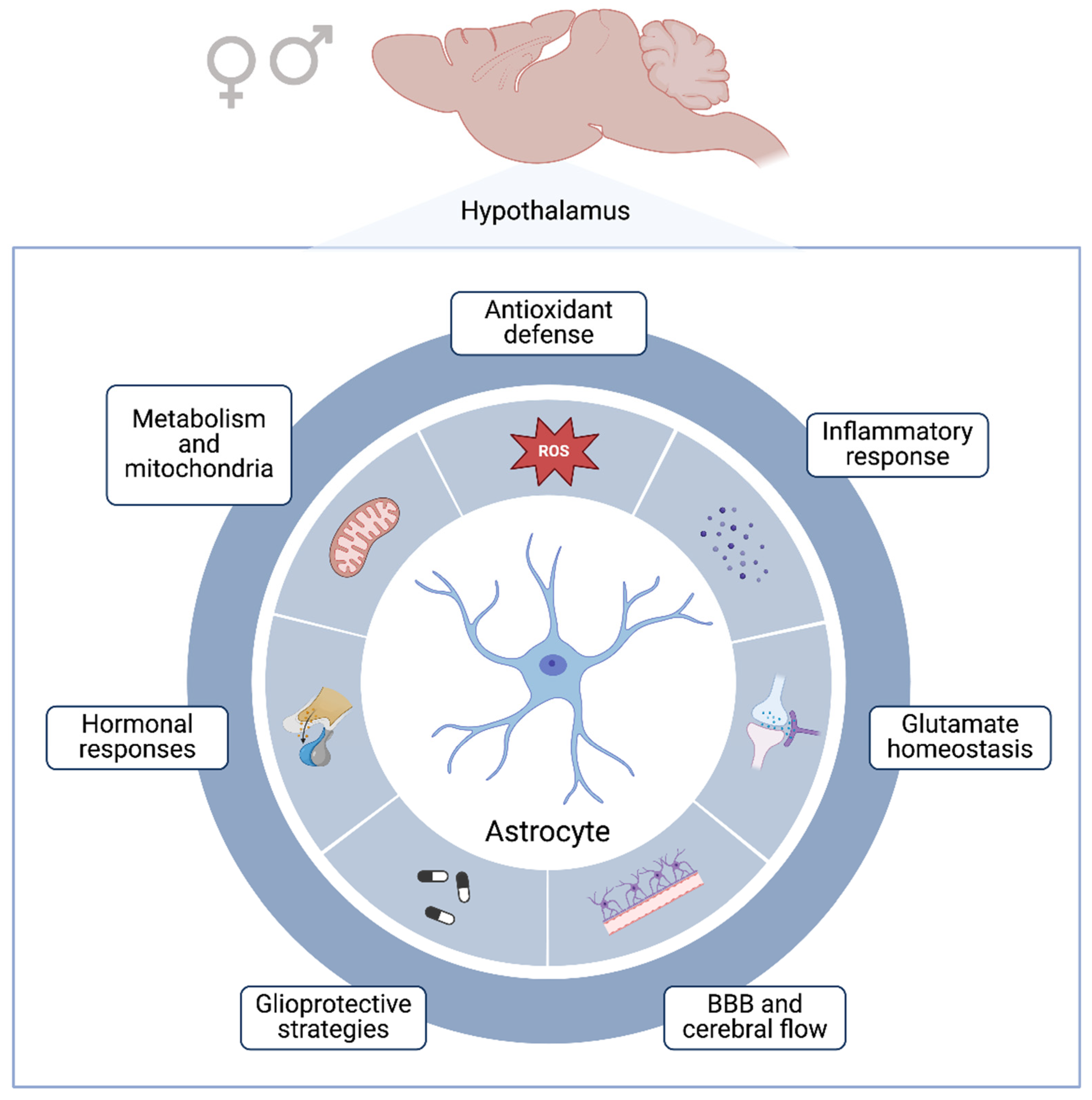
6. Glioprotective Mechanisms Associated with Hypothalamus

The physiological role of astrocytes has already been previously addressed (Figure 1); however, these cells can also react to insults through a process named “astrocyte reactivity”, a phenotype that is potentially harmful since it is associated with inflammatory processes and oxidative stress [122]. On the other hand, astrocytes are also capable of protecting the CNS. Thus, the term “glioprotection” is defined as a response originating from glia to preserve the functional properties of other glial cells and/or neurons [38]. In this review, we highlighted the glioprotective mechanisms associated with steroids as well as non-hormonal molecules, that have been described by our group. Estrogen and progesterone negatively regulate the release of pro-inflammatory mediators and increase autophagy in astrocytes through the suppression of mTOR/nuclear factor kappa B (NFκB) signaling [123,124]. In addition, estrogen has been shown to be beneficial in conditions of oxidative stress and excitotoxicity in experimental models of stroke, Parkinson’s disease, and multiple sclerosis, with glioprotective actions [125]. Moreover, steroid hormones regulate the expression of aquaporins by astrocytes, preserving the tissue and reducing post-injury edema [126,127,128]. It is noteworthy that astrocytes and neurons from male mice appear to be more susceptible (3 to 4 times) to toxicity caused by oxidative stress than cells from female mice [129].
Hormone therapy appears to be a promising alternative for CNS functional restoration. Steroidal or non-steroidal compounds can directly or indirectly affect astrocytes in both physiological and pathological conditions [130]. Thus, currently, the use of estrogenic compounds has been investigated from a therapeutic perspective, since these molecules stimulate glioprotection by astrocytes. Notably, phytoestrogens are molecules of plant origin, including isoflavones (such as genistein) and stilbenes (such as resveratrol), which have significant biological effects on glial cells [131]. Both isoflavones and resveratrol are estrogen receptor modulators [132]. The mechanisms underlying their glioprotective actions involve antioxidant and anti-inflammatory activities, improvement of mitochondrial function, Nrf2/heme oxygenase 1 (HO-1) activation, and NFκB inhibition, as well as glutamate clearance and metabolism [38,133]. Additionally, we recently demonstrated that metformin and simvastatin, drugs used in metabolic disorders (diabetes and hyperlipidemia, respectively), modulate gene expression in neonatal hypothalamic astrocytes obtained from an immunocompromised mice model [134,135]. However, their roles in sexual dimorphism are still poorly known, although the estrogen receptor is required for specific biological effects of metformin and simvastatin [136,137]. In this sense, it is important to note that hypothalamic astrocytes regulate glucose metabolism and cholesterol synthesis in a sex-dependent manner, along with immunity, indicating the relevance of our works about glioprotection. In addition, studies in progress in our Lab (LABGLIO) will confirm and expand the knowledge about glioprotective mechanisms in female rodents [2,3,36].

7. Concluding Remarks and Future Directions

Although there are significant differences between the sexes, most experimental studies use male animals. In the CNS, especially in hypothalamic astrocytes, important sexually dimorphic differences were observed in rodents, indicating that sex hormones have a significant influence on such differences. Therefore, it is important to highlight the use of both sexes in scientific research (preclinical and clinical studies). This approach can promote a better understanding of the pathomechanisms involved in dimorphic patterns, including in the CNS, and improve effective therapeutic and glioprotective strategies, that must take hormonal variables into consideration. Thus, sexual equity in animal models and studies on astrocytes can contribute to this relevant topic and provide essential information regarding obesity, aging, neurodegeneration, and neuropsychiatric disorders.

Author Contributions

N.K.T., L.D.B. and A.Q.-S. conceived, wrote, and revised the manuscript. All authors have read and agreed to the published version of the manuscript.

Funding

This research received no external funding.

Acknowledgments

The authors are supported by the Universidade Federal do Rio Grande do Sul (UFRGS), Conselho Nacional de Desenvolvimento Científico e Tecnológico (CNPq), Coordenação de Aperfeiçoamento de Pessoal de Nível Superior (CAPES), and Fundação de Amparo à Pesquisa do Estado do Rio Grande do Sul (FAPERGS).

Conflicts of Interest

The authors declare no conflicts of interest.

References

  1. Sovrani, V.; Bobermin, L.D.; Santos, C.L.; Brondani, M.; Gonçalves, C.-A.; Leipnitz, G.; Quincozes-Santos, A. Effects of Long-Term Resveratrol Treatment in Hypothalamic Astrocyte Cultures from Aged Rats. Mol. Cell. Biochem. 2023, 478, 1205–1216. [Google Scholar] [CrossRef]
  2. Fong, H.; Zheng, J.; Kurrasch, D. The Structural and Functional Complexity of the Integrative Hypothalamus. Science 2023, 382, 388–394. [Google Scholar] [CrossRef]
  3. Maroso, M.; Stern, P. Small and Mighty: The Hypothalamus. Science 2023, 382, 386–387. [Google Scholar] [CrossRef]
  4. Nampoothiri, S.; Nogueiras, R.; Schwaninger, M.; Prevot, V. Glial Cells as Integrators of Peripheral and Central Signals in the Regulation of Energy Homeostasis. Nat. Metab. 2022, 4, 813–825. [Google Scholar] [CrossRef]
  5. Luengo-Mateos, M.; González-Vila, A.; Vicente Dragano, N.R.; Ohinska, N.; Silveira-Loureiro, M.; González-Domínguez, M.; Estévez-Salguero, Á.; Novelle-Rodríguez, P.; López, M.; Barca-Mayo, O. Hypothalamic Astrocytic-BMAL1 Regulates Energy Homeostasis in a Sex-Dependent Manner. Cell Rep. 2023, 42, 112949. [Google Scholar] [CrossRef]
  6. Chowen, J.A.; Frago, L.M.; Fernández-Alfonso, M.S. Physiological and Pathophysiological Roles of Hypothalamic Astrocytes in Metabolism. J. Neuroendocrinol. 2019, 31, e12671. [Google Scholar] [CrossRef] [PubMed]
  7. Hatcher, K.M.; Royston, S.E.; Mahoney, M.M. Modulation of Circadian Rhythms through Estrogen Receptor Signaling. Eur. J. Neurosci. 2020, 51, 217–228. [Google Scholar] [CrossRef]
  8. Davis, F.C.; Darrow, J.M.; Menaker, M. Sex Differences in the Circadian Control of Hamster Wheel-Running Activity. Am. J. Physiol.-Regul. Integr. Comp. Physiol. 1983, 244, R93–R105. [Google Scholar] [CrossRef]
  9. Becker, J.B.; Arnold, A.P.; Berkley, K.J.; Blaustein, J.D.; Eckel, L.A.; Hampson, E.; Herman, J.P.; Marts, S.; Sadee, W.; Steiner, M.; et al. Strategies and Methods for Research on Sex Differences in Brain and Behavior. Endocrinology 2005, 146, 1650–1673. [Google Scholar] [CrossRef]
  10. Heberden, C. Sex Steroids and Neurogenesis. Biochem. Pharmacol. 2017, 141, 56–62. [Google Scholar] [CrossRef]
  11. Mong, J.A.; Glaser, E.; McCarthy, M.M. Gonadal Steroids Promote Glial Differentiation and Alter Neuronal Morphology in the Developing Hypothalamus in a Regionally Specific Manner. J. Neurosci. 1999, 19, 1464–1472. [Google Scholar] [CrossRef]
  12. Goodfellow, P.N.; Lovell-Badge, R. SRY and Sex Determination in Mammals. Annu. Rev. Genet. 1993, 27, 71–92. [Google Scholar] [CrossRef]
  13. Weisz, J.; Ward, I.L. Plasma Testosterone and Progesterone Titers of Pregnant Rats, Their Male and Female Fetuses, and Neonatal Offspring. Endocrinology 1980, 106, 306–316. [Google Scholar] [CrossRef]
  14. Bakker, J. The Role of Steroid Hormones in the Sexual Differentiation of the Human Brain. J. Neuroendocrinol. 2022, 34, e13050. [Google Scholar] [CrossRef]
  15. Singh, G.; Singh, V.; Schneider, J.S. Post-Translational Histone Modifications and Their Interaction with Sex Influence Normal Brain Development and Elaboration of Neuropsychiatric Disorders. Biochim. Biophys. Acta (BBA)-Mol. Basis Dis. 2019, 1865, 1968–1981. [Google Scholar] [CrossRef]
  16. Arambula, S.E.; McCarthy, M.M. Neuroendocrine-Immune Crosstalk Shapes Sex-Specific Brain Development. Endocrinology 2020, 161, bqaa055. [Google Scholar] [CrossRef]
  17. Deverman, B.E.; Patterson, P.H. Cytokines and CNS Development. Neuron 2009, 64, 61–78. [Google Scholar] [CrossRef]
  18. Nelson, L.H.; Lenz, K.M. The Immune System as a Novel Regulator of Sex Differences in Brain and Behavioral Development. J. Neurosci. Res. 2017, 95, 447–461. [Google Scholar] [CrossRef]
  19. Schwarz, J.M.; Sholar, P.W.; Bilbo, S.D. Sex Differences in Microglial Colonization of the Developing Rat Brain. J. Neurochem. 2012, 120, 948–963. [Google Scholar] [CrossRef]
  20. VanRyzin, J.W.; Marquardt, A.E.; Argue, K.J.; Vecchiarelli, H.A.; Ashton, S.E.; Arambula, S.E.; Hill, M.N.; McCarthy, M.M. Microglial Phagocytosis of Newborn Cells Is Induced by Endocannabinoids and Sculpts Sex Differences in Juvenile Rat Social Play. Neuron 2019, 102, 435–449.e6. [Google Scholar] [CrossRef]
  21. Garcia-Segura, L.M.; Suarez, I.; Segovia, S.; Tranque, P.A.; Calés, J.M.; Aguilera, P.; Olmos, G.; Guillamón, A. The Distribution of Glial Fibrillary Acidic Protein in the Adult Rat Brain Is Influenced by the Neonatal Levels of Sex Steroids. Brain Res. 1988, 456, 357–363. [Google Scholar] [CrossRef] [PubMed]
  22. Been, L.E.; Sheppard, P.A.S.; Galea, L.A.M.; Glasper, E.R. Hormones and Neuroplasticity: A Lifetime of Adaptive Responses. Neurosci. Biobehav. Rev. 2022, 132, 679–690. [Google Scholar] [CrossRef] [PubMed]
  23. Dafny, N.; Feldman, S. Unit Responses and Convergence of Sensory Stimuli in the Hypothalamus. Brain Res. 1970, 17, 243–257. [Google Scholar] [CrossRef] [PubMed]
  24. Sominsky, L.; Jasoni, C.L.; Twigg, H.R.; Spencer, S.J. Hormonal and Nutritional Regulation of Postnatal Hypothalamic Development. J. Endocrinol. 2018, 237, R47–R64. [Google Scholar] [CrossRef]
  25. Carrer, H.F.; Cambiasso, M.J. Sexual Differentiation of the Brain: Genes, Estrogen, and Neurotrophic Factors. Cell. Mol. Neurobiol. 2002, 22, 479–500. [Google Scholar] [CrossRef]
  26. Silva, M.S.B.; Giacobini, P. New Insights into Anti-Müllerian Hormone Role in the Hypothalamic–Pituitary–Gonadal Axis and Neuroendocrine Development. Cell. Mol. Life Sci. 2021, 78, 1–16. [Google Scholar] [CrossRef] [PubMed]
  27. Marcondes, F.K.; Bianchi, F.J.; Tanno, A.P. Determination of the Estrous Cycle Phases of Rats: Some Helpful Considerations. Braz. J. Biol. 2002, 62, 609–614. [Google Scholar] [CrossRef] [PubMed]
  28. Bridges, R.S. Neuroendocrine Regulation of Maternal Behavior. Front. Neuroendocrinol. 2015, 36, 178–196. [Google Scholar] [CrossRef]
  29. Frederich, R.C.; Hamann, A.; Anderson, S.; Löllmann, B.; Lowell, B.B.; Flier, J.S. Leptin Levels Reflect Body Lipid Content in Mice: Evidence for Diet-Induced Resistance to Leptin Action. Nat. Med. 1995, 1, 1311–1314. [Google Scholar] [CrossRef]
  30. Koebele, S.V.; Bimonte-Nelson, H.A. Modeling Menopause: The Utility of Rodents in Translational Behavioral Endocrinology Research. Maturitas 2016, 87, 5–17. [Google Scholar] [CrossRef]
  31. Qian, J.; Morris, C.J.; Caputo, R.; Wang, W.; Garaulet, M.; Scheer, F.A.J.L. Sex Differences in the Circadian Misalignment Effects on Energy Regulation. Proc. Natl. Acad. Sci. USA 2019, 116, 23806–23812. [Google Scholar] [CrossRef] [PubMed]
  32. Novozhilova, M.; Mishchenko, T.; Kondakova, E.; Lavrova, T.; Gavrish, M.; Aferova, S.; Franceschi, C.; Vedunova, M. Features of Age-Related Response to Sleep Deprivation: In Vivo Experimental Studies. Aging 2021, 13, 19108–19126. [Google Scholar] [CrossRef] [PubMed]
  33. Wyse, A.T.; Siebert, C.; Bobermin, L.D.; Dos Santos, T.M.; Quincozes-Santos, A. Changes in Inflammatory Response, Redox Status and Na+, K+-ATPase Activity in Primary Astrocyte Cultures from Female Wistar Rats Subject to Ovariectomy. Neurotox. Res. 2020, 37, 445–454. [Google Scholar] [CrossRef] [PubMed]
  34. Sumien, N.; Cunningham, J.T.; Davis, D.L.; Engelland, R.; Fadeyibi, O.; Farmer, G.E.; Mabry, S.; Mensah-Kane, P.; Trinh, O.T.P.; Vann, P.H.; et al. Neurodegenerative Disease: Roles for Sex, Hormones, and Oxidative Stress. Endocrinology 2021, 162, bqab185. [Google Scholar] [CrossRef] [PubMed]
  35. Razmara, A.; Duckles, S.P.; Krause, D.N.; Procaccio, V. Estrogen Suppresses Brain Mitochondrial Oxidative Stress in Female and Male Rats. Brain Res. 2007, 1176, 71–81. [Google Scholar] [CrossRef] [PubMed]
  36. Shepherd, R.; Cheung, A.S.; Pang, K.; Saffery, R.; Novakovic, B. Sexual Dimorphism in Innate Immunity: The Role of Sex Hormones and Epigenetics. Front. Immunol. 2021, 11, 604000. [Google Scholar] [CrossRef]
  37. Sohrabji, F.; Okoreeh, A.; Panta, A. Sex Hormones and Stroke: Beyond Estrogens. Horm. Behav. 2019, 111, 87–95. [Google Scholar] [CrossRef]
  38. Quincozes-Santos, A.; Santos, C.L.; de Souza Almeida, R.R.; da Silva, A.; Thomaz, N.K.; Costa, N.L.F.; Weber, F.B.; Schmitz, I.; Medeiros, L.S.; Medeiros, L.; et al. Gliotoxicity and Glioprotection: The Dual Role of Glial Cells. Mol. Neurobiol. 2021, 58, 6577–6592. [Google Scholar] [CrossRef] [PubMed]
  39. Camandola, S. Astrocytes, Emerging Stars of Energy Homeostasis. Cell Stress 2018, 2, 246–252. [Google Scholar] [CrossRef]
  40. Linnerbauer, M.; Wheeler, M.A.; Quintana, F.J. Astrocyte Crosstalk in CNS Inflammation. Neuron 2020, 108, 608–622. [Google Scholar] [CrossRef]
  41. Verkhratsky, A.; Nedergaard, M. Physiology of Astroglia. Physiol. Rev. 2018, 98, 239–389. [Google Scholar] [CrossRef] [PubMed]
  42. Collignon, A.; Dion-Albert, L.; Ménard, C.; Coelho-Santos, V. Sex, Hormones and Cerebrovascular Function: From Development to Disorder. Fluids Barriers CNS 2024, 21, 2. [Google Scholar] [CrossRef] [PubMed]
  43. Micevych, P.; Sinchak, K. Estradiol Regulation of Progesterone Synthesis in the Brain. Mol. Cell. Endocrinol. 2008, 290, 44–50. [Google Scholar] [CrossRef] [PubMed]
  44. Ubuka, T.; Tsutsui, K. Neuropeptidergic Control of Neurosteroids Biosynthesis. Front. Neuroendocrinol. 2022, 65, 100976. [Google Scholar] [CrossRef]
  45. Chen, C.; Kuo, J.; Wong, A.; Micevych, P. Estradiol Modulates Translocator Protein (Tspo) and Steroid Acute Regulatory Protein (StAR) via Protein Kinase A (PKA) Signaling in Hypothalamic Astrocytes. Endocrinology 2014, 155, 2976–2985. [Google Scholar] [CrossRef] [PubMed]
  46. Pawlak, J.; Karolczak, M.; Krust, A.; Chambon, P.; Beyer, C. Estrogen Receptor-α Is Associated with the Plasma Membrane of Astrocytes and Coupled to the MAP/Src-Kinase Pathway. Glia 2005, 50, 270–275. [Google Scholar] [CrossRef]
  47. Heemers, H.V.; Tindall, D.J. Androgen Receptor (AR) Coregulators: A Diversity of Functions Converging on and Regulating the AR Transcriptional Complex. Endocr. Rev. 2007, 28, 778–808. [Google Scholar] [CrossRef] [PubMed]
  48. Foradori, C.D.; Weiser, M.J.; Handa, R.J. Non-Genomic Actions of Androgens. Front. Neuroendocrinol. 2008, 29, 169–181. [Google Scholar] [CrossRef] [PubMed]
  49. Gore, A.C.; Oung, T.; Yung, S.; Flagg, R.A.; Woller, M.J. Neuroendocrine Mechanisms for Reproductive Senescence in the Female Rat: Gonadotropin-Releasing Hormone Neurons. Endocrine 2000, 13, 315–323. [Google Scholar] [CrossRef]
  50. Illarionova, N.B.; Illarionava, N.B.; Brismar, H.; Aperia, A.; Gunnarson, E. Role of Na,K-ATPase A1 and A2 Isoforms in the Support of Astrocyte Glutamate Uptake. PLoS ONE 2014, 9, e98469. [Google Scholar] [CrossRef]
  51. Chakraborti, A.; Gulati, K.; Banerjee, B.D.; Ray, A. Possible Involvement of Free Radicals in the Differential Neurobehavioral Responses to Stress in Male and Female Rats. Behav. Brain Res. 2007, 179, 321–325. [Google Scholar] [CrossRef] [PubMed]
  52. Guevara, R.; Santandreu, F.M.; Valle, A.; Gianotti, M.; Oliver, J.; Roca, P. Sex-Dependent Differences in Aged Rat Brain Mitochondrial Function and Oxidative Stress. Free Radic. Biol. Med. 2009, 46, 169–175. [Google Scholar] [CrossRef] [PubMed]
  53. Sobočanec, S.; Balog, T.; Kušić, B.; Šverko, V.; Šarić, A.; Marotti, T. Differential Response to Lipid Peroxidation in Male and Female Mice with Age: Correlation of Antioxidant Enzymes Matters. Biogerontology 2008, 9, 335–343. [Google Scholar] [CrossRef] [PubMed]
  54. Liddell, J. Are Astrocytes the Predominant Cell Type for Activation of Nrf2 in Aging and Neurodegeneration? Antioxidants 2017, 6, 65. [Google Scholar] [CrossRef] [PubMed]
  55. Gudkov, S.V.; Burmistrov, D.E.; Kondakova, E.V.; Sarimov, R.M.; Yarkov, R.S.; Franceschi, C.; Vedunova, M.V. An Emerging Role of Astrocytes in Aging/Neuroinflammation and Gut-Brain Axis with Consequences on Sleep and Sleep Disorders. Ageing Res. Rev. 2023, 83, 101775. [Google Scholar] [CrossRef] [PubMed]
  56. Santos-Galindo, M.; Acaz-Fonseca, E.; Bellini, M.J.; Garcia-Segura, L.M. Sex Differences in the Inflammatory Response of Primary Astrocytes to Lipopolysaccharide. Biol. Sex Differ. 2011, 2, 7. [Google Scholar] [CrossRef] [PubMed]
  57. García-Cáceres, C.; Quarta, C.; Varela, L.; Gao, Y.; Gruber, T.; Legutko, B.; Jastroch, M.; Johansson, P.; Ninkovic, J.; Yi, C.-X.; et al. Astrocytic Insulin Signaling Couples Brain Glucose Uptake with Nutrient Availability. Cell 2016, 166, 867–880. [Google Scholar] [CrossRef]
  58. Fernandez-Galaz, M.D.C.; Naftolin, F.; Garcia-Segura, L.M. Phasic Synaptic Remodeling of the Rat Arcuate Nucleus during the Estrous Cycle Depends on Insulin-like Growth Factor-I Receptor Activation. J. Neurosci. Res. 1999, 55, 286–292. [Google Scholar] [CrossRef]
  59. Björnström, L.; Sjöberg, M. Signal Transducers and Activators of Transcription as Downstream Targets of Nongenomic Estrogen Receptor Actions. Mol. Endocrinol. 2002, 16, 2202–2214. [Google Scholar] [CrossRef]
  60. Childs, G.V.; Odle, A.K.; MacNicol, M.C.; MacNicol, A.M. The Importance of Leptin to Reproduction. Endocrinology 2021, 162, bqaa204. [Google Scholar] [CrossRef]
  61. Bélanger, M.; Allaman, I.; Magistretti, P.J. Brain Energy Metabolism: Focus on Astrocyte-Neuron Metabolic Cooperation. Cell Metab. 2011, 14, 724–738. [Google Scholar] [CrossRef] [PubMed]
  62. Ciofi, P.; Garret, M.; Lapirot, O.; Lafon, P.; Loyens, A.; Prévot, V.; Levine, J.E. Brain-Endocrine Interactions: A Microvascular Route in the Mediobasal Hypothalamus. Endocrinology 2009, 150, 5509–5519. [Google Scholar] [CrossRef]
  63. Kaczkurkin, A.N.; Raznahan, A.; Satterthwaite, T.D. Sex Differences in the Developing Brain: Insights from Multimodal Neuroimaging. Neuropsychopharmacology 2019, 44, 71–85. [Google Scholar] [CrossRef]
  64. Liu, Y.; Zhu, X.; Feinberg, D.; Guenther, M.; Gregori, J.; Weiner, M.W.; Schuff, N. Arterial Spin Labeling MRI Study of Age and Gender Effects on Brain Perfusion Hemodynamics. Magn. Reson. Med. 2012, 68, 912–922. [Google Scholar] [CrossRef]
  65. Chen, Z.; Yuhanna, I.S.; Galcheva-Gargova, Z.; Karas, R.H.; Mendelsohn, M.E.; Shaul, P.W. Estrogen Receptor α Mediates the Nongenomic Activation of Endothelial Nitric Oxide Synthase by Estrogen. J. Clin. Investig. 1999, 103, 401–406. [Google Scholar] [CrossRef]
  66. Yilmaz, C.; Karali, K.; Fodelianaki, G.; Gravanis, A.; Chavakis, T.; Charalampopoulos, I.; Alexaki, V.I. Neurosteroids as Regulators of Neuroinflammation. Front. Neuroendocrinol. 2019, 55, 100788. [Google Scholar] [CrossRef] [PubMed]
  67. Mellon, S.H.; Griffin, L.D. Neurosteroids: Biochemistry and Clinical Significance. Trends Endocrinol. Metab. 2002, 13, 35–43. [Google Scholar] [CrossRef] [PubMed]
  68. Melcangi, R.C.; Garcia-Segura, L.M.; Mensah-Nyagan, A.G. Neuroactive Steroids: State of the Art and New Perspectives. Cell. Mol. Life Sci. 2008, 65, 777–797. [Google Scholar] [CrossRef]
  69. Mauch, D.H.; Nägler, K.; Schumacher, S.; Göritz, C.; Müller, E.C.; Otto, A.; Pfrieger, F.W. CNS Synaptogenesis Promoted by Glia-Derived Cholesterol. Science 2001, 294, 1354–1357. [Google Scholar] [CrossRef]
  70. Meffre, D.; Labombarda, F.; Delespierre, B.; Chastre, A.; de Nicola, A.F.; Stein, D.G.; Schumacher, M.; Guennoun, R. Distribution of Membrane Progesterone Receptor Alpha in the Male Mouse and Rat Brain and Its Regulation after Traumatic Brain Injury. Neuroscience 2013, 231, 111–124. [Google Scholar] [CrossRef]
  71. Lorenz, B.; Garcia-Segura, L.M.; DonCarlos, L.L. Cellular Phenotype of Androgen Receptor-Immunoreactive Nuclei in the Developing and Adult Rat Brain. J. Comp. Neurol. 2005, 492, 456–468. [Google Scholar] [CrossRef] [PubMed]
  72. Stone, D.J.; Song, Y.; Anderson, C.P.; Krohn, K.K.; Finch, C.E.; Rozovsky, I. Bidirectional Transcription Regulation of Glial Fibrillary Acidic Protein by Estradiol in Vivo and in Vitro. Endocrinology 1998, 139, 3202–3209. [Google Scholar] [CrossRef] [PubMed]
  73. Day, J.R.; Laping, N.J.; Lampert-Etchells, M.; Brown, S.A.; O’Callaghan, J.P.; McNeill, T.H.; Finch, C.E. Gonadal Steroids Regulate the Expression of Glial Fibrillary Acidic Protein in the Adult Male Rat Hippocampus. Neuroscience 1993, 55, 435–443. [Google Scholar] [CrossRef] [PubMed]
  74. Nilsson, M.; Pekny, M. Enriched Environment and Astrocytes in Central Nervous System Regeneration. J. Rehabil. Med. 2007, 39, 345–352. [Google Scholar] [CrossRef] [PubMed]
  75. Johnson, R.T.; Schneider, A.; DonCarlos, L.L.; Breedlove, S.M.; Jordan, C.L. Astrocytes in the Rat Medial Amygdala Are Responsive to Adult Androgens. J. Comp. Neurol. 2012, 520, 2531–2544. [Google Scholar] [CrossRef] [PubMed]
  76. Borrás, C.; Sastre, J.; García-Sala, D.; Lloret, A.; Pallardó, F.V.; Viña, J. Mitochondria from Females Exhibit Higher Antioxidant Gene Expression and Lower Oxidative Damage than Males. Free Radic. Biol. Med. 2003, 34, 546–552. [Google Scholar] [CrossRef] [PubMed]
  77. Morken, T.S.; Brekke, E.; Håberg, A.; Widerøe, M.; Brubakk, A.-M.; Sonnewald, U. Altered Astrocyte–Neuronal Interactions After Hypoxia-Ischemia in the Neonatal Brain in Female and Male Rats. Stroke 2014, 45, 2777–2785. [Google Scholar] [CrossRef] [PubMed]
  78. Torrens-Mas, M.; Pons, D.-G.; Sastre-Serra, J.; Oliver, J.; Roca, P. Sexual Hormones Regulate the Redox Status and Mitochondrial Function in the Brain. Pathological implications. Redox Biol. 2020, 31, 101505. [Google Scholar] [CrossRef]
  79. Wu, J.; Williams, D.; Walter, G.A.; Thompson, W.E.; Sidell, N. Estrogen Increases Nrf2 Activity through Activation of the PI3K Pathway in MCF-7 Breast Cancer Cells. Exp. Cell Res. 2014, 328, 351–360. [Google Scholar] [CrossRef]
  80. Chistyakov, D.V.; Azbukina, N.V.; Astakhova, A.A.; Goriainov, S.V.; Chistyakov, V.V.; Sergeeva, M.G. Sex-Mediated Differences in LPS Induced Alterations of TNFα, IL-10 Expression, and Prostaglandin Synthesis in Primary Astrocytes. Int. J. Mol. Sci. 2018, 19, 2793. [Google Scholar] [CrossRef]
  81. Jha, M.K.; Jo, M.; Kim, J.-H.; Suk, K. Microglia-Astrocyte Crosstalk: An Intimate Molecular Conversation. Neuroscientist 2019, 25, 227–240. [Google Scholar] [CrossRef]
  82. Lennol, M.P.; Canelles, S.; Guerra-Cantera, S.; Argente, J.; García-Segura, L.M.; de Ceballos, M.L.; Chowen, J.A.; Frago, L.M. Amyloid-Β1-40 Differentially Stimulates Proliferation, Activation of Oxidative Stress and Inflammatory Responses in Male and Female Hippocampal Astrocyte Cultures. Mech. Ageing Dev. 2021, 195, 111462. [Google Scholar] [CrossRef]
  83. Musatov, S.; Chen, W.; Pfaff, D.W.; Mobbs, C.V.; Yang, X.-J.; Clegg, D.J.; Kaplitt, M.G.; Ogawa, S. Silencing of Estrogen Receptor Alpha in the Ventromedial Nucleus of Hypothalamus Leads to Metabolic Syndrome. Proc. Natl. Acad. Sci. USA 2007, 104, 2501–2506. [Google Scholar] [CrossRef]
  84. Frye, C.A. Neurosteroids’ Effects and Mechanisms for Social, Cognitive, Emotional, and Physical Functions. Psychoneuroendocrinology 2009, 34 (Suppl. 1), S143–S161. [Google Scholar] [CrossRef]
  85. Jais, A.; Brüning, J.C. Hypothalamic Inflammation in Obesity and Metabolic Disease. J. Clin. Investig. 2017, 127, 24–32. [Google Scholar] [CrossRef] [PubMed]
  86. Louwe, M.C.; van der Hoorn, J.W.A.; van den Berg, S.A.A.; Jukema, J.W.; Romijn, J.A.; van Dijk, K.W.; Rensen, P.C.N.; Smit, J.W.A.; Steendijk, P. Gender-Dependent Effects of High-Fat Lard Diet on Cardiac Function in C57Bl/6J Mice. Appl. Physiol. Nutr. Metab. 2012, 37, 214–224. [Google Scholar] [CrossRef]
  87. Morselli, E.; Criollo, A.; Rodriguez-Navas, C.; Clegg, D.J. Chronic High Fat Diet Consumption Impairs Metabolic Health of Male Mice. Inflamm. Cell Signal. 2014, 1, e561. [Google Scholar] [CrossRef] [PubMed]
  88. Bonvento, G.; Bolaños, J.P. Astrocyte-Neuron Metabolic Cooperation Shapes Brain Activity. Cell Metab. 2021, 33, 1546–1564. [Google Scholar] [CrossRef] [PubMed]
  89. Zhang, Y.; Reichel, J.M.; Han, C.; Zuniga-Hertz, J.P.; Cai, D. Astrocytic Process Plasticity and IKKβ/NF-κB in Central Control of Blood Glucose, Blood Pressure, and Body Weight. Cell Metab. 2017, 25, 1091–1102.e4. [Google Scholar] [CrossRef]
  90. Wu, X.; Huang, J.; Shen, C.; Liu, Y.; He, S.; Sun, J.; Yu, B. NRF2 Deficiency Increases Obesity Susceptibility in a Mouse Menopausal Model. PLoS ONE 2020, 15, e0228559. [Google Scholar] [CrossRef]
  91. Hidalgo-Lanussa, O.; González Santos, J.; Barreto, G.E. Sex-Specific Vulnerabilities in Human Astrocytes Underpin the Differential Impact of Palmitic Acid. Neurobiol. Dis. 2024, 195, 106489. [Google Scholar] [CrossRef] [PubMed]
  92. Allison, M.B.; Myers, M.G. 20 Years of Leptin: Connecting Leptin Signaling to Biological Function. J. Endocrinol. 2014, 223, T25–T35. [Google Scholar] [CrossRef] [PubMed]
  93. Banks, W.A. Brain Meets Body: The Blood-Brain Barrier as an Endocrine Interface. Endocrinology 2012, 153, 4111–4119. [Google Scholar] [CrossRef] [PubMed]
  94. Santos, C.L.; Roppa, P.H.A.; Truccolo, P.; Fontella, F.U.; Souza, D.O.; Bobermin, L.D.; Quincozes-Santos, A. Age-Dependent Neurochemical Remodeling of Hypothalamic Astrocytes. Mol. Neurobiol. 2018, 55, 5565–5579. [Google Scholar] [CrossRef] [PubMed]
  95. García-Cáceres, C.; Fuente-Martín, E.; Burgos-Ramos, E.; Granado, M.; Frago, L.M.; Barrios, V.; Horvath, T.; Argente, J.; Chowen, J.A. Differential Acute and Chronic Effects of Leptin on Hypothalamic Astrocyte Morphology and Synaptic Protein Levels. Endocrinology 2011, 152, 1809–1818. [Google Scholar] [CrossRef] [PubMed]
  96. Pinto, S.; Roseberry, A.G.; Liu, H.; Diano, S.; Shanabrough, M.; Cai, X.; Friedman, J.M.; Horvath, T.L. Rapid Rewiring of Arcuate Nucleus Feeding Circuits by Leptin. Science 2004, 304, 110–115. [Google Scholar] [CrossRef] [PubMed]
  97. Burguera, B.; Couce, M.E.; Curran, G.L.; Jensen, M.D.; Lloyd, R.V.; Cleary, M.P.; Poduslo, J.F. Obesity Is Associated with a Decreased Leptin Transport across the Blood-Brain Barrier in Rats. Diabetes 2000, 49, 1219–1223. [Google Scholar] [CrossRef] [PubMed]
  98. Thaler, J.P.; Yi, C.-X.; Schur, E.A.; Guyenet, S.J.; Hwang, B.H.; Dietrich, M.O.; Zhao, X.; Sarruf, D.A.; Izgur, V.; Maravilla, K.R.; et al. Obesity Is Associated with Hypothalamic Injury in Rodents and Humans. J. Clin. Investig. 2012, 122, 153–162. [Google Scholar] [CrossRef] [PubMed]
  99. Santos, C.L.; Bobermin, L.D.; Souza, D.O.; Quincozes-Santos, A. Leptin Stimulates the Release of Pro-Inflammatory Cytokines in Hypothalamic Astrocyte Cultures from Adult and Aged Rats. Metab. Brain Dis. 2018, 33, 2059–2063. [Google Scholar] [CrossRef]
  100. Kelly, M.J.; Qiu, J. Estrogen Signaling in Hypothalamic Circuits Controling Reproduction. Brain Res. 2010, 1364, 44–52. [Google Scholar] [CrossRef]
  101. Kamitakahara, A.; Bouyer, K.; Wang, C.-H.; Simerly, R. A Critical Period for the Trophic Actions of Leptin on AgRP Neurons in the Arcuate Nucleus of the Hypothalamus. J. Comp. Neurol. 2018, 526, 133–145. [Google Scholar] [CrossRef] [PubMed]
  102. Young, E.A.; Lopez, J.F.; Murphy-Weinberg, V.; Watson, S.J.; Akil, H. Hormonal Evidence for Altered Responsiveness to Social Stress in Major Depression. Neuropsychopharmacology 2000, 23, 411–418. [Google Scholar] [CrossRef] [PubMed]
  103. Kajantie, E.; Phillips, D.I.W. The Effects of Sex and Hormonal Status on the Physiological Response to Acute Psychosocial Stress. Psychoneuroendocrinology 2006, 31, 151–178. [Google Scholar] [CrossRef] [PubMed]
  104. Huang, Y.-C.; Hung, C.-F.; Lin, P.-Y.; Lee, Y.; Wu, C.-C.; Hsu, S.-T.; Chen, C.-C.; Chong, M.-Y.; Lin, C.-H.; Wang, L.-J. Gender Differences in Susceptibility to Schizophrenia: Potential Implication of Neurosteroids. Psychoneuroendocrinology 2017, 84, 87–93. [Google Scholar] [CrossRef] [PubMed]
  105. Manoli, D.S.; Tollkuhn, J. Gene Regulatory Mechanisms Underlying Sex Differences in Brain Development and Psychiatric Disease. Ann. N. Y. Acad. Sci. 2018, 1420, 26–45. [Google Scholar] [CrossRef] [PubMed]
  106. Trotman, H.D.; Holtzman, C.W.; Ryan, A.T.; Shapiro, D.I.; MacDonald, A.N.; Goulding, S.M.; Brasfield, J.L.; Walker, E.F. The Development of Psychotic Disorders in Adolescence: A Potential Role for Hormones. Horm. Behav. 2013, 64, 411–419. [Google Scholar] [CrossRef] [PubMed]
  107. Patchev, V.K.; Hayashi, S.; Orikasa, C.; Almeida, O.F.X. Implications of Estrogen-Dependent Brain Organization for Gender Differences in Hypothalamo-Pituitary-Adrenal Regulation. FASEB J. 1995, 9, 419–423. [Google Scholar] [CrossRef] [PubMed]
  108. Zorumski, C.F.; Paul, S.M.; Izumi, Y.; Covey, D.F.; Mennerick, S. Neurosteroids, Stress and Depression: Potential Therapeutic Opportunities. Neurosci. Biobehav. Rev. 2013, 37, 109–122. [Google Scholar] [CrossRef] [PubMed]
  109. Wang, M.; Hu, S.; Fu, X.; Zhou, H.; Yang, S.; Yang, C. Neurosteroids: A Potential Target for Neuropsychiatric Disorders. J. Steroid Biochem. Mol. Biol. 2024, 239, 106485. [Google Scholar] [CrossRef]
  110. Guennoun, R.; Labombarda, F.; Gonzalez Deniselle, M.C.; Liere, P.; de Nicola, A.F.; Schumacher, M. Progesterone and Allopregnanolone in the Central Nervous System: Response to Injury and Implication for Neuroprotection. J. Steroid Biochem. Mol. Biol. 2015, 146, 48–61. [Google Scholar] [CrossRef] [PubMed]
  111. Schüle, C.; Nothdurfter, C.; Rupprecht, R. The Role of Allopregnanolone in Depression and Anxiety. Prog. Neurobiol. 2014, 113, 79–87. [Google Scholar] [CrossRef]
  112. Vallée, M. Structure-Activity Relationship Studies on Neuroactive Steroids in Memory, Alcohol and Stress-Related Functions: A Crucial Benefit from Endogenous Level Analysis. Psychopharmacology 2014, 231, 3243–3255. [Google Scholar] [CrossRef] [PubMed]
  113. Tizabi, Y.; Getachew, B.; Hauser, S.R.; Tsytsarev, V.; Manhães, A.C.; da Silva, V.D.A. Role of Glial Cells in Neuronal Function, Mood Disorders, and Drug Addiction. Brain Sci. 2024, 14, 558. [Google Scholar] [CrossRef] [PubMed]
  114. Chivero, E.T.; Sil, S.; Kumar, M.; Buch, S. Substance Use, Microbiome and Psychiatric Disorders. Pharmacol. Biochem. Behav. 2022, 219, 173432. [Google Scholar] [CrossRef]
  115. Schmitz, I.; da Silva, A.; Bobermin, L.D.; Gonçalves, C.-A.; Steiner, J.; Quincozes-Santos, A. The Janus Face of Antipsychotics in Glial Cells: Focus on Glioprotection. Exp. Biol. Med. 2023, 248, 2120–2130. [Google Scholar] [CrossRef]
  116. Ostrom, Q.T.; Kinnersley, B.; Wrensch, M.R.; Eckel-Passow, J.E.; Armstrong, G.; Rice, T.; Chen, Y.; Wiencke, J.K.; McCoy, L.S.; Hansen, H.M.; et al. Sex-Specific Glioma Genome-Wide Association Study Identifies New Risk Locus at 3p21.31 in Females, and Finds Sex-Differences in Risk at 8q24.21. Sci. Rep. 2018, 8, 7352. [Google Scholar] [CrossRef]
  117. Daswani, B.; Khan, Y. Insights into the Role of Estrogens and Androgens in Glial Tumorigenesis. J. Carcinog. 2021, 20, 10. [Google Scholar] [CrossRef]
  118. Kokras, N.; Dioli, C.; Paravatou, R.; Sotiropoulos, M.G.; Delis, F.; Antoniou, K.; Calogeropoulou, T.; Charalampopoulos, I.; Gravanis, A.; Dalla, C. Psychoactive Properties of BNN27, a Novel Neurosteroid Derivate, in Male and Female Rats. Psychopharmacology 2020, 237, 2435–2449. [Google Scholar] [CrossRef] [PubMed]
  119. Powrie, Y.S.L.; Smith, C. Central Intracrine DHEA Synthesis in Ageing-Related Neuroinflammation and Neurodegeneration: Therapeutic Potential? J. Neuroinflamm. 2018, 15, 289. [Google Scholar] [CrossRef]
  120. Pike, C.J. Sex and the Development of Alzheimer’s Disease. J. Neurosci. Res. 2017, 95, 671–680. [Google Scholar] [CrossRef]
  121. Gillies, G.E.; Murray, H.E.; Dexter, D.; McArthur, S. Sex Dimorphisms in the Neuroprotective Effects of Estrogen in an Animal Model of Parkinson’s Disease. Pharmacol. Biochem. Behav. 2004, 78, 513–522. [Google Scholar] [CrossRef] [PubMed]
  122. Escartin, C.; Galea, E.; Lakatos, A.; O’Callaghan, J.P.; Petzold, G.C.; Serrano-Pozo, A.; Steinhäuser, C.; Volterra, A.; Carmignoto, G.; Agarwal, A.; et al. Reactive Astrocyte Nomenclature, Definitions, and Future Directions. Nat. Neurosci. 2021, 24, 312–325. [Google Scholar] [CrossRef] [PubMed]
  123. Cerciat, M.; Unkila, M.; Garcia-Segura, L.M.; Arevalo, M.-A. Selective Estrogen Receptor Modulators Decrease the Production of Interleukin-6 and Interferon-γ-inducible Protein-10 by Astrocytes Exposed to Inflammatory Challenge in Vitro. Glia 2010, 58, 93–102. [Google Scholar] [CrossRef] [PubMed]
  124. Giraud, S.N.; Caron, C.M.; Pham-Dinh, D.; Kitabgi, P.; Nicot, A.B. Estradiol Inhibits Ongoing Autoimmune Neuroinflammation and NFκB-Dependent CCL2 Expression in Reactive Astrocytes. Proc. Natl. Acad. Sci. USA 2010, 107, 8416–8421. [Google Scholar] [CrossRef] [PubMed]
  125. Marin, R. Estrogen Receptors in Lipid Raft Signalling Complexes for Neuroprotection. Front. Biosci. 2012, E4, 1420–1433. [Google Scholar] [CrossRef]
  126. Rutkowsky, J.M.; Wallace, B.K.; Wise, P.M.; O’Donnell, M.E. Effects of Estradiol on Ischemic Factor-Induced Astrocyte Swelling and AQP4 Protein Abundance. Am. J. Physiol.-Cell Physiol. 2011, 301, C204–C212. [Google Scholar] [CrossRef] [PubMed]
  127. Habib, P.; Dang, J.; Slowik, A.; Victor, M.; Beyer, C. Hypoxia-Induced Gene Expression of Aquaporin-4, Cyclooxygenase-2 and Hypoxia-Inducible Factor 1α in Rat Cortical Astroglia Is Inhibited by 17β-Estradiol and Progesterone. Neuroendocrinology 2014, 99, 156–167. [Google Scholar] [CrossRef] [PubMed]
  128. Gu, F.; Hata, R.; Toku, K.; Yang, L.; Ma, Y.; Maeda, N.; Sakanaka, M.; Tanaka, J. Testosterone Up-regulates Aquaporin-4 Expression in Cultured Astrocytes. J. Neurosci. Res. 2003, 72, 709–715. [Google Scholar] [CrossRef] [PubMed]
  129. Giordano, G.; Tait, L.; Furlong, C.E.; Cole, T.B.; Kavanagh, T.J.; Costa, L.G. Gender Differences in Brain Susceptibility to Oxidative Stress Are Mediated by Levels of Paraoxonase-2 Expression. Free Radic. Biol. Med. 2013, 58, 98–108. [Google Scholar] [CrossRef]
  130. Dhandapani, K.M.; Brann, D.W. Neuroprotective Effects of Estrogen and Tamoxifen In Vitro: A Facilitative Role for Glia? Endocrine 2003, 21, 59–66. [Google Scholar] [CrossRef]
  131. Martin-Jiménez, C.; Gaitán-Vaca, D.M.; Areiza, N.; Echeverria, V.; Ashraf, G.M.; González, J.; Sahebkar, A.; Garcia-Segura, L.M.; Barreto, G.E. Astrocytes Mediate Protective Actions of Estrogenic Compounds after Traumatic Brain Injury. Neuroendocrinology 2019, 108, 142–160. [Google Scholar] [CrossRef] [PubMed]
  132. Baker, M.E.; Lathe, R. The Promiscuous Estrogen Receptor: Evolution of Physiological Estrogens and Response to Phytochemicals and Endocrine Disruptors. J. Steroid Biochem. Mol. Biol. 2018, 184, 29–37. [Google Scholar] [CrossRef] [PubMed]
  133. Zhao, H.; Wang, Q.; Cheng, X.; Li, X.; Li, N.; Liu, T.; Li, J.; Yang, Q.; Dong, R.; Zhang, Y.; et al. Inhibitive Effect of Resveratrol on the Inflammation in Cultured Astrocytes and Microglia Induced by Aβ1–42. Neuroscience 2018, 379, 390–404. [Google Scholar] [CrossRef] [PubMed]
  134. Bobermin, L.D.; Sesterheim, P.; da Costa, D.S.; Rezena, E.; Schmitz, I.; da Silva, A.; de Moraes, A.D.M.; Souza, D.O.; Wyse, A.T.; Leipnitz, G.; et al. Simvastatin Differentially Modulates Glial Functions in Cultured Cortical and Hypothalamic Astrocytes Derived from Interferon α/β Receptor Knockout Mice. Neurochem. Res. 2024, 49, 732–743. [Google Scholar] [CrossRef] [PubMed]
  135. Bobermin, L.D.; da Costa, D.S.; de Moraes, A.D.M.; da Silva, V.F.; de Oliveira, G.T.; Sesterheim, P.; Tramontina, A.C.; Basso, L.A.; Leipnitz, G.; Quincozes-Santos, A.; et al. Effect of Metformin in Hypothalamic Astrocytes from an Immunocompromised Mice Model. Biochimie 2024, 223, S0300908424000804. [Google Scholar] [CrossRef]
  136. Sekar, A.P.; Nurmala, S.; Matsuura, E.; Tan, X.W.; Rahmasari, R.; Sauriasari, R. Estrogen Receptor Is Required for Metformin-Induced Apoptosis in Breast Cancer Cells Under Hyperglycemic Conditions. Breast Cancer 2024, 18, 11782234241240173. [Google Scholar] [CrossRef]
  137. Menze, E.T.; Ezzat, H.; Shawky, S.; Sami, M.; Selim, E.H.; Ahmed, S.; Maged, N.; Nadeem, N.; Eldash, S.; Michel, H.E. Simvastatin Mitigates Depressive-like Behavior in Ovariectomized Rats: Possible Role of NLRP3 Inflammasome and Estrogen Receptors’ Modulation. Int. Immunopharmacol. 2021, 95, 107582. [Google Scholar] [CrossRef]
Figure 1. Astrocyte functions deserve attention regarding sexual dimorphism. Hypothalamus is a crucial brain region involved in maintaining body homeostasis. Astrocytes display several functions including antioxidant defense, inflammatory response, glutamate homeostasis, BBB formation and cerebral flow regulation, metabolic support, and particularly in hypothalamus, hormonal responses. Therefore, astrocytes can be promising cellular targets for glioprotective effects. Considering all these crucial roles of astrocytes, investigating the functional differences between female and male cells is fundamental for understanding patterns of aging and neurodegenerative and neuropsychiatric disorders, as well as improving therapeutic strategies.
Neuroglia 05 00019 g001
Table 1. Sex differences between female and male brains.
Experimental Model/SubjectsMain FindingsReferences
Hypothalamic tissueAstrocytes from females and males are morphologically different [11]
Long-term sleep deprivation in miceWeight loss and cellular adaptive processes to increase stress resistance in young but not in adult female mice[30]
Cultured astrocytes from adult female ovariectomized ratsDecreased Na+, K+-ATPase activity
Increased pro-inflammatory cytokine release
[31]
MRI in human brainHigher cerebral flow in females[42]
Brain tissue and cultured astrocytesExpression of receptors for estrogen, progesterone and testosterone in astrocytes
Steroid hormones modulate GFAP expression in astrocytes
[43,44,45,46]
[47,48]
Rodent brainFemale brains present lower oxidative damage and increased antioxidant capacity[49,50,51,52]
Neonatal hypoxia-ischemiaAstrocytes from female rats show an enhanced mitochondrial metabolism, but male rats have higher recovery[53]
Primary astrocyte culturesFemale and male cells show different responses to an inflammatory stimulus
β-amyloid peptides induce an increased inflammatory response in females compared with males
[54,55]
[56]
Rat hypothalamic tissue Hypothalamic plasticity is prominent in female brain[57,58]
Human astrocytesCells from males showed increased reactive oxygen species production in response to palmitic acid[59]
Leptin deficient (ob/ob) miceFemale animals are more responsive to the restorative effects of leptin on hypothalamus than male [60]
Disclaimer/Publisher’s Note: The statements, opinions and data contained in all publications are solely those of the individual author(s) and contributor(s) and not of MDPI and/or the editor(s). MDPI and/or the editor(s) disclaim responsibility for any injury to people or property resulting from any ideas, methods, instructions or products referred to in the content.

