Review

The Antiglioma Potential of Plant Lectins: Molecular Targets, Mechanisms, and Future Directions

by
Rodrigo Bainy Leal
1,*,
Vanir Reis Pinto-Junior
2,
Messias Vital Oliveira
2,
Vinicius Jose Silva Osterne
2,
Nicole Sartori
1,
Ana Carolina dos Santos
1,
Ricardo Castilho Garcez
1,
Kyria Santiago Nascimento
2 and
Benildo Sousa Cavada
2,*
1
Department of Biochemistry and Graduate Program in Biochemistry, Biological Sciences Center, University Campus, Federal University of Santa Catarina, Florianopolis 88040-900, Santa Catarina, Brazil
2
BioMolLab, Department of Biochemistry and Molecular Biology, Federal University of Ceara, Fortaleza 60020-181, Ceara, Brazil
*
Authors to whom correspondence should be addressed.
Neuroglia 2025, 6(1), 5; https://doi.org/10.3390/neuroglia6010005
Submission received: 7 December 2024 / Revised: 11 January 2025 / Accepted: 21 January 2025 / Published: 2 February 2025

Abstract

:
Gliomas, ranging from low-grade pilocytic astrocytomas to highly malignant glioblastomas, are primary brain tumors that originate from neural or glial stem cells. Classified by the WHO into grades 1 to 4, these tumors exhibit varying prognoses, with oligodendrogliomas and astrocytomas having better and intermediate outcomes, respectively, while glioblastomas are associated with a poor prognosis. Despite advancements in molecular and genetic research that have improved diagnosis and the development of targeted therapies, treating high-grade gliomas remains a significant challenge due to their diffuse nature. In this context, lectins, carbohydrate-binding proteins, have shown promise as diagnostic and therapeutic agents for cancer, including gliomas. Plant lectins, particularly those from legumes, exhibit significant antiproliferative effects on glioma cells. These effects include decreased cell viability and migration, alongside the induction of autophagy and apoptosis, suggesting their potential as therapeutic agents. Although the mechanisms underlying these effects are not yet fully understood, molecular targets and pathways involved in the antiglioma activity of lectins have been identified. Key targets include matrix metalloproteinases (MMPs), epidermal growth factor receptor (EGFR), CD98 (xc- system), AMPA receptor, and CD73. This review focuses on the antiglioma potential of legume lectins, their applications, and the main molecular targets based on their functions, structures, and associated molecular mechanisms.

1. Gliomas

Gliomas are the most common primary malignant brain tumors originating from glial cells or their precursors in the central nervous system (CNS). In 2021, the World Health Organization (WHO) introduced several changes to the classification of gliomas, incorporating a range of molecular markers [1]. Gliomas are currently classified into four main groups: (1) diffuse gliomas, characterized by their infiltrative growth and the presence of specific genetic alterations, (2) circumscribed gliomas, where tumors are typically more localized and have distinct histopathological and molecular features, (3) gliomas with a mixed molecular profile, and (4) other glioma subtypes (see Table 1).
In the WHO 2021 update, the integration of molecular markers with histology was implemented for a more comprehensive diagnosis, allowing for a clearer distinction between adult and pediatric gliomas. In this new classification, glioblastomas are specifically defined as the aggressive form of adult diffuse astrocytoma that is IDH-wildtype, with methylation status of the O6-methylguanine-DNA methyltransferase (MGMT) promoter serving as a prognostic factor. Despite these advances, glioma remains an incurable disease with a low survival rate (approximately 15 months) owing to its high lethality and aggressiveness [1,2].

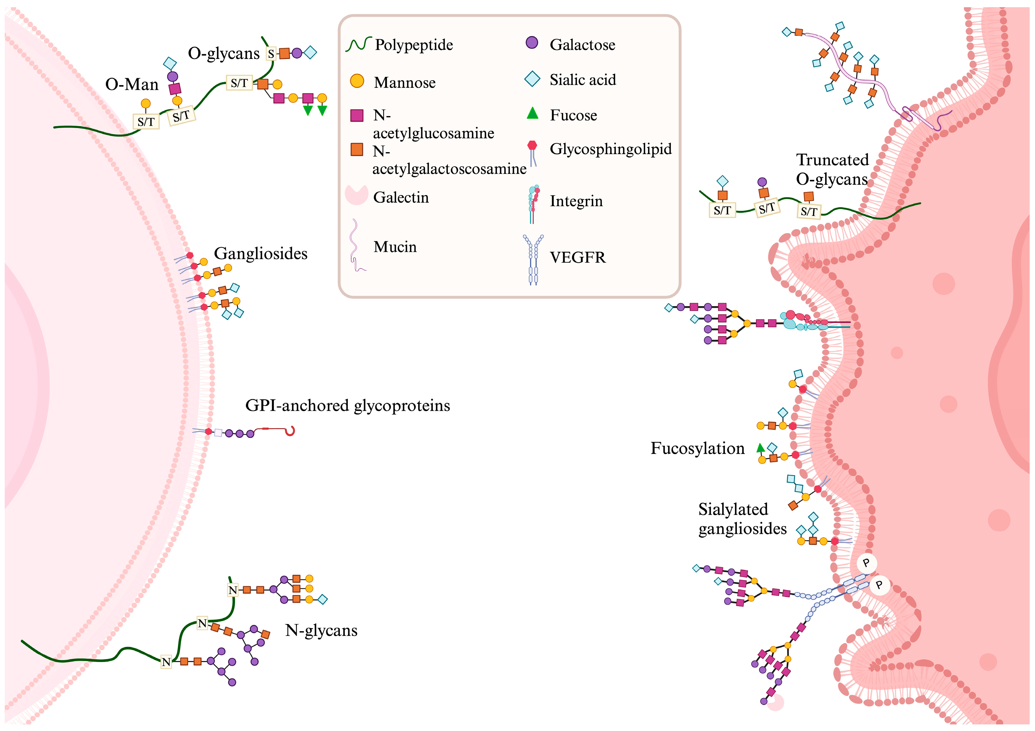
2. Glycobiology of Gliomas, Tumor Microenvironment, and Cell Death

In gliomas, glycobiology has emerged as an area of significant relevance. Studies focused on the structure, function, and biology of glycans have gained increasing attention in cancer research owing to their critical role in tumor progression. In gliomas, alterations in glycan structures are a hallmark of malignancy (Figure 1). These modifications, which occur on glycoproteins, glycolipids, and proteoglycans, influence a wide array of cellular processes, including cell adhesion, migration, immune evasion, and angiogenesis [3]. In proteins, glycosylation occurs on serine/threonine residues (O-glycosylation) or asparagine residues (N-glycosylation). Gliomas exhibit aberrant glycan structures, such as truncated O-glycans (Tn and sialyl-Tn antigens), hyper sialylation, and highly branched N-glycans, which enhance invasiveness, resistance, and serve as biomarkers or therapeutic targets [4].
Glycosylation on proteins, such as integrins and E-cadherins, impacts cell adhesion and extracellular matrix (ECM) interactions, facilitating tumor invasion and migration. Growth receptors, like epidermal growth factor receptors (EGFRs), are also affected, promoting proliferation and survival [3]. Terminal sialylation and carbohydrates, like sialyl Lewis X, aid tumor dissemination by enhancing adhesion to endothelial cells and immune evasion. Changes in ECM glycoproteins, such as fibronectin and laminin, further support progression [5,6].
These glycosylation changes stem from dysregulated glycosyltransferase enzymes, altered chaperones, and glycosidases, often driven by increased glucose metabolism in tumors [7].
The tumor microenvironment (TME) in glioblastoma consists of tumor cells, immune cells (e.g., macrophages, microglia, NK cells, and T cells), astrocytes, neural progenitor cells, blood vessels, and the ECM (Figure 2), along with signaling molecules and exosomes that promote tumorigenesis and proliferation [8,9,10]. Glycans are key mediators of cell–cell and cell–ECM interactions, but aberrant glycosylation in gliomas alters these interactions, promoting immune evasion, therapy resistance, and tumor progression [3,8].
Glioma-associated macrophages and microglia interact with glioma cells through sialylated glycans, suppressing immune activation via Siglecs and facilitating immune evasion [6,11]. Altered glycosylation of ECM molecules enhances glioma cell adhesion, migration, and invasion by modulating integrin signaling [12]. Tumor-associated macrophages, including antitumoral M1 and pro-tumoral M2 subtypes, influence tumor progression through cytokine secretion (e.g., interleukin (IL)-4, IL-10, and transforming growth factor-β (TGF-β)) and angiogenesis via vascular endothelial growth factor (VEGF) and IL-8 (Figure 2). Glioma-associated macrophages also activate immune checkpoints (e.g., PD-L1) and disrupt metabolism [13,14,15,16,17].
Endothelial cells are affected by aberrant glycosylation of VEGF and glycosaminoglycans, enhancing angiogenesis and tumor growth [18]. Astrocytes contribute to tumor progression by secreting ECM proteins and modulating matrix metalloproteinase (MMP) activity via glycosylation, promoting invasion [19].
Glycosylation processes are central to glioblastoma progression, and disruptions in these interactions can lead to cell death, indicating the importance of studying glycan-mediated mechanisms in the glioma microenvironment.
Cell death in glioblastoma is crucial for the development of effective therapies, involving such mechanisms as apoptosis, autophagy, ferroptosis, necroptosis, pyroptosis, and necrosis, each with distinct characteristics that influence the tumor response. Temozolomide, a chemotherapeutic agent primarily used in the treatment of brain tumors, particularly glioblastoma multiforme, acts as an alkylating agent. As a prodrug, it is metabolized in the body to form an active compound that alkylates DNA bases, especially guanine, resulting in breaks in the DNA strands and the formation of crosslinks, impeding accurate DNA replication. This process leads tumor cells to undergo apoptosis or experience severe cellular stress that results in apoptosis. Furthermore, temozolomide is often used in combination with radiotherapy, as it can enhance the sensitivity of tumor cells to radiation-induced damage [20,21]. Glycobiology alterations in gliomas can directly impact key types of cell death. Aberrant glycosylation of death receptors, such as Fas (CD95) and tumor necrosis factor receptor (TNFR), disrupts apoptosis signaling. For example, excessive sialylation of Fas impairs the formation of the death-inducing signaling complex (DISC), reducing apoptosis effectiveness in cancer cells [22]. Similarly, aberrant glycosylation of TNFR and tumor necrosis factor-related apoptosis-inducing ligand (TRAIL) receptors hinders necroptosis by interfering with ligand binding and downstream signaling. Hypersialylation, for instance, inhibits the activation of necroptotic machinery [23,24]. Glycosylation also regulates apoptosis and ferroptosis by modifying key signaling receptors, such as EGFR [25], which is overexpressed in diffuse glioblastoma, IDH-wildtype (see Table 1).
Ferroptosis, induced by the accumulation of lipid peroxides, is being explored as a novel therapeutic approach for cancer and degenerative diseases [26]. The role of glycosylation in the regulation of ferroptosis has been described [27,28]. Ferroptosis has been explored as an alternative strategy to damage glioma cells and glioblastoma stem cells (GSC), which are typically resistant to other therapies [29,30,31].
Autophagy is a process responsible for degradation and recycling the majority of long-lived or aggregated proteins and cellular organelles. It plays an important role in maintaining cell survival under stress in some cellular contexts. Moreover, when apoptosis is defective, autophagy induction can mediate a second form of programmed cell death [32,33,34]. It is interesting to highlight that lectins via glycan interactions may trigger autophagic cell death.
Targeting abnormal glycosylation represents a promising strategy to enhance glioma sensitivity to cell death. Potential approaches include inhibiting sialyltransferases, blocking glycan-mediated signaling pathways, and using lectins to disrupt glycan–receptor interactions. These strategies could restore apoptosis, regulate autophagy, or promote necroptosis, potentially improving treatment outcomes when combined with existing therapies. Integrating glycosylation-targeted therapies with chemotherapy and immunotherapy offers a personalized approach to modulating cell death mechanisms, overcoming resistance, and improving glioblastoma prognosis [35,36,37].

3. Lectins

The altered glycosylation patterns observed in glioma are being exploited by researchers using lectins. Due to their specific glycan-binding properties, lectins show potential as diagnostic tools and in the development of targeted therapies for this disease [38]. The following topics highlight the main functions of plant lectins in combating glioma cells, their possible mechanisms of action, and potential applications in this field of research. The identification and understanding of carbohydrate–lectin interactions can unravel the cellular and molecular mechanisms involved in a variety of human pathologies, such as neoplasia [39,40,41], viral infections [42,43], and inflammatory processes [44]. In the case of cancer, this approach allows the discovery of new markers, as well as new therapeutic targets [3]. Lectins are diverse proteins present in all kingdoms of life. Still, it is in plants that research with lectins is most prevalent, especially those belonging to the Leguminosae family.
The identification of the first lectin (ricin) occurred just over a century ago. After its discovery, a growing and continuous number of studies have been undertaken to understand the properties of this class of proteins, their role in cellular physiology, as well as their potential biological activities [45,46]. Lectins are present in different plant tissues and can be classified according to their abundance in plant tissues/organelles and their molecular structure, sequence, and families [47]. According to the properties that lectins assume when binding to carbohydrates, they are defined as proteins of non-immune origin that reversibly interact with carbohydrates without altering their structures. Recently, Cavada and colleagues summarized the main biological properties of Leguminosae lectins, including their antiproliferative activity and/or binding to cancer cells or cancer-specific antigens that may indicate these proteins as agents for diagnosis and/or treatment of various types of cancer [48,49,50,51,52]. Although numerous studies have explored the antitumor properties of lectins, the effect and mechanisms arising from the specific interaction between plant lectins and glycosylated targets of glioblastoma cells has not been comprehensively reviewed. A focused review in this area could contribute to our understanding of the potential activity of lectins against glioblastoma, clarifying possible mechanisms and informing future research on novel therapeutic approaches.

3.1. Antiglioma Lectins

Given the importance of glioblastoma as a disease, the potential of plant lectins in glioma research is a growing area of interest. By reviewing the literature, it becomes clear that the ConA-like lectins, predominantly associated with the genera Canavalia and Dioclea, are the most extensively studied in this context. Considering the available data, this review highlights glioma studies with 12 plant lectins. Among these, mannose/glucose-specific lectins are the most extensively tested, compared to those binding to galactosides and other carbohydrates. Almost all lectins used in experiments with glioma cells were obtained from the plant material through protein chemistry protocols. The only exception is the Dioclea sclerocarpa lectin, which was tested in recombinant form (rDSL) and compared with its native counterpart (DSL; Table 2).
In cell-based assays, the ideal glioblastoma model should accurately reflect human cellular properties, including morphological characteristics, invasive patterns and capabilities, cellular behavior, and immunological microenvironment. The most commonly used cell lines for glioblastoma studies include human-derived cell lines, such as U251 and U87, as well as rodent-derived cell lines like GL261 from mice and 9L/LacZ, F98, RG2, CNS-1, and C6 from rats. Studies involving lectins notably focus on specific cell lines, particularly the rat-derived C6 and human-derived U87, SF-295, and GBM-1 strains. The rat glioma model, C6, is an experimental model utilized in many studies reporting on the antiglioma activity of numerous compounds, and it has been employed for many studies with lectins [60,68]. The U87 strain is largely used in culture models, but it has also been used in xenograft models of tumor growth in immunocompromised rodents [69]. The SF-295 cell line is also published in some studies of chemotherapy treatments for glioblastoma, representing an additional cellular model for evaluating drugs and molecular modulators for their effect on glioblastomas [70].
In assays involving lectins, glioma cells are exposed to varying concentrations to assess their viability, morphology, migration, and cell death after 24 and 48 h of contact. These effects are analyzed through parameters such as mitochondrial membrane potential, acid vesicular organelle formation, and the activation of programmed cell-death processes. Plant lectins have demonstrated toxicity at concentrations ranging from 30 to 100 µg/mL, primarily by reducing cell viability and migration rates, while inducing autophagic and apoptotic processes, the latter mediated by the activation of caspases 3 and 8 [55,68]. The modulatory effects and potential mechanisms of action of plant lectins against glioma cells are discussed in greater detail in later sections, in addition to the findings on lectins from the Leguminosae, Malvaceae, and Solanaceae families. Further studies are needed to explore the underlying mechanisms behind cytotoxicity and the role of carbohydrate-binding domains in interactions with glioma cells.

3.2. Suggested Mechanisms

The precise mechanisms underlying the antiglioma activity of lectins are not yet fully understood, though emerging evidence has begun to shed light on potential pathways involved. While literature data provide fragments of the overall picture, much of the available information still suggests hypotheses that require further experimental validation. This review aims to compile the main findings regarding the mechanisms through which lectins exert their effects on glioma, highlighting both established evidence and areas for future investigation.
Membrane-type 1 matrix metalloproteinase (MT1-MMP) expression and matrix metalloproteinase-2 (MMP2) activation by ConA were described originally in MDA-MB-231 human breast cancer cells [71] and glioma cells [72] (Figure 3). ConA positively regulates MT1-MMP mRNA expression, facilitating MMP-2 activation, as demonstrated by Northern blotting and Gelatin Zymography. Furthermore, ConA acts at a non-transcriptional level, exerting rapid and transcription-independent effects, since these effects persisted even after inhibition of MT1-MMP mRNA transcription, as evidenced by observing the fragments derived from the proteolytic breakdown of proMMP-2 to form MMP2 through Western blotting. Moreover, this reported activity of ConA on glioma cells was subsequently extended upon by other researchers. For example, the work in [73] represents one of the main catalysts for investigating the mechanisms of antiglioma lectins. This work suggests that ConA may interact with MT1-MMP, triggering the activation of autophagy. Autophagosome formation was found by acridine orange staining and immunoblotting, while silencing of the MT1-MMP gene by siRNA reversed ConA-induced autophagy, highlighting the central role of MT1-MMP in this process [73,74]. The authors tested a panel of lectins, including ConA, peanut agglutinin (PNA), Phaseolus max lectin, and wheat germ agglutinin (WGA), and found that ConA induced significantly increased formation of acidic vesicles, an indicator of autophagy activation. Based on these observations, they used qRT-PCR and immunoblotting to examine the role of MT1-MMP in cultures of U87 glioma cells. Previous findings on the upregulation of MT1-MMP expression by ConA prompted the authors to investigate the relationship between MT1-MMP and ConA in the context of the lectin’s antiglioma effects [57,75]. The results clearly indicated that the binding between ConA and MT1-MMP is required for the activation of autophagy. However, this activity depends on the intracellular domain of the enzyme rather than its extracellular catalytic function. Evaluation of the expression of MT1-MMP and autophagy-related genes by qRT-PCR was performed in untreated U87 cells or in cells treated with siRNA for MT1-MMP knockdown, allowing the identification of genes associated with the effect promoted by ConA in these cells. The analysis revealed the activation of the BCL2 and adenovirus E1B 19-kDa-interacting protein 3 (BNIP3), ATG3, ATG12, and ATG16-L1 genes, these being autophagy-related proteins. Furthermore, the activation of autophagy induced by ConA required MT1-MMP and was followed by the activation of the Janus kinase 2/signal transducer and activator of transcription 3 (JAK2/STAT3) pathway and the expression of BNIP3 [56]. These findings were reinforced by experiments in human U87 glioblastoma cells. In these experiments, transient knockdown of the MT1-MMP gene by siRNA prevented SNAIL induction by ConA. Activation of signaling pathways was analyzed by phosphorylation assays, which revealed that ConA induced phosphorylation of the Src tyrosine kinase and activated the STAT3 pathway. Furthermore, RNA-seq and differential gene array analyses were performed, confirming that SNAIL induction by TGF-β and ConA was associated with the modulation of several signaling pathways, including those mediated by STAT3 and Src. These results suggested that these pathways are involved in the MT1-MMP-mediated signaling axis that promotes SNAIL induction and epithelial-to-mesenchymal transition [76]. Other studies have explored the potential of the legume lectins ConBr [55] and ConGf [60] in modulating MMP1 via glycan interaction. Hence, using predictive bioinformatics data, it was proposed that ConGf inhibits MMP1 and impairs PAR1 (protease-activated receptor 1) signaling, thereby inhibiting AKT and ERK pathways [60]. While these findings are based on computational models, further experimental studies, such as co-immunoprecipitation assays or MMP1 activity assays in lectin-treated cells, are needed to confirm this hypothesis.
A series of studies involving different mannose-binding lectins has attempted to understand the effects and expand our knowledge of the mechanisms underlying the antiglioma potential of legume lectins [54,58,59]. Overall, it has been observed that all tested lectins exhibited cytotoxicity against glioblastoma cell lines, likely attributed to their binding with glycoconjugates, particularly high-mannose N-glycans. The data obtained from these studies revealed an increase in acidic vesicles and the lipidated form of LC3 (LC3II; 14 kDa), clearly indicating autophagy activation. For Dioclea violacea lectin (DVL), as well as for Canavalia brasiliensis lectin (ConBr), several events have been identified, including inhibition of Akt, ERK1/2, and mTORC1 phosphorylation—three cell signaling proteins well known to be upregulated in glioma (Figure 4A). Moreover, disruption of mitochondrial membrane potential in parallel with the activation of stress-associated MAPKs, p38MAPK, and JNK (P46/p54) were also observed. The downregulation of Akt/mTORC1, recognized as an autophagy repressor system, and the phosphorylation/activation of P38MAPK and JNK (P46/p54), reported to promote cell death, are suggested to be significant pieces in the puzzle [77,78,79,80].
ConBr demonstrated the ability to induce cell death in C6 glioma cells and was associated with a significant increase in the production of reactive oxygen species (ROS) and a reduction in the levels of reduced glutathione (GSH), as determined by non-protein thiol content quantification assays. Molecular dynamics studies suggested that ConBr may act by regulating the xc- system through its interaction with glycans present in the CD98hc subunit (Figure 4B). These findings indicated the potential of ConBr as a ferroptosis-promoting agent in glioma cells [81].
Other studies also arrived with Kar and colleagues (2021), who reported that ConA-induced apoptosis in C6 glioblastoma cells occurs concurrently with an imbalance in thiol/disulfide homeostasis, leading to oxidative damage and increased levels of proinflammatory cytokines, such as interleukin-6 and tumor necrosis factor-α [68].
Overall, the mechanisms of action of lectins are an active area of investigation, with experimental and in silico data providing valuable insights. However, more directed research is needed to confirm or refute the suggested mechanisms and advance the research of antiglioma lectins toward the preclinical step.

3.3. Suggested Targets

Lectins exert their biological activities by interacting with glycosylated target molecules, selectively binding to specific carbohydrates on the surface of cancer cells, activating pathways that contribute to their antitumor effects. However, many of these mechanisms remain unclear or have primarily been studied in non-glioma cell lines. Potential molecular targets can be identified based on their function, presence, glycosylation patterns, and associated mechanisms linked to lectin-induced cytotoxicity. This section will highlight possible targets characterized by their glycosylation composition and interaction with plant lectins.

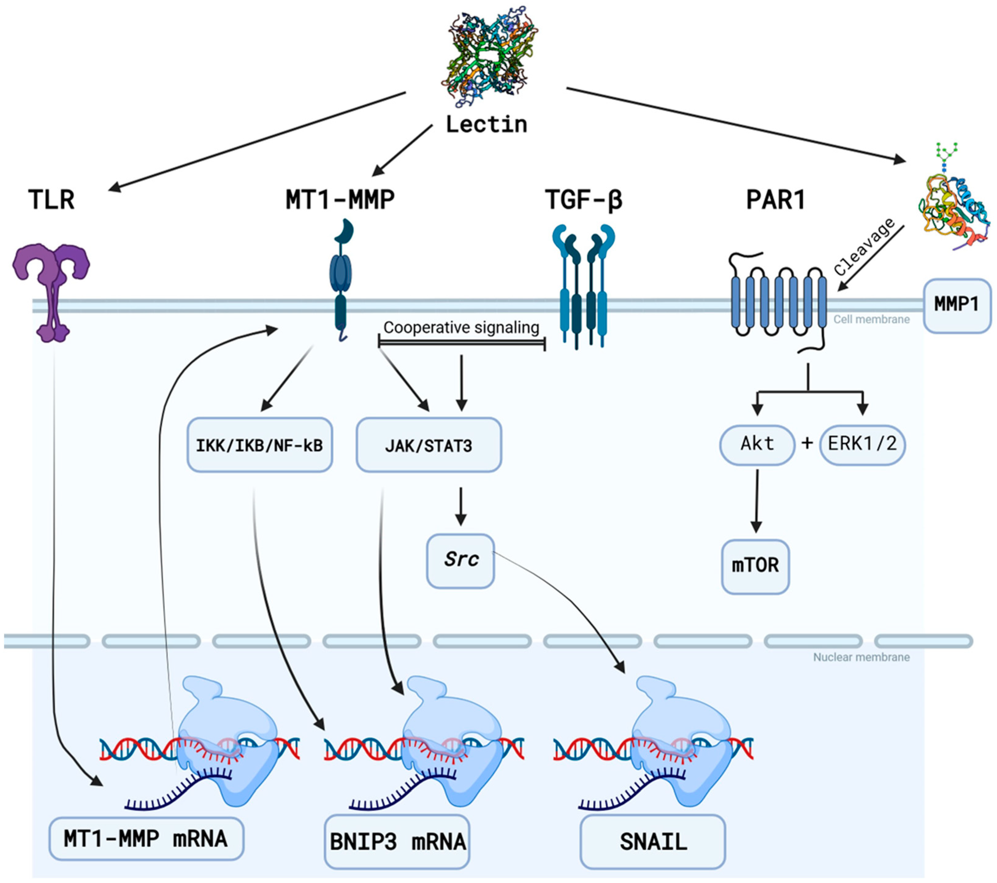
3.3.1. Matrix Metalloproteinases

Matrix metalloproteinases (MMPs) exhibit altered expression levels in many human glioma cell lines, such as SNB-19, GaMG, U251, U87, U373, U343, U138, A172, and T98G, compared to normal glial cells [82]. Additionally, the glycosylation patterns of these proteins can be altered due to malignancy. Data on the alteration of these patterns in glioma cells are scarce, but such changes have been observed in other cell lines. The MMPs that will be mentioned as targets are suggested based on glycosylation data and their relevance to the onset or progression of glioblastomas. For the latter, MMPs 8, 10, 16, 17, 20, 23, 26, 27, and 28 appear to exhibit no significant expression variations in human glioma lineages to justify being considered molecular targets [83].
MMP-1, in contrast, contains two glycosylation sites that are predicted to be located at Asn120 and Asn143 in its structure. However, experimental evidence indicates that glycosylation only occurs at the Asn120 site. This identification was only possible using immobilized ConA on Sepharose-4B to identify glycosylated forms of this enzyme, highlighting the capacity of mannose-specific legume lectins to interact with this target [84,85]. Changes in MMP-1 glycosylation were observed in the HT-1080 fibrosarcoma cell line compared to human skin fibroblasts. The N-glycans of MMP-1 in fibroblasts were predominantly of the α2,3-sialylated complex type, but in HT-1080 cells, some variations increased glycosylation heterogeneity. Motifs, such as GalNAcβ1,4(Fucα1,3)GlcNAc, LacdiNAc, sialylated LacdiNAc, and Lewis X, were observed [83]. The antiglioma lectin from Canavalia grandiflora (ConGF) interacted favorably with MMP-1 glycans in molecular docking studies, consistently interacting through mannosyl motifs and promoting antiglioma activity via autophagy against glioma C6 cells [60].
MMP-3, much like MMP-1, has the potential to undergo glycosylation in fibroblasts, but the specific composition of its glycans remains unknown. It is speculated that N-glycosylation may occur at Asn120 and Asn398, while O-glycosylation sites are likely to be present at Ser57 (within the pro-peptide), Ser269, and Thr277 [86,87]. However, lectin array assays have shown that lectins from Agrocybe cylindracea (ACG), Agaricus bisporus (ABA), Amaranthus caudatus (ACA), and Artocarpus integrifolia (Jacalin) can all interact with MMP-3 via a cluster of O-glycans present in the protein [88]. Since ACG recognizes α2,3-sialylated motifs, and since ABA, ACA, and Jacalin recognize patterns similar to T antigen (Galβ1-3GalNAc), the interaction profile was similar to that found in type 1 glycosylated mucins [89,90,91].
MMP-9 contains three potential N-glycosylation sites, two of which—Asn38 and Asn120—have been experimentally confirmed. These sites are characterized by biantennary glycans with partially sialylated and fucosylated terminals. Additionally, fucosylation may also occur in the glycan core [87]. The N-glycans found in these sites are closely linked to secretion mechanisms of the protease (Asn120), as well as its activation (Asn38) by other MMPs [83].
Furthermore, MMP-9 exhibits a distinctive domain known as OG, encompassing 14 O-glycosylation sites featuring a range of glycan structures, spanning from Galβ1-3GalNAc to Galβ1-3GlcNAcβ1-6GalNAc. These glycans can subsequently undergo further extension into larger structures [92]. This OG domain is responsible for the structural flexibility of MMP-9, influencing the recognition, binding, and processing of substrates and cellular receptors [93,94]. The overexpression of MMP-9 is associated with increased cell growth in U87 glioblastoma cell lines [95]. This protease localizes to the cytoplasm of tumor cells and, alongside MMP-2, plays a crucial part in the invasion process of gliomas by mediating ECM degradation and angiogenesis. Therefore, targeting MMP-9 represents a promising therapeutic strategy for the treatment of malignant gliomas [96].
MMP-13 exhibits a significant presence of glycosylation, with over 10% of its molecular mass comprising glycans. This protease harbors two potential N-glycosylation sites (Asn117 and Asn162) and two O-glycosylation sites (Ser24 and Ser62), albeit only the glycosylation at Asn117 has been experimentally verified. The precise role of these glycosylations remains unclear. However, catalytic assays comparing glycosylated and non-glycosylated forms have demonstrated that glycosylations are not essential for catalytic activity [97]. Up to the present moment, we have not found data on the glycan profile of MMP-13. However, investigations involving the 4T1 breast cancer cell line have identified the presence of glycosylated MMP-13 through chromatography employing ConAimmobilized on Sepharose 4B. These studies have elucidated that those legume lectins exhibiting mannose specificity are capable of interacting with this protease [98].
MMP-14, also known as MT1-MMP, shares similarities with MMP-9 because it possesses a cluster of O-glycans within a domain containing four O-glycosylation sites (Thr291, Thr299, Thr300, and/or Ser301) [99,100]. This glycosylation apparently does not affect the activation of the protease, its catalytic activity, or its autolysis process. However, it appears to impact the formation of the complex with TIMP2 (tissue inhibitor of metalloproteinase-2), which is necessary for activating MMP-2, an event crucial for tumor cell invasion and metastasis [87,101].

3.3.2. Epidermal Growth Factor Receptor (EGFR)

The EGFR is a highly glycosylated membrane receptor with 10 N-glycosylation sites. Overexpression and the presence of aberrant forms of this receptor are common in gliomas, occurring in more than 40% of tumors [102,103]. The glycosylation sites of EGFR are distributed as follows: Asn328, Asn337, and Asn599 contain high-mannose glycans, while Asn32, Asn151, Asn389, Asn420, Asn504, Asn544, and Asn579 are associated with complex bi-, tri-, or tetra-antennary glycans. These complex glycans are fucosylated and may include one or more sialylations [103,104,105]. The ratio between complex-type glycans and high-mannose-type glycans is 2:1 [103]. In a smaller proportion, EGFR presents O-glycosylations, such as O-GalNAc and O-GlcNAc, many of which are predicted to be the Tn antigen [106,107].
Glycosylation allows EGFR to exert its binding activity with EGF, as demonstrated by the 50% decrease in complex formation rates with EGF following pretreatment with N-glycosylation inhibitors [108]. Furthermore, ConA and WGA (wheat germ agglutinin) could block the interaction of this receptor with EGF [109]. The lectin PHA-E (Phaseolus vulgaris) could interact with EGFR expressed in U373 MG cells, and this interaction could inhibit binding with EGF and cell proliferation [110].
The glycosylation pattern profile of EGFR can be altered by the differential expression of enzymes in malignant cells, which can influence its function and lectin recognition. For example, Golgi phosphoprotein 3 (GOLPH3), a peripheral membrane protein located in the trans-Golgi network, is overexpressed in glioblastoma multiforme. Silencing GOLPH3 in T98G glioma cells decreases the sialylation and fucosylation of EGFR, resulting in negative regulation of EGFR activity and reduced cell proliferation in this line, demonstrating the important extent of glycosylation in EGFR regulation [111,112].

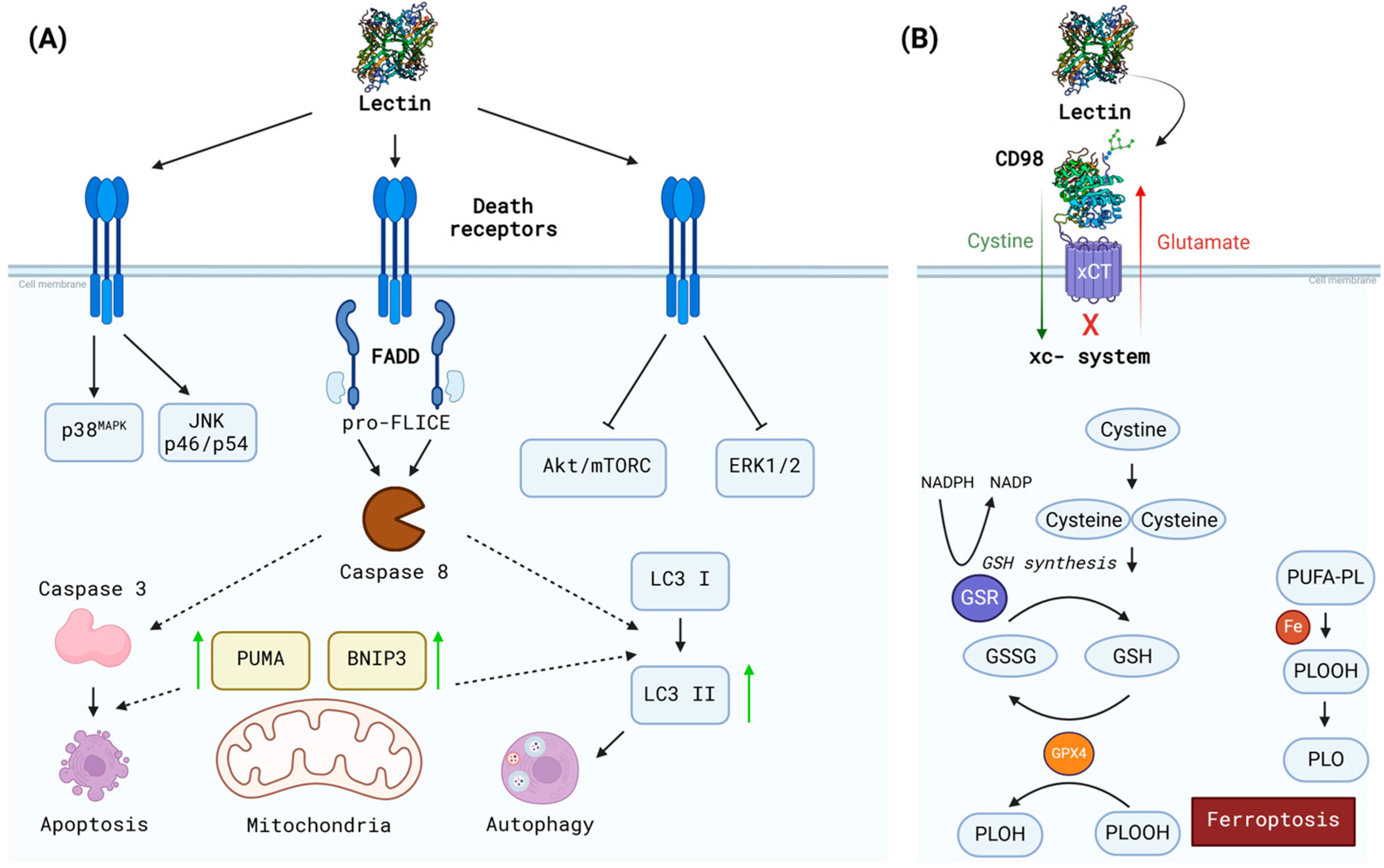
3.3.3. CD98hc (4F2hc)

System xc- belongs to the SoLute Carrier 7 (SLC7) family of membrane antiporter amino acid transporters, which import cystine in exchange for intracellular glutamate. This system is a heterodimer composed of a light chain (xCT or SLC7A11) and a heavy chain, 4F2hc (also called CD98hc or SLC3A2). The heavy chain 4F2hc is a glycoprotein located in the extracellular face of the plasma membrane, with a single transmembrane connected to the xCT subunit by a disulfide bond. Cystine is incorporated into the intracellular milieu via the xc- system and reduced to cysteine for the synthesis of glutathione (GSH), the most abundant intracellular antioxidant. This system is positively regulated in gliomas since its overexpression confers a tumor growth advantage by increasing extracellular glutamate levels and facilitating tumor expansion. Moreover, by increasing GSH production, tumor cells maintain redox potential and neutralize ROS in the intracellular environment, suppressing ferroptotic cell death. Some glioma cell lineages have increased expression of the xCT transporter, which confers resistance to oxidative stress and some pharmacological therapies upon these tumor cells [113,114,115]. CD98 can also form complexes with other members of the SLC7 family, in addition to xCT, such as (a) L system transporters, like LAT1 and LAT2, (b) y + L system transporters, like y + LAT1 and y + LAT2, and (c) asc system transporters, like asc-1 [116].
CD98 displays four glycosylation sites, including Asn365, Asn381, Asn424, and Asn506, where it carries N-linked glycans, as demonstrated in the three-dimensional structure of the xc- complex (PDB ID: 7P9V) [116,117]. In a recent study, glycosylation analysis of CD98 from HeLa cells showed that CD98 in complex with xCT is rich in complex-type glycans, with a higher frequency of poly-LacNAc chains. The Asn381 and Asn424 sites have larger tetra-antennary glycans than other sites, and at Asn424, N-glycans were more frequently extended with linear poly-LacNAc chains. Asn506 glycans were mainly bi- and tri-antennary, while Asn365 exhibited tri- and tetra-antennary structures. The total LacNAc content ranked by site was Asn424 > Asn381~Asn365 > Asn506 [118]. The role of CD98 glycosylation has been appreciated in a recent study of pancreatic ductal adenocarcinoma (PDAC). It was reported that knockdown of SLC3A2 (gene name of 4F2hc) or blocking the N-glycosylation of 4F2hc potentiates ferroptosis sensitization of pancreatic ductal adenocarcinoma cells by impairing the activity of system xc-, as manifested by a marked decrease in intracellular glutathione. Mechanistically, glycosylation of CD98 appears to stabilize the protein and increase the interaction between CD98 and xCT [26].
Furthermore, the CD98 glycoprotein is naturally recognized by a type-S lectin, galectin-3, which is known to regulate macrophage function and phenotype through this interaction [119]. Thus, lectins that recognize the glycosylations of CD98 can affect the activity of many transporters that are complex with this protein, many of which are molecular targets for glioma therapy [120,121]. As an example of a plant lectin application that can interact with CD98, we can mention a resin containing two immobilized isolectins from Maackia amurensis (MAA and MHA) that bind to sialic acid, which were used to isolate glycoproteins from HeLa cell lysates, and one of the identified proteins was CD98 [122]. Recently, a study evaluated the effect of ConBr on C6 glioma cells. ConBr showed dose-dependent cell death induction, accompanied by an increase in the production of ROS and a decrease in reduced glutathione. The analysis by molecular dynamics assessments show that ConBr may be effective in regulating the xc- system through interaction with CD98hc glycans, suggesting that the lectin has the potential to promote ferroptosis in glioma cells [81]. This observation is in line with the description that monoclonal antibodies, directed against glycans that decorate the CD98hc surface in multiple myeloma cells, promoted selective tumor cell death [123]. This indicates that changes in the structure of CD98 glycans expressed in tumor cells could represent a target for the selective induction of tumor cell death.

3.3.4. AMPA Receptor (AMPA-R)

Glutamate receptors (GluRs) are largely expressed in the central nervous system (CNS). They are divided into two main families, namely, metabotropic glutamate receptors (mGluRs) and ionotropic glutamate receptors (iGluRs). The mGluRs are a family of G-protein-coupled receptors (GPCRs) and mediate downstream signaling through G-proteins. On the other hand, ionotropic glutamate receptors are cation-permeable ligand-gated ion channels, which include α-amino-3-hydroxy-5-methyl-4-isoxazolepropionic acid receptors (AMPARs), Kainate receptors (KARs), and N-methyl-D-aspartate receptors (NMDARs). Notably, the subunits of NMDA (GluN1-3), AMPA (GluA1-4), and Kainate (GluK1-4) receptors are glycosylated, and lectins have been applied for their purification [124], as well as to study the structure and activity of the NMDA [125], AMPA [126,127], and Kainate [128] receptors in the CNS.
Glutamatergic transmission has been widely studied in gliomas, considering the importance of this system for tumor cell survival and infiltration into brain tissue [129]. Hence, the glutamatergic signaling elements, including glutamate receptors and transporters, may represent druggable targets to counteract glioma progression and to control epileptiform symptoms, secondary to the increment of extracellular glutamate caused by xc- antiporter activity of glioma cells [129,130].
AMPA receptors have received special attention in gliomas, and many reports suggest they are a promising target to mitigate glioma infiltration into the brain tissue [131,132,133].
AMPA glutamate receptors are tetrameric ion channels formed by combinations of four subunits (GluA1-4; in the past named GluR1-4) that, depending on the composition, may permeate Na+ or Na+/Ca2+. The N-glycosylations present in this receptor are essential for tetramer formation in the endoplasmic reticulum and their translocation to the cell membrane. Therefore, it is likely that AMPAR subunits follow a Golgi-apparatus-dependent pathway [134]. GluA1 has six experimentally verified glycosylation sites located at Asn63, Asn249, Asn257, Asn363, Asn401, and Asn406, while GluA2 contains four sites located at Asn256, Asn370, Asn406, and Asn413 [135]. GluA2 and GluA4 possess N-glycans with mature and immature mannosyl moieties, as demonstrated by their binding to the mannose-specific lectins ConA, NPL (Narcissus pseudonarcissus lectin), and HHL (Hippeastrum hybrid lectin). Additionally, LEL (Lycopersicon esculentum lectin), a lectin specific to GlcNAc, was able to interact with GluA2 and GluA4, indicating the possible presence of complex glycans. However, PNA (peanut agglutinin), a lectin specific to GalNAc, was unable to interact with any subunit [136,137]. In another similar study with lectins, ConA was able to interact with all AMPA-R subunits. The lectin from Aleuria aurantia (AAL) strongly interacted with GluA3 and, to a lesser extent, with GluA2 and GluA4, revealing a higher abundance of Fucα6GlcNAc motifs in GluA3 compared to other subunits. Interactions with LEL and DSL (Datura stramonium lectin) demonstrated that the abundance of GlcNAc motifs was surprisingly higher for the GluA1 subunit than for other subunits, but with a lower intensity of labeling in GluA4. The lectin from Moclura pomifera (MPL), which shows specificity for GalNAc, was the only one that could interact among other GalNAc-specific lectins tested, and then, only with GluA3 and with low intensity [136].
GluA2 also contains the human natural killer-1 epitope (HNK-1, HSO3-3GlcAβ1-3Galβ1-4GlcNAc-R) at Asn413, which is crucial for the expression of AMPA-R on the cell surface [138,139]. These data demonstrated that all AMPA-R subunits are glycosylated, yet they exhibit different N-glycans compositions. Furthermore, lectins have proven to be molecules capable of distinguishing these glycosylations and are able to recognize and differentiate subunits.
In the CNS, excitatory neurons generally express calcium-impermeable AMPARs that contain the edited GluA2 subunit, whereas other neural cells, including inhibitory parvalbumin interneurons, which express receptors lacking the GluA2 subunit, are calcium-permeable [140]. Noteworthy, glioma cells usually express calcium-permeable AMPARs (CP-AMPARs), where glutamate binding promotes calcium influx, improving glioma viability and invasiveness [141,142]. The expression of the calcium-permeable GluA1 subunit has been associated with MAPK and Akt activation and cell proliferation [142,143], as well as enhancement of tumor invasion via beta1-integrin-dependent cell adhesion to the ECM [144]. Notably, propofol (a sedative hypnotic drug) decreased cell viability, invasiveness, and migration of C6 glioma cells via upregulation of the AMPA receptor GluA2 subunit, which prevented Ca2+ influx through CP-AMPARs, causing downregulation of the xc- system, thus inhibiting glutamate release and cystine uptake [145]. Moreover, a study with U87MG cells showed that inhibition of ERK signaling specifically downregulated the expression of calcium-permeable GluA1/GluA4 AMPAR subunits and upregulated calcium-impermeable AMPAR subunit GluA2, which promoted a decrease in the invasion capability of U87MG cells [146]. Considering these studies, it is interesting to highlight that ConBr and DVL lectins can decrease ERK1/2 and Akt signaling [55,59], which theoretically could decrease GluA1/GluA4 AMPA subunits’ expression and, consequently, reduce glioma calcium signaling, dampening glioma viability and invasiveness. Despite all this evidence, there are no studies addressing direct or indirect modulation of AMPA receptor subunits by lectins in glioma cell models. Nonetheless, the capability of legume lectins (e.g., ConA) to interact with glycans present on AMPA subunits in neurons or brain tissue has been well demonstrated [126,127,128]. Moreover, a blockage of AMPA receptor GluA1 subunit phosphorylation by ConBr was shown in mice hippocampus, an effect possibly associated with downregulation of synaptic AMPA receptors [147]. In conclusion, lectin effects on AMPA receptors in gliomas deserve a deeper study.

3.3.5. CD73 (Ecto-5′-nucleotidase)

Enzymes involved in adenosine metabolism, such as CD73 (ecto-5′-nucleotidase) and CD39 (ectonucleoside triphosphate diphosphohydrolase 1, E-NTPDase1), are highly expressed in glioma cells and have been shown to promote tumor progression and induce immunosuppression [148]. The crystal structure of a soluble form of human CD73 (sCD73) with ecto-5′-nucleotidase activity has been determined, and a consistent binding of ConA and Galanthus nivalis agglutinin (GNA) suggested high levels of mannosylation on sCD73 glycans [149]. These findings were consistent with previous studies that indicated CD73 glycosylation [150,151]. Noteworthy, ConA appeared to inhibit the ecto-enzyme activity in many systems analyzed, including in human melanoma cells, in which ConA strongly inhibited the enzyme in a noncompetitive manner and increased the enzyme association with the heavy cytoskeletal complexes [152].
CD73 has four N-glycosylation sites located at Asn53, Asn311, Asn333, and Asn403. The latter three are present in the C-terminal catalytic domain and potentially influence the activity of CD73. Glycomic analyses of CD73 through mass spectrometry have revealed that Asn53 contains only biantennary complex glycans that can be fucosylated and sialylated. The Asn311 site can contain mannose-rich glycans, either complex or hybrid, which are bi-antennary and may be branched. In the case of complex glycans, fucosylation and sialylation may be present. Asn333 is predominantly associated with complex-type glycans that can be bi-, tri-, or tetra-antennary and may contain fucosylation and sialylations. Lastly, Asn403 can harbor hybrid or branched complex glycans, which can be bi-, tri-, or tetra-antennary and may have fucosylation and sialylations [153]. The strong binding of lectins ConA and Galanthus nivalis agglutinin (GNA) also suggests a high abundance of N-glycans containing mannose moieties in CD73, thereby affecting its activity [150,151].

3.4. Other Applications of Lectins

To date, only a limited number of studies have explored the application of plant lectins in the research, diagnosis, and treatment of glioma, highlighting the need for further research in this area. Presently, the majority of studies focus on the use of lectins for biomarker discovery. For instance, a work by Park and colleagues (2018) attempted to pinpoint specific surface glycoproteins differentially expressed in low- and high-grade glioma cells through a combination of lectin array and lectin-affinity chromatography and LC-MS/MS [154]. A total of eight lectins with variable specificities were applied in the arrays, in which the galactose-specific Griffonia simplicifolia lectin was selected for its differential binding properties. Affinity chromatography using this lectin identified 931 proteins, among which 105 exhibited statistically significant increases in high-grade glioma.
Besides glioblastoma, efforts have been made to identify glioblastoma stem cells (GSCs) using plant lectins. In a study by Tucker-Burden and colleagues (2012), a panel of 20 lectins with different carbohydrate specificities was employed to recognize surface glycans of CD133+ glioblastoma stem cells [155]. The results demonstrated that GlcNAc- and GalNAc-specific lectins had the potential to interact differently, depending on the cellular differentiation state. The authors observed that the expression of GlcNAc- and GalNAc-containing glycans accessible to lectins was high in undifferentiated cells but significantly decreased during a 12-day differentiation period, highlighting the suitability of lectins for such applications. In another study, a lectin array combined with an LC-MS/MS approach was employed to identify cell-surface glycoprotein markers on glioblastoma stem cells (GSCs). Two galactose-specific lectins were able to distinguish GSCs from conventional glioblastoma cells. These lectins were immobilized on agarose gels to capture glycoconjugates, resulting in the identification of approximately 12 glycoproteins, most of which were membrane proteins [155]. A recent study using lectin fluorescence staining demonstrated that Maackia amurensis lectin II (MAL-II), a plant-derived protein that binds to α-2,3-sialylated glycans, showed strong binding to GSCs, but this binding decreased significantly as the cells differentiated into glioma cells. Furthermore, treatment of GSCs with MAL-II significantly suppressed cell viability and sphere formation by inducing cell cycle arrest and apoptosis, indicating that MAL-II could have potential as a therapeutic agent for glioma treatment [156].
Besides biomarker discovery, lectins, such as ConA, WGA, Ricinus communis agglutinin 1 (RCA-1), PNA, and Ulex europaeus agglutinin 1 (UEA-1), have been used to perform histochemical analyses on fixed paraffin-embedded samples of 24 human gliomas. ConA and WGA showed positive staining for astrocytomas, oligodendrogliomas, and ependymomas, while PNA and UEA-1 did not. For glioblastomas, only ConA and WGA exhibited positive staining. The authors observed different staining patterns for different lectins across various glioma cells. Furthermore, a correlation was found between the binding of certain lectins, such as RCA-1, and the degree of malignancy. Based on these findings, ConA emerged as a promising lectin for marking gliomas. These results suggest an abundance of mannose-containing glycans and an overall absence of galactoside-containing glycans in glioma cells [157]. Similarly, Figols et al. (1991) investigated the potential of ConA, RCA-1, PNA, and WGA as markers of differentiation [158]. A total of 21 paraffin-embedded glial tumors of various types were utilized for the experiments. The results revealed an intriguing binding pattern, whereby ConA and RCA-1 interacted with most of the tested cells, while PNA and WGA selectively interacted with the plasma membrane of well-differentiated oligodendrogliomas. This led the authors to conclude that these lectins could be of interest in differentiating between gliomas based on the cellular differentiation stage.

4. Conclusions

Glioma cells exhibit distinct glycosylation profiles that influence tumor progression, invasion, and immune evasion. Lectins, with their capacity to selectively recognize and bind to glycans, show promise in identifying glioma-specific glycosylation patterns. This selectivity highlights their potential not only for diagnostic applications but also for new therapies, as their binding can trigger mechanisms leading to cell death and alter cell adhesion properties, both critical in glioma pathology. Crucially, however, significant gaps remain in understanding the specific targets and the nature of their glycan modifications. Addressing these gaps is essential for advancing the application of lectins in glioma treatment and should be the focus of future research. Lectin-based tools offer a promising avenue for purifying and studying these targets with greater precision. In conclusion, the integration of lectin-based approaches, especially in the context of nanotechnology, holds the potential to revolutionize glioma research, enabling more effective therapeutic strategies and, ultimately, improving patient outcomes.

Author Contributions

Conceptualization, R.B.L., K.S.N. and B.S.C.; funding acquisition, R.B.L., K.S.N. and B.S.C.; writing—original draft preparation, R.B.L., V.R.P.-J., M.V.O., V.J.S.O., N.S., A.C.d.S., R.C.G., K.S.N. and B.S.C.; writing—review and editing, R.B.L., V.R.P.-J., M.V.O., V.J.S.O., N.S., A.C.d.S., R.C.G., K.S.N. and B.S.C. All authors have read and agreed to the published version of the manuscript.

Funding

This study was supported by grants from the Conselho Nacional de Desenvolvimento Científico e Tecnológico (CNPq), Coordenação de Aperfeiçoamento de Pessoal de Nível Superior (CAPES), and Fundação Cearense de Apoio ao Desenvolvimento Científico e Tecnológico (FUNCAP).

Informed Consent Statement

Not applicable.

Data Availability Statement

The data supporting the findings of this study are available within the article and the references therein.

Acknowledgments

R.B.L., K.S.N., and B.S.C. are senior investigators of CNPq. David Martin helped with the English editing of the manuscript.

Conflicts of Interest

The authors declare no conflicts of interest.

References

  1. Louis, D.N.; Perry, A.; Wesseling, P.; Brat, D.J.; Cree, I.A.; Figarella-Branger, D.; Hawkins, C.; Ng, H.K.; Pfister, S.M.; Reifenberger, G.; et al. The 2021 WHO Classification of Tumors of the Central Nervous System: A Summary. Neuro-Oncology 2021, 23, 1231–1251. [Google Scholar] [CrossRef] [PubMed]
  2. Felistia, Y.; Wen, P.Y. Molecular Profiling and Targeted Therapies in Gliomas. Curr. Neurol. Neurosci. Rep. 2023, 23, 627–636. [Google Scholar] [CrossRef] [PubMed]
  3. Yue, J.; Huang, R.; Lan, Z.; Xiao, B.; Luo, Z. Abnormal Glycosylation in Glioma: Related Changes in Biology, Biomarkers and Targeted Therapy. Biomark. Res. 2023, 11, 54. [Google Scholar] [CrossRef]
  4. Sun, Y.-F.; Zhang, L.-C.; Niu, R.-Z.; Chen, L.; Xia, Q.-J.; Xiong, L.-L.; Wang, T.-H. Predictive Potentials of Glycosylation-Related Genes in Glioma Prognosis and Their Correlation with Immune Infiltration. Sci. Rep. 2024, 14, 4478. [Google Scholar] [CrossRef]
  5. Fuster, M.M.; Brown, J.R.; Wang, L.; Esko, J.D. A Disaccharide Precursor of Sialyl Lewis X Inhibits Metastatic Potential of Tumor Cells. Cancer Res. 2003, 63, 2775–2781. [Google Scholar]
  6. Cuello, H.A.; Ferreira, G.M.; Gulino, C.A.; Toledo, A.G.; Segatori, V.I.; Gabri, M.R. Terminally Sialylated and Fucosylated Complex N-Glycans Are Involved in the Malignant Behavior of High-Grade Glioma. Oncotarget 2020, 11, 4822–4835. [Google Scholar] [CrossRef] [PubMed]
  7. He, M.; Zhou, X.; Wang, X. Glycosylation: Mechanisms, Biological Functions and Clinical Implications. Signal Transduct. Target. Ther. 2024, 9, 194. [Google Scholar] [CrossRef] [PubMed]
  8. Peixoto, A.; Relvas-Santos, M.; Azevedo, R.; Santos, L.L.; Ferreira, J.A. Protein Glycosylation and Tumor Microenvironment Alterations Driving Cancer Hallmarks. Front. Oncol. 2019, 9, 380. [Google Scholar] [CrossRef]
  9. González-Tablas Pimenta, M.; Otero, Á.; Arandia Guzman, D.A.; Pascual-Argente, D.; Ruíz Martín, L.; Sousa-Casasnovas, P.; García-Martin, A.; Roa Montes de Oca, J.C.; Villaseñor-Ledezma, J.; Torres Carretero, L.; et al. Tumor Cell and Immune Cell Profiles in Primary Human Glioblastoma: Impact on Patient Outcome. Brain Pathol. 2021, 31, 365–380. [Google Scholar] [CrossRef]
  10. Ma, J.; Chen, C.C.; Li, M. Macrophages/microglia in the Glioblastoma Tumor Microenvironment. Int. J. Mol. Sci. 2021, 22, 5775. [Google Scholar] [CrossRef]
  11. Dusoswa, S.A.; Verhoeff, J.; Abels, E.; Méndez-Huergo, S.P.; Croci, D.O.; Kuijper, L.H.; de Miguel, E.; Wouters, V.M.C.J.; Best, M.G.; Rodriguez, E.; et al. Glioblastomas Exploit Truncated O-Linked Glycans for Local and Distant Immune Modulation via the Macrophage Galactose-Type Lectin. Proc. Natl. Acad. Sci. USA 2020, 117, 3693–3703. [Google Scholar] [CrossRef] [PubMed]
  12. Sethi, M.K.; Downs, M.; Shao, C.; Hackett, W.E.; Phillips, J.J.; Zaia, J. In-Depth Matrisome and Glycoproteomic Analysis of Human Brain Glioblastoma versus Control Tissue. Mol. Cell. Proteom. 2022, 21, 100216. [Google Scholar] [CrossRef] [PubMed]
  13. Khan, F.; Pang, L.; Dunterman, M.; Lesniak, M.S.; Heimberger, A.B.; Chen, P. Macrophages and Microglia in Glioblastoma: Heterogeneity, Plasticity, and Therapy. J. Clin. Investig. 2023, 133, e163446. [Google Scholar] [CrossRef]
  14. Zhang, W.; Wang, M.; Ji, C.; Liu, X.; Gu, B.; Dong, T. Macrophage Polarization in the Tumor Microenvironment: Emerging Roles and Therapeutic Potentials. Biomed. Pharmacother. 2024, 177, 116930. [Google Scholar] [CrossRef] [PubMed]
  15. Basak, U.; Sarkar, T.; Mukherjee, S.; Chakraborty, S.; Dutta, A.; Dutta, S.; Nayak, D.; Kaushik, S.; Das, T.; Sa, G. Tumor-Associated Macrophages: An Effective Player of the Tumor Microenvironment. Front. Immunol. 2023, 14, 1295257. [Google Scholar] [CrossRef]
  16. Taiarol, L.; Formicola, B.; Fagioli, S.; Sierri, G.; D’Aloia, A.; Kravicz, M.; Renda, A.; Viale, F.; Dal Magro, R.; Ceriani, M.; et al. The 3.0 Cell Communication: New Insights in the Usefulness of Tunneling Nanotubes for Glioblastoma Treatment. Cancers 2021, 13, 4001. [Google Scholar] [CrossRef] [PubMed]
  17. Sharma, N.; Mallela, A.N.; Shi, D.D.; Tang, L.W.; Abou-Al-Shaar, H.; Gersey, Z.C.; Zhang, X.; McBrayer, S.K.; Abdullah, K.G. Isocitrate Dehydrogenase Mutations in Gliomas: A Review of Current Understanding and Trials. Neurooncol. Adv. 2023, 5, vdad053. [Google Scholar] [CrossRef]
  18. Pace, A.; Scirocchi, F.; Napoletano, C.; Zizzari, I.G.; D’Angelo, L.; Santoro, A.; Nuti, M.; Rahimi, H.; Rughetti, A. Glycan-Lectin Interactions as Novel Immunosuppression Drivers in Glioblastoma. Int. J. Mol. Sci. 2022, 23, 6312. [Google Scholar] [CrossRef] [PubMed]
  19. Kim, Y.; Kang, H.; Powathil, G.; Kim, H.; Trucu, D.; Lee, W.; Lawler, S.; Chaplain, M. Role of Extracellular Matrix and Microenvironment in Regulation of Tumor Growth and LAR-Mediated Invasion in Glioblastoma. PLoS ONE 2018, 13, e0204865. [Google Scholar] [CrossRef]
  20. Sun, X.; Turcan, S. From Laboratory Studies to Clinical Trials: Temozolomide Use in IDH-Mutant Gliomas. Cells 2021, 10, 1225. [Google Scholar] [CrossRef] [PubMed]
  21. Jezierzański, M.; Nafalska, N.; Stopyra, M.; Furgoł, T.; Miciak, M.; Kabut, J.; Gisterek-Grocholska, I. Temozolomide (TMZ) in the Treatment of Glioblastoma Multiforme-A Literature Review and Clinical Outcomes. Curr. Oncol. 2024, 31, 3994–4002. [Google Scholar] [CrossRef] [PubMed]
  22. Schultz, M.J.; Swindall, A.F.; Bellis, S.L. Regulation of the Metastatic Cell Phenotype by Sialylated Glycans. Cancer Metastasis Rev. 2012, 31, 501–518. [Google Scholar] [CrossRef]
  23. Estornes, Y.; Dondelinger, Y.; Weber, K.; Bruggeman, I.; Peall, A.; MacFarlane, M.; Lebecque, S.; Vandenabeele, P.; Bertrand, M.J.M. N-Glycosylation of Mouse TRAIL-R Restrains TRAIL-Induced Apoptosis. Cell Death Dis. 2018, 9, 494. [Google Scholar] [CrossRef]
  24. Micheau, O. Regulation of TNF-Related Apoptosis-Inducing Ligand Signaling by Glycosylation. Int. J. Mol. Sci. 2018, 19, 715. [Google Scholar] [CrossRef] [PubMed]
  25. Li, H.-W.; Liu, M.-B.; Jiang, X.; Song, T.; Feng, S.-X.; Wu, J.-Y.; Deng, P.-F.; Wang, X.-Y. GALNT14 Regulates Ferroptosis and Apoptosis of Ovarian Cancer through the EGFR/mTOR Pathway. Future Oncol. 2022, 18, 149–161. [Google Scholar] [CrossRef]
  26. Stockwell, B.R. Ferroptosis Turns 10: Emerging Mechanisms, Physiological Functions, and Therapeutic Applications. Cell 2022, 185, 2401–2421. [Google Scholar] [CrossRef] [PubMed]
  27. Zhou, X.; Wang, Y.; Li, X.; Zhou, J.; Yang, W.; Wang, X.; Jiao, S.; Zuo, W.; You, Z.; Ying, W.; et al. O-GlcNAcylation Regulates the Stability of Transferrin Receptor (TFRC) to Control the Ferroptosis in Hepatocellular Carcinoma Cells. Redox Biol. 2024, 73, 103182. [Google Scholar] [CrossRef] [PubMed]
  28. Ma, H.; Chen, X.; Mo, S.; Zhang, Y.; Mao, X.; Chen, J.; Liu, Y.; Tong, W.-M.; Lu, Z.; Yu, S.; et al. Targeting N-Glycosylation of 4F2hc Mediated by Glycosyltransferase B3GNT3 Sensitizes Ferroptosis of Pancreatic Ductal Adenocarcinoma. Cell Death Differ. 2023, 30, 1988–2004. [Google Scholar] [CrossRef] [PubMed]
  29. de Souza, I.; Ramalho, M.C.C.; Guedes, C.B.; Osawa, I.Y.A.; Monteiro, L.K.S.; Gomes, L.R.; Rocha, C.R.R. Ferroptosis Modulation: Potential Therapeutic Target for Glioblastoma Treatment. Int. J. Mol. Sci. 2022, 23, 6879. [Google Scholar] [CrossRef] [PubMed]
  30. Teng, W.; Ling, Y.; Long, N.; Cen, W.; Zhang, H.; Jiang, L.; Liu, J.; Zhou, X.; Chu, L. Repurposing Flubendazole for Glioblastoma Ferroptosis by Affecting xCT and TFRC Proteins. J. Cell. Mol. Med. 2024, 28, e70188. [Google Scholar] [CrossRef] [PubMed]
  31. Wei, Y.; Xu, Y.; Sun, Q.; Hong, Y.; Liang, S.; Jiang, H.; Zhang, X.; Zhang, S.; Chen, Q. Targeting Ferroptosis Opens New Avenues in Gliomas. Int. J. Biol. Sci. 2024, 20, 4674–4690. [Google Scholar] [CrossRef] [PubMed]
  32. Debnath, J.; Gammoh, N.; Ryan, K.M. Autophagy and Autophagy-Related Pathways in Cancer. Nat. Rev. Mol. Cell Biol. 2023, 24, 560–575. [Google Scholar] [CrossRef] [PubMed]
  33. Noonan, J.; Zarrer, J.; Murphy, B.M. Targeting Autophagy in Glioblastoma. Crit. Rev. Oncog. 2016, 21, 241–252. [Google Scholar] [CrossRef] [PubMed]
  34. Xu, Y.; Wan, W. Acetylation in the Regulation of Autophagy. Autophagy 2023, 19, 379–387. [Google Scholar] [CrossRef] [PubMed]
  35. Nie, H.; Saini, P.; Miyamoto, T.; Liao, L.; Zielinski, R.J.; Liu, H.; Zhou, W.; Wang, C.; Murphy, B.; Towers, M.; et al. Targeting Branched N-Glycans and Fucosylation Sensitizes Ovarian Tumors to Immune Checkpoint Blockade. Nat. Commun. 2024, 15, 2853. [Google Scholar] [CrossRef] [PubMed]
  36. Khan, T.; Cabral, H. Abnormal Glycosylation of Cancer Stem Cells and Targeting Strategies. Front. Oncol. 2021, 11, 649338. [Google Scholar] [CrossRef] [PubMed]
  37. Cai, L.; Gu, Z.; Zhong, J.; Wen, D.; Chen, G.; He, L.; Wu, J.; Gu, Z. Advances in Glycosylation-Mediated Cancer-Targeted Drug Delivery. Drug Discov. Today 2018, 23, 1126–1138. [Google Scholar] [CrossRef]
  38. Thomas, D.; Rathinavel, A.K.; Radhakrishnan, P. Altered glycosylation in cancer: A promising target for biomarkers and therapeutics. Biochim. Biophys. Acta Rev. Cancer 2021, 1875, 188464. [Google Scholar] [CrossRef]
  39. Yau, T.; Dan, X.; Ng, C.C.W.; Ng, T.B. Lectins with Potential for Anti-Cancer Therapy. Molecules 2015, 20, 3791–3810. [Google Scholar] [CrossRef]
  40. Cavada, B.S.; Oliveira, M.V.; Osterne, V.J.S.; Pinto-Junior, V.R.; Correia-Neto, C.; Nascimento, K.S. Lectins Applied to Diagnosis and Treatment of Prostate Cancer and Benign Hyperplasia: A Review. Int. J. Biol. Macromol. 2021, 190, 543–553. [Google Scholar] [CrossRef] [PubMed]
  41. Cavada, B.S.; de Oliveira, M.V.; Osterne, V.J.S.; Pinto-Junior, V.R.; Martins, F.W.V.; Correia-Neto, C.; Pinheiro, R.F.; Leal, R.B.; Nascimento, K.S. Recent Advances in the Use of Legume Lectins for the Diagnosis and Treatment of Breast Cancer. Biochimie 2022, 208, 100–116. [Google Scholar] [CrossRef] [PubMed]
  42. Hopper, J.T.S.; Ambrose, S.; Grant, O.C.; Krumm, S.A.; Allison, T.M.; Degiacomi, M.T.; Tully, M.D.; Pritchard, L.K.; Ozorowski, G.; Ward, A.B.; et al. The Tetrameric Plant Lectin BanLec Neutralizes HIV through Bidentate Binding to Specific Viral Glycans. Structure 2017, 25, 773–782.e5. [Google Scholar] [CrossRef] [PubMed]
  43. Barre, A.; Simplicien, M.; Benoist, H.; Van Damme, E.J.M.; Rougé, P. Mannose-Specific Lectins from Marine Algae: Diverse Structural Scaffolds Associated to Common Virucidal and Anti-Cancer Properties. Mar. Drugs 2019, 17, 440. [Google Scholar] [CrossRef] [PubMed]
  44. Marques, G.F.O.; Osterne, V.J.S.; Almeida, L.M.; Oliveira, M.V.; Brizeno, L.A.C.; Pinto-Junior, V.R.; Santiago, M.Q.; Neco, A.H.B.; Mota, M.R.L.; Souza, L.A.G.; et al. Contribution of the Carbohydrate-Binding Ability of Vatairea Guianensis Lectin to Induce Edematogenic Activity. Biochimie 2017, 140, 58–65. [Google Scholar] [CrossRef] [PubMed]
  45. Stillmark, H. Ueber Ricin, Ein Giftiges Ferment Aus Den Samen von Ricinus Comm. L. Und Einigen Anderen Euphorbiaceen: Inaugural-Dissertation; Schnakenburg: Dorpat, Estonia, 1888. [Google Scholar]
  46. Tsaneva, M.; Van Damme, E.J.M. 130 Years of Plant Lectin Research. Glycoconj. J. 2020, 37, 533–551. [Google Scholar] [CrossRef] [PubMed]
  47. De Coninck, T.; Van Damme, E.J.M. Review: The Multiple Roles of Plant Lectins. Plant Sci. 2021, 313, 111096. [Google Scholar] [CrossRef]
  48. Cavada, B.S.; Osterne, V.J.S.; Oliveira, M.V.; Pinto-Junior, V.R.; Silva, M.T.L.; Bari, A.U.; Lima, L.D.; Lossio, C.F.; Nascimento, K.S. Reviewing Mimosoideae Lectins: A Group of under Explored Legume Lectins. Int. J. Biol. Macromol. 2020, 154, 159–165. [Google Scholar] [CrossRef] [PubMed]
  49. Cavada, B.S.; Pinto-Junior, V.R.; Osterne, V.J.S.; Oliveira, M.V.; Lossio, C.F.; Silva, M.T.L.; Bari, A.U.; Lima, L.D.; Souza-Filho, C.H.D.; Nascimento, K.S. Comprehensive Review on Caelsalpinioideae Lectins: From Purification to Biological Activities. Int. J. Biol. Macromol. 2020, 162, 333–348. [Google Scholar] [CrossRef]
  50. Cavada, B.S.; Pinto-Junior, V.R.; Oliveira, M.V.; Osterne, V.J.S.; Lossio, C.F.; Nascimento, K.S. A Review of Vicieae Lectins Studies: End of the Book or a Story in the Writing? Int. J. Biol. Macromol. 2021, 181, 1104–1123. [Google Scholar] [CrossRef] [PubMed]
  51. Nascimento, K.S.; Silva, M.T.L.; Oliveira, M.V.; Lossio, C.F.; Pinto-Junior, V.R.; Osterne, V.J.S.; Cavada, B.S. Dalbergieae Lectins: A Review of Lectins from Species of a Primitive Papilionoideae (leguminous) Tribe. Int. J. Biol. Macromol. 2020, 144, 509–526. [Google Scholar] [CrossRef] [PubMed]
  52. Cavada, B.S.; Pinto-Junior, V.R.; Osterne, V.J.S.; Nascimento, K.S. ConA-Like Lectins: High Similarity Proteins as Models to Study Structure/Biological Activities Relationships. Int. J. Mol. Sci. 2018, 20, 30. [Google Scholar] [CrossRef] [PubMed]
  53. Musthafa, S.A.; Muthu, K.; Vijayakumar, S.; George, S.J.; Murali, S.; Govindaraj, J.; Munuswamy-Ramanujam, G. Lectin Isolated from Abelmoschus Esculentus Induces Caspase Mediated Apoptosis in Human U87 Glioblastoma Cell Lines and Modulates the Expression of Circadian Clock Genes. Toxicon 2021, 202, 98–109. [Google Scholar] [CrossRef]
  54. Cavada, B.S.; Silva, M.T.L.; Osterne, V.J.S.; Pinto-Junior, V.R.; Nascimento, A.P.M.; Wolin, I.A.V.; Heinrich, I.A.; Nobre, C.A.S.; Moreira, C.G.; Lossio, C.F.; et al. Canavalia bonariensis Lectin: Molecular Bases of Glycoconjugates Interaction and Antiglioma Potential. Int. J. Biol. Macromol. 2018, 106, 369–378. [Google Scholar] [CrossRef]
  55. Wolin, I.A.V.; Heinrich, I.A.; Nascimento, A.P.M.; Welter, P.G.; Sosa, L.D.V.; De Paul, A.L.; Zanotto-Filho, A.; Nedel, C.B.; Lima, L.D.; Osterne, V.J.S.; et al. ConBr Lectin Modulates MAPKs and Akt Pathways and Triggers Autophagic Glioma Cell Death by a Mechanism Dependent upon Caspase-8 Activation. Biochimie 2021, 180, 186–204. [Google Scholar] [CrossRef] [PubMed]
  56. Pratt, J.; Annabi, B. Induction of Autophagy Biomarker BNIP3 Requires a JAK2/STAT3 and MT1-MMP Signaling Interplay in Concanavalin-A-Activated U87 Glioblastoma Cells. Cell. Signal. 2014, 26, 917–924. [Google Scholar] [CrossRef] [PubMed]
  57. Sina, A.; Proulx-Bonneau, S.; Roy, A.; Poliquin, L.; Cao, J.; Annabi, B. The Lectin Concanavalin-A Signals MT1-MMP Catalytic Independent Induction of COX-2 through an IKKgamma/NF-kappaB-Dependent Pathway. J. Cell Commun. Signal. 2010, 4, 31–38. [Google Scholar] [CrossRef]
  58. Nascimento, A.P.M.; Knaut, J.L.; Rieger, D.K.; Wolin, I.A.V.; Heinrich, I.A.; Mann, J.; Juarez, A.V.; Sosa, L.D.V.; De Paul, A.L.; Moreira, C.G.; et al. Anti-Glioma Properties of DVL, a Lectin Purified from Dioclea violacea. Int. J. Biol. Macromol. 2018, 120, 566–577. [Google Scholar] [CrossRef]
  59. Nascimento, A.P.M.; Wolin, I.A.V.; Welter, P.G.; Heinrich, I.A.; Zanotto-Filho, A.; Osterne, V.J.S.; Lossio, C.F.; Silva, M.T.L.; Nascimento, K.S.; Cavada, B.S.; et al. Lectin from Dioclea violacea Induces Autophagy in U87 Glioma Cells. Int. J. Biol. Macromol. 2019, 134, 660–672. [Google Scholar] [CrossRef]
  60. Leal, R.B.; Mann, J.; Pinto-Junior, V.R.; Oliveira, M.V.; Osterne, V.J.S.; Wolin, I.A.V.; Nascimento, A.P.M.; Welter, P.G.; Ferreira, V.M.S.; Silva, A.A.; et al. Structural Prediction and Characterization of Canavalia grandiflora (ConGF) Lectin Complexed with MMP1: Unveiling the Antiglioma Potential of Legume Lectins. Molecules 2022, 27, 89. [Google Scholar] [CrossRef] [PubMed]
  61. Osterne, V.J.S.; Silva-Filho, J.C.; Santiago, M.Q.; Pinto-Junior, V.R.; Almeida, A.C.; Barreto, A.A.G.C.; Wolin, I.A.V.; Nascimento, A.P.M.; Amorim, R.M.F.; Rocha, B.A.M.; et al. Structural Characterization of a Lectin from Canavalia virosa Seeds with Inflammatory and Cytotoxic Activities. Int. J. Biol. Macromol. 2017, 94, 271–282. [Google Scholar] [CrossRef] [PubMed]
  62. Sasaki, T.; Yamazaki, K.; Yamori, T.; Endo, T. Inhibition of Proliferation and Induction of Differentiation of Glioma Cells with Datura Stramonium Agglutinin. Br. J. Cancer 2002, 87, 918–923. [Google Scholar] [CrossRef] [PubMed]
  63. Sasaki, T.; Endo, T. Both Cell-Surface Carbohydrates and Protein Tyrosine Phosphatase Are Involved in the Differentiation of Astrocytes In Vitro. Glia 2000, 32, 60–70. [Google Scholar] [CrossRef] [PubMed]
  64. Nascimento, K.S.; Santiago, M.Q.; Pinto-Junior, V.R.; Osterne, V.J.S.; Martins, F.W.V.; Nascimento, A.P.M.; Wolin, I.A.V.; Heinrich, I.A.; Martins, M.G.Q.; Silva, M.T.L.; et al. Structural Analysis of Dioclea lasiocarpa Lectin: A C6 Cells Apoptosis-Inducing Protein. Int. J. Biochem. Cell Biol. 2017, 92, 79–89. [Google Scholar] [CrossRef]
  65. Leal, R.B.; Pinto-Junior, V.R.; Osterne, V.J.S.; Wolin, I.A.V.; Nascimento, A.P.M.; Neco, A.H.B.; Araripe, D.A.; Welter, P.G.; Neto, C.C.; Correia, J.L.A.; et al. Crystal Structure of DlyL, a Mannose-Specific Lectin from Dioclea lasiophylla Mart. Ex Benth Seeds That Display Cytotoxic Effects against C6 Glioma Cells. Int. J. Biol. Macromol. 2018, 114, 64–76. [Google Scholar] [CrossRef] [PubMed]
  66. Nascimento, K.S.; Andrade, M.L.L.; Silva, I.B.; Domingues, D.L.; Chicas, L.S.; Silva, M.T.L.; Bringel, P.H.S.F.; Marques, G.F.O.; Martins, M.G.Q.; Lóssio, C.F.; et al. Heterologous Production of α-Chain of Dioclea sclerocarpa Lectin: Enhancing the Biological Effects of a Wild-Type Lectin. Int. J. Biol. Macromol. 2020, 156, 1–9. [Google Scholar] [CrossRef] [PubMed]
  67. Varmes Fernandes, A.; Viana Ramos, M.; Maria Vasconcelos, I.; Cristina Oliveira Monteiro Moreira, A.; Bruno Moreno, F.; Odair Pereira, J.; Francisco de Carvalho Goncalves, J. Purification and Characterization of a Lectin of the Swartzieae Legume Taxa. Protein Pept. Lett. 2012, 19, 1082–1088. [Google Scholar] [CrossRef]
  68. Kar, F.; Kacar, S.; Hacioglu, C.; Kanbak, G.; Sahinturk, V. Concanavalin A Induces Apoptosis in a Dose-Dependent Manner by Modulating Thiol/disulfide Homeostasis in C6 Glioblastoma Cells. J. Biochem. Mol. Toxicol. 2021, 35, e22742. [Google Scholar] [CrossRef] [PubMed]
  69. Giakoumettis, D.; Kritis, A.; Foroglou, N. C6 Cell Line: The Gold Standard in Glioma Research. Hippokratia 2018, 22, 105–112. [Google Scholar] [PubMed]
  70. Costa Nunes, F.; Silva, L.B.; Winter, E.; Silva, A.H.; de Melo, L.J.; Rode, M.; Martins, M.A.P.; Zanatta, N.; Feitosa, S.C.; Bonacorso, H.G.; et al. Tacrine Derivatives Stimulate Human Glioma SF295 Cell Death and Alter Important Proteins Related to Disease Development: An Old Drug for New Targets. Biochim. Biophys. Acta Gen. Subj. 2018, 1862, 1527–1536. [Google Scholar] [CrossRef]
  71. Yu, M.; Sato, H.; Seiki, M.; Thompson, E.W. Complex Regulation of Membrane-Type Matrix Metalloproteinase Expression and Matrix Metalloproteinase-2 Activation by Concanavalin A in MDA-MB-231 Human Breast Cancer Cells. Cancer Res. 1995, 55, 3272–3277. [Google Scholar]
  72. Gingras, D.; Pagé, M.; Annabi, B.; Béliveau, R. Rapid Activation of Matrix Metalloproteinase-2 by Glioma Cells Occurs through a Posttranslational MT1-MMP-Dependent Mechanism. Biochim. Biophys. Acta 2000, 1497, 341–350. [Google Scholar] [CrossRef] [PubMed]
  73. Pratt, J.; Roy, R.; Annabi, B. Concanavalin-A-Induced Autophagy Biomarkers Requires Membrane Type-1 Matrix Metalloproteinase Intracellular Signaling in Glioblastoma Cells. Glycobiology 2012, 22, 1245–1255. [Google Scholar] [CrossRef]
  74. Huldani, H.; Rashid, A.I.; Turaev, K.N.; Opulencia, M.J.C.; Abdelbasset, W.K.; Bokov, D.O.; Mustafa, Y.F.; Al-Gazally, M.E.; Hammid, A.T.; Kadhim, M.M.; et al. Concanavalin A as a Promising Lectin-Based Anti-Cancer Agent: The Molecular Mechanisms and Therapeutic Potential. Cell Commun. Signal. 2022, 20, 167. [Google Scholar] [CrossRef]
  75. Annabi, B.; Laflamme, C.; Sina, A.; Lachambre, M.-P.; Béliveau, R. A MT1-MMP/NF-kappaB Signaling Axis as a Checkpoint Controller of COX-2 Expression in CD133+ U87 Glioblastoma Cells. J. Neuroinflamm. 2009, 6, 8. [Google Scholar] [CrossRef] [PubMed]
  76. Djediai, S.; Gonzalez Suarez, N.; El Cheikh-Hussein, L.; Rodriguez Torres, S.; Gresseau, L.; Dhayne, S.; Joly-Lopez, Z.; Annabi, B. MT1-MMP Cooperates with TGF-β Receptor-Mediated Signaling to Trigger SNAIL and Induce Epithelial-to-Mesenchymal-like Transition in U87 Glioblastoma Cells. Int. J. Mol. Sci. 2021, 22, 13006. [Google Scholar] [CrossRef] [PubMed]
  77. Hwang, Y.K.; Lee, D.-H.; Lee, E.C.; Oh, J.S. Importance of Autophagy Regulation in Glioblastoma with Temozolomide Resistance. Cells 2024, 13, 1332. [Google Scholar] [CrossRef] [PubMed]
  78. Ravikumar, B.; Sarkar, S.; Davies, J.E.; Futter, M.; Garcia-Arencibia, M.; Green-Thompson, Z.W.; Jimenez-Sanchez, M.; Korolchuk, V.I.; Lichtenberg, M.; Luo, S.; et al. Regulation of Mammalian Autophagy in Physiology and Pathophysiology. Physiol. Rev. 2010, 90, 1383–1435. [Google Scholar] [CrossRef] [PubMed]
  79. Dikic, I.; Elazar, Z. Mechanism and Medical Implications of Mammalian Autophagy. Nat. Rev. Mol. Cell Biol. 2018, 19, 349–364. [Google Scholar] [CrossRef] [PubMed]
  80. Wang, B.; Ma, Y.; Zhang, Y.; Yin, X. Therapeutic Potential of ASK1 Activators in Cancer Treatment: Current Insights and Future Directions. Biomed. Pharmacother. 2024, 178, 117214. [Google Scholar] [CrossRef]
  81. Pinto-Junior, V.R.; Seeger, R.L.; Souza-Filho, C.H.D.; França, A.P.; Sartori, N.; Oliveira, M.V.; Osterne, V.J.S.; Nascimento, K.S.; Leal, R.B.; Cavada, B.S. Xc- System as a Possible Target for ConBr Lectin Interaction in Glioma Cells. Neuroglia 2024, 5, 202–222. [Google Scholar] [CrossRef]
  82. Hagemann, C.; Anacker, J.; Ernestus, R.-I.; Vince, G.H. A Complete Compilation of Matrix Metalloproteinase Expression in Human Malignant Gliomas. World J. Clin. Oncol. 2012, 3, 67–79. [Google Scholar] [CrossRef] [PubMed]
  83. Boon, L.; Ugarte-Berzal, E.; Vandooren, J.; Opdenakker, G. Glycosylation of Matrix Metalloproteases and Tissue Inhibitors: Present State, Challenges and Opportunities. Biochem. J. 2016, 473, 1471–1482. [Google Scholar] [CrossRef] [PubMed]
  84. Saarinen, J.; Welgus, H.G.; Flizar, C.A.; Kalkkinen, N.; Helin, J. N-Glycan Structures of Matrix Metalloproteinase-1 Derived from Human Fibroblasts and from HT-1080 Fibrosarcoma Cells. Eur. J. Biochem. 1999, 259, 829–840. [Google Scholar] [CrossRef]
  85. Wilhelm, S.M.; Eisen, A.Z.; Teter, M.; Clark, S.D.; Kronberger, A.; Goldberg, G. Human Fibroblast Collagenase: Glycosylation and Tissue-Specific Levels of Enzyme Synthesis. Proc. Natl. Acad. Sci. USA 1986, 83, 3756–3760. [Google Scholar] [CrossRef] [PubMed]
  86. Wilhelm, S.M.; Collier, I.E.; Kronberger, A.; Eisen, A.Z.; Marmer, B.L.; Grant, G.A.; Bauer, E.A.; Goldberg, G.I. Human Skin Fibroblast Stromelysin: Structure, Glycosylation, Substrate Specificity, and Differential Expression in Normal and Tumorigenic Cells. Proc. Natl. Acad. Sci. USA 1987, 84, 6725–6729. [Google Scholar] [CrossRef] [PubMed]
  87. Madzharova, E.; Kastl, P.; Sabino, F.; Auf dem Keller, U. Post-Translational Modification-Dependent Activity of Matrix Metalloproteinases. Int. J. Mol. Sci. 2019, 20, 3077. [Google Scholar] [CrossRef]
  88. Takeshita, M.; Kuno, A.; Suzuki, K.; Matsuda, A.; Shimazaki, H.; Nakagawa, T.; Otomo, Y.; Kabe, Y.; Suematsu, M.; Narimatsu, H.; et al. Alteration of Matrix Metalloproteinase-3 O-Glycan Structure as a Biomarker for Disease Activity of Rheumatoid Arthritis. Arthritis Res. Ther. 2016, 18, 112. [Google Scholar] [CrossRef]
  89. Yagi, F.; Miyamoto, M.; Abe, T.; Minami, Y.; Tadera, K.; Goldstein, I.J. Purification and Carbohydrate-Binding Specificity of Agrocybe Cylindracea Lectin. Glycoconj. J. 1997, 14, 281–288. [Google Scholar] [CrossRef]
  90. Kuno, A.; Kato, Y.; Matsuda, A.; Kaneko, M.K.; Ito, H.; Amano, K.; Chiba, Y.; Narimatsu, H.; Hirabayashi, J. Focused Differential Glycan Analysis with the Platform Antibody-Assisted Lectin Profiling for Glycan-Related Biomarker Verification. Mol. Cell. Proteom. 2009, 8, 99–108. [Google Scholar] [CrossRef]
  91. Matsuda, A.; Kuno, A.; Nakagawa, T.; Ikehara, Y.; Irimura, T.; Yamamoto, M.; Nakanuma, Y.; Miyoshi, E.; Nakamori, S.; Nakanishi, H.; et al. Lectin Microarray-Based Sero-Biomarker Verification Targeting Aberrant O-Linked Glycosylation on Mucin 1. Anal. Chem. 2015, 87, 7274–7281. [Google Scholar] [CrossRef]
  92. Mattu, T.S.; Royle, L.; Langridge, J.; Wormald, M.R.; Van den Steen, P.E.; Van Damme, J.; Opdenakker, G.; Harvey, D.J.; Dwek, R.A.; Rudd, P.M. O-Glycan Analysis of Natural Human Neutrophil Gelatinase B Using a Combination of Normal Phase-HPLC and Online Tandem Mass Spectrometry: Implications for the Domain Organization of the Enzyme. Biochemistry 2000, 39, 15695–15704. [Google Scholar] [CrossRef] [PubMed]
  93. Rosenblum, G.; Van den Steen, P.E.; Cohen, S.R.; Grossmann, J.G.; Frenkel, J.; Sertchook, R.; Slack, N.; Strange, R.W.; Opdenakker, G.; Sagi, I. Insights into the Structure and Domain Flexibility of Full-Length pro-Matrix Metalloproteinase-9/gelatinase B. Structure 2007, 15, 1227–1236. [Google Scholar] [CrossRef] [PubMed]
  94. Vandooren, J.; Knoops, S.; Aldinucci Buzzo, J.L.; Boon, L.; Martens, E.; Opdenakker, G.; Kolaczkowska, E. Differential Inhibition of Activity, Activation and Gene Expression of MMP-9 in THP-1 Cells by Azithromycin and Minocycline versus Bortezomib: A Comparative Study. PLoS ONE 2017, 12, e0174853. [Google Scholar] [CrossRef] [PubMed]
  95. Duellman, T.; Burnett, J.; Yang, J. Functional Roles of N-Linked Glycosylation of Human Matrix Metalloproteinase 9. Traffic 2015, 16, 1108–1126. [Google Scholar] [CrossRef] [PubMed]
  96. Wang, M.; Wang, T.; Liu, S.; Yoshida, D.; Teramoto, A. The Expression of Matrix Metalloproteinase-2 and -9 in Human Gliomas of Different Pathological Grades. Brain Tumor Pathol. 2003, 20, 65–72. [Google Scholar] [CrossRef] [PubMed]
  97. Knäuper, V.; López-Otin, C.; Smith, B.; Knight, G.; Murphy, G. Biochemical Characterization of Human Collagenase-3. J. Biol. Chem. 1996, 271, 1544–1550. [Google Scholar] [CrossRef] [PubMed]
  98. Phang, W.-M.; Tan, A.-A.; Gopinath, S.C.B.; Hashim, O.H.; Kiew, L.V.; Chen, Y. Secretion of N- and O-Linked Glycoproteins from 4T1 Murine Mammary Carcinoma Cells. Int. J. Med. Sci. 2016, 13, 330–339. [Google Scholar] [CrossRef]
  99. Wu, Y.I.; Munshi, H.G.; Sen, R.; Snipas, S.J.; Salvesen, G.S.; Fridman, R.; Stack, M.S. Glycosylation Broadens the Substrate Profile of Membrane Type 1 Matrix Metalloproteinase. J. Biol. Chem. 2004, 279, 8278–8289. [Google Scholar] [CrossRef] [PubMed]
  100. Wu, Y.I.; Munshi, H.G.; Snipas, S.J.; Salvesen, G.S.; Fridman, R.; Stack, M.S. Activation-Coupled Membrane-Type 1 Matrix Metalloproteinase Membrane Trafficking. Biochem. J. 2007, 407, 171–177. [Google Scholar] [CrossRef]
  101. Kinoshita, T.; Sato, H.; Okada, A.; Ohuchi, E.; Imai, K.; Okada, Y.; Seiki, M. TIMP-2 Promotes Activation of Progelatinase A by Membrane-Type 1 Matrix Metalloproteinase Immobilized on Agarose Beads. J. Biol. Chem. 1998, 273, 16098–16103. [Google Scholar] [CrossRef]
  102. Hatanpaa, K.J.; Burma, S.; Zhao, D.; Habib, A.A. Epidermal Growth Factor Receptor in Glioma: Signal Transduction, Neuropathology, Imaging, and Radioresistance. Neoplasia 2010, 12, 675–684. [Google Scholar] [CrossRef]
  103. Zhen, Y.; Caprioli, R.M.; Staros, J.V. Characterization of Glycosylation Sites of the Epidermal Growth Factor Receptor. Biochemistry 2003, 42, 5478–5492. [Google Scholar] [CrossRef] [PubMed]
  104. Rao, T.C.; Beggs, R.R.; Ankenbauer, K.E.; Hwang, J.; Ma, V.P.-Y.; Salaita, K.; Bellis, S.L.; Mattheyses, A.L. ST6Gal-I-Mediated Sialylation of the Epidermal Growth Factor Receptor Modulates Cell Mechanics and Enhances Invasion. J. Biol. Chem. 2022, 298, 101726. [Google Scholar] [CrossRef] [PubMed]
  105. Liu, Y.-C.; Yen, H.-Y.; Chen, C.-Y.; Chen, C.-H.; Cheng, P.-F.; Juan, Y.-H.; Chen, C.-H.; Khoo, K.-H.; Yu, C.-J.; Yang, P.-C.; et al. Sialylation and Fucosylation of Epidermal Growth Factor Receptor Suppress Its Dimerization and Activation in Lung Cancer Cells. Proc. Natl. Acad. Sci. USA 2011, 108, 11332–11337. [Google Scholar] [CrossRef] [PubMed]
  106. Lin, M.-C.; Chien, P.-H.; Wu, H.-Y.; Chen, S.-T.; Juan, H.-F.; Lou, P.-J.; Huang, M.-C. C1GALT1 Predicts Poor Prognosis and Is a Potential Therapeutic Target in Head and Neck Cancer. Oncogene 2018, 37, 5780–5793. [Google Scholar] [CrossRef] [PubMed]
  107. Stateva, S.R.; Villalobo, A. O-GlcNAcylation of the Human Epidermal Growth Factor Receptor. Org. Biomol. Chem. 2015, 13, 8196–8204. [Google Scholar] [CrossRef]
  108. Soderquist, A.M.; Carpenter, G. Glycosylation of the Epidermal Growth Factor Receptor in A-431 Cells. The Contribution of Carbohydrate to Receptor Function. J. Biol. Chem. 1984, 259, 12586–12594. [Google Scholar] [CrossRef] [PubMed]
  109. Zeng, F.Y.; Benguría, A.; Kafert, S.; André, S.; Gabius, H.J.; Villalobo, A. Differential Response of the Epidermal Growth Factor Receptor Tyrosine Kinase Activity to Several Plant and Mammalian Lectins. Mol. Cell. Biochem. 1995, 142, 117–124. [Google Scholar] [CrossRef]
  110. Rebbaa, A.; Yamamoto, H.; Moskal, J.R.; Bremer, E.G. Binding of Erythroagglutinating Phytohemagglutinin Lectin from Phaseolus Vulgaris to the Epidermal Growth Factor Receptor Inhibits Receptor Function in the Human Glioma Cell Line, U373 MG. J. Neurochem. 1996, 67, 2265–2272. [Google Scholar] [CrossRef]
  111. Arriagada, C.; Cavieres, V.A.; Luchsinger, C.; González, A.E.; Muñoz, V.C.; Cancino, J.; Burgos, P.V.; Mardones, G.A. GOLPH3 Regulates EGFR in T98G Glioblastoma Cells by Modulating Its Glycosylation and Ubiquitylation. Int. J. Mol. Sci. 2020, 21, 8880. [Google Scholar] [CrossRef] [PubMed]
  112. Sechi, S.; Frappaolo, A.; Belloni, G.; Colotti, G.; Giansanti, M.G. The Multiple Cellular Functions of the Oncoprotein Golgi Phosphoprotein 3. Oncotarget 2015, 6, 3493–3506. [Google Scholar] [CrossRef]
  113. Nawashiro, H.; Otani, N.; Shinomiya, N.; Fukui, S.; Nomura, N.; Yano, A.; Shima, K.; Matsuo, H.; Kanai, Y. The Role of CD98 in Astrocytic Neoplasms. Hum. Cell 2002, 15, 25–31. [Google Scholar] [CrossRef] [PubMed]
  114. Digomann, D.; Kurth, I.; Tyutyunnykova, A.; Chen, O.; Löck, S.; Gorodetska, I.; Peitzsch, C.; Skvortsova, I.-I.; Negro, G.; Aschenbrenner, B.; et al. The CD98 Heavy Chain Is a Marker and Regulator of Head and Neck Squamous Cell Carcinoma Radiosensitivity. Clin. Cancer Res. 2019, 25, 3152–3163. [Google Scholar] [CrossRef] [PubMed]
  115. Digomann, D.; Linge, A.; Dubrovska, A. SLC3A2/CD98hc, Autophagy and Tumor Radioresistance: A Link Confirmed. Autophagy 2019, 15, 1850–1851. [Google Scholar] [CrossRef]
  116. Console, L.; Scalise, M.; Salerno, S.; Scanga, R.; Giudice, D.; De Bartolo, L.; Tonazzi, A.; Indiveri, C. N-Glycosylation Is Crucial for Trafficking and Stability of SLC3A2 (CD98). Sci. Rep. 2022, 12, 14570. [Google Scholar] [CrossRef] [PubMed]
  117. Parker, J.L.; Deme, J.C.; Kolokouris, D.; Kuteyi, G.; Biggin, P.C.; Lea, S.M.; Newstead, S. Molecular Basis for Redox Control by the Human Cystine/glutamate Antiporter System Xc. Nat. Commun. 2021, 12, 7147. [Google Scholar] [CrossRef]
  118. Zhang, C.; Shafaq-Zadah, M.; Pawling, J.; Ng, D.W.J.; Hesketh, G.G.; Dransart, E.; Pacholczyk, K.; Longo, J.; Gingras, A.-C.; Penn, L.Z.; et al. SLC3A2 N-Glycosylation and Alternate Evolutionary Trajectories for Amino Acid Metabolism. bioRxiv 2022. [Google Scholar] [CrossRef]
  119. Mukaro, V.R.; Bylund, J.; Hodge, G.; Holmes, M.; Jersmann, H.; Reynolds, P.N.; Hodge, S. Lectins Offer New Perspectives in the Development of Macrophage-Targeted Therapies for COPD/emphysema. PLoS ONE 2013, 8, e56147. [Google Scholar] [CrossRef]
  120. Cappoli, N.; Jenkinson, M.D.; Dello Russo, C.; Dickens, D. LAT1, a Novel Pharmacological Target for the Treatment of Glioblastoma. Biochem. Pharmacol. 2022, 201, 115103. [Google Scholar] [CrossRef]
  121. Anagnostakis, F.; Kokkorakis, M.; Markouli, M.; Piperi, C. Impact of Solute Carrier Transporters in Glioma Pathology: A Comprehensive Review. Int. J. Mol. Sci. 2023, 24, 9393. [Google Scholar] [CrossRef] [PubMed]
  122. McDonald, C.A.; Yang, J.Y.; Marathe, V.; Yen, T.-Y.; Macher, B.A. Combining Results from Lectin Affinity Chromatography and Glycocapture Approaches Substantially Improves the Coverage of the Glycoproteome. Mol. Cell. Proteom. 2009, 8, 287–301. [Google Scholar] [CrossRef] [PubMed]
  123. Hasegawa, K.; Ikeda, S.; Yaga, M.; Watanabe, K.; Urakawa, R.; Iehara, A.; Iwai, M.; Hashiguchi, S.; Morimoto, S.; Fujiki, F.; et al. Selective Targeting of Multiple Myeloma Cells with a Monoclonal Antibody Recognizing the Ubiquitous Protein CD98 Heavy Chain. Sci. Transl. Med. 2022, 14, eaax7706. [Google Scholar] [CrossRef] [PubMed]
  124. Clark, R.A.; Gurd, J.W.; Bissoon, N.; Tricaud, N.; Molnar, E.; Zamze, S.E.; Dwek, R.A.; McIlhinney, R.A.; Wing, D.R. Identification of Lectin-Purified Neural Glycoproteins, GPs 180, 116, and 110, with NMDA and AMPA Receptor Subunits: Conservation of Glycosylation at the Synapse. J. Neurochem. 1998, 70, 2594–2605. [Google Scholar] [CrossRef]
  125. Hemelikova, K.; Kolcheva, M.; Skrenkova, K.; Kaniakova, M.; Horak, M. Lectins Modulate the Functional Properties of GluN1/GluN3-Containing NMDA Receptors. Neuropharmacology 2019, 157, 107671. [Google Scholar] [CrossRef]
  126. Jang, S.; Yu, J.-Y.; Ahn, J.-H.; Oh, S. Modulation of NMDA Receptor Subunits Expression by Concanavalin A. Neurochem. Res. 2016, 41, 1887–1898. [Google Scholar] [CrossRef] [PubMed]
  127. Tucholski, J.; Pinner, A.L.; Simmons, M.S.; Meador-Woodruff, J.H. Evolutionarily Conserved Pattern of AMPA Receptor Subunit Glycosylation in Mammalian Frontal Cortex. PLoS ONE 2014, 9, e94255. [Google Scholar] [CrossRef] [PubMed]
  128. Gangwar, S.P.; Yelshanskaya, M.V.; Nadezhdin, K.D.; Yen, L.Y.; Newton, T.P.; Aktolun, M.; Kurnikova, M.G.; Sobolevsky, A.I. Kainate Receptor Channel Opening and Gating Mechanism. Nature 2024, 630, 762–768. [Google Scholar] [CrossRef] [PubMed]
  129. Biegański, M.; Szeliga, M. Disrupted Glutamate Homeostasis as a Target for Glioma Therapy. Pharmacol. Rep. 2024, 76, 1305–1317. [Google Scholar] [CrossRef] [PubMed]
  130. Kumaria, A.; Ashkan, K. Novel Therapeutic Strategies in Glioma Targeting Glutamatergic Neurotransmission. Brain Res. 2023, 1818, 148515. [Google Scholar] [CrossRef]
  131. Venkataramani, V.; Tanev, D.I.; Strahle, C.; Studier-Fischer, A.; Fankhauser, L.; Kessler, T.; Körber, C.; Kardorff, M.; Ratliff, M.; Xie, R.; et al. Glutamatergic Synaptic Input to Glioma Cells Drives Brain Tumour Progression. Nature 2019, 573, 532–538. [Google Scholar] [CrossRef]
  132. Venkatesh, H.S.; Morishita, W.; Geraghty, A.C.; Silverbush, D.; Gillespie, S.M.; Arzt, M.; Tam, L.T.; Espenel, C.; Ponnuswami, A.; Ni, L.; et al. Electrical and Synaptic Integration of Glioma into Neural Circuits. Nature 2019, 573, 539–545. [Google Scholar] [CrossRef] [PubMed]
  133. Taylor, K.R.; Barron, T.; Hui, A.; Spitzer, A.; Yalçin, B.; Ivec, A.E.; Geraghty, A.C.; Hartmann, G.G.; Arzt, M.; Gillespie, S.M.; et al. Glioma Synapses Recruit Mechanisms of Adaptive Plasticity. Nature 2023, 623, 366–374. [Google Scholar] [CrossRef] [PubMed]
  134. Morise, J.; Yamamoto, S.; Midorikawa, R.; Takamiya, K.; Nonaka, M.; Takematsu, H.; Oka, S. Distinct Cell Surface Expression Patterns of N-Glycosylation Site Mutants of AMPA-Type Glutamate Receptor under the Homo-Oligomeric Expression Conditions. Int. J. Mol. Sci. 2020, 21, 5101. [Google Scholar] [CrossRef] [PubMed]
  135. Everts, I.; Villmann, C.; Hollmann, M. N-Glycosylation Is Not a Prerequisite for Glutamate Receptor Function but Is Essential for Lectin Modulation. Mol. Pharmacol. 1997, 52, 861–873. [Google Scholar] [CrossRef] [PubMed]
  136. Tucholski, J.; Simmons, M.S.; Pinner, A.L.; Haroutunian, V.; McCullumsmith, R.E.; Meador-Woodruff, J.H. Abnormal N-Linked Glycosylation of Cortical AMPA Receptor Subunits in Schizophrenia. Schizophr. Res. 2013, 146, 177–183. [Google Scholar] [CrossRef] [PubMed]
  137. Kandel, M.B.; Yamamoto, S.; Midorikawa, R.; Morise, J.; Wakazono, Y.; Oka, S.; Takamiya, K. N-Glycosylation of the AMPA-Type Glutamate Receptor Regulates Cell Surface Expression and Tetramer Formation Affecting Channel Function. J. Neurochem. 2018, 147, 730–747. [Google Scholar] [CrossRef] [PubMed]
  138. Morita, I.; Kakuda, S.; Takeuchi, Y.; Itoh, S.; Kawasaki, N.; Kizuka, Y.; Kawasaki, T.; Oka, S. HNK-1 Glyco-Epitope Regulates the Stability of the Glutamate Receptor Subunit GluR2 on the Neuronal Cell Surface. J. Biol. Chem. 2009, 284, 30209–30217. [Google Scholar] [CrossRef]
  139. Morise, J.; Takematsu, H.; Oka, S. The Role of Human Natural Killer-1 (HNK-1) Carbohydrate in Neuronal Plasticity and Disease. Biochim. Biophys. Acta Gen. Subj. 2017, 1861, 2455–2461. [Google Scholar] [CrossRef] [PubMed]
  140. Hong, I.; Kim, J.; Hainmueller, T.; Kim, D.W.; Keijser, J.; Johnson, R.C.; Park, S.H.; Limjunyawong, N.; Yang, Z.; Cheon, D.; et al. Calcium-Permeable AMPA Receptors Govern PV Neuron Feature Selectivity. Nature 2024, 635, 398–405. [Google Scholar] [CrossRef] [PubMed]
  141. Yoshida, Y.; Tsuzuki, K.; Ishiuchi, S.; Ozawa, S. Serum-Dependence of AMPA Receptor-Mediated Proliferation in Glioma Cells. Pathol. Int. 2006, 56, 262–271. [Google Scholar] [CrossRef] [PubMed]
  142. Corsi, L.; Mescola, A.; Alessandrini, A. Glutamate Receptors and Glioblastoma Multiforme: An Old “Route” for New Perspectives. Int. J. Mol. Sci. 2019, 20, 1796. [Google Scholar] [CrossRef]
  143. de Groot, J.F.; Piao, Y.; Lu, L.; Fuller, G.N.; Yung, W.K.A. Knockdown of GluR1 Expression by RNA Interference Inhibits Glioma Proliferation. J. Neurooncol. 2008, 88, 121–133. [Google Scholar] [CrossRef] [PubMed]
  144. Piao, Y.; Lu, L.; de Groot, J. AMPA Receptors Promote Perivascular Glioma Invasion via beta1 Integrin-Dependent Adhesion to the Extracellular Matrix. Neuro-Oncology 2009, 11, 260–273. [Google Scholar] [CrossRef]
  145. Wang, X.-Y.; Li, Y.-L.; Wang, H.-Y.; Zhu, M.; Guo, D.; Wang, G.-L.; Gao, Y.-T.; Yang, Z.; Li, T.; Yang, C.-Y.; et al. Propofol Inhibits Invasion and Proliferation of C6 Glioma Cells by Regulating the Ca2+ Permeable AMPA Receptor-System Xc- Pathway. Toxicol. In Vitro 2017, 44, 57–65. [Google Scholar] [CrossRef]
  146. Ramaswamy, P.; Dalavaikodihalli Nanjaiah, N.; Prasad, C.; Goswami, K. Transcriptional Modulation of Calcium-Permeable AMPA Receptor Subunits in Glioblastoma by MEK-ERK1/2 Inhibitors and Their Role in Invasion. Cell Biol. Int. 2020, 44, 830–837. [Google Scholar] [CrossRef]
  147. Russi, M.A.; Vandresen-Filho, S.; Rieger, D.K.; Costa, A.P.; Lopes, M.W.; Cunha, R.M.S.; Teixeira, E.H.; Nascimento, K.S.; Cavada, B.S.; Tasca, C.I.; et al. ConBr, a Lectin from Canavalia brasiliensis Seeds, Protects against Quinolinic Acid-Induced Seizures in Mice. Neurochem. Res. 2012, 37, 288–297. [Google Scholar] [CrossRef] [PubMed]
  148. Goswami, S.; Walle, T.; Cornish, A.E.; Basu, S.; Anandhan, S.; Fernandez, I.; Vence, L.; Blando, J.; Zhao, H.; Yadav, S.S.; et al. Immune Profiling of Human Tumors Identifies CD73 as a Combinatorial Target in Glioblastoma. Nat. Med. 2020, 26, 39–46. [Google Scholar] [CrossRef] [PubMed]
  149. Heuts, D.P.H.M.; Weissenborn, M.J.; Olkhov, R.V.; Shaw, A.M.; Gummadova, J.; Levy, C.; Scrutton, N.S. Crystal Structure of a Soluble Form of Human CD73 with Ecto-5′-Nucleotidase Activity. Chembiochem 2012, 13, 2384–2391. [Google Scholar] [CrossRef]
  150. Burgemeister, R.; Danescu, I.; Gutensohn, W. Glycosylation and Processing of Carbohydrate Side Chains of Ecto-5′-Nucleotidase in Cultured Human Chorionic Cells. Biol. Chem. Hoppe Seyler 1990, 371, 355–361. [Google Scholar] [CrossRef]
  151. Zimmermann, H. 5′-Nucleotidase: Molecular Structure and Functional Aspects. Biochem. J. 1992, 285 Pt 2, 345–365. [Google Scholar] [CrossRef] [PubMed]
  152. Sadej, R.; Spychala, J.; Skladanowski, A.C. Ecto-5′-Nucleotidase (eN, CD73) Is Coexpressed with Metastasis Promoting Antigens in Human Melanoma Cells. Nucleosides Nucleotides Nucleic Acids 2006, 25, 1119–1123. [Google Scholar] [CrossRef] [PubMed]
  153. Alcedo, K.P.; Guerrero, A.; Basrur, V.; Fu, D.; Richardson, M.L.; McLane, J.S.; Tsou, C.-C.; Nesvizhskii, A.I.; Welling, T.H.; Lebrilla, C.B.; et al. Tumor-Selective Altered Glycosylation and Functional Attenuation of CD73 in Human Hepatocellular Carcinoma. Hepatol. Commun. 2019, 3, 1400–1414. [Google Scholar] [CrossRef] [PubMed]
  154. Park, Y.E.; Yeom, J.; Kim, Y.; Lee, H.J.; Han, K.-C.; Lee, S.-T.; Lee, C.; Lee, J.E. Identification of Plasma Membrane Glycoproteins Specific to Human Glioblastoma Multiforme Cells Using Lectin Arrays and LC-MS/MS. Proteomics 2018, 18, 1700302. [Google Scholar] [CrossRef]
  155. Tucker-Burden, C.; Chappa, P.; Krishnamoorthy, M.; Gerwe, B.A.; Scharer, C.D.; Heimburg-Molinaro, J.; Harris, W.; Usta, S.N.; Eilertson, C.D.; Hadjipanayis, C.G.; et al. Lectins Identify Glycan Biomarkers on Glioblastoma-Derived Cancer Stem Cells. Stem Cells Dev. 2012, 21, 2374–2386. [Google Scholar] [CrossRef] [PubMed]
  156. Putthisen, S.; Silsirivanit, A.; Panawan, O.; Niibori-Nambu, A.; Nishiyama-Ikeda, Y.; Ma-In, P.; Luang, S.; Ohta, K.; Muisuk, K.; Wongkham, S.; et al. Targeting alpha2,3-Sialylated Glycan in Glioma Stem-like Cells by Maackia Amurensis Lectin-II: A Promising Strategy for Glioma Treatment. Exp. Cell Res. 2022, 410, 112949. [Google Scholar] [CrossRef] [PubMed]
  157. Wang, X.C.; Kochi, N.; Tani, E.; Kaba, K.; Matsumoto, T.; Shindo, H. Lectin Histochemistry of Human Gliomas. Acta Neuropathol. 1989, 79, 176–182. [Google Scholar] [CrossRef] [PubMed]
  158. Figols, J.; Madrid, J.F.; Cervós-Navarro, J. Lectins as Differentiation Markers of Human Gliomas. Histol. Histopathol. 1991, 6, 79–85. [Google Scholar] [PubMed]
Figure 1. Alterations in glycosylation in tumor cell membranes. In the cell membranes of normal cells, glycan structures attached to proteins and lipids are displayed, such as O-glycans and O-Man, represented by chains linked to serine/threonine residues; gangliosides, represented by glycosphingolipids containing sialic acids; GPI-anchored glycoproteins, represented by glycoproteins anchored by glycosylphosphatidylinositol; N-glycans, represented by chains linked to asparagine residues. Meanwhile, in tumor cells, modifications in cellular interactions and signaling occur, such as truncated O-glycans, fucosylation (through the addition of fucose to glycans), and sialylated gangliosides, represented by the enrichment of sialic acids.
Neuroglia 06 00005 g001
Figure 2. Glioma microenvironment and its influence on immunosuppression and tumor progression. The glioma tumor microenvironment comprises various cell types, such as microglia, macrophages, natural killer cells, lymphocytes, astrocytes, granulocytes, and dendritic cells. This microenvironment promotes a range of immunosuppressive and pro-tumor effects. CD4+ and CD8+ T cells are influenced by tumorigenesis due to the immunosuppressive environment, leading to immune exhaustion and tumor tolerance. Tumor-associated macrophages polarize into M1 (proinflammatory, with antitumoral activity) or M2 (anti-inflammatory, promoting tumor progression). M2 tumor-associated macrophages produce anti-inflammatory cytokines (IL-4, IL-10, and TGF-β) and pro-tumoral factors (IGF-1, EGF, and PDGF). They also promote angiogenesis (VEGF and IL-8), dysregulated tumor metabolism (ARG1 and IDO), and activate immune checkpoints (PD-L1 and CD39), thereby deactivating the host’s immune response against glioblastoma. Straight black arrows indicate signals of stimulation (→) and inhibition (⊥).
Neuroglia 06 00005 g002
Figure 3. The MT1-MMP-JAK-STAT signaling axis regulates BNIP3 expression, playing a relevant role in glioblastoma. MT1-MMP is activated by stimuli that trigger signaling pathways, such as JAK/STAT3, Src, and IKK/IkB/NF-κB. These pathways, in an integrated manner, can induce autophagy, partially reflected by the increased expression of BNIP3 and SNAIL. MT1-MMP-mediated signal transduction functionally cooperates with the TGF-β receptor in glioblastoma cells. Lectins are able to induce MT1-MMP transcription and expression, potentially involving Toll-like receptor (TLR) signaling [56,76]. The PAR1 receptor is activated by MMP1-mediated proteolytic cleavage. Signaling pathways activated downstream of PAR1 include MAPKs (such as ERK1/2) and PI3K/AKT/mTOR, which are often associated with inhibition of autophagy and promote glioma proliferation and migration. Lectins can interact with glycans associated with MMP1, inhibiting its activity. This effect may lead to the induction of cell death in glioma cells and the activation of autophagy [60].
Neuroglia 06 00005 g003
Figure 4. (A) Lectins can interact with glycans present on death receptors located on the surface of C6 glioma cells, resulting in reduced cell viability. This effect is associated with increased phosphorylation of p38MAPK and JNK (p46/54), accompanied by a decrease in ERK1/2 and Akt activity. Furthermore, these alterations lead to inhibition of cell migration and survival. The suppression of the Akt/mTORC1 pathway by lectins may be related to the activation of autophagic processes, evidenced by increasing LC3II, which culminates in cell death. Interestingly, this process also appears to inhibit the production of mitochondrial reactive oxygen species (ROS). The antiglioma activity of lectins is multifaceted, involving a complex crosstalk mechanism between caspase-8 activation and autophagic pathways, resulting in glioma cell death [55]. (B) Lectins also demonstrate the ability to interact with glycans present on CD98, inhibiting cystine/glutamate transport mediated by the xCT transporter. This inhibition reduces glutathione (GSH) synthesis, leading to increased oxidative stress, lipid peroxidation, and cell death by ferroptosis [81]. Straight black arrows indicate signals of stimulation ( ) and inhibition ( ). Green straight arrows () indicate increased expression. Curved arrows ( ) indicate reactions. Dashed straight arrows ( ) indicate stimulated event.
Neuroglia 06 00005 g004
Table 1. Classification of gliomas carried out by the World Health Organization (WHO) in 2021.
Glioma TypeSubgroup
(1) Diffuse gliomas, characterized by their infiltrative growth and the presence of specific genetic alterationsAdult-type diffuse glioblastoma, isocitrate dehydrogenase (IDH)-wildtype (CNS WHO grade 4): Characterized by mutations such as telomerase reverse transcriptase (TERT) promoter mutations, EGFR amplification, and/or +7/−10 chromosome (RG1) changes.
Adult-type diffuse astrocytoma, IDH-mutant (grades 2, 3, or 4): Defined by the presence of an IDH mutation and the absence of 1p/19q co-deletion. Grading depends on histological and molecular features, including CDKN2A/B homozygous deletion for grade 4.
Pediatric-type diffuse high-grade gliomas: Includes entities like diffuse midline glioma, H3 K27-altered and diffuse hemispheric glioma, and H3 G34-mutant.
Pediatric-type diffuse low-grade gliomas: Characterized by alterations such as MYB or MYBL1 rearrangements.
(2) Circumscribed gliomas, tumors are typically more localized and have distinct histopathological and molecular featuresPilocytic astrocytoma (grade 1): Frequently associated with mitogen-activated protein kinase (MAPK) pathway alterations (e.g., KIAA1549-BRAF fusion).
Pleomorphic xanthoastrocytoma (grade 2): Often with BRAF V600E mutations.
(3) Gliomas with a mixed molecular profileOligodendroglioma, IDH-mutant, 1p/19q co-deleted (grades 2 or 3): Defined by the presence of both IDH mutation and 1p/19q co-deletion, which distinguish it from astrocytic gliomas.
(4) Other glioma subtypesEpendymomas: Now classified based on anatomic location and molecular markers (e.g., RELA Fusion-positive ependymoma).
Gliomas with histone mutations: Highlighting specific subtypes with poor prognosis, such as H3 K27M-mutant gliomas.
Table 2. Main plant lectins tested against different cell lines of glioma. The cytotoxic concentration of the tested lectins was evaluated between 24 and 48 h with mouse and human cell lines.
SpecieLectinSpecificityCell LineConcentration
(μg/mL)
Effects
Abelmoschus esculentus (L.) Moench AELGalactosidesU8721
  • Apoptosis induced by modulation of caspase-3 and -7. Bmal1 and clock gene expression.
  • Generation of intracellular reactive oxygen species (ROS) [53].
Canavalia bonariensis Lindl.CaBoGlucose
Mannose
C6100
  • Decreased the cellular viability and migration by induction of autophagy and cell death [54].
Canavalia brasiliensis Mart. ex Benth.ConBrGlucose
Mannose
C6, U87, and GBM-130–50
  • Increased p38MAPK and JNK and decreased extracellular signal-regulated kinase (ERK1/2) and Akt phosphorylation.
  • Inhibited mammalian target of rapamycin complex 1 (mTORC1) phosphorylation associated with accumulation of acidic vacuoles and microtubule-associated protein light chain (LC3) cleavage.
  • Inhibition of early steps of autophagy with 3-methyl-adenine (3-MA) partially protected, whereas the later autophagy inhibitor chloroquine (CQ) had no protective effect upon ConBr cytotoxicity.
  • Augmented caspase-3 activation without affecting mitochondrial function. The caspase-8 inhibitor IETF-fmk attenuated ConBr-induced autophagy and C6 glioma cell death.
  • No cytotoxicity against primary astrocytes [55].
Canavalia ensiformis (L.) DC.ConAGlucose
Mannose
C6 and U8730
  • Modulation of membrane-type 1 matrix metalloproteinase (MT1-MMP), providing increased expression of cyclooxygenase-2 (COX-2), via IKK/NFκB (nuclear factor kappa B).
  • Inhibition of Akt phosphorylation, suggestive of cell death [56,57].
  • Cytotoxic with morphological and molecular changes inducing cell death by autophagy.
  • Inhibition of cell migration, proliferation, and clonogenic capacity [58,59].
Canavalia grandiflora Benth.ConGFGlucose
Mannose
C630, 50, and 100
  • Impair the mitochondrial transmembrane potential.
  • Reduce cell viability.
  • Induce morphological changes.
  • Induce massive autophagy.
  • The possible mechanism of action is the interaction of ConGF with the glycan structures of matrix metalloproteinase 1 (MMP1) [60].
Canavalia virosa (Roxb.) Wight & Arn.ConVGlucose
Mannose
C630–100
  • Induction of autophagy, thereby impairing cell migration and decreasing cell viability of glioma cells [61].
Datura stramonium (L.)DSAN-acetyllactosamineC61 #
  • Induced irreversible differentiation of tumor cells after a short exposure [62].
  • Did not affect neuronal cell migration or axonal extension, and did not induce any morphological change of neurons [63].
Dioclea lasiocarpa Mart. ex Benth.DLLGlucose/mannoseC6100
  • Strong antiglioma activity by mechanisms involving activation of caspase-3 [64].
Dioclea lasiophylla Mart. ex Benth.DlyLGlucose/mannoseC630–100
  • Reduction of cell migration, inducing autophagy and cell death via activation of caspase-3 [65].
Dioclea sclerocarpa DuckeDSL and rDSL *Glucose/mannoseC6 and U87100
  • Decreased cell viability only on C6 glioma cells [66].
Dioclea violacea Mart. ex Benth.DVLGlucose/mannoseC6 and U8730–100
  • Caspase-3 activation, apoptotic cell death, and cellular membrane damage.
  • Decreased mitochondrial membrane potential.
  • Increased the number of acidic vesicles and cleavage of LC3, indicating activation of autophagic processes.
  • Decreased glioma cell viability and migration ability.
  • Induced autophagy process by decreasing Akt, mTORC1, and ERK1/2 phosphorylation and augmenting JNK(p54) and p38MAPK phosphorylation [59].
Swartzia laevicarpa AmshoffSLLGalactosidesSF-295>100
  • IC50 was hit only after 72 h of contact with the lectin above a 100 μg/mL concentration [67].
* Recombinant lectin. # Lectin concentration (μm).
Disclaimer/Publisher’s Note: The statements, opinions and data contained in all publications are solely those of the individual author(s) and contributor(s) and not of MDPI and/or the editor(s). MDPI and/or the editor(s) disclaim responsibility for any injury to people or property resulting from any ideas, methods, instructions or products referred to in the content.

