Article

Biodegradation of Malathion in Amended Soil by Indigenous Novel Bacterial Consortia and Analysis of Degradation Pathway

Department of Environmental Science, School of Earth Sciences, Central University of Rajasthan, Bandar Sindri, Ajmer 305817, Rajasthan, India
*
Author to whom correspondence should be addressed.
Soil Syst. 2023, 7(4), 81; https://doi.org/10.3390/soilsystems7040081
Submission received: 19 July 2023 / Revised: 11 September 2023 / Accepted: 20 September 2023 / Published: 26 September 2023
(This article belongs to the Special Issue Soil Pollution: Monitoring, Risk Assessment and Remediation)

Abstract

:
The capabilities of pure bacterial strains and their consortia isolated from agricultural soil were evaluated during a bioremediation process of the organophosphate pesticide malathion. The pure bacterial strains efficiently degraded 50.16–68.47% of the pesticide within 15 days of incubation, and metabolites were observed to accumulate in the soil. The consortia of three bacterial species [Micrococcus aloeverae (MAGK3) + Bacillus cereus (AGB3) + Bacillus paramycoides (AGM5)] degraded the malathion more effectively, and complete malathion removal was observed by the 15th day in soils inoculated with that consortium. In contrast, the combined activity of any two of these strains was lower than the mixed consortium of all of the strains. Individual mixed consortia of Micrococcus aloeverae (MAGK3) + Bacillus cereus (AGB3); Micrococcus aloeverae (MAGK3) + Bacillus paramycoides (AGM5); and Bacillus cereus (AGB3) + Bacillus paramycoides (AGM5) caused 76.58%, 70.95%, and 88.61% malathion degradation in soil, respectively. Several intermediate metabolites like malaoxon, malathion monocarboxylic acid, diethyl fumarate, and trimethyl thiophosphate were found to accumulate and be successively degraded during the bioremediation process via GC–MS detection. Thus, inoculating with a highly potent bacterial consortium isolated from in situ soil may result in the most effective pesticide degradation to significantly relieve soils from pesticide residues, and could be considered a prospective approach for the degradation and detoxification of environments contaminated with malathion and other organophosphate pesticides. This study reports the use of a mixed culture of Indigenous bacterial species for successful malathion degradation.

1. Introduction

Pesticides control pests, and become pollutants as they enter the environment in different ways like accidental spills during manufacturing and transport, direct application in agricultural fields, waste from container cleaning, etc. [1]. Pesticides are dispersed across the soil, water, and air, influencing the ecosystem and human well-being [2]. The arbitrary usage of pesticides worldwide has culminated in the contamination of all environmental compartments with residues from pesticides [3]. Dar et al. [4] described numerous forms of matrices found to be polluted with different organophosphate pesticides (OPPs) such as soil, water, vegetables, etc. Pesticides are comprehensively arranged into three categories, i.e., organochlorines, organophosphates (OPs), and carbamides. However, among them, OPs are the largest and most commonly used category of pesticides globally [5,6]. However, the persistent usage of vast amounts of OPPs contributes to their introduction into the environment and agricultural crops. All OPPs are strong inhibitors of cholinesterase (ChE), and are powerful neurotoxins that may contribute to paralysis/death and prostate cancer in people who spray such pesticides [7,8]. Some OPPs are exceptionally harmful, and are being still utilized broadly for pest control. The widespread usage of OPPs triggers serious concerns about food safety and environmental pollution [9].
Malathion, a synthetic OPP, is commonly utilized to protect crops and livestock from pests [10]. Its use in farming to safeguard crops has grown substantially, so its residues have been detected in soil [11], water [12], vegetables [13], and breast milk [14]. Thus, the presence of OPPs like malathion in the environment is causing concern worldwide due to its impact on ChE activity and the nervous system, and is considered a main hazard to public health [4]. Malathion is categorized as carcinogenic, both to humans and animals, and high exposure levels can disrupt nerve cells and cause neurotoxicity in animals, as well as impair higher vertebrate immunity [15]. Due to its effects on the adrenal glands and central nervous systems of higher vertebrates and invertebrates, malathion is highly toxic to aquatic and terrestrial animals [16]. Pesticides like malathion have an impact on photosynthesis, plant cell growth, biosynthetic reactions, etc. [17]. Malathion has a half-life of months/years in water, and the WHO has set the pesticide limit in drinking water at 0.1 µgL−1 [18]. Malathion’s presence in the environment provides a significant threat to humans and other organisms, since it is mitogenic and cytogenic at low and higher exposure levels, respectively, and a significant rise in DNA destruction has been detected for exposure to 24 mM [19]. Malathion is very soluble, making typical treatment procedures such as sand filtration and coagulation ineffective [20]. As a result, new cost-effective technologies are needed to effectively remove malathion from contaminated surroundings.
Numerous conventional treatment techniques, for example, adsorption, incineration, advanced oxidation, and volatilization, are accessible for the diminution of pesticide pollution from aqueous/soil phases [3]; however, due to the advantages of biological treatments over physicochemical processes such as cost-effectiveness, energy efficiency, and the production of fewer toxic byproducts and sludge, bioremediation techniques are preferred. Bioremediation tends to be an inexpensive and ecologically sustainable process, as it is obvious from the literature that different microbes have the capability to degrade various pesticide groups. Therefore, unique attention has been paid to the emergence of bacteria that are capable of metabolizing OPPs in recent decades, since they were products of an evolutionary phase of indigenous bacteriological populations that have adapted themselves to such toxins, and their utilization has been proposed for the in-situ detoxification of highly polluted environments [21]. Remediation methods are a realistic choice for pollution management, owing to the harmful substances used in numerous operations. These methods utilize plants and microorganisms to transform or break down toxic substances into less toxic or non-toxic components that have been progressively investigated [4]. Bioremediation improves the pace of the natural biodegradation of contaminants in the environment, by supplementing potent microbes and sufficient nutrients in the contaminated environments [3]. Diverse bacterial strains have been isolated and documented for OPP biodegradation in recent years. Acinetobacter johnsonii can degrade malathion co-metabolically, as it cannot devour malathion as a sole carbon source [22]. Similarly, bacterial species, such as Bacillus licheniformis [23], Pseudomonas putida, Rhodoccocus rhodochrous, Sphingomonas species [10], Enterobacter amnigenus [24], and Bacillus species [25], isolated from diverse samples, were found to be exceptionally proficient in malathion degradation.
The biodegradation of OPPs is thought to include a variety of bacterial species. We have identified and assessed the malathion degradation ability of bacterial species in liquid culture in some of our earlier publications [6,15]. The bacterial strains that caused higher malathion degradation were labeled as effective malathion degraders, and were identified as Micrococcus aloeverae MAGK3 (GenBank acc # MZ220366), Bacillus cereus AGB3 (GenBank acc # ON150776), and Bacillus paramycoides AGM5 (GenBank acc # OM021874) by the National Center for Biotechnology Information (NCBI) after taxonomic characterization. Research on malathion biodegradation has mostly been carried out using single strains. Hence, it is important to establish a biosystem (consortia) that can efficiently degrade such pesticides, because mixed cultures of microbial consortia typically display improved productivity and substrate tolerance compared to pure cultures of a single strain. No doubt, this is due to interactions between the cells in the microbial consortium and the metabolites produced by one strain that may control the development and metabolism of another strain. Therefore, the present study developed a highly efficient OPP degrading bacterial consortium, and evaluated the bioremediation of malathion in soil by the bacterial consortium and by individual pure strains previously isolated. The use of consortia of native soil bacterial strains to remediate malathion contaminated soils has never been reported previously.

2. Material and Methods

2.1. Chemicals and Media

Malathion 50% EC (Sikko Industries Ltd., Ahmedabad, India) was used in all of the microbial studies, and was obtained from the local market. However, reference standards of malathion were acquired from Sigma Aldrich (Bangalore, India). All other culture reagents and chemicals used were of analytical and HPLC grade. Nutrient broth (NB) was utilized for the growth of the bacterial strains and to preserve them. When required, the NB was solidified with 2.5% agar to make NB agar medium, and was sterilized by autoclaving at 121 °C and 15 psi for 20 min.

2.2. Microorganisms

Micrococcus aloeverae (MAGK3), Bacillus cereus (AGB3), and Bacillus paramycoides (AGM5) capable of degrading malathion were isolated from agricultural soils located in Ajmer district in Rajasthan, state of India, from our previous studies [15,16]; they degraded (50–1000 mgL−1) malathion in liquid cultures. The current study examined the biodegradation capacity of these bacterial strains and their consortia in the soil.

2.3. Identification of Malathion-Degrading Bacteria

The isolates were identified via their morphological and biochemical properties, and were further confirmed based on 16S rRNA gene sequencing. As stated by Dar and Kaushik [15], PCR amplification and sequencing of 16S rDNA genes were carried out, and the sequence similarity was assessed on 17 May 2021 via the BLAST function in NCBI Genbank (https://www.ncbi.nlm.nih.gov).

2.4. Inoculum Preparation

The seed culture of three pure strains was prepared by growing them discretely in NB medium overnight (25 °C) on a rotary shaker (200 rpm) followed by harvesting after 24 h through centrifuging (5000 rpm) for 10 min. The culture was washed with sterilized NaCl saline and resuspended in the same fresh saline. Two percent of the prepared suspensions was utilized as the inoculum for the biodegradation of the selected pesticide until mentioned otherwise.

2.5. Experimental Design for Pot Experiment

Soil from the surface layer (0–20 cm) of barren land near agricultural fields around the Central University of Rajasthan campus was collected with no known history of pesticide application. Before commencing the experiment, physicochemical characteristics like the pH, EC, organic carbon/matter, major anions, and cations were determined. Table S1 shows the physicochemical parameters of the soil utilized in this study. The soil was sieved via a 60-mesh screen, and sterilized (3 consecutive sterilizations) via autoclaving (121 °C, 15 psi, 20 min) with 24 h intervals between each cycle in glass containers. The effect of various bacterial treatments on malathion degradation was studied using a complete randomized block design in a pot culture experiment. Thirty days of pot experiments were carried out in individual plastic cups (9.5 cm external diameter, 12 cm height), and 100 g of soil was distributed to each cup, fortified with a measured amount of malathion in triplicates. There were four treatments at a malathion concentration of 500 mgkg−1 soil: 1. Malathion-augmented sterilized soil, but devoid of bacterial inoculations (control); 2. Malathion-augmented unsterilized soil but without bacterial inoculations; 3. Malathion-augmented soil inoculated discretely with individual strains; 4. Malathion-augmented soil with microbial consortia of different combinations. The individual cups were inoculated with 2 mL of overnight cultures of the strains, and had their moisture content restored with sterile Milli-Q water. All of the cups were wrapped with sterile aluminum foil and cultured at room temperature (observed to be optimal) for up to 30 days, with regular watering at 10-day intervals to maintain moisture content. The residual malathion from the soil was extracted via the QuEChERS method, as stated in Section 2.6, and the residual analysis was evaluated using UHPLC (Ultra-high-performance liquid chromatography).
The biodegradation rate was calculated using the following equation, where Mi is the initial and Mf is the final malathion concentration:
B i o d e g r a d a t i o n   R a t e   % = M i M f M i 100

2.6. Estimation of Malathion and Its Metabolites

Malathion was extracted from the soil as outlined below. A representative portion of the sample, i.e., 10 g of soil, was transferred to screwed glass tubes in which the tubes were kept closed (with screwed caps) for most of the time, thus minimizing volatile chemical losses. Then, 5 mL of ultrapure water was added to produce sample pores which made them more accessible to the extraction solvent, and the mixture was vortexed for 1 min. The next step involved the addition of 10 mL of ACN into all of the tubes as an extraction solvent, followed by vortexing to ensure that the solvent interacted with the entire sample. Then, MgSO4 (4 g) and C2H3NaO2 (1 g) were added to the tubes, which were vortexed immediately to avoid salt accumulation before centrifuging for a minute. Then, the top clear extract was shifted to a new tube containing MgSO4 (150 mg) and PSA sorbent (25 mg), followed by vortexing and centrifugation [26]. The cleaned sample extracts were filtered with 0.22 µm filters for UHPLC and GC–MS (gas chromatography mass spectrometry) analyses.

2.7. Analysis of Extracts for Malathion Estimation

The final filtered extracts were analyzed for malathion and its metabolites via UHPLC and GC–MS. The UHPLC analysis was executed on an Agilent LC system (Model—1290 infinity 2) armed with an easy-to-use autosampler (G7129B), a binary pump (Agilent 1290, flexible pump, G7104A) with a pressure range up to 1300 bar, a column oven, and a multiple wavelength flexibility DAD detector (Agilent 1260, DAD WR, G7115A). Separation was accomplished via a C18 column (Agilent, EP C18, 5 µm, 4.6 × 250 mm i.d) with a 35 °C column oven temperature. The C18 column used water and ACN as mobile phases A and B, respectively, in the ratio of 30:70, with a 1 mLmin−1 flow rate. For the 10 µL sample, the detection wavelength was set to 210 nm for analysis. The entire duration for the chromatographic run was 11 min. By comparing the sample retention duration and the area under the curve with those of standards analyzed under the same operating conditions, the compound in the samples was identified and quantified. Chem-Station Control software was used for processing and analyzing the data.

2.8. Confirmation–Identification of Degradation Metabolites

GC–MS (Shimadzu Corporation, Kyoto, Japan), was employed to analyze the extracted samples to confirm the degradation of malathion and its metabolites. The GC–MS was equipped with a standard column Rtx-1MS. The temperature of the GC column oven was configured for an initial hold of 2 min at 70 °C, then raised at 25 °C min−1 to 150 °C for a 4 min hold, then up to 200 °C at 3 °C min−1 with a 5 min hold time, then up to 280 °C at 8 °C min−1, and then a final hold for 10 min at 280 °C. In the split-less mode, the gas flow was 1 mL min−1 and the injection temperature was 280 °C. The MS analysis was achieved with the following conditions: MS ion source temperature of 230 °C, interface temperature of 280 °C, electron impact ionization at 1.34 kV, selective ion monitoring mode with a dwell time of 45 min, and solvent delay of 4.5 min. The ions with an m/z of 330 correspond to malathion. The data were processed through GC–MS real-time analytical software, and the compounds were identified in SIM mode based on the m/z ratio. The chromatograms attained were compared, and the individual peaks were identified by comparing the mass spectra to NIST11 mass spectra library references.

2.9. Degradation Kinetics

The kinetics of malathion degradation were determined by graphing the period against the residual malathion concentration to produce correlation coefficient and best-fit curve equations that were determined using the squared correlation coefficient [27]. The dissipation kinetics of the malathion residues were confirmed using a graphical depiction of the time and LogC.

3. Results and Discussion

3.1. Comparative Analysis of Malathion Degradation by Bacterial Species in Liquid Culture

The relative malathion degradation analysis found the bacterial species Micrococcus aloeverae (MAGK3), Bacillus cereus (AGB3), and Bacillus paramycoides (AGM5) to be the most potent malathion degraders; they caused efficient malathion degradation at different concentrations [15,16]. Thus, the malathion degradation capabilities of these isolates were ranked as Bacillus cereus (AGB3) > Micrococcus aloeverae (MAGK3) > Bacillus paramycoides (AGM5).

3.2. Bioremediation Evaluation of Individual Strains and Mixed Strains Consortia

The malathion metabolization potential of Micrococcus aloeverae (MAGK3), Bacillus cereus (AGB3), and Bacillus paramycoides (AGM5) strains and their consortia were ascertained from residues of malathion during their inoculation in malathion-supplemented pre-sterilized soil samples. Table 1, Table 2 and Table 3 describe the results of malathion degradation by these strains and their various consortia combinations over the course of 30 days in soil samples with an initial malathion content of 500 mgkg−1. Almost all of the treatments showed a progressive decrease in malathion concentration over time.
The individual strains of Micrococcus aloeverae (MAGK3), Bacillus cereus (AGB3), and Bacillus paramycoides (AGM5) in inoculated cultures decreased the malathion residues to 132.15, 52.70, and 83.58 mgkg−1, respectively, in 15 days, corresponding to soil samples with an initial malathion concentration of 500 mgkg−1 soil. This reduction in malathion was maintained as the incubation time was increased, to the point that the residue levels were lowered to 3.15, 1.424, and 11.61 mgkg−1, respectively, in 30 days (Table 1). This amounted to total malathion degradation rates of 98%, 99%, and 95%, respectively, after 30 days in soil. The malathion, regardless of its initial concentration, was degraded linearly, eventually resulting in residual malathion concentrations of 3–12 mgkg−1 by 30 days. However, the degradation percentages in single-strain treatments were remarkably less as compared to the consortia treatments.
The maximum malathion residues obtained at 15 days were decreased to 62 mgkg−1 when sterilized soil was injected with a mixed culture of Micrococcus aloeverae (MAGK3) + Bacillus cereus (AGB3), matched to soil samples with an initial malathion concentration of 500 mgkg−1 (Table 2). However, in 20 days, the malathion remnants were reduced to undetectable levels in soil with an initial malathion concentration of 500 mgkg−1; 100% malathion degradation was achieved in 20 days. Upon amending the soil with Micrococcus aloeverae (MAGK3) + Bacillus paramycoides (AGM5) for 15 days, the malathion residues were reduced to 77 mgkg−1 (Table 2). After 20 days, the residues were decreased further to 22 mgkg−1, and completely degraded in 30 days. This represented 100% malathion degradation in 30 days in soils with an initial malathion concentration of 500 mgkg−1. The highest malathion residues obtained at 15 days were diminished to 30 mgkg−1 when soil with an initial malathion concentration of 500 mgkg−1 was treated with a mixed culture of Bacillus cereus (AGB3) + Bacillus paramycoides (AGM5) (Table 2). However, in 20 days, the malathion was reduced further to undetectable levels. Thus, in soils with an initial malathion concentration of 500 mgkg−1, when treated with this consortium, resulted in 100% malathion degradation in 20 days. The degradation effect was evident, and a substantial decline in malathion content was noted in the treatment, augmented with a consortium of all three of the strains [Micrococcus aloeverae (MAGK3) + Bacillus cereus (AGB3) + Bacillus paramycoides (AGM5)] as compared to other treatments, with a maximum malathion concentration (79 mgkg−1) detected at the 10th day (Table 2). By 15 days, the residues were reduced to undetectable levels, amounting to 100% malathion degradation within 15 days in soils amended with a 500 mgkg−1 initial malathion concentration.
The malathion degraded slowly in a linear way for the control conditions (uninoculated unsterilized soil), regardless of its starting concentration (500 mgkg−1), resulting in a total decrease of 81% in 30 days. In sterilized uninoculated soil, the malathion deteriorated slowly in a linear manner, resulting in a 52% overall decrease from an initial malathion concentration of 500 mgkg−1 (Table 3). Low malathion degradation (52% in 30 days) was observed in the uninoculated sterilized controls, in contrast to a greater rate of malathion degradation in the unsterilized control soil (81% in 30 days) in this study. The malathion concentration in the controls was also observed to decrease to some extent, which can be owing to the indigenous microflora of the soil. The degradation can also be influenced by physiochemical processes like volatilization and photodegradation. However, other variables like temperature, pH, and moisture content all have an impact on the decomposition of malathion in soil. Since the moisture content in the pots was consistently maintained, it is possible that hydrolysis is responsible for the decline in malathion in the controls [28]. Additionally, the soil used had a pH of 7.3, which is slightly alkaline. It is well-known that alkalinity favors the hydrolysis of organophosphates [29].
Soil sterilization is most commonly used way of inhibiting microbial activity in soil [30], although it has drawbacks that can change the soil’s physical and chemical properties, thereby changing the quantitative and qualitative interactions between the soil, pesticide, and microbes. The increased malathion degradation rate in the uninoculated, sterilized control may be due to autoclaving, because this method did not eliminate all of the microbes from the soils [27]. The higher malathion degradation in autoclaved soils may be related to the activity of numerous spores producing bacterial and fungal taxa that had avoided death in similarly sterilized soils, according to the current study [31]. Autoclaving, on the other hand, is a frequently favored method, since it can be conducted on-site in experimental containers, is affordable, and does not generate significant amounts of contaminated soil that are difficult to dispose of [32]. Temperature, pH, pesticide concentration, OM quantity, and bioavailability are some of the abiotic parameters of soil that have been shown to influence pesticide breakdown by bacteria [33,34]. Several pesticides, including phorate [27], diazinon [35], chlorpyrifos [36], and triazophos [37], have been successfully degraded using bioaugmentation. Microorganisms that degrade OPPs under culture conditions can likewise degrade them in soil [27,38,39]. Various soil-isolated bacterial species have been shown to thrive on malathion as the only carbon source in aqueous media, and cause active malathion degradation to varying degrees [10,23,40,41]. In soil containing 1.5% malathion, Lysinibacillus species KB1 caused a 68% decline in malathion contents within 7 days [38]. Singh, et al. [42] performed bioremediation of malathion in contaminated sterile soil, and observed that 74.11% and 57.14% of malaoxon from malathion (1.5% kg−1 soil) were degraded by Bacillus cereus strain PU and Brevibacillus species strain KB2, respectively, after a week. After inoculating the sterilized soil with a bacterial consortium consisting of Lysinibacillus species KB1 + Bacillus cereus PU + Brevibacillus species KB2, the degradation of malathion further increased to 80%, which showed that mixed cultures have greater efficiency to degrade malathion [42].
Different bacterial species have been isolated that show an ability to thrive on different pesticides and actively degrade them. Zhao et al. [43] observed 60% degradation of β-cypermethrin at 100 mgL−1 of β-cypermethrin within 7 days by the bacterium Bacillus cereus (GW-01) isolated from the rumen chyme of a sheep. During the degradation of β-cypermethrin, the GW-01 strain momentarily accumulated 3-phenoxybenzoic acid, phenol, and benzoic acid. Birolli et al. [44] employed a commercially available bioinsecticide Bacillus thuringiensis Berliner for cypermethrin biodegradation. During the study, it was observed that the strain grew well in the presence of cypermethrin, and 37% of the compound in the soil was degraded within 28 days at 30 °C by the strain in conjunction with the native microbes, showing that the degradation of cypermethrin was significantly increased by the bioaugmentation of this strain. Bacillus megaterium (HLJ7 strain) has shown excellent biodegradation capacity of a pyrethroid insecticide, allethrin, in the soil. After 15 days of treatment, the soil’s half-life of allethrin was dramatically shortened, and roughly 71% of the 50 mgkg−1 of allethrin was eliminated and transformed into harmless intermediate metabolites [45].

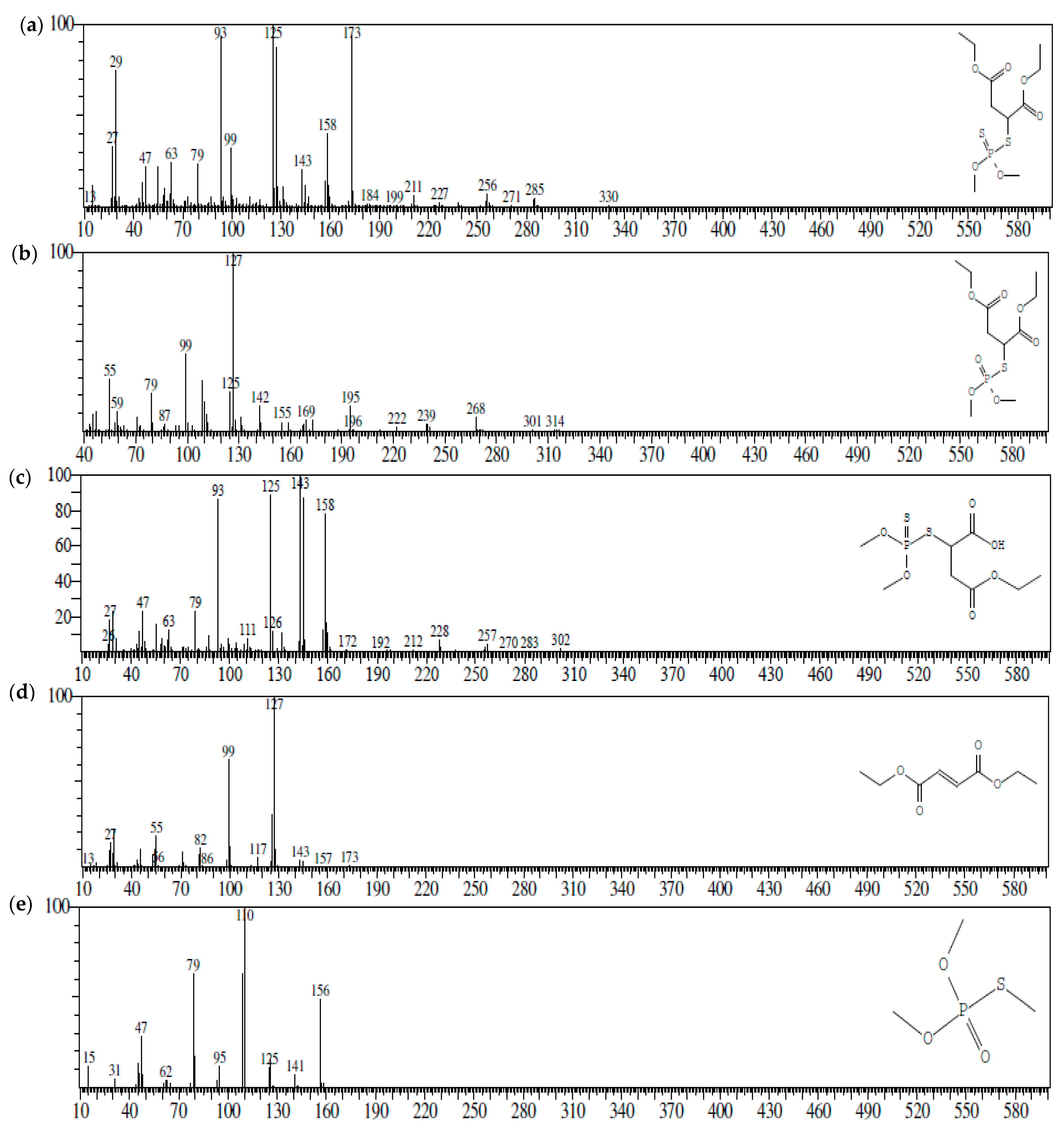
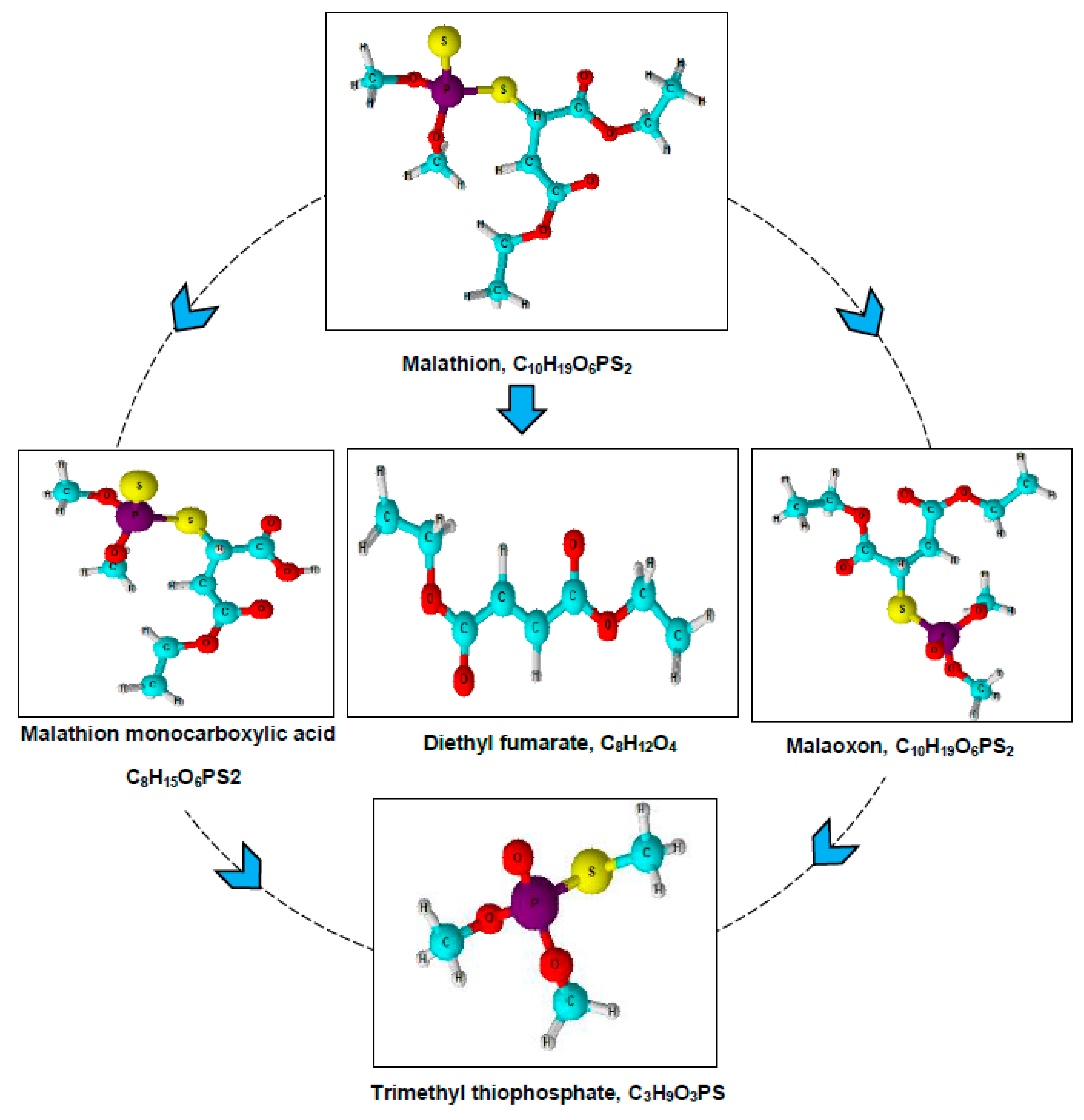
3.3. Metabolites Analysis

The metabolites formed during the malathion degradation were characterized via GC–MS. At the 15th day, soil with 500 mg malathion kg−1 cultured with a bacterial consortium consisting of Micrococcus aloeverae (MAGK3) + Bacillus cereus (AGB3) + Bacillus paramycoides (AGM5) was extracted and submitted for GC–MS analysis. Five types of products including malathion were detected due to the inoculated microbial activity in the soil. The fragmentation patterns of malathion, malaoxon, malathion monoacid, diethyl fumarate, and trimethyl thiophosphate detected in the soil are detailed in Table 4. The malathion spectrum pattern (without inoculums) displays a parent ion peak at m/z 330, which was feeble (Figure 1a).
The parent ions’ S-CH bonds cleaved, yielding two extremely abundant peaks at m/z 173 and 125. Two more peaks at m/z 256 and 184 in the mass spectrum correspond to the loss of 1–2 fragments of -COOC2H5 from the parent ion. With the addition of H, another signal was noticed at m/z 285, owing to CO2 evolution from the parent ion. The peak for malaoxon may be seen in the mass spectrum at m/z 314 (Figure 1b). The spectrum in Figure 1c exhibits the typical characteristics of malathion monocarboxylic acid (MMA), m/z 302, with a peak at m/z 257 owing to CO2 evolution from the parent ion. The parent ion’s S-CH is cleaved, resulting in a very abundant peak emerging at m/z 143. The common characteristics of diethyl fumarate m/z 173 are shown in Figure 1d, and the fragments of trimethyl thiophosphate m/z 156 are shown in Figure 1e. These finding revealed that malathion was degraded and transformed into four metabolites throughout the degradation process in the current study, which is consistent with prior findings [10,15,40,41,46]. Malathion degradation was likewise linked to the emergence of malaoxon, MMA, diethyl fumarate, and trimethyl thiophosphate in our study; therefore, a possible degradation pathway was proposed (Figure 2) because documenting metabolites and degradation pathways may lead to a better understanding of degradation processes [47]. However, from the standpoint of degradation processes, a single bacterial species is unlikely to completely mineralize the secondary metabolites. As a result of synergistic interactions that permit full mineralization, consortia are more effective for the degradation of environmental contaminants [48]. In several studies, microorganisms have been shown to degrade both original compounds and their metabolites [39,49,50]. However, only a few bacterial species are more effective in metabolite degradation than the primary compound [36]. The degradation rate of parent compounds was reported to be higher than their hydrolytic metabolites [37].

3.4. Kinetic Studies

The malathion degradation model, which better explains the kinetic data results, is discussed in this section. The kinetics of malathion degradation were computed by plotting the malathion residues against time (Figure 3) to produce correlation coefficients, and the maximum squared correlation coefficients were used to identify the best fit curve equations [27]. The kinetics were validated by graphing the time against LogC. Pesticide determination is generally described in terms of half-life (T1/2)/DT50, which is the time it takes for a pesticide to dissipate to 50% of its initial concentration. In soil inoculated with Micrococcus aloeverae (MAGK3), Bacillus cereus (AGB3), Bacillus paramycoides (AGM5), Micrococcus aloeverae (MAGK3) + Bacillus cereus (AGB3), Micrococcus aloeverae (MAGK3) + Bacillus paramycoides (AGM5), Bacillus cereus (AGB3) + Bacillus paramycoides (AGM5), and Micrococcus aloeverae (MAGK3) + Bacillus cereus (AGB3) + Bacillus paramycoides (AGM5), the DT50 values of total malathion were determined to be 9.75, 7.29, 9.26, 6.23, 7.63, 5.97, and 3.25 days, respectively, in soil with an initial malathion content of 500 mgkg−1. In the uninoculated sterilized and unsterilized control soil, the DT50 was observed to be 28.17 and 15.95 days, respectively (Table 5). Micrococcus aloeverae (MAGK3) + Bacillus cereus (AGB3) + Bacillus paramycoides (AGM5) microbial activity caused a decrease in DT50 to roughly 3.25 days. These finding showed that this unique mixed consortium may be suitable for the bioremediation of soil from malathion. The DT50 values in the kinetic model demonstrate that malathion was less persistent in the presence of the mixed consortium, which means that this consortium was better in the bioremediation of malathion compared to the pure strains, as depicted in Figure 3.

4. Conclusions

Three bacterial strains from Micrococcus and Bacillus species previously isolated via an enrichment approach were employed to investigate malathion degradation in soil-based pot experiments. Degradation kinetics revealed that, in addition to individual bacterial inoculum, a specific consortium was particularly successful in remediating malathion-contaminated soil. For a concentration of 500 mgkg−1 in the soil, a bacterial consortium consisting of Micrococcus aloeverae (MAGK3) + Bacillus cereus (AGB3) + Bacillus paramycoides (AGM5) was reported to completely degrade malathion by the 15th day. These findings highlight the potential of using bacterial consortia as the most optimal approach for the bioremediation of organophosphates in soil. The DT50 values, as suggested by the kinetics model, also demonstrated that mixed consortia were better in the bioremediation of malathion. The detection of metabolic products in the biodegradation process also validated the efficacy of the consortium, and indicated this mixed culture as one of the most effective ways to treat contaminated soils.

Supplementary Materials

The following supporting information can be downloaded at: https://www.mdpi.com/article/10.3390/soilsystems7040081/s1, Table S1: Results of physicochemical parameters of soil samples.

Author Contributions

The study plan was designed by M.A.D. and G.K. M.A.D. carried out the experiments, compiled the data, and wrote the manuscript. The manuscript was revised and finalized by G.K. All authors have read and agreed to the published version of the manuscript.

Funding

This research received no external funding.

Institutional Review Board Statement

Not applicable.

Informed Consent Statement

Not applicable.

Data Availability Statement

All the data that support the finding of this study are included in the manuscript.

Acknowledgments

Mohd Ashraf Dar acknowledges the support from DST, New Delhi, for providing a fellowship under the INSPIRE (IF170501) scheme.

Conflicts of Interest

The authors declare that they have no conflict of interest.

References

  1. Ortiz-Hernández, M.L.; Sánchez-Salinas, E.; Dantán-González, E.; Castrejón-Godínez, M.L. Pesticide biodegradation: Mechanisms, genetics and strategies to enhance the process. Biodegrad.-Life Sci. 2013, 10, 251–287. [Google Scholar]
  2. Morillo, E.; Villaverde, J. Advanced technologies for the remediation of pesticide-contaminated soils. Sci. Total Environ. 2017, 586, 576–597. [Google Scholar] [CrossRef]
  3. Krishna, K.R.; Philip, L. Biodegradation of lindane, methyl parathion and carbofuran by various enriched bacterial isolates. J. Environ. Sci. Health Part B-Pestic. Food Contam. Agric. Wastes 2008, 43, 157–171. [Google Scholar] [CrossRef] [PubMed]
  4. Dar, M.A.; Kaushik, G.; Chiu, J.F.V. Pollution status and biodegradation of organophosphate pesticides in the environment. In Abatement of Environmental Pollutants; Elsevier: Amsterdam, The Netherlands, 2020; pp. 25–66. [Google Scholar]
  5. Dar, M.A.; Kaushik, G.; Villarreal-Chiu, J.F. Pollution status and bioremediation of chlorpyrifos in environmental matrices by the application of bacterial communities: A review. J Environ. Manag. 2019, 239, 124–136. [Google Scholar] [CrossRef] [PubMed]
  6. Dar, M.A.; Baba, Z.A.; Kaushik, G. A review on phorate persistence, toxicity and remediation by bacterial communities. Pedosphere 2022, 32, 171–183. [Google Scholar] [CrossRef]
  7. Aardema, H.; Meertens, J.; Ligtenberg, J.J.M.; Peters-Polman, O.M.; Tulleken, J.E.; Zijlstra, J.G. Organophosphorus pesticide poisoning: Cases and developments. Neth. J. Med. 2008, 66, 149–153. [Google Scholar]
  8. Mahajan, R.; Bonner, M.R.; Hoppin, J.A.; Alavanja, M.C.R. Phorate exposure and incidence of cancer in the agricultural health study. Environ. Health Perspect. 2006, 114, 1205–1209. [Google Scholar] [CrossRef]
  9. Deng, S.Y.; Chen, Y.; Wang, D.S.; Shi, T.Z.; Wu, X.W.; Ma, X.; Li, X.Q.; Hua, R.M.; Tang, X.Y.; Li, Q.X. Rapid biodegradation of organophosphorus pesticides by Stenotrophomonas sp. G1. J. Hazard. Mater. 2015, 297, 17–24. [Google Scholar] [CrossRef]
  10. Geed, S.; Kureel, M.; Shukla, A.; Singh, R.; Rai, B. Biodegradation of malathion and evaluation of kinetic parameters using three bacterial species. Resour.-Effic. Technol. 2016, 2, S3–S11. [Google Scholar] [CrossRef]
  11. Kumari, B.; Madan, V.K.; Kathpal, T.S. Status of insecticide contamination of soil and water in Haryana, India. Environ. Monit. Assess. 2008, 136, 239–244. [Google Scholar] [CrossRef]
  12. Fadaei, A.; Dehghani, M.H.; Nasseri, S.; Mahvi, A.H.; Rastkari, N.; Shayeghi, M. Organophosphorous Pesticides in Surface Water of Iran. Bull. Environ. Contam. Toxicol. 2012, 88, 867–869. [Google Scholar] [CrossRef] [PubMed]
  13. Harinathareddy, A.; Prasad, N.; Devi, L. Pesticide residues in vegetable and fruit samples from Andhra Pradesh, India. J. Biol. Chem. Res 2014, 31, 1005–1015. [Google Scholar]
  14. Sanghi, R.; Pillai, M.K.K.; Jayalekshmi, T.R.; Nair, A. Organochlorine and organophosphorus pesticide residues in breast milk from Bhopal, Madhya Pradesh, India. Hum. Exp. Toxicol. 2003, 22, 73–76. [Google Scholar] [CrossRef] [PubMed]
  15. Dar, M.A.; Kaushik, G. Optimizing the malathion degrading potential of a newly isolated Bacillus sp. AGM5 based on Taguchi design of experiment and elucidation of degradation pathway. Biodegradation 2022, 33, 419–439. [Google Scholar] [CrossRef]
  16. Dar, M.A.; Chanwala, J.; Meena, P.R.; Singh, A.P.; Kaushik, G. Biodegradation of malathion by Micrococcus sp. strain MAGK3: Kinetics and degradation fragments. Arch. Microbiol. 2022, 204, 482. [Google Scholar] [CrossRef]
  17. Dar, M.A.; Kaushik, G. Phytotoxic effects and persistence of malathion on fenugreek (Trigonella foenum-graecum), and assessment of health risks. Acta Ecol. Sin. 2023, 43, 742–750. [Google Scholar] [CrossRef]
  18. WHO. World Health Organisation Staff. Guidelines for Drinking-Water Quality; WHO: Geneva, Switzerland, 2004; Volume 1, pp. 370–373. [Google Scholar]
  19. Moore, P.D.; Yedjou, C.G.; Tchounwou, P.B. Malathion-induced oxidative stress, cytotoxicity, and genotoxicity in human liver carcinoma (HepG2) cells. Environ. Toxicol. 2010, 25, 221–226. [Google Scholar] [CrossRef]
  20. Ohno, K.; Minami, T.; Matsui, Y.; Magara, Y. Effects of chlorine on organophosphorus pesticides adsorbed on activated carbon: Desorption and oxon formation. Water Res. 2008, 42, 1753–1759. [Google Scholar] [CrossRef]
  21. Ortiz-Hernandez, M.L.; Sanchez-Salinas, E. Biodegradation of the organophosphate pesticide tetrachlorvinphos by bacteria isolated from agricultural soils in mexico. Rev. Int. De Contam. Ambient. 2010, 26, 27–38. [Google Scholar]
  22. Xie, S.; Liu, J.X.; Li, L.; Qiao, C.L. Biodegradation of malathion by Acinetobacter johnsonii MA19 and optimization of cometabolism substrates. J. Environ. Sci. 2009, 21, 76–82. [Google Scholar] [CrossRef] [PubMed]
  23. Khan, S.; Zaffar, H.; Irshad, U.; Ahmad, R.; Khan, A.R.; Shah, M.M.; Bilal, M.; Iqbal, M.; Naqvi, T. Biodegradation Of Malathion By Bacillus Licheniformis Strain Ml-1. Arch. Biol. Sci. 2016, 68, 51–59. [Google Scholar] [CrossRef]
  24. Jimenez-Torres, C.; Ortiz, I.; San-Martin, P.; Hernandez-Herrera, R.I. Biodegradation of malathion, α-and β-endosulfan by bacterial strains isolated from agricultural soil in Veracruz, Mexico. J. Environ. Sci. Health Part B 2016, 51, 853–859. [Google Scholar] [CrossRef]
  25. Ishag, A.S.A.; Abdelbagi, A.O.; Hammad, A.M.A.; Elsheikh, E.A.E.; Elsaid, O.E.; Hur, J.H.; Laing, M.D. Biodegradation of Chlorpyrifos, Malathion, and Dimethoate by Three Strains of Bacteria Isolated from Pesticide-Polluted Soils in Sudan. J. Agric. Food Chem. 2016, 64, 8491–8498. [Google Scholar] [CrossRef]
  26. Dar, M.A.; Kaushik, G. Optimization of process parameters for biodegradation of malathion by Micrococcus aloeverae MAGK3 using Taguchi methodology and metabolic pathway analysis. Biocatal. Agric. Biotechnol. 2022, 42, 102362. [Google Scholar] [CrossRef]
  27. Jariyal, M.; Jindal, V.; Mandal, K.; Gupta, V.K.; Singh, B. Bioremediation of organophosphorus pesticide phorate in soil by microbial consortia. Ecotoxicol. Environ. Saf. 2018, 159, 310–316. [Google Scholar] [CrossRef] [PubMed]
  28. Rasool, S.; Rasool, T.; Gani, K.M. A review of interactions of pesticides within various interfaces of intrinsic and organic residue amended soil environment. Chem. Eng. J. Adv. 2022, 11, 100301. [Google Scholar] [CrossRef]
  29. Hong, F.; Pehkonen, S.O.; Brooks, E. Pathways for the hydrolysis of phorate: Product studies by 31P NMR and GC-MS. J. Agric. Food Chem. 2000, 48, 3013–3017. [Google Scholar] [CrossRef]
  30. Carter, D.O.; Yellowlees, D.; Tibbett, M. Autoclaving kills soil microbes yet soil enzymes remain active. Pedobiologia 2007, 51, 295–299. [Google Scholar] [CrossRef]
  31. Nowak, A.; Wronkowska, H. On the efficiency of soil sterilization in autoclave. Zent. Mikrobiol. 1987, 142, 521–525. [Google Scholar] [CrossRef]
  32. Trevors, J. Sterilization and inhibition of microbial activity in soil. J. Microbiol. Methods 1996, 26, 53–59. [Google Scholar] [CrossRef]
  33. Singh, B.K.; Walker, A. Microbial degradation of organophosphorus compounds. Fems Microbiol. Rev. 2006, 30, 428–471. [Google Scholar] [CrossRef] [PubMed]
  34. Karpouzas, D.; Walker, A. Factors influencing the ability of Pseudomonas putida strains epI and II to degrade the organophosphate ethoprophos. J. Appl. Microbiol. 2000, 89, 40–48. [Google Scholar] [CrossRef] [PubMed]
  35. Cycon, M.; Zmijowska, A.; Wojcik, M.; Piotrowska-Seget, Z. Biodegradation and bioremediation potential of diazinon-degrading Serratia marcescens to remove other organophosphorus pesticides from soils. J. Environ. Manag. 2013, 117, 7–16. [Google Scholar] [CrossRef] [PubMed]
  36. Maya, K.; Singh, R.S.; Upadhyay, S.N.; Dubey, S.K. Kinetic analysis reveals bacterial efficacy for biodegradation of chlorpyrifos and its hydrolyzing metabolite TCP. Process Biochem. 2011, 46, 2130–2136. [Google Scholar] [CrossRef]
  37. Liang, B.; Yang, C.L.; Gong, M.B.; Zhao, Y.F.; Zhang, J.; Zhu, C.X.; Jiang, J.D.; Li, S.P. Adsorption and degradation of triazophos, chlorpyrifos and their main hydrolytic metabolites in paddy soil from Chaohu Lake, China. J. Environ. Manag. 2011, 92, 2229–2234. [Google Scholar] [CrossRef]
  38. Karpouzas, D.G.; Singh, B.K. Microbial degradation of organophosphorus xenobiotics: Metabolic pathways and molecular basis. In Advances in Microbial Physiology; Poole, R.K., Ed.; Advances in Microbial Physiology: Cambridge, MA, USA, 2006; Volume 51, pp. 119–185. [Google Scholar]
  39. Lakshmi, C.V.; Kumar, M.; Khanna, S. Biotransformation of chlorpyrifos and bioremediation of contaminated soil. Int. Biodeterior. Biodegrad. 2008, 62, 204–209. [Google Scholar] [CrossRef]
  40. Goda, S.K.; Elsayed, I.E.; Khodair, T.A.; El-Sayed, W.; Mohamed, M.E. Screening for and isolation and identification of malathion-degrading bacteria: Cloning and sequencing a gene that potentially encodes the malathion-degrading enzyme, carboxylestrase in soil bacteria. Biodegradation 2010, 21, 903–913. [Google Scholar] [CrossRef]
  41. Singh, B.; Kaur, J.; Singh, K. Transformation of malathion by Lysinibacillus sp. isolated from soil. Biotechnol. Lett. 2012, 34, 863–867. [Google Scholar] [CrossRef]
  42. Singh, B.; Kaur, J.; Singh, K. Bioremediation of malathion in soil by mixed Bacillus culture. Adv. Biosci. Biotechnol. 2013, 4, 674–678. [Google Scholar] [CrossRef]
  43. Zhao, J.; Jiang, Y.; Gong, L.; Chen, X.; Xie, Q.; Jin, Y.; Liu, G. Mechanism of β-cypermethrin metabolism by Bacillus cereus GW-01. Chem. Eng. J. 2022, 430, 132961. [Google Scholar] [CrossRef]
  44. Birolli, W.G.; Dos Santos, A.; Pilau, E.; Rodrigues-Filho, E. New role for a commercially available bioinsecticide: Bacillus thuringiensis Berliner biodegrades the pyrethroid cypermethrin. Environ. Sci. Technol. 2021, 55, 4792–4803. [Google Scholar] [CrossRef] [PubMed]
  45. Huang, Y.; Chen, W.J.; Li, J.; Ghorab, M.A.; Alansary, N.; El-Hefny, D.E.; Chen, S. Novel mechanism and degradation kinetics of allethrin using Bacillus megaterium strain HLJ7 in contaminated soil/water environments. Environ. Res. 2022, 214, 113940. [Google Scholar] [CrossRef] [PubMed]
  46. Kim, Y.H.; Ahn, J.Y.; Moon, S.H.; Lee, J. Biodegradation and detoxification of organophosphate insecticide, malathion by Fusarium oxysporum f. sp pisi cutinase. Chemosphere 2005, 60, 1349–1355. [Google Scholar] [CrossRef]
  47. Sirajuddin, S.; Khan, M.A.; Ul Qader, S.A.; Iqbal, S.; Sattar, H.; Ansari, A. A comparative study on degradation of complex malathion organophosphate using of Escherichia coli IES-02 and a novel carboxylesterase. Int. J. Biol. Macromol. 2020, 145, 445–455. [Google Scholar] [CrossRef]
  48. Xu, B.; Xue, R.; Zhou, J.; Wen, X.; Shi, Z.; Chen, M.; Xin, F.; Zhang, W.; Dong, W.; Jiang, M. Characterization of acetamiprid biodegradation by the microbial consortium ACE-3 enriched from contaminated soil. Front. Microbiol. 2020, 11, 1429. [Google Scholar] [CrossRef]
  49. Hong, Q.; Zhang, Z.H.; Hong, Y.F.; Li, S.P. A microcosm study on bioremediation of fenitrothion-contaminated soil using Burkholderia sp. FDS-1. Int. Biodeterior. Biodegrad. 2007, 59, 55–61. [Google Scholar] [CrossRef]
  50. Cycon, M.; Wojcik, M.; Piotrowska-Seget, Z. Biodegradation kinetics of the benzimidazole fungicide thiophanate-methyl by bacteria isolated from loamy sand soil. Biodegradation 2011, 22, 573–583. [Google Scholar] [CrossRef]
Figure 1. Mass spectra of (a) malathion, (b) malaoxon, (c) malathion monocarboxylic acid, (d) diethyl fumarate, and (e) trimethyl thiophosphate formed during malathion biodegradation.
Soilsystems 07 00081 g001
Figure 2. Possible degradation pathways of malathion.
Soilsystems 07 00081 g002
Figure 3. Semi-logarithmic graph showing dissipation kinetics of malathion residues in amended soil. (a) M. aloeverae, B. cereus, and B. paramycoides; (b) M. aloeverae + B. cereus, M. aloeverae + B. paramycoides, B. cereus + B. paramycoides, and M. aloeverae + B. cereus + B. paramycoides; (c) control.
Soilsystems 07 00081 g003
Table 1. Effect of period of single-strain growth on residual malathion levels (mgkg−1) in soils fortified with malathion (500 mgkg−1).
Days after TreatmentMicrococcus aloeveraeBacillus cereusBacillus paramycoides
Malathion ResiduesPercent ReductionMalathion ResiduesPercent ReductionMalathion ResiduesPercent Reduction
0265.1270265.1270265.1270
5237.44010.442204.79622.755216.87718.198
10184.59530.37492.59165.076168.73536.356
15132.14750.15652.69080.12683.58268.474
2051.344080.63432.09287.89540.41084.757
303.150198.8111.42499.46211.61595.618
Table 2. Effect of period of consortia growth on residual malathion (mgkg−1) in soils fortified with malathion (500 mgkg−1).
Days after TreatmentMicrococcus aloeverae + Bacillus cereusMicrococcus aloeverae + Bacillus paramycoidesBacillus cereus + Bacillus paramycoidesMicrococcus aloeverae + Bacillus cereus + Bacillus paramycoides
Malathion ResiduesPercent ReductionMalathion ResiduesPercent ReductionMalathion ResiduesPercent ReductionMalathion ResiduesPercent Reduction
0265.1270265.1270265.1270265.1270
5193.37127.064179.33232.360213.44319.494149.71643.530
10114.69956.738132.16450.150127.41851.94079.69969.939
1562.09676.57877.02470.94830.21188.605ND 100
20ND10022.79091.404ND 100--
30--ND 100----
ND = Not detected
Table 3. Effect of period of unsterilized and sterilized soils on residual malathion (mgkg−1) in soils fortified with malathion (500 mgkg−1).
Days after TreatmentUninoculated UnsterilizedUninoculated Sterilized
Malathion ResiduesPercent ReductionMalathion ResiduesPercent Reduction
0265.1270265.1270
5237.41510.452247.7956.537
10213.54619.455223.53915.686
15173.78334.452187.87429.137
20117.78855.572159.34639.898
3048.02181.887126.04652.458
Table 4. Intermediate metabolites formed during biodegradation of malathion.
S. NoCompoundChemical FormulaCAS No.Molecular Weight (m/z)
1MalathionC10H19O6PS2CAS:121-75-5330
2MalaoxonC10H19O7PSCAS:1634-78-2314
3Malathion monocarboxylic acidC8H15O6PS2CAS:1190-29-0302
4Diethyl fumarate C8H12O4CAS:623-91-6173
5Trimethyl thiophosphateC3H9O3PSCAS: 152-18-1156
Table 5. Kinetic parameters and half-lives for the dissipation of malathion in soil amended with bacterial strains and their consortia.
TreatmentRegression Equation (y)Half-Life (Days)Correlation Coefficient (R2)
Micrococcus aloeverae−0.1438x + 0.70969.7530.8472
Bacillus cereus−0.1684x + 0.53447.2880.9187
Bacillus paramycoides−0.1087x + 0.31359.2590.9634
Micrococcus aloeverae + Bacillus cereus−0.2305x + 0.7426.2250.8725
Micrococcus aloeverae + Bacillus paramycoides−0.1903x + 0.75937.6310.8834
Bacillus cereus + Bacillus paramycoides−0.2153x + 0.59185.9670.9013
Micrococcus aloeverae + Bacillus cereus + Bacillus paramycoides−0.2333x + 0.06483.2480.8503
Uninoculated unsterilized−0.0565x + 0.208215.9500.915
Uninoculated sterilized−0.0261x + 0.042228.1680.9865
Disclaimer/Publisher’s Note: The statements, opinions and data contained in all publications are solely those of the individual author(s) and contributor(s) and not of MDPI and/or the editor(s). MDPI and/or the editor(s) disclaim responsibility for any injury to people or property resulting from any ideas, methods, instructions or products referred to in the content.

