Article

Genetic Diversity and Plant Growth-Promoting Activities of Root-Nodulating Bacteria in Guar Plants Across Jazan Province

1
Laboratory of Biodiversity and Valorization of Bioresources in Arid Zones (LR18ES36), Faculty of Sciences of Gabes, University of Gabes, Gabes 6029, Tunisia
2
Department of Biology, College of Science, Jazan University, P.O. Box 114, Jazan 45142, Saudi Arabia
3
Environment and Nature Research Center, Jazan University, P.O. Box 114, Jazan 45142, Saudi Arabia
*
Authors to whom correspondence should be addressed.
Soil Syst. 2025, 9(2), 39; https://doi.org/10.3390/soilsystems9020039
Submission received: 19 February 2025 / Revised: 10 April 2025 / Accepted: 21 April 2025 / Published: 24 April 2025

Abstract

:
Guar (Cyamopsis tetragonoloba L. Taub.) is a significant summer legume used as food for both humans and livestock. In Saudi Arabia, the root nodule bacteria of guar have not been studied. The present work investigated the phenotypic and genetic diversity of guar microsymbionts. Eighty-eight bacterial strains were isolated from the root nodules of guar grown in different locations of Jazan region of Saudi Arabia. The strains were analyzed based on their phenotypic characteristics and variations in their 16S rRNA gene sequences. A significant proportion of the isolates (90%) were fast-growing rhizobia, with 77% showing tolerance to 3–4% NaCl and 91% capable of thriving at temperatures reaching 40 °C. Several isolates exhibited strong plant growth-promoting traits, particularly in IAA production and phosphate solubilization. Genetic analysis indicated considerable diversity, with isolates classified under the genera Rhizobium, Ensifer, Mesorhizobium, Bradyrhizobium, and Agrobacterium. To the best of our knowledge, this study is the first to report on the phenotypic and genetic diversity of guar microsymbionts in Saudi Arabia.

1. Introduction

Increasing food production has become a top research priority for most countries worldwide to meet the demands of a rapidly growing global population, which is projected to reach 10 billion within the next 50 years [1]. Abiotic stresses caused by complex environmental conditions pose significant constraints on crop productivity and influence the geographic distribution of plant species across ecosystems [2]. The scientific community has sounded the alarm on the impact of climate change on rainfall patterns, salinization of farmland through irrigation, and the need to sustain or increase agricultural productivity on marginal lands [3,4,5].
Plants, under both natural and agricultural conditions, are exposed to various environmental stresses, either simultaneously or sequentially throughout their life cycles. Among these stresses, drought and salinity are particularly critical factors that reduce crop productivity by inhibiting photosynthesis and reducing plant growth [6]. This issue has become increasingly urgent as the area of salinity- and drought-affected lands con-tinues to expand.
Under natural conditions, plants often develop symbiotic relationships with the soil microorganisms, such as arbuscular mycorrhizal fungi (AMF) and nitrogen-fixing bacteria, notably rhizobia. Legumes, in particular, play a significant economic and ecological role as natural sources of fertilizer (reduced nitrogen) and human food. They also provide high-quality forage, contribute to soil stabilization, and prevent erosion. Additionally, perennial woody legumes are a major source of timber, phytochemicals, phytomedicines, and nitrogen fertility in agroecosystems, due to their ability to form nitrogen-fixing symbioses with rhizobia [7,8].
Rhizobia belong primarily to the α- and β-Proteobacteria classes, encompassing over 98 species across 13 genera, including Rhizobium, Mesorhizobium, Ensifer (formerly Sinorhizobium), and Bradyrhizobium. Recent studies also report non-classical rhizobia belonging to the γ-Proteobacteria class; however, their effectiveness in nodulating legumes remains inconclusive [9,10,11].
Rhizobia are soil-dwelling bacteria that establish nitrogen-fixing symbiotic relationships with both domesticated and wild leguminous plants. As plant growth-promoting rhizobacteria (PGPR), they enhance plant development by synthesizing indole-3-acetic acid (IAA), facilitating phosphate solubilization, generating ammonia, and producing siderophores. The plant hormone IAA promotes root growth and development im-proving root system architecture and enhancing the plant’s ability to absorb water and nutrients [12,13]. Furthermore, rhizobia solubilize inorganic soil phosphate, a critical mineral for energy transfer and root development, making it more accessible to plants [14,15]. Certain rhizobia strains produce ammonia, which plants can use as a nitrogen source, enhancing nutrition and growth [16]. They also exude siderophores, small, high-affinity iron-chelating compounds that bind soil iron, making it available to the plant [17,18,19]. Additionally, rhizobia can suppress soil-borne pathogens through competition for nutrients, antimicrobial compound release, or induction of plant defense mechanisms [20,21]. These plant growth-promoting activities play a crucial role in enhancing plant health, stimulating growth, and improving resilience to adverse environmental conditions [22,23,24].
Guar (Cyamopsis tetragonoloba L.) is a drought-tolerant legume widely cultivated in parts of Asia, valued for human consumption and cattle forage [25]. Despite numerous studies on its use as an alternative forage legume [26,27] and its potential for salt-affected soils [28], limited research exists on its symbiotic relationships with rhizobia.
Previous studies [29,30] have demonstrated that rhizobia isolated from native guar in Sudan and India exhibit fast growth and tolerance to high salinity and temperature levels. These stress-tolerant rhizobia have potential applications in biofertilization programs for legume seedlings [31]. However, the genetic diversity of rhizobia associated with guar remains poorly documented, particularly in Saudi Arabia.
Research shows that rhizobia-nodulating guar primarily belong to the genus Bradyrhizobium [32]. This study aims to enhance our understanding of legume-nodulating bacteria (LNB) in guar by analyzing a collection of rhizobia associated with the legume. To accomplish this, the taxonomic diversity of 88 nodule isolates from guar grown in various soil samples from Jazan was examined through both phenotypic and genotypic approaches. Additionally, their plant growth-promoting properties were assessed.

2. Materials and Methods

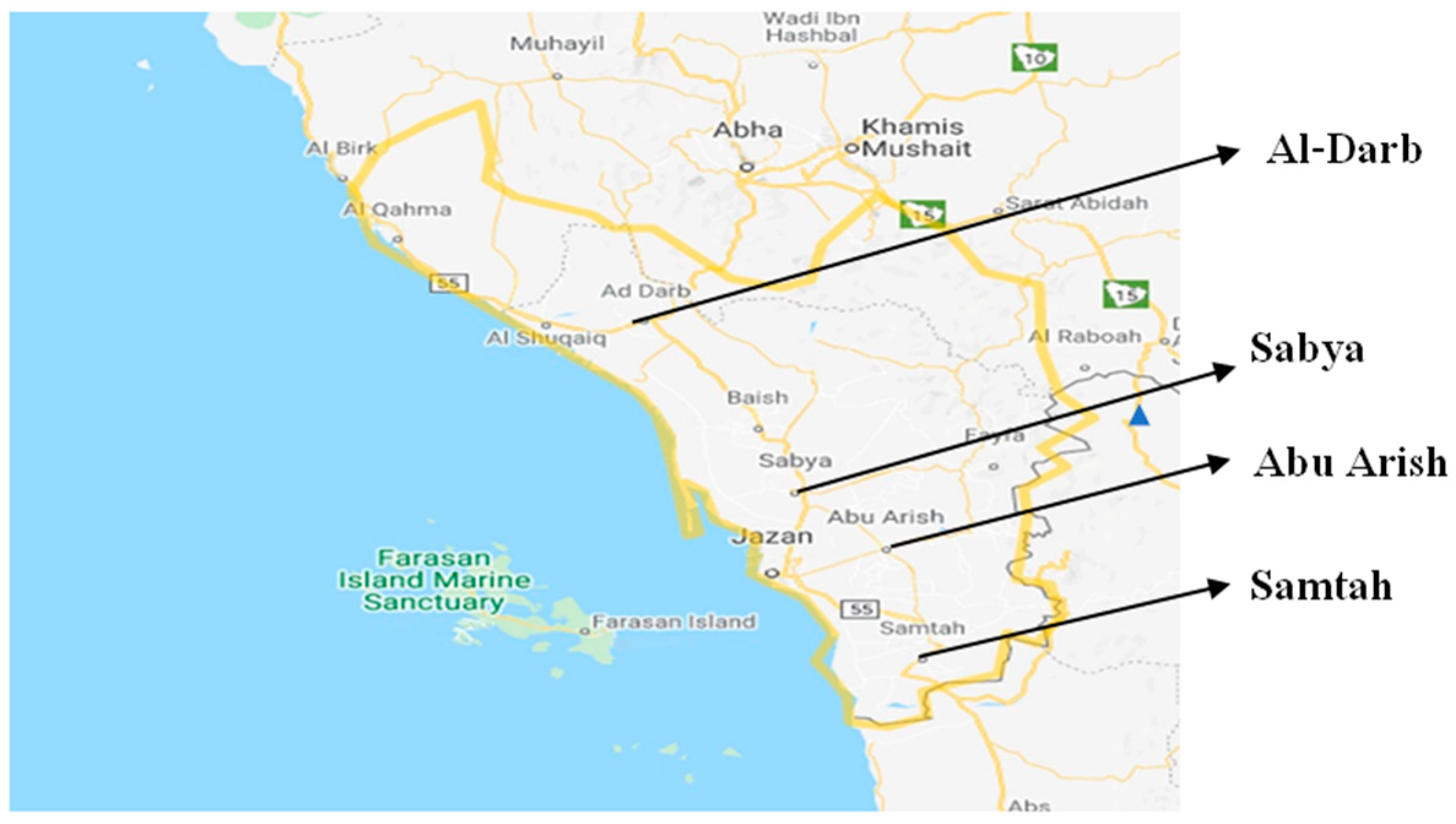
2.1. Collection of Soil Samples

Soil samples were collected from four agricultural areas in Jazan province: Samtah (16°36′0.2″ N, 42°55′50″ E), Abu Arish (16°57′14″ N, 42°50′12″ E), Al-Darb (17°41′18.1″ N, 42°19′31.4″ E), and Sabya (17°11′53.7″ N, 42°36′54.5″ E) (Figure 1). The altitude of all sites is below 85 m. At each site, soil samples were taken from a depth of 10–20 cm at multiple points and combined to form a composite sample. In the laboratory, the soil was passed through a 2 mm sieve, homogenized and stored in a cool room before the experiment. The pH and electrical conductivity values (EC) were measured using a pH and conductivity meter, respectively. Soil organic carbon (Corg) was determined by the Walkley and Black method, and total nitrogen (TN) was determined using the Kjeldahl method.

2.2. Plant Growth, Nodule Collection, and Bacteria Isolation

Guar seeds were surface-sterilized using 70% ethanol for one minute before being germinated in petri dishes. The resulting seedlings were individually transplanted into 1 L plastic pots filled with soil samples from four different locations. These plants were cultivated in a greenhouse under controlled conditions, maintaining temperatures of 24–26 °C during the day and 19–22 °C at night, with a 14-h photoperiod and 60–70% relative humidity. Each site was represented by four replicates, with one plant per pot. After a two-month growth period, the plants were uprooted, and nodulation was assessed. Nodules were collected, preserved in calcium chloride (CaCl2) to maintain their structural integrity, and transported to the laboratory for further analysis.
For rhizobia isolation, nodules were first rehydrated in sterile water and surface-sterilized by immersing them in 95% ethanol for 30 s, followed by treatment with 0.2% mercuric chloride for 20 s. They were then rinsed five times in sterile distilled water. A single sterilized nodule was crushed using a sterile glass rod in 0.1 mL of sterile distilled water within a sterile tube. A loopful of the resulting suspension was streaked onto Yeast Mannitol Agar (YEMA) plates supplemented with 0.0025% (w/v) Congo red to differentiate rhizobial colonies from other bacteria [34]. The plates were incubated at 28 °C in an inverted position and observed daily. Following incubation, individual colonies were selected and transferred onto fresh YEMA plates to ensure purity. Pure cultures were preserved on YEMA slants at 4 °C. An additional set of cultures was stored at −80 °C in 25% glycerol for long-term use.

2.3. Phenotypic Characterization of Bacteria and Plant Nodulation Tests

The isolates were characterized based on their colony color and morphology on YEMA plates incubated at 28 °C. Their growth rate was assessed spectrophotometrically, while generation time was determined by culturing in 50 mL of YEM broth within 250-mL Erlenmeyer flasks [35]. The cultures were incubated on a gyratory shaker at 180 g and 28 °C, with optical density measurements taken at 600 nm every two hours. Generation time was calculated from the exponential growth phase.
To evaluate environmental tolerance, the isolates were tested for their ability to grow under varying NaCl concentrations (1–5%), pH levels (4–12), and temperature conditions ranging from 28 °C to 45 °C on YEM agar plates [36]. Acid and alkali production were examined using YEM agar medium supplemented with bromothymol blue (0.0025%, w/v) as an indicator. All phenotypic assessments were conducted in triplicate.
Nodulation ability was tested by surface-sterilizing and germinating guar seeds in Petri dishes before transferring them to plastic pots filled with autoclaved vermiculite. The seedlings were inoculated with bacterial isolates and cultivated in a growth chamber for one month under controlled conditions—24 °C during the day, 18 °C at night, with a 14 h photoperiod and 70% relative humidity. Periodic sub-irrigation was performed using a nitrogen-free nutrient solution (Jensen’s medium). Three replicates of each isolate, along with three non-inoculated controls, were included. After a month, the plants were uprooted, and nodulation was evaluated.

2.4. Screening of PGPR for Multiple Plant Growth-Promoting Activities

To assess IAA production by bacterial isolates, individual colonies were inoculated into Jensen’s broth supplemented with 2 mg/mL L-tryptophan [37]. The cultures were incubated at 30 °C for 48–72 h on a gyratory shaker set at 120 rpm. Following incubation, 2 mL of the culture was centrifuged at 25,155× g for one min. A 1 mL aliquot of the resulting supernatant was combined with 2 mL of Salkowski’s reagent (a solution containing concentrated H2SO4, distilled water, and FeCl3.6H2O) and left to react in the dark at room temperature for 20 min. The appearance of a pink-red coloration signified IAA production, which was then quantified using a spectrophotometer (Shimadzu Corporation, Kyoto, Japan) [38].
Siderophore production was evaluated using Chrome Azurol Sulfate (CAS) agar medium. Bacterial isolates were streaked onto CAS agar plates and incubated at 30 °C. The formation of an orange or brown halo around the bacterial colonies was considered a positive result, indicating siderophore production [39].
Ammonia production was tested in peptone water by inoculating bacterial isolates into tubes containing 10 mL of peptone water. The cultures were incubated for 48–72 h, after which 1 mL of Nessler’s reagent was added. A color change to brown or yellow was indicative of ammonia production [40].
The ability of isolates to solubilize phosphate was examined using Pikovskaya medium [41]. After two weeks of incubation, the clear zone surrounding the colonies was measured. The phosphate solubilization efficiency was calculated using the following formula: (size of colony + size of clear zone)/diameter of colony.

2.5. Extraction of DNA and PCR Amplifications

DNA was extracted by simple boiling. The process involved suspending bacterial pellets in 30 μL sterile water and subjecting the mix to seven heat shocks (96 °C-2 min, 4 °C-10 s) [42]. After boiling, the microfuge tubes were centrifuged at 5000 rpm for 5 min, and the resulting supernatant containing DNA was carefully transferred to fresh tubes and stored at −20 °C for further analysis.
For PCR amplification of the 16S rRNA gene, the primers fD1 (5′ CCA GCA GCC GCG GTA ATA CG 3′) and rD1 (5′ AAG GAG GGG ATC CAG CCG CA 3′) [43] were utilized. The PCR reaction was conducted in a total volume of 20 μL, which included 10 μL of 1X innuMix Standard PCR, 1 μL of each primer at a concentration of 10 μM, 6 μL of sterile Milli-Q water, and 2 μL of template DNA. A negative control, omitting the DNA template, was included in each PCR run to ensure the accuracy of the reaction. The PCR was carried out in a Gene Amp PCR System 9700 (Applied Biosystem, Waltham, MA, USA) using the following program: initial denaturation of 5 min at 95 °C, followed by 35 cycles of 1 min at 94 °C, 1 min at 59 °C, and 2 min at 72 °C, and a final extension of 3 min at 72 °C. The PCR products were analyzed through horizontal electrophoresis on a 1% agarose gel (Type II agarose: Medium EEO, Sigma, St. Louis, MO, USA). A 1 kb Ladder marker (GibcoBRL, Waltham, MA, USA) was included to estimate band sizes. The gel was run in 0.5X Tris-acetate (TAE) buffer at 100 V for 30 min. Following electrophoresis, the gel was stained with ethidium bromide (2 μg/mL) for 30 min, then visualized using a UV transilluminator and documented with a digital camera.

2.6. 16S rRNA Gene Sequencing and Analysis

The amplified 16S rRNA gene fragment (approximately 1500 bp) was excised and purified using a gel extraction kit (Invitrogen, Darmstadt, Germany). DNA concentration was determined by measuring optical density and confirmed through electrophoresis on a 1% agarose gel. Purified DNA (more than 10 ng) was used for a sequencing reaction (Sanger method) using the ABI Prism BigDye kitTerminator (Applied Biosystems). Two primers, FGPS6 and FGPS1509, were used to sequence the 16S rRNA gene. The partial sequences of the 16S rRNA gene were assembled using ChromasPro software (version 1.34 Applied Biosystems) in one single sequence representing almost the entire gene.
The obtained sequences were aligned using Clustal X 1.83 and GeneDoc 2.7 software. To determine the closest relatives, a BLAST search was conducted on the National Center for Biotechnology Information (NCBI) database, comparing sequence similarities.
The 16S rRNA gene sequences analyzed in this study have been assigned GenBank accession numbers MW186843 to MW186856. Phylogenetic analyses were carried out using MEGA 3.1 software [44]. A neighbor-joining tree was constructed based on the Kimura two-parameter model of evolution [45], and the reliability of internal branches was evaluated through 1000 bootstrap replications.

3. Results

3.1. Constitution of a Local Collection of Rhizobium and Soil Analysis

The characteristics of the soil of the study sites are presented in Table 1. The samples’ pH and EC were remarkably similar among all four sites, with all soils being basic (pH > 7). The highest total nitrogen content was observed in the Sabya region (0.13%). After two months of growth, nodule formation was observed for all sites. However, the nodules were small, with a maximum of eight nodules per plant. All collected nodules were considered for bacteria isolation with a maximum of 28 nodules per site. Bacteria were isolated from these nodules resulting in a total of 88 isolates obtained (Table 2). Only one isolate was considered from each nodule. In cases where several colony types were detected, the most abundant colony type was chosen for isolation.

3.2. Phenotypic Characterization

Nodulation test was performed for all the isolates. Results showed that only seven isolates, which were classified as Agrobacterium and Rhizobium by 16S rRNA gene sequencing analyses (see below), failed to nodulate their host plant of origin. The number of nodules per plant varied between 2 and 11. For phenotypic test, the study examined several morphological criteria for the bacterial colonies, including shape, relief, size, granulation, and color. Most isolates (73%) had a non-circular colony shape. The predominant relief was convex, with only 10% of isolates exhibiting a flat surface. Colony diameter ranged from 0.5 to 5 mm, with size depending on the quantity of extracellular polysaccharides (EPS), produced by the bacteria. A diffuse, grainy granulation was most common. On solid growth medium, all isolates appeared between 3 and 8 days after subculturing. When streaked on YEMA-BTB medium, most of the isolates exhibited acid production, as evidenced by the BTB indicator turning from deep green to yellow after 3 to 5 days of dark incubation. These isolates also showed rapid growth, with an average generation time of 3 h. Nine isolates were identified as slow-growing, with generation times exceeding 6 h. The majority (80%) of the isolates were capable of growing in 2% NaCl, while 7 isolates demonstrated even higher levels of halotolerance, thriving in 5% NaCl. Based on NaCl tolerance, the isolates were classified into three groups: the first group included 19 isolates resistant to high NaCl concentrations (4%), the second group consisted of 56 isolates with moderate resistance (2–3% NaCl), and the third group had 13 isolates sensitive to NaCl with only 1% tolerance. Tolerance to temperature varied, with 77% of the isolates being able to grow at 40 °C. Only 14 isolates continued to proliferate at 42 °C but not at 45 °C. The influence of pH on the growth of guar isolates was carried out on YEMA medium. At pH 6, 7, 9, and 12, the majority of the isolates exhibited optimal growth. At pH 4, 90% of the isolates were unable to grow.

3.3. Characterization for Plant Growth-Promoting Traits

The plant growth-promoting characteristics of all isolates were evaluated, and the results are summarized in Table 3. For IAA production, the qualitative test revealed that 30% of guar isolates were capable of producing indole compounds in 1% tryptone broth. In contrast, most of the Rhizobium isolates produced IAA. Among these, the highest IAA production was observed in Rhizobium isolate GS1, which produced 120 µg/mL after 72 h, likely due to its more efficient utilization of medium components for IAA production compared to other isolates. The lowest IAA-producing isolate was GSA17, with only 15 µg/mL. Two Ensifer isolates, GA18 and GD8, were also IAA producers. Ammonia production, which indicates ammonification occurring in the rhizosphere, was detected in 76 bacterial isolates, as evidenced by a change in colony color from white to brownish-yellow. However, all Agrobacterium isolates, three Rhizobium isolates, and five Ensifer isolates did not produce ammonia. Siderophore production was observed in 15 isolates, indicated by the formation of clear zones around colonies, suggesting their ability to produce siderophores. Eight Bradyrhizobim isolates and six Rhizobium isolates formed an orange-colored zone around the bacterial colonies. Many Rhizobium and Ensifer isolates are able to solubilize phosphate and, thus, are able to form the halo zone with different areas. The other strain does not form the halo zone. The high phosphate dissolution ability (phosphate dissolution rate) was observed in isolate GS1 (3.88) (Table 3).

3.4. Molecular Characterization

In this study, we sequenced the 16S rRNA gene of all bacterial isolates obtained from the nodules of guar plants. The sequences were compared to those available in the GenBank database. Our result revealed a high degree of genetic diversity among the isolates in the bacterial collection. The majority of the isolates belonged to the following genera: Rhizobium (39 isolates), Ensifer (23 isolates), Mesorhizobium (13 isolates), Bradyrhizobium (9 isolates). Four isolates grouped with the genus Agrobacterim, which is not known to induce nodule formation. The comparison of the obtained sequences revealed that they were grouped into 15 different sequences. Based on 16 rRNA gene sequence comparisons, including reference bacterial sequences, we distinguished 15 phylogenetic clusters (Figure 2). Clusters 1, 2, and 3 were closely related to Ensifer medicae 11-3 21a (NR_104719), Ensifer arboris LMG 14919 (NR_114988), and Ensifer fredii USDA205 (NR_115181), respectively. Members of Clusters 4 and 5 were grouped with 3 Mesorhizobium species (Mesorhizobium plurifarium NBRC 102498, Mesorhizobium plurifarium LMG 11892, and Mesorhizobium atlanticum CNPSo 3140). Clusters 7, 8, 9, 10, 11, 12, and 13 were grouped with reference types strains: Rhizobium glycinendophyticum, Rhizobium tropici, Rhizobium mongolense, Rhizobium indicum, Rhizobium aegyptiacum, Rhizobium galegae, and Rhizobium huautlense, respectively. Cluster 14 (6 isolates) and Cluster 15 (3 isolates) were associated with reference strains of Bradyrhizobium (Bradyrhizobium cajani, Bradyrhizobium denitrificans, Bradyrhizobium elkanii, Bradyrhizobium ripae, and Bradyrhizobium pachyrhizi). GS13, representative of 16S rRNA gene type 10, formed with 2 reference strains of Agrobacterium (Agrobacterium tumefaciens and Agrobacterium radiobacter) and Rhizobium nepotum 39/7 (NR_117203) common cluster (Cluster 6).

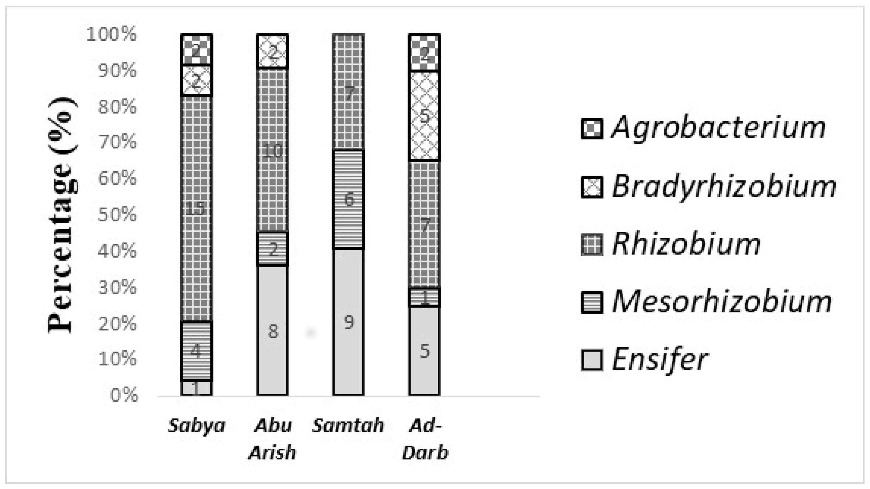
3.5. Taxonomic Affiliation of the Rhizobial Isolates and Their Distribution Across Sites of Origin

The taxonomic classification of the isolates, based on their site of origin, is shown in Figure 3. Three genera—Rhizobium, Ensifer, and Mesorhizobium—were present at all the sites examined in this study. The Rhizobium genus was widely distributed among guar nodules collected from different regions, with frequences ranging from 62% in Sabia to 35% in Al-Darb. Agrobacterium bacteria were detected only in guar nodules from Sabia region (2 isolates) and Al-Darb region (2 isolates). Bradyrhizobium bacteria were isolated from Al-Darb (5 isolates), Sabia (2 isolates), and Abu Arish (2 isolates), but not found in guar nodules from Samtah region.

4. Discussion

In this study, we characterized 88 legume-nodulating bacteria (LNB) strains associated with guar (Cyamopsis tetragonoloba) plants growing at various locations in Jazan province. The success of rhizobia inoculants in promoting plant growth and nitrogen fixation depends on their ability to adapt to the soil environment. When selecting rhizobia strains for inoculants, it is crucial to consider their adaptability to various environmental conditions and also their PGPR activities such as phytohormones, siderophore production, phosphate solubilization, and production of antibiotics or antagonistic compounds. This can be assessed by examining the strains’ ability to utilize various substrates and withstand different environmental conditions. The growth parameter analysis allowed for the differentiation of slow- and fast-growing isolates. About 90% of the isolates obtained from guar nodules were classified as fast growers, while only 10% were slow-growing. A similar distinction between fast and slow-growing strains has been noted in rhizobia isolated from cowpea, soybean [46], and Acacia species in Saudi Arabian ecosystems [42]. However, Ibrahim et al. [32] reported only slow-growing strains in guar nodules. It is important to note that classifying rhizobia as fast or slow growers based on agar growth does not necessarily predict their growth rate in soil or nodules, as pointed out by Sarma et al. [47]. Additionally, it has been previously noted that fast-growing rhizobia, particularly those in the genus Rhizobium, change color from deep green to yellow on YE-MA-BTB within 3–5 days of growth [48,49]. In this study, all isolates (both fast and slow growers) were found to be acid producers when grown on Bromothymol Blue media. This property has been mentioned by several authors [50,51].
It is well established that salinity and temperature significantly impact the survival and growth of roots nodulating and their nitrogen fixation capabilities [52,53]. In our study, most tested isolates exhibited high tolerance to NaCl (3–4%) and high temperatures (40–42 °C). This adaptation may be specific to arid regions characterized by high soil temperatures and salinity, as described by Karanja and Wood [54] and Lebrazi et al. [55]. However, the legume–rhizobium symbiosis and nodule formation are generally more sensitive to salt or osmotic stress than free-living rhizobia [56]. As a result, rhizobial strains that show tolerance to high salinity in the laboratory may not necessarily be effective in nitrogen fixation in the field. Similar findings were reported by Ibrahim et al. [32] and Mishra et al. [28] for bacterial rhizobia isolated from guar in Sudan and India, respectively. Other studies [42,57] have found that rhizobia isolated from Acacia species in Saudi Arabia exhibit specific adaptations to high soil temperatures and salinity. Salt- and temperature-tolerant strains may have an advantage in surviving and multiplying in saline, arid soils, but nodulation and nitrogen fixation in such environments depend more on the guar plant than on the rhizobia themselves. The use of salt-tolerant rhizobial strains as inoculants for guar could enhance nodulation and nitrogen content under salt stress in reclaimed soils. Therefore, further research is recommended to identify salt-tolerant and effective rhizobial strains. As suggested by previous studies [36,42], we found that fast-growing strains were generally more tolerant to high NaCl concentrations than slow-growing rhizobia.
pH is a critical factor that significantly affects the growth and survival of microorganisms. Even slight variations in the pH of the growth medium can have substantial effects on organism development. A study by Jordan [58] indicated that the optimal pH range for rhizobial growth is typically between 6.0 and 7.0. In the current study, most of the guar isolates exhibited a neutral to baso-tolerant tendency, which may be linked to the alkaline pH that characterizes soils in southwestern Saudi Arabia. This finding aligns with a study by Mishra et al. [28], which found that rhizobia isolated from guar in India were tolerant to pH levels ranging from 6.0 to 8.0. Jordan [59] also reported that slow-growing rhizobial strains tend to be more tolerant to low pH conditions compared to fast-growing strains. However, some fast-growing strains, such as Rhizobium tropici, can grow at pH levels as low as 4.0 [59]. In this study, most of the guar isolates were fast-growing and were unable to grow at pH 4.
Many of the rhizobial strains isolated in this study demonstrated significant Plant Growth-Promoting Rhizobacteria (PGPR) activities. These strains were found to produce IAA, which can enhance root development and overall plant growth. Phytohormones produced by rhizobia, including auxins, cytokinins, and gibberellins, play vital roles in regulating processes like root elongation, cell division, and overall plant vitality [60,61]. In this study, most of the Rhizobium isolates were IAA producers, as described by Spaepen et al. [62] and Ghosh et al. [63], who noted that the biosynthesis of IAA in rhizobia primarily involves the indole-3-pyruvate pathway (IPyA), which has been identified in Bradyrhizobium, Rhizobium, and Azospirillum. Additionally, some of our guar strains demonstrated the ability to produce siderophores. Many reports have shown that several rhizobial strains from both Rhizobium and Bradyrhizobium species are capable of producing siderophores [64]. The siderophores released by rhizobia may also stimulate plant growth by making iron more available to both the rhizobia and the host plant, particularly under conditions of iron deficiency [65,66]. Many Rhizobium and Ensifer strains isolated in this study are able to solubilize phosphate. These findings suggest that rhizobia could be a promising approach for enhancing phosphorus nutrition in plants. Recent studies [12,67,68] have emphasized the role of rhizobia in increasing phosphorus availability through phosphate solubilization, especially when co-inoculated with phosphate-solubilizing bacteria (PSB). This synergistic interaction has been shown to improve phosphorus availability and optimize biological nitrogen fixation (BNF), resulting in more efficient and sustainable plant growth. For example, co-inoculating PSB and rhizobia has significantly boosted phosphorus availability and promoted the growth of forage legumes [69].
In our study, the majority of guar isolates are ammonia producers, which can play a key role in their ability to enhance plant growth. The production of ammonia by rhizobia contributes to the nitrogen supply for the host plant, especially in environments where nitrogen availability is limited [70,71]. Further studies—such as the detection of the nifH gene, which plays a crucial role in nitrogen fixation—could provide more robust evidence of the strains’ potential for nitrogen fixation. These PGPR activities suggest that the guar rhizobial strains not only promote nitrogen fixation but also support plant growth through multiple mechanisms. Nodule formation in legumes is a key symbiotic process that facilitates nitrogen fixation, providing the plant with essential nitrogen in nutrient-poor soils. The ability to form nodules is often accompanied by additional plant growth-promoting traits, including phosphate solubilization, phytohormone production (e.g., indole-3-acetic acid), and siderophore synthesis [70,72,73]. These traits work synergistically with nodule formation by enhancing nutrient uptake, regulating root architecture, and improving overall plant stress tolerance. For instance, phosphate solubilization increases the availability of phosphorus, a nutrient critical for both nodule development and overall plant growth [74], while phytohormones promote root proliferation, aiding in the formation of more nodules and better nutrient absorption [75]. This combined effect strengthens the plant’s growth, especially in challenging environmental conditions [76]. The PGPR activities of rhizobia are also of interest for improving the efficiency of biofertilizers. By selecting or engineering these rhizobial strains, researchers aim to enhance the growth of leguminous crops, particularly under suboptimal soil conditions.
For the identification of rhizobia, 16S rRNA gene sequencing was utilized, a widely accepted method for classifying bacterial genera [77,78,79,80]. This approach is also essential for the characterization of new bacterial species [81]. While Bradyrhizobium strains are commonly associated with nodulating guar [32], the present study found that most guar isolates belonged to the genera Rhizobium, Ensifer, Mesorhizobium, and Bradyrhizobium, based on genotypic analysis. The diversity of rhizobial strains in Jazan soils increases the likelihood of guar forming compatible symbioses for effective nodulation and nitrogen fixation. The high diversity of guar rhizobia observed in this study aligns with previous research, which has reported a broad diversity of rhizobial isolates from leguminous plants in the Jazan region [42].
The results further indicated that the majority of guar isolates in this study belonged to the genera Rhizobium and Ensifer, both of which are known symbionts of many legumes growing in arid regions [82,83]. To better resolve their taxonomic classification, further phylogenetic analysis based on housekeeping genes is recommended. Additionally, numerous Agrobacterium-like isolates have been obtained from root nodules, although most fail to induce nodulation in their original host [84,85,86]. While the mechanism of nodule invasion by Agrobacterium remains unclear, studies suggest that these strains act as endophytic bacteria within nodules. In our study, four isolates were identified as belonging to the genus Agrobacterium, which is not typically associated with nodule formation. These isolates can be considered opportunistic, as previously reported by Zakhia et al. [77], and no definitive explanation of the presence of Agrobacteria inside the nodules could be demonstrated. In addition, the genera Agrobacterium and Rhizobium are taxonomically very closely related. The current status of Rhizobium and Agrobacterium reflects ongoing efforts to clarify their taxonomy, as these genera share significant genetic and phenotypic similarities, complicating their differentiation. Advances in molecular techniques have helped, but the 16S rRNA gene, commonly used for bacterial classification, often lacks sufficient resolution at the species level for these closely related genera [87]. This is due to the high sequence similarity in the 16S rRNA gene among species within Rhizobium and Agrobacterium.

5. Conclusions

This study is the first to document the isolation of rhizobial bacteria from guar nodules in Saudi Arabia. The findings demonstrate that most of the LNB (legume-nodulating bacteria) originating from nodules of Cyamopsis tetragonoloba in four Jazan soils show significant genetic diversity. The majority of the isolates belong to the genera Ensifer and Rhizobium, with some potentially representing new species. Further taxonomic studies, such as DNA–DNA hybridization and sequencing of additional genes, are necessary to confirm their classification. Beyond their nitrogen-fixing ability, some guar isolates exhibit notable PGPR activities and may serve as promising candidates for inoculation trials. These combined mechanisms highlight the potential of rhizobial strains as valuable tools in sustainable agriculture, enhancing plant growth and productivity in an environmentally friendly manner.

Author Contributions

Conceptualization: M.A.A., H.K., M.M., and B.Y.; Data curation: M.A.A., H.K., M.M., and B.Y.; Formal analysis: H.K. and M.M.; Funding acquisition: H.K. and M.M.; Project administration: H.K.; Investigation: M.M., H.K., and B.Y.; Methodology: M.A.A., H.K., M.M., and B.Y.; Resources: M.A.A., H.K., M.M., and B.Y.; Supervision: H.K. and M.M.; Writing—original draft: H.K., M.M., and B.Y.; Writing—review and editing: M.A.A., H.K., M.M., E.A., and B.Y. All authors have read and agreed to the published version of the manuscript.

Funding

The research project was funded by the Deanship of Graduate Studies and Scientific Research, Jazan University, Saudi Arabia (project number: RG24-S057).

Institutional Review Board Statement

Not applicable.

Informed Consent Statement

Not applicable.

Data Availability Statement

The data set used and analyzed in this study, along with related materials, can be obtained from the corresponding author (M.M.) upon reasonable request.

Acknowledgments

The authors gratefully acknowledge the funding of the Deanship of Graduate Studies and Scientific Research, Jazan University, Saudi Arabia, through project number (RG24-S057).

Conflicts of Interest

The authors declare that there are no conflicts of interest related to this article.

References

  1. Food and Agriculture Organization of the United Nations (FAO). How to Feed the World in 2050; FAO: Rome, Italy, 2009; Available online: https://www.fao.org/publications (accessed on 20 February 2021).
  2. Araus, J.L.; Slafer, G.A.; Reynolds, M.P.; Royo, C. Plant breeding and drought in C-3 cereals: What should we breed for? Ann. Bot. 2002, 89, 925–940. [Google Scholar] [CrossRef] [PubMed]
  3. Mittler, R. Abiotic stress, the field environment and stress combination. Trends Plant Sci. 2006, 11, 15–19. [Google Scholar] [CrossRef] [PubMed]
  4. Shao, H.B.; Chu, L.Y.; Jaleel, C.A.; Zhao, C.X. Water-deficit stress-induced anatomical changes in higher plants. Comptes Rendus Biol. 2008, 331, 215–225. [Google Scholar] [CrossRef] [PubMed]
  5. Patanè, C.; Saita, A.; Sortino, O. Eco-physiological response of a drought-tolerant and a drought-sensitive tomato genotype during water stress and recovery. Plant Biosyst. 2012, 146, 100–109. [Google Scholar] [CrossRef]
  6. Verslues, P.E.; Agarwal, M.; Katiyar-Agarwal, S.; Zhu, J.; Zhu, J.K. Methods and concepts in quantifying resistance to drought, salt, and freezing, abiotic stresses that affect plant water status. Plant J. 2006, 45, 523–539. [Google Scholar] [CrossRef]
  7. Graham, P.H.; Vance, C.P. Nitrogen fixation in perspective: An overview of research and extension needs. Field Crops Res. 2000, 65, 93–106. [Google Scholar] [CrossRef]
  8. Graham, P.H.; Vance, C.P. Legumes: Importance and constraints to greater use. Plant Physiol. 2003, 131, 872–877. [Google Scholar] [CrossRef]
  9. Benhizia, Y.; Benhizia, H.; Benguedouar, A.; Muresu, R.; Giacomini, A.; Squartini, A. Gamma proteobacteria can nodulate legumes of the genus Hedysarum. Syst. Appl. Microbiol. 2004, 27, 462–468. [Google Scholar] [CrossRef]
  10. Muresu, R.; Polone, E.; Sulas, L.; Baldan, B.; Tondello, A.; Delogu, G.; Benguedouar, A. Coexistence of predominantly nonculturable rhizobia with diverse, endophytic bacterial taxa within nodules of wild legumes. FEMS Microbiol. Ecol. 2008, 63, 383–400. [Google Scholar] [CrossRef]
  11. Mahdhi, M.; Fterich, A.; Rejili, M.; Rodriguez-Llorente, I.D.; Mars, M. Legume-Nodulating Bacteria (LNB) from three pasture legumes (Vicia sativa, Trigonella maritima and Hedysarum spinosissimum) in Tunisia. Ann. Microbiol. 2012, 62, 61–68. [Google Scholar] [CrossRef]
  12. Datta, C.; Basu, P.S. Indole acetic acid production by a Rhizobium species from root nodules of a leguminous shrub, Cajanus cajan. Microbiol Res. 2000, 155, 123–127. [Google Scholar] [CrossRef] [PubMed]
  13. Lebrazi, S.; Fadil, M.; Chraibi, M.; Fikri-Benbrahim, K. Screening and optimization of indole-3-acetic acid production by Rhizobium sp. strain using response surface methodology. J. Genet. Eng. Biotechnol. 2020, 18, 21. [Google Scholar] [CrossRef] [PubMed]
  14. Sridevi, M.; Mallaiah, K.V. Phosphate solubilization by Rhizobium strains. Indian J. Microbiol. 2009, 49, 98–102. [Google Scholar] [CrossRef]
  15. Shome, S.; Barman, A.; Solaiman, Z.M. Rhizobium and Phosphate Solubilizing Bacteria Influence the Soil Nutrient Availability, Growth, Yield, and Quality of Soybean. Agriculture 2022, 12, 1136. [Google Scholar] [CrossRef]
  16. Manasa, K.R.; Subhash Reddy, S.; Triveni, B.; Kranthi, K.; Gowri Priya, N. Characterization of Rhizobium Isolates and their Potential PGPR Characteristics of different Rhizosphere Soils of Telangana Region. Int. J. Curr. Microbiol. Appl. Sci. 2017, 6, 2808–2813. [Google Scholar] [CrossRef]
  17. Saikia, S.P.; Bora, D.; Goswami, A.; Mudoi, K.D. Role of siderophores in chickpea (Cicer arietinum L.)—Rhizobium symbiosis. Microbiol. Res. 1998, 153, 47–53. [Google Scholar] [CrossRef]
  18. Deryło, M.; Choma, A.; Puchalski, B.; Suchanek, W. Siderophore activity in Rhizobium species isolated from different legumes. Acta Biochim. Pol. 1994, 41, 7–11. [Google Scholar] [CrossRef]
  19. Datta, B.; Chakrabartty, P.K. Siderophore biosynthesis genes of Rhizobium sp. isolated from Cicer arietinum L. 3 Biotech 2013, 4, 391–401. [Google Scholar] [CrossRef]
  20. Chandra, N.; Jack, K.J.; Wozniak, S.S.; Porter, M.L.; Friesen, M.L. Rhizobia protect their legume hosts against soil-borne microbial antagonists in a host-genotype-dependent manner. Rhizosphere 2019, 9, 47–55. [Google Scholar]
  21. Goyal, R.K.; Habtewold, J.Z. Evaluation of Legume–Rhizobial Symbiotic Interactions Beyond Nitrogen Fixation That Help the Host Survival and Diversification in Hostile Environments. Microorganisms 2023, 11, 1454. [Google Scholar] [CrossRef]
  22. Sijilmassi, B.; Filali-Maltouf, A.; Fahde, S.; Ennahli, Y.; Boughribil, S.; Kumar, S.; Amri, A. In-Vitro Plant Growth Promotion of Rhizobium Strains Isolated from Lentil Root Nodules under Abiotic Stresses. Agronomy 2020, 10, 1006. [Google Scholar] [CrossRef]
  23. Tulumello, J.; Chabert, N.; Rodriguez, J.; Long, J.; Nalin, R.; Achouak, W.; Heulin, T. Rhizobium alamii improves water stress tolerance in a non-legume. Sci. Total. Environ. 2021, 797, 148895. [Google Scholar] [CrossRef] [PubMed]
  24. Barquero, M.; Poveda, J.; Laureano-Marín, A.M.; Ortiz-Liébana, N.; Brañas, J.; González-Andrés, F. Mechanisms involved in drought stress tolerance triggered by rhizobia strains in wheat. Front. Plant Sci. 2022, 13, 1036973. [Google Scholar] [CrossRef] [PubMed]
  25. Alshameri, A.; Al-Qurainy, F.; Khan, S.; Nadeem, M.; Gaafar, A.R.; Tarroum, M.; Ashraf, M. Appraisal of guar [Cyamopsis tetragonoloba (L.) Taub.] accessions for forage purpose under the typical Saudi Arabian environmental conditions encompassing high temperature, salinity and drought. Pak. J. Bot. 2017, 49, 1405–1413. [Google Scholar]
  26. Gomaa, M.S.; Simons, C.; Brancale, A. Homology model of 1α, 25-dihydroxyvitamin D3 24-hydroxylase cytochrome P450 24A1 (CYP24A1): Active site architecture and ligand binding. J. Steroid Biochem. Mol. Biol. 2007, 104, 53–60. [Google Scholar] [CrossRef]
  27. Rao, N.K.; Shahid, M. Potential of cowpea [Vigna unguiculata (L.) Walp.] and guar [Cyamopsis tetragonoloba (L.) Taub.] as alternative forage legumes for the United Arab Emirates. Emir. J. Food Agric. 2011, 23, 147–156. [Google Scholar]
  28. Rasheed, M.J.Z.; Ahmad, K.; Qurainy, F.A.; Khan, S.; Athar, H.U.R. Screening of diverse local germplasm of guar [Cyamopsis tetragonoloba (L.) Taub.] for salt tolerance: A possible approach to utilize salt-affected soils. Pak. J. Bot. 2015, 47, 1721–1726. [Google Scholar]
  29. Mishra, B.K.; Yadav, V.; Vishal, M.K.; Kant, K. Physiological and molecular characterization of clusterbean (Cyamopsis tetragonoloba (L.) Taub) rhizobia isolated from different areas of Rajasthan, India. Legume Res. 2013, 36, 299–305. [Google Scholar]
  30. El Hussein, A.A. Characterization of Native Guar Rhizobia and Their Cross Inoculation Abilities Among Different Guar (Cyamopsis tetragonoloba (L.) Taub.) Lines. Master’s Thesis, University of Khartoum, Khartoum, Sudan, 2015. [Google Scholar]
  31. Subha, D.; Rajesh, G. Assessing Stress Tolerant Rhizobial Isolates of Clusterbean (Cymopsis tetragonoloba (L.) Taub.) Retrieved from Semi- Arid Regions of Haryana, India. Int. J. Curr. Microbiol. Appl. Sci. 2017, 6, 744–753. [Google Scholar]
  32. Ibrahim, K.A.; Naeim, E.A.; El Naim, A.M.; Elsheikh, M.A. Response of guar (Cyamopsis teteragonolopa L.) to Bradyrhizobium inoculations in semi-arid Environment. Int. J. Agric. For. 2016, 6, 137–141. [Google Scholar]
  33. Yami, B. Phenotypic and Genetic Diversity of Root-Nodulating Bacteria Associated with Guar (Cyamopsis tetragonoloba L. Taub.) Growing in Different Regions of Jazan. Master’s Thesis, Jazan University Saudi Arabia, Jazan, Saudi Arabia, 2021; p. 18. [Google Scholar]
  34. Vincent, J.M. A Manual for the Practical Study of Root Nodule Bacteria; Blackwell Scientific Publications: Oxford, UK, 1970. [Google Scholar]
  35. Yelton, M.M.; Buckley, D.W.; Post, J.A. Growth and nitrogen fixation of Rhizobium japonicum and its mutant strains. Appl. Environ. Microbiol. 1983, 45, 602–605. [Google Scholar]
  36. Mohamed, M.A.H.; Harris, P.J.C.; Henderson, J. In vitro selection and characterisation of a drought tolerant clone of Tagetes minuta. Plant Sci. 2000, 159, 213–222. [Google Scholar] [CrossRef]
  37. Bric, J.M.; Bostock, R.M.; Silverstone, S.E. Rapid in situ assay for indoleacetic acid production by bacteria immobilized on a nitrocellulose membrane. Appl. Environ. Microbiol. 1991, 57, 535–538. [Google Scholar] [CrossRef]
  38. Islam, S.; Akanda, A.M.; Prova, A.; Islam, M.T.; Hossain, M.M. Isolation and Identification of Plant Growth Promoting Rhizobacteria from Cucumber Rhizosphere and Their Effect on Plant Growth Promotion and Disease Suppression. Front. Microbiol. 2016, 6, 1360. [Google Scholar] [CrossRef] [PubMed]
  39. Schwyn, B.; Neilands, J.B. Universal chemical assay for the detection and determination of siderophores. Anal. Biochem. 1987, 160, 47–56. [Google Scholar] [CrossRef] [PubMed]
  40. Cappuccino, J.G.; Sherman, N. Microbiology: A Laboratory Manual, 4th ed.; Benjamin/Cummings: San Francisco, CA, USA, 1992. [Google Scholar]
  41. Pikovskaya, R.I. Mobilization of phosphorus in soil in connection with the vital activity of some microbial species. Microbiol. 1948, 17, 362–370. [Google Scholar]
  42. Mahdhi, M.; Tounekti, T.; Khemira, H. Phenotypic and Genotypic Characterization of Microsymbionts of Acacia Species Plants Grown in South-Western Saudi Arabia. J. Soil. Sci. Plant Nutr. 2019, 19, 631–638. [Google Scholar] [CrossRef]
  43. Weisburg, W.G.; Barns, S.M.; Pelletier, D.A.; Lane, D.J. 16S ribosomal DNA amplification for phylogenetic study. J. Bacteriol. 1991, 173, 697–703. [Google Scholar] [CrossRef]
  44. Kumar, S.; Tamura, K.; Jakobsen, I.B.; Nei, M. MEGA2: Molecular evolutionary genetics analysis software. Bioinformatics 2001, 17, 1244–1245. [Google Scholar] [CrossRef]
  45. Kimura, M. A simple method for estimating evolutionary rates of base substitutions through comparative studies of nucleotide sequences. J. Mol. Evol. 1980, 16, 111–120. [Google Scholar] [CrossRef]
  46. Hernandez, B.S.; Focht, D.D. Invalidity of the concept of slow growth and alkali production in cowpea Rhizobia. Appl. Environ. Microbiol. 1984, 48, 206–210. [Google Scholar] [CrossRef]
  47. Sarma, R.K.; Saikia, R.; Thakur, D. Impact of plant growth-promoting endophytic bacteria on nutrient uptake and growth enhancement of aromatic rice and maize under low fertility condition. Microbiol. Res. 2021, 250, 126789. [Google Scholar]
  48. Alemayehu, W. The effect of indigenous Root Nodulating Bacteria on Nodulation and Growth of faba bean (Vicia faba) in low input agricultural systems of Tigray Highlands, Northern Ethopia. Momona Ethiop. J Sci. 2009, 1, 30–43. [Google Scholar]
  49. Al-mujahidy, S.J.; Hassan, M.; Rahman, M.; Mamun-Or-Rashid, A.N.M. Isolation and characterization of Rhizobium spp. and determination of their potency for growth factor production. Int. Res. J. Biotechnol. 2013, 4, 117–123. [Google Scholar]
  50. Jebara, M.; Mhamdi, R.; Aouani, M.E.; Ghrir, R.; Mars, M. Genetic diversity of Sinorhizobium populations recovered from different Medicago varieties cultivated in Tunisia soils. Can. J. Microbiol. 2001, 47, 139–147. [Google Scholar] [CrossRef] [PubMed]
  51. Odee, D.W.; Haukka, K.; Sprent, J.I.; Sutherland, J.M.; Young, J.P.W. Genetic and symbiotic characterization of rhizobia isolated from tree and herbaceous legumes grown in soils from ecologically diverse sites in Kenya. Soil Biol. Biochem. 2002, 34, 801–811. [Google Scholar] [CrossRef]
  52. Mutumba, F.A.; Zagal, E.; Gerding, M.; Castillo-Rosales, D.; Paulino, L.; Schoebitz, M. Plant growth promoting rhizobacteria for improved water stress tolerance in wheat genotypes. J. Soil Sci. Plant Nutr. 2018, 18, 1080–1096. [Google Scholar] [CrossRef]
  53. Saghafi, D.; Ghorbanpour, M.; Lajayer, B.A. Efficiency of Rhizobium strains as plant growth promoting rhizobacteria on morphophysiological properties of Brassica napus L. under salinity stress. J. Soil Sci. Plant Nutr. 2018, 18, 253–268. [Google Scholar]
  54. Karanja, N.K.; Wood, M. Selecting Rhizobium phaseoli strains for use with beans (Phaseolus vulgaris L.) in Kenya: Tolerance of high temperature and antibiotic resistance. Plant Soil 1988, 112, 15–22. [Google Scholar] [CrossRef]
  55. Lebrazi, S.; Chraibi, M.; Fadil, M.; Barkai, H.; Fikri-Benbrahim, K. Phenotypic, Genotypic and Symbiotic Characterization of Rhizobial Isolates Nodulating Acacia sp. in Morocco. J. Pure Appl. Microbiol. 2018, 12, 249–263. [Google Scholar] [CrossRef]
  56. Coba de la Peña, T.; Fedorova, E.; Pueyo, J.J.; Lucas, M.M. The Symbiosome: Legume and Rhizobia Co-evolution toward a Nitrogen-Fixing Organelle? Front. Plant Sci. 2018, 8, 2229. [Google Scholar] [CrossRef]
  57. Shetta, D.; EL-Sayed, A.W.B.; Nasr, T.A.; Shaarawy, N.M. Influences of mineral fertilization with NPK, inoculation and methods of inoculation on seedling growth of two woody legume trees. World Appl. Sci. J. 2014, 29, 825–834. [Google Scholar]
  58. Jordan, D.C. Family III Rhizobiacae. In Bergey’s Manual of Systematic Bacteriology; krieg, N.R., Holt, J.G., Eds.; Williams & Wilkins: Baltimore, ML, USA, 1984; pp. 234–242. [Google Scholar]
  59. Graham, P.H. Stress tolerance in Rhizobium and Bradyrhizobium, and nodulation under adverse soil conditions. Can. J. Microbiol. 1992, 38, 475–484. [Google Scholar] [CrossRef]
  60. Glick, B.R. Plant Growth-Promoting Bacteria: Mechanisms and Applications. Sci. Hortic. 2012, 128, 963401. [Google Scholar] [CrossRef] [PubMed]
  61. Hossain, M.D.; Ghosh, A.K.; Kaur, S. Role of Rhizobium in Plant Growth Promotion and Nitrogen Fixation. Biotechnol. Lett. 2014, 36, 1399–1408. [Google Scholar] [CrossRef]
  62. Spaepen, S.; Vanderleyden, J.; Remans, R. Indole-3-acetic acid in microbial and microorganism-plant signaling. FEMS Microbiol. Rev. 2007, 31, 425–448. [Google Scholar] [CrossRef] [PubMed]
  63. Ghosh, S.; Ghosh, P.; Maiti, T.K. Production and Metabolism of Indole Acetic Acid (IAA) by Root Nodule Bacteria (Rhizobium): A Review. Appl. Microbiol. 2011, 5, 523–540. [Google Scholar]
  64. Ramesh, B.D.; Subramanian, K.R.V. Siderophore production by Rhizobium and Bradyrhizobium species and its role in iron acquisition. Microbial. Ecol. 2014, 67, 667–676. [Google Scholar]
  65. Tahir, M.; Sarwar, M.A. Plant Growth Promoting Rhizobacteria (PGPR): A Budding Complement of Synthetic Fertilizers for Improving Crop Production. Group 2013, 19, 79–87. [Google Scholar]
  66. Igiehon, N.O.; Babalola, O.O.; Cheseto, X. Siderophores from rhizosphere bacteria as a potential biocontrol tool for managing plant pathogens. Environ. Sustain. 2019, 2, 57–71. [Google Scholar]
  67. Cubillos-Hinojosa, J.G.; de Sá, E.L.S.; Muniz, A.W.; Dick, D.P. Combined application of humic substances and PGPR inoculated and co-inoculated in plants of Phaseolus lunatus (L.) and Leucaena leucocephala (Lam.) de Wit. Rev. Colomb. Cienc. Hort. 2024, 18, e17621. [Google Scholar] [CrossRef]
  68. Ríos-Ruiz, W.F.; Castro-Tuanama, R.; Valdez-Nuñez, R.A.; Torres-Bernal, L.; Jave-Concepción, H.G.; Daza-Pérez, A.C.; Barrera-Lozano, M.; Archentti-Reátegui, F. Co-Inoculation of Phosphate-Solubilizing Bacteria and Rhizobia Increases Phosphorus Availability and Promotes the Development of Forage Legumes. Agronomy 2024, 14, 2493. [Google Scholar] [CrossRef]
  69. Damo, J.L.C.; Pedro, M.; Sison, M.L. Phosphate Solubilization and Plant Growth Promotion by Enterobacter sp. Isolate. Appl. Microbiol. 2024, 4, 1177–1192. [Google Scholar] [CrossRef]
  70. Gopalakrishnan, S.; Sathya, A.; Vijayabharathi, R.; Varshney, R.K.; Gowda, C.L.; Krishnamurthy, L. Plant growth promoting rhizobia: Challenges and opportunities. 3 Biotech 2015, 5, 355–377. [Google Scholar] [CrossRef] [PubMed]
  71. Laslo, É.; Mara, G. Is PGPR an Alternative for NPK Fertilizers in Sustainable Agriculture? In Microbial Interventions in Agriculture and Environment; Singh, D.P., Gupta, V.K., Prabha, R., Eds.; Springer: Singapore, 2019; pp. 51–62. [Google Scholar]
  72. Glick, B.R.; de-Bashan, L.E. Enhanced Plant Growth by ACC Deaminase-Producing Bacteria. Plant Physiol. Biochem. 2017, 119, 57–65. [Google Scholar]
  73. Chibeba, A.M.; Kyei-Boahen, S.; de Fátima Guimarães, M.; Nogueira, M.A.; Hungria, M. Isolation and Screening of Elite Inoculant Strains of Bradyrhizobium spp. for Soybean Cultivation in Mozambique. Appl. Soil Ecol. 2017, 120, 55–67. [Google Scholar]
  74. Alikhani, H.A.; Saleh-Rastin, N.; Antoun, H. Phosphate Solubilization Activity of Rhizobia Native to Iranian Soils. Plant Soil 2006, 287, 35–41. [Google Scholar] [CrossRef]
  75. Fukaki, H.; Tasaka, M. Hormone Interactions During Lateral Root Formation. Plant Mol. Biol. 2020, 104, 73–83. [Google Scholar] [CrossRef]
  76. Atieno, M.; Herrmann, L.; Okalebo, R.W.; Lesueur, D. Improvement of Maize Growth and Nutrient Uptake by Co-inoculation of Nitrogen Fixing and Phosphate Solubilizing Rhizobacteria under Field Conditions in Kenya. Soil Sci. Plant Nutr. 2017, 63, 398–409. [Google Scholar]
  77. Zakhia, F.; Jeder, H.; Domergue, O.; Willems, A.; Cleyet-Marel, J.-C.; Gillis, M.; Dreyfus, B.; De Lajudie, P. Characterisation of Legume-Nodulating Bacteria (LNB) in arid regions of Tunisia. Syst. Appl. Microbiol. 2004, 27, 380–395. [Google Scholar] [CrossRef]
  78. Zakhia, F.; Jeder, H.; Willems, A.; Gillis, M.; Dreyfus, B.; de Lajudie, P. Diverse bacteria associated with root nodules of spontaneous legumes in Tunisia and first report for nifH-like gene within the genera Microbacterium and Starkeya. Microb. Ecol. 2006, 51, 375–393. [Google Scholar] [CrossRef]
  79. Khalid, R.; Zhang, X.X.; Hayat, R.; Ahmed, M. Molecular Characteristics of Rhizobia Isolated from Arachis hypogaea Grown under Stress Environment. Sustainability 2020, 12, 6259. [Google Scholar] [CrossRef]
  80. Singh, R.P.; Manchanda, G.; Singh, R.N.; Srivastava, A.K.; Dubey, R.C. Selection of alkalotolerant and symbiotically efficient chickpea nodulating rhizobia from North-West Indo Gangetic Plains. J. Basic Microbiol. 2016, 56, 14–25. [Google Scholar] [CrossRef] [PubMed]
  81. Yarza, P.; Richter, M.; Peplies, J.; Euzeby, J.; Amann, R.; Schleifer, K.-H.; Ludwig, W.; Glöckner, F.O.; Rosselló-Móra, R. The All-Species Living Tree project: A 16S rRNA-based phylogenetic tree of all sequenced type strains. Syst. Appl. Microbiol. 2008, 31, 241–250. [Google Scholar] [CrossRef] [PubMed]
  82. Zahran, H.H. Rhizobia from wild legumes: Diversity, taxonomy, ecology, nitrogen fixation and biotechnology. J. Biotechnol. 2001, 91, 143–153. [Google Scholar] [CrossRef]
  83. Mahdhi, M.; Nzoué, A.; Gueye, F.; Merabet, C.; De Lajudie, P.; Mars, M. Phenotypic and genotypic diversity of Genista saharae microsymbionts from the infra-arid region of Tunisia. Lett. Appl. Microbiol. 2007, 45, 604–609. [Google Scholar] [CrossRef] [PubMed]
  84. de Lajudie, P.; Laurent-Fulele, E.; Willems, A.; Torck, U.; Coopman, R.; Collins, M.D.; Kersters, K.; Dreyfus, B.; Gillis, M. Allorhizobium undicola gen. nov., sp. nov., nitrogen-fixing bacteria that efficiently nodulate Neptunia natans in Senegal. Int. J. Syst. Evol. 1998, 4, 1277–1290. [Google Scholar] [CrossRef]
  85. Mhamdi, R.; Laguerre, G.; Aouani, M.E.; Mars, M.; Amarger, N. Different species and symbiotic genotypes of field rhizobia can nodulate Phaseolus vulgaris in Tunisian soils. FEMS Microbiol. Ecol. 2002, 41, 77–84. [Google Scholar] [CrossRef]
  86. Liu, J.; Wang, E.T.; Chen, W.X. Diverse rhizobia associated with woody legumes Wisteria sinensis, Cercis. Syst. Appl. Microbiol. 2005, 28, 465–477. [Google Scholar] [CrossRef]
  87. Ma, T.; Xue, H.; Piao, C.; Jiang, N.; Li, Y. Phylogenomic reappraisal of the family Rhizobiaceae at the genus and species levels, including the description of Ectorhizobium quercum gen. nov., sp. nov. Front. Microbiol. 2023, 14, 1207256. [Google Scholar] [CrossRef]
Figure 1. Sampling site locations. (Yami [33]).
Soilsystems 09 00039 g001
Figure 2. A neighbor-joining tree illustrating the phylogenetic relationships of guar nodule isolates to closely related reference strains was constructed using nearly full-length 16S rRNA gene sequences. Bootstrap values, based on 1000 replications, are shown at the branching points, with values greater than 80% highlighted. The accession numbers for the sequences are provided in parentheses. The number of nucleotide positions used in the analysis is 1152.
Soilsystems 09 00039 g002
Figure 3. The composition of bacterial symbionts at the genus level and their relative abundance (%) in guar nodules collected from four sites in Jazan province is presented. The number of isolates from each genus is depicted in each bar of the chart.
Soilsystems 09 00039 g003
Table 1. Soil characteristics of the study sites.
Soil TexturepHEC 1 (dS/m)Total N 2 (%)
Sabyasilt7.3 ± 0.071.51 ± 0.040.13 ± 0.01
Abu Arishsilt7.6 ± 0.061.42 ± 0.040.10 ± 0.01
Al-Darbsandy7.5 ± 0.061.39 ± 0.040.11 ± 0.01
Samtahsandy7.4 ± 0.101.53 ± 0.050.09 ± 0.01
1: Electrical conductivity; 2: Nitrogen.
Table 2. Bacterial isolates used in this study.
IsolateSite of Origin16S rRNA Gene TypeGenus
Level
Nodulation TestIsolateSite of Origin16S rRNA Gene TypesGenus
Level
Nodulation Test
GD1Ad Darb14Bradyrhizobium+(3)GSA1Samtah4Mesorhizobium+(8)
GD2Ad Darb3Ensifer+(6)GSA2Samtah4Mesorhizobium+(2)
GD3Ad Darb3Ensifer+(7)GSA3Samtah2Ensifer+(9)
GD4Ad Darb14Bradyrhizobium+(5)GSA4Samtah4Mesorhizobium+(3)
GD5Ad Darb14Bradyrhizobium+(4)GSA5Samtah4Mesorhizobium+(7)
GD6Ad Darb4Mesorhizobium+(5)GSA6Samtah5Mesorhizobium+(2)
GD7Ad Darb11Rhizobium-GSA7Samtah3Ensifer+(5)
GD8Ad Darb1Ensifer+(4)GSA8Samtah3Ensifer+(2)
GD9Ad Darb2Ensifer+(4)GSA9Samtah3Ensifer+(11)
GD10Ad Darb1Ensifer+(3)GSA10Samtah3Ensifer+(2)
GD11Ad Darb15Bradyrhizobium+(8)GSA11Samtah3Ensifer+(2)
GD12Ad Darb8Rhizobium+(7)GSA12Samtah7Rhizobium+(4)
GD13Ad Darb8Rhizobium+(2)GSA13Samtah9Rhizobium+(4)
GD14Ad Darb7Rhizobium+(6)GSA14Samtah9Rhizobium+(6)
GD15Ad Darb9Rhizobium+(4)GSA15Samtah1Ensifer+(7)
GD16Ad Darb9Rhizobium+(7)GSA16Samtah5Mesorhizobium+(5)
GD17Ad Darb10Agrobacterium-GSA17Samtah12Rhizobium+(9)
GD18Ad Darb14Bradyrhizobium+(2)GSA18Samtah12Rhizobium+(2)
GD19Ad Darb10Agrobacterium-GSA19Samtah13Rhizobium+(10)
GD20Ad Darb7Rhizobium-GSA20Samtah13Rhizobium+(2)
GS1Sabya9Rhizobium+(3)GSA21Samtah3Ensifer+(8)
GS2Sabya9Rhizobium+(7)GSA22Samtah3Ensifer+(4
GS3Sabya9Rhizobium+(3)GA1Abu Arish5Mesorhizobium+(3)
GS4Sabya6Rhizobium+(9)GA2Abu Arish2Ensifer+(7)
GS5Sabya12Rhizobium+(3)GA3Abu Arish1Ensifer+(5)
GS6Sabya2Ensifer+(5)GA4Abu Arish1Ensifer+(3)
GS7Sabya11Rhizobium-GA5Abu Arish1Ensifer+(2)
GS8Sabya11Rhizobium-GA6Abu Arish2Ensifer+(6)
GS9Sabya6Rhizobium+(4)GA7Abu Arish14Bradyrhizobium+(2)
GS10Sabya6Rhizobium+(4)GA8Abu Arish14Bradyrhizobium+(9)
GS11Sabya15Bradyrhizobium+(7)GA9Abu Arish8Rhizobium+(2)
GS12Sabya15Bradyrhizobium+(7)GA10Abu Arish8Rhizobium+(2)
GS13Sabya10Agrobacterium-GA11Abu Arish8Rhizobium+(7)
GS14Sabya10Agrobacterium-GA12Abu Arish5Mesorhizobium+(7)
GS15Sabya6Rhizobium+(6)GA13Abu Arish12Rhizobium+(8)
GS16Sabya13Rhizobium+(2)GA14Abu Arish12Rhizobium+(3)
GS17Sabya4Mesorhizobium+(6)GA15Abu Arish6Rhizobium+(4)
GS18Sabya4Mesorhizobium+(4)GA16Abu Arish6Rhizobium+(6
GS19Sabya8Rhizobium+(2)GA17Abu Arish2Ensifer+(9)
GS20Sabya6Rhizobium+(3)GA18Abu Arish3Ensifer+(7)
GS21Sabya13Rhizobium+(8)GA19Abu Arish3Ensifer+(5)
GS22Sabya5Mesorhizobium+(7)GA20Abu Arish7Rhizobium+(5)
GS23Sabya9Rhizobium+(9)GA21Abu Arish7Rhizobium+(4)
GS24Sabya5Mesorhizobium+(7)GA22Abu Arish13Rhizobium+(2)
G = Guar; A = Abu Arish; S = Sabya; D = Ad Darb; SA = Samtah.
Table 3. Main plant growth-promoting (PGP) traits for which the bacteria were screened.
IsolateIAA Production
(µg/mL)
Siderophore
Production
Ammonia ProductionPhosphate SolubilizationIsolateIAA Production
(µg/mL)
Siderophore
Production
Ammonia ProductionPhosphate Solubilization
GD1-++-GSA1--+-
GD2---1.33GSA2--+-
GD3---1.22GSA3----
GD4-++-GSA4--+-
GD5-++-GSA5--+-
GD6--+-GSA6--+-
GD7----GSA7--+1.34
GD855-+2.02GSA8--+1.37
GD9--+-GSA9--+1.54
GD10--+2.11GSA10--+1.66
GD11--+-GSA11----
GD12--+1.4GSA1236-+2.19
GD13--+1.55GSA1384-+-
GD14--+1.61GSA1493-+-
GD1573++2.33GSA15--+-
GD1639++2.-5GSA16--+-
GD17----GSA1715-+-
GD18-+--GSA1848-+-
GD19- --GSA19--+-
GD20--+-GSA20--+-
GS1120 ++3.88GSA21--+-
GS245-+2.22GSA22--+1.04
GS350-+2.4GA1--+-
GS471-+2.44GA2--+-
GS5--+-GA3--+1.22
GS6----GA4--+1.44
GS7----GA5--+1.88
GS8----GA6--+-
GS982++3.11GA7-++-
GS10- +3.25GA8-++-
GS11--+-GA988-+-
GS12-++-GA1076-+2.18
GS13----GA1190-+2.03
GS14----GA12--+-
GS15--+-GA1355-+1.77
GS1692++3.44GA1462-+-
GS17--+-GA1578-+2.33
GS18--+-GA1669-+2.55
GS1953-+2.92GA17--+-
GS2070-+2.77GA1885-+2.88
GS2167-+2.03GA19--+1.21
GS22--+-GA2094-+1.66
GS23--+-GA2137-+1.11
GS24--+-GA22--+-
G = Guar; A = Abu Arish; S = Sabya; D = Ad Darb; SA = Samtah; IAA: Indole-3-acetic acid.
Disclaimer/Publisher’s Note: The statements, opinions and data contained in all publications are solely those of the individual author(s) and contributor(s) and not of MDPI and/or the editor(s). MDPI and/or the editor(s) disclaim responsibility for any injury to people or property resulting from any ideas, methods, instructions or products referred to in the content.

