Abstract
Hepaticojejunostomy is an essential component of many surgical procedures, including pancreaticoduodenectomy. Biliary leaks after HJS represent a major complication leading to relevant clinical problems: the postoperative mortality rate could reach 70% for surgical re-intervention, whereas endoscopic management is technically difficult due to the postoperative anatomy. Interventional Radiology plays a pivotal role for these patients. The case of a percutaneous biliary rendez-vous procedure performed to treat an HJA dehiscence after duodeno-cephalo-pancreasectomy is presented, which is successfully guaranteed to avoid a new surgical approach.
1. Introduction
Hepaticojejunostomy (HJS) is an essential component of many surgical procedures, including pancreaticoduodenectomy for benign and malignant neoplasms, resection of bile duct tumors, liver transplantation, palliative treatment for unresectable obstructive tumors, and surgical procedures for chronic pancreatitis or choledocholithiasis [1]. Biliary leaks after HJS represent a major complication: intraabdominal abscesses, cholangitis, biliary peritonitis, sepsis, and secondary biliary cirrhosis due to chronic strictures may prolong hospital stay, with high post-surgical death incidence [1,2,3,4,5]. Their management includes endoscopic, percutaneous, and open surgical interventions based on the type of injury [6,7,8,9]. Surgical and endoscopic management have been demonstrated to be effective in the treatment of biliary leaks [7,8], although the postoperative mortality rate could reach 70% for surgical re-intervention [1,7], whereas endoscopic management represents an effective and less invasive alternative [10,11,12]. Nonetheless, endoscopic treatment is technically difficult to perform following a Billroth II gastrectomy or Roux-en-Y bilio-enteric reconstruction due to the postoperative anatomy, and it is not available in many centers [13,14]. Interventional Radiology plays a pivotal role in these cases, with several procedures that can be performed to treat different types of complications after pancreatic resection (i.e., percutaneous drainage of fluid collections, percutaneous transhepatic biliary procedures, and fistula embolization) with fewer complications compared with re-look surgery, shorter hospital stays, and faster recovery [15,16,17,18].
We present the case of a percutaneous biliary rendez-vous procedure performed to treat an HJA dehiscence, which successfully ensured the avoidance of a new surgical approach.
2. Case Presentation
In 2020 a 70-year-old male underwent a duodeno-cephalo-pancreasectomy due to a pancreatic head adenocarcinoma. Three days after, a re-look surgery was performed due to a complete HJS dehiscence.
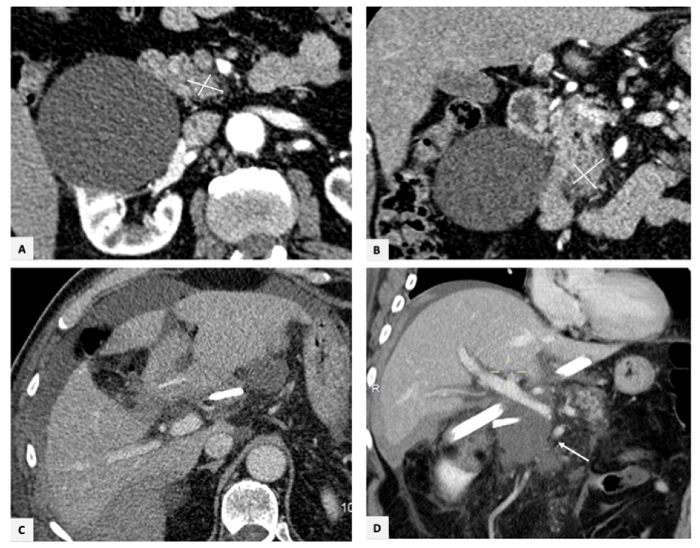
Two days later, due to an increase in direct bilirubin levels (9 mg/dL), a Computed Tomography (CT) scan of the abdomen was performed, which showed a fluid collection near the hepatic hilum (Figure 1).
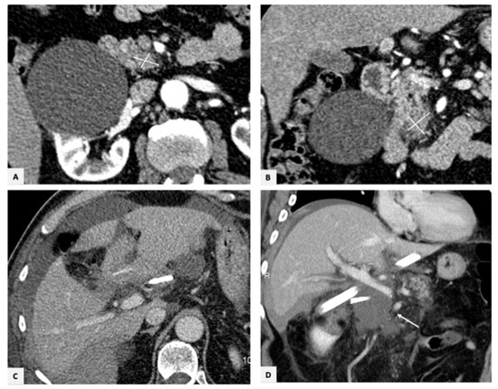
Figure 1.
(A,B) Axial and coronal CT scan showing a pancreatic head adenocarcinoma. (C,D) Axial and coronal CT images in portal phase performed 2 days after re-look surgery showing peri-hepatic fluid collections (arrow) near the surgical drainages.
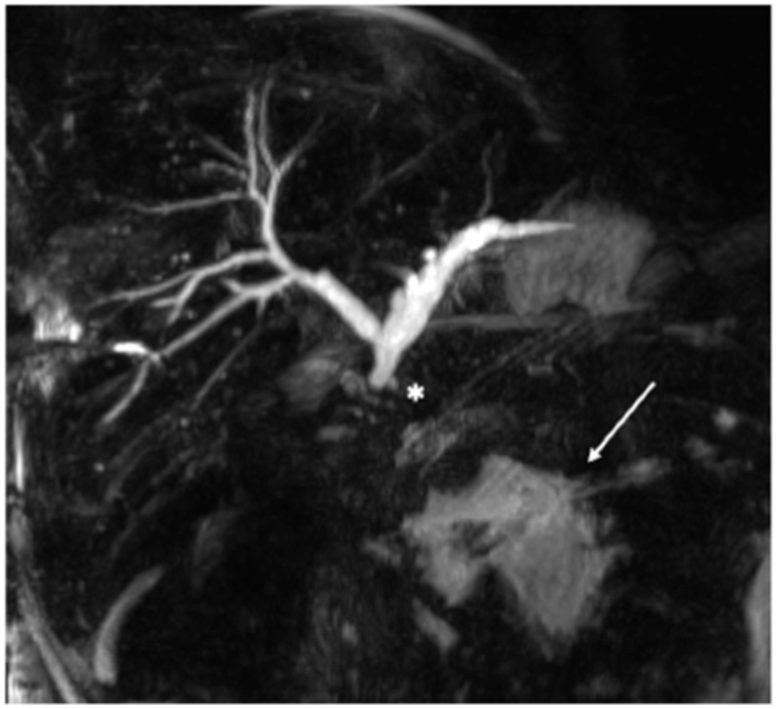
Thus, a Magnetic Resonance Cholangiopancreatography (MRCP) was acquired, which confirmed the presence of HJS stenosis with an associated biliary leak (Figure 2).
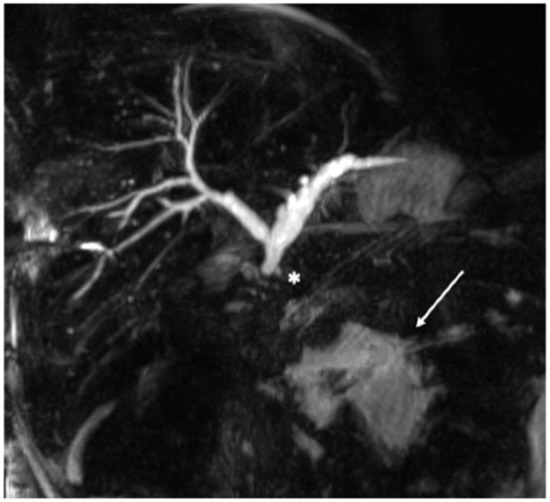
Figure 2.
MRCP exam revealing a stenosis (*) of the HJS with a collection near the hepatic hilum (arrow).
The patient was referred to our Interventional Radiology department to perform a percutaneous cholangiography and differentiate between the presence of a leakage originating from a bile duct injury or from the bilio-enteric anastomosis.
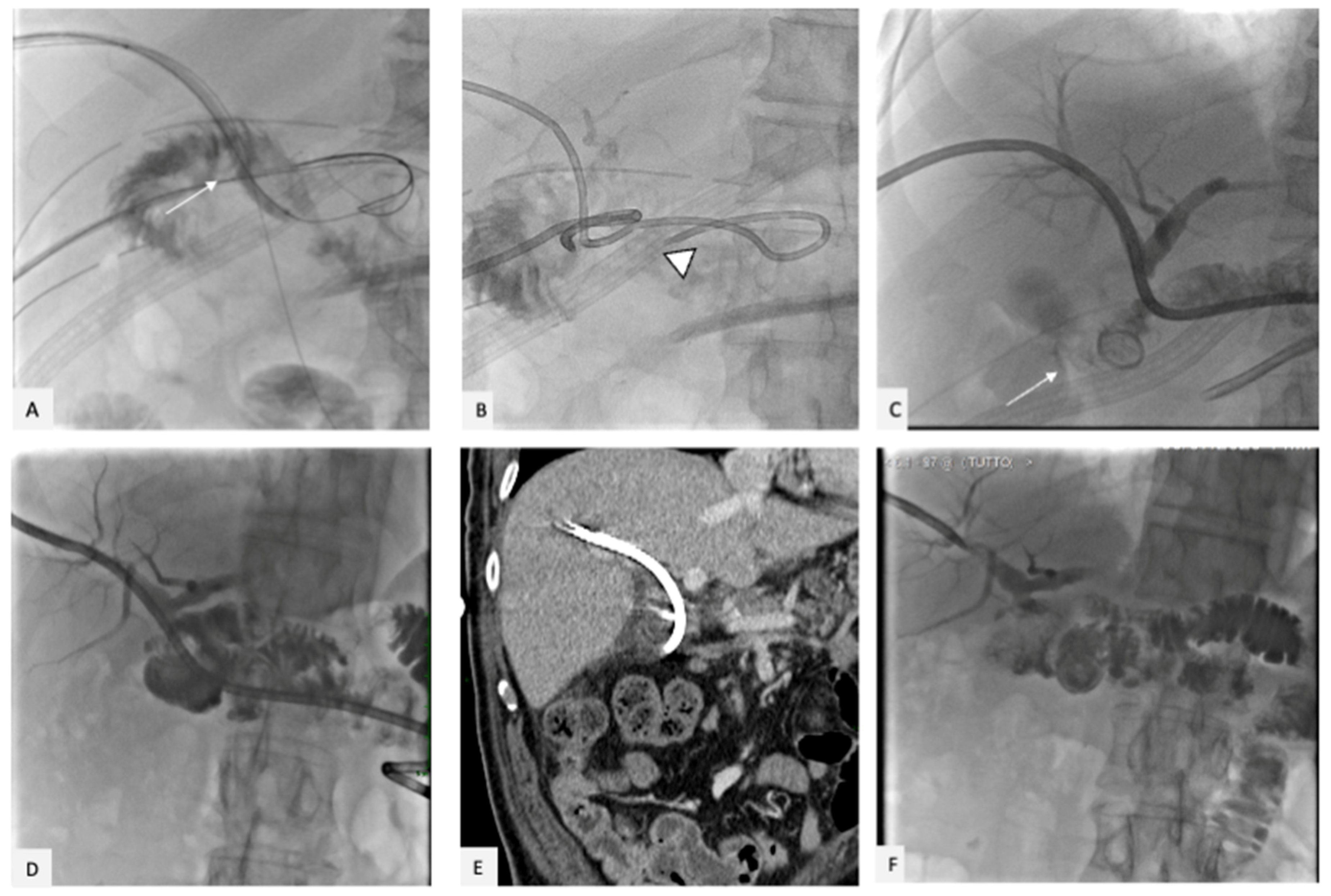
The puncture of a bile duct of the right hepatic lobe was made under ultrasound (US) guidance with a Chiba needle, and the cholangiography confirmed the HJA stenosis (Figure 3), with an associated bile leak into the peritoneal cavity; these findings justified the increased bilirubin levels and the peri-hepatic biloma.

Figure 3.
(A) Percutaneous cholangiography confirming the HJA stenosis (*) and the small bile leak into the peritoneal cavity (B). (C–E) A percutaneous drainage (8 Fr) was positioned into the biliary tree (arrowhead), and a hydrophilic 4 Fr angiographic catheter (arrows) was placed, passing through the biliary leak into the peritoneal cavity.
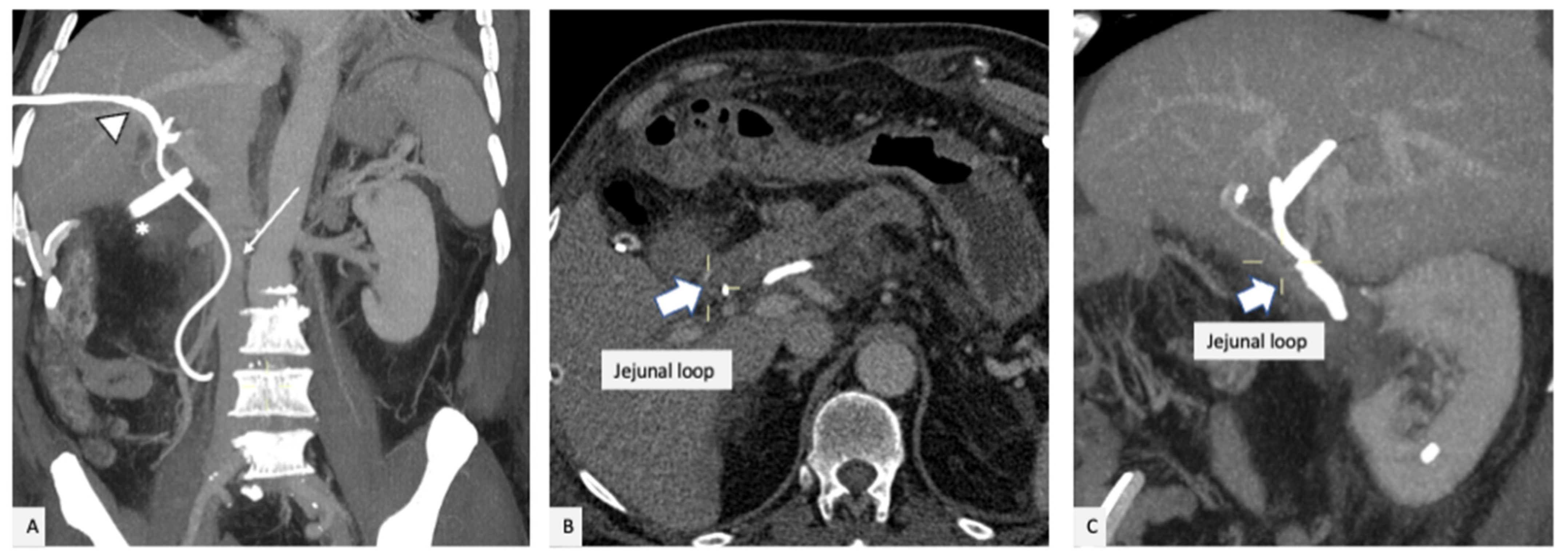
Several unsuccessful attempts to cross the stenosis and reach the jejunal loop were made with different guidewires; consequently, percutaneous drainage (8 Fr) was positioned at the level of the intrahepatic biliary junction in order to reduce the pressure along the biliary tract and try to redirect the bile flow (Figure 3). Moreover, a hydrophilic 4 Fr angiographic catheter was advanced into the biliary tree and positioned freely into the biloma, passing through the HSJ dehiscence, and a new CT scan was acquired (Figure 4).
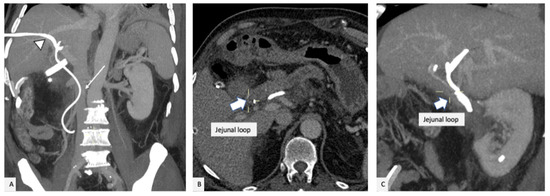
Figure 4.
(A) coronal MIP reconstruction displaying the percutaneous drainage into the biliary tree (arrowhead) and the angiographic catheter (arrow) into the peritoneal cavity; surgical drainage at the hepatic hilum (*). (B,C) axial and sagittal images confirming that the anastomotic jejunal loop was located anteriorly to the angiographic catheter positioned into the biliary tree.
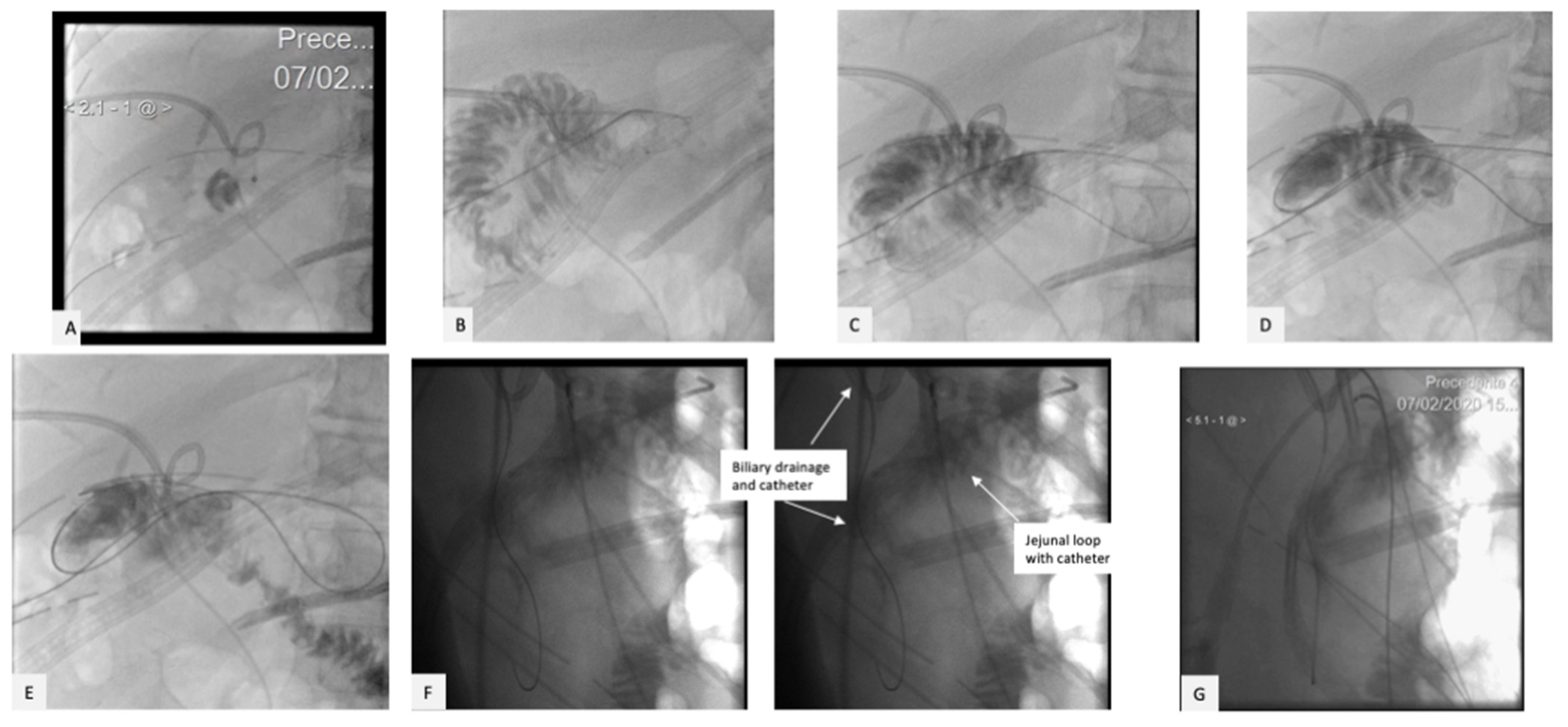
The CT scan confirmed that the anastomotic jejunal loop was anterior to the angiographic catheter. Therefore, a direct puncture of the afferent loop was performed under US guidance, and the biloma was reached with a guidewire passing through the jejunal dehiscence (Figure 5, Video S1).
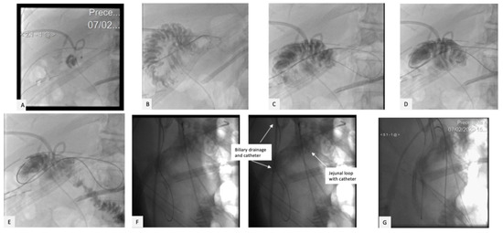
Figure 5.
The afferent jejunal loop was then punctured with US and fluoroscopic control (A), and the bile collection was reached with a hydrophilic guide and an angiographic catheter (B–E) passing through the jejunal dehiscence. Latero-lateral projection (F) and CBCT acquisition (G) confirmed that the jejunal loop was anterior to the angiographic catheter positioned into the biliary tree.
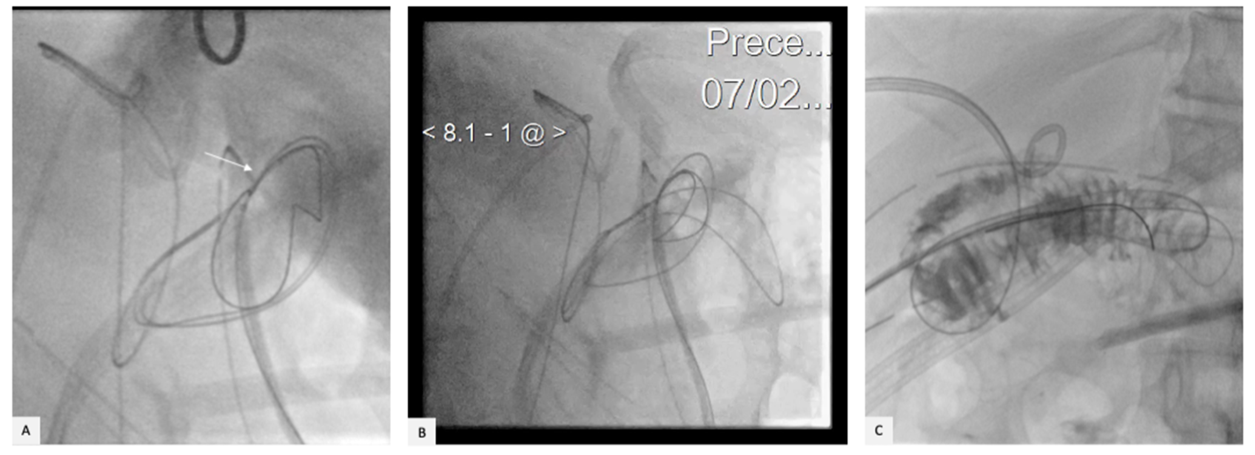
A snare loop catheter (Amplatz GooseNeck Snare Kit) was then advanced through the jejunal loop into the biloma: the transhepatic hydrophilic wire was then captured with the snare and pulled into the jejunal loop (Video S2, Figure 6).
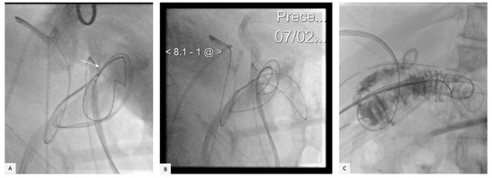
Figure 6.
A snare loop catheter (arrow) was then advanced through the afferent jejunal loop into the collection (A): the transhepatic hydrophilic wire was captured (B,C) and pulled into the jejunal loop from the peritoneal bile collection.
Thereafter, a percutaneous bilioplasty was performed with a 10 × 70 mm balloon to solve the anastomotic stenosis (Figure 7). Finally, a transhepatic biliary drainage (8 Fr) was positioned into the jejunal loop, completing the biliary rendez-vous and recreating the HJA connection. Another 8 Fr drainage was left into the jejunal loop and replaced after one week with a 4 Fr pig-tail catheter to avoid the creation of an enteric fistula.
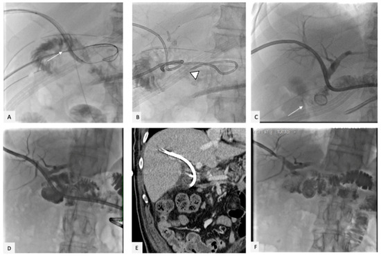
Figure 7.
(A) Percutaneous bilioplasty with a 10 × 70 mm balloon (arrow) performed to solve the stenosis. (B) Transhepatic biliary drainage (8 Fr) positioned into the jejunal loop (arrowhead), completing the biliary rendez-vous. Another 8 Fr drainage was left into the jejunal loop and then (C) replaced after one week with a 4 Fr pig-tail catheter (arrow) to avoid the creation of an enteric fistula. One month after the rendez-vous, the percutaneous cholangiography (D) showed no evidence of residual stenosis. (E) CT scan performed two months after the rendez-vous displaying the absence of collections and no dilatation of the biliary tree. (F) Five months after the rendez-vous, the percutaneous cholangiography showed no residual stenosis.
One month after the rendez-vous, the percutaneous cholangiography showed no evidence of residual stenosis, and the biliary drainage was left in place, removing the other drainages. The CT scan performed two months after the rendez-vous displayed the absence of collections without the dilatation of the biliary tree.
Five months after the rendez-vous, the percutaneous cholangiography showed no evidence of residual stenosis. Hence, the biliary drainage was removed, and 12 months after the procedure, the patient did not have dilatation of the biliary tree, with bilirubin levels in the normal range.
3. Discussion
The postoperative complication rate associated with a complex surgical procedure such as DCP is still relevant [16]. Bile anastomotic leaks after pancreatic surgery are a serious complication, and rates of 0 to 5% have been described [19,20]. Partial injury can usually be managed with a combination of percutaneous and/or endoscopic techniques. However, the management of complete HJA dehiscence is challenging due to the post-surgical anatomy. A rendez-vous technique classically includes a combination of surgical, endoscopic, and percutaneous approaches to reach one point in the body through two access points. It is often employed in the management of hepatobiliary dysfunction, such as biliary stenosis, stones, injury, and leakage, when either endoscopic retrograde cholangio-pancreatography (ERCP) or percutaneous drainage alone is not sufficient [21].
There are a few case reports presenting a totally percutaneous rendez-vous technique used to treat biliary strictures [21], bile injury and leakages [22], or to reestablish the hepaticojejunostomy anastomosis following pediatric liver transplant [23].
The potential risks of percutaneous biliary drainage include biloma, transitory haemobilia, or cutaneous hematomas; in addition, the main drawback of the rendez-vous technique is the fact that if the leak is secondary to a large defect, endoscopic or percutaneous techniques alone may fail in reestablishing the anatomic continuity due to the technical inability to traverse the large resection defect [21].
In these cases, the main options are the combined radiologic–endoscopic rendez-vous technique [24] or a new surgery. More recently, percutaneous cholangioscopy-assisted guidewire placement was reported in post-liver transplant patients with severe biliary anastomotic strictures and failed endoscopic and conventional percutaneous approaches [25,26].
In conclusion, the percutaneous biliary rendez-vous technique, planned in cooperation with hepatobiliary surgeons and executed by a multi-disciplinary team, provides a potentially safer option to treat HJA dehiscence and avoid a re-look surgery.
Supplementary Materials
The following supporting information can be downloaded at: https://www.mdpi.com/article/10.3390/gidisord5010007/s1, Video S1: jejunal loop puncture, Video S2: snare-loop capture.
Funding
This research received no external funding.
Institutional Review Board Statement
The study was conducted in accordance with the Declaration of Helsinki; Institutional review board approval for this case was not required (retrospective, de-identified data).
Informed Consent Statement
Informed consent was obtained from all individual participants included in the study.
Data Availability Statement
Data is contained within the article or Supplementary Materials.
Conflicts of Interest
The authors declare no conflict of interest.
References
- de Castro, S.; Kuhlmann, K.; Busch, O.; van Delden, O.; Lameris, J.; van Gulik, T.; Obertop, H.; Gouma, D. Incidence and management of biliary leakage after hepaticojejunostomy. J. Gastrointest. Surg. 2005, 9, 1163–1173. [Google Scholar] [CrossRef] [PubMed]
- Farooqui, W.; Penninga, L.; Burgdorf, S.K.; Storkholm, J.H.; Hansen, C.P. Biliary Leakage Following Pancreatoduodenectomy: Experience from a High-Volume Center. J. Pancreat. Cancer 2021, 7, 80–85. [Google Scholar] [CrossRef] [PubMed]
- El Nakeeb, A.; El Sorogy, M.; Hamed, H.; Said, R.; Elrefai, M.; Ezzat, H.; Askar, W.; Elsabbagh, A. Biliary leakage following pancreaticoduodenectomy: Prevalence, risk factors and management. Hepatobiliary Pancreat. Dis. Int. 2019, 18, 67–72. [Google Scholar] [CrossRef] [PubMed]
- Righi, D.; Franchello, A.; Ricchiuti, A.; Breatta, A.D.; Versace, K.; Calvo, A.; Romagnoli, R.; Fonio, P.; Gandini, G.; Salizzoni, M. Safety and efficacy of the percutaneous treatment of bile leaks in hepaticojejunostomy or split-liver transplantation without dilatation of the biliary tree. Liver Transpl. 2008, 14, 611–615. [Google Scholar] [CrossRef]
- Booij, K.A.; Coelen, R.J.; de Reuver, P.R.; Besselink, M.G.; van Delden, O.M.; Rauws, E.A.; Busch, O.R.; van Gulik, T.M.; Gouma, D.J. Long-term follow-up and risk factors for strictures after hepaticojejunostomy for bile duct injury: An analysis of surgical and percutaneous treatment in a tertiary center. Surgery 2018, 163, 1121–1127. [Google Scholar] [CrossRef]
- de Reuver, P.R.; Sprangers, M.A.G.; Rauws, E.A.J.; Lameris, J.S.; Busch, O.R.; Van Gulik, T.M.; Gouma, D.J. Impact of bile duct injury after laparoscopic cholecystectomy on quality of life: A longitudinal study after multidisciplinary treatment. Endoscopy 2008, 40, 637–643. [Google Scholar] [CrossRef]
- de Reuver, P.R.; Rauws, E.A.; Bruno, M.J.; Lameris, J.S.; Busch, O.R.; van Gulik, T.M.; Gouma, D.J. Survival in bile duct injury patients after laparoscopic cholecystectomy: A multidisciplinary approach of gastroenterologists, radiologists, and surgeons. Surgery 2007, 142, 1–9. [Google Scholar] [CrossRef]
- Henry, A.C.; Smits, F.J.; van Lienden, K.; Heuvel, D.A.V.D.; Hofman, L.; Busch, O.R.; van Delden, O.M.; Zijlstra, I.A.; Schreuder, S.M.; Lamers, A.B.; et al. Biliopancreatic and biliary leak after pancreatoduodenectomy treated by percutaneous transhepatic biliary drainage. HPB (Oxf.) 2022, 24, 489–497. [Google Scholar] [CrossRef]
- May, K.; Hunold, P. Leakage of Hepaticojejunal Anastomosis: Radiological Interventional Therapy. Visc. Med. 2017, 33, 192–196. [Google Scholar] [CrossRef]
- Lau, W.Y.; Lai, E.C.; Lau, S.H. Management of bile duct injury after laparoscopic cholecystectomy: A review. ANZ J. Surg 2010, 80, 75–81. [Google Scholar] [CrossRef]
- Kaffes, A.J.; Hourigan, L.; De Luca, N.; Byth, K.; Williams, S.J.; Bourke, M.J. Impact of endoscopic intervention in 100 patients with suspected postcholecystectomy bile leak. Gastrointest. Endosc. 2005, 61, 269–275. [Google Scholar] [CrossRef] [PubMed]
- Sandha, G.S.; Bourke, M.J.; Haber, G.B.; Kortan, P.P. Endoscopic therapy for bile leak based on a new classification: Results in 207 patients. Gastrointest. Endosc. 2004, 60, 567–574. [Google Scholar] [CrossRef] [PubMed]
- Yang, M.J.; Kim, A.R.; Hwang, J.C.; Yoo, B.M.; Kim, J.H. Long-type double-balloon enteroscopy-assisted ERCP using hand-made accessories in Roux-en-Y hepaticojejunostomy (with video). Hepatobiliary Pancreat. Dis. Int. 2021, 20, 407–408. [Google Scholar] [CrossRef]
- Farina, E.; Cantù, P.; Cavallaro, F.; Iori, V.; Rosa-Rizzotto, E.; Cavina, M.; Tontini, G.E.; Nandi, N.; Scaramella, L.; Sassatelli, R.; et al. Effectiveness of double-balloon enteroscopy-assisted endoscopic retrograde cholangiopancreatography (DBE-ERCP): A multicenter real-world study. Dig. Liver Dis. 2022. ahead of print. [Google Scholar] [CrossRef] [PubMed]
- Sandha, G.S.; Bourke, M.J.; Haber, G.B.; Kortan, P.P. Percutaneous transhepatic biliary drainage in the management of postsurgical biliary leaks in patients with nondilated intrahepatic bile ducts. Cardiovasc. Intervent. Radiol. 2006, 29, 380–388. [Google Scholar]
- Mosconi, C.; Cocozza, M.A.; Piacentino, F.; Fontana, F.; Cappelli, A.; Modestino, F.; Coppola, A.; Palumbo, D.; Marra, P.; Maffi, P.; et al. Interventional Radiological Management and Prevention of Complications after Pancreatic Surgery: Drainage, Embolization and Islet Auto-Transplantation. J. Clin. Med. 2022, 11, 6005. [Google Scholar] [CrossRef]
- Sohn, T.A.; Yeo, C.J.; Cameron, J.L.; Geschwind, J.F.; Mitchell, S.E.; Venbrux, A.C.; Lillemoe, K.D. Pancreaticoduodenectomy: Role of interventional radiologists in managing patients and complications. J. Gastrointest. Surg. 2003, 7, 209–219. [Google Scholar] [CrossRef]
- Mauri, G.; Mattiuz, C.; Sconfienza, L.M.; Pedicini, V.; Poretti, D.; Melchiorre, F.; Rossi, U.; Lutman, F.R.; Montorsi, M. Role of interventional radiology in the management of complications after pancreatic surgery: A pictorial review. Insights Imaging 2015, 6, 231–239. [Google Scholar] [CrossRef]
- Büchler, M.W.; Wagner, M.; Schmied, B.M.; Uhl, W.; Friess, H.; Z’Graggen, K. Changes in the morbidity after pancreatic resection. Arch. Surg. 2003, 138, 1310–1314. [Google Scholar] [CrossRef]
- House, M.G.; Cameron, J.L.; Schulick, R.D.; Campbell, K.A.; Sauter, P.K.; Coleman, J.; Lillemoe, K.D.; Yeo, C.J. Incidence and outcome of biliary strictures after pancreaticoduodenectomy. Ann. Surg. 2006, 243, 571–578. [Google Scholar] [CrossRef]
- De Robertis, R.; Contro, A.; Zamboni, G.; Mansueto, G. Totally percutaneous rendezvous techniques for the treatment of bile strictures and leakages. J. Vasc. Interv. Radiol. 2014, 25, 650–654. [Google Scholar] [CrossRef] [PubMed]
- Meek, J.; Fletcher, S.; Crumley, K.; Culp, W.C.; Meek, M. Percutaneous rendezvous technique for the management of a bile duct injury. Radiol. Case Rep. 2018, 13, 175–178. [Google Scholar] [CrossRef] [PubMed]
- Huespe, P.E.; Oggero, S.; de Santibañes, M.; Boldrini, G.; D’agostino, D.; Pekolj, J.; de Santibañes, E.; Ciardullo, M.; Hyon, S.H. Percutaneous patency recovery and biodegradable stent placement in a totally occluded hepaticojejunostomy after paediatric living donor liver transplantation. Cardiovasc. Intervent. Radiol. 2019, 42, 466–470. [Google Scholar] [CrossRef] [PubMed]
- Martin, D.F. Combined percutaneous and endoscopic procedures for bile duct obstruction. Gut 1994, 35, 1011–1012. [Google Scholar] [CrossRef] [PubMed]
- Martins, F.P.; Ferrari, A.P. Cholangioscopy-assisted guidewire placement in post-liver transplant anastomotic biliary stricture: Efficient and potentially also cost-effective. Endoscopy 2017, 49, E283–E284. [Google Scholar] [CrossRef]
- Woo, Y.S.; Lee, J.K.; Noh, D.H.; Park, J.K.; Lee, K.H.; Lee, K.T. SpyGlass cholangioscopy-assisted guidewire placement for post-LDLT biliary strictures: A case series. Surg. Endosc. 2016, 30, 3897–3903. [Google Scholar] [CrossRef]
Disclaimer/Publisher’s Note: The statements, opinions and data contained in all publications are solely those of the individual author(s) and contributor(s) and not of MDPI and/or the editor(s). MDPI and/or the editor(s) disclaim responsibility for any injury to people or property resulting from any ideas, methods, instructions or products referred to in the content. |
© 2023 by the authors. Licensee MDPI, Basel, Switzerland. This article is an open access article distributed under the terms and conditions of the Creative Commons Attribution (CC BY) license (https://creativecommons.org/licenses/by/4.0/).

