Communication

Sex Differences and Bmal1/Acetylcholine- or Bmal1/Noradrenaline-Mediated Effects of Blue Light Irradiation on Dextran-Sodium-Sulfate-Induced Ulcerative Colitis Model Mice

1
Department of Pharmaceutical Sciences, Suzuka University of Medical Science, Suzuka 513-8670, Japan
2
Research Department, Daiichi Sankyo Healthcare Co., Ltd., Chuo-ku, Tokyo 140-8170, Japan
*
Author to whom correspondence should be addressed.
Gastrointest. Disord. 2024, 6(3), 720-732; https://doi.org/10.3390/gidisord6030048
Submission received: 13 May 2024 / Revised: 25 July 2024 / Accepted: 2 August 2024 / Published: 5 August 2024

Abstract

:
Humans are exposed to significant amounts of blue light from computers and smartphones. However, the effects of blue light on ulcerative colitis remain unclear. In this study, we assessed blue-light-irradiation-induced alterations in colonic symptoms using a dextran sodium sulfate (DSS)-induced ulcerative colitis model mice. Both male and female institute of cancer research (ICR) mice were administered DSS (5%) ad libitum for 5 days while irradiated with 40 kJ/m2 blue light daily. Additionally, tranexamic acid (TA) was administered daily throughout the study. Male mice treated with DSS/blue light exhibited exacerbated colitis compared to those treated with DSS alone. In contrast, female mice treated with DSS/blue light exhibited enhanced symptoms compared to those treated with DSS alone. Additionally, in male mice exposed to blue light, the clock/brain and muscle Arndt-like 1 (Bma1)/noradrenaline/macrophage or beta2-adrenergic receptor (β2-AR) pathways were activated. In female mice, the Bmal1/acetylcholine/macrophage/nicotinic acetylcholine receptor alpha 7 (α7nAChR) pathway was activated. These findings highlight sex differences in the effects of blue light on DSS-induced ulcerative colitis. Moreover, the worsening of symptoms in males was ameliorated through TA administration.

1. Introduction

Inflammatory bowel disease (IBD) is a chronic inflammatory disease that affects the gastrointestinal tract. It refers to ulcerative colitis and Crohn’s disease (CD). Ulcerative colitis results in inflammation, ulcers, and lesions in the mucous membrane of the large intestine, and it affects over 170,000 Japanese people, with increasing numbers annually [1,2]. Additionally, the number of patients in Europe is approximately 2.1 million, making it a global concern [3]. Chronic inflammation of the large intestine triggers ulcerative colitis, resulting in symptoms, such as abdominal pain, diarrhea, bloody stool, and fever, with an increased risk of colorectal cancer over time [1]. CD causes inflammatory ulcers throughout the gastrointestinal tract, from the mouth to the buttocks. Ulcerative colitis affects both sexes equally, with peak incidence among individuals aged between 20 and 24 years, which reduces with age. The male-to-female ratio of CD is approximately twice as high [2].
Ulcerative colitis causes continuous and diffuse lesions in the colonic mucosa starting from the rectum. The effectiveness of corticosteroids and immunosuppressive treatments indicates the involvement of immune abnormalities in the colonic mucosal injury. Additionally, ulcerative colitis is caused by organ-specific autoimmunity, accompanied by systemic immune abnormalities [4]. Specific colonic mucosal injury produces various autoantibodies in the serum and causes extraintestinal complications [5,6,7]. The concept of a disease is central.
Recently, Anti-tumor necrosis factor-alpha (TNF-α) antibody preparations have become popular for the treatment of IBD. In 1993, the efficacy of a chimeric TNF-α-specific antibody (infliximab) was reported in severe cases of refractory CD [8]. Subsequently, humanized versions of adalimumab [9] and golimumab [10] were administered. Additionally, usatekinumab, which is conjugated to p40, a protein shared with interleukin (IL) 12 and IL23 expression that suppresses its effects, has been developed for administration to TNF-α antibody-refractory cases [11].
We previously assessed the effects of ultraviolet (UV) irradiation on ulcerative colitis using a dextran sulfate sodium (DSS)-induced colitis model mice [12]. Therefore, UV rays aggravated DSS-induced ulcerative colitis through the regulation of pituitary-derived hormones (urocortin 2 [UCN2], corticotropin-releasing hormone [CRH], adrenocorticotropic hormone [ACTH]) and clock genes (clock/brain and muscle arnt-like 1 [Bmal1]). Alterations in clock genes occur because of UV exposure and lifestyle disturbances. In modern times, the prolonged use of computers and smartphones that emit blue light has adverse effects on the human body [13]. Additionally, blue light exposure disrupts circadian rhythms, thereby inducing alterations in the immune system [12]. Moreover, blue light induces neutrophil accumulation and neutrophil extracellular traposis (NETosis) [14]. These findings indicate that blue light exposure may affect immune diseases, including ulcerative colitis. In addition, it has been reported that blue light irradiation alleviates DSS-induced ulcerative colitis, and it has been shown that this is due to the reduction of colonic inflammation due to the activation and polarization of macrophages [15]. However, the detailed relationship between blue light and DSS-induced ulcerative colitis is not known.
In this study, we assessed blue-light-irradiation-induced alterations in symptoms using DSS-induced ulcerative colitis model mice. Additionally, because of sex differences in CD and the varying effects of UV irradiation on DSS-induced ulcerative colitis [16], we assessed potential sex differences in this study. Moreover, to enhance DSS-induced ulcerative colitis, we assessed the effects of blue light irradiation using tranexamic acid (TA) [17,18], which is useful for treating immune diseases.

2. Results

2.1. Effects of Blue Light Irradiation on DSS-Treated Male and Female Mice

DSS treatment reduced body weight and colon length in both sexes (Figure 1A–F). In male mice, blue light irradiation further reduced both body weight and colon length. In female mice, blue light irradiation ameliorated the DSS-treatment-induced reduction in body weight and colon length. In contrast, when TA was administered, males experienced an enhanced blue light irradiation group-induced reduction, with no effect on the females. Additionally, diarrhea and fecal bleeding were observed in DSS-treated mice (Figure 1G,H). Blue light/DSS-treated male mice exhibited higher disease activity scores than those of DSS-treated mice, with lower scores in females. Additionally, in males, the score was reduced to the value observed after DSS treatment when subjected to TA treatment. However, in females, there was no difference between blue light/DSS-treated mice. Moreover, colon damage was more severe in blue light/DSS-treated male mice than in DSS-treated mice, which was enhanced through TA administration (Figure 1I). In contrast, colon damage was enhanced in blue light/DSS-treated female mice compared to DSS-treated mice, with no further enhancement observed through TA administration (Figure 1J).

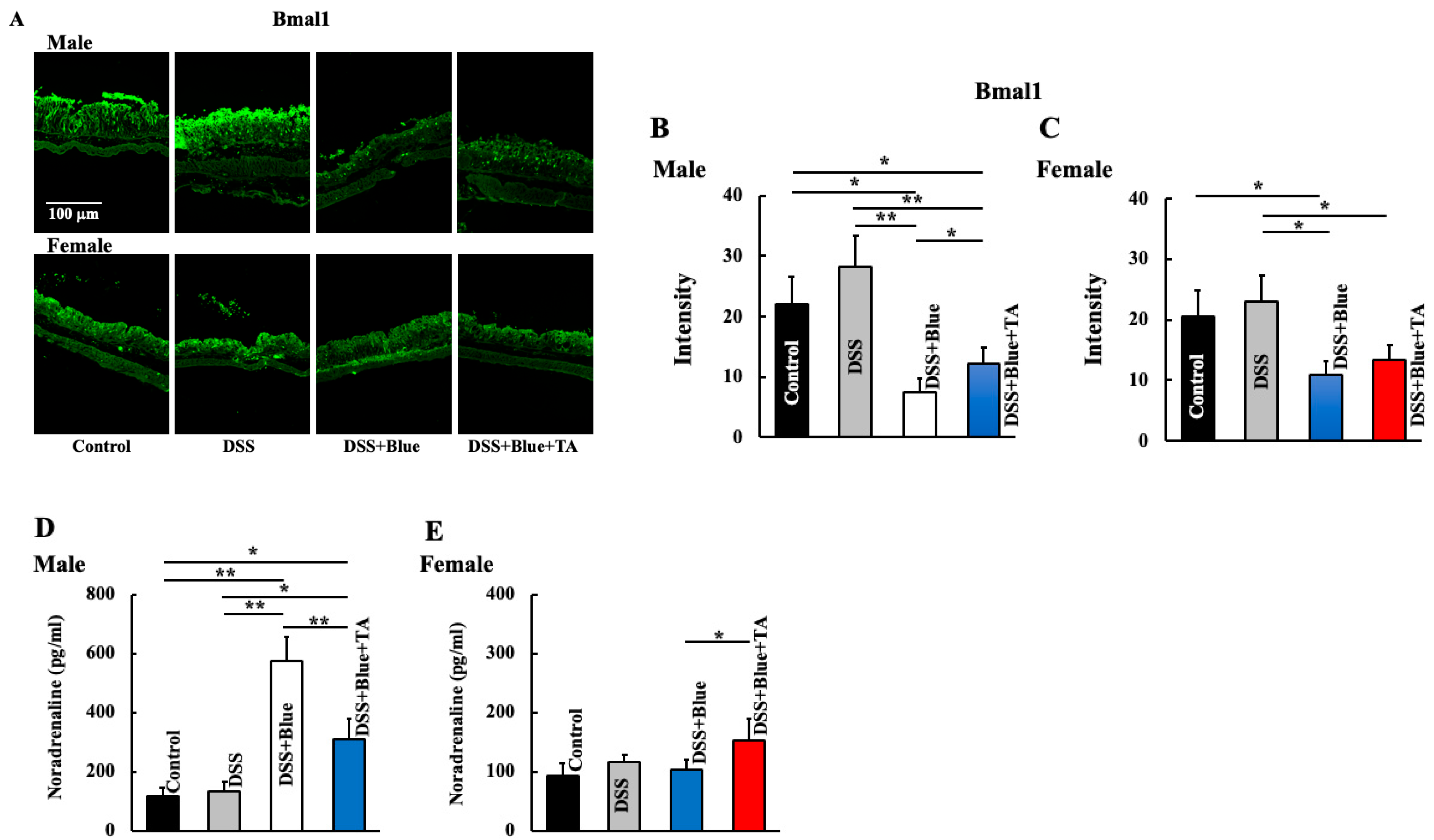
2.2. Effect of Blue Light Irradiation on the Expression of Bmal1 and Noradrenaline Levels in DSS-Treated Male and Female Mice

We assessed the expression of blue-light-associated clock genes in the colon. The level of Bmal1 in the colon was reduced in DSS/blue-light-treated mice (male and female) (Figure 2A–C). In TA-treated mice, the Bmal1 levels were similar to those in DSS/blue-light-treated mice. In contrast, noradrenaline levels were significantly increased in DSS/blue-light-treated male mice and reduced through TA administration (Figure 2D). In females, no changes were observed between treatments (Figure 2E).

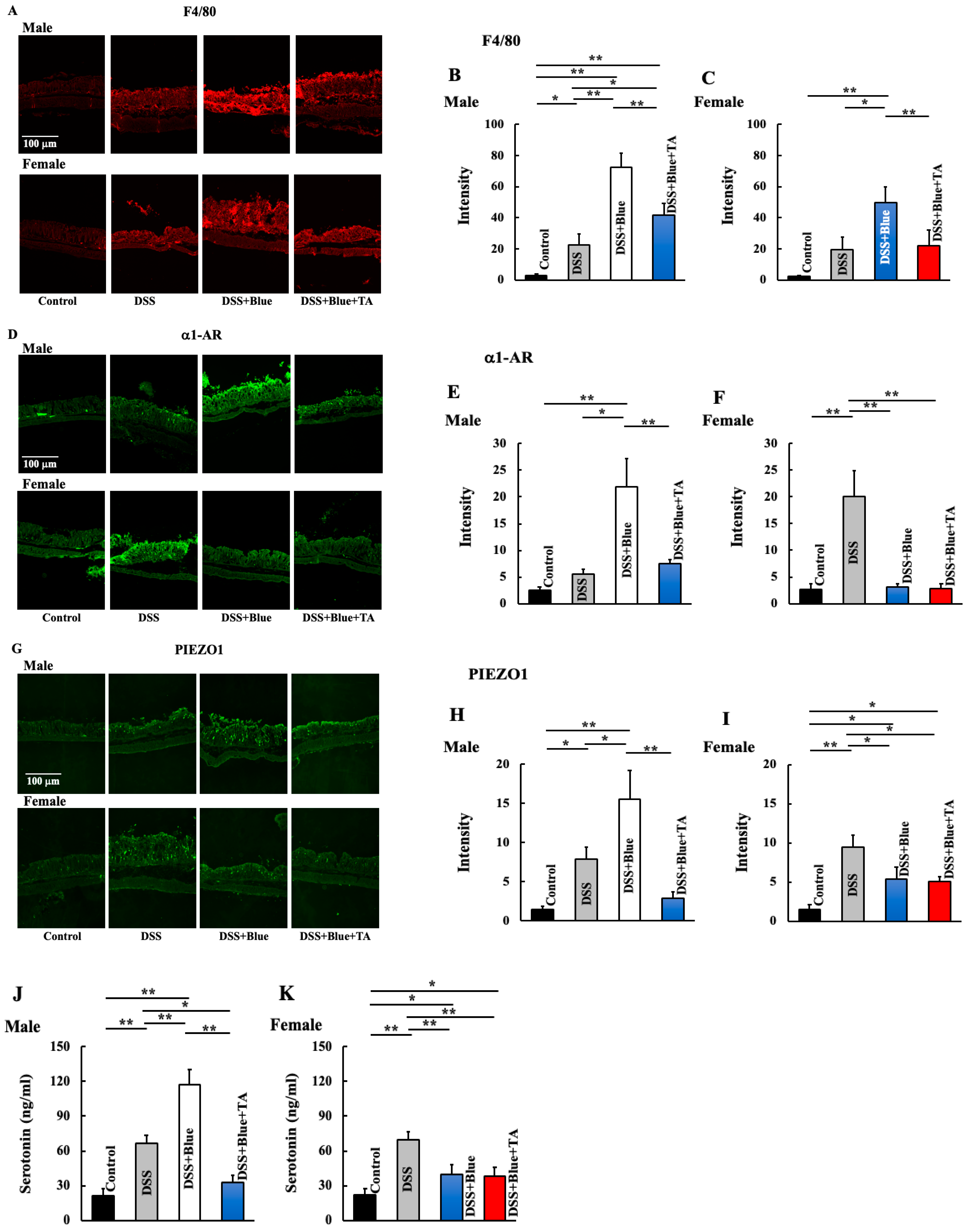
2.3. Effects of Blue Light Irradiation on the Expression of Macrophages, Piezo-Type Mechanosensitive Ion Channel Component 1 (PIEZO1), Alpha-1 Adrenergic Receptor (a1-AR), and Plasma Serotonin Levels in DSS-Treated Male and Female Mice

Subsequently, we assessed the expression levels of macrophages, α1-AR, PIEZO1, and serotonin. In the large intestine, DSS/blue light treatment significantly increased the expression of macrophages in both males and females, which was reduced through TA administration (Figure 3A–C). The expression of α1-AR in the large intestine was highest in DSS/blue-light-treated male mice, and reduced to the levels observed in DSS-treated mice when subjected to TA administration (Figure 3D,E). In contrast, it was highest in DSS-treated female mice, and no difference was observed between the control, DSS/blue light-treated, and DSS/blue light/TA-treated mice (Figure 3D,F). PIEZO1 expression in the colon and plasma serotonin levels were increased in DSS-treated male mice, which were highest in DSS/blue-light-treated mice (Figure 3G,H,J). Additionally, TA administration reduced these levels compared to DSS-treated mice. In females, these levels increased with DSS treatment and reduced with DSS/blue light treatment (Figure 3G,I,K). No difference was observed between TA administration and DSS/blue-light-treated mice.

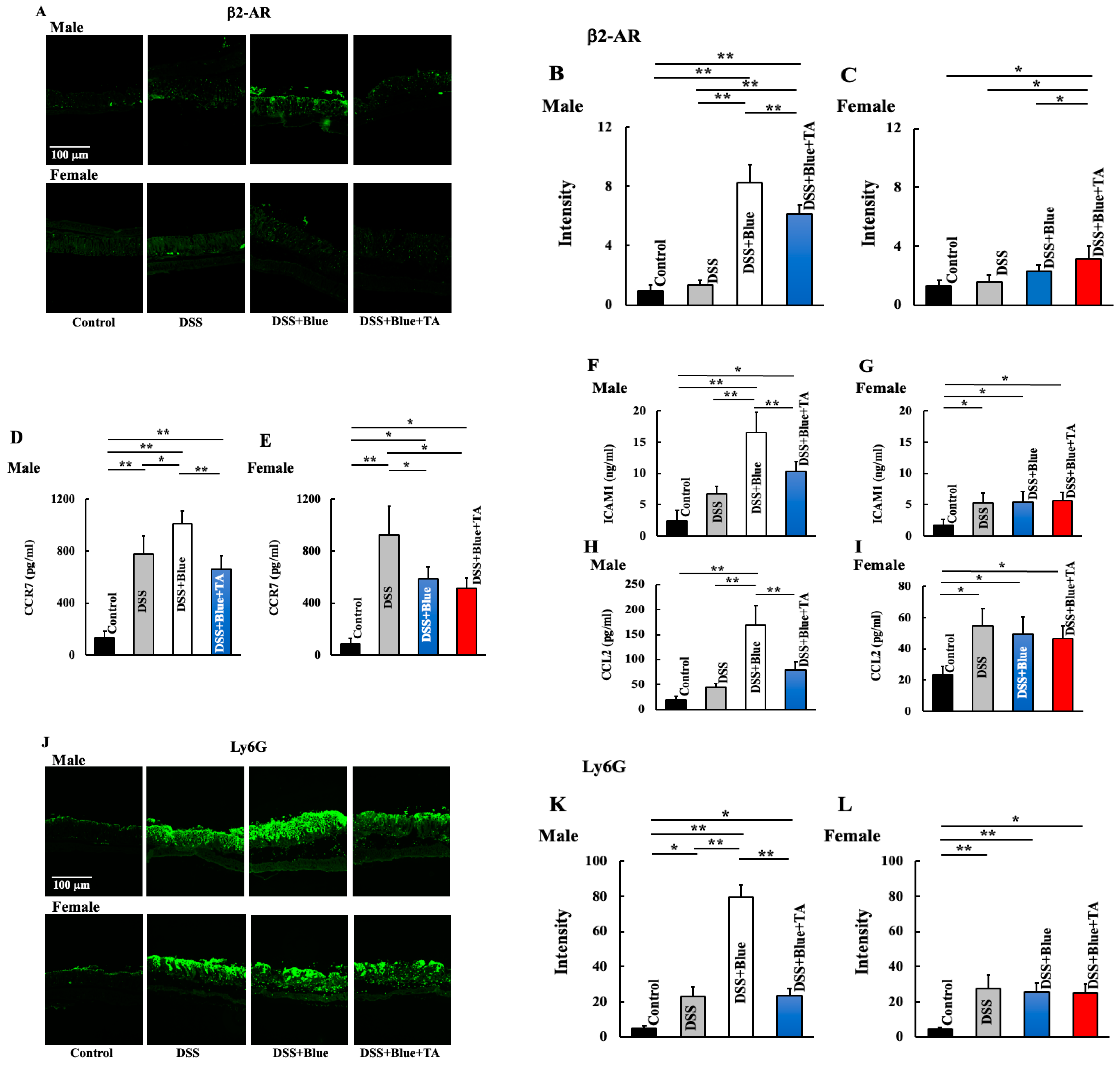
2.4. Effect of Blue Light Irradiation on the Expression of Beta 2 Adrenergic Receptor (β2-AR), Neutrophils, and Plasma Levels of C-C Motif Chemokine Receptor (CCR7), Intercellular Adhesion Molecule 1 (ICAM1), and C-C Motif Chemokine Ligand (CCL2) in DSS-Treated Male and Female Mice

Additionally, we assessed the neutrophil-associated pathways. In males, β2-AR expression was the highest in DSS/blue-light-treated mice, which was reduced through TA administration (Figure 4A,B). In females, no changes were observed between the treatments (Figure 4A,C). Moreover, the levels of CCR7, ICAM1, and CCL2, which are involved in neutrophil accumulation and expression, were highest in DSS/blue-light-treated male mice and reduced to the levels observed in DSS-treated mice when subjected to TA administration (Figure 4D,F,H,J,K). In contrast, in females, CCR7 levels were increased in DSS-treated mice and reduced in DSS/blue-light-treated mice (Figure 4E). ICAM1 and CCL2 levels and neutrophil expression were unaltered between DSS- and DSS/blue-light-treated mice (Figure 4G,I,J,L). Furthermore, TA-treated mice did not differ from DSS/blue-light-treated mice for any of the genes.

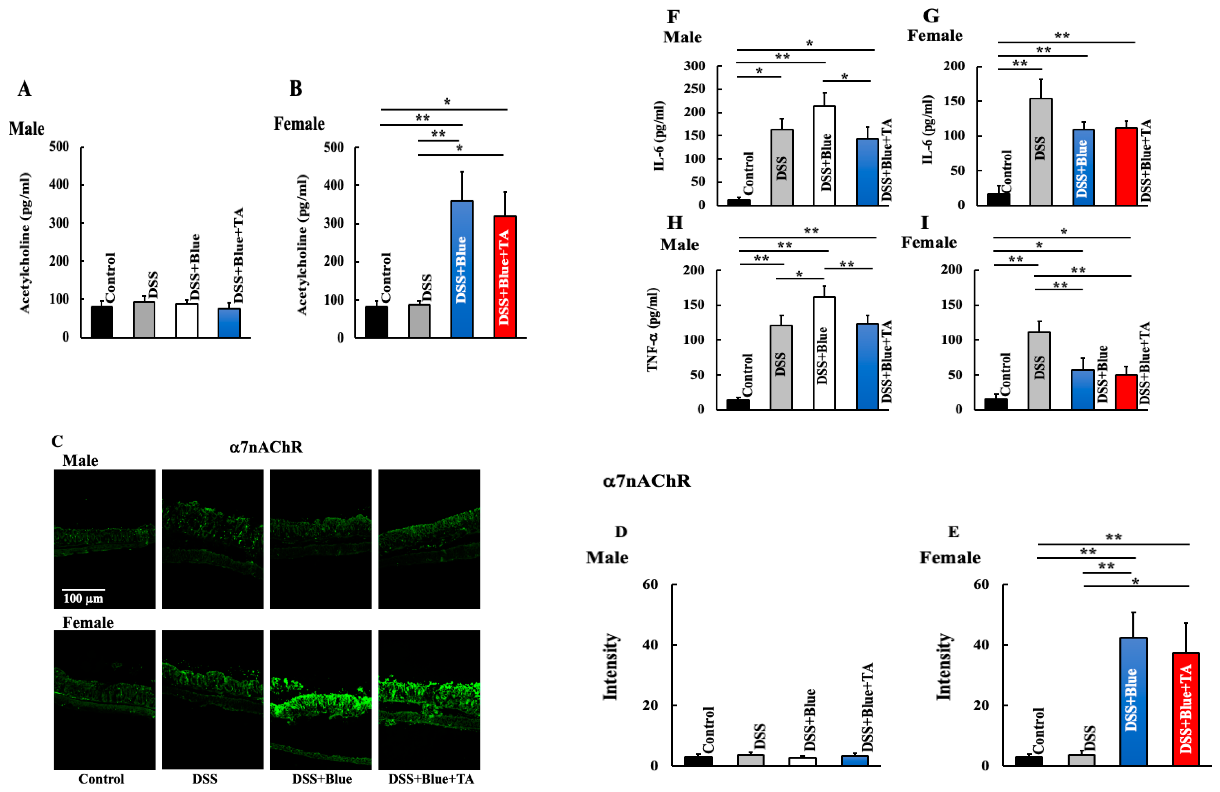
2.5. Effect of Blue Light Irradiation on the Expression of Nicotinic Acetylcholine Receptor Alpha 7 (α7nAChR) and Plasma Levels of Interleukin (IL)-6 and TNF-α in DSS-Treated Male and Female Mice

We assessed the expression of clock-gene-associated acetylcholine and α7nAChR and inflammatory cytokine levels, which exacerbate DSS. In males, acetylcholine levels and α7nAChR expression were unaltered between treatments; however, they were significantly increased in DSS/blue-light- and DSS/blue-light/TA-treated female mice (Figure 5A–E). Additionally, inflammatory cytokine (IL-6 and TNF-α) levels were highest in DSS/blue-light-treated male mice, reduced in DSS/blue-light/TA-treated mice, and became comparable to those in DSS-treated mice (Figure 5F,H). In females, these levels were the highest in DSS-treated mice and reduced in DSS/blue-light- and DSS/blue-light/TA-treated mice, but there was no difference between these groups (Figure 5G,I).

3. Discussion

In this study, when DSS-induced ulcerative colitis model mice were exposed to blue light, symptoms were exacerbated in males, while symptoms were enhanced in females. Additionally, in males, TA administration enhanced the exacerbation of blue-light-exposure-induced symptoms, while symptoms in females remained unaltered from those in DSS/blue-light-treated mice. Moreover, blue light irradiation reduced Bmal1 levels and increased noradrenaline levels in both males and females. Furthermore, in males, there was an increase in macrophage, neutrophil, and inflammatory cytokine production. In females, an increase in acetylcholine levels was observed. Additionally, it was observed that a7nAChR expression in macrophages increased, thereby suppressing the production of inflammatory cytokines.
Blue light irradiation reduces Bmal1 expression [19]. Bmal1 and sympathetic nerves exhibit an inverse relationship, where reduction in Bmal1 expression increases norepinephrine levels [20]. In this study, a reduction in Bmal1 expression and an increase in noradrenaline levels were observed in the DSS/blue-light-treated group. Noradrenaline induces the expression of CCL2 and ICAM1 in vascular endothelial cells through β2-adrenergic receptors, facilitating neutrophil migration from blood to tissues [21,22]. This increase in tissue neutrophil levels (large intestine) was anticipated to exacerbate colitis in DSS/blue-light-treated male mice. Additionally, it was anticipated that TA administration may have an enhancing effect by suppressing the increase in neutrophil levels. In females, the expression level of neutrophils in DSS/blue-light-treated mice did not differ from that in DSS-treated mice. Additionally, the expression level was not altered even after TA administration. This indicates that neutrophils are not involved in DSS/blue-light-associated ulcerative colorectal cancer in females.
In contrast, noradrenaline induces the production of inflammatory cytokines from macrophages and facilitates inflammatory responses [22,23] through α1 or α2 adrenergic receptors expressed on macrophages. DSS treatment increased the expression of α1 adrenergic receptors in both of the sexes. However, in males, the increase was significant in DSS/blue-light-treated mice, and TA treatment reduced it to levels similar to those observed in DSS-treated mice. In females, both DSS/blue-light- and DSS/blue-light/TA-treated mice demonstrated no differences from the control group. These results indicate that the macrophage/α1 adrenergic receptor/inflammatory cytokine pathway may contribute to the aggravation of DSS-induced ulcerative colitis caused by blue light in males.
Additionally, PIEZO1 plays a crucial role in intestinal inflammation [24]. PIEZO1 is a Ca-permeable ion channel that is activated in response to mechanical stimulation [25]. In this study, an increase in macrophages was observed in DSS/blue-light-treated mice. Macrophages demonstrate increased PIEZO1 expression, and PIEZO1 acts on macrophages in an autocrine manner to increase the secretion of inflammatory cytokines [24,26,27]. Additionally, PIEZO1 increases intestinal serotonin levels. Serotonin is a monoamine neurotransmitter, approximately 98% of which is biosynthesized in the body by enterochromaffin cells present in the gastrointestinal mucosa [28]. It facilitates intestinal peristalsis and exacerbates enteritis [24,29,30]. Therefore, an increase in PIEZO1 expression exacerbates colitis. In this study, the expression of PIEZO1 and serotonin was increased in both male and female DSS-treated mice, demonstrating a correlation with the exacerbation of colitis. In males, further increases in PIEZO1 and serotonin levels were observed after DSS/blue light treatment. Additionally, both PIEZO1 and serotonin were reduced with TA administration, which is one of the factors contributing to the enhancement of TA-administration-induced colitis. In contrast, in females, expression was reduced in both DSS/blue-light- and DSS/blue-light/TA-treated groups compared to that in DSS-treated mice. This was anticipated as one of the factors.
The nervous system regulates immunity. Noradrenaline is released from the sympathetic nerve endings, whereas acetylcholine is released from the parasympathetic nerve endings. Acetylcholine is mediated by α7nAChRs expressed on macrophages [31]. This prevents the nuclear translocation of NF-κB in macrophages through various signal transduction pathways and suppresses the production of inflammatory cytokines, including TNF-α [32,33]. An increase in acetylcholine is caused by certain CD4-positive T cells that produce acetylcholine in response to noradrenaline stimulation, which serves as a source of acetylcholine [34]. In this study, an increase in norepinephrine and acetylcholine was observed in blue-light-treated female mice, indicating that the secretion of anti-inflammatory cytokines from macrophages enhanced the symptoms of DSS/blue-light-treated colitis. In contrast, males exhibited no increase in acetylcholine, and DSS/blue light treatment did not enhance colitis symptoms.
In this study, we have investigated the effects of blue light irradiation and neutrophils on DSS-induced ulcerative colitis. On the other hand, it has been reported that irradiating DSS-induced ulcerative colitis with blue light activates and polarizes macrophages, modulates T cells, and reduces inflammatory cytokines [15]. Furthermore, Bmal1 is involved in the progression of colitis, and blue light reverses the decreased expression of Bmal1 caused by colitis [15]. The mechanism of action of Bmal1 is to reduce the production of IL-1β, a proinflammatory cytokine, by macrophages. Therefore, blue light is involved in the inhibition of proinflammatory cytokines by increasing the expression of Bmal1. Thus, blue light/Bmal1/macrophage/IL-1β signaling alleviates DSS-induced ulcerative colitis. This study does not mention macrophages, and it is necessary to consider the comprehensive involvement of Bmal1 with the immune system, including neutrophils and macrophages.

4. Materials and Methods

4.1. Animal Experiments

We used 8-week-old male and female specific-pathogen-free (SPF) mice from the Institute of Cancer Research (ICR) (SLC, Hamamatsu, Shizuoka, Japan). They were individually bred in cages in an air-conditioned room at 23 ± 1 °C with a 12 h light/dark cycle under SPF and stress-free conditions. The mice were divided into four groups, each consisting of five mice: control, DSS treatment, DSS treatment + blue light irradiation (DSS/blue), and DSS treatment + blue light irradiation + tranexamic acid (TA) administration (DSS/blue/TA). The following light sources were used: fluorescent lamp (control) or light-emitting diode (LED) blue light (wavelength 380–500 nm, peak emission 479 nm, and irradiation 40 kJ/m2; ISLM-150X150-BB; CCS, Kyoto, Japan). The LED light energy content was measured using a light analyzer (LA-105; Nippon Medical & Chemical Instruments, Osaka, Japan). The control group was irradiated using the fluorescent lamp, which is normally used for breeding purposes. The mice in a specific treatment group were irradiated daily with blue LED light (10 min/day) at 10 a.m. This study was approved by the Suzuka University of Medical Science Animal Experiment Ethics Committee on 25 September 2014, and strictly adhered to the Guide for the Care and Use of Laboratory Animals of Suzuka University of Medical Science (approval number: 34). Surgeries were conducted under pentobarbital anesthesia, with measures to minimize animal suffering.

4.2. DSS-Induced Colitis

To induce ulcerative colitis, mice in the DSS treatment group were administered 5.0% (w/v) DSS (36,000–50,000 Da; MP Biomedicals, Solon, OH, USA) in drinking water for five consecutive days. The onset of colitis was monitored by observing the fecal condition of each mouse following administration. The severity of colitis was determined based on the fecal condition and postmortem colon length. Fecal condition scores were determined using two parameters: stool consistency (0 = negative; 1 = soft; 2 = very soft, but formed; and 3 = liquid) and fecal bleeding (0 = negative; 1 = faintly blue; 2 = moderately blue; 3 = dark blue; and 4 = blood visible using the guaiac paper test). The sum of the two parameters was considered the individual disease activity score [35].

4.3. TA Treatment

During the study period, mice were orally administered approximately 12 mg/kg of TA in distilled water daily. The solvent-treated animals received distilled water (other than the TA-treated group) [36].

4.4. Preparation and Staining of the Colon

Mice were euthanized, and their colons were collected for histological analysis. The colon was fixed in 4% phosphate-buffered paraformaldehyde and embedded in Tissue-Tek O.C.T. compound (Sakura Finetek, Co., Ltd., Tokyo, Japan). Subsequently, 5 μm thick sections were cut and air-dried at room temperature. The sections were stained with hematoxylin and eosin (H&E) using standard protocols. For immunofluorescence analysis, skin sections were stained using the aforementioned antibodies [37]. The following primary antibodies (1:100 dilution) were used: rabbit monoclonal anti-Bmal1 (Cell Signaling Technology Inc., Danvers, MA, USA), rabbit polyclonal anti-piezo-type mechanosensitive ion channel component 1 (PIEZO1) (Novus Biologicals, LLC., Centennial, CO, USA), rabbit polyclonal anti-beta 2 adrenergic receptor (β2-AR) (Santa Cruz Biotechnology, Santa Cruz, CA, USA), mouse monoclonal anti-alpha-1 adrenergic receptor (α1-AR) (Santa Cruz Biotechnology), rat monoclonal anti-F4/80 (macrophage marker), mouse monoclonal anti-lymphocyte antigen 6 family member G (Ly6G) (neutrophil marker; BD Biosciences, Franklin Lakes, NJ, USA), and rabbit polyclonal anti-nicotinic acetylcholine receptor alpha 7 (α7nAChR) (Abcam, Cambridge, MA, USA). Subsequently, the sections were coated with appropriate secondary antibodies (1:30 dilution; fluorescein isothiocyanate-conjugated anti-rabbit, anti-mouse, anti-rat, or anti-goat secondary antibodies [Dako Cytomation, Glostrup, Denmark]) and incubated for 2 h in the dark. PIEZO1, β2-AR, α1-AR, F4/80, Ly6G, and α7nAChR were calculated from five random fields of constant area using the ImageJ software ver. 2.1.53 (National Institutes of Health, Bethesda, MD, USA). The original file was converted to a monochrome 8-bit file. A luminosity threshold was arbitrarily set. The area above the threshold was measured for each sample. In this study, these areas were referred to as intensity.

4.5. Measurement of Noradrenaline, CCR7, ICAM1, Serotonin, IL-6, TNF-a α, and CCL2 Levels in the Colon

Colon samples were collected on the final day of the experiment. The colon was isolated and homogenized in lysis buffer (Kurabo, Osaka, Japan). Tissue extracts were centrifuged (Tomy MX-201, TOMY DIGITAL BIOLOGY Co., Ltd., Nerima-ku, Tokyo, Japan) at 10,000 rpm. Supernatants were collected for the assay. Commercial enzyme-linked immunosorbent assay (ELISA) kits were used, following the manufacturer’s protocol, to measure noradrenaline (ab287789, Abcam), CCR7 (Biocompare Aviva Systems Biology, San Diego, CA, USA), ICAM1 (Proteintech, Rosemont, IL, USA), serotonin (Enzo Life Sciences Inc., Farmingdale, NY, USA), IL-6 (431304, BioLegend, San Diego, CA, USA), TNF-α (KE10002, Proteintech), and CCL2 (MyBiosource, San Diego, CA, USA). Optical density was measured using a microplate reader (Molecular Devices; Sunnyvale, CA, USA).

4.6. Statistical Analysis

The data obtained from the experiments are presented as the mean ± standard deviation (SD). Data were analyzed using Microsoft Excel for Mac ver. 16.78 (Microsoft Corp., Redmond, WA, USA). One-way analysis of variance (ANOVA) followed by Tukey’s post hoc test were performed using SPSS version 20 (SPSS, Chicago, IL, USA). Significance was determined with p-values * < 0.05 and ** < 0.01.

5. Conclusions

Our results indicate sex differences in the effects of blue light on DSS-induced ulcerative colitis, with symptoms exacerbated in males and a reduction in females. It has been demonstrated that clock genes and nervous-system-derived hormones play crucial roles in these symptom alterations (Figure 6). Additionally, TA administration has been demonstrated to counteract the negative effects of blue light in males. In contrast, in blue-light-irradiated female mice, no alteration was observed even after TA was administered. Although TA has various effects, the precise mechanism underlying DSS-induced ulcerative colitis remains unclear. Ulcerative colitis is an idiopathic disease. Although this study was conducted in a mouse model, elucidating the mechanism of blue-light-induced symptom alterations may help in the development of novel treatment and prevention methods.

Author Contributions

K.H. and S.K. performed the experiments and analyzed the data; K.H. and D.S. provided new tools and reagents; H.H. conceived and supervised the study; and K.H. and K.T. designed the experiments and wrote the manuscript. All authors have read and agreed to the published version of the manuscript.

Funding

This study was supported by a JSPS KAKENHI (Grant No. 23K06074). This study was also supported by a grant from Daiichi Sankyo Healthcare Co., Ltd.

Institutional Review Board Statement

All experimental procedures described in the present study were conducted in accordance with the recommendations of the Guide for the Care and Use of Laboratory Animals of the Suzuka University of Medical Science (Approval number: 34). All surgeries were performed under pentobarbital anesthesia, and efforts were made to minimize animal suffering.

Informed Consent Statement

Not applicable.

Data Availability Statement

Data are available within the article.

Acknowledgments

Hamano contributed the essential reagents and tools. We would like to thank Prabh Grewal for English editing.

Conflicts of Interest

Authors S. Kubo, K. Tsuji, D. Sugiyama, and H. Hamano were employed by the company Daiichi Sankyo Healthcare Co., Ltd. The authors declare that this study received funding from Daiichi Sankyo Healthcare Co., Ltd. The funder was not involved in the study design, collection, analysis, interpretation of data, the writing of this article, or the decision to submit it for publication.

References

  1. Hirmatsu, T. Recent advances in the diagnosis and management of inflammatory bowel disease. Gastroenterol. Endosc. 2019, 61, 1523–1537. [Google Scholar]
  2. Kobayashi, T.; Hibi, T. Concept, definition and epidemiology of inflammatory bowel diseases. Nihon Naika Gakkai Zasshi 2009, 98, 5–11. [Google Scholar] [CrossRef] [PubMed]
  3. Kakiuchi, N.; Yoshida, K.; Uchino, M.; Kihara, T.; Akaki, K.; Inoue, Y.; Ogawa, S. Frequent mutations that converge on the NFKBIZ pathway n ulcerative colitis. Nature 2020, 577, 260–265. [Google Scholar] [CrossRef]
  4. Inoue, N.; Watanabe, M.; Sato, T.; Okazawa, A.; Yamazaki, M.; Kanai, T.; Hibi, T. Restricted VH)gene usage in lamina propria B cells producing anticolon antibody from patients with ulcerative colitis. Gastroenterology 2001, 121, 15–23. [Google Scholar] [CrossRef] [PubMed]
  5. Nakazawa, A.; Watanabe, M.; Kanai, T.; Yajima, T.; Yamazaki, M.; Ogata, H.; Hibi, T. Functional expression of costimulatory molecule CD86 on epithelial cells in the inflamed colonic mucosa. Gastroenterology 1999, 117, 536–545. [Google Scholar] [CrossRef]
  6. Watanabe, M.; Ueno, Y.; Yajima, T.; Okamoto, S.; Hayashi, T.; Yamazaki, M.; Hibi, T. Interleukin 7 transgenic mice develop chronic colitis with decreased interleukin 7 protein accumulation in the colonic mucosa. J. Exp. Med. 1998, 187, 389–402. [Google Scholar] [CrossRef]
  7. Ogata, H.; Hibi, T. Inflammatory bowel disease: Current progress and future aspect. Nihon Shokakibyo Gakkai Zasshi 2007, 104, 1155–1164. [Google Scholar]
  8. Derkx, B.; Taminiau, J.; Rdema, S.; Stronkhorst, A.; Wortel, C.; Tytgat, G.; van Deventer, S. Tumour-necrosis-factor antibody treatment in Crohn’s disease. Lancet 1993, 342, 173–174. [Google Scholar] [CrossRef]
  9. Suzuki, Y.; Motoya, S.; Hanai, H.; Matsumoto, T.; Hibi, T.; Robinson, A.M.; Watanabe, M. Efficacy and safety of adalimumab in Japanese patients with moderately to severely active ulcerative colitis. J. Gastroenterol. 2014, 49, 283–294. [Google Scholar] [CrossRef]
  10. Sandborn, W.J.; Feagan, B.G.; Marano, C.; Zhang, H.; Strauss, R.; Johanns, J.; PURSUIT-SC Study Group. Subcutaneous golimumab induces clinical response and remission in patients with moderate-to-severe ulcerative colitis. Gastroenterology 2014, 146, 85–95. [Google Scholar] [CrossRef]
  11. Sandborn, W.J.; Gasink, C.; Gao, L.L.; Blank, M.A.; Johanns, J.; Guzzo, C.; Feagan, B.G. Ustekinumab induction and maintenance therapy in refractory Crohn’s disease. N. Engl. J. Med. 2012, 367, 1519–1528. [Google Scholar] [CrossRef] [PubMed]
  12. Hiramoto, K.; Yamate, Y.; Kasahara, E.; Sato, E.F. An inhibitor of casein kinase 1e/d (PF670462) prevents the deterioration of dextran sodium sulfate-induced ulcerative colitis caused by UVB eye irradiation. Int. J. Biol. Sci. 2018, 14, 992–999. [Google Scholar] [CrossRef] [PubMed]
  13. Touitou, Y.; Touitou, D.; Reinberg, A. Disruption of adolescents’ circadian clock: The vicious circle of media use, exposure to light at night, sleep loss and risk behaviors. J. Physiol. Paris 2016, 110, 467–479. [Google Scholar] [CrossRef] [PubMed]
  14. Neubert, E.; Bach, K.M.; Busse, J.; Bogeski, I.; Schon, M.P.; Kruss, S.; Erpenbeck, L. Blue and long-wave ultraviolet light induce in vitro neutrophil extracellular trap (NET) formation. Front. Immunol. 2019, 10, 476466. [Google Scholar] [CrossRef] [PubMed]
  15. Qin, Q.; Guo, X.; Su, X.; Liu, Y.; Zhang, X.; Peng, X.; Qiu, C.H. Blue light irradiation alleviated dextran sulfate sodium-induced colitis mediated by the Bmal1 pathway in macrophages. J. Photochem. Photobiol. 2022, 12, 100156. [Google Scholar] [CrossRef]
  16. Hiramoto, K.; Yamate, Y.; Sato, E.F. The effects of ultraviolet eye irradiation on dextran sodium sulfate-induced ulcerative colitis in mice. Photochem. Photobiol. 2016, 92, 728–734. [Google Scholar] [CrossRef]
  17. Hiramoto, K.; Yamate, Y.; Sugiyama, D.; Matsuda, K.; Iizuka, Y.; Yamaguchi, T. Ameriolative effect of tranexamic acid on physiological skin aging and its sex difference in mice. Ach. Dermatol. Res. 2019, 311, 545–553. [Google Scholar] [CrossRef] [PubMed]
  18. Hiramoto, K.; Yamate, Y.; Sugiyama, D.; Matsuda, K.; Iizuka, Y.; Yamaguchi, T. Tranexamic acid ameliorates nonmelanoma skin cancer induced by long-term ultraviolet A irradiation. Photochem. Photobiol. 2019, 95, 612–617. [Google Scholar] [CrossRef]
  19. Sugiyama, A.; Yujiri, T.; Tanaka, M.; Tanaka, Y.; Nakamura, Y.; Tanizawa, Y. Altered expression of circadian clock genes during peripheral blood stem cell mobilization induced by granulocyte colony-stimulating factor. Chronobiol. Int. 2015, 32, 934–941. [Google Scholar] [CrossRef]
  20. Scheiermann, C.; Kunisaki, Y.; Frenette, P.S. Circadian control of the immune system. Nat. Rev. Immunol. 2013, 13, 190–198. [Google Scholar] [CrossRef]
  21. Scheiermann, C.; Kunisaki, Y.; Lucas, D.; Chow, A.; Jang, J.E.; Zhang, D.; Frenette, P.S. Adrenergic nerves govern circadian leukocyte recruitment to tissues. Immunity 2012, 37, 290–301. [Google Scholar] [CrossRef] [PubMed]
  22. Shaked, I.; Hanna, R.N.; Shaked, H.; Chodaczek, G.; Nowyhed, H.N.; Tweet, G.; Hedrick, C.C. Transcription factor Nr4a1 couples sympathetic and inflammatory cues in CNS-recruited macrophages to limit neuroinflammation. Nat. Immunol. 2015, 16, 1228–1234. [Google Scholar] [CrossRef] [PubMed]
  23. Flierl, M.A.; Rittirsch, D.; Nadeau, B.A.; Chen, A.J.; Sarma, J.V.; Zetoune, F.S.; Ward, P.A. Phagocyte-derived catecholamines enhance acute inflammatory injury. Nature 2007, 449, 721–725. [Google Scholar] [CrossRef] [PubMed]
  24. Sugisawa, E.; Takayama, Y.; Takemura, N.; Kondo, T.; Hatakeyama, S.; Kumagai, Y.; Maruyama, K. RNA sensing by gut piezo1 is essential for systemic serotonin synthesis. Cell 2020, 182, 609–624. [Google Scholar] [CrossRef]
  25. Coste, B.; Mathur, J.; Schmidt, M.; Early, T.J.; Ranada, S.; Petrus, M.J.; Patapoutian, A. Piezo1 and piezo2 are essential components of distinct mechanically activated cation channels. Science 2010, 330, 55–60. [Google Scholar] [CrossRef]
  26. Atcha, H.; Jairaman, A.; Holt, J.R.; Meli, V.S.; Nagalla, R.R.; Veerasubramanian, P.K.; Liu, W.F. Mechanically activated ion channel Piezo1 modulates macrophage polarization and stiffness sensing. Nat. Commun. 2021, 12, 3256. [Google Scholar] [CrossRef] [PubMed]
  27. Williams, E.R. PIEZO1 promotes inflammation. Sci. Signal. 2019, 12, 4154. [Google Scholar] [CrossRef]
  28. Talley, N.J. Serotoninergic neuroenteric modulators. Lancet 2001, 358, 2061–2068. [Google Scholar] [CrossRef]
  29. Coates, M.D.; Mahoney, C.R.; Linden, D.R.; Sampson, J.E.; Chen, J.; Blaszyk, H.; Moses, P.L. Molecular defects in mucosal serotonin content and decreased serotonin reuptake transporter in ulcerative colitis and irritable bowel syndrome. Gastroenterology 2004, 126, 1657–1664. [Google Scholar] [CrossRef]
  30. El-Salhy, M.; Danielsson, A.; Stenling, R.; Grimelius, L. Colonic endocrine cells in inflammatory bowel disease. J. Intern. Med. 1997, 242, 413–419. [Google Scholar] [CrossRef]
  31. Wang, H.; Yu, M.; Ochani, M.; Amella, C.A.; Tanovic, M.; Susarla, S.; Tracey, K.J. Nicotinic acetylcholine receptor α7 subunit is an essential regulator of inflammation. Nature 2003, 421, 384–388. [Google Scholar] [CrossRef] [PubMed]
  32. Andersson, U.; Tracey, K.J. Reflex principles of immunological homeostasis. Annu. Rev. Immunol. 2012, 30, 313–335. [Google Scholar] [CrossRef]
  33. Tracey, K.J. Reflex control of immunity. Nat. Rev. Immunol. 2009, 9, 418–428. [Google Scholar] [CrossRef] [PubMed]
  34. Rosas-Ballina, M.; Olofsson, P.S.; Ochani, M.; Valdes-Ferrer, S.I.; Levine, Y.A.; Reardon, C.; Tracey, K.J. Acetylcholine-synthesizing T cells relay neural signals in a vagus nerve circuit. Science 2011, 334, 98–101. [Google Scholar] [CrossRef] [PubMed]
  35. Nishimura, T.; Andoh, A.; Hashimoto, T.; Kobori, A.; Tsujikawa, T.; Fujiyama, Y. Cellobiose prevents the development of dextran sulfate sodium (DSS)-induced experimental colitis. J. Clin. Biochem. Nutr. 2010, 46, 105–110. [Google Scholar] [CrossRef]
  36. Boudreau, R.M.; Johnson, M.; Veile, R.; Friend, L.A.; Goetzman, H.; Pritts, T.A.; Goodman, M.D. Impact of tranexamic acid on coagulation and inflammation in murine models of traumatic brain injury and hemorrhage. J. Surg. Res. 2017, 215, 47–54. [Google Scholar] [CrossRef]
  37. Yokoyama, S.; Hiramoto, K.; Koyama, M.; Ooi, K. Skin disruption is associated with indomethacin-induced small intestine injury in mice. Exp. Dermatol. 2014, 23, 659–663. [Google Scholar] [CrossRef]
Figure 1. Effects of blue light irradiation on dextran sodium sulfate (DSS)-induced ulcerative colitis. Body weights (A,B), quantified length of the large intestine and images (CF), temporal response of the colitis scores and images (G,H), and hematoxylin and eosin (H&E) staining of the colon tissue sections (I,J) in the experimental groups of mice. All images were acquired using similar settings within a section. Scale bar = 100 μm. Data are presented as the mean ± standard deviation (SD) (n = 5/group). * p < 0.05 and ** p < 0.01. TA: tranexamic acid.
Gastrointestdisord 06 00048 g001
Figure 2. Effects of blue light irradiation on Bmal1 expression (AC) and noradrenaline levels (D,E) in dextran sodium sulfate (DSS)-treated mice. Scale bar = 100 μm. All images were acquired using similar settings within a section. Intensity was calculated from five random visual fields with a constant area using the ImageJ software ver. 1.53. Noradrenaline was measured using an ELISA kit (Abcam, Cambridge, MA, USA). Data are presented as the mean ± standard deviation (SD) (n = 5/group). * p < 0.05 and ** p < 0.01. TA: tranexamic acid.
Gastrointestdisord 06 00048 g002
Figure 3. Effects of blue light irradiation on the expression of F4/80 (neutrophil marker) (AC), α1-AR (DF), PIEZO1 (GI), and plasma serotonin levels (J,K) in the DSS-treated mice. Scale bar = 100 μm. All images were acquired using similar settings within a section. Intensity is calculated from five random visual fields with a constant area using the ImageJ software. Serotonin levels are measured using an ELISA kit. Data are presented as the mean ± standard deviation (SD) (n = 5/group). * p < 0.05 and ** p < 0.01. TA: tranexamic acid.
Gastrointestdisord 06 00048 g003
Figure 4. Effects of blue light irradiation on the expression of β2-AR (AC), Ly6G (macrophage marker) (JL), and levels of CCR7 (D,E), ICAM1 (F,G), and CCL2 (H,I) in the DSS-treated mice. Scale bar = 100 μm. All images were acquired using similar settings within a section. Intensity is calculated from five random visual fields with a constant area using the ImageJ software. CCR7, ICAM1, and CCL2 levels are measured using an ELISA kit. Data are presented as the mean ± standard deviation (SD) (n = 5/group). * p < 0.05 and ** p < 0.01. TA: tranexamic acid.
Gastrointestdisord 06 00048 g004
Figure 5. Effects of blue light irradiation on the expression of α7nAChR (CE) and levels of acetylcholine (A,B), IL-6 (F,G), and TNF-α (H,I) in the DSS-treated mice. Scale bar = 100 μm. All images were acquired using similar settings within a section. Intensity is calculated from five random visual fields with a constant area using the ImageJ software. Acetylcholine, IL-6, and TNF-α levels are measured using an ELISA kit. Data are presented as the mean ± standard deviation (SD) (n = 5/group). * p < 0.05 and ** p < 0.01. TA: tranexamic acid.
Gastrointestdisord 06 00048 g005
Figure 6. Mechanism of sex differences in the effects of blue light irradiation on DSS-induced ulcerative colitis.
Gastrointestdisord 06 00048 g006
Disclaimer/Publisher’s Note: The statements, opinions and data contained in all publications are solely those of the individual author(s) and contributor(s) and not of MDPI and/or the editor(s). MDPI and/or the editor(s) disclaim responsibility for any injury to people or property resulting from any ideas, methods, instructions or products referred to in the content.

