Review

Paradoxical Roles of Carbon Nanotubes in Cancer Therapy and Carcinogenesis

by
Bohan Xu
1,2,†,
Shunjie Wu
1,†,
Yiyang Wang
2,†,
Yuhe Ji
2,
Shufeng Liang
3,
Chunyan Wang
3,* and
Xin Tian
1,*
1
State Key Laboratory of Radiation Medicine and Protection, School of Radiation Medicine and Protection, School for Radiological and Interdisciplinary Sciences (RAD-X), Collaborative Innovation Center of Radiation Medicine of Jiangsu Higher Education Institutions, Soochow University, Suzhou 215123, China
2
School of Biology & Basic Medical Sciences, Soochow University, Suzhou 215123, China
3
Department of Clinical Laboratory, Shanxi Province Cancer Hospital, Shanxi Hospital Affiliated to Cancer Hospital, Chinese Academy of Medical Sciences, Cancer Hospital Affiliated to Shanxi Medical University, Taiyuan 030013, China
*
Authors to whom correspondence should be addressed.
These authors contributed equally to this work.
J. Nanotheranostics 2024, 5(3), 84-98; https://doi.org/10.3390/jnt5030006
Submission received: 27 May 2024 / Revised: 23 June 2024 / Accepted: 3 July 2024 / Published: 8 July 2024

Abstract

:
Carbon nanotubes (CNTs), members of the nanomaterial family, are increasingly being used in consumer products and extensively studied for various biomedical applications. Due to their benign elemental composition, large surface area, and chemical and biological activities, CNTs demonstrate great potential in cancer therapy, including drug delivery, imaging analysis, photothermal therapy, photodynamic therapy, and radiotherapy. However, there is still a major knowledge gap when it comes to transitioning from research to clinical applications. One of the important issues is that the biological toxicity of CNTs, especially in terms of carcinogenesis, and the underlying mechanisms are not fully understood. Therefore, a thorough evaluation of toxicity and the underlying mechanisms of carcinogenesis is essential to enable the wide application of CNTs. In this review, we summarize the recent progress of CNTs as multifunctional therapeutics in cancer therapy. Furthermore, a detailed discussion is provided on the carcinogenesis and potential mechanisms of CNTs. Finally, the review ends with further challenges and prospects for CNTs with the expectation of facilitating their broader utilization.

1. Introduction

Carbon nanotubes (CNTs) are cylindrical nanostructures made up of carbon atoms arranged in a hexagonal lattice. Based on the number of graphitic layers, CNTs can be divided into single-walled carbon nanotubes (SWCNTs) and multi-walled carbon nanotubes (MWCNTs). In recent decades, significant progress has been made in the research and application of CNTs, making them one of the research hotspots in nanoscience and nanotechnology. Due to their unique structures and excellent properties, CNTs have broad application prospects in biomedical fields, particularly in tumor diagnosis and treatment. For instance, Pd-decorated SWCNTs developed by Aasi et al. can effectively identify specific volatile organic compounds in the exhaled gas of liver cancer patients, providing an “early warning” of liver cancer based on the differences in exhaled gas between patients and normal controls [1]. In addition, CNTs can be used as carriers for various chemotherapeutic drugs such as doxorubicin and 5-fluorouracil, significantly enhancing the targeted efficacy and reducing the side effects of chemotherapy [2,3].
However, the extensive production and wide use of CNTs have led to much concern about their biocompatibility. Recent studies have revealed that CNTs can induce toxicity through various mechanisms, such as physical damage, oxidative stress, inflammation, and genotoxicity. Notably, some studies have suggested that CNTs have potential carcinogenic effects [4,5,6]. There is strong evidence indicating that long-term exposure to MWCNTs can significantly increase the risk of lung carcinomas in rats [7]. In addition, animal studies have shown that inhaled CNTs can be taken up by macrophages and induce inflammation in the respiratory tract [8,9,10]. Long-term deposition of CNTs in the lungs can lead to the expression of pro-angiogenic factors, thus promoting breast cancer metastatic cascades [11,12].
As demonstrated by the above studies, CNTs have the potential to serve as both cancer therapeutics and carcinogens [13,14]. Therefore, a thorough assessment of their toxicity and a clear understanding of the mechanisms of carcinogenesis are essential to ensure the widespread utilization of CNTs. To gain a better understanding of the toxic damage mechanisms and carcinogenic effect of CNTs, as well as to develop safe CNTs for medical applications, this review will focus on the recent research progress of CNTs in the field of cancer carcinogenesis and therapy. In this review, we conducted a thorough search in PubMed and EMBASE. The keyword search included CNTs, drug delivery, cancer therapy, cancer diagnosis, inflammation, and carcinogenesis. A total of 91 documents were cited and analyzed in this review.

2. Applicability of CNTs in Cancer Diagnosis and Treatment

CNTs have attracted significant research interest and show great promise for many biomedical applications due to their unique properties. They have been investigated as drug delivery vehicles for cancer therapy due to their mechanical, electrical, and chemical properties [15,16]. In addition, CNTs demonstrate an excellent absorption ability for near-infrared (NIR) light, making them suitable for photothermal therapy (PTT) [17,18]. Here, we summarized the recent research progress on functionalized CNTs for drug delivery, cancer therapy, diagnosis, and immune regulation.

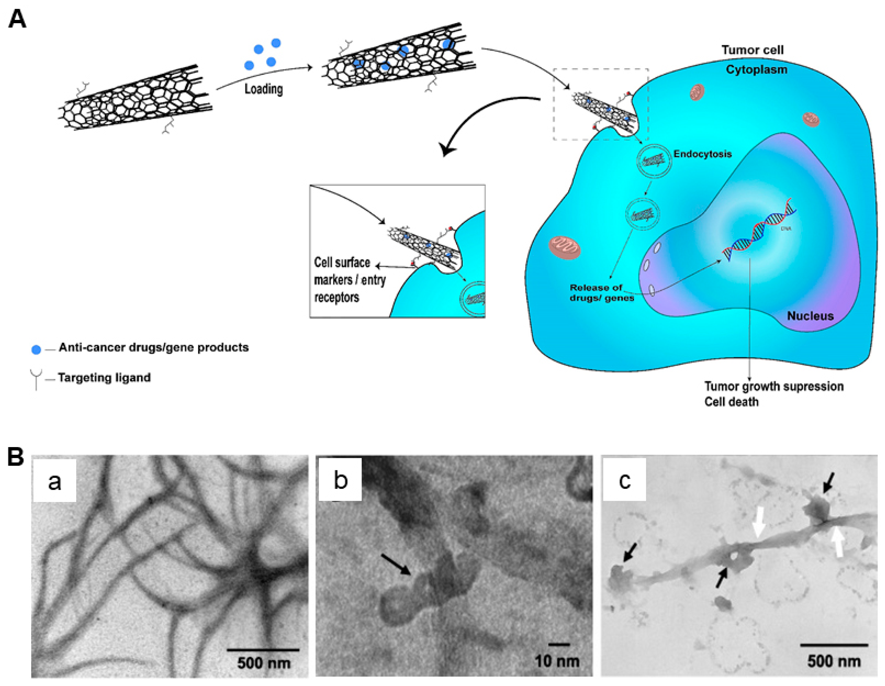
2.1. CNTs as Drug Delivery Vehicles

The application of CNTs in drug delivery can be classified into drug packaging, drug delivery/targeted delivery, sustained and controlled release, and delivery monitoring. In drug packaging, CNTs can be packaged by covalent adhesion, noncovalent adsorption, capillary effect filling, and other methods. Covalent adhesion involves the covalent bonding of drug molecules to the surface functional groups of CNTs (such as carboxyl groups and amino groups) [19]. This method ensures tight binding of drugs to CNTs, thereby improving the stability and controlled release of the drug. Noncovalent adsorption, on the other hand, attracts drug molecules through interaction forces such as hydrophobicity and π-π bonds on the surface of CNTs, offering the advantages of simple operation and low cost. For example, SWCNTs are functionalized with PEI through noncovalent bonds, which can achieve higher transfer efficiency [20]. However, the connection of noncovalent adsorption may not be strong enough, potentially leading to earlier drug release compared to covalent adhesion.
Targeted drug delivery of CNTs involves passive targeting and active targeting. Passive targeting relies on the enhanced penetration and retention effect (EPR). In this mode, drugs are delivered to target organs through systemic circulation, which is the most traditional mode of drug delivery, but it is often difficult to achieve optimal drug concentration in the lesion. Active targeting, on the other hand, relies on the binding of specific ligands to receptors, such as ligand-receptor interactions, antibody-antigen interactions, and coagulin–sugar interactions, to deliver drugs to targeted cells [21]. CNTs are commonly used for active targeted drug delivery, wherein functionalized CNTs could achieve local drug enrichment and sustained release by combining specific ligands with tumor cell receptors, thereby reducing drug consumption and toxic side effects in surrounding normal tissues (Figure 1). For example, CNTs functionalized with anti-HER-2 antibodies have higher affinity and specificity for breast cancer cells [22]. The combination of SWCNTs and antibodies achieved a remarkable 97% inhibition rate on breast cancer cells, which was 16 times higher than the rate achieved by antibodies alone. This significant improvement can be attributed to the overexpression of receptors in breast cancer cells. Western blot analysis confirmed that HER-2 protein levels had increased more than threefold in breast cancer cells overexpressing ERβ1, further explaining the efficacy of this combined treatment approach [23]. In colorectal cancer, CNTs modified by anti-EGFR and chemotherapy drugs show higher cytotoxicity [24]. Therefore, CNTs combined with chemotherapy drugs have great application potential in targeted cancer therapy.

2.2. CNTs for Cancer Therapy

Radiation is one of the most widely used treatments for cancer. One of the key challenges is to maximize the damage to tumor tissues without increasing the radiation dose to normal tissues. Radiosensitizers play a significant role in increasing the sensitivity of tumors to radiation by promoting DNA double-strand breaks, inhibiting damage repair, and affecting the cell cycle, thus enhancing the specific damage to tumor tissues [27]. However, small-molecule radiosensitizers such as nitroimidazole and capecitabine often have insufficient specificity and off-target effects [28,29]. Macromolecular sensitizers like proteins and miRNAs face challenges such as high immunogenicity and poor stability [30,31,32]. The emergence of nanomaterials provides a new approach for developing new radiosensitizers. Nanomaterials can accumulate in the tumor tissues and enhance the radiosensitivity of cells through retention effect and functionalization. Functionalized CNTs are preferred due to their good drug loading, permeability, biocompatibility, and targeting specificity. CNTs are often combined with metallic nanomaterials with high atomic number (Z) elements such as gold, platinum, and bismuth to form radiosensitizers to achieve complementary advantages [33,34,35,36]. For instance, Aghaei et al. loaded gold nanoparticles on SWCNTs and modified them with folic acid and bovine serum albumin to enhance their targeting specificity and stability (Figure 2A) [33]. This led to a significant improvement in the sensitivity of cancer cells to X-rays, thereby enhancing the effect of radiotherapy.
CNTs have also emerged as promising agents in PTT, a non-invasive treatment approach for various diseases, including cancer. Due to their exceptional optical properties and high absorption of light in the NIR region, CNTs can efficiently convert light energy into heat, effectively destroying targeted cancer cells. In a recent study, doxorubicin was combined with MWCNTs for PTT, resulting in a rapid increase in the temperature of the tumor area, causing irreversible tumor damage [37]. Another study utilized oxidized MWCNTs (ox-MWCNTs) in combination with PTT for treating breast cancer [38]. An increased infiltration of lymphocytes and macrophages in the tumor microenvironment was observed, leading to a higher survival rate in rats.
In addition to PTT, photodynamic therapy (PDT) is another type of tumor phototherapy in which photosensitizers are activated by specific wavelengths of light to produce photochemical reactions, leading to the destruction of tumor cells. While CNTs do not have obvious photodynamic effects, they can combine with other photosensitizers to enter tumor tissues and produce reactive oxygen species (ROS) through a photochemical reaction, causing cell damage. Zhang et al. developed Ru (II) complex-functionalized SWCNTs (Ru@SWCNTs) as nanotemplates for PTT and PDT (Figure 2B) [36]. SWCNTs possess the capability to load a significant quantity of Ru (II) complexes (Ru1 or Ru2) via noncovalent π−π interactions. These loaded Ru (II) complexes can be effectively released through the photothermal effect triggered by irradiation. Based on the combination of PTT and PDT, Ru@SWCNTs have greater anticancer efficacies than either PDT using Ru (II) complexes or PTT using SWCNTs in vitro and in vivo. These findings open up new possibilities for cancer radiotherapy, which are expected to improve treatment efficacy and reduce damage to normal cells.

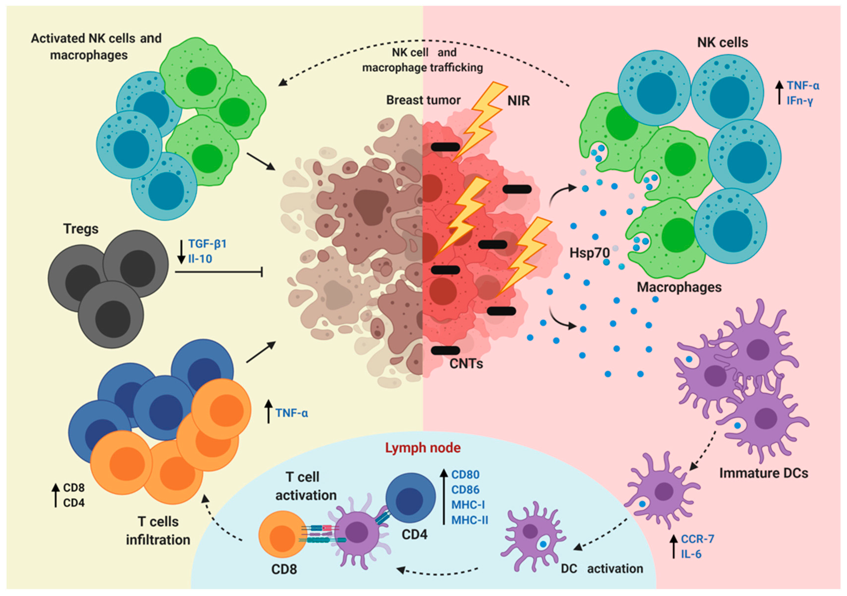
2.3. CNTs for the Regulation of Immunity

Tumor cells interact with the immune system in a complex manner that can help them evade recognition by the immune system or reduce immune clearance (Figure 3). Many studies have demonstrated that CNTs can regulate the immune response through a variety of mechanisms, including promoting or inhibiting the activity of immune cells and regulating the production of cytokines [38,39,40]. Niu et al. developed a durvalumab/CNT/PEI nanoagent with high permeability in tumor tissue and effective drug delivery for the immunotherapy of hepatocellular carcinoma [40]. Compared to the use of CNTs or durvalumab alone, the use of durvalumab/CNT/PEI significantly enhanced the proliferation and infiltration of CD8+ T cells.
Interestingly, CNTs can also be functionalized with specific antibodies to directly inhibit tumors or be used as carriers to transport antigen fragments to induce specific immune responses against tumor tissues, offering a new approach to treatment.
A recent study developed a DNA vaccine based on the GCRV-vp4-3 epitope using a SWCNT-loaded plasmid (pcDNA-vp4-3). It significantly increased the expression of IgM and other antibodies, prolonged the retention time of DNA vaccines such as eukaryotic plasmids, prevented premature degradation, and improved immune efficiency [41]. In addition, MWCNTs combined with hyperthermia were demonstrated to increase the expression of MCH-Ⅰ [42] and CD80+ [43] molecules and promote the maturation of dendritic cells in lymph nodes. Strikingly, the synergistic effect of this combination therapy further enhanced the infiltration of NK cells and macrophages in tumor tissues compared with hyperthermia alone, thus triggering a downstream response in the lymphocyte lineage. However, for dendritic cells, the modification of CNTs and the synergistic effect of “partners” may be more important. Researchers found that ox-MWCNTs combined with the antigen complex group expressed more “tentacles”, but its single effect did not distinctly affect the maturation and activation of dendritic cells [44]. The synergistic mechanism still needs to be further explored.

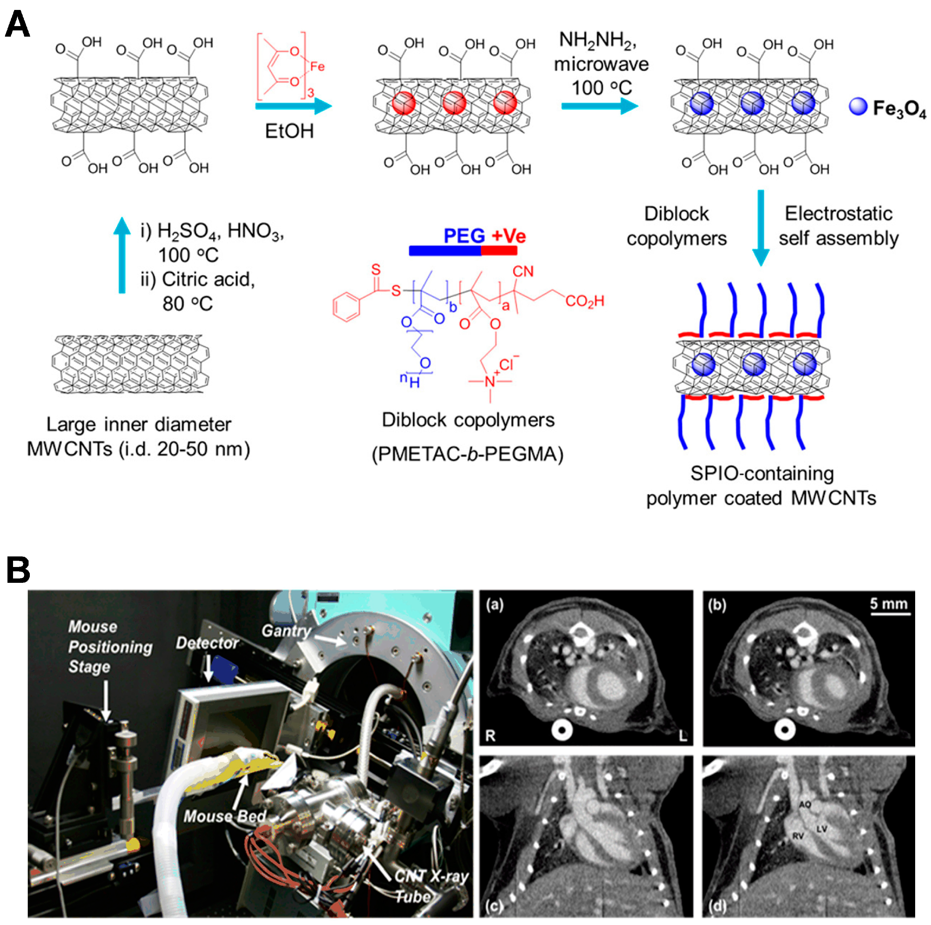
2.4. CNTs for Cancer Diagnosis

CNTs possess unique physical properties that make them suitable for cancer diagnosis. For example, SWCNTs display a strong resonance in Raman scattering, allowing for the quantitative detection of functionalization levels [45]. In addition, CNTs have been widely used in photoacoustic (PA) imaging due to their excellent tissue penetration and spatial resolution [46,47]. Magnetic MWCNTs can be used as a contrast agent for magnetic resonance molecular imaging (MRI) to enhance the magnetic labeling of tumor cells [48].
Karmakar et al. have covalently cross-linked SWCNTs and epidermal growth factor (EGF) to construct EGF-SWCNTs [49]. Raman spectroscopy and an enzyme-linked immunosorbent assay were used to study the mechanism of EGF in the targeted transport of SWCNTs to the surface and internalization of pancreatic cancer cells (PANC-1). In this process, the interaction between EGF-bound nanotubes and cells can be monitored by analyzing the concentration differences of SWCNTs in the complex cellular environment using two-dimensional mapping of Raman spectroscopy, aiming at the specific distribution of EGF-SWCNT conjugates on the surface of single cell membranes. Meanwhile, EGF specifically targeted coupled SWCNTs were analyzed as highly sensitive nanoagents, which could precisely locate the positions of various tumor-associated receptors on the cell membrane. They perfectly demonstrate the dual role of carbon nanotubes as “communicators” and “attackers”. In recent years, superparamagnetic iron oxide (SPIO), a widely studied targeted MRI agent, has been explored in combination with MWCNTs. Liu et al. explored microwave reactions for encapsulating SPIO inside nanotubes (Figure 4A) [48]. The CNT-Fe3O4-polymer hybrid materials demonstrated a notable 55% enhancement in the tumor-to-liver contrast ratio, accompanied by a substantial reduction in cytotoxicity owing to their exceptional sealing capabilities.
CNTs are capable of containing heavy atoms such as barium or gadolinium to serve as X-ray contrast agents. Due to their fast time switching mode and effective spatial modulation advantages, CNT X-ray sources have been widely used in biomedical fields such as microcomputed tomography (micro-CT) imaging in vivo (Figure 4B) [50]. Micro-CT using a CNT X-ray source can be precisely timed to the physiological stages needed by the respiratory or cardiac cycle of mice [51]. The trigger signal from the sensor is transmitted to the dynamic CNT micro-CT scanner, triggering X-rays and exposure to produce images. Compared with traditional CT, its radiation risk can be reduced by more than 85% [52]. Taken together, these studies provide novel possibilities for assessing the biodistribution of CNTs and developing a new generation of imaging monitoring systems.

3. Carcinogenicity and Potential Mechanisms of CNTs

Although CNTs show promise in cancer therapy, the toxicity of CNTs, especially the potential carcinogenic threat, has been a major concern. Compared to micron-sized carbon-based particles, CNTs are more easily taken up and transported by cells [53]. Many studies have demonstrated that CNTs can induce carcinogenesis through various pathways, including oxidative stress [54], inflammation [54,55], genotoxicity [56,57], and interference with cellular signaling pathways [58,59]. A recent study has shown that the production of ROS induced by CNTs can lead to oxidative damage to cells and DNA mutations, and trigger proinflammatory responses, all of which contribute to tumor initiation and progression [9]. In addition, CNT exposure has been linked to the promotion of epithelial–mesenchymal transition (EMT), a process that enhances the migratory and invasive capabilities of cells, thereby promoting tumor metastasis [60,61]. Furthermore, CNTs have been found to disrupt the integrity of endothelium, leading to increased vascular permeability and leakage [62,63]. This can facilitate the extravasation of cancer cells and promote cancer metastasis.

3.1. ROS

The biological effects of various nanomaterials are closely linked to the induction of intracellular oxidative stress. Many studies have shown that exposure to CNTs, both in the short term and long term, causes cells to produce a large amount of ROS. This disrupts the oxidative balance in cells, leading to DNA damage and mutations in tumor suppressor genes [64]. Moreover, the disruption of intracellular oxidative and antioxidant systems can cause oxidative stress, which can trigger an elevation of cytosolic calcium levels. This, in turn, can induce the migration of transcription factors such as NF-κB to the nucleus, leading to the upregulation of proinflammatory genes such as TNF-α and iNOS [65]. Prolonged inflammation can lead to the development of granuloma, fibrosis, and cancer.
Mitochondria play a crucial role in consuming 90% of the oxygen in cells for aerobic respiration to produce ATP. Meanwhile, mitochondria are also responsible for the scavenging of ROS generated under normal and pathological conditions. According to a recent study, MWCNTs not only stimulate cells to produce ROS but also damage mitochondria, hinder the removal of ROS, and ultimately disrupt the oxidative balance in cells, leading to DNA damage and cell death [66]. Another study suggests that mitochondria in cells treated with SWCNTs exhibit vacuolation and swelling, irregular and disordered lamellar ridges, resulting in mitochondrial dysfunction [67]. Therefore, the oxidative imbalance caused by mitochondrial damage and ROS production is one of the most fundamental mechanisms of CNT toxicity and carcinogenesis (Figure 5).

3.2. Inflammation and Fibrosis

Inflammation is a complex biological response triggered by harmful stimuli such as pathogens, damaged cells, or irritants. The process involves a cascade of events orchestrated by immune cells, such as neutrophils, macrophages, and lymphocytes, as well as signaling molecules like cytokines and chemokines. Previous studies have shown that exposure to CNTs can lead to chronic inflammation and tissue damage, especially in the lungs, with a massive accumulation of cytokines and chemokines [69,70]. For instance, when pleural mesothelial cells are cocultured with MWCNTs, it can lead to chronic inflammation and malignant pleural mesothelioma [71]. In addition, MWCNTs can increase the number of fibroblast-specific-protein-1-positive cells, produce large amounts of collagen, and cause collagen deposition, which is an important step in fibrosis. Pulmonary fibrosis is characterized by the excessive accumulation of extracellular matrix (ECM) in lung tissue. There are two reasons for the excessive accumulation of ECM: an increase in transcriptional activation leading to increased fibro-collagen mRNA levels, and the effect of the ECM degradation inhibitor. Wang et al. found that MWCNTs can upregulate the mRNA transcription of type I/III collagen and induce the transcription of the ECM protease inhibitors timo-1 and PAI-1, resulting in the excessive aggregation of ECM and promoting fibrosis [72].
Inflammatory factors are involved throughout this process, but their roles are different. For instance, MWCNTs are engulfed by pulmonary macrophages when they enter the lungs. This process induces macrophages to secrete transforming growth factor-β (TGF-β) and activates the TGF-β/Smad signaling pathway, ultimately leading to pulmonary fibrosis [73]. The levels of inflammatory molecules such as CXCL2, LIX, and S100A9 in the alveolar lavage fluid of CNT-exposed mice are significantly increased [74], and these factors are essential for the recruitment and adhesion of neutrophils. Using transgenic mouse models and single-cell transcriptomic technologies, researchers have shown that CD8+ T cells can produce a variety of cytokines, such as interferon-γ (IFN-γ) and tumor necrosis factor-α (TNF-α). They regulate the inflammatory response and inhibit the growth and spread of tumor cells, thereby inhibiting the development of cancer. In addition, in the serum of CNT-exposed mice, the concentrations of vascular endothelial growth factor A, basic fibroblast growth factor (bFGF), and NF-κB are also increased [72]. However, if the immune system cannot effectively regulate the inflammatory response, the inflammation may persist or even deteriorate [75]. The ongoing inflammatory response may contribute to the development of cancers, particularly colorectal [75], esophageal [76], and nasopharyngeal cancers [77].

3.3. EMT

The EMT is a fundamental process by which epithelial cells undergo biochemical changes and transform into a mesenchymal cell phenotype. This transition involves the loss of cell–cell adhesion and polarity, as well as the acquisition of migratory and invasive properties [78]. The EMT plays an important role in embryonic development, wound healing, and tissue regeneration. However, a dysregulated EMT is implicated in pathological conditions such as cancer progression and fibrosis [79]. The process of the EMT allows tumor cells to acquire invasive and metastatic properties, leading to tumor spread and resistance to treatment. It is worth noting that there is increasing evidence that CNTs can induce the transformation of epithelial cells into fibroblasts or myoblasts [80]. Furthermore, SWCNTs can interact with lung epithelial cells to induce tumor stem cells (CSCs), which tend to form tumor spheres, indicating their new plasticity and ability to self-renew [81].
Many studies have shown that exposure to MWCNTs and SWCNTs can trigger the EMT in various cell types, particularly in bronchial epithelial cells. This transition involves a complex interplay of signaling pathways, including the TGF-β/Smad pathway, Akt/GSK-3β/SNAIL-1 signaling, and DNA methylation changes. A recent study has shown that long-term exposure to SWCNTs changes the morphology and growth mode of bronchial epithelial cells (Figure 6) [80]. Meanwhile, the proliferation ability of bronchial epithelial cells exposed to SWCNTs was much higher than that of normal cells in the control group.
There is growing evidence that the polarization of alveolar macrophages is the key “transit station” in the regulation of the EMT [82]. Exposure to CNTs promotes the polarization of alveolar macrophages toward the M2 phenotype, which facilitates the EMT by secreting TGF-β. Therefore, this suggests that CNTs can lead to malignant transformation by indirectly affecting the immune system. Notably, a recent study has shown that functionalized CNTs (CPG-CNTs) have the ability to inhibit colon cancer and significantly reduce the degree of the EMT [83]. This indicates that the effects of CNTs on epithelial cells may depend on the different functionalization of the material and the tissue microenvironment.

3.4. Endothelial Leakiness

In recent years, a new mechanism of nanomaterial biotoxicity has been widely studied [84,85]. Many studies have found that exposure to various nanoparticles can induce endothelial leakiness, where the integrity of the endothelial barrier is compromised, allowing substances to leak through (Figure 7) [86]. This effect has been observed for different types of nanoparticles, including anionic nanoplastics [87], titanium dioxide nanomaterials [88], gold nanoparticles [89], etc. The degree of leakiness appears to be influenced by factors such as the size of the nanoparticles and the origin of the endothelial cells. For instance, gold nanoparticles within the range of 10–30 nm have been shown to effectively induce leakiness in human mammary and skin endothelial cells [89]. Meanwhile, nanoplastics disrupt vascular endothelial cadherin junctions in a dose-dependent manner. It has been confirmed that anionic nanomaterials and titanium dioxide nanomaterials can cause leakage through VE-Cadherin-triggered cascade reactions [88].
CNTs have a larger specific surface area, which means they can come into more contact with cells, thus increasing their impacts on the tissue endothelium. In addition, their nanoscale size makes it easier for CNTs to penetrate cell membranes or the space between the blood vessel endothelium [90]. This can lead to the destruction or degeneration of cell-to-cell connections, increasing the exchange of materials inside and outside the cell. On the other hand, functionalized SWCNTs can be more easily taken up and retained by tumor tissues for a longer period due to the EPR effect [91]. Therefore, this property of CNTs is a “double-edged sword”, as it is expected to carry targeted drugs to kill tumors but may also cause damage to normal tissues.

4. Conclusions and Perspectives

The utilization of CNTs in cancer therapy presents promising solutions to numerous obstacles encountered by conventional treatments. Extensive studies have successfully used CNTs as drug delivery carriers, radiosensitizers, immunomodulators, and more for cancer treatment and diagnosis. This review offers an overview of the application of CNTs for cancer therapy and diagnosis from a broad perspective, summarizing recent research advancements of functionalized CNTs for drug delivery, cancer therapy, diagnosis, and immune regulation. Nevertheless, the clinical advancement of CNTs has been hindered by biosafety concerns. Therefore, this review places more emphasis on the biological toxicity of CNTs, especially carcinogenesis, and the underlying mechanisms. Moreover, while CNTs exhibit immense potential in targeted drug delivery, achieving optimal drug concentrations at tumor sites remains a challenge. Both passive and active targeting strategies aim to address this issue, yet further research is needed to enhance their efficacy. In the future, we need to gain a deeper understanding of how CNTs interact with tissues, cells, and biomolecules to uncover the mechanisms of their biological toxicity. In addition, the behavior of CNTs in complex biological microenvironments and their long-term biocompatibility should be thoroughly investigated. Due to the unique physicochemical properties of CNTs, establishing toxicity assessment methodologies suitable for CNTs is an essential for the precise and reproducible measurement of their toxicity.

Funding

This work was supported by the Interdisciplinary Basic Frontier Innovation Program of Suzhou Medical College of Soochow University (YXY2304033), a project funded by the Priority Academic Program Development of Jiangsu Higher Education Institutions (PAPD), Jiangsu Provincial Key Laboratory of Radiation Medicine and Protection, and Undergraduate Training Program for Innovation and Entrepreneurship (025), Soochow University.

Conflicts of Interest

The authors declare that they have no conflicts of interest.

References

  1. Aasi, A.; Aasi, E.; Aghaei, S.M.; Panchapakesan, B. CNT biodevices for early liver cancer diagnosis based on biomarkers detection-a promising platform. J. Mol. Graph. Model. 2022, 114, 108208. [Google Scholar] [CrossRef]
  2. Yaghoubi, A.; Ramazani, A. Anticancer DOX delivery system based on CNTs: Functionalization, targeting and novel technologies. J. Control. Release 2020, 327, 198–224. [Google Scholar] [CrossRef]
  3. Dehaghani, M.Z.; Yousefi, F.; Sajadi, S.M.; Munir, M.T.; Abida, O.; Habibzadeh, S.; Mashhadzadeh, A.H.; Rabiee, N.; Mostafaci, E.; Saeb, M.R. Theoretical encapsulation of fluorouracil (5-FU) anti-cancer chemotherapy drug into carbon nanotubes (CNT) and boron nitride nanotubes (BNNT). Molecules 2021, 26, 4920. [Google Scholar] [CrossRef]
  4. Aoki, K.; Saito, N. Biocompatibility and carcinogenicity of carbon nanotubes as biomaterials. Nanomaterials 2020, 10, 264. [Google Scholar] [CrossRef]
  5. Barbarino, M.; Giordano, A. Assessment of the carcinogenicity of carbon nanotubes in the respiratory system. Cancers 2021, 13, 1318. [Google Scholar] [CrossRef] [PubMed]
  6. Pu, Z.; Wei, Y.; Sun, Y.; Wang, Y.; Zhu, S. Carbon nanotubes as carriers in drug delivery for non-small cell lung cancer, mechanistic analysis of their carcinogenic potential, safety profiling and identification of biomarkers. Int. J. Nanomed. 2022, 17, 6157. [Google Scholar] [CrossRef] [PubMed]
  7. Fukushima, S.; Kasai, T.; Umeda, Y.; Makoto, O.; Sasaki, T.; Matsumoto, M. Carcinogenicity of multi-walled carbon nanotubes: Challenging issue on hazard assessment. J. Occup. Health 2018, 60, 10–30. [Google Scholar] [CrossRef]
  8. Yang, M.; Zhang, M. Biodegradation of carbon nanotubes by macrophages. Front. Mater. 2019, 6, 225. [Google Scholar] [CrossRef]
  9. Dong, J. Signaling pathways implicated in carbon nanotube-induced lung inflammation. Front. Immunol. 2020, 11, 552613. [Google Scholar] [CrossRef]
  10. Hadrup, N.; Knudsen, K.B.; Carriere, M.; Mayne-L’Hermite, M.; Bobyk, L.; Allard, S.; Miserque, F.; Pibaleau, B.; Pinault, M.; Wallin, H.; et al. Safe-by-design strategies for lowering the genotoxicity and pulmonary inflammation of multiwalled carbon nanotubes: Reduction of length and the introduction of COOH groups. Environ. Toxicol. Pharmacol. 2021, 87, 103702. [Google Scholar] [CrossRef]
  11. Lu, X.; Zhu, Y.; Bai, R.; Wu, Z.; Qian, W.; Yang, L.; Cai, R.; Yan, H.; Li, T.; Pandey, V.; et al. Long-term pulmonary exposure to multi-walled carbon nanotubes promotes breast cancer metastatic cascades. Nat. Nanotechnol. 2019, 14, 719–727. [Google Scholar] [CrossRef] [PubMed]
  12. Posypanova, G.A.; Gayduchenko, I.A.; Moskaleva, E.Y.; Fedorov, G.E. Neuronal differentiation of PC12 cells and mouse neural stem cells on carbon nanotube films. Cell Tissue Biol. 2016, 10, 194–201. [Google Scholar] [CrossRef]
  13. Öner, D.; Moisse, M.; Ghosh, M.; Duca, R.; Poels, K.; Luyts, K.; Putzeys, E.; Cokic, S.; Landuyt, K.L.V.; Lambrechts, D.; et al. Epigenetic effects of carbon nanotubes in human monocytic cells. Mutagenesis 2017, 32, 181–191. [Google Scholar] [CrossRef]
  14. Panigrahi, B.K.; Nayak, A.K. Carbon nanotubes: An emerging drug delivery carrier in cancer therapeutics. Curr. Drug Deliv. 2020, 17, 558–576. [Google Scholar] [CrossRef]
  15. Kofoed, A.C.; Khatri, S.; Hansen, J.; Slott, S.; Parvathaneni, R.P.; Mendes, A.C.; Chronakis, L.S.; Hung, S.; Rajasekaran, N.; Ma, Z.; et al. Carbon nanotubes—Potent carriers for targeted drug delivery in rheumatoid arthritis. Pharmaceutics 2021, 13, 453. [Google Scholar] [CrossRef] [PubMed]
  16. Dubey, R.; Dutta, D.; Sarkar, A.; Chattopadhyay, P. Functionalized carbon nanotubes: Synthesis, properties and applications in water purification, drug delivery, and material and biomedical sciences. Nanoscale Adv. 2021, 3, 5722–5744. [Google Scholar] [CrossRef]
  17. Zhao, Y.; Zhao, T.; Cao, Y.; Sun, J.; Zhou, Q.; Chen, H.; Guo, S.; Wang, Y.; Zhen, Y.; Liang, X.; et al. Temperature-sensitive lipid-coated carbon nanotubes for synergistic photothermal therapy and gene therapy. ACS Nano 2021, 15, 6517–6529. [Google Scholar] [CrossRef]
  18. Naief, M.F.; Mohammed, S.N.; Mayouf, H.J.; Mohammed, A.M. A review of the role of carbon nanotubes for cancer treatment based on photothermal and photodynamic therapy techniques. J. Organomet. Chem. 2023, 999, 122819. [Google Scholar] [CrossRef]
  19. Ijaz, H.; Mahmood, A.; Abdel-Daim, M.M.; Sarfraz, R.M.; Zaman, M.; Zafar, N.; Alshehery, S.; Salem-Bekhit, M.M.; Ali, M.A.; Eltayeb, L.B.; et al. Review on carbon nanotubes (CNTs) and their chemical and physical characteristics, with particular emphasis on potential applications in biomedicine. Inorg. Chem. Commun. 2023, 155, 111020. [Google Scholar] [CrossRef]
  20. Behnam, B.; Shier, W.T.; Nia, A.H.; Abnous, K.; Ramezani, M. Non-covalent functionalization of single-walled carbon nanotubes with modified polyethyleneimines for efficient gene delivery. Int. J. Pharm. 2013, 454, 204–215. [Google Scholar] [CrossRef]
  21. Canal, F.; Vicent, M.J.; Pasut, G.; Schiavon, O. Relevance of folic acid/polymer ratio in targeted PEG–epirubicin conjugates. J. Control. Release 2010, 146, 388–399. [Google Scholar] [CrossRef] [PubMed]
  22. Xiao, Y.; Gao, X.; Taratula, O.; Treado, S.; Urbas, A.; Holbrook, R.D.; E Cavicchi, R.; Avedisian, C.T.; Mitra, S.; Savla, R.; et al. Anti-HER2 IgY antibody-functionalized single-walled carbon nanotubes for detection and selective destruction of breast cancer cells. BMC Cancer 2009, 9, 351. [Google Scholar] [CrossRef] [PubMed]
  23. Lattrich, C.; Juhasz-boess, I.; Ortmann, O.; Treeck, O. Detection of an elevated HER2 expression in MCF-7 breast cancer cells overexpressing estrogen receptor ß1. Oncol. Rep. 2008, 19, 811–817. [Google Scholar] [PubMed]
  24. Lee, P.C.; Chiou, Y.C.; Wong, J.M.; Peng, C.L.; Shieh, M.J. Targeting colorectal cancer cells with single-walled carbon nanotubes conjugated to anticancer agent SN-38 and EGFR antibody. Biomaterials 2013, 34, 8756–8765. [Google Scholar] [CrossRef] [PubMed]
  25. Kiran, A.R.; Kumari, G.K.; Krishnamurthy, P.T. Carbon nanotubes in drug delivery: Focus on anticancer therapies. J. Drug Deliv. Sci. Technol. 2020, 59, 101892. [Google Scholar] [CrossRef]
  26. Pantarotto, D.; Singh, R.; McCarthy, D.; Erhardt, M.; Briand, J.P.; Prato, M.; Kostarelos, K.; Bianco, A. Functionalized carbon nanotubes for plasmid DNA gene delivery. Angew. Chem. Int. Ed. 2004, 43, 5242–5246. [Google Scholar] [CrossRef] [PubMed]
  27. Schaue, D.; McBride, W.H. Opportunities and challenges of radiotherapy for treating cancer. Nat. Rev. Clin. Oncol. 2015, 12, 527–540. [Google Scholar] [CrossRef] [PubMed]
  28. Jia, Y.; Weng, Z.; Wang, C.; Zhu, M.; Lu, Y.; Ding, L.; Wang, Y.; Cheng, X.; Lin, Q.; Wu, K. Increased chemosensitivity and radiosensitivity of human breast cancer cell lines treated with novel functionalized single-walled carbon nanotubes. Oncol. Lett. 2017, 13, 206–214. [Google Scholar] [CrossRef]
  29. Gong, L.; Zhang, Y.; Liu, C.; Zhang, M.; Han, S. Application of radiosensitizers in cancer radiotherapy. Int. J. Nanomed. 2021, 16, 1083–1102. [Google Scholar] [CrossRef]
  30. Harvey, J.D.; Jena, P.V.; Baker, H.A.; Zerze, G.H.; Williams, R.M.; Galassi, T.V.; Roxbury, D.; Mittal, J.; Heller, D.A. A carbon nanotube reporter of microRNA hybridization events in vivo. Nat. Biomed. Eng. 2017, 1, 0041. [Google Scholar] [CrossRef]
  31. Zhou, Q.; Zhang, L.; Wu, H. Nanomaterials for cancer therapies. Nanotechnol. Rev. 2017, 6, 473–496. [Google Scholar] [CrossRef]
  32. Comparetti, E.J.; Pedrosa, V.D.A.; Kaneno, R. Carbon nanotube as a tool for fighting cancer. Bioconjugate Chem. 2017, 29, 709–718. [Google Scholar] [CrossRef] [PubMed]
  33. Aghaei, A.; Shaterian, M.; Danafar, H.; Likozar, B.; Šuligoj, A.; Gyergyek, S. The role of single-walled carbon nanotubes functionalized with gold to increase radiosensitivity of cancer cells to X-ray radiation. Appl. Organomet. Chem. 2023, 37, e7265. [Google Scholar] [CrossRef]
  34. Berber, M.R.; Elkhenany, H.; Hafez, I.H.; El-Badawy, A.; Essawy, M.; El-Badri, N. Efficient tailoring of platinum nanoparticles supported on multiwalled carbon nanotubes for cancer therapy. Nanomedicine 2020, 15, 793–808. [Google Scholar] [CrossRef] [PubMed]
  35. Tang, L.; Xiao, Q.; Mei, Y.; He, S.; Zhang, Z.; Wang, R.; Wang, W. Insights on functionalized carbon nanotubes for cancer theranostics. J. Nanobiotechnol. 2021, 19, 423. [Google Scholar] [CrossRef] [PubMed]
  36. Zhang, P.; Huang, H.; Huang, J.; Chen, H.; Wang, J.; Qiu, K.; Zhao, D.; Ji, L.; Chao, H. Noncovalent ruthenium (II) complexes–single-walled carbon nanotube composites for bimodal photothermal and photodynamic therapy with near-infrared irradiation. ACS Appl. Mater. Interfaces 2015, 7, 23278–23290. [Google Scholar] [CrossRef] [PubMed]
  37. Pai, C.L.; Chen, Y.C.; Hsu, C.Y.; Su, H.L.; Lai, P.S. Carbon nanotube-mediated photothermal disruption of endosomes/lysosomes reverses doxorubicin resistance in MCF-7/ADR cells. J. Biomed. Nanotechnol. 2016, 12, 619–629. [Google Scholar] [CrossRef] [PubMed]
  38. Radzi, M.R.M.; Johari, N.A.; Zawawi, W.F.A.W.M.; Zawawi, N.A.; Latiff, N.A.; Malek, N.A.N.N.; Wahab, A.A.; Salim, M.I.; Jemon, K. In vivo evaluation of oxidized multiwalled-carbon nanotubes-mediated hyperthermia treatment for breast cancer. Biomater. Adv. 2022, 134, 112586. [Google Scholar] [CrossRef] [PubMed]
  39. Modo, M.M.; Bulte, J.W. What is molecular and cellular imaging. In Molecular and Cellular MR Imaging; CRC Press: Boca Raton, FL, USA, 2007; pp. 1–9. [Google Scholar]
  40. Niu, Q.; Lv, W.; Yan, T.; Wang, J.; Yan, B.; Zhou, D. Construction of Durvalumab/carbon nanotube/PEI/aptamer-siRNA chimera for the immunotherapy of hepatocellular carcinoma. Biomed. Mater. 2022, 17, 025015. [Google Scholar]
  41. Zheng, Y.Y.; Qiu, D.K.; Guo, Z.R.; Gong, Y.M.; Wang, G.X.; Zhu, B. Evaluation of SWCNTs-loaded DNA vaccine encoding predominant antigen epitope VP4-3 against type II GCRV. Aquaculture 2021, 534, 736197. [Google Scholar] [CrossRef]
  42. Xia, Q.; Gong, C.; Gu, F.; Wang, Z.; Hu, C.; Zhang, L.; Qiang, L.; Ding, X.; Gao, S.; Gao, Y. Functionalized multi-walled carbon nanotubes for targeting delivery of immunostimulatory CpG oligonucleotides against prostate cancer. J. Biomed. Nanotechnol. 2018, 14, 1613–1626. [Google Scholar] [CrossRef] [PubMed]
  43. Wang, C.; Xu, L.; Liang, C.; Xiang, J.; Peng, R.; Liu, Z. Immunological responses triggered by photothermal therapy with carbon nanotubes in combination with anti-CTLA-4 therapy to inhibit cancer metastasis. Adv. Mater. 2014, 48, 8154–8162. [Google Scholar] [CrossRef] [PubMed]
  44. Dizaji, B.F.; Khoshbakht, S.; Farboudi, A.; Azarbaijan, M.H.; Irani, M. Far-reaching advances in the role of carbon nanotubes in cancer therapy. Life Sci. 2020, 257, 118059. [Google Scholar] [CrossRef] [PubMed]
  45. Hwang, Y.; Park, S.H.; Lee, J.W. Applications of functionalized carbon nanotubes for the therapy and diagnosis of cancer. Polymers 2017, 9, 13. [Google Scholar] [CrossRef] [PubMed]
  46. Welsher, K.; Sherlock, S.P.; Dai, H. Deep-tissue anatomical imaging of mice using carbon nanotube fluorophores in the second near-infrared window. Proc. Natl. Acad. Sci. USA 2011, 108, 8943–8948. [Google Scholar] [CrossRef] [PubMed]
  47. Kim, H.; Chung, K.; Lee, S.; Kim, D.H.; Lee, H. Near-infrared light-responsive nanomaterials for cancer theranostics. Wiley Interdiscip. Rev. Nanomed. Nanobiotechnol. 2016, 8, 23–45. [Google Scholar] [CrossRef]
  48. Liu, Y.; Muir, B.W.; Waddington, L.J.; Hinton, T.M.; Moffat, B.A.; Hao, X.; Hughes, T.C. Colloidally stabilized magnetic carbon nanotubes providing MRI contrast in mouse liver tumors. Biomacromolecules 2015, 16, 790–797. [Google Scholar] [CrossRef] [PubMed]
  49. Karmakar, A.; Iancu, C.; Bartos, D.M.; Mahmood, M.W.; Ghosh, A.; Xu, Y.; Dervishi, E.; Collom, S.L.; Khodakovskaya, M.; Mustafa, T.; et al. Raman spectroscopy as a detection and analysis tool for in vitro specific targeting of pancreatic cancer cells by EGF-conjugated, single-walled carbon nanotubes. J. Appl. Toxicol. 2012, 32, 365–375. [Google Scholar] [CrossRef]
  50. Cao, G. Biomedical X-ray imaging enabled by carbon nanotube X-ray sources. Chin. J. Chem. Phys. 2018, 31, 529–536. [Google Scholar] [CrossRef]
  51. Dalby, M.J.; Giannaras, D.; Riehle, M.O.; Gadegaard, N.; Affrossman, S.; Curtis, A.S.G. Rapid fibroblast adhesion to 27 nm high polymer demixed nano-topography. Biomaterials 2004, 25, 77–83. [Google Scholar] [CrossRef]
  52. Silva, G.A.; Czeisler, C.; Niece, K.L.; Beniash, E.; Harrington, D.A.; Kessler, J.A.; Stupp, S.I. Selective differentiation of neural progenitor cells by high-epitope density nanofibers. Science 2004, 303, 1352–1355. [Google Scholar] [CrossRef]
  53. Kobayashi, N.; Izumi, H.; Morimoto, Y. Review of toxicity studies of carbon nanotubes. J. Occup. Health 2017, 59, 394–407. [Google Scholar] [CrossRef]
  54. Cammisuli, F.; Giordani, S.; Gianoncelli, A.; Rizzardi, C.; Radillo, L.; Zweyer, M.; Da Ros, T.; Salomé, M.; Melato, M.; Pascolo, L. Iron-related toxicity of single-walled carbon nanotubes and crocidolite fibres in human mesothelial cells investigated by Synchrotron XRF microscopy. Sci. Rep. 2018, 8, 706. [Google Scholar] [CrossRef]
  55. Digifico, E.; Belgiovine, C.; Mantovani, A.; Allavena, P. Microenvironment and immunology of the human pleural malignant mesothelioma. In Mesothelioma: From Research to Clinical Practice; Spring: Berlin/Heidelberg, Germany, 2019; pp. 69–84. [Google Scholar]
  56. Mittal, V.; El Rayes, T.; Narula, N.; McGraw, T.E.; Altorki, N.K.; Barcellos-Hoff, M.H. The microenvironment of lung cancer and therapeutic implications. Adv. Exp. Med. Biol. 2016, 890, 75–110. [Google Scholar]
  57. Jiang, T.; Amadei, C.A.; Gou, N.; Lin, Y.; Lan, J.; Vecitis, C.D.; Gu, A.Z. Toxicity of single-walled carbon nanotubes (SWCNTs): Effect of lengths, functional groups and electronic structures revealed by a quantitative toxicogenomics assay. Environ. Sci. Nano 2020, 7, 1348–1364. [Google Scholar] [CrossRef]
  58. Ghosh, M.; Janssen, L.; Martens, D.S.; Öner, D.; Vlaanderen, J.; Pronk, A.; Kuijpers, E.; Vermeulen, R.; Nawrot, T.S.; Godderis, L.; et al. Increased telomere length and mtDNA copy number induced by multi-walled carbon nanotube exposure in the workplace. J. Hazard. Mater. 2020, 394, 122569. [Google Scholar] [CrossRef]
  59. Di Cristo, L.; Bianchi, M.G.; Chiu, M.; Taurino, G.; Donato, F.; Garzaro, G.; Bussolati, O.; Bergamaschi, E. Comparative in Vitro Cytotoxicity of Realistic Doses of Benchmark Multi-Walled Carbon Nanotubes towards Macrophages and Airway Epithelial Cells. Nanomaterials 2019, 9, 982. [Google Scholar] [CrossRef]
  60. Rubio, L.; El Yamani, N.; Kazimirova, A.; Dusinska, M.; Marcos, R. Multi-walled carbon nanotubes (NM401) induce ROS-mediated HPRT mutations in Chinese hamster lung fibroblasts. Environ. Res. 2016, 146, 185–190. [Google Scholar] [CrossRef]
  61. Kuempel, E.D.; Jaurand, M.C.; Møller, P.; Morimoto, Y.; Kobayashi, N.; Pinkerton, K.E.; Sargent, L.M.; Vermeulen, R.C.; Fubini, B.; Kane, A.B. Evaluating the mechanistic evidence and key data gaps in assessing the potential carcinogenicity of carbon nanotubes and nanofibers in humans. Crit. Rev. Toxicol. 2017, 47, 1–58. [Google Scholar] [CrossRef]
  62. Kiratipaiboon, C.; Stueckle, T.A.; Ghosh, R.; Rojanasakul, L.W.; Chen, Y.C.; Dinu, C.Z.; Rojanasakul, Y. Acquisition of cancer stem cell-like properties in human small airway epithelial cells after a long-term exposure to carbon nanomaterials. Environ. Sci. Nano 2019, 6, 2152–2170. [Google Scholar] [CrossRef]
  63. Walker, V.G.; Li, Z.; Hulderman, T.; Schwegler-Berry, D.; Kashon, M.L.; Simeonova, P.P. Potential in vitro effects of carbon nanotubes on human aortic endothelial cells. Toxicol. Appl. Pharmacol. 2009, 236, 319–328. [Google Scholar] [CrossRef]
  64. Møller, P.; Christophersen, D.V.; Jensen, D.M.; Kermanizadeh, A.; Roursgaard, M.; Jacobsen, N.R.; Hemmingsen, J.G.; Danielsen, P.H.; Cao, Y.; Jantzen, K.; et al. Role of oxidative stress in carbon nanotube-generated health effects. Arch. Toxicol. 2014, 88, 1939–1964. [Google Scholar] [CrossRef]
  65. Castranova, V. Signaling pathways controlling the production of inflammatory mediators in response to crystalline silica exposure: Role of reactive oxygen/nitrogen species. Free. Radic. Biol. Med. 2004, 37, 916–925. [Google Scholar] [CrossRef]
  66. He, X.; Young, S.H.; Schwegler-Berry, D.; Chisholm, W.P.; Fernback, J.E.; Ma, Q. Multiwalled carbon nanotubes induce a fibrogenic response by stimulating reactive oxygen species production, activating NF-κB signaling, and promoting fibroblast-to-myofibroblast transformation. Chem. Res. Toxicol. 2011, 24, 2237–2248. [Google Scholar] [CrossRef]
  67. Ma, D.D.; Yang, W.X. Engineered nanoparticles induce cell apoptosis: Potential for cancer therapy. Oncotarget 2016, 7, 40882. [Google Scholar] [CrossRef]
  68. Liu, Y.; Zhao, Y.; Sun, B.; Chen, C. Understanding the toxicity of carbon nanotubes. Acc. Chem. Res. 2013, 46, 702–713. [Google Scholar] [CrossRef]
  69. Soltani, R.; Guo, S.; Bianco, A.; Ménard-Moyon, C. Carbon nanomaterials applied for the treatment of inflammatory diseases: Preclinical evidence. Adv. Ther. 2020, 3, 2000051. [Google Scholar] [CrossRef]
  70. Lim, C.S.; Porter, D.W.; Orandle, M.S.; Green, B.J.; Barnes, M.A.; Croston, T.L.; Wolfarth, M.G.; Battelli, L.A.; Andrew, M.E.; Beezhold, D.H.; et al. Resolution of pulmonary inflammation induced by carbon nanotubes and fullerenes in mice: Role of macrophage polarization. Front. Immunol. 2020, 11, 1186. [Google Scholar] [CrossRef]
  71. Huang, X.; Tian, Y.; Shi, W.; Chen, J.; Yan, L.; Ren, L.; Zhang, X.; Zhu, J. Role of inflammation in the malignant transformation of pleural mesothelial cells induced by multi-walled carbon nanotubes. Nanotoxicology 2020, 14, 947–967. [Google Scholar] [CrossRef]
  72. Wang, P.; Nie, X.; Wang, Y.; Li, Y.; Ge, C.; Zhang, L.; Wang, L.; Bai, R.; Chen, Z.; Zhao, Y.; et al. Multiwall carbon nanotubes mediate macrophage activation and promote pulmonary fibrosis through TGF-β/Smad signaling pathway. Small 2013, 9, 3799–3811. [Google Scholar] [CrossRef]
  73. Li, X.; Chen, G.; Wang, Y.; Su, L.; Chen, B.; Wu, K.; Xing, Y.; Song, Z.; Dai, R.; Liu, T.; et al. Systematic co-delivery of dual agonists to enhance cancer immunotherapy. Nano Res. 2022, 15, 8326–8335. [Google Scholar] [CrossRef]
  74. Böttcher, J.P.; Bonavita, E.; Chakravarty, P.; Blees, H.; Cabeza-Cabrerizo, M.; Sammicheli, S.; Rogers, N.C.; Sahai, E.; Zelenay, S.; Reise Sousa, C. NK cells stimulate recruitment of cDC1 into the tumor microenvironment promoting cancer immune control. Cell 2018, 172, 1022–1037. [Google Scholar] [CrossRef]
  75. Long, A.G.; Lundsmith, E.T.; Hamilton, K.E. Inflammation and colorectal cancer. Curr. Color. Cancer Rep. 2017, 13, 341–351. [Google Scholar] [CrossRef]
  76. Jiang, Y.; Xu, D.; Song, H.; Qiu, B.; Tian, D.; Li, Z.; Ji, Y.; Wang, J. Inflammation and nutrition-based biomarkers in the prognosis of oesophageal cancer: A systematic review and meta-analysis. BMJ Open 2021, 11, e048324. [Google Scholar] [CrossRef]
  77. He, W.Z.; Jiang, C.; Liu, L.L.; Yin, C.X.; Rong, Y.M.; Hu, W.M.; Yang, L.; Wang, L.; Jin, Y.N.; Lin, X.P.; et al. Association of body composition with survival and inflammatory responses in patients with non-metastatic nasopharyngeal cancer. Oral Oncol. 2020, 108, 104771. [Google Scholar] [CrossRef]
  78. Polimeni, M.; Gulino, G.R.; Gazzano, E.; Kopecka, J.; Marucco, A.; Fenoglio, I.; Cesano, F.; Campagnolo, L.; Magrini, A.; Pietroiusti, A.; et al. Multi-walled carbon nanotubes directly induce epithelial-mesenchymal transition in human bronchial epithelial cells via the TGF-β-mediated Akt/GSK-3β/SNAIL-1 signalling pathway. Part. Fibre Toxicol. 2015, 13, 27. [Google Scholar] [CrossRef]
  79. Wang, P.; Wang, Y.; Nie, X.; Braïni, C.; Bai, R.; Chen, C. Multiwall carbon nanotubes directly promote fibroblast–myofibroblast and epithelial–mesenchymal transitions through the activation of the TGF-β/Smad signaling pathway. Small 2015, 11, 446–455. [Google Scholar] [CrossRef]
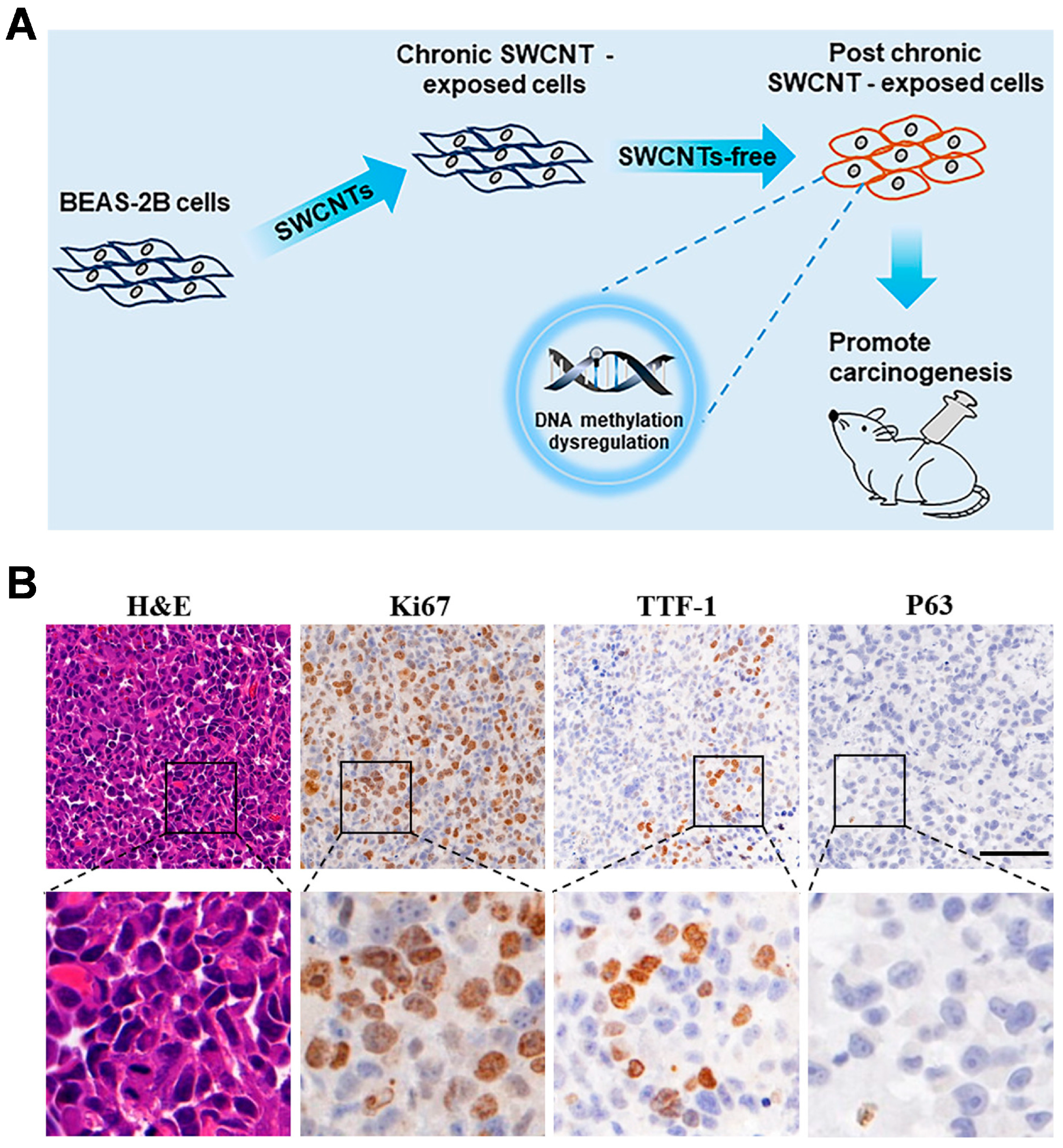
  80. Wang, J.; Tian, X.; Zhang, J.; Tan, L.; Ouyang, N.; Jia, B.; Chen, C.; Ge, C.; Li, J. Postchronic single-walled carbon nanotube exposure causes irreversible malignant transformation of human bronchial epithelial cells through DNA methylation changes. ACS Nano 2021, 15, 7094–7104. [Google Scholar] [CrossRef]
  81. Luanpitpong, S.; Wang, L.; Castranova, V.; Rojanasakul, Y. Induction of stem-like cells with malignant properties by chronic exposure of human lung epithelial cells to single-walled carbon nanotubes. Part. Fibre Toxicol. 2014, 11, 22. [Google Scholar] [CrossRef]
  82. Zhang, X.; Luo, M.; Zhang, J.; Yao, Z.; Zhu, J.; Yang, S.; Zhu, Q.; Shen, T. Carbon nanotubes promote alveolar macrophages toward M2 polarization mediated epithelial-mesenchymal transition and fibroblast-to-myofibroblast transdifferentiation. Nanotoxicology 2021, 15, 588–604. [Google Scholar] [CrossRef]
  83. Jin, H.; Gao, S.; Song, D.; Liu, Y.; Chen, X. Intratumorally CpG immunotherapy with carbon nanotubes inhibits local tumor growth and liver metastasis by suppressing the epithelial–mesenchymal transition of colon cancer cells. Anti-Cancer Drugs 2021, 32, 278. [Google Scholar] [CrossRef]
  84. Yang, D.; Shen, J.; Fan, J.; Chen, Y.; Guo, X. Paracellular permeability changes induced by multi-walled carbon nanotubes in brain endothelial cells and associated roles of hemichannels. Toxicology 2020, 440, 152491. [Google Scholar] [CrossRef]
  85. Guo, Q.; Li, L.; Gao, G.; Zhao, Q.; Huang, X.; Wang, H.; Liu, B.; Zhi, J. Nanodiamonds inhibit the proliferation and migration of endothelial cells in a tumor/endothelial cells co-culture microfluidic system. Carbon 2024, 218, 118671. [Google Scholar] [CrossRef]
  86. Lasak, M.; Ciepluch, K. Overview of mechanism and consequences of endothelial leakiness caused by metal and polymeric nanoparticles. Beilstein J. Nanotechnol. 2023, 14, 329–338. [Google Scholar] [CrossRef]
  87. Wei, W.; Li, Y.; Lee, M.; Andrikopoulos, N.; Lin, S.; Chen, C.; Leong, D.T.; Ding, F.; Song, Y.; Ke, P.C. Anionic nanoplastic exposure induces endothelial leakiness. Nat. Commun. 2022, 13, 4757. [Google Scholar] [CrossRef]
  88. Setyawati, M.I.; Tay, C.Y.; Chia, S.L.; Goh, S.L.; Fang, W.; Neo, M.J.; Chong, H.C.; Tan, S.M.; Loo, S.C.; Ng, K.W.; et al. Titanium dioxide nanomaterials cause endothelial cell leakiness by disrupting the homophilic interaction of VE–cadherin. Nat. Commun. 2013, 4, 1673. [Google Scholar] [CrossRef]
  89. Setyawati, M.I.; Wang, Q.; Ni, N.; Tee, J.K.; Ariga, K.; Ke, P.C.; Ho, H.K.; Wang, Y.; Leong, D.T. Engineering tumoral vascular leakiness with gold nanoparticles. Nat. Commun. 2023, 14, 4269. [Google Scholar] [CrossRef]
  90. Zhang, Y.; Zhang, Y.; Wu, J.; Liu, J.; Kang, Y.; Hu, C.; Feng, X.; Liu, W.; Luo, H.; Chen, A.; et al. Effects of carbon-based nanomaterials on vascular endothelia under physiological and pathological conditions: Interactions, mechanisms and potential therapeutic applications. J. Control. Release 2021, 330, 945–962. [Google Scholar] [CrossRef]
  91. Liu, Z.; Chen, K.; Davis, C.; Sherlock, S.; Cao, Q.; Chen, X.; Dai, H. Drug delivery with carbon nanotubes for in vivo cancer treatment. Cancer Res. 2008, 68, 6652–6660. [Google Scholar] [CrossRef]
Figure 1. (A) Functionalized SWCNTs loaded with drugs or gene products enter the cell, releasing the drugs or gene products into the cytoplasm [25]. Reproduced with permission from ref. [25]. Copyright © 2020 Elsevier. (B) TEM images of SWCNTs loaded with plasmid DNA [26]. (a) TEM image of SWCNTs. (b,c) TEM images of SWCNTs loaded with plasmid DNA. Globular and supercoiled structures of plasmid DNA (black arrows in (b,c)). Tighter packing of the SWCNTs (white arrows in (c)). Reproduced with permission from ref. [26]. Copyright © 2004 WILEY-VCH Verlag GmbH & Co. KGaA, Weinheim, respectively.
Jnt 05 00006 g001
Figure 2. CNTs for cancer therapy. (A) Au combined with SWCNTs enhances the sensitivity of cancer cells to X-rays [33]. Reproduced with permission from ref. [33]. Copyright © 2020 Elsevier. (B) Ru@SWCNTs are developed for bimodal PTT and PDT [36]. Reproduced with permission from ref. [36]. Copyright © 2015 American Chemical Society.
Jnt 05 00006 g002
Figure 3. Schematic diagram of CNTs for the regulation of immunity [38]. Reproduced with permission from ref. [38]. Copyright © 2024 Formstack, LLC.
Jnt 05 00006 g003
Figure 4. Molecular imaging methods based on CNTs. (A) Encapsulation of SPIO in the center of polymer-modified MWCNTs by a microwave reaction [48]. Reproduced with permission from ref. [48]. Copyright © 2015, American Chemical Society. (B) An inner view of a CNT dynamic micro-CT scanner [50]. (a,c) CT slice images at the diastolic phase. (b,d) CT slice images at the systolic phase. Reproduced with permission from ref. [50]. Copyright © 2018, AIP Publishing, respectively.
Jnt 05 00006 g004
Figure 5. Schematic illustration of how CNTs promote ROS production and induce cell damage. Reprinted from Accounts of Chemical Research [68]. Reproduced with permission from ref. [68]. Copyright © 2012 American Chemical Society.
Jnt 05 00006 g005
Figure 6. Chronic exposure to SWCNTs induces the malignant transformation of epithelial cells [80]. (A) Dysregulation of DNA methylation leads to carcinogenesis. (B) H&E staining and IHC staining (Ki67, TTF-1, and P63) of malignant transformed tissues. Reproduced with permission from ref. [80]. Copyright © 2021 American Chemical Society.
Jnt 05 00006 g006
Figure 7. Schematic illustration of nanoparticles penetrating the endothelium of a blood vessel [86]. Reproduced with permission from ref. [86]. Copyright © 2023 Lasak and Ciepluch.
Jnt 05 00006 g007
Disclaimer/Publisher’s Note: The statements, opinions and data contained in all publications are solely those of the individual author(s) and contributor(s) and not of MDPI and/or the editor(s). MDPI and/or the editor(s) disclaim responsibility for any injury to people or property resulting from any ideas, methods, instructions or products referred to in the content.

