Review

Application of Metal Oxide Nanoparticles in Different Carcinomas

1
Department of Chemistry, Miranda House, University of Delhi, Patel Chest Marg, New Delhi 110007, India
2
Department of Chemistry, Kirori Mal College, University of Delhi, New Delhi 110007, India
3
Integrated Nanosystems Development Institute, Indiana University, Indianapolis, IN 46202, USA
4
Department of Chemistry and Chemical Biology, Indiana University, Indianapolis, IN 46202, USA
*
Authors to whom correspondence should be addressed.
J. Nanotheranostics 2024, 5(4), 253-272; https://doi.org/10.3390/jnt5040015
Submission received: 31 October 2024 / Revised: 8 December 2024 / Accepted: 13 December 2024 / Published: 20 December 2024

Abstract

:
Metal oxide nanoparticles (MONPs) have recently attracted much attention from researchers due to their use in cancer chemotherapy, targeted drug delivery, and diagnosis/MRI imaging. Various studies have demonstrated that different metal oxide NPs show cytotoxic effects by inducing apoptosis in cancerous cells and do not have any toxic impact on normal cells. The mechanism of cytotoxicity is shown through reactive oxygen species (ROS) generated by (MONPs) in the cancerous cell. In vitro and in vivo studies reveal that in some cases metal oxide NPs are used alone and somewhere these NPs are used in combination with other therapies such as photodynamic therapy and with anticancer nanomedicines as drug carriers or drug conjugates. The phenomenon of enhanced permeability and retention (EPR) effect has been the basis of targeted drug delivery to cancerous tumors. Finally, we also provide a simple and comparative analysis of the major apoptosis pathways proposed to increase beginner understanding of anti-cancer nanomaterials. Herein, we have reviewed the most important antitumor results obtained with different metal oxide nanoparticles such as ZnO, Fe2O3/Fe3O4, CuO/Cu2O, TiO2, CeO2, and HfO2, respectively. These NPs can be applied to treat cancer by either passive or active processes. A passive process uses the enhanced permeability and retention (EPR) effect. Superparamagnetic iron oxide nanoparticles (SPIONs), due to their unique magnetic and physiochemical properties have been used in magnetic fluid hyperthermia (MFH) and magnetic resonance imaging (MRI) in vitro as well as in vivo. Now, the research has reached the stage of clinical trials for the treatment of various types of cancer. ZnO NPs have been used very vastly in cytotoxic as well as in targeted drug delivery. These NPs are also used for loading anticancer drugs such as doxorubicin. Herein, in this review, we have examined current advances in utilizing MONPs and their analogs as cancer therapeutic, diagnostic, and drug-delivery agents.

Graphical Abstract

1. Introduction

Cancer is a worldwide problem, responsible for an estimated 10 million deaths in 2020. Alarmingly, the World Cancer Report 2020 of the WHO suggests that approximately one in ten Indians will develop cancer, and one in fifteen will succumb to it [1]. Cancer is an uncontrolled growth of abnormal cells that causes a solid tumor in any organ or tissue of the body. The tumors are of two types benign non-cancerous and malignant cancerous. The benign tumors usually grow slowly and cannot be spread to other tissues. Malignant tumors are cancerous and can develop slowly or quickly depending on the metabolites and micro-environment of the tumor. Malignant tumors carry the risk of metastasis, or we can say the spreading to multiple tissues and organs. However, the actual reasons for tumor growth are still being investigated. In general, cancerous tumor growth is triggered by DNA mutations within the cells. As we know DNA contains genes that give information to the cells on how they have to operate, grow, and divide. When the DNA changes within the cells, these cells do not work properly. The most common types of cancer are melanoma (mainly skin cancer), lymphoma (cancer of lymphocytes), carcinoma, leukemia (cancer of bone marrow where blood cells generate), and sarcoma, respectively. Herein, sarcoma is a cancer of connective tissues like muscles, cartilage, blood vessels, and bones. As we know carcinoma occurs in body organs such as the breast, lungs, pancreas (pancreatic cancer), prostate (prostatic cancer), colon (colorectal cancer), and so on. The hallmarks of cancer consist of six biological distinct and complementary capabilities that are acquired by human tumors during their multistep development. These hallmarks are such as sustaining proliferative signalling, evading growth suppressors, enabling replicative immortality, inducing angiogenesis, resisting cell death, and activating invasion and metastasis, respectively. These hallmarks are schematically given in Figure 1. Sustaining proliferative signalling is the most fundamental characteristic of cancer cells that involves their capability to sustain chronic proliferation. The normal tissues control the production and release of growth-promoting signals. These growth-promoting signals lead the progression through the cell growth and division cycles. Thus, this process causes the homeostasis of cells.
The cytotoxic impact of different metal and metal oxide nanoparticles on varied cancer cell lines remains uncertain in terms of the precise molecular mechanism. Yet, apoptosis has been regarded as the main mechanism of cell death [2].
Chemotherapy is a common cancer treatment method relying on the use of drugs to mitigate or kill malignant cell division in cancerous tissues. These drugs rely on different mechanisms and can be grouped as DNA-interactive agents (e.g., cis-platin, doxorubicin, ifosfamide), antimetabolites (e.g., methotrexate, 5-fluorouracil), molecular targeting agents, anti-tubulin agents, etc. There are a lot of challenges in the use of these drugs in chemotherapy such as low solubility, high toxicity, multi-drug resistance, poor selectivity, and low bioavailability for many years. Nano-medicine can provide solutions to many of these challenges. Nanomaterials have dimensions of 1–100 nm and their large surface area-to-volume ratio provides a higher degree of functionality and diverse avenues for fine-tuning in comparison to bulk materials. It is not surprising that nanomaterials have found applications in fields from medicine to material science. Nano-biotechnology and nano-medicine explore the use of nanomaterials in enhancing medical outcomes in various diseases and conditions. Metal oxide nanoparticles (MONPs) have been extensively used in biomedical applications due to their size, high surface area, and functional tunability, so they can be used in anti-cancer imaging to therapy. The use of metal oxide nanoparticles in cancer treatment has received a lot of attention because of their unique physicochemical properties, which include a large surface area, customizable size, capacity for novel interactions with biological molecules, and functional tunability. Therefore, metal oxide nanoparticles have lately been studied as potential diagnostic and therapeutic tools.
Nanoparticles are smaller than human cells and have higher penetration power which makes them ideal for therapeutic and diagnostic tasks. Similarly, magnetic MONPs such as those of Fe3O4 have been used for MRI-based imaging or targeted delivery. The ZnO NPs are also widely used because of their inherent cytotoxicity due to ROS production within the cell. Metal oxide nanoparticles are prepared from metal precursors. These nanoparticles can be synthesized by various methods such as chemical wet, electrochemical, sol-gel, co-precipitation, hydrothermal, solvothermal, micro-emulsion technique, laser ablation technique, thermal decomposition, high-energy ball milling technique, and green method. In chemical methods, the metal nanoparticles are obtained by reducing the metal-ion precursors in solution with chemical reducing agents. These MONPs can adsorb small molecules and have high surface energy. Several researchers have reviewed the MONPs for oncological applications. These MONPs are used in chemotherapy, photodynamic therapy, MRI imaging, drug delivery, etc. El-Boubbou et al. have used iron oxide mesoporous magnetic nanostructures (IO-MMN) in selective drug delivery for breast and colon cancerous cells [3]. These mesoporous nano-vehicles have been synthesized with the nano replication wet-etching technique. This strategy is required for reducing cytotoxicity in normal cells. Folic acid-coated SPIONPs have been conjugated with poloxamer 407. These NPs have been used in vivo for active targeting of the folic acid receptor and in MR imaging of cancerous tissues. The anticancer activity of metal oxide nanoparticles might be either due to intrinsic effects or external stimuli such as hyperthermia. The intrinsic antitumor effect such as the antioxidant nature of nanoparticles is due to specific physicochemical properties. Surface-modified SPIONs (Fe3O4) show a very good heating efficiency for hyperthermia tumor therapy. Herein, reactive oxygen species are generated in the presence of an externally applied magnetic field or infrared radiation and due to oxidative stress, the tumor cell is killed. Anzai et al. have used SPIONs coated with dextran (BMS 180549; Squibb Diagnostics, Princeton, NJ, USA) in MR imaging of lymph nodes in the neck and head of healthy male volunteers [4]. Herein, these NPs have been investigated as a contrast agent in phase-1 clinical trials. Zinc oxide nanoparticles (ZnO NP) and iron oxide NPs show selective cytotoxicity against tumor cells through the generation of reactive oxygen species (ROS). These nanoparticles are promising therapeutic nano-medicine because these NPs are non-toxic, low-cost, biocompatible, and safe. The cytotoxicity can be delayed with a silica shell which can help in the future for safe transport in the bloodstream. The cellular damage can be an induced apoptotic cell death when treated with ZnO NPs at incubation for 48 h. Metal oxide NPs are formulated with various types of molecules such as micelles, protein molecules, anticancer medicine, antibodies, and conjugated ligands for selective cytotoxicity and targeted drug delivery to reduce the side effects on normal tissues. However, in some research papers, it is reported that the targeted drug delivery also shows a toxic effect if inflamed tissues have vasculature spaces. In that condition targeted drug leaks through vasculature space in the inflamed tissue and creates side effects. Since 2007, iron oxide nanoparticles have been utilized in biomedical applications such as cancer treatment and diagnosis like targeted drug delivery, thermo-ablation, magnetic fluid hyperthermia, and contrast agents in MRI. These demonstrated the abovementioned oncological applications due to their unique magnetic and semiconductor properties. TiO2 NPs synthesized with the ball milling method are used in vitro models of colon cancer cell lines (HCT116) [5]. Spherical-shaped copper oxide NPs are used in an in vitro study of A549 cell lines for cytotoxicity [6]. Due to its substantially fewer side effects, cancer immunotherapy is regarded as one of the most effective and promising treatment approaches.
Cancer immunotherapy uses immune system activation to target and kill/destroy cancer cells, potentially producing long-lasting antitumor effects and preventing relapse and metastasis. Cancer immunotherapy using nanoparticles has a lot of potential. Several copper-doped TiO2 nanoparticles have been developed by Hesemans et al. for improved immunotherapy [7]. In another study, Bai et al. reported the multifunctional ultra-small iron-doped titanium oxide nanodots sonosensitizer for chemodynamic therapy [8]. Furthermore, the treated animals (mice) show no discernible long-term toxicity since most of these ultra-small Fe-TiO2 nanodots were efficiently eliminated in a month.
In the last few decades, nano-immunotherapy-which combines immunotherapy and nanotechnology to fight tumors- has emerged as a very potential chemotherapy approach. Hu et al. reported polyacrylic acid-coated ultrasmall superparamagnetic iron-oxide nanoparticles with doxorubicin liposomes as chemo–-immunotherapeutic agent for chemo-resistance and metastasis of triple-negative breast cancer (TNBC) [9]. The authors observed that these nanoparticles have the potential to target tumors, bone, liver, lungs, and tumor-draining lymph nodes (TDLNs). In another investigation, after receiving local treatment, iron nanoparticles hold substantial potential for combining magnetic hyperthermia with immunotherapy to produce a long-lasting systemic therapeutic effect [10]. Similarly, Chen et al. reported a pH-activated promodulator of iron oxide nanoparticles that respond to tumor acidic microenvironment for photothermal-enhanced chemodynamic immunotherapy of cancer. These nanoparticles induce immunogenic cell death and directly destroy the cancer cells [11].
In this review, we explore recent developments in the utilization of MONPs and their derivatives as diagnostic, therapeutic, and drug-delivery agents to combat cancer. The study of metal oxide nanoparticles (MONPs) in various carcinomas is significant in multiple fields of cancer research, diagnosis, and therapy. This study has the potential to revolutionize cancer management by increasing the accuracy, effectiveness, and safety of cancer treatments. It helps create a more individualized, less harmful, and more successful cancer treatment in the future, which will ultimately increase patient survival and quality of life around the globe. Even if issues like toxicity and regulatory constraints still need to be resolved, ongoing research is addressing these issues and clearing the way for more personalized and effective cancer treatments in the future. Promising prospects to improve patient outcomes in the fight against carcinoma are presented by the continuous developments in nanotechnology and cancer treatment. The schematic representation of oncological applications of metal-oxide nanoparticles has been given in Figure 2.

2. Metal Oxide Nanoparticles for Therapeutic Effect

A vast amount of work has been done on the cytotoxic effect of MONPs against cancerous cells. They have been used against the most common types of carcinomas such as prostatic cancer, colorectal cancer, pancreatic cancer, and so on. Zinc oxide nanoparticles (ZnO NPs) are non-toxic, chemically stable, have a high drug-loading capacity, and are highly biocompatible. In recent years, the ZnO NPs have been used in anticancer activities such as drug carriers/targeting drug delivery, biosensors, imaging agents, and cytotoxic toward various cancer cell lines. The ZnO has been an attractive candidate for anti-cancer therapeutics due to its ability to induce cell death with/without drug loading. This is because of its ability to increase reactive oxygen species (ROS) production within the cell causing oxidative stress which leads to apoptosis. Mahanta et al. have synthesized ZnO NPs with a chemical route and further, it is functionalized with Bovine α-lactalbumin (BLA) via crosslinking [12]. These functionalized ZnO NPs are more cytocompatible and show improved hemocompatibility. These ZnO NPs demonstrate an enhanced antiproliferative activity in breast cancer cells. Thus, surface functionalization of ZnO with BLA leads us to develop excellent therapeutic strategies for the treatment of cancer. Surface-functionalized superparamagnetic iron oxide nanoparticles (SPIOs) have been synthesized and used as nanomedicines to treat liver cancer via magnetic fluid hyperthermia (MFH)-based thermotherapy. Herein, an in vitro study has been done on liver cancer cells (HepG2) via trypan blue assay at two different times (24 h and 48 h). The HepG2 cancer cells (3.5 × 10⁴) are seeded in 24-well plates and incubated for 24 h. After that, these SPIOs at specific concentrations (5, 10, 15, 20, and 25 μg Fe) per well are incubated in triplicates at 37 °C under a 5% CO2 atmosphere. Park et al. reported the formation of doxorubicin (DOX)-loaded liposomal IONP (Lipo-IONP/DOX) and their efficacy for combined chemo-photothermal cancer treatment was assessed in both in vitro and in vivo conditions. Figure 3 represents the scheme for Lipo-IONP/DOX for combined chemo-photothermal cancer therapy [13] (open access).
In one study, Liang et al. reported core-shell NPs of hollow Au nanocages with MnO2 layer for activating the H2O2 reaction, which released O2 in the tumor environment (Figure 4) [14]. In the presence of NIR, this system generates ROS that triggers PDT for metastatic triple-negative breast cancer (mTNBC). Furthermore, the impact of PDT not only efficiently eliminates the primary tumor but also triggers immunogenic cell death (ICD).
Altering the noxious tumor microenvironment (TME) rather than directly eliminating/killing cancer cells may enhance the therapeutic advantages of cancer treatment. To modify the TME and improve tumor radiation and immunotherapy, Gong et al. designed the FeWOX bimetallic nanosheets as cascade bioreactors [15]. The synthesized bimetallic nanosheet system could generate ROS, which ultimately enhances the oxidative stress in the tumor microenvironment and is applied for chemodynamic therapy. Similarly, to attain better tumor therapeutic efficacy, Koo et al. synthesized a heterogeneous copper–iron peroxide nanoparticles-based chemodynamic therapy (CDT) system [16]. Radiation therapy is widely used in tumor treatment. However, severe adverse effects are observed in the nearby normal tissues during radiation-induced treatment. Ma et al. have developed gadolinium-intercalated carbon dots (Gd@C-dots) as radiation sensitizers for non-small cell lung cancer treatment [17].
Zinc oxide nanoparticles (ZnO NP) and iron oxide NPs show selective cytotoxicity against tumor cells through the generation of reactive oxygen species (ROS). Various studies have reported that the green synthesized ZnO NPs, made via fungi, algae, and plant-mediated platforms are highly selective towards cancer cell lines and exhibit high cytotoxicity. ZnO NPs synthesized with the waste of Rheum rhaponticum (MCF-7; breast), R. radix extract (MG63; bone cancer), A. lebbeck stem bark (MCF-7, MDAMB231; breast), M. indica leaves (A549; lung), T. castanifolia flower (A549; lung) G. sylvestre leaves (A498; kidney), S. muticum algae extract (HepG2; liver), G. edulis algae extract (SiHa; cervical), etc. are some recent designs in this area [18]. The IC50 values of NPs are enormously large for healthy cells as compared to cancer cell lines, which indicate their selective cytotoxicity. These NPs produce ROS like O2−, OH, and H2O2, consequently the oxidative stress increases in the cell which induces cell death.
Although, very few in vivo studies of ZnO NPs have been reported in the literature. The NPs synthesized with the leaves of H. officinalis are also tested in a mouse model to control tumor growth in liver and spleen cancers and also in vitro study on HUH7 and HepG2 human liver cancer cell lines. Herein, these green synthesized ZnO NPs demonstrate the synergistic effect on cytotoxicity. Further, apart from increased ROS concentration, the NPs also activate the expression of pro-apoptotic genes p53 and Bax [19].
Similarly, several groups have also exploited other metal oxide nanoparticles. MgO NPs showed appreciable cytotoxicity with a low IC50 of 37.5 µg/mL against A549 lung cancer cell lines. They also indicate poor hemolytic activity, which allows IV-based treatments [20]. CuO NPs synthesized from Cordia myxa L. extract showed the effective killing of breast cancer lines MCF-7 and AMJ13, as Cu (II), bind to the mitochondrial DNA by disrupting the mitochondrial membrane. However, these NPs show relatively little damage to healthy cell lines (HBL-100) [21]. Even though bulk Cu is toxic to humans, this study is an example of how nanomedicine can overcome the challenges faced in traditional chemotherapy.
Manimaran et al. reported the cytotoxic effect of FeONPs synthesized using an aqueous extract of P. citrinopileatus on the MG-63 cell lines [22]. The study’s findings demonstrated that an inhibition range with an IC50 value of 59.11 was observed at 55.63 µg/mL extract concentration. Mohsin et al. synthesized core/shell nanocomposite of hexagonal boron nitride (h-BN) and doped with gadolinium oxide (Gd2O3) [23]. The authors studied the anticancer activity of the synthesized nanocomposites on various cell lines namely human colon adenocarcinoma cells (HT-29), human breast cancer cells (MCF-7), and normal breast cell line (MCF-10A). The proliferation of HT-29 and MCF-7 was reduced whereas no effect was observed on the proliferation of MCF-10A. In another report, TiO2 nanoparticles were utilized by Hadi et al. to study their antiproliferative efficacy against prostate cancer cell lines [24]. The laser ablation technique was used for the synthesis of TiO2 nanoparticles. Phalake et al. investigated the effectiveness of chitosan-coated manganese-iron oxide nanoparticles functionalized with doxorubicin and an aptamer (AS1411) in a 3-D breast cancer model [25]. The synthesized nanoparticles have a crystallite size of 16.88 nm. The anticancer viability was decreased to about 92.2% in the 3-D model. This study investigated sophisticated 3D in vitro tumor models as a viable substitute to imitate the entire range of tumor features.
In another investigation, four different surface formulations of Fe3O4 nanoparticles using different polysaccharide polymers (Gum Arabic, beta-cyclodextrin (β-CD), Alginate, and Chitosan) were reported for their application in cancer therapy [26]. The doxorubicin drug was loaded on these modified Fe3O4 nanoparticles. The quantum mechanics and molecular dynamics simulation studies were performed to identify the interaction mechanism between the polymer and the drug doxorubicin. The loading efficiency and capacity were found to be maximum for β-CD-Fe2O3 formulation. Furthermore, compared to MCF-10A normal cells, DOX-loaded β-CD-Fe2O3 NPs showed greater toxicity in MDA-MB468 malignant cells.
Surface-functionalized TiO2 NPs have been studied for targeted cancer therapy. Herein, these functionalized TiO2 NPs have been used to show the cytotoxic effect on cancerous cell lines of T-24, HeLa, and U937 cells, respectively [27]. These NPs combine with the cell membrane and enter the cytoplasm. TiO2 has been used in the photodynamic therapy of cancer. Herein, photodynamic therapy has been found to be less effective in the presence of UV-visible light as compared to near-infrared (NIR) light. Because UV-visible light has a limited penetration power in cancerous tissue compared to near-infrared (NIR) light. The maximum penetration into cancerous tissue has been reported in the range of near-infrared (NIR) from 700 nm to 1000 nm. So, for the treatment of deep cancerous tumors, TiO2 can be used in the presence of the NIR light in photodynamic therapy. Lu et al. studied the efficacy of Salmonella (Sal) in microbial immunotherapy against tumors [28]. This precise remodelling indicates a fundamental change in the tumor immune microenvironment. In addition to introducing a treatment approach that combines Sal with MnO2 NPs to boost efficacy synergistically, the study sheds light on the crucial but little-studied role of neutrophils in bacteria-mediated tumor therapy. The authors also reported that the neutrophil population increased within the tumor microenvironment due to the colonization of Sal. Du et al. reported the synthesis of metal-organic framework nanoparticles loaded with D-arginine [29]. These nanoparticles were extremely effective at enabling osteosarcoma to be highly sensitive to radiation therapy while being comparatively safe. They also stopped mice from developing lung metastasis after receiving radiation treatment.
Metal oxide nanoparticles have significant benefits over metal- and carbon-based nanomaterials in anti-tumor applications, since they have unique characteristics and functions targeted to cancer treatment. Metal oxide nanoparticles have advantages such as biocompatibility and stability, intrinsic antitumor properties, versatility in functionalization, optical and photothermal properties, magnetic and imaging capabilities, cost and scalability, etc. For instance, iron oxide nanoparticles are employed in contrast-enhanced magnetic resonance imaging (MRI) as a diagnostic tool [30,31,32].
The comparison of metal oxide nanoparticles, metal nanoparticles, and carbon-based nanomaterials is shown in Table 1 [33,34,35,36,37,38,39,40,41,42].

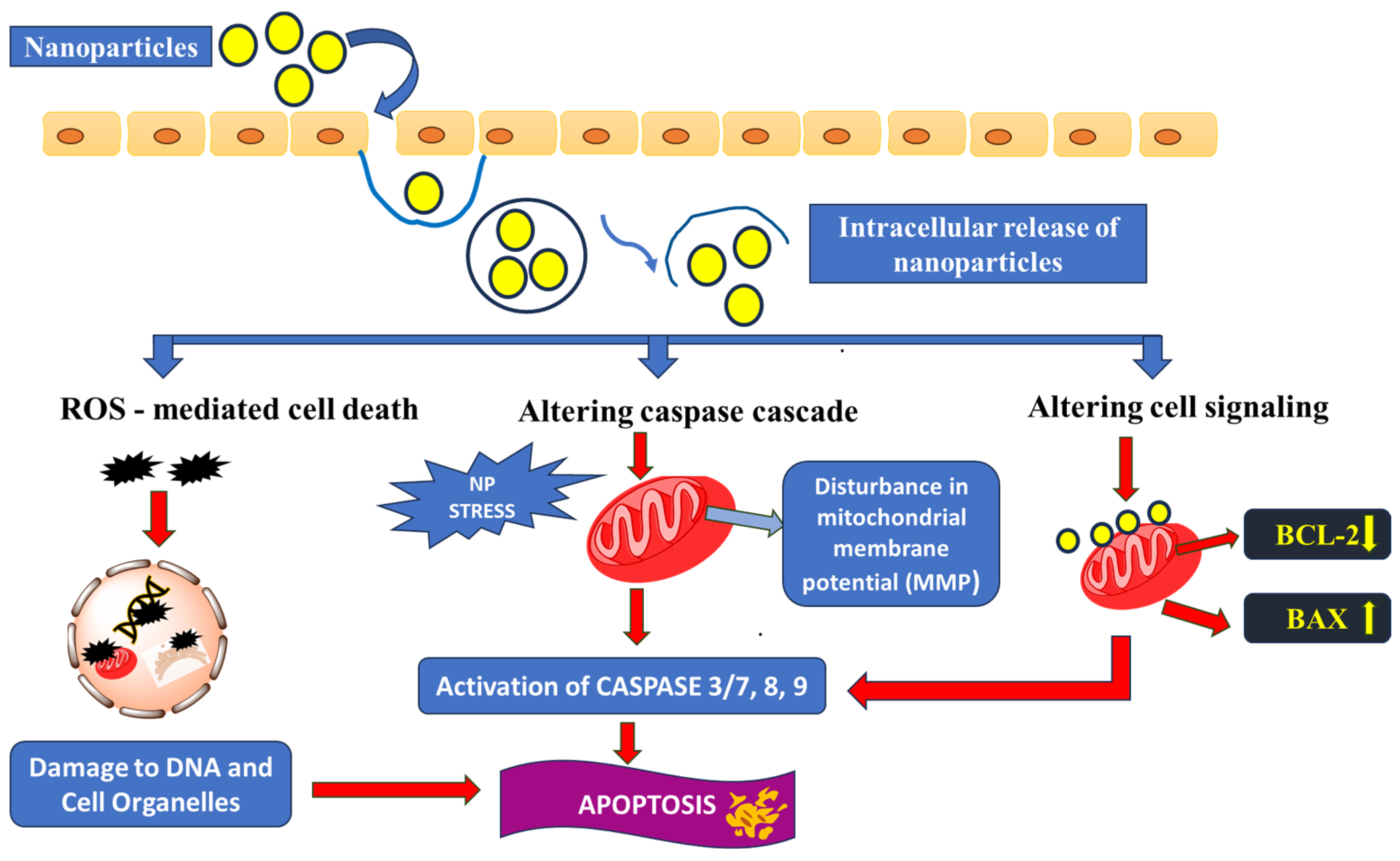
3. Major Apoptosis Mechanisms

Throughout our literature survey, we came across suggested pathways and mechanisms that induce cell death in cancer cells and how NPs are designed to take advantage of and manipulate them. Major apoptosis mechanisms triggered by nanoparticles have been shown in Figure 5.

3.1. Oxidative Stress Caused by Enhanced ROS Production

Metal oxide NPs and their derivatives have band gaps that can produce reactive oxygen species (ROS) within the cell. Although ROS levels are required for healthy functioning of the cell, excessive ROS levels can lead to lipid peroxidation and end up in cell death. ZnO, Au, Ag NPs, and many more function on this basis. Many studies reported here measure ROS levels during cytotoxicity assays to verify this mechanism. A recent study also explained the cytotoxicity of iron oxide NPs due to increased ROS expression. They synthesized ultra-small PEG-modified polydopamine NPs and chelated Fe(II) and Fe(III) ions to them. This led to ferroptosis in the cell, which is a phenomenon described by Dixon et al. in 2012 [43]. Ferroptosis is a type of programmed cell death dependent on iron and characterized by the accumulation of lipid peroxides, and is genetically and biochemically distinct from other forms of regulated cell death such as apoptosis. Lipophilic antioxidants and iron chelators can prevent ferroptosis cell death. The iron ions are also able to convert the H2O2 produced by band gap catalysis into the most harmful OH via the Fenton reaction. Photoexcited nanostructured TiO2 has been used as a nano-medicine for cancer diseases [44]. Herein, photo activated TiO2 induces the production of pairs of holes and electrons, respectively. These pairs of electrons and holes react with water and oxygen molecules and give reactive oxygen species (ROS) which destroy the cancerous cells and initiate the controlled growth of cells.

3.2. Caspase Cascade

ZnO NPs were known to cause cell death in gingival squamous cell carcinoma (GSCC) lines but the mechanism was not fully understood. A study by Wang et al. reported the role of caspases in inducing apoptosis [45]. When Ca9-22 and OECM-1 oral cancer cells are treated with ZnO NPs, superoxide production increases along with increased expression of caspases 8 and 9 which are initiator caspases. Caspases are cysteine acid proteases that regulate apoptosis. The cleavage of caspase-9 due to ZnO NP treatment leads to cleavage of caspase-3 (effect of caspase) as a result of the expression of cytochrome C. This in turn leads to the expression of poly-(ADP-ribose) polymerase (PARP) which causes cell death. Due to the expression and cleavage of downstream substrates, this process has been termed a cascade.

3.3. Disrupting Cell-Signalling Pathways

Cell signalling genes are important in cell functioning as well as proliferation. Some well-known targets in this regard are p53 signalling pathways, Bcl-2 genes, p70S6K signalling pathway, histone expression, etc. For instance, in bladder cancer, methylation of histone leads to epigenetic changes which lead to tumor and T24 cell proliferation and migration. Histone methyltransferase is responsible for promoting the methylation of histone. In a study, ZnO NPs have been used to treat bladder cancer cells and it is seen that EZH2 expression is greatly reduced and minimum amounts of ROS increase. This points to the fact that the suppression of EZH2 levels is responsible for controlling tumor growth [46]. Maspin expression increases when MCF-7 (breast) cell lines are treated with ZnO NPs. Maspin is a mammary serine protease inhibitor that is toxic to T-cancer cells [47].
Another aspect worth mentioning is the interplay of BCL-2 and BAX gene levels. BCL-2 is pro-survival while BAX is pro-apoptosis. ZnO NPs produced with R. rhaponticum waste caused cytotoxicity in MCF-7 cancer cells [48]. Herein, a comparative study has been done with MCF7 breast cancer and normal Human HFF and HDF cells. Herein, the apoptotic activities of ZnO NPs have been tested via the determination of the cell morphology, BCL2- BAX genes expression profile, and AO/PI-fluorescent cell staining. All cell lines were incubated for 24, 48 and 72 h, respectively. This study reveals that the ZnO NP dose (7.5 μg/mL, 15 μg/mL, 30 μg/mL) decreased gene expression of BCL-2 whereas gene expression of BAX increased, which is the cause of cell death. Thus, this effect works in synergy with ROS induction. Many of the NPs reported here show a similar increase in the BAX/BCL-2 ratio, which ultimately explains their cytotoxic properties.

4. Metal Oxide Nanoparticles for Drug Delivery

Employing nanoparticles for the delivery of cancer drugs is a fertile area of research. Low solubility, high toxicity, multi-drug resistance, poor selectivity, and low bioavailability often restrict the use of drugs in treatment or cause detrimental effects. Apart from cancer drug delivery, they are also used in the targeted delivery of anti-microbial and bactericidal compounds.
Drug targeting conventionally has been based on passive targeting. Uncontrolled multiplication of cancerous cells causes nanometre-range lesions leading to a ‘leaky’ tumor. This gives rise to the enhanced permeability and retention (EPR) effect, where either cancer drugs such as doxorubicin and cisplatin, or cytotoxic nanoparticles, accumulate at the ‘leak’ and cause damage. The enhanced permeability and retention (EPR) effect works as the foundation of anticancer nano-medicine and its design by using various drug formulations. Drug delivery based on the EPR effect is an effective strategy for most solid tumors [49]. The vascular mediators including prostaglandins, bradykinin, and nitric oxide play a vital role in facilitating and maintaining EPR effect dynamics. The advanced stage of cancers may induce activated blood coagulation cascades, which cause thrombus formation in tumor vasculature. The drug delivery, as well as the EPR effect, will be enhanced by restoring obstructed tumor blood flow and improving tumor vascular permeability through vascular mediators. Moreover, the efficiency of the EPR effect depends on the pathophysiological conditions of tumors, drug formulations, and other factors such as the tumor microenvironment. In the physiological location, research on magnetite nanoparticles coupled with lipids reveals a low selectivity for the targeted organ/tissue. However, accumulation in tumor tissues would result from passive targeting, which is facilitated by the ERP effect in tumors via fenestrated arteries (Figure 6) [50].
The microenvironment of tumors is mainly affected by blood flow in cancerous tissue which also plays a critical role in the effective EPR effect. However, this approach has many drawbacks principally that of damage to other cells as the drug/nanoparticles need to be in circulation for increased periods to allow sufficient concentration in the tumor, and also there is a lack of specificity. These significant failures and potential side effects call the effectiveness of EPR into question. Therefore, the renewed focus has been on targeted delivery. Ligands or antigens are used to bind to receptors that are typically present or over-expressed in a tumor environment. Biotin, folic acid, aptamers, antibodies, and peptides are common binding agents that are incorporated into the design for active targeting [51]. Physical phenomena such as electromagnetic radiation, pH changes, and magnetic fields are also used to induce active targeting.
Magnetic nanoparticles, such as oxides of iron, cobalt, and nickel have been exploited to enhance biomedical applications for decades as they can be controlled via a magnetic field and directed to the cancer site. Fe3O4 has been widely studied in this regard often associated with polymer or biomaterials to enhance its bioavailability and safety. Recently, Fe3O4 NPs were modified with chitosan (CS) and loaded with Imatinib as a kinase inhibitor. The system showed a pH-responsive drug release at pH 5.4 which is ideal as the tumor micro-environment is slightly acidic. As compared to pure Imatinib, the Fe3O4@CS/Imatinib had a 49% reduction in IC50 dose [52].
Curcumin is another proposed anti-cancer drug performing poorly on ADMET (Absorption, Distribution, Metabolism, Excretion, and Toxicity) parameters and pharmacokinetics/pharmacodynamics (PK/PD) standards, which determine the suitability of a drug and thus having a short half-life. As a result, improving the bioavailability of curcumin is a challenging task. A group used musk melon seeds to produce ZnO NPs which were then conjugated with dialdehyde cellulose to create a chitosan-based nanocomposite hydrogel for curcumin delivery. At pH 4.0, the hydrogel swelled and was loaded with a high loading efficiency of 89.68%. At pH 7.0, a slow sustained first-order release was seen. When tested on A431 skin carcinoma cells, the highest cytotoxicity was seen with the curcumin-loaded nanocomposite, which was greater than pure curcumin and demonstrated the vital function of targeted delivery. The IC50 for pure curcumin was 23 µg/mL whereas 16 µg/mL for the curcumin-loaded nanocomposite. Moreover, the biocompatibility of the nanocomposite was also better [53].
Alternatively, another study used casein, a protein found in milk as a capping agent during the synthesis of ZnO NPs, and loaded it with curcumin. The casein-ZnO-curcumin composite was also conjugated with folic acid to target folic receptors in cancer cell lines. In HeLa (cervical) cell lines, there was a 10% higher cytotoxicity when folic acid was incorporated. Further, drug release studies showed a pH-controlled release of curcumin at pH 5.0 in addition to a 15% increase in cytotoxicity. The increased cytotoxicity in comparison to naked ZnO NPs was attributed to the additive killing effects of both ZnO NPs and curcumin. The nanocomposite showed no hemolysis. Similar cytotoxic effects were seen in MCF-7 (breast), MDA-MB-231 (breast), and MG63 (bone) cancer cells. Ginsenoside Rh2, the active component of ginseng was loaded on hyaluronic acid (HA)-functionalized ZnO NPs. Ginsenosides in general suffer from poor solubility, poor bioavailability due to hydrophobic groups, and non-targeted cytotoxicity. However, the functionalized ZnO NPs showed impressive cytotoxicity against A549 (lung), HT29 (colon), and MCF-7 (breast) cancer cells [54].
Another way to improve the targeted action of nanoparticles is by incorporating them into composites. These nanocomposites often contain compounds such as folic acids which can bind to the tumor site and thus prevent collateral damage to healthy cells. Other metals such as Ag and Au, popularly used in traditional medicine, have shown remarkable therapeutic action via increasing intracellular oxidative stress against a variety of mutated cell lines and have been extensively studied.
Mesoporous silica NPs (SiO2) have been used in targeted drug delivery. Herein, various stimuli such as pH-sensitive, thermo-sensitive, light-sensitive, enzyme-sensitive, redox-sensitive, magnetic field-sensitive, and ultrasound-responsive drug delivery systems have been discussed [55]. Various targeted drug delivery systems based on different metal oxide nanoparticles are given in Table 2 [56,57,58,59,60,61,62,63,64,65,66].

5. Metal Oxide Nanoparticles for Cancer Diagnosis/Imaging

5.1. Magnetic Resonance Imaging (MRI)

The imaging of tumors and cancer cell proliferation is dynamic in tracking the growth of cancer and helps in changing the strategy of treatment in the early stage. In both cases, differentiating between healthy and cancerous cells is a challenge and thus both these applications are closely connected. Magnetic nanomaterials and composites are especially favored in this field due to their high sensitivity, high signal-to-noise ratio, low limit of detection, and reusability. Iron-based NPs are already widely studied and reviewed as MRI contrast agents. For synergistic therapy and multimodal imaging, He et al. offered a novel approach using silver core/AIE (aggregation-induced emission) shell nanoparticles [67]. Wang et al. reported using superparamagnetic Fe2O3 NPs coupled with anti-CD133 monoclonal antibodies which can bind to cell-marker antigen CD133 in vitro model of human brain glioblastoma cancer stem cells and be used for fluorescence and MRI imaging. This is especially significant as the blood-brain barrier is notoriously difficult to cross. Tiny and previously undetected lymph node metastases in prostate cancer patients can now be identified through a macrophage-specific magnetic resonance (MR) contrast agent. This has a significant clinical effect since it will result in a better accurate diagnosis that requires less intrusive testing. Thus, this will consequently lower morbidity and medical expenses. Ultra-small superparamagnetic iron oxide (USPIO) particles have been used in getting high-resolution MRI images. Herein, these USPIO have been used as a lymph node-specific contrast agent for nodal stages in prostate cancer. This study has been performed on 24 healthy volunteer men. When these USPIO particles are injected intravenously into the patients, they are transported by macrophages to only normal lymph node tissue as macrophage activity, but these particles are not transported in metastatic tissue. These particles change the magnetic properties and consequently signal intensity alters, which is detected by MRI. Therefore, normal functioning lymph nodes look black in MRI after 24–36 h administration of USPIO. However, in the metastatic node, the signal intensity does not change due to the absence of iron particles. Ferumoxytol (Fe3O4, Feraheme) is commonly used in contrast-enhanced magnetic resonance imaging (MRI) as a diagnostic tool. Ferumoxytol is a non-stoichiometric magnetite (superparamagnetic iron oxide) that is coated with polyglucose sorbitol carboxy-methyl ether. Ferumoxytol is the better alternative material as compared to the standard gadolinium-based contrast agents because it is more feasible in patients with impaired renal function. Ferumoxytol is available in the market of USA as the only intravascular MRI contrast agent.
Early diagnosis of cancer can vastly improve patient outcomes due to interventions in the primary stages. Peptide-targeted gadolinium oxide-based multifunctional NPs have been used for the diagnosis of prostate cancer using magnetic resonance imaging as well as fluorescent molecular imaging [68]. Herein, synthesized Gd2O3-FI-PEG-BBN NPs have uniformity in size and are spherically shaped with a 53.2 nm diameter. The longitudinal relaxivity (r1) of Gd2O3-FIPEG-BBN (r1 = 4.23 mM−1 s−1) matches with the clinically used gadolinium-diethylenetriamine pentaacetic acid (Gd-DTPA) which is known as Magnevist in the market. Herein, in vitro cellular, MRI is found receptor-specific for gastrin-releasing-peptide (GRP). Also, enhanced cellular uptake of the Gd2O3-FI-PEG-BBN is found to be in PC-3 tumor cells. Cellular MRI has been performed with composites of amino dextran-coated Fe3O4 NPs with graphene oxide (GO) by Chen et al. [69]. Herein, this nanocomposite of Fe3O4 has been used as a T2-weighted contrast agent. These nanocomposites of Fe3O4 show very low cytotoxicity and very good physiological stability in MRI diagnosis. Herein, HeLa cells are treated with this composite of Fe3O4 and their staining has been done with Prussian blue. The staining analysis demonstrates that the Fe3O4-GO nanocomposites are internalized in the HeLa cells very efficiently. Thangudu et al. effectively designed biocompatible FeCO3 nanoparticles as an MRI agent for in vivo lung tumors [70]. Guan et al. reported the synthesis of glycol-quantum dots (glycol-QDs) as an efficient tumor bio imaging agent [71]. These glycol-QDs can deeply penetrate tumors. Table 3 summarizes several metal oxide nanoparticles employed as a contrast agent in Magnetic Resonance Imaging (MRI) [72,73,74,75,76,77,78].

5.2. Computed Tomography (CT) Scan Imaging

Formerly a CT scan was known as computed axial tomography (CAT scan), an imaging technique in which X-rays are used as a source. It is a diagnostic tool for finding images of the body non-invasively. Hafnium oxide-PEG (HfO2-PEG) based nanocrystals have been designed and tested in vivo in a mouse model. The nanocrystals have been administered intravenously and accumulated in the 4T1 breast cancer tumor which has been imaged using a CT scan. The nanocrystals, therefore, provided targeted imaging preventing radiation damage to surrounding normal cells. Moreover, the nanocrystals have been excreted quickly and do not show unhealthy deposits in the heart, liver, and other vital organs without any side effects over a 90-day window [79]. Similarly, nanostructured cerium oxide with polyethylene glycol-α-maleimide-ω-n-hydroxide succinimide (NHS-PEG-Mal CeO2) bioconjugated with anti-HER2 has been used to detect abnormal concentrations of HER2 which are over-expressed in breast cancer patients. Blood serum samples have been tested and the electrochemical study has also been used to ascertain HER2 concentrations using the bioconjugate which is more selective and sensitive than most other HER2 nano-immunosensors [80].

6. Conclusions

We have given a brief outlook on the contemporary research exploiting metal oxide nanoparticles for anti-cancer applications. The area and the work of researchers therein, prove the diverse potential of the field. We have documented recent work whereby therapeutic metal oxide nanoparticles have shown promising cytotoxic activity toward various cell lines. The incorporation of green methods, phytochemical-mediated and bio-functionalized nanoparticle synthesis also opens avenues for reducing present challenges posed by the medical use of nanoparticles in terms of toxicity and excessive retention. Combined with other issues such as environmental impact, it becomes clear that toxicity must be an essential focus area during the design of new nanoparticle candidates.
The field of imaging already employs MONPs such as superparamagnetic iron oxide nanoparticles (SPIONs) for MRI-based imaging. Clinical trials have been done on younger patients and children having brain tumors for viewing the vessels of the brain. Herein, a comparative study has been done for getting MRI images using paramagnetic iron oxides as well as gadolinium as contrast agents. Initially, ferumoxytol has been used as an MRI contrast agent because of its efficacy in shortening T1 and T2 relaxation times. Preclinical and clinical studies on ferumoxytol have reported the overall safety and effectiveness of this novel contrast agent, which is used in MRI imaging. There is no evidence of allergic reactions after injection in the patients. A combinatorial approach whereby imaging and drug delivery or imaging or therapy are amalgamated and made more efficient will enrich real-time monitoring of the prognosis of the cancer. Immunosensors, such as those mentioned earlier, can be path-breaking in early identification and intervention and thus enhance patient outcomes. Small-molecule drug delivery is a well-established application for nanoparticles in cancer treatment research.
Finally, our summary of the prevalent apoptosis mechanisms gives a brief overview to help picture the various processes. We hope that it will prove beneficial to researchers and students beginning their exploration into this immensely vital and diverse area, contributing to treatments and strategies against cancer which was responsible for potentially one in six deaths worldwide.

Author Contributions

N.R.: writing—original draft; Y.K.: conceptualization, writing—original draft. S.Y.: reference editing; K.S.: supervision, review, and editing; D.M.: review and editing. All authors have read and agreed to the published version of the manuscript.

Funding

Sapna Yadav would like to express her great appreciation to the CSIR, New Delhi, for JRF (CSIR, File No. 08/700/(0004)/2019-EMR-1).

Acknowledgments

The authors would like to thank Principal, Miranda House, University of Delhi for providing a lab facility to carry out the research work. Yousuf Khan would like to thank his dearest friends for their constant encouragement, time, and support.

Conflicts of Interest

The authors declare no conflicts of interest.

Abbreviations

ADMET, absorption, distribution, metabolism, excretion and toxicity; CA125, cancer antigen 125; CS, chitosan; CT, computed tomography; Dox, doxorubicin; Endo-G, endonuclease-G, mitochondrial; EPR, enhanced permeability and retention; EZH2, enhancer of zeste homolog 2; GPX4, glutathione peroxidase 4; HA, hyaluronic acid; HER2, Human epidermal growth factor receptor 2; MONPs, metal oxide nanoparticles; MRI, magnetic resonance imaging; NPs, nanoparticles; PARP, poly-(ADP-ribose) polymerase; PEG, poly(ethylene) glycol; PK/PD, pharmacokinetics/pharmacodynamics; ROS, reactive oxygen species; SPIONs, superparamagnetic iron oxide nanoparticles.

References

  1. WHO. World Cancer Report: Cancer Research for Cancer Prevention; International Agency for Research on Cancer, WHO: Lyon, France, 2020. [Google Scholar]
  2. Pistritto, G.; Trisciuoglio, D.; Ceci, C.; Garufi, A.; D’Orazi, G. Apoptosis as anticancer mechanism: Function and dysfunction of its modulators and targeted therapeutic strategies. Aging 2016, 8, 603–619. [Google Scholar] [CrossRef] [PubMed]
  3. El-Boubbou, K.; Ali, R.; Al-Humaid, S.; Alhallaj, A.; Lemine, O.M.; Boudjelal, M.; AlKushi, A. Iron Oxide mesoporous magnetic nanostructures with high surface area for enhanced and selective drug delivery to metastatic cancer cells. Pharmaceutics 2021, 13, 553. [Google Scholar] [CrossRef] [PubMed]
  4. Anzai, Y.; Mclachlan, S.; Morris, M.; Saxton, R.; Lufkin, R.B. Dextran-coated superparamagnetic iron oxide, an MR contrast agent for assessing lymph nodes in the head and neck. Am. J. Neuroradiol. 1994, 15, 87–94. [Google Scholar] [PubMed]
  5. Verma, S.K.; Jha, E.; Panda, P.K.; Thirumurugan, A.; Parashar, S.K.S.; Patro, S.; Suar, M. Mechanistic Insight into Size-Dependent Enhanced Cytotoxicity of Industrial Antibacterial Titanium Oxide Nanoparticles on Colon Cells Because of Reactive Oxygen Species Quenching and Neutral Lipid Alteration. ACS Omega 2018, 3, 1244–1262. [Google Scholar] [CrossRef] [PubMed]
  6. Shwetha, U.R.; Latha, M.S.; Kumar, C.R.; Kiran, M.S.; Onkarappa, H.S.; Betageri, V.S. Potential antidiabetic and anticancer activity of copper oxide nanoparticles synthesised using Areca catechu leaf extract. Adv. Nat. Sci. Nanosci. Nanotechnol. 2021, 12, 025008. [Google Scholar] [CrossRef]
  7. Hesemans, E.; Saffarzadeh, N.; Maksoudian, C.; Izci, M.; Chu, T.; Rios Luci, C.; Wang, Y.; Naatz, H.; Thieme, S.; Richter, C.; et al. Cu-doped TiO2 nanoparticles improve local antitumor immune activation and optimize dendritic cell vaccine strategies. J. Nanobiotechnol. 2023, 21, 87. [Google Scholar] [CrossRef]
  8. Bai, S.; Yang, N.; Wang, X.; Gong, F.; Dong, Z.; Gong, Y.; Liu, Z.; Cheng, L. Ultrasmall iron-doped titanium oxide nanodots for enhanced sonodynamic and chemodynamic cancer therapy. ACS Nano 2020, 14, 15119–15130. [Google Scholar] [CrossRef]
  9. Hu, H.; Yu, L.; Ding, Z.; Ding, J.; Hu, Y.; Yin, Z. Chemo-immunotherapy for chemo-resistance and metastasis of triple-negative breast cancer by combination of iron-oxide nanoparticles and dual-targeting doxorubicin liposomes. Chin. Chem. Lett. 2023, 34, 108592. [Google Scholar] [CrossRef]
  10. Chao, Y.; Chen, G.; Liang, C.; Xu, J.; Dong, Z.; Han, X.; Wang, C.; Liu, Z. Iron nanoparticles for low-power local magnetic hyperthermia in combination with immune checkpoint blockade for systemic antitumor therapy. Nano Lett. 2019, 19, 4287–4296. [Google Scholar] [CrossRef]
  11. Chen, S.; Lv, Y.; Wang, Y.; Kong, D.; Xia, J.; Li, J.; Zhou, Q. Tumor acidic microenvironment-responsive promodulator iron oxide nanoparticles for photothermal-enhanced chemodynamic immunotherapy of cancer. ACS Biomater. Sci. Eng. 2023, 9, 773–783. [Google Scholar] [CrossRef]
  12. Mahanta, S.; Prathap, S.; Ban, D.K.; Paul, S. Protein functionalization of ZnO nanostructure exhibits selective and enhanced toxicity to breast cancer cells through oxidative stress-based cell death mechanism. J. Photochem. Photobiol. B Biol. 2017, 173, 376–388. [Google Scholar] [CrossRef] [PubMed]
  13. Park, T.; Amatya, R.; Min, K.A.; Shin, M.C. Liposomal iron oxide nanoparticles loaded with doxorubicin for combined chemo-photothermal cancer therapy. Pharmaceutics 2023, 15, 292. [Google Scholar] [CrossRef] [PubMed]
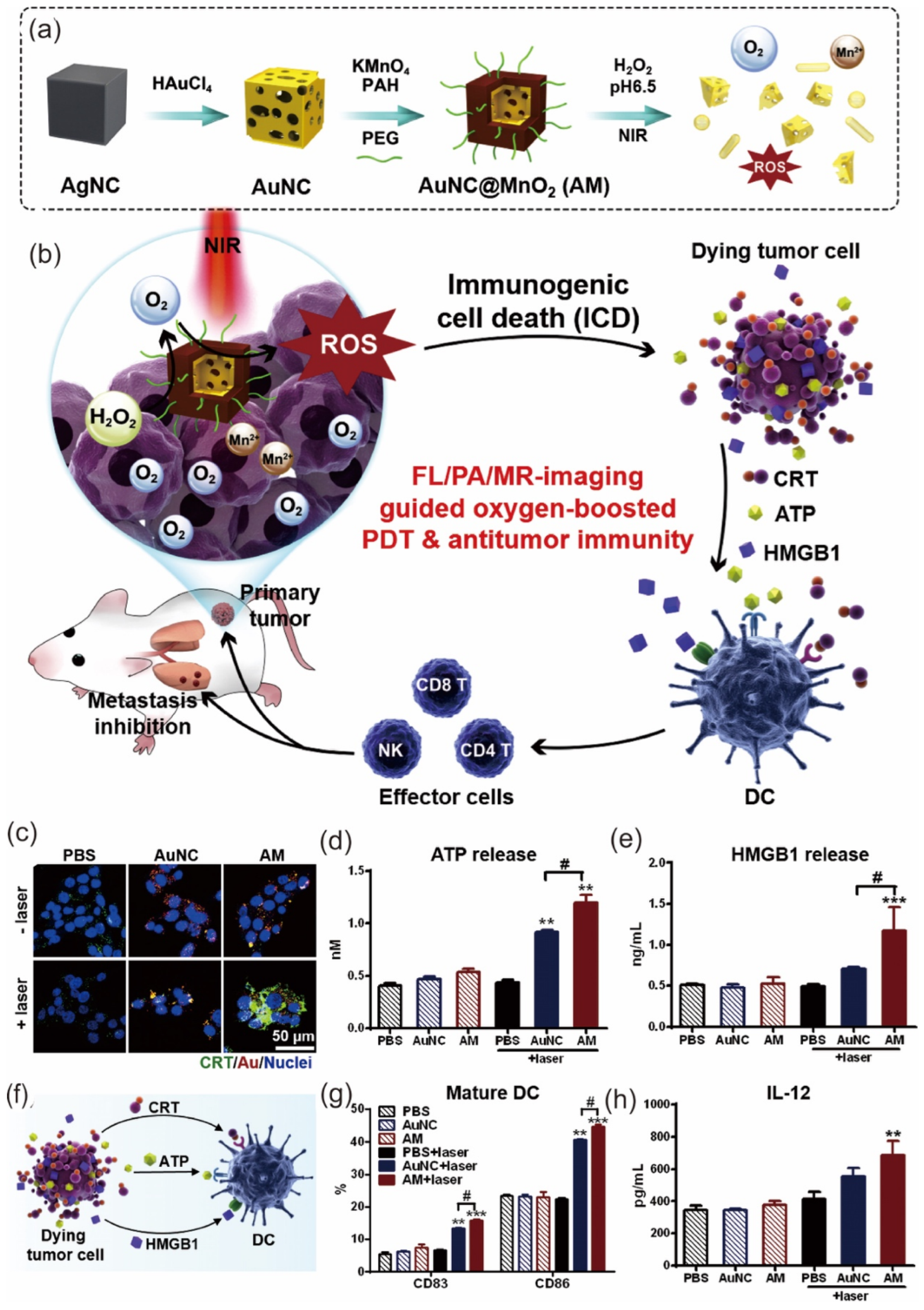
  14. Liang, R.; Liu, L.; He, H.; Chen, Z.; Han, Z.; Luo, Z.; Wu, Z.; Zheng, M.; Ma, Y.; Cai, L. Oxygen-boosted immunogenic photodynamic therapy with gold nanocages@ manganese dioxide to inhibit tumor growth and metastases. Biomaterials 2018, 177, 149–160. [Google Scholar] [CrossRef]
  15. Gong, F.; Chen, M.; Yang, N.; Dong, Z.; Tian, L.; Hao, Y.; Zhuo, M.; Liu, Z.; Chen, Q.; Cheng, L. Bimetallic oxide FeWOX nanosheets as multifunctional cascade bioreactors for tumor microenvironment-modulation and enhanced multimodal cancer therapy. Adv. Funct. Mater. 2020, 30, 2002753. [Google Scholar] [CrossRef]
  16. Koo, S.; Park, O.K.; Kim, J.; Han, S.I.; Yoo, T.Y.; Lee, N.; Kim, Y.G.; Kim, H.; Lim, C.; Bae, J.S.; et al. Enhanced chemodynamic therapy by Cu–Fe peroxide nanoparticles: Tumor microenvironment-mediated synergistic Fenton reaction. ACS Nano 2022, 16, 2535–2545. [Google Scholar] [CrossRef]
  17. Ma, X.; Lee, C.; Zhang, T.; Cai, J.; Wang, H.; Jiang, F.; Wu, Z.; Xie, J.; Jiang, G.; Li, Z. Image-guided selection of Gd@ C-dots as sensitizers to improve radiotherapy of non-small cell lung cancer. J. Nanobiotechnol. 2021, 19, 284. [Google Scholar] [CrossRef]
  18. Gowdhami, B.; Jaabir, M.; Archunan, G.; Suganthy, N. Anticancer potential of zinc oxide nanoparticles against cervical carcinoma cells synthesized via biogenic route using aqueous extract of Gracilaria edulis. Mater. Sci. Eng. C 2019, 103, 109840. [Google Scholar]
  19. Rahimi Kalateh Shah Mohammad, G.; Seyedi, S.M.R.; Karimi, E.; Homayouni-Tabrizi, M. The cytotoxic properties of zinc oxide nanoparticles on the rat liver and spleen, and its anticancer impacts on human liver cancer cell lines. J. Biochem. Mol. Toxicol. 2019, 33, e22324. [Google Scholar] [CrossRef]
  20. Pugazhendhi, A.; Prabhu, R.; Muruganantham, K.; Shanmuganathan, R.; Natarajan, S. Anticancer, antimicrobial and photocatalytic activities of green synthesized magnesium oxide nanoparticles (MgONPs) using aqueous extract of Sargassum wightii. J. Photochem. Photobiol. B Biol. 2019, 190, 86–97. [Google Scholar] [CrossRef]
  21. Thamer, N.A.; Barakat, N.T. In Cytotoxic activity of green synthesis copper oxide nanoparticles using cordia myxa L. aqueous extract on some breast cancer cell lines. J. Phys. Conf. Ser. 2019, 1294, 062104. [Google Scholar] [CrossRef]
  22. Manimaran, K. Synthesis and characterization of Pleurotus citrinopileatus extract–mediated iron oxide (FeO) nanoparticles and its antibacterial and anticancer activity. Biomass Conv. Bioref. 2024, 14, 15011–15020. [Google Scholar] [CrossRef]
  23. Mohsin, M.H.; Khashan, K.S.; Sulaiman, G.M.; Mohammed, H.A.; Qureshi, K.A.; Aspatwar, A. A novel facile synthesis of metal nitride@metal oxide (BN/Gd2O3) nanocomposite and their antibacterial and anticancer activities. Sci. Rep. 2023, 13, 22749. [Google Scholar] [CrossRef] [PubMed]
  24. Hadi, A.J.; Nayef, U.M.; Jabir, M.S.; Mutlak, F.A. Titanium dioxide nanoparticles prepared via laser ablation: Evaluation of their antibacterial and anticancer activity. Surf. Rev. Lett. 2023, 30, 2350066. [Google Scholar] [CrossRef]
  25. Phalake, S.S.; Somvanshi, S.B.; Tofail, S.A.; Thorat, N.D.; Khot, V.M. Functionalized manganese iron oxide nanoparticles: A dual potential magneto-chemotherapeutic cargo in a 3D breast cancer model. Nanoscale 2023, 15, 15686–15699. [Google Scholar] [CrossRef] [PubMed]
  26. Hasani, M.; Ghanbarzadeh, S.; Hajiabadi, H.; Mortezazadeh, T.; Yoosefian, M.; Akbari Javar, H. In vitro and in silico characteristics of doxorubicin-loaded four polymeric-based polysaccharides-modified super paramagnetic iron oxide nanoparticles for cancer chemotherapy and magnetic resonance imaging. Int. J. Polym. Mater. Polym. Biomater. 2024, 73, 117–130. [Google Scholar] [CrossRef]
  27. Thevenot, P.; Cho, J.; Wavhal, D.; Timmons, R.B.; Tang, L. Surface chemistry influences cancer killing effect of TiO2 nanoparticles. Nanomed. Nanotechnol. Biol. Med. 2008, 4, 226–236. [Google Scholar] [CrossRef]
  28. Lu, S.; Mi, Z.; Liu, P.; Ding, J.; Ma, Y.; Yang, J.; Rong, P.; Zhou, W. Repolarizing neutrophils via MnO2 nanoparticle-activated STING pathway enhances Salmonella-mediated tumor immunotherapy. J. Nanobiotechnol. 2024, 22, 443. [Google Scholar] [CrossRef]
  29. Du, C.; Zhou, M.; Jia, F.; Ruan, L.; Lu, H.; Zhang, J.; Zhu, B.; Liu, X.; Chen, J.; Chai, Z.; et al. D-arginine-loaded metal-organic frameworks nanoparticles sensitize osteosarcoma to radiotherapy. Biomaterials 2021, 269, 120642. [Google Scholar] [CrossRef]
  30. Wang, X.; Li, B.; Li, R.; Yang, Y.; Zhang, H.; Tian, B.; Cui, L.; Weng, H.; Wei, F. Anti-CD133 monoclonal antibody conjugated immunomagnetic nanosensor for molecular imaging of targeted cancer stem cells. Sens. Actuators B Chem. 2018, 255, 3447–3457. [Google Scholar] [CrossRef]
  31. Barentsz, J.O.; Fütterer, J.J.; Takahashi, S. Use of ultrasmall superparamagnetic iron oxide in lymph node MR imaging in prostate cancer patients. Eur. J. Radiol. 2007, 63, 369–372. [Google Scholar] [CrossRef]
  32. Bashir, M.R.; Bhatti, L.; Marin, D.; Nelson, R.C. Emerging applications for ferumoxytol as a contrast agent in MRI. J. Magn. Reson. Imaging 2015, 41, 884–898. [Google Scholar] [CrossRef] [PubMed]
  33. Tanino, R.; Amano, Y.; Tong, X.; Sun, R.; Tsubata, Y.; Harada, M.; Fujita, Y.; Isobe, T. Anticancer Activity of ZnO Nanoparticles against Human Small-Cell Lung Cancer in an Orthotopic Mouse Model. Mol. Cancer Ther. 2020, 19, 502–512. [Google Scholar] [CrossRef] [PubMed]
  34. Yang, N.; Qiu, F.; Zhu, F.; Qi, L. Therapeutic Potential of Zinc Oxide-Loaded Syringic Acid Against in vitro and in vivo Model of Lung Cancer. Int. J. Nanomed. 2020, 15, 8249–8260. [Google Scholar] [CrossRef] [PubMed]
  35. Chabattula, S.C.; Gupta, P.K.; Tripathi, S.K.; Gahtori, R.; Padhi, P.; Mahapatra, S.; Biswal, B.K.; Singh, S.K.; Dua, K.; Ruokolainen, J.; et al. Anticancer therapeutic efficacy of biogenic Am-ZnO nanoparticles on 2D and 3D tumor models. Mater. Today Chem. 2021, 22, 100618. [Google Scholar] [CrossRef]
  36. Latha, T.S.; Reddy, M.C.; Muthukonda, S.V.; Srikanth, V.V.S.S.; Lomada, D. In vitro and in vivo evaluation of anti-cancer activity: Shape-dependent properties of TiO2 nanostructures. Mater. Sci. Eng. C 2017, 78, 969–977. [Google Scholar] [CrossRef]
  37. Chen, M.; Xiong, F.; Ma, L.; Yao, H.; Wang, Q.; Wen, L.; Wang, Q.; Gu, N.; Chen, S. Inhibitory effect of magnetic Fe3O4 nanoparticles coloaded with homoharringtonine on human leukemia cells in vivo and in vitro. Int. J. Nanomed. 2016, 11, 4413–4422. [Google Scholar] [CrossRef]
  38. Zhao, F.; Wang, C.; Yang, Q.; Han, S.; Hu, Q.; Fu, Z. Titanium dioxide nanoparticle stimulating pro-inflammatory responses in vitro and in vivo for inhibited cancer metastasis. Life Sci. 2018, 202, 44–51. [Google Scholar] [CrossRef]
  39. Ruiz, A.L.; Garcia, C.B.; Gallón, S.N.; Webster, T.J. Novel Silver-Platinum Nanoparticles for Anticancer and Antimicrobial Applications. Int. J. Nanomed. 2020, 15, 169–179. [Google Scholar] [CrossRef]
  40. Kuppusamy, P.; Ichwan, S.J.A.; Al-Zikri, P.N.H.; Suriyah, W.H.; Soundharrajan, I.; Govindan, N.; Maniam, G.P.; Yusoff, M.M. In Vitro Anticancer Activity of Au, Ag Nanoparticles Synthesized Using Commelina nudiflora L. Aqueous Extract Against HCT-116 Colon Cancer Cells. Biol. Trace Elem. Res. 2016, 173, 297–305. [Google Scholar] [CrossRef]
  41. Markovic, Z.M.; Harhaji-Trajkovic, L.M.; Todorovic-Markovic, B.M.; Kepic, D.P.; Arsikin, K.M.; Jovanovic, S.P.; Pantovic, A.C.; Dramicanin, M.D.; Trajkovic, V.S. In vitro comparison of the photothermal anticancer activity of graphene nanoparticles and carbon nanotubes. Biomaterials 2011, 32, 1121–1129. [Google Scholar] [CrossRef]
  42. Shao, W.; Paul, A.; Zhao, B.; Lee, C.; Rodes, L.; Prakash, S. Carbon nanotube lipid drug approach for targeted delivery of a chemotherapy drug in a human breast cancer xenograft animal model. Biomaterials 2013, 34, 10109–10119. [Google Scholar] [CrossRef] [PubMed]
  43. Dixon, S.J.; Lemberg, K.M.; Lamprecht, M.R.; Skouta, R.; Zaitsev, E.M.; Gleason, C.E.; Patel, D.N.; Bauer, A.J.; Cantley, A.M.; Yang, W.S. Ferroptosis: An iron-dependent form of nonapoptotic cell death. Cell 2012, 149, 1060–1072. [Google Scholar] [CrossRef] [PubMed]
  44. Lagopati, N.; Evangelou, K.; Falaras, P.; Tsilibary, E.P.C.; Vasileiou, P.V.; Havaki, S.; Angelopoulou, A.; Pavlatou, E.A.; Gorgoulis, V.G. Nanomedicine: Photo-activated nanostructured titanium dioxide, as a promising anticancer agent. Pharmacol. Ther. 2021, 222, 107795. [Google Scholar] [CrossRef] [PubMed]
  45. Wang, S.W.; Lee, C.H.; Lin, M.S.; Chi, C.W.; Chen, Y.J.; Wang, G.S.; Liao, K.W.; Chiu, L.P.; Wu, S.H.; Huang, D.M.; et al. ZnO nanoparticles induced caspase-dependent apoptosis in gingival squamous cell carcinoma through mitochondrial dysfunction and p70S6K signalling pathway. Int. J. Mol. Sci. 2020, 21, 1612. [Google Scholar] [CrossRef]
  46. Zhang, T.; Du, E.; Liu, Y.; Cheng, J.; Zhang, Z.; Xu, Y.; Qi, S.; Chen, Y. Anticancer effects of zinc oxide nanoparticles through altering the methylation status of histone on bladder cancer cells. Int. J. Nanomed. 2020, 15, 1457–1468. [Google Scholar] [CrossRef]
  47. Khorsandi, L.; Farasat, M. Zinc oxide nanoparticles enhance expression of maspin in human breast cancer cells. Environ. Sci. Pollut. Res. 2020, 27, 38300–38310. [Google Scholar] [CrossRef]
  48. Salari, S.; Neamati, A.; Tabrizi, M.H.; Seyedi, S.M.R. Green-synthesized Zinc oxide nanoparticle, an efficient safe anticancer compound for human breast MCF7 cancer cells. Appl. Organomet. Chem. 2020, 34, e5417. [Google Scholar] [CrossRef]
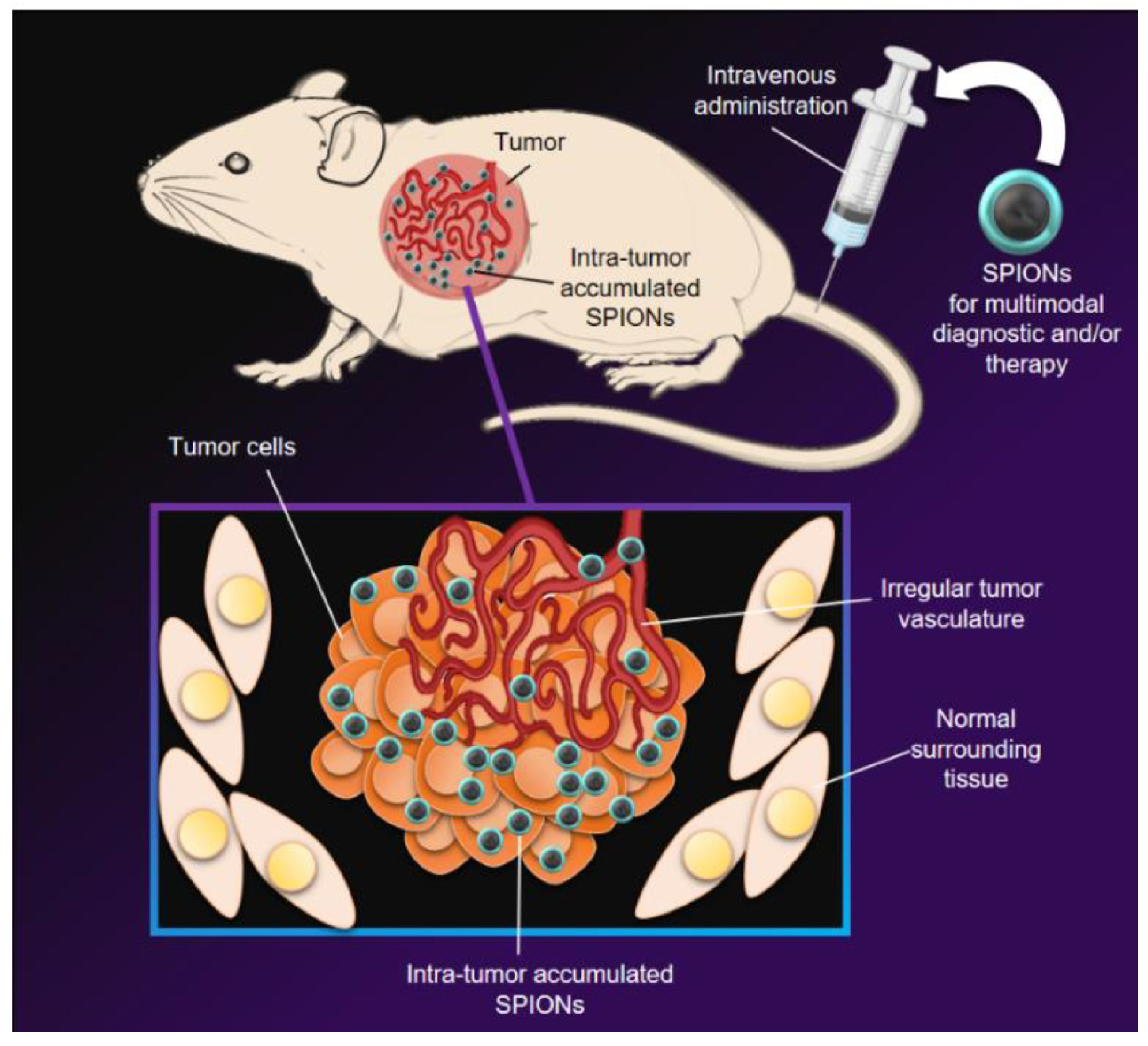
  49. Islam, R.; Maeda, H.; Fang, J. Factors affecting the dynamics and heterogeneity of the EPR effect: Pathophysiological and pathoanatomic features, drug formulations and physicochemical factors. Expert Opin. Drug Deliv. 2022, 19, 199–212. [Google Scholar] [CrossRef]
  50. Schneider, M.G.M.; Martín, M.J.; Otarola, J.; Vakarelska, E.; Simeonov, V.; Lassalle, V.; Nedyalkova, M. Biomedical applications of iron oxide nanoparticles: Current insights progress and perspectives. Pharmaceutics 2022, 14, 204. [Google Scholar] [CrossRef]
  51. Mu, Q.; Yan, B. Nanoparticles in Cancer Therapy-Novel Concepts, Mechanisms, and Applications. Front. Pharmacol. 2019, 9, 1552. [Google Scholar] [CrossRef]
  52. Karimi Ghezeli, Z.; Hekmati, M.; Veisi, H. Synthesis of Imatinib-loaded chitosan-modified magnetic nanoparticles as an anti-cancer agent for pH responsive targeted drug delivery. Appl. Organomet. Chem. 2019, 33, e4833. [Google Scholar] [CrossRef]
  53. George, D.; Maheswari, P.U.; Sheriffa Begum, K.M.M.; Arthanareeswaran, G. Biomass-derived dialdehyde cellulose cross-linked chitosan-based nanocomposite hydrogel with phytosynthesized zinc oxide nanoparticles for enhanced curcumin delivery and bioactivity. J. Agric. Food Chem. 2019, 67, 10880–10890. [Google Scholar] [CrossRef] [PubMed]
  54. Kim, Y.J.; Perumalsamy, H.; Castro-Aceituno, V.; Kim, D.; Markus, J.; Lee, S.; Kim, S.; Liu, Y.; Yang, D.C. Photoluminescent and self-assembled hyaluronic acid-zinc oxide-ginsenoside Rh2 nanoparticles and their potential caspase-9 apoptotic mechanism towards cancer cell lines. Int. J. Nanomed. 2019, 14, 8195. [Google Scholar] [CrossRef] [PubMed]
  55. Gulzar, A.; Gai, S.; Yang, P.; Li, C.; Ansari, M.B.; Lin, J. Stimuli responsive drug delivery application of polymer and silica in biomedicine. J. Mater. Chem. B 2015, 3, 8599–8622. [Google Scholar] [CrossRef] [PubMed]
  56. Zadeh, F.A.; Jasim, S.A.; Atakhanova, N.E.; Majdi, H.S.; Jawad, M.A.; Hasan, M.K.; Borhani, F.; Khatami, M. Drug delivery and anticancer activity of biosynthesised mesoporous Fe2O3 nanoparticles. IET Nanobiotechnol. 2022, 16, 85–91. [Google Scholar] [CrossRef]
  57. Wang, J.; Zhu, F.; Li, K.; Xu, J.; Li, P.; Fan, Y. pH-responsive mesoporous Fe2O3-Au nanomedicine delivery system with magnetic targeting for cancer therapy. Med. Nov. Technol. Devices 2022, 15, 100127. [Google Scholar] [CrossRef]
  58. Al-Ajmi, M.F.; Hussain, A.; Ahmed, F. Novel synthesis of ZnO nanoparticles and their enhanced anticancer activity: Role of ZnO as a drug carrier. Ceram. Int. 2016, 42, 4462–4469. [Google Scholar] [CrossRef]
  59. Cai, X.; Luo, Y.; Zhang, W.; Du, D.; Lin, Y. pH-Sensitive ZnO Quantum Dots–Doxorubicin Nanoparticles for Lung Cancer Targeted Drug Delivery. ACS Appl. Mater. Interfaces 2016, 8, 22442–22450. [Google Scholar] [CrossRef]
  60. Marcu, A.; Pop, S.; Dumitrache, F.; Mocanu, M.; Niculite, C.M.; Gherghiceanu, M.; Lungu, C.P.; Fleaca, C.; Ianchis, R.; Barbut, A.; et al. Magnetic iron oxide nanoparticles as drug delivery system in breast cancer. Appl. Surf. Sci. 2013, 281, 60–65. [Google Scholar] [CrossRef]
  61. Han, J.; Jang, E.-K.; Ki, M.-R.; Son, R.G.; Kim, S.; Choe, Y.; Pack, S.P.; Chung, S. pH-responsive phototherapeutic poly(acrylic acid)-calcium phosphate passivated TiO2 nanoparticle-based drug delivery system for cancer treatment applications. J. Ind. Eng. Chem. 2022, 112, 258–270. [Google Scholar] [CrossRef]
  62. Fadeel, D.A.A.; Hanafy, M.S.; Kelany, N.A.; Elywa, M.A. Novel greenly synthesized titanium dioxide nanoparticles compared to liposomes in drug delivery: In vivo investigation on Ehrlich solid tumor model. Heliyon 2021, 7, e07370. [Google Scholar] [CrossRef] [PubMed]
  63. Singh, S.; Pal, K. Exploration of polydopamine capped bimetallic oxide (CuO-NiO) nanoparticles inspired by mussels for enhanced and targeted paclitaxel delivery for synergistic breast cancer therapy. Appl. Surf. Sci. 2023, 626, 157266. [Google Scholar] [CrossRef]
  64. Binu, N.M.; Prema, D.; Prakash, J.; Balagangadharan, K.; Balashanmugam, P.; Selvamurugan, N.; Venkatasubbu, G.D. Folic acid decorated pH sensitive polydopamine coated honeycomb structured nickel oxide nanoparticles for targeted delivery of quercetin to triple negative breast cancer cells. Colloids Surf. A Physicochem. Eng. Asp. 2021, 630, 127609. [Google Scholar] [CrossRef]
  65. Sudakaran, S.V.; Venugopal, J.R.; Vijayakumar, G.P.; Abisegapriyan, S.; Grace, A.N.; Ramakrishna, S. Sequel of MgO nanoparticles in PLACL nanofibers for anti-cancer therapy in synergy with curcumin/β-cyclodextrin. Mater. Sci. Eng. C 2017, 71, 620–628. [Google Scholar] [CrossRef] [PubMed]
  66. Jaisankar, E.; Azarudeen, R.S.; Thirumarimurugan, M. A Study on the Effect of Nanoscale MgO and Hydrogen Bonding in Nanofiber Mats for the Controlled Drug Release along with In Vitro Breast Cancer Cell Line and Antimicrobial Studies. ACS Appl. Bio Mater. 2022, 5, 4327–4341. [Google Scholar] [CrossRef] [PubMed]
  67. He, X.; Peng, C.; Qiang, S.; Xiong, L.H.; Zhao, Z.; Wang, Z.; Kwok, R.T.; Lam, J.W.; Ma, N.; Tang, B.Z. Less is more: Silver-AIE core@ shell nanoparticles for multimodality cancer imaging and synergistic therapy. Biomaterials 2020, 238, 119834. [Google Scholar] [CrossRef]
  68. Cui, D.; Lu, X.; Yan, C.; Liu, X.; Hou, M.; Xia, Q.; Xu, Y.; Liu, R. Gastrin-releasing peptide receptor-targeted gadolinium oxide-based multifunctional nanoparticles for dual magnetic resonance/fluorescent molecular imaging of prostate cancer. Int. J. Nanomed. 2017, 12, 6787–6797. [Google Scholar] [CrossRef]
  69. Chen, W.; Yi, P.; Zhang, Y.; Zhang, L.; Deng, Z.; Zhang, Z. Composites of amino dextran-coated Fe3O4 nanoparticles and graphene oxide for cellular magnetic resonance imaging. ACS Appl. Mater. Interfaces 2011, 3, 4085–4091. [Google Scholar] [CrossRef]
  70. Thangudu, S.; Yu, C.C.; Lee, C.L.; Liao, M.C.; Su, C.H. Magnetic, biocompatible FeCO3 nanoparticles for T2-weighted magnetic resonance imaging of in vivo lung tumors. J. Nanobiotechnol. 2022, 20, 157. [Google Scholar] [CrossRef]
  71. Guan, X.; Zhang, L.; Lai, S.; Zhang, J.; Wei, J.; Wang, K.; Zhang, W.; Li, C.; Tong, J.; Lei, Z. Green synthesis of glyco-CuInS2 QDs with visible/NIR dual emission for 3D multicellular tumor spheroid and in vivo imaging. J. Nanobiotechnol. 2023, 21, 118. [Google Scholar] [CrossRef]
  72. Barick, K.C.; Singh, S.; Bahadur, D.; Lawande, M.A.; Patkar, D.P.; Hassan, P.A. Carboxyl decorated Fe3O4 nanoparticles for MRI diagnosis and localized hyperthermia. J. Colloid Interface Sci. 2014, 418, 120–125. [Google Scholar] [CrossRef] [PubMed]
  73. Hong, R.Y.; Feng, B.; Chen, L.L.; Liu, G.H.; Li, H.Z.; Zheng, Y.; Wei, D.G. Synthesis, characterization and MRI application of dextran-coated Fe3O4 magnetic nanoparticles. Biochem. Eng. J. 2008, 42, 290–300. [Google Scholar] [CrossRef]
  74. Zheng, Y.; Zhang, H.; Hu, Y.; Bai, L.; Xue, J. MnO nanoparticles with potential application in magnetic resonance imaging and drug delivery for myocardial infarction. Int. J. Nanomed. 2018, 13, 6177–6188. [Google Scholar] [CrossRef] [PubMed]
  75. Sakai, N.; Zhu, L.; Kurokawa, A.; Takeuchi, H.; Yano, S.; Yanoh, T.; Wada, N.; Taira, S.; Hosokai, Y.; Usui, A.; et al. Synthesis of Gd2O3 nanoparticles for MRI contrast agents. J. Phys. Conf. Ser. 2012, 352, 012008. [Google Scholar] [CrossRef]
  76. Yuan, M.; Xu, S.; Zhang, Q.; Zhao, B.; Feng, B.; Ji, K.; Yu, L.; Chen, W.; Hou, M.; Xu, Y.; et al. Bicompatible porous Co3O4 nanoplates with intrinsic tumor metastasis inhibition for multimodal imaging and DNA damage–mediated tumor synergetic photothermal/photodynamic therapy. Chem. Eng. J. 2020, 394, 124874. [Google Scholar] [CrossRef]
  77. Gao, L.; Meng, Y.; Luo, X.; Chen, J.; Wang, X. ZnO Nanoparticles Induced MRI Alterations to the Rat Olfactory Epithelium and Olfactory Bulb after Intranasal Instillation. Toxics 2024, 12, 724. [Google Scholar] [CrossRef]
  78. Babayevska, N.; Florczak, P.; Wozniak-Budych, M.; Jarek, M.; Nowaczyk, G.; Zalewski, T.; Jurga, S. Functionalized multimodal ZnO@Gd2O3 nanosystems to use as perspective contrast agent for MRI. Appl. Surf. Sci. 2017, 404, 129–137. [Google Scholar] [CrossRef]
  79. Li, Y.; Qi, Y.; Zhang, H.; Xia, Z.; Xie, T.; Li, W.; Zhong, D.; Zhu, H.; Zhou, M. Gram-scale synthesis of highly biocompatible and intravenous injectable hafnium oxide nanocrystal with enhanced radiotherapy efficacy for cancer theranostic. Biomaterials 2020, 226, 119538. [Google Scholar] [CrossRef]
  80. Hartati, Y.W.; Letelay, L.K.; Gaffar, S.; Wyantuti, S.; Bahti, H.H. Cerium oxide-monoclonal antibody bioconjugate for electrochemical immunosensing of HER2 as a breast cancer biomarker. Sens. Bio-Sens. Res. 2020, 27, 100316. [Google Scholar] [CrossRef]
Figure 1. Six pathological hallmarks of cancer.
Jnt 05 00015 g001
Figure 2. Oncological applications of metal-oxide nanoparticles.
Jnt 05 00015 g002
Figure 3. Schematic representation of Lipo-IONP/DOX for combined chemo-photothermal cancer therapy [13] (open access).
Jnt 05 00015 g003
Figure 4. Represented the working principle of AuNCs@MnO2 (AM) nanomaterial for ICD (immunogenic cell death) (a) synthesis procedure for AM and generation of O2 (b) Therapeutic approach of AM for inducing ICD by stimulating more dead tumor cells (ce) Identification of different ICD signal molecules during AM and laser treatments (c) fluorescence microscopy images showing CRT expression in 4T1 cells (d,e) liberated ATP and HMGB1 in the supernatant (f) diagrammatic illustration of ICD-induced DC activation (g) CD83 and CD86 expression after DC maturation (h) liberated IL-12 in the culture supernatant. Asterisks (*) denote statistically significant differences between PBS and other treatments. ** p < 0.01, *** p < 0.001. # p < 0.05 (n = 5) indicates that the differences between the two groups are statistically significant. Reproduced with permission from [14], Copyright 2018, Elsevier.
Jnt 05 00015 g004
Figure 5. Major apoptosis mechanisms triggered by nanoparticles.
Jnt 05 00015 g005
Figure 6. SPIONs are administered intravenously (IV) for multimodal diagnostic and/or therapeutic uses [50] (open access).
Jnt 05 00015 g006
Table 1. The comparison of metal oxide nanoparticles, metal nanoparticles, and carbon-based nanomaterials.
NanoparticlesCellsSize of NpsConcentration of NpsExposure DurationResultsReference
ZnON417, H82, H187 Human small-cell Lung cancer20 nm10 μg/mL120 minLow viability, even in cells orthotopically grafted onto mouse models[33]
ZnO-Loaded Syringic AcidA549 cells120 nm12.5 μM24 hThe ZnO-Loaded Syringic Acid indued ROS have induced the cell death in A549 cancer cells[34]
Annona muricata-ZnOA549 and MOLT4 Cells80 nm0–500 μg/mL24 hAm-ZnO treated cancer cells underwent programmed cell death with depolarization in their MMP.[35]
TiO2MDAMB231 cells140 nm100 μg/mL72 hTiO2 nanostructures inhibited the migration and colony formation of breast cancer MDAMB231 cells.[36]
Fe3O4 nanoparticles coloaded with homoharringtonineK562, HL-60, SHI-1, and NB4 cells-1.875 μg/mL24 hFe3O4 nanoparticles coloaded with homoharringtonine had cooperative effect in suppression of tumor cell growth[37]
TiO24T1 cells (Mice)21 nm1 μg/mL21 daysPeritoneal macrophage exposed to P-25 TiO2 NPs displayed activated M1 macrophage response[38]
AgPtDetroit 551-CCL-110, #A375 and U 87 cells~42 nm10–250 μg/mL1–3 daysAgPt nanoparticles demonstrated a remarkable and statistically significant ability to reduce the viability of cancer cells[39]
AuHCT-116 cells50 nm200 μg/mL1 htarget the abnormal growth of HCT-116 colon cancer cells[40]
AgHCT-116 cells24–80 nm100 μg/mL1 htarget the abnormal growth of HCT-116 colon cancer cells[40]
GrapheneU251 human glioma cells50 nm,2.5–10 mg/mL24 hgraphene nanoparticles performed significantly better than CNT in inducing photothermal death of U251 cells[41]
Carbon nanotubesU251 human glioma cells60 nm,2.5–10 mg/mL24 hgraphene nanoparticles performed significantly better than CNT in inducing photothermal death of U251 cells[41]
Carbon nanotubeMCF-7 breast cancer cells1.5 nm in diameter and 200 nm in length10 mg/ml48 hSWNT-drug showed target specificity in vitro[42]
Table 2. Various targeted drug delivery system based on different metal oxide nanoparticles.
NanoparticlesSynthesis MethodCancer CellsDrug DeliveredReference
Fe2O3Biosynthesis methodMCF7 cancer cellsDoxorubicin[56]
Fe2O3-Au-A549 Human lung cancer cellsDoxorubicin[57]
ZnOOrganic precursor methodMCF7 cancer cells5-Fluorouracil[58]
ZnO-CD44 cancer cellsDoxorubicin[59]
Fe3O4Laser pyrolysisMCF7 cancer cellsViolamycine B1[60]
Poly(acrylic acid)-calcium phosphate passivated TiO2Hydrothermal synthesisMCF7 cancer cellsDoxorubicin[61]
TiO2Green synthesisHSF and MCF-7 cancer cellsDoxorubicin[62]
CuO-NiOCo-precipitation methodMCF-7 cancer cellsPaclitaxel[63]
NiOHydrothermal synthesisMDA-MB-231 breast cancer cellsQuercetin[64]
MgOSol-gel methodMCF-7 cancer cellsCurcumin/β-cyclodextrin[65]
MgOCo-precipitation methodMDA-MB-231 cancer cells5-fluorouracil[66]
Table 3. Several metal oxide nanoparticles employed as a contrast agent in Magnetic Resonance Imaging (MRI).
NanoparticlesSizeMRI Contrast Agent TypeAdvantagesReference
Carboxyl decorated-Fe3O4~10 nmT2 contrast agentGood colloidal stability, high r2 value, and high-efficiency[72]
dextran-coated Fe3O4~13 nmT2 contrast agentImproved MRI images of liver, marrow and lymph[73]
MnO~28 nmT1 contrast agentHigh r1 relaxivity, favored infarcted myocardium retention characteristic, and good biocompatibility[74]
Gd2O318–66 nmBoth T1 and T2 contrast agentT1- and T2-shortening MRI contrast agents, especially with their large T1 relaxation rate.[75]
Co3O4-T2 contrast agentHigh photothermal conversion efficiency, excellent colloidal stability, biocompatibility and multifunctional groups[76]
ZnO~20 nmT2 contrast agentDirectly and dynamically[77]
ZnO/Gd2O3~50 nmT2 contrast agentHigh adsorption and water stability, and high-efficiency[78]
Disclaimer/Publisher’s Note: The statements, opinions and data contained in all publications are solely those of the individual author(s) and contributor(s) and not of MDPI and/or the editor(s). MDPI and/or the editor(s) disclaim responsibility for any injury to people or property resulting from any ideas, methods, instructions or products referred to in the content.

