Article

Investigating Grape Seed Extract as a Natural Antibacterial Agent for Water Disinfection in Saudi Arabia: A Pilot Chemical, Phytochemical, Heavy-Metal, Mineral, and CB-Dock Study Employing Water and Urine Samples

by
Shifa Felemban
1 and
Asmaa Fathi Hamouda
2,*
1
Department of Chemistry, Faculty of Applied Science, University College—Al Leith, University of Umm Al-Qura, Makkah 21955, Saudi Arabia
2
Department of Biochemistry, Faculty of Science, University of Alexandria, Alexandria 21111, Egypt
*
Author to whom correspondence should be addressed.
Chemistry 2024, 6(5), 852-898; https://doi.org/10.3390/chemistry6050051
Submission received: 10 July 2024 / Revised: 27 August 2024 / Accepted: 29 August 2024 / Published: 1 September 2024

Abstract

:
Microorganisms remain in water from various sources after desalination and other treatments, posing health risks. We explored alternative natural disinfection agents, focusing on grape seed extract (GSE). We collected local grape seeds in Saudi Arabia and analyzed their chemical components. Using gas chromatography–mass spectrometry and inductively coupled plasma mass spectrometry, we identified essential phytochemicals in the GSE, including polyphenols, flavonoids, and alkaloids. Notably, the GSE was free from bacteria and heavy-metal contamination and rich in beneficial nutrient metals. We conducted qualitative analyses on local water and urine samples to detect bacterial infections, heavy metals, and minerals. To assess GSE’s antibacterial potential, we performed molecular docking analysis. Our results reveal a strong binding energy between GSE and bacterial protein receptors, parallel to that of standard antibiotics. Additionally, the results of the laboratory pilot investigations align with those of computational analyses, confirming GSE’s efficacy. Agar well diffusion tests demonstrated significantly greater zones of inhibition for the crude oil extract compared with both diluted GSE and the positive control against the bacteria detected in the water and urine samples. Furthermore, we identified contamination by four bacterial strains and heavy metals in water samples and female urine samples, highlighting the need for effective water disinfectants. GSE shows promise as a safe and potent natural water disinfectant.

1. Introduction

Many microorganisms, such as Pseudomonas aeruginosa, can be found in seawater and in petroleum oil in the coastal areas of Saudi Arabia; they have an impact on the biodegradation of oil and infect the water supply. Furthermore, many bacterial populations, such as Klebsiella pneumoniae, Pseudomonas aeruginosa, Staphylococcus epidermidis, Staphylococcus aureus, Streptococcus faecalis, Proteus vulgaris, and Proteus mirabilis, have been detected in tap water, wells, and local desalinated water [1,2,3]. Many countries, including Saudi Arabia, need to find ways to meet the growing need for clean water, especially in the current context of global warming-related issues that affect water consumption. Water is essential to human life, and its quality is vital to human health. Therefore, research on water safety is very important. As presented in previous publications, many studies have revealed intestinal contamination with microorganisms that are present in many water samples from Saudi Arabia and are related to many waterborne diseases [4,5,6].
Saudi Arabia relies on many technologies for the desalination of seawater and the disinfection of well water to supply fresh water for domestic and agricultural purposes that is safe to use. However, many microorganisms remain in the water from various sources after local desalination and other treatments [7,8]. Various technological devices are used for detecting and removing water microorganisms in Saudi Arabia, such as the QIAstat-Dx platform and Luminex 200 [5]. For Staphylococcus aureus, Pseudomonas aeruginosa, Klebsiella pneumoniae, and Staphylococcus epidermidis, there are many disinfection agents, including chloramines, ozone, chlorine, and chlorine dioxide. Chlorine is used as a pre-treatment disinfection chemical to control microorganism growth and biofouling in desalination plants in Saudi Arabia. However, its use has a side effect: chlorine reacts with organic material, yielding trihalomethanes [THMs], haloacetic acids [HAAs], and haloacetonitriles [HANs], which cause many health issues and various consequences [9,10].
Nanoparticles, due to their unique physicochemical properties, have emerged as promising therapeutic agents in the fight against antibiotic-resistant bacteria, and those made of gold, silver, zinc, copper, and iron support the delivery of novel antimicrobial agents. These antimicrobial nanoproducts physically disrupt cell membranes, preventing the emergence of drug-resistant microorganisms, and interfere with DNA replication and protein synthesis. Additionally, these nanosized particles can serve as diagnostic agents, targeted drug delivery vehicles, and noninvasive imaging tools [11,12,13,14]. For example, silver nanoparticles are known for their ability to be combined with broad-spectrum antibacterial agents. Oxide nanoparticles have strong effects against Gram-positive and Gram-negative bacteria. Copper nanoparticles can encapsulate antibiotics for targeted delivery and are used in wound healing and infection prevention; they also have many applications in addition to antimicrobial action, such as coatings for medical devices to reduce infection risk. Despite the many benefits of the application of nanoparticles, it is essential to consider both their efficacy and potential side effects; for example, Ag nanoparticles (AgNPs) have adverse effects including nephrotoxicity, renal insufficiency, and cardiotoxicity [12,13,14].
Currently, finding alternative natural disinfection agents is vital to providing fresh, clean water for safe use and consumption in Saudi Arabia. Grape fruit seed extract is used to disinfect wounds, sterilize fruits and vegetables, sanitize dishes, and inhibit bacterial and viral growth [15,16]. Furthermore, grape seeds, considered agricultural and wine by-products, are used as fuel, cattle feed, and a source of oil for human consumption and skin treatment [15,16]. Furthermore, the authors of a previous publication reported on green nanoparticles produced from grape seeds using the extract of Ives cultivar (Vitis labrusca) pomace that can be used as a natural antimicrobial water agent and water cleaner [17]. In this context, we collected local grape seeds in Saudi Arabia, identified their chemical components, and analyzed their phytochemical characteristics with gas chromatography–mass spectrometry and inductively coupled plasma mass spectrometry. We further performed qualitative analyses on local water samples and urine samples for the detection of bacteria, heavy metals, and minerals. With this work, we aimed to determine the possibility of studying grape seed oil extract as an antibacterial receptor, and we propose its potential use as a natural disinfection agent based on the potential binding energy results obtained via the molecular docking analysis. Figure 1 shows the experimental design.

2. Materials and Methods

2.1. Plant Sample Collection

We obtained samples of local grapes (Vitis vinifera) from Saudi Arabia for this investigation. Grape seeds were crushed and extracted with a solvent (1: 6 w/v), where the solvent components were hexane and ethanol (1:1). After solvent extraction for 20 min at room temperature (25 ± 3 °C), the extracts were concentrated with a 40–45 °C rotary evaporator [16,23]; we obtained a total GSE (grape (Vitis vinifera) extract) yield of 15% [16,23]. Figure 1 demonstrates the experimental design.

2.2. Water Sample Collection

We obtained three samples of desalinated water and three samples of tap water from different places in Saudi Arabia and stored them separately in sterile syringes at 25 °C for further studies.

2.3. Urine Sample Collection and Preparation

As part of our previous work [22], we collected 100 urine samples from 100 females (21–45 years of age) with a body mass index (BMI) of 32.45 ± 3.5. We excluded participants affected by a known disease, pregnant females, and smokers from this study. The relevant institution approved and accepted our protocol, which satisfied the Declaration of Helsinki as revised in 2013 (protocol code HAPO-02-K-012-2022-06-1110; 12 June 2022). We obtained consent from all of the female volunteers.

2.4. Gas Chromatography–Mass Spectrometry Analysis

We employed the same method we recently described for the GC–MS analysis in this study to determine the phytochemical content of the GSE with a gas chromatography system (G3440B; Agilent Technologies, Santa Clara, CA, USA). We re-dissolved the GSE in ethanol/hexane (1:5) and then purified the mixture by using a nylon membrane filter with a pore size of 0.45 µm. Then, we added 5 µL of the purified GSE into the GC-MS system and analyzed the samples by using helium as a carrier gas with a 1 mL/min flow rate. Then, we used the WILEY and National Institute of Standards and Technology (NIST) mass spectral libraries to determine the phytochemicals in the GSE, as reported in detail in our previous publication; see Figure 1 [16,23].

2.5. Phytochemical Determination

We analyzed the phytochemical contents of the GSE, including flavonoids, total phenolics, tannins, and alkaloids, with a UV–Vis spectrophotometer (1000 series; CECIL Instruments Limited, Milton Technical Centre Cambridge, UK) according to the method described in our previous reports [24,25].

2.6. Metal Analyses

We determined the heavy metals and minerals present in the GSE, urine, and water samples according to our previous publication by using inductively coupled plasma–mass spectrometry (ICP–MS; 7500 cx; Agilent Technologies, Santa Clara, CA, USA) [22]. We pre-treated the GSE and urine samples with nitric acid (1:1 v/v) in a microwave digestion system (Ethos 1; Milestone, Fremont, CA, USA); then, we diluted the GSE, urine, and water samples (1:4 v/v) with nitric acid to analyze the contents of lead (Pb), cadmium (Cd), aluminum (Al), potassium (K,) sodium (Na), calcium (Ca), iron (Fe), zinc (Zn), and chromium (Cr) [16,22,23,24,25,26].

2.7. Bacterial Population Analysis

We incubated 1 mL each of water, urine, and GSE samples in 5 mL MacConkey broth-containing tubes (Sigma–Aldrich, St. Louis, MO, USA) at 37 °C for 48 h to observe bacterial growth. We identified both Gram-positive and -negative bacteria in each sample following the 48 h incubation by using standard analytical profile index (API) identification kits (Biomerieux, Marcy L’etoil, France) according to the manufacturer’s instructions [14,16] and the aid of the ID System manual; see Figure 1.
The API provides a standardized, miniaturized version of existing identification techniques; until now, these techniques were complicated to perform and their results difficult to read. In API 20E, the plastic strip holds twenty mini-test chambers containing dehydrated media having chemically defined compositions for each test. Figure 1 shows the experimental design. Each test corresponds to a biochemical reaction for the detection of enzymatic activity related to the fermentation of carbohydrates or the catabolism of proteins or amino acids performed by the bacterial organisms incubated in the suspension. During incubation, these metabolic reactions produce color changes that are either spontaneous or revealed following the addition of reagents. All positive and negative test results are put together to obtain a profile number, which is then compared with the profile numbers in a commercial manual codebook to determine the identification of the bacterial species, as demonstrated in Figure 1 [18,19,20,21,22].

2.8. Docking Analysis

The preparation of the modeled bacterial protein receptor based on the Protein Data Bank (PDB) is reported in Supplementary Table S1. Additionally, the preparation of grape seed oil extract chemical components and ligands for docking is illustrated in Supplementary Table S2, which includes the properties of the 15 ligands studied and the chemical structure of standard antibiotics derived from PubChem compound identifier (CID) [16].
We obtained bacterial protein receptors (Staphylococcus aureus, Pseudomonas aeruginosa, Klebsiella pneumoniae, and Staphylococcus epidermidis) from the PDB portal site (https://www.rcsb.org/), accessed on 1 January 2024 (Supplementary Tables S1 and S2) and used the protein–ligand docking method on CB-Dock (https://www.rcsb.org/), a web server for cavity detection-guided protein–ligand blind docking. The protein–ligand docking method on this website (http://cao.labshare.cn/cb-dock/) is based on AutoDock Vina (accessed on 15 May 2024) (Figure 1). The program is designed to accurately detect potential binding cavities on a protein and sort them according to their binding energy rather than just performing docking on the protein surface as a whole. The procedure includes performing the following nine times: sorting the detected blind protein cavities according to their binding energy and cavity size according to AutoDock Vina and then performing molecular docking; this exhaustive procedure gave us more chances of determining the best binding cavity size with the highest binding energy, which is indicated by the ordering of the Vina scores. Then, we compared the chemical composition of the GSE with a standard antibiotic (doxycycline) against the detected bacterial receptors (Staphylococcus aureus, Pseudomonas aeruginosa, Klebsiella pneumoniae, and Staphylococcus epidermidis) [16,22,27,28,29,30,31].

2.9. Antibacterial Activity

The antimicrobial activity was evaluated by using a modified agar well diffusion technique with the disk diffusion assay, which showed the production of zones of inhibition. In this study, we tested the bacteria in 10 mL of fresh medium. Next, we applied 100 μL of the bacterial culture (102 cells/mL of bacteria) onto nutrient agar plates to assess susceptibility by using the well diffusion method. Nutrient agar promotes bacterial growth, and agarose provides structure to solid media for well diffusion assays. Circular holes with a diameter of six millimeters were created in the agarose gel, and crude grape seed oil extract was added to the wells at doses of 10, 20, 30, 50, and 100 mg/mL. The Petri dishes were then incubated for 24–48 h at 37 °C for bacterial strains and for 48 h at 28 °C for other microbes, such as fungi. Inhibition zones were measured in millimeters to determine the antimicrobial efficiency. Additionally, we included positive controls based on conventional medications, i.e., 30 mcg of the standard antibiotic doxycycline, an example of an antibacterial drug [32].

2.10. Statistical Analysis

We analyzed the data with one-way analysis of variance (ANOVA) by using IBM SPSS software package version 20.0. (Armonk, NY, USA: IBM Corp). The values were expressed as the means ± standard deviation (SD) [22].

3. Results

3.1. GC-MS and Analyses of GSE

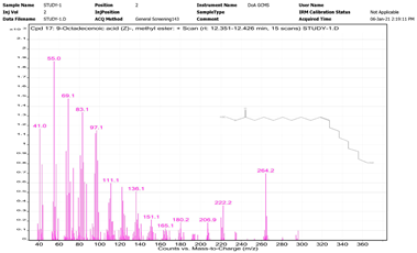
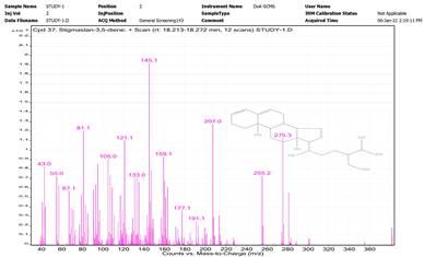
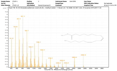
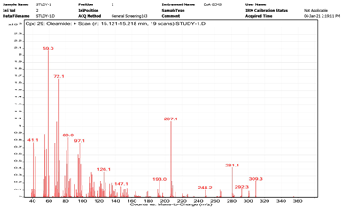
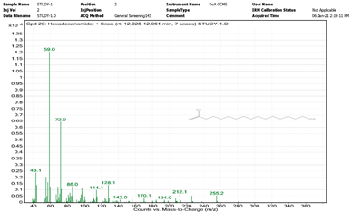
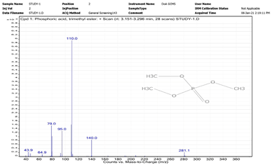
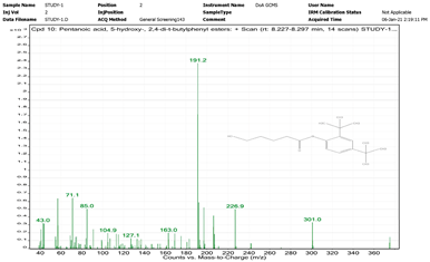
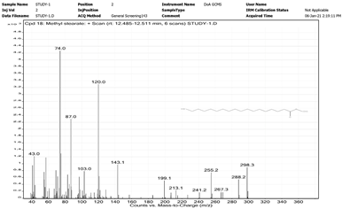
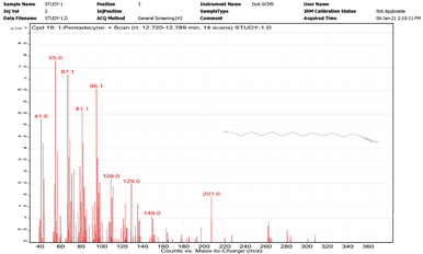
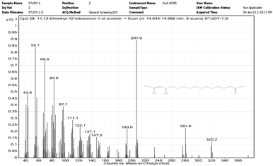
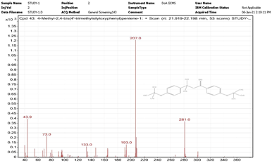
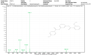
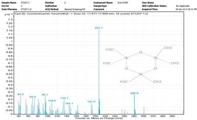
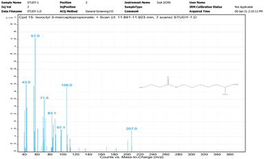
Table 1 shows the chemical composition of GSE (grape (Vitis vinifera) seed oil extract) identified by using GC-MS [16].

3.2. Phytochemical Components and Metal Contents of GSE

Supplementary Table S3 shows the quantified phytochemical components and metal contents of GSE (grape seed oil extract). As indicated in the table, phytochemicals such as tannins (in mg/100 g TAE) were not detected, while the content of polyphenols was 125.1 mg/100 g GAE, that of flavonoids 57.0 mg/100 g CE, and that of alkaloids 22.5 mg/g AE. Furthermore, the content of Na was 21.0 µg/g, that of Zn 9.1 µg/g, that of K 172.0 µg/g, that of Ca 27.2 µg/g, and that of Fe 29.1 µg/g, while Cr, Cd, Pb, and Al were not detected.

3.3. Metal Analysis in Water Samples

Figure 2A shows the concentrations in µg/L of lead (Pb), cadmium (Cd), aluminum (Al), potassium (K,) sodium (Na), calcium (Ca), iron (Fe), zinc (Zn), and chromium (Cr) in the water samples. These concentrations were analyzed in each of the water samples separately in the present study (sample 1 = Tap Water 1; sample 2 = Tap Water 2; sample 3 = Tap Water 3; sample 4 = Desalinated Water 1; sample 5 = Desalinated Water 2; sample 6 = Desalinated Water 3). The outcomes analyzed according to the Pareto curve rule (80/20) [22] showed that the greatest concentration, among the metals and minerals considered, in the studied samples was that of Zn, followed by that of K, Fe, Na, Ca, and Cr, in this order (Figure 2B).

3.4. Analysis of Bacterial Species in Water and GSE Samples

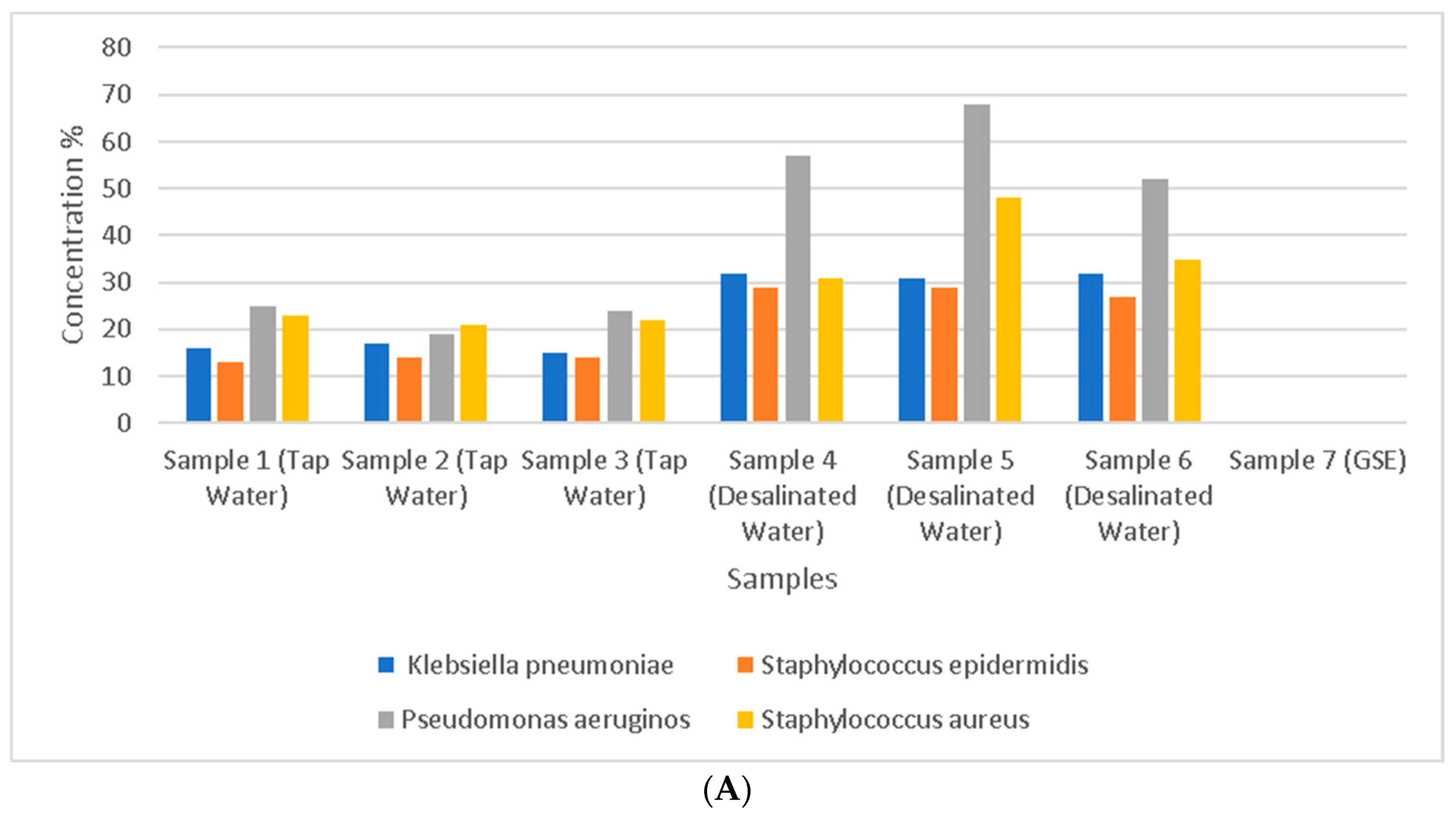
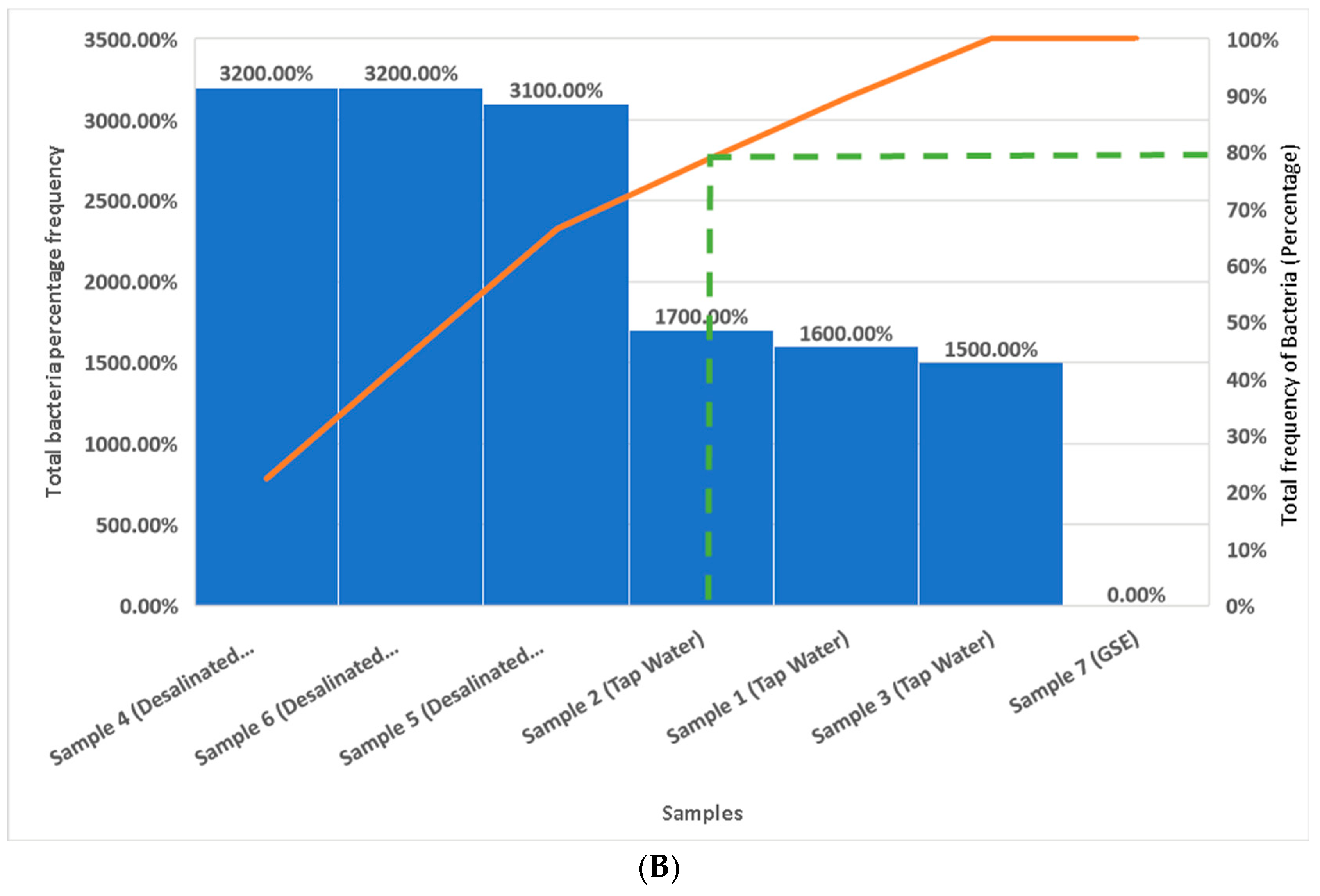
The results of the analysis of the bacterial species in the water samples indicate the presence of Staphylococcus aureus, Pseudomonas aeruginosa, Klebsiella pneumoniae, and Staphylococcus epidermidis as the main bacteria identified; their presence in the urine samples further supported the bacterial contamination of the water samples. Furthermore, the present outcomes also indicate the absence of bacteria in the GSE. The results indicate that Klebsiella pneumoniae is a Gram-negative bacterium (5215773), Staphylococcus epidermidis a Gram-positive bacterium (6706113), Pseudomonas aeruginosa a Gram-negative bacterium (2200004), and Staphylococcus aureus a Gram-positive bacterium (6736153); no other organisms or fungi were detected. Figure 3 shows the percentage of Klebsiella pneumoniae, Staphylococcus epidermidis, Pseudomonas aeruginosa, and Staphylococcus aureus in each water sample and in the GSE, which were separately analyzed in the present study (sample 1 = Tap Water 1; sample 2 = Tap Water 2; sample 3 = Tap Water 3; sample 4 = Desalinated Water 1; sample 5 = Desalinated Water 2; sample 6 = Desalinated Water (Brand) 3; sample 7 = GSE). The report outcomes indicate that, according to the Pareto curve cumulative frequency rule (80/20), the frequency percentage of contamination by the studied bacteria in water samples 1 to 6 and the GSE in descending order was as follows—sample 4, sample 6, sample 5, sample 2, sample 1, and sample 3—with the GSE showing 0%.

3.5. Screening of Urine Biological Samples for Heavy Metals, Other Elements, and Bacteria

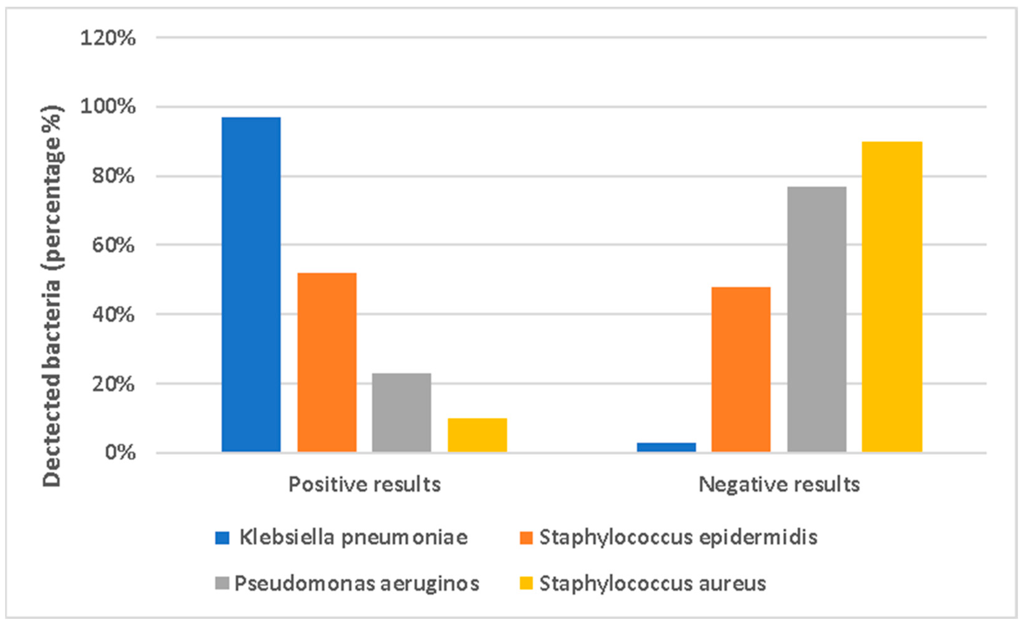
Supplementary Table S4 shows the data, represented as means ± SD, for the concentrations of heavy metals and elements in µg/L as follows: Na, 0.007 ± 0.001 µg/L; Cr, not detected; Zn, 0.0032 ± 0.002 µg/L; K, 0.0045 ± 0.021 µg/L; Cd, not detected; Pb, 0.0023 ± 0.031 µg/L; Al, 0.0012 ± 0.021 µg/L; Ca, 0.0029 ± 0.011 µg/L; Fe, not detected. Moreover, Supplementary Table S5 and Figure 4 illustrate the presence of bacteria (expressed as a percentage) detected in the urine samples; group 1 was positive for Klebsiella pneumoniae, Staphylococcus epidermidis, Pseudomonas aeruginosa, and Staphylococcus aureus, while group 2 was negative for the same species. Specifically, in the urine analysis study of 100 volunteers (n = 100), the results were as follows: for Klebsiella pneumoniae, we obtained 97% positive vs. 3% negative results; for Staphylococcus epidermidis, we obtained 52% positive vs. 48% negative results; for Pseudomonas aeruginosa, we obtained 23% positive vs. 77% negative results; and finally, for Staphylococcus aureus, we obtained 10% positive vs. 90% negative results.

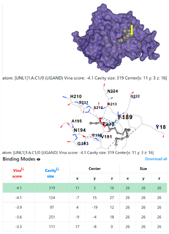
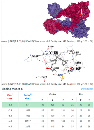
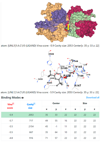
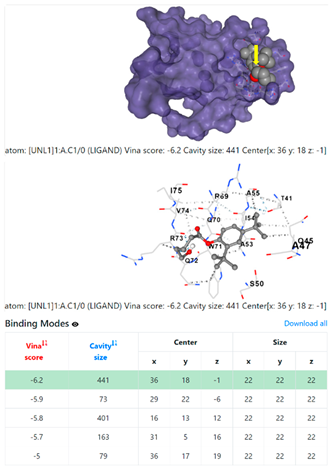
3.6. CB-Dock Analysis

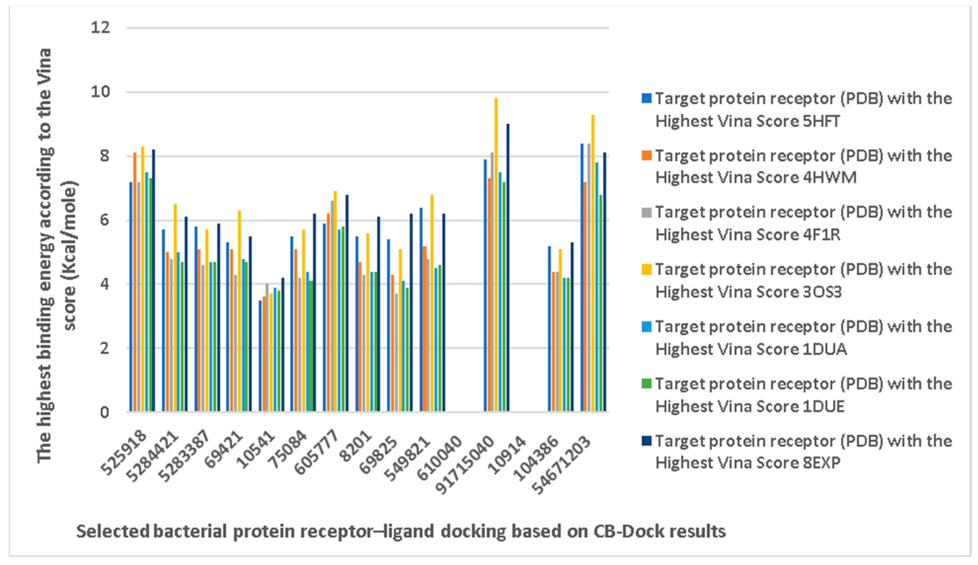
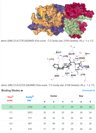
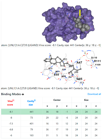
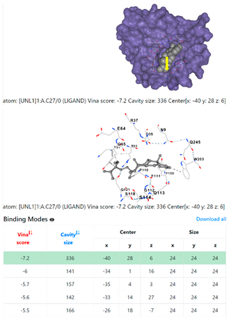
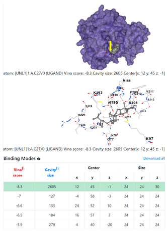
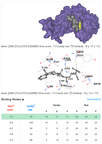
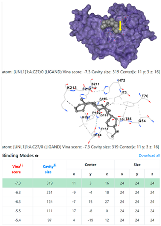
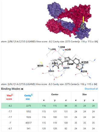
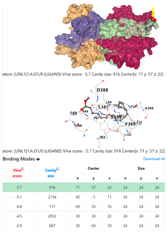
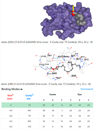
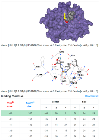
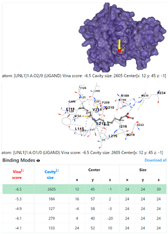
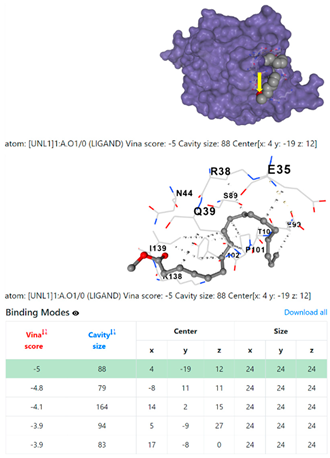
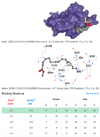
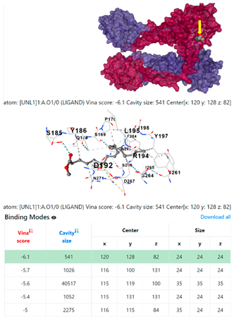
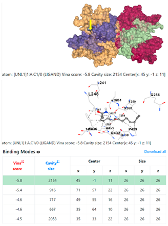
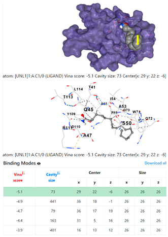
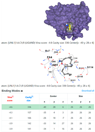
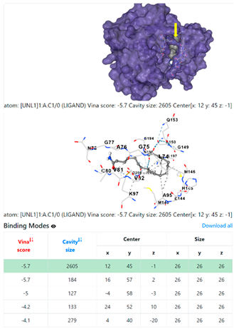
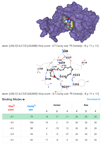
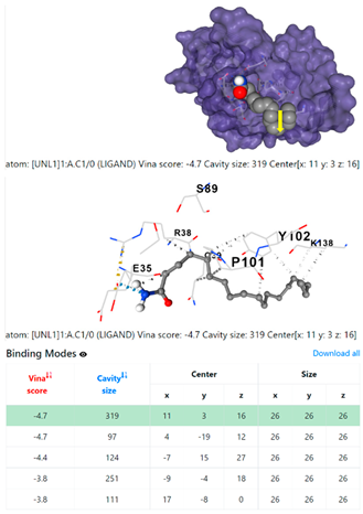
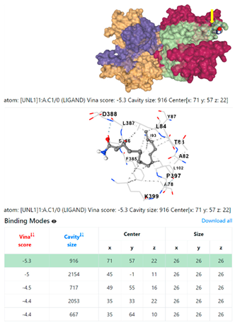
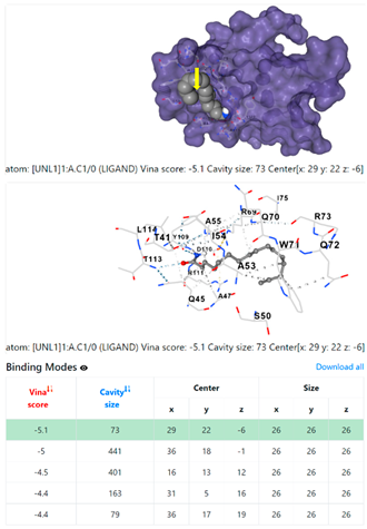
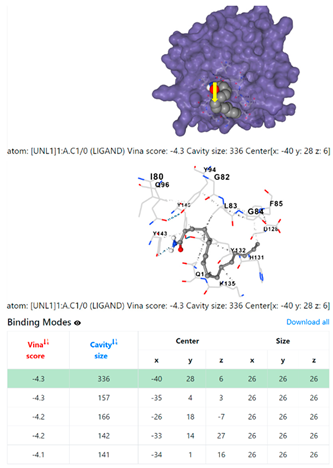
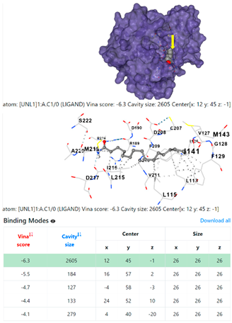
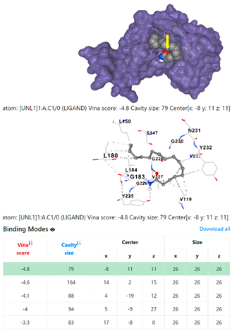
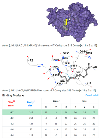
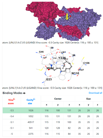
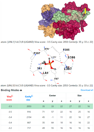
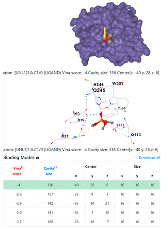
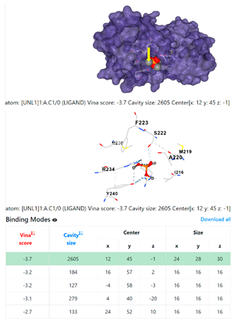
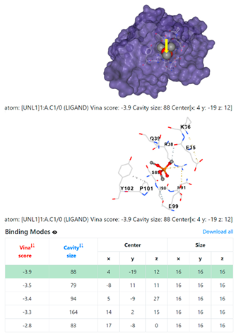
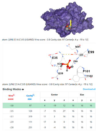
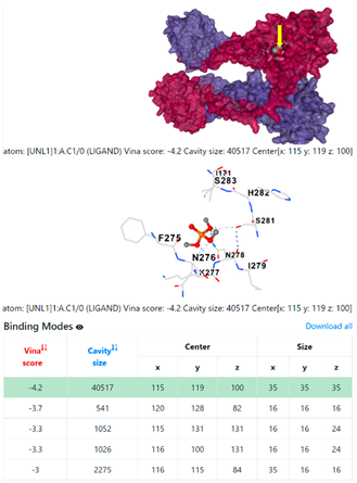
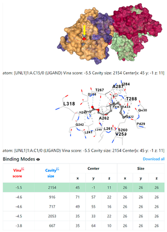
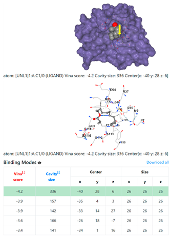
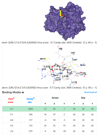
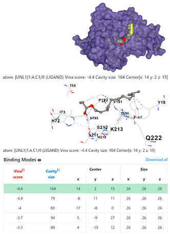
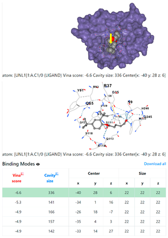
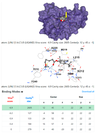
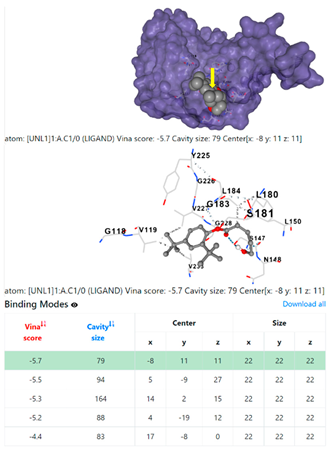
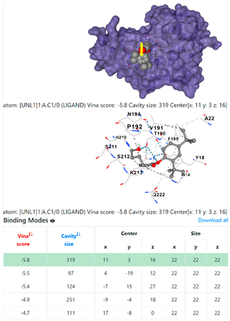
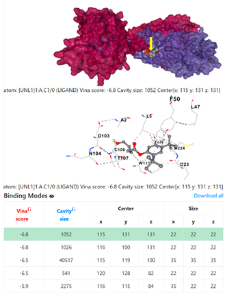
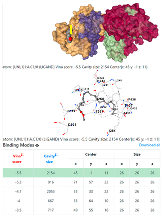
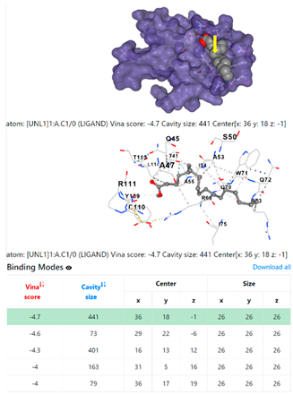
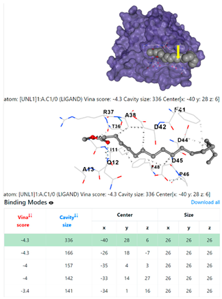
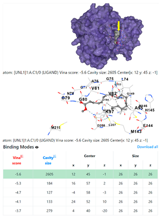
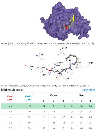
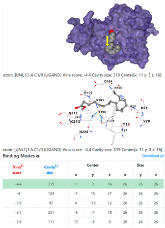
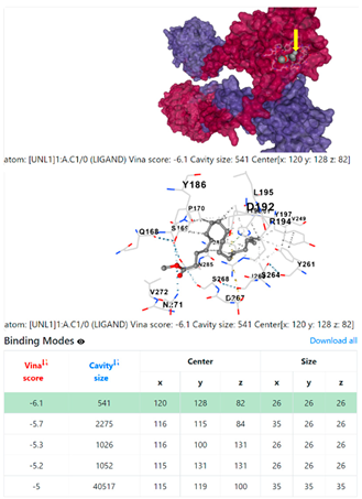
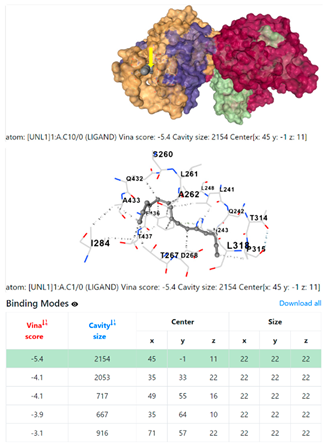
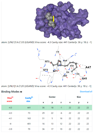
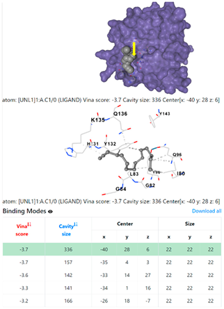
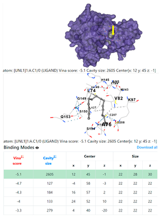
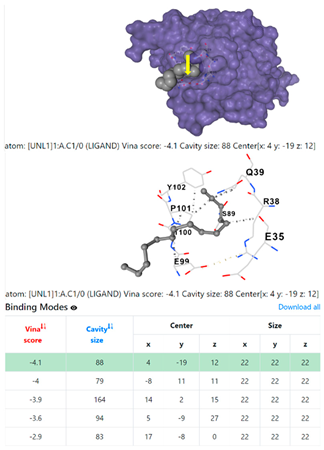
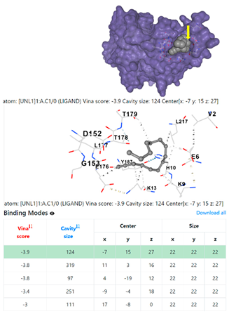
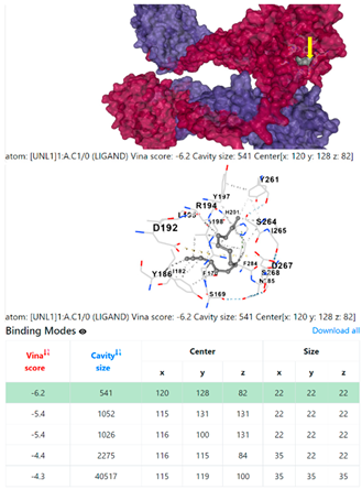
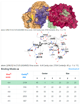
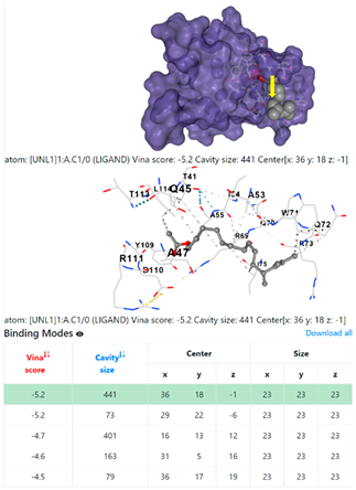
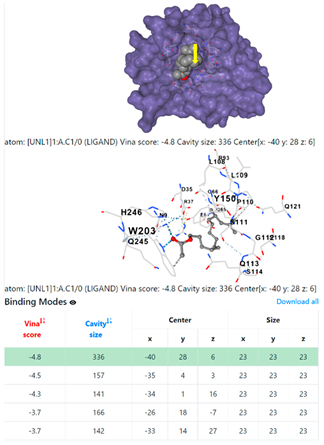
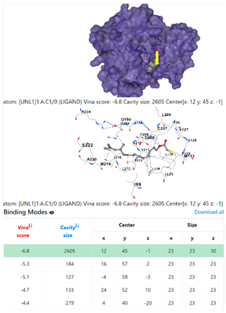
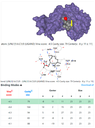
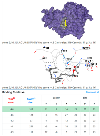
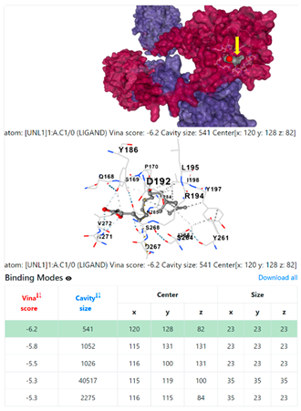
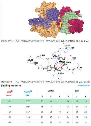
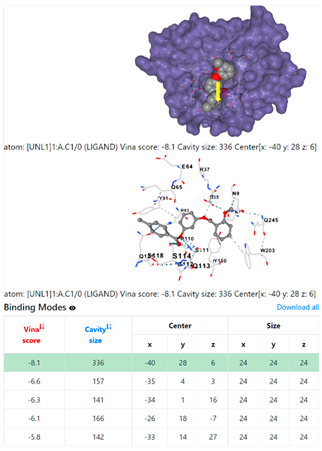
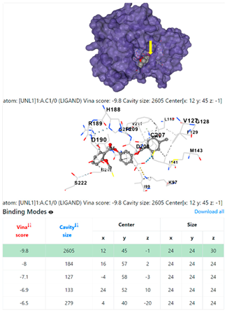
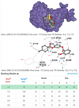
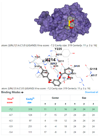
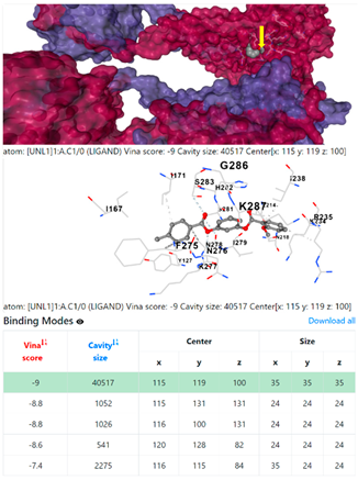
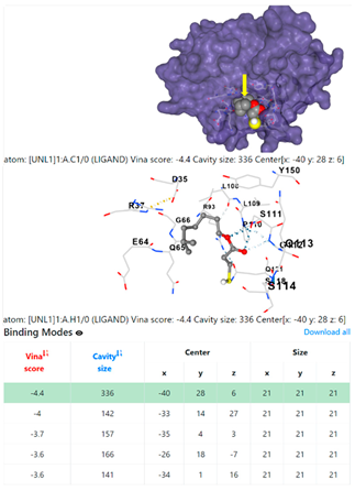
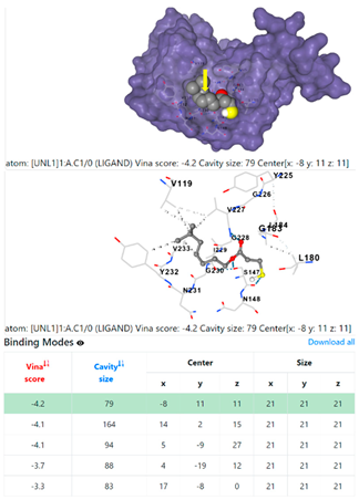
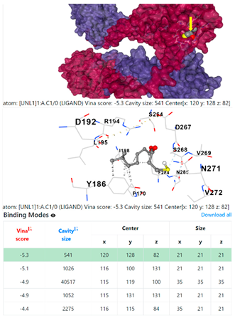
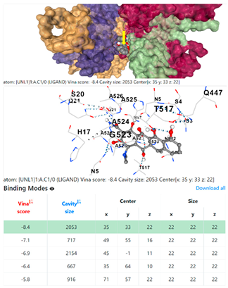
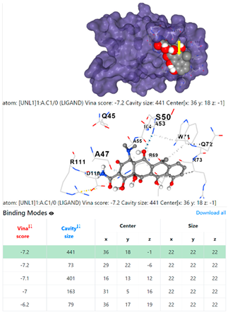
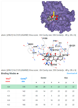
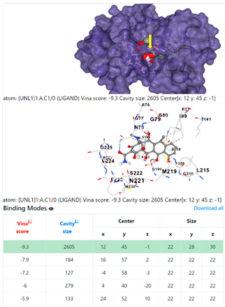
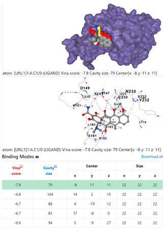
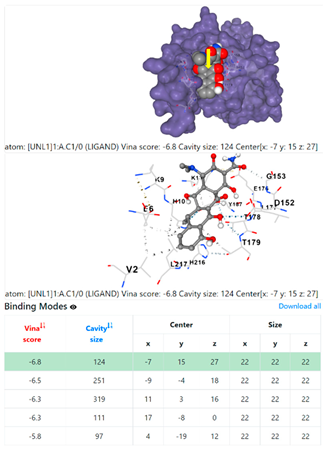
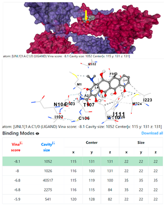
Table 2 shows potential CB-Dock interaction scenarios according to the chemical composition of GSE (grape seed oil extract) identified with GC-MS. The extract components were compared with a standard antibiotic (doxycycline) in the interaction with selected proteins and receptors in the detected bacterial species identified in the water and urine samples. The docking interaction was simulated with CB-Dock and AutoDock Vina based on the binding energy potential of the ligands. The procedure included three stages: (1) curvature determination of the protein surface, (2) cavity detection based on clustering, and (3) docking with AutoDock Vina. The results were sorted by the Vina score according to the cavity size as well as accuracy in predicting the binding sites of the PDB target whole proteins, not only surface binding. Figure 5 shows the highest binding energy between the studied bacterial receptor proteins and the ligands. The higher the binding energy, represented with negative symbols, the higher the binding affinity (kcal/mole).
The binding energy values between Klebsiella pneumoniae target protein receptors PDB:5HFT and PDB:4HWM (Table 2, Figure 5) and the studied ligands, i.e., CID 525918 (stigmastan-3,5-diene), CID 5284421 (9,12-octadecadienoic acid (Z,Z)-, methyl ester), CID 5283387 (oleamide), CID 69421 (hexadecanamide), CID 10541 (phosphoric acid, trimethyl ester), CID 75084 (dodecyl acrylate), CID 605777 (pentanoic acid, 5-hydroxy-, 2,4-di-t-butylphenyl esters), CID 8201 (methyl stearate), CID 69825 (1-pentadecyne), CID 549821 (11,13-dimethyl-12-tetradecen-1-ol acetate), CID 610040 (4-methyl-2,4-bis (4′-trimethylsilyloxyphenyl) pentene-1), CID 91715040 (1,3-benzenediol, o-(4-methylbenzoyl)-o′-(2-methoxybenzoyl)), CID 10914 (cyclotrisiloxane, hexamethyl-), CID 104386 (isooctyl 3-mercaptopropionate), and CID 54671203 (standard antibiotic (doxycycline)), were as follows: for PDB:5HFT, 7.2, −5.7, −5.8, −5.3, −3.5, −5.5, −5.9, −5.5, −5.4, −6.4, 0, −7.9, 0, −5.2, and −8.4, respectively; for PDB:4HWM, −8.1,−5,−5.1,−5.1,−3.6,−5.1,−6.2,−4.7,−4.3,−5.2, 0, −7.3, 0, −4.4, and −7.2, respectively.
Additionally, the binding energy values between the Pseudomonas aeruginosa target protein receptor PDB:4F1R (Table 2, Figure 5) and ligands CID 525918, CID 5284421, CID 5283387, CID 69421, CID 10541, CID 75084, CID 605777, CID 8201, CID 69825, CID 549821, CID 610040, CID 91715040, CID 10914, CID 104386, and CID 54671203 were as follows: −7.2, −4.8, −4.6, −4.3, −4, −4.2, −6.6, −4.3, −3.7, −4.8, 0, −8.1, 0, −4.4, and −8.4, respectively.
Furthermore, the prokaryotic ribosome (binding unit of antibiotic) PDB:3OS3 (Table 2, Figure 5) showed the following binding energy values with ligands CID 525918, CID 5284421, CID 5283387, CID 69421, CID 10541, CID 75084, CID 605777, CID 8201, CID 69825, CID 549821, CID 610040, CID 91715040, CID 10914, CID 104386, and CID 54671203: −8.3, −6.5, −5.7, −6.3, −3.7, −5.7, −6.9, −5.6, −5.1, −6.8, 0, −9.8, 0, −5.1, and −9.3, respectively.
Finally, the Staphylococcus aureus target protein receptors PDB:1DUA, PDB:1DUE, and PDB:8EXP (Table 2, Figure 5) showed the following binding energy values with ligands CID 525918, CID 5284421, CID 5283387, CID 69421, CID 10541, CID 75084, CID 605777, CID 8201, CID 69825, CID 549821, CID 610040, CID 91715040, CID 10914, CID 104386, and CID 54671203: for PDB:1DUA, −7.5, −5, −4.7, −4.8, −3.9, −4.4, −5.7, −4.4, −4.1, −4.5, 0, −7.5, 0, −4.2, and −7.8, respectively; for PDB:1DUE −7.3, −4.7, −4.7, −4.7, −3.8, −4.1, −5.8, −4.4, −3.9, −4.6, 0, −7.2, 0, −4.2, and −6.8, respectively; and for PDB:8EXP, −8.2, −6.1, −5.9, −5.5, −4.2, −6.2, −6.8, −6.1, −6.2, −6.2, 0, −9, 0, −5.3, and −8.1, respectively.

3.7. Antibacterial Effects at Different Concentrations of GSE

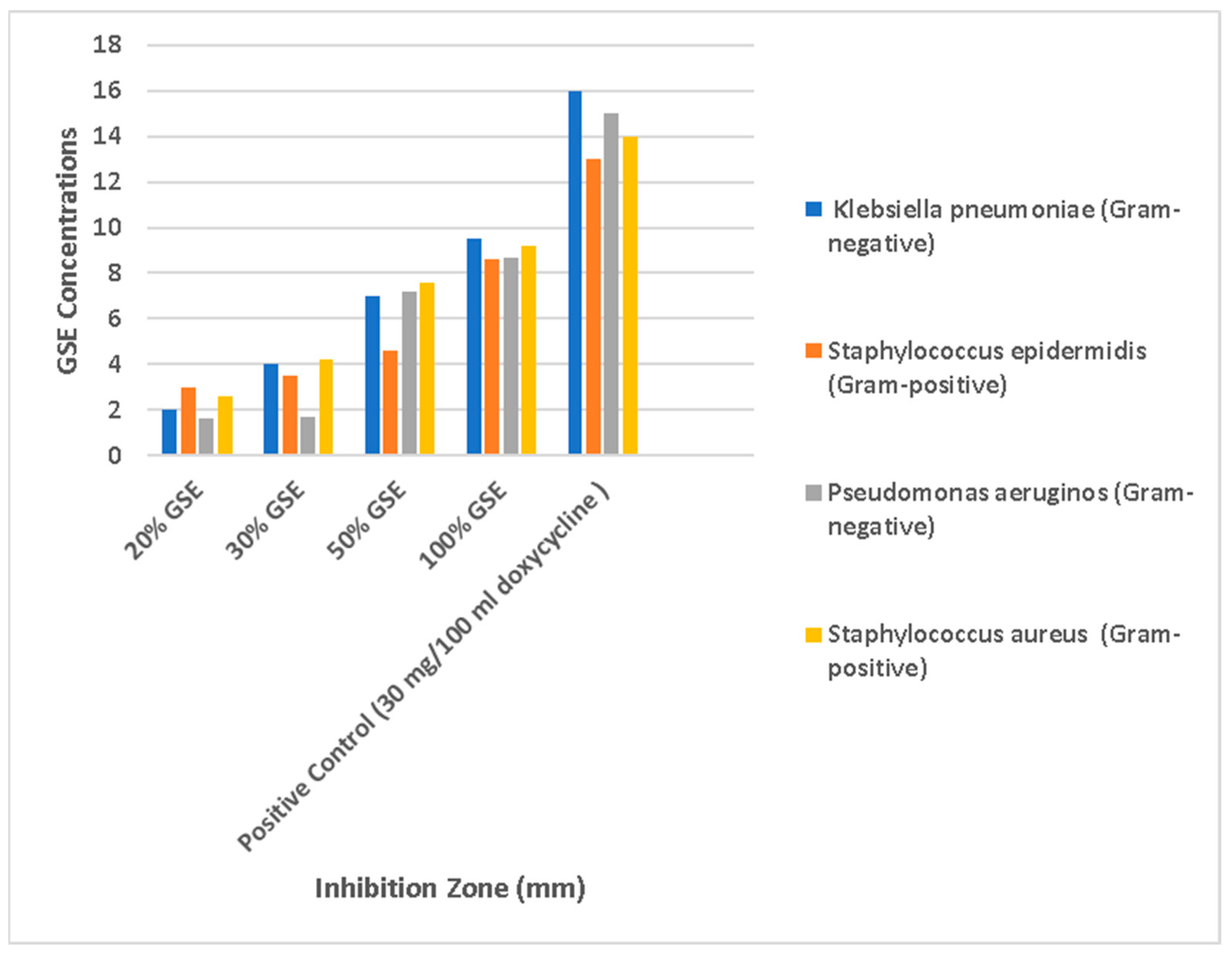
We analyzed the production of zones of inhibition (in mm). The results indicate that the highest antimicrobial effect was achieved with GSE at the crude concentration of 100% against Klebsiella pneumoniae (Gram-negative), where the inhibition zone was 9.5 mm, followed by Staphylococcus aureus (Gram-positive; inhibition zone of 9.2 mm), Pseudomonas aeruginosa (Gram-negative; inhibition zone of 8.7 mm), and Staphylococcus epidermidis (Gram-positive; inhibition zone of 8.6 mm), with the lowest effect being obtained at a GSE concentration equal to 10% (Figure 6).

4. Discussion

Plant waste, including seeds, represents a significant amount of biomass. Specifically, industrial waste such as grape seeds stands out due to its antioxidant properties. Research on recycling such waste is gaining traction, especially in the context of green treatment approaches [16,17,23,25,33,34].
In our previous publication [16], we highlighted the importance of unlocking the potential of fruit seed waste, given the substantial waste generated during fruit processing, as a green approach to developing potential anti-coronavirus ligands. The discarded stones from fruits such as grapes, prunes, apple, Ajwa dates, pomegranate, and avocado contribute significantly to this waste stream, posing environmental challenges. However, these seemingly overlooked remnants harbor untapped dietary and therapeutic potential, which could alleviate the burden of environmental pollution, including river water toxicity. We also reported that oil extract has many health benefits, such as anti-inflammatory, anti-diabetic, anti-obesity, and anti-apoptosis properties, as well as protective skincare properties. Additionally, we suggest that the studied seed oil could serve as a promising candidate for future research on its use as a nanoparticle against viruses and its use in vaccines and psychiatric drugs. By harnessing the power of these underutilized waste materials, we contribute to both environmental sustainability and scientific advancement. Previous research has explored the potential of grape seed extract as a stabilizing–reducing agent, transforming grape waste into green chemistry. This approach relies on less harmful chemicals from renewable sources. Notably, grape seed extract has demonstrated synergistic antimicrobial activity against both Gram-positive and Gram-negative bacteria in water. Our results agree with previous findings [16,17,23,25,33,34].
In the context of maintaining a clean water supply worldwide, in our study, we determined the presence of phytochemicals in grape seeds, and by using chromatography–mass spectrometry and inductively coupled plasma mass spectrometry, we identified these compounds. Furthermore, we conducted qualitative analyses on local water and urine samples to detect bacterial infections, heavy metals, and minerals. Our findings suggest a potential scenario for investigating grape seed oil extract as an antibacterial agent that targets bacterial receptors. Additionally, we analyzed binding energy with CB-Dock molecular docking analysis, and the results highlight the potential disinfection properties of natural agents derived from grape seed oil.
Our results indicate that grape seed extract is free from heavy-metal contamination (such as Cr, Cd, Pb, and Al) and rich in essential phytochemicals such as polyphenols, flavonoids, and alkaloids. Additionally, GSE contains healthy nutrient metals (Na, K, Zn, Ca, and Fe; Table 1 and Table 2), making it a safe choice for use in applied scientific fields, particularly as a water disinfectant. The strong binding energy observed between grape seed oil extract and microbes, confirmed based on CB-Dock analysis (Table 2 and Figure 5), underscores its potential efficacy. In the present study, we identified contamination by four bacterial strains and heavy metals in the studied water samples and in the urine samples from female individuals, a result that confirms the presence of harmful microorganisms and substances in water and the human body (Supplementary Tables S3 and S4, Figure 3 and Figure 4). In the agar well diffusion tests, the crude oil extract exhibited significantly greater zones of inhibition than the diluted GSE, parallel with the positive control. Furthermore, our chemical identification based on GC-MS revealed GSE’s effectiveness against specific bacteria: Klebsiella pneumoniae (Gram-negative; ID: 5215773), Staphylococcus epidermidis (Gram-positive; ID: 6706113), Pseudomonas aeruginosa (Gram-negative; ID: 2200004), and Staphylococcus aureus (Gram-positive; ID: 6736153). These findings emphasize GSE’s antimicrobial benefits and its relevance for water quality and human health (Table 2, Figure 5 and Figure 6).
CB-Dock, a computational tool for blind docking, is used for predicted binding sites within proteins. The workflow includes the following steps: (1) cavity detection, where CB-Dock identifies potential binding sites on the protein surface by selecting larger cavities for further analysis; (2) the sorting of the detected cavities, with the top candidates being chosen based on size, serving as potential binding sites for the ligand; (3) center and size calculations, where the docking center coordinates are determined and the docking box size is adjusted for subsequent molecular docking; (4) the AutoDock Vina method, where the ligand docks into the selected cavities, generating bound poses; (5) docking score reranking, where the bound poses are ranked based on docking scores, with the highest-scoring conformation representing the optimal binding pose; and (6) optimal binding site identification, where the site associated with the top-ranking conformation is determined to be the best binding site for the query ligand. CB-Dock’s Perl script automates the entire process after submitting the input files. As the number of elucidated protein structures continues to grow, the efficient utilization of structural information becomes crucial for biological and pharmaceutical purposes. Predicting binding sites and affinity between proteins and ligands is essential for computer-aided drug discovery. While existing docking tools often rely on preset binding sites provided by users, CB-Dock takes a different approach, as described above. Furthermore, small-molecule binding involves binding inside protein pockets or cavities as a result of high affinity, which can only be simulated with sufficiently large interaction interfaces. CB-Dock searches for concave surfaces to detect these cavities. Briefly, the tool generates a set of points representing the solvent-accessible surface and calculates the curvature factor for each point by using the method described earlier. These points on the concave surface (curvature factor > 8) are clustered by using a density-peak-based algorithm. As a result, several clusters of points representing cavities on the protein surface are obtained. In this study, we present our work on identifying ligands from the GSE database based on GC-MS and comparing them with an antibiotic to study their potential interactions as antibacterial agents in water and urine samples [30,31].
Our results indicate strong binding energy between all the studied ligands and bacterial protein receptors, compared with the binding site of the standard antibiotic. However, an exception was observed for 1,3-benzenediol, o-(4-methylbenzoyl)-o’-(2-methoxybenzoyl)-, and cyclotrisiloxane, hexamethyl-. Figure 5 and Table 2 indicate that grape seed extract (GSE) can be considered an excellent antibacterial agent for further chemical analysis investigation. Furthermore, our laboratory pilot investigation results align with CB-Dock’s analysis results in terms of the global binding energy of the highest binding affinity (kcal/mole) of the studied receptors, with the greatest enlarged angle for ligand binding, as shown in the protein visual representations. Our findings demonstrate that the highest antimicrobial effect occurred when GSE was used at a crude concentration of 100%. Specifically, against Klebsiella pneumoniae (Gram-negative), the inhibition zone was 9.5 mm; for Staphylococcus aureus (Gram-positive), it was 9.2 mm. Pseudomonas aeruginosa (Gram-negative) followed, with an inhibition zone of 8.7 mm, and Staphylococcus epidermidis (Gram-positive) exhibited an inhibition zone of 8.6 mm. Conversely, the lowest effect was observed at a GSE concentration of 10% (see Figure 6), which may have been due to the phytochemical components of the GSE, such as polyphenols, flavonoids, and alkaloids, and their individual chemical composition as identified with GC-MS (Table 1 and Table 2).
Heavy metals, often considered pseudo-elements, are hazardous due to their negative impact on the body’s metabolic processes. Toxic effects depend on exposure duration, dose, and route. These metals can lead to lipid peroxidation, genetic mutations, and interactions with proteins such as albumin and cell receptors. Heavy metals are implicated in human toxicity, infertility, chronic diseases, immune disorders, DNA alterations, and other health issues. Regular screening and monitoring for environmental toxicity, especially heavy metals, remain crucial [22,35]. Reference ranges vary based on factors such as population, race, age, lifestyle, sex, geographical region, and analytical methods [22,35].
In our previous investigation [22], we confirmed the presence of heavy metals in the serum, hair, and nails of female university students and in selected food, drink, water, and cosmetic products. Notably, we observed a relationship between heavy metals and the biological profile of complete blood count (CBC) and the cholinesterase enzyme, as confirmed with AutoDock Interaction [22,35]. Furthermore, we highlighted the interactions between heavy metals and the human body. These metals, through electron loss and the formation of metal cations, bind to various proteins, genes, and DNA. The affinity sites on macromolecules are affected, impacting tissues and organs. Direct exposure to heavy metals from sources such as food, beverages, cosmetics, soil, air, and water can disrupt internal biological systems, leading to acute and chronic health issues. Neurological, brain, liver, kidney, bone, skin, and fetal diseases may result due to the interactions of metals with different protein receptors. In our previous studies based on AutoDock analysis, we specifically investigated the effects of the strong binding energy of heavy metals on cholinesterase, P53, dopamine, metallothionein, estrogen, keratin, protein kinase enzymes, beta-amyloid, ATPase, albumin, MAO, adrenaline, cortisol, TNF-α, IL-1β, COX-2, and LOX-1, shedding light on potential health consequences [22,34].
The present results indicate that GSE may be considered a good choice for water disinfection from free bacteria and heavy metals. Therefore, it could be used to prevent side effects of bacteria such as inflammation. Bacterial diseases, such as pneumonia, skin infections, kidney issues, and bone inflammation, are often associated with Gram-negative bacteria. Klebsiella pneumoniae and Pseudomonas aeruginosa, particularly in hospital-acquired cases, can cause pneumonia, especially when the body’s immune system is not powerful. Moreover, Staphylococcus aureus, a Gram-positive pathogen, leads to skin infections, pneumonia, endocarditis, and osteomyelitis. Some Staphylococcus aureus strains produce toxins that result in health issues such as gastroenteritis and toxic shock syndrome. Additionally, Pseudomonas aeruginosa, another Gram-negative bacterium, is commonly associated with pneumonia, and Staphylococcus epidermidis, a Gram-positive bacterium found on human skin, is generally harmless but can cause infections, especially in immunocompromised individuals. It is crucial to be aware that antibiotics may have adverse effects, including kidney issues, skin reactions, and bone inflammation. Therefore, it is essential to find natural, side-effect-free ways, such as using grape seed extract, in agreement with the present results, to combat bacteria in water and the human body [36,37,38,39].
Many reports [16,40,41,42,43,44,45], including our previous publication, indicate that grape seed extract (GSE) contains various bioactive compounds, including 9-octadecenamide; 9,12-octadecadienoic acid; oleamide; and hexadecanamide. These compounds exhibit a sedative effect by interacting with neurotransmitters during sleep deprivation, making them potential treatments for mood, depression, and sleep disorders [16,40,41,42,43,44,45]. Additionally, they bind to cannabinoid receptor type 1 (CB1) as full agonists. GSE also contains stigmastane, which serves as a biomarker for initial eukaryotes, and stigmastanol derivatives, which inhibit cholesterol absorption. Acrylates found in GSE have multifunctional properties, including use in the polymer industry and as antimicrobial agents. The extract also contains non-genotoxic, non-carcinogenic phosphoric acids used in water treatment and other applications. Valeric acid, also present in GSE, is produced by gut microbiota and has implications for gut health. Methyl stearate, another component, is associated with reduced LDL cholesterol. GSE’s secondary metabolites, such as sulfurous acid and 4-methyl-2,4-bis(4-hydroxyphenyl)pent-1-ene (MBP), exhibit antibacterial, antifungal, and anti-diabetic properties. Overall, GSE offers a diverse range of bioactive compounds with potential health benefits [16,40,41,42,43,44,45].
Grape seed extract (GSE) contains antioxidants, including oligomeric proanthocyanidins (OPCs), which indirectly support detoxification. While GSE is not specifically used as a heavy-metal removal agent, its antioxidant properties may help manage heavy-metal exposure. Research [46,47] suggests that GSE can ameliorate the effects of heavy metals such as cadmium and lead by reducing the number apoptotic cells and regulating protein levels. However, for targeted heavy-metal removal, specialized methods or products may be more effective. Researchers found that GSE effectively inhibits corrosion in mild steel exposed to acidic conditions. As the concentration of GSE increases, the inhibition efficiency improves, suggesting the chemisorption of inhibitive species. A surface analysis revealed reduced quantities of corrosion products and enhanced surface film integrity, likely attributed to GSE’s antioxidant activity [46,47].
Grapes, widely consumed worldwide, offer not only vitamins and fiber but also valuable bioactive compounds found in their skin and seeds. Among these, proanthocyanidins stand out as potent polyphenols present in grape seeds. GSE, derived from these seeds, has garnered attention for its potential health benefits, and grape seed extract has been used as a wound disinfectant, fruit and vegetable sanitizer, and cattle feed. In this study, we collected local grape seeds from Saudi Arabia and analyzed their chemical components. By using gas chromatography–mass spectrometry and inductively coupled plasma mass spectrometry, we identified essential phytochemicals in GSE, including polyphenols, flavonoids, and alkaloids. Notably, GSE was free from bacterial and heavy-metal contamination (e.g., Cr, Cd, Pb, and Al) and rich in beneficial nutrient metals (Na, K, Zn, Ca, and Fe). Our results are in agreement with previous findings [48,49], according to which GSE has demonstrated beneficial effects against various diseases. It reduces inflammation, prevents cardiovascular disease, and helps to manage blood pressure and glycemic control. It also exhibits antioxidant properties, as demonstrated by its use in cancer patients’ diets during chemotherapy to decrease the side effects of the latter, such as gastroenteritis and constipation, thanks to its gastroprotective effects. Additionally, it showed antimicrobial activity [48,49]. Furthermore, GSE contains proanthocyanidins, flavonoids, and other bioactive compounds and, as confirmed by the results of this study, is free from heavy-metal contamination (e.g., chromium, cadmium, lead, and aluminum) and provides essential minerals (sodium, potassium, zinc, calcium, and iron). Beyond its use as a nutraceutical or cosmeceutical, GSE could complement existing drugs. It is a safe and potent natural extract that could be used to develop pharmaceutical formulations, holding promise for better health outcomes [48,49].
The findings of this study highlight the potential of grape seed extract (GSE) as a natural antibacterial agent for water disinfection in Saudi Arabia. However, several avenues for further research and practical implementation have to be explored. First, comprehensive laboratory toxicity assessments are essential in in vivo animal studies to ensure GSE’s safety for human consumption and environmental impact. Long-term studies should evaluate any potential adverse effects and address concerns related to its widespread use. Additionally, field trials and real-world applications are necessary to validate GSE’s efficacy in large-scale water treatment facilities, for which a pilot investigation was conducted in the present study. Collaborations with relevant financial institutions, including government agencies, public health organizations, and industry partners, can facilitate the adoption of GSE as a green, sustainable, and safe water disinfection solution. While GSE itself is not a new substance, and its use draws upon traditional knowledge and practices, its application in water disinfection represents an innovative approach aligning with the global trend toward natural and eco-friendly solutions. By integrating indigenous wisdom (i.e., GSE’s historical use) with modern scientific methods, we can create innovative approaches to address pressing environmental and health challenges. Water scarcity and contamination remain critical global challenges. In Saudi Arabia, where water desalination is vital, finding safe and effective disinfection methods is crucial. Researchers, policymakers, and practitioners should collaborate to bridge the gap between traditional practices and evidence-based scientific methods. In doing so, we can unlock the full potential of natural resources such as grape seeds while safeguarding public health and promoting sustainable water management practices [8,15,16,19,35].
In conclusion, GSE boasts antibacterial properties and is rich in phytochemicals such as polyphenols and flavonoids. GSE’s efficacy against bacteria, its safety profile, and its rich phytochemical composition make it a promising candidate for water treatment. In our study, we explored GSE’s potential as a water disinfectant, revealing strong binding energy with bacterial receptors, and agar well diffusion tests confirmed its efficacy against the detected bacteria. GSE thus represents a promising, eco-friendly alternative for clean water access [8,15,16,19,35].

5. Conclusions

In this study, we investigated grape seed extract (GSE) as a potential natural antibacterial agent for water disinfection in Saudi Arabia. Our findings indicate that GSE exhibits a strong binding energy with bacterial protein receptors compared with standard antibiotics. Additionally, GSE is free from heavy-metal contamination and is rich in essential phytochemicals. Agar well diffusion tests confirmed its efficacy against bacteria. Given its safety profile and promising antibacterial properties, GSE holds potential as an effective water disinfectant. Further research and practical applications are warranted to harness GSE’s benefits for clean water supply in Saudi Arabia.

Supplementary Materials

The following supporting information can be downloaded at: https://www.mdpi.com/article/10.3390/chemistry6050051/s1, Table S1: Target protein receptor (PDB) results of studied bacteria receptors; Table S2: The chemical structure from PubChem CID results of GSE components; Table S3: Analysis of phytochemical content and metals in grape seed extract using ICP-MS; Table S4. Urine analysis (metals); Table S5. Urine analysis (bacteria) N = 100.

Author Contributions

A.F.H. contributed to the creation of the study; conceptualizing; work idea designing; performing all the experiments; analyzing the data; writing, revising, and editing the manuscript; and explaining the results. Both A.F.H. and S.F. equally contributed to the present study by interpreting and analyzing the data; writing, revising, and editing the manuscript; and explaining the results. All authors have read and agreed to the published version of the manuscript.

Funding

This research study received no external funding.

Institutional Review Board Statement

This study was conducted according to the guidelines of the Declaration of Helsinki and was approved by the National committee of Bio Ethics of UMM Al-Qura University, Al-Lith University College (protocol code HAPO-02-K-012-2022-06-1110; 12 June 2022).

Informed Consent Statement

Informed consent was obtained from all subjects involved in the study.

Data Availability Statement

The original contributions presented in the study are included in the article/Supplementary Materials, further inquiries can be directed to the corresponding author.

Acknowledgments

The authors are grateful to the universities and all the doctors who helped in this research study and for the willingness of the volunteers to take part in this study.

Conflicts of Interest

The authors declare no conflicts of interest.

References

  1. El-Sayed, A.A.H.; Al-Blehed, M.S. Bacterial Isolate from Arabian Gulf Coast Soils in Saudi Arabia Able to Degrade Arab Crude Oil. J. King Saud Univ. Eng. Sci. 1999, 11, 251–260. [Google Scholar] [CrossRef]
  2. Abdalwahab, M.A.; Dawood, S.A.; Abouhosh, E. Evaluation of Microbial or Bacterial Quality of the Drinking Water of Duba Province North Saudi Arabia. Asian J. Agric. Food Sci. 2017, 5, 13–18. Available online: https://ajouronline.com/index.php/AJAFS/article/view/4447#google_vignette (accessed on 12 January 2023).
  3. AlOtaibi, E.L.S. Bacteriological assessment of urban water sources in Khamis Mushait Governorate, southwestern Saudi Arabia. Int. J. Health Geogr. 2009, 8, 16. [Google Scholar] [CrossRef] [PubMed]
  4. Saati, A.; Faidah, H. Environmental Prevalence of Pathogens in Different Drinking Water Resources in Makkah City (Kingdom of Saudi Arabia). Curr. World Environ. J. 2013, 8, 37–53. [Google Scholar] [CrossRef]
  5. Al-Wohaib, F.A.; Al-Sharif, I.; Al-Zain, H.; Murad, D.; Al-Harbi, L.; Al-Mozaini, M. Evaluation of two molecular detection platforms for gastroenteritis pathogens in treated sewage water in the Eastern province of Saudi Arabia. Sci. Rep. 2022, 12, 21744. [Google Scholar] [CrossRef]
  6. Alqahtani, J.M.; Asaad, A.M.; Ahmed, E.M.; Qureshi, M.A. Drinking water quality and public health in Southwestern Saudi Arabia: The need for a national monitoring program. J. Fam. Community Med. 2015, 22, 19–24. [Google Scholar] [CrossRef]
  7. Alaa el-Din, M.N.; Madany, I.M.; Al-Tayaran, A.; Al-Jubair, A.H.; Gomaa, A. Trends in water quality of some wells in Saudi Arabia, 1984–1989. Sci. Total Environ. 1994, 143, 173–181. [Google Scholar] [CrossRef]
  8. Pritchard, M.; Mkandawire, T.; O’Neill, J.G. Biological, Chemical and Physical Drinking Water Quality from Shallow Wells in Malawi: Case Study of Blantyre, Chiradzulu and Mulanje. Phys. Chem. Earth Parts A/B/C 2007, 32, 1167–1177. [Google Scholar] [CrossRef]
  9. Agus, E.; Voutchkov, N.; Sedlak, D. Disinfection by-products and their potential impact on the quality of water produced by desalination systems: A literature review. Desalination 2009, 237, 214–237. [Google Scholar] [CrossRef]
  10. Kim, D.; Amy, G.L.; Karanfil, T. Disinfection by-product formation during seawater desalination: A review. Water Research 2015, 81, 343–355. [Google Scholar] [CrossRef]
  11. Fatima, F.; Siddiqui, S.; Khan, W.A. Nanoparticles as Novel Emerging Therapeutic Antibacterial Agents in the Antibiotics Resistant Era. Biol. Trace Elem. Res. 2021, 199, 2552–2564. [Google Scholar] [CrossRef]
  12. Xin, X.; Qi, C.; Xu, L.; Gao, Q.; Liu, X. Green synthesis of silver nanoparticles and their antibacterial effects. Front. Chem. Eng. 2022, 4, 941240. [Google Scholar] [CrossRef]
  13. Wang, Y.; Gu, M.; Cheng, J.; Wan, Y.; Zhu, L.; Gao, Z.; Jiang, L. Antibiotic Alternatives: Multifunctional Ultra-Small Metal Nanoclusters for Bacterial Infectious Therapy Application. Molecules 2024, 29, 3117. [Google Scholar] [CrossRef] [PubMed]
  14. Crisan, C.M.; Mocan, T.; Manolea, M.; Lasca, L.I.; Tăbăran, F.-A.; Mocan, L. Review on Silver Nanoparticles as a Novel Class of Antibacterial Solutions. Appl. Sci. 2021, 11, 1120. [Google Scholar] [CrossRef]
  15. Al Bahri, M.; Calvo, L.; Gilarranz, M.A.; Rodriguez, J.J. Activated carbon from grape seeds upon chemical activation with phosphoric acid: Application to the adsorption of diuron from water. Chem. Eng. J. 2012, 203, 348–356. [Google Scholar] [CrossRef]
  16. Hamouda, A.F.; Farawilla, T.-L.M.; Attafi, I.M.; Khardali, I.A.; Attafi, M.A.; Oraiby, M.E.; Abualsail, F.M. Screening Pilot Study of Fruit Seed Compositions by GC-MS and Their Potential Scenario Anti ACE2 and 2rh1 Receptors as a Recycling Possibility in the Coronavirus Pandemic. J. Clin. Med. Res. 2021, 2, 1–65. [Google Scholar] [CrossRef]
  17. Raota, C.S.; Cerbaro, A.F.; Salvador, M.; Delamare, A.P.L.; Echeverrigaray, S.; Crespo, J.d.S.; da Silva, T.B.; Giovanela, M. Green synthesis of silver nanoparticles using an extract of Ives cultivar (Vitis labrusca) pomace: Characterization and application in wastewater disinfection. J. Environ. Chem. Eng. 2019, 7, 103383. [Google Scholar] [CrossRef]
  18. Munther Al Agha, A.G.; Muslih Al Khafaji, N.J.; Al Azawi, A.K.S. Isolation and Identification of Klebsiella pneumoniae using API-20E analytical system and conventional PCR assay. Int. J. Curr. Microbiol. Appl. Sci. 2017, 6, 203–210. [Google Scholar] [CrossRef]
  19. Rice, E.E.W.; Baird, R.B.; Eaton, A.D. Standard Methods for the Examination of Water and Wastewater. 2017. Available online: https://yabesh.ir/wp-content/uploads/2018/02/Standard-Methods-23rd-Perv.pdf (accessed on 18 February 2023).
  20. Al-Abdalall, A.H.A. Isolation and identification of microbes associated with mobile phones in Dammam in eastern Saudi Arabia. J. Fam. Community Med. 2010, 17, 11–14. [Google Scholar] [CrossRef]
  21. O’hara, C.M. Manual and automated instrumentation for identification of Enterobacteriaceae and other aerobic gram-negative bacilli. Clin. Microbiol. Rev. 2005, 18, 147–162. [Google Scholar] [CrossRef]
  22. Hamouda, A.F.; Felemban, S. A Bio-Indicator Pilot Study Screening Selected Heavy Metals in Female Hair, Nails, and Serum from Lifestyle Cosmetic, Canned Food, and Manufactured Drink Choices. Molecules 2023, 28, 5582. [Google Scholar] [CrossRef]
  23. Hamouda, A.F.; Felemban, S. Biochemical Pilot Study on Effects of Pomegranate Seed Oil Extract and Cosmetic Cream on Neurologically Mediated Skin Inflammation in Animals and Humans: A Comparative Observational Study. Molecules 2023, 28, 903. [Google Scholar] [CrossRef]
  24. Bolajoko, E.; Onyeaghala, A.A.; Akinosun, O.; Attah, A.; Moody, J.O.; Khine, A.A. Comparative proximate, phytochemical and mineral contents of freeze-dried and aqueous extracts of vernonia amygdalina del. leaves and garcinia kola heckel seeds. Int. J. Herbs Pharmacol. Res. 2020, 9, 2–13. [Google Scholar]
  25. Felemban, S.; Hamouda, A.F. A Pilot Study of the Effects of Ajwa Date Seed Extract in a Diabetic Animal with Parallel Observations on Human Subjects. J. Pharm. Res. Int. 2022, 34, 23–33. [Google Scholar] [CrossRef]
  26. Hamouda, A.F.; Hassan, A.A.M.; Khardali, I.A.; Attafi, I.M.; Oraiby, M.E.; Attafi, M.A.; Mahmoud, M.H.; Salem, S.F.A.; Sahli, K.A.Y.; Hamdi, F.Y.; et al. A Screening Pilot Study on the Relation between Body Mass Index, and Heavy Metal and Mineral Levels in College Students. Electron. J. Biol. 2019, 15, 79–89. [Google Scholar] [CrossRef]
  27. Aamir, M.; Singh, V.K.; Dubey, M.K.; Meena, M.; Kashyap, S.P.; Katari, S.K.; Upadhyay, R.S.; Umamaheswari, A.; Singh, S. In silico Prediction, Characterization, Molecular Docking, and Dynamic Studies on Fungal SDRs as Novel Targets for Searching Potential Fungicides Against Fusarium Wilt in Tomato. Front. Pharmacol. 2018, 9, 1038. [Google Scholar] [CrossRef] [PubMed]
  28. Tao, X.; Huang, Y.; Wang, C.; Chen, F.; Yang, L.; Ling, L.; Che, Z.; Chen, X. Recent developments in molecular docking technology applied in food science: A review. Int. J. Food Sci. Technol. 2020, 55, 33–45. [Google Scholar] [CrossRef]
  29. Cattaneo, A.; Macchi, F.; Plazzotta, G.; Veronica, B.; Bocchio-Chiavetto, L.; Riva, M.A.; Pariante, C.M. Inflammation and neuronal plasticity: A link between childhood trauma and depression pathogenesis. Front. Cell. Neurosci. 2015, 9, 40. [Google Scholar] [CrossRef] [PubMed]
  30. Liu, Y.; Grimm, M.; Dai, W.-T.; Hou, M.-C.; Xiao, Z.-X.; Cao, Y. CB-Dock: A web server for cavity detection-guided protein-ligand blind docking. Acta Pharmacol. Sin. 2020, 41, 138–144. [Google Scholar] [CrossRef]
  31. Cao, Y.; Li, L. Improved protein-ligand binding affinity prediction by using a curvature-dependent surface-area model. Bioinformatics 2014, 30, 1674–1680. [Google Scholar] [CrossRef]
  32. Mohamed, A.A.; Ali, S.I.; EL-Baz, F.K.; Hegazy, A.K.; Kord, M.A. Chemical composition of essential oil and in vitro antioxidant and antimicrobial activities of crude extracts of Commiphora myrrha resin. Ind. Crops Prod. 2014, 57, 10–16. [Google Scholar] [CrossRef]
  33. El-Shazly, H.A.M.; Mohamed, E.S.A.; Abdelatif, S.H. Effect of Pomegranate Seed Oil on Certain Food Spoilage Microorganisms and its Role in Shelf Life of Cream. J. Food Dairy Sci. 2017, 8, 173–178. [Google Scholar] [CrossRef]
  34. Hamouda, A.F.; Felemban, S. A Pilot Study of the Amelioration of Avocado Seed Oil in Obese Female Rats Induced by Carbon Tetrachloride and Alloxan Monohydrate. J. Drug Alcohol Res. 2022, 11, 236177. [Google Scholar] [CrossRef]
  35. Witkowska, D.; Słowik, J.; Chilicka, K. Heavy Metals and Human Health: Possible Exposure Pathways and the Competition for Protein Binding Sites. Molecules 2021, 26, 6060. [Google Scholar] [CrossRef] [PubMed]
  36. Cilloniz, C.; Martin-Loeches, I.; Garcia-Vidal, C.; San Jose, A.; Torres, A. Microbial Etiology of Pneumonia: Epidemiology, Diagnosis and Resistance Patterns. Int. J. Mol. Sci. 2016, 17, 2120. [Google Scholar] [CrossRef]
  37. Ginsburg, A.S.; Izadnegahdar, R.; Klugman, K.P. World Pneumonia Day 2016: Pulse oximetry and oxygen. Lancet Glob. Heal. 2016, 4, e893–e894. [Google Scholar] [CrossRef]
  38. Huang, S.S.; Singh, R.; McKinnell, J.A.; Park, S.; Gombosev, A.; Eells, S.J.; Gillen, D.L.; Kim, D.; Rashid, S.; Macias-Gil, R.; et al. Decolonization to Reduce Postdischarge Infection Risk among MRSA Carriers. N. Engl. J. Med. 2019, 380, 638–650. [Google Scholar] [CrossRef]
  39. Bratzler, D.W.; Dellinger, E.P.; Olsen, K.M.; Perl, T.M.; Auwaerter, P.G.; Bolon, M.K.; Fish, D.N.; Napolitano, L.M.; Sawyer, R.G.; Slain, D.; et al. Clinical practice guidelines for antimicrobial prophylaxis in surgery. Surg. Infect. 2013, 14, 73–156. [Google Scholar] [CrossRef]
  40. Cravatt, B.F.; Prospero-Garcia, O.; Siuzdak, G.; Gilula, N.B.; Henriksen, S.J.; Boger, D.L.; Lerner, R.A. Chemical characterization of a family of brain lipids that induce sleep. Science 1995, 268, 1506–1509. [Google Scholar] [CrossRef]
  41. Thomas, A.; A Stevenson, L.; Wease, K.N.; Price, M.R.; Baillie, G.; A Ross, R.; Pertwee, R.G. Evidence that the plant cannabinoid Delta9-tetrahydrocannabivarin is a cannabinoid CB1 and CB2 receptor antagonist. Br. J. Pharmacol. 2005, 146, 917–926. [Google Scholar] [CrossRef]
  42. Markowiak-Kopeć, P.; Śliżewska, K. The Effect of Probiotics on the Production of Short-Chain Fatty Acids by Human Intestinal Microbiome. Nutrients 2020, 12, 1107. [Google Scholar] [CrossRef] [PubMed]
  43. McDonald, J.A.; Mullish, B.H.; Pechlivanis, A.; Liu, Z.; Brignardello, J.; Kao, D.; Holmes, E.; Li, J.V.; Clarke, T.B.; Thursz, M.R.; et al. Inhibiting Growth of Clostridioides difficile by Restoring Valerate, Produced by the Intestinal Microbiota. Gastroenterology 2018, 155, 1495–1507.e15. [Google Scholar] [CrossRef] [PubMed]
  44. Sharma, D.; Pramanik, A.; Agrawal, P.K. Evaluation of bioactive secondary metabolites from endophytic fungus Pestalotiopsis neglecta BAB-5510 isolated from leaves of Cupressus torulosa D.Don. 3 Biotech 2016, 6, 210. [Google Scholar] [CrossRef] [PubMed]
  45. Huang, B.; Ban, X.; He, J.; Tong, J.; Tian, J.; Wang, Y. Comparative analysis of essential oil components and antioxidant activity of extracts of Nelumbo nucifera from various areas of China. J. Agric. Food Chem. 2010, 58, 441–448. [Google Scholar] [CrossRef]
  46. Khan, R.; Ali, S.; Mumtaz, S.; Kanwal, L.; Nauroze, T. Ameliorating and pharmacological intervention potential of grape seed extract against lead- and cadmium-induced toxicity. Int. J. Environ. Sci. Technol. 2021, 19, 10441–10456. [Google Scholar] [CrossRef]
  47. Marhamati, F.; Mahdavian, M.; Bazgir, S. Corrosion mitigation of mild steel in hydrochloric acid solution using grape seed extract. Sci. Rep. 2021, 11, 18374. [Google Scholar] [CrossRef]
  48. Gupta, M.; Dey, S.; Marbaniang, D.; Pal, P.; Ray, S.; Mazumder, B. Grape seed extract: Having a potential health benefits. J. Food Sci. Technol. 2020, 57, 1205–1215. [Google Scholar] [CrossRef] [PubMed]
  49. Hamouda, A.F. Approach to Cope with Cancer Treatment: I Will Rise Again—Our Voices Must Be Heard, 1st ed.; B P International: London, UK, 2024; pp. 1–105. ISBN 978-81-976377-8-0. [Google Scholar] [CrossRef]
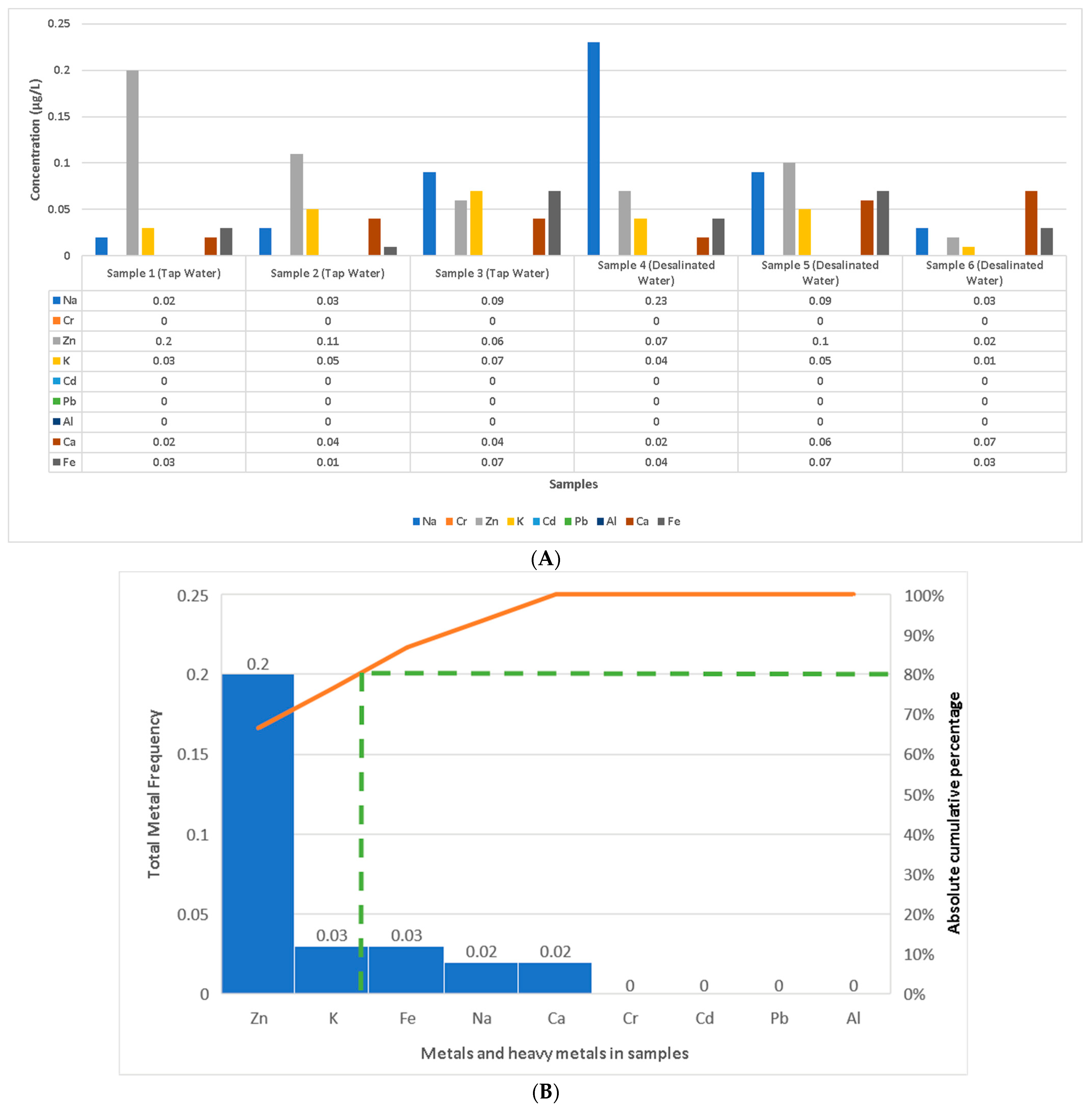
Figure 1. Experimental design [16,18,19,20,21,22].
Chemistry 06 00051 g001
Figure 2. Comparison of heavy metal and minerals, with concentrations expressed in µg/L: lead (Pb), cadmium (Cd), aluminum (Al), potassium (K) sodium (N), calcium (Ca), iron (Fe), zinc (Zn), and chromium (Cr). (A) Water samples 1–6 (sample 1 = Tap Water 1; sample 2 = Tap Water 2; sample 3 = Tap Water 3; sample 4 = Desalinated Water 1; sample 5 = Desalinated Water 2; sample 6 = Desalinated Water 3); (B) Pareto curve of heavy metals and minerals in descending order of concentration in water samples 1 to 6 according to the cumulative frequency rule (80/20). The orange line represents the cumulative effect of the studied samples in descending order. The green color represents the slope, which, according to the Pareto principle (or the law of the vital few and trivial many), states that for many heavy-metal concentrations, roughly 80% of the toxicity effects come from 20% of the samples on the left of the slope. This means that the water samples include many elements such as Zn, K, Fe, Na, and Ca but no heavy metals.
Chemistry 06 00051 g002
Figure 3. Results of the analysis of bacterial species in water samples and GSE: (A) percentages of Klebsiella pneumoniae, Staphylococcus epidermidis, Pseudomonas aeruginosa, and Staphylococcus aureus in each water sample and GSE, as separately analyzed in the present study, from sample 1 to sample 7 (sample 1 = Tap Water 1; sample 2 = Tap Water 2; sample 3 = Tap Water 3; sample 4 = Desalinated Water 1; sample 5 = Desalinated Water 2; sample 6 = Desalinated Water 3; sample 7 = GSE); (B) Pareto curve of contamination by studied bacteria accumulated in water samples 1 to 6 and GSE according to the cumulative frequency rule (80/20) in descending order: sample 4, sample 6, sample 5, sample 2, sample 1, and sample 3. The orange line represents the cumulative effect of the studied samples in descending order. The green color represents the slope, which, according to the Pareto principle (or the law of the vital few and trivial many), states that for many heavy-metal concentrations, roughly 80% of the toxicity effects come from 20% of the samples on the left of the slope.
Chemistry 06 00051 g003aChemistry 06 00051 g003b
Figure 4. Percentage of detected bacteria in urine samples (n = 100).
Chemistry 06 00051 g004
Figure 5. A comparison of the CB-Dock results of the highest binding energy between the studied ligands and the target receptors of the detected bacteria according to the Vina score.
Chemistry 06 00051 g005
Figure 6. Antimicrobial activity of different grape seed oil concentrations against detected bacterial strains.
Chemistry 06 00051 g006
Table 1. Results of gas chromatography–mass spectrometry analysis of GSE (grape seed oil extract).
No.NameBase PeakRT (min)Chromatogram
1.9-Octadecenamide, (Z)-5912.351–12.426Chemistry 06 00051 i001
2.Stigmastan-3,5-diene396.418.213–18.272Chemistry 06 00051 i002
3.9,12-Octadecadienoic acid (Z,Z)-, methyl ester81.112.308–12.351Chemistry 06 00051 i003
4.Oleamide5915.121–15.218Chemistry 06 00051 i004
5.Hexadecanamide5912.928–12.961Chemistry 06 00051 i005
6.Phosphoric acid, trimethyl ester1103.151–3.296Chemistry 06 00051 i006
7.Dodecyl acrylate559.564–9.591Chemistry 06 00051 i007
8.Pentanoic acid, 5-hydroxy-, 2,4-di-t-butylphenyl esters191.28.227–8.297Chemistry 06 00051 i008
9.Methyl stearate7412.485–12.511Chemistry 06 00051 i009
10.1-Pentadecyne5512.720–12.789Chemistry 06 00051 i010
11.11,13-Dimethyl-12-tetradecen-1-ol acetate20714.645–14.688Chemistry 06 00051 i011
12.4-Methyl-2,4-bis(4′-trimethylsilyloxyphenyl)pentene-120721.919–22.198Chemistry 06 00051 i012
13.1,3-Benzenediol, o-(4-methylbenzoyl)-o′-(2-methoxybenzoyl)-135.15.644–5.697Chemistry 06 00051 i013
14.Cyclotrisiloxane, hexamethyl-20717.411–17.469Chemistry 06 00051 i014
15.Isooctyl 3-mercaptopropionate5711.891–11.923Chemistry 06 00051 i015
RT: retention time (minutes); PA: peak area (%). The GC–MS investigation was performed on hexane/ethanol (1:6) extracts with Agilent Technologies equipment (G3440B; Santa Clara, CA, USA). The phytochemical contents of the GSE were standardized with computer simulations in the commercial libraries of Wiley and the NIST (National Institute of Standards and Technology).
Table 2. Comparison between the chemical components of GSE (grape (Vitis vinifera) seed oil extract) and standard antibiotic (doxycycline) in potential CB-Dock interaction scenarios with selected protein receptors in the bacterial species detected.
LigandTarget Protein Receptor (PDB)
1.CID 525918
Stigmastan-3,5-diene
Klebsiella pneumoniae
5HFT
Chemistry 06 00051 i016
4HWM
Chemistry 06 00051 i017
Pseudomonas aeruginosa3OS prokaryotic ribosoma (binding unit of antibiotic)
4F1R
Chemistry 06 00051 i018
3OS3
Chemistry 06 00051 i019
Staphylococcus aureus
1DUA
Chemistry 06 00051 i020
1DUE
Chemistry 06 00051 i021
8EXP
Chemistry 06 00051 i022
LigandTarget Protein Receptor (PDB)
2.CID 5284421
9,12-Octadecadienoic acid (Z,Z)-, methyl ester
Klebsiella pneumoniae
5HFT
Chemistry 06 00051 i023
4HWM
Chemistry 06 00051 i024
Pseudomonas aeruginosa3OS prokaryotic ribosoma (binding unit of antibiotic)
4F1R
Chemistry 06 00051 i025
3OS3
Chemistry 06 00051 i026
Staphylococcus aureus
1DUA
Chemistry 06 00051 i027
1DUE
Chemistry 06 00051 i028
8EXP
Chemistry 06 00051 i029
LigandTarget Protein Receptor (PDB)
3.CID 5283387
Oleamide
Klebsiella pneumoniae
5HFT
Chemistry 06 00051 i030
4HWM
Chemistry 06 00051 i031
Pseudomonas aeruginosa3OS prokaryotic ribosoma (binding unit of antibiotic)
4F1R
Chemistry 06 00051 i032
3OS3
Chemistry 06 00051 i033
Staphylococcus aureus
1DUA
Chemistry 06 00051 i034
1DUE
Chemistry 06 00051 i035
8EXP
Chemistry 06 00051 i036
LigandTarget Protein Receptor (PDB)
4.CID 69421
Hexadecanamide
Klebsiella pneumoniae
5HFT
Chemistry 06 00051 i037
4HWM
Chemistry 06 00051 i038
Pseudomonas aeruginosa3OS prokaryotic ribosoma (binding unit of antibiotic)
4F1R
Chemistry 06 00051 i039
3OS3
Chemistry 06 00051 i040
Staphylococcus aureus
1DUA
Chemistry 06 00051 i041
1DUE
Chemistry 06 00051 i042
8EXP
Chemistry 06 00051 i043
LigandTarget Protein Receptor (PDB)
5.CID 10541
Phosphoric acid, trimethyl ester
Klebsiella pneumoniae
5HFT
Chemistry 06 00051 i044
4HWM
Chemistry 06 00051 i045
Pseudomonas aeruginosa3OS prokaryotic ribosoma (binding unit of antibiotic)
4F1R
Chemistry 06 00051 i046
3OS3
Chemistry 06 00051 i047
Staphylococcus aureus
1DUA
Chemistry 06 00051 i048
1DUE
Chemistry 06 00051 i049
8EXP
Chemistry 06 00051 i050
LigandTarget Protein Receptor (PDB)
6.CID 75084
Dodecyl acrylate
Klebsiella pneumoniae
5HFT
Chemistry 06 00051 i051
4HWM
Chemistry 06 00051 i052
Pseudomonas aeruginosa3OS prokaryotic ribosoma (binding unit of antibiotic)
4F1R
Chemistry 06 00051 i053
3OS3
Chemistry 06 00051 i054
Staphylococcus aureus
1DUA
Chemistry 06 00051 i055
1DUE
Chemistry 06 00051 i056
8EXP
Chemistry 06 00051 i057
LigandTarget Protein Receptor (PDB)
7.CID 605777
Pentanoic acid, 5-hydroxy-, 2,4-di-t-butylphenyl esters
Klebsiella pneumoniae
5HFT
Chemistry 06 00051 i058
4HWM
Chemistry 06 00051 i059
Pseudomonas aeruginosa3OS prokaryotic ribosoma (binding unit of antibiotic)
4F1R
Chemistry 06 00051 i060
3OS3
Chemistry 06 00051 i061
Staphylococcus aureus
1DUA
Chemistry 06 00051 i062
1DUE
Chemistry 06 00051 i063
8EXP
Chemistry 06 00051 i064
LigandTarget Protein Receptor (PDB)
8.CID 8201
Methyl stearate
Klebsiella pneumoniae
5HFT
Chemistry 06 00051 i065
4HWM
Chemistry 06 00051 i066
Pseudomonas aeruginosa3OS prokaryotic ribosoma (binding unit of antibiotic)
4F1R
Chemistry 06 00051 i067
3OS3
Chemistry 06 00051 i068
Staphylococcus aureus
1DUA
Chemistry 06 00051 i069
1DUE
Chemistry 06 00051 i070
8EXP
Chemistry 06 00051 i071
LigandTarget Protein Receptor (PDB)
9.CID 69825
1-Pentadecyne
Klebsiella pneumoniae
5HFT
Chemistry 06 00051 i072
4HWM
Chemistry 06 00051 i073
Pseudomonas aeruginosa3OS prokaryotic ribosoma (binding unit of antibiotic)
4F1R
Chemistry 06 00051 i074
3OS3
Chemistry 06 00051 i075
Staphylococcus aureus
1DUA
Chemistry 06 00051 i076
1DUE
Chemistry 06 00051 i077
8EXP
Chemistry 06 00051 i078
LigandTarget Protein Receptor (PDB)
10.CID 549821
11,13-Dimethyl-12-tetradecen-1-ol acetate
Klebsiella pneumoniae
5HFT
Chemistry 06 00051 i079
4HWM
Chemistry 06 00051 i080
Pseudomonas aeruginosa3OS prokaryotic ribosoma (binding unit of antibiotic)
4F1R
Chemistry 06 00051 i081
3OS3
Chemistry 06 00051 i082
Staphylococcus aureus
1DUA
Chemistry 06 00051 i083
1DUE
Chemistry 06 00051 i084
8EXP
Chemistry 06 00051 i085
LigandTarget Protein Receptor (PDB)
11.CID 91715040
1,3-Benzenediol, o-(4-methylbenzoyl)-o′-(2-methoxybenzoyl)-
Klebsiella pneumoniae
5HFT
Chemistry 06 00051 i086
4HWM
Chemistry 06 00051 i087
Pseudomonas aeruginosa3OS prokaryotic ribosoma (binding unit of antibiotic)
4F1R
Chemistry 06 00051 i088
3OS3
Chemistry 06 00051 i089
Staphylococcus aureus
1DUA
Chemistry 06 00051 i090
1DUE
Chemistry 06 00051 i091
8EXP
Chemistry 06 00051 i092
LigandTarget Protein Receptor (PDB)
12.CID 104386
Isooctyl 3-mercaptopropionate
Klebsiella pneumoniae
5HFT
Chemistry 06 00051 i093
4HWM
Chemistry 06 00051 i094
Pseudomonas aeruginosa3OS prokaryotic ribosoma (binding unit of antibiotic)
4F1R
Chemistry 06 00051 i095
3OS3
Chemistry 06 00051 i096
Staphylococcus aureus
1DUA
Chemistry 06 00051 i097
1DUE
Chemistry 06 00051 i098
8EXP
Chemistry 06 00051 i099
LigandTarget Protein Receptor (PDB)
13.CID 54671203
Standard antibiotic (doxycycline)
Klebsiella pneumoniae
5HFT
Chemistry 06 00051 i100
4HWM
Chemistry 06 00051 i101
Pseudomonas aeruginosa3OS prokaryotic ribosoma (binding unit of antibiotic)
4F1R
Chemistry 06 00051 i102
3OS3
Chemistry 06 00051 i103
Staphylococcus aureus
1DUA
Chemistry 06 00051 i104
1DUE
Chemistry 06 00051 i105
8EXP
Chemistry 06 00051 i106
Disclaimer/Publisher’s Note: The statements, opinions and data contained in all publications are solely those of the individual author(s) and contributor(s) and not of MDPI and/or the editor(s). MDPI and/or the editor(s) disclaim responsibility for any injury to people or property resulting from any ideas, methods, instructions or products referred to in the content.