Article

Extended Chalcones: Synthesis, In Vitro Analysis, and In Vivo Testing Against a Drosophila melanogaster Alzheimer’s Disease Model

Massachusetts College of Pharmacy and Health Sciences, Boston, MA 02115, USA
*
Author to whom correspondence should be addressed.
Chemistry 2024, 6(6), 1477-1494; https://doi.org/10.3390/chemistry6060089
Submission received: 17 October 2024 / Revised: 19 November 2024 / Accepted: 20 November 2024 / Published: 22 November 2024
(This article belongs to the Section Medicinal Chemistry)

Abstract

:
Alzheimer’s Disease (AD) is the most common form of dementia in individuals over the age of 65. There is no known prevention for the progression of the disease, although the FDA recently approved two drugs for AD. The exact etiology of AD is still under debate; however, it is commonly associated with the aggregation of amyloid-beta (Aβ) plaques in the brain. Recently some extended chalcones were reported to be potential anti-amyloidogenic agents. In this study, a larger library of extended chalcone analogs were synthesized with modifications on both rings. These were tested using the Thioflavin T fluorescence assay to measure their anti-Aβ aggregation properties. Three notably active compounds were further evaluated for potential neurotoxicity and neuroprotection using an MTT cell viability assay. These compounds were non-neurotoxic and displayed a trend toward neuroprotection. These were further assessed in a Drosophila melanogaster animal AD model. A forced climbing assay revealed statistically significant changes in flies’ movement by ~30% when fed these anti-amyloidogenic agents.

Graphical Abstract

1. Introduction

Alzheimer’s Disease (AD) is clinically distinguished by the progressive and gradual decline in cognitive function of those afflicted, and ultimately leads to death [1]. The damage to the brain may precede the visible symptoms by up to 20 years [2]. As the disease progresses, symptoms such as depression, mood swings, decreased physical function, and impaired cognition appear, ultimately leading to death [3]. The pathophysiological hallmarks of AD include a reduction in levels of acetylcholine (ACh), intracellular flame-shaped neurofibrillary tangles (NFTs) of hyperphosphorylated tau proteins, and extracellular deposits of the amyloid-β peptide (Aβ), which form senile plaques [4,5].
One of the main hallmarks of AD is the accumulation of neurotoxic aggregates of Aβ plaques. The amyloid cascade hypothesis suggests that the Aβ plaques initiate mitochondrial oxidative stress, promoting hyperphosphorylation of tau, resulting in the aforementioned neurotoxicity [5]. The Aβ peptide is derived from the catabolism of the amyloid precursor protein (APP), a type 1 transmembrane glycoprotein presents mainly in the neuronal and glial cells of the brain [6]. APP can be catabolized via two alternative pathways: the non-amyloidogenic (physiological conditions) or the amyloidogenic pathway (pathological conditions) [7,8]. In the former pathway, APP proteolysis occurs via the membrane-bound enzyme, α-secretase, to release sAPPα, leaving behind an 83 amino acid C-terminal APP fragment (CTFα) [7]. Further cleavage of the CTFα by γ-secretase yields an extracellular non-toxic soluble fragment (P3) and the APP intracellular domain (AICD). However, under pathological conditions, APP undergoes cleavage by β-secretase (BACE-1) then γ-secretase at the N and C termini, respectively. When β-secretase cleaves APP, it leads to the release of sAPPβ and a membrane-tethered, 99 amino acid, C-terminal (CTFβ) unit [9]. Subsequently, CTFβ is cleaved by γ-secretase, liberating AICD and extracellular Aβ peptides [10,11]. The Aβ monomers can aggregate into oligomers to form insoluble fibrils, which further arrange themselves into a β-sheet conformation. The β-sheets cumulatively form ordered fibrils, known as β-plaques [12]. The presence of β-amyloid plaques in the brain marks a characteristic feature of AD. These plaques interfere in neuronal signaling, which disrupts brain cell functions. The result is a loss of neuronal synapses, progressive decline in neurotransmitter activity, inflammation, and neuronal cell death [13]. Additionally, Aβ can affect microtubule stability by increasing Ca2+ import and activating protein kinases, leading to the aforementioned tau hyperphosphorylation [14,15].
Amyloid inhibitor therapies have been attempted to reduce Aβ peptide production, either via: α-secretase stimulation, inhibition of γ-secretase, and/or inhibition of β-secretase [16]. Another approach being taken is to develop amyloid antiaggregant therapies, either to inhibit Aβ fibrillation and/or oligomerization, or to clear out existing plaques. In 2022, aducanumab, a human immunoglobulin gamma 1 (IgG1) monoclonal antibody targeting Aβ oligomers, developed by Biogen, was approved by the FDA and became the first drug to treat AD [17]. More recently, the FDA has approved Biogen’s lecanemab, another monoclonal antibody, and Eli Lilly’s, donanemb, both of which bind to and remove the aggregated Aβ fibrils [18]. On the small molecule front, homotaurine and curcumin have shown mixed results. Recently, we developed a library of extended chalcones that displayed low µM inhibition of plaque formation (Figure 1). It was reported that having a dialkylamino substituent on Ring B was a necessity (although a full study had not been conducted) [5]. Only a small library of compounds with varying substituents on Ring A had been assessed. Thus, we began this study with a simple hypothesis that altering the substituents on Ring A would affect the anti-amyloidogenic nature of these extended chalcones. It was unknown whether the electronic, steric, or lipophilic/hydrophilic nature of the substituent would increase or decrease this effect; however, we began with the intent to utilize the compound(s) with the most promising in vitro results and see if this would translate over to a tangible physiological benefit in animal models. Herein is the expansion of this prior work, with the first set of animal studies on our most promising analogs [5].

2. Results and Discussion

Based on our previously published work, extended chalcones 1, 2, and 3 (Figure 1A) were found to have the lowest IC50 values as anti-amyloidogenic compounds [5]. In order to expand upon the library of compounds, we focused first on a more thorough analysis of the substitution and pattern on Ring A, with some subsequent analysis of the amino group on Ring B (Figure 1B) in the aim to develop more potent anti-amyloidogenic agents. As a result of our earlier work, analogs were designed to maintain an electron donating group on Ring B, while with Ring A, different substituents at varying positions were synthesized to assess the electronics and steric constraints.
To synthesize the library of compounds with different substituents on Ring A, we followed the procedure of our previous work [5]. Briefly, an aldol condensation reaction was carried out between acetophenone derivatives and 4-(dimethylamino)cinnamaldehyde dissolved in ethanol, using 10% NaOH as the base (Scheme 1). The product precipitated out of the solution as the reaction progressed. After 24 h, ice chips were added to the reaction mixture to increase precipitation and improve the yield. The solid was collected by vacuum filtration and washed with a mixture of cold water and ethanol to produce the desired extended chalcone in good to excellent yields, without any need of further purification. Some reactions produced oils rather than solids. In those cases, the oil was concentrated and purified using flash column chromatography. 1H-NMR was utilized to confirm the success of reactions, as well as their purity.
The acetophenone derivatives used had varying electronic and steric effects to provide an understanding of the structure activity relationship (SAR). To study the effect of lipophilicity, alkyl analogs were synthesized ranging from the previously synthesized methyl to isopropyl, isobutyl, and cyclohexyl groups at the para position (Table 1). Additionally, to investigate the electronic effects of substituent nature on Ring A, both electron withdrawing groups (EWGs) and electron donating groups (EDGs) were incorporated at the para position. The EWGs included nitro, cyano, and acetyl substituents, while the EDGs included methoxy, ethoxy, and methylthio groups. Halogens, as well as trifluoromethyl analogs, which impacted both solubility and electronics, were synthesized. To explore the effects of position of the substituents, chloro, and trifluoromethyl group analogs were additionally synthesized at the ortho and meta position of Ring A. To further explore the electronic and steric effects, a small number of di-substituted extended chalcones were also synthesized.
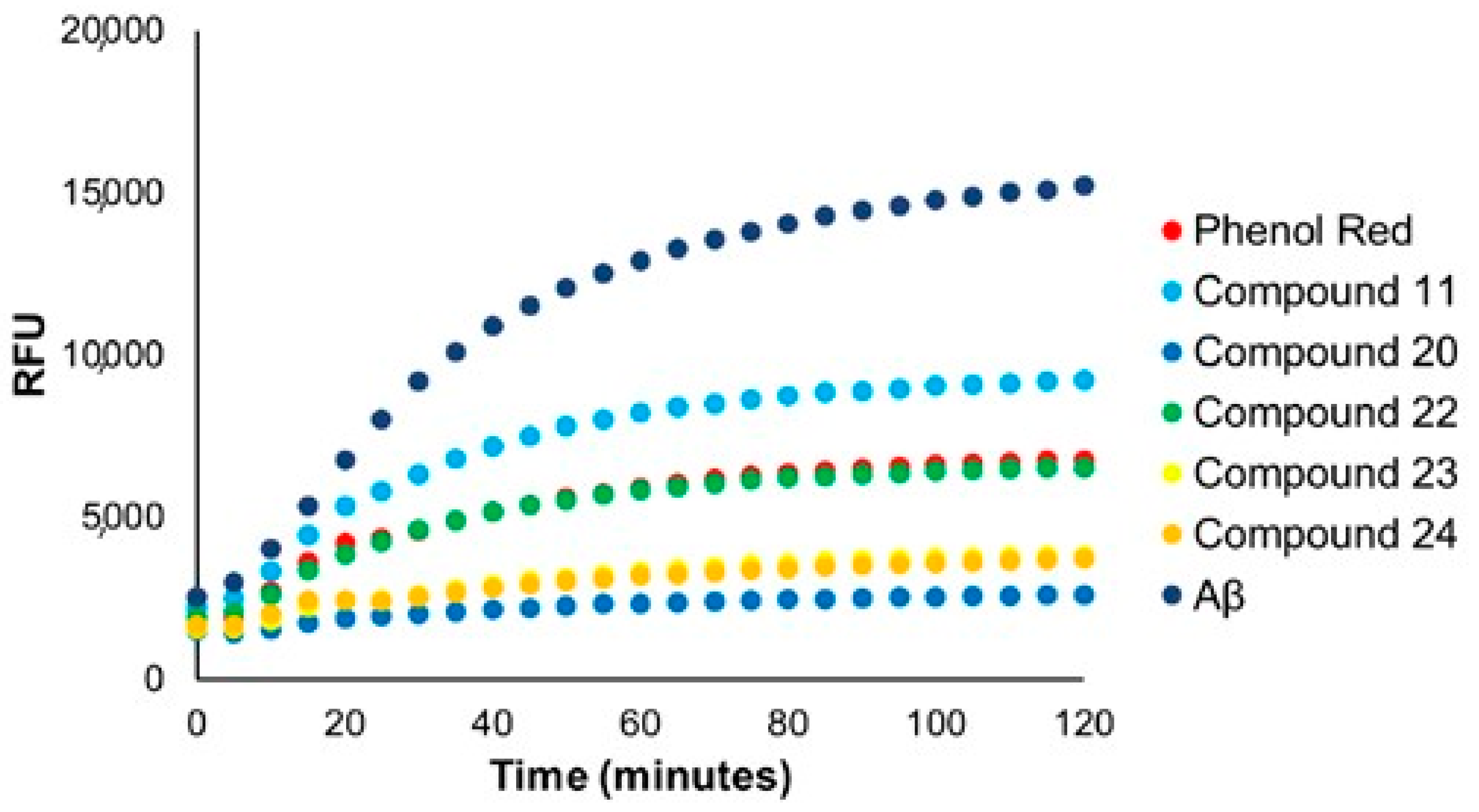
All newly synthesized compounds were screened for their anti-amyloidogenic activity on Aβ1-42 peptides, using Phenol Red as the internal standard—all at 100 μM [5]. Ultimately, our compounds were significantly superior to control, necessitating the need to lower their concentration to 10 μM and compare to Phenol Red at 100 μM (Figure 2). To obtain the IC50 values, all compounds were tested at a minimum of five different concentrations (Table 1).
Comparing Compounds 1, 2, 4, and 5, upon increasing the length of the alkyl substituent (–H, –CH3, ethyl, n-propyl) and subsequent clogP values [19], a decrease in the IC50 value was observed, suggesting that as lipophilicity increases, anti-aggregation activity of the compound improves. Comparing Compound 5 to 6, going from n-propyl to i-propyl, a slight decrease in activity was observed. However, Compound 8 with the cyclohexyl substituent and a clogP value of 5.59 [19], displayed a reduction in anti-aggregation activity. This suggested that a lipophilic, unbranched substituent was favorable.
Halogen substituents also improved aggregation inhibition in comparison to the unsubstituted extended chalcone (1). The p-chloro analog (3) had the highest activity compared to the other halogen derivatives, followed by p-bromo (9), and lastly p-iodo (10). Since Compounds 3, 9, and 10 have similar lipophilicities, the decrease in activity may be a result of the decrease in electronegativity. Other highly electron-withdrawing groups such as trifluoromethyl (11) and cyano (13) at the para position, although better than the unsubstituted chalcone (1), displayed higher IC50 values than many others.
The evaluation of electron donating groups was possible when comparing Compounds 15 to 16. The latter with an ethoxy group displayed better anti-amyloidogenic activity than the former, containing a methoxy substituent. As indicated earlier, increasing lipophilicity tends to increase activity, up to a certain point. Switching from an oxygen atom (15) to sulfur in Compound 18, led to a drop in the IC50 value, from 3.2 to 1.5 μM. This could be owed to the progressive increase in lipophilicity, suggesting a p-ethylthio containing analog should be synthesized and tested.
To test the effect of the substituent position, m-trifluoromethyl (20), o-trifluoromethyl (22), m-chloro (21), and o-chloro (24) containing analogs were synthesized. It was observed that for both the substituents, the meta substituted analog had a noticeable decrease in its IC50 values. Comparing para vs. ortho substituted compounds, Compound 2 (p-methyl) versus 20 (o-methyl), the ortho substitutions displayed better activity. From these data, it is suggested that the ideal mono substituted position is meta > ortho > para.
Lastly, the effect of di-substituted derivatives on Ring A was studied. Compounds 25 and 26, both di-substituted chloro analogs, showed comparable or better activity than their mono-substituted counterparts (i.e., 3, 21, and 24). The 3,5-dichloro analog (25) had the lowest IC50 value of 0.91 μM of all the Ring A modified analogs. This suggests that di-substitution at the meta-position may lead to significant improvement in anti-aggregation activity. Collectively, these data suggest that increasing lipophilicity leads to an improvement in anti-aggregation activity of extended chalcones, up to a certain point. Furthermore, di-substituted extended chalcones could lead to further enhancement of activity.
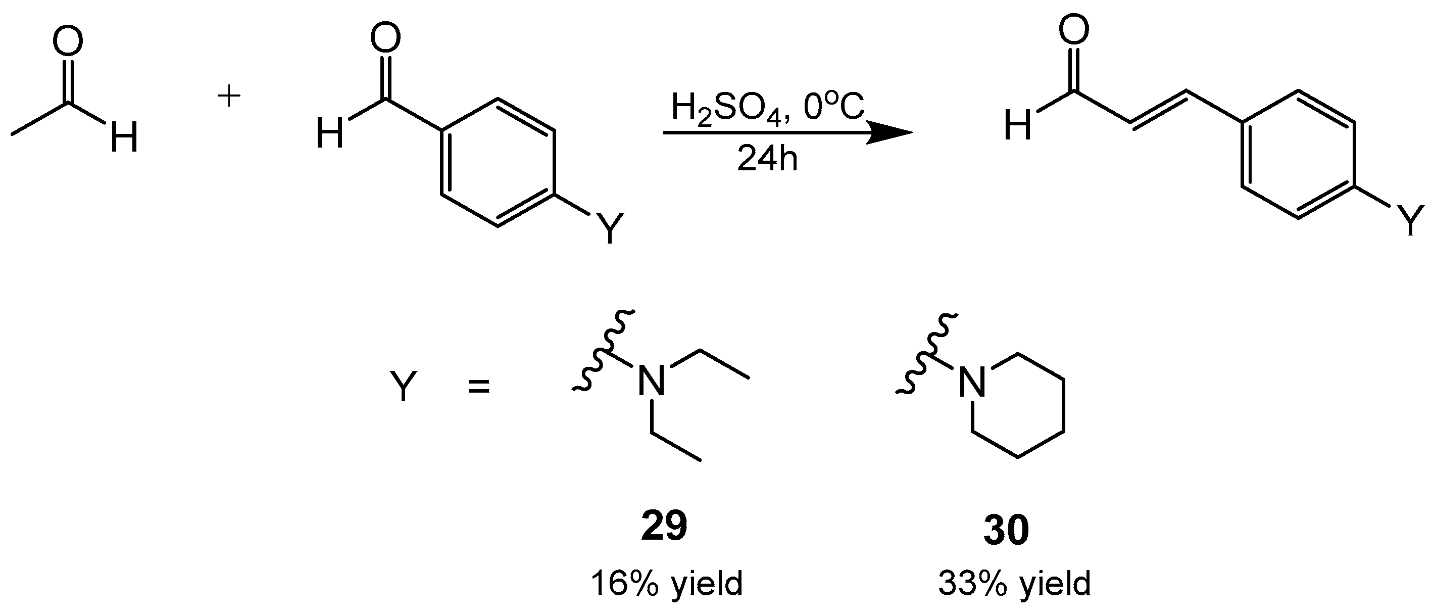
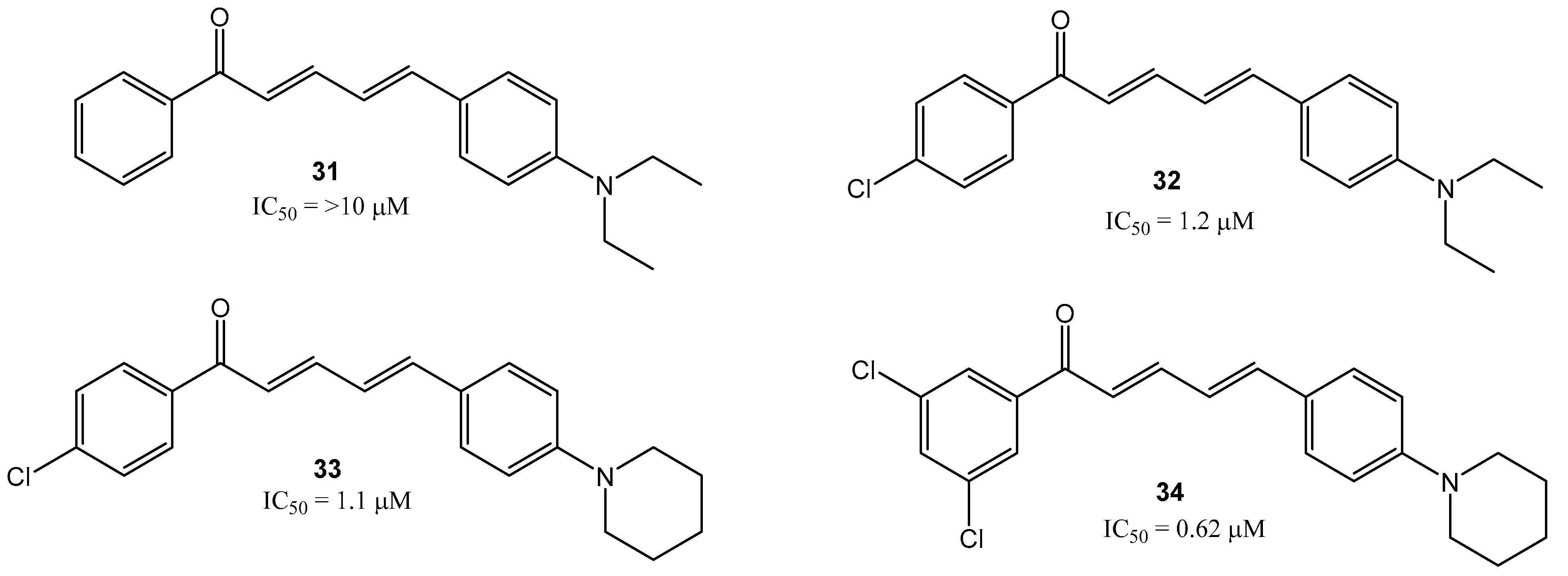
Next modifications on Ring B were made to add more lipophilic groups while maintaining the electron donating nature of the amino substituent. It was thus necessary to first synthesize para-amino substituted cinnamaldehydes. Initially, a Wittig reaction was attempted; however, a lower than ideal yield was produced. Ultimately, an acid catalyzed aldol condensation reaction was used for the synthesis of cinnamaldehyde derivatives (Scheme 2). This acid catalyzed condensation reaction involved initially dissolving a p-aminobenzaldehyde in concentrated H2SO4 under vigorous stirring and cooling to 0 °C. The acetaldehyde was then added dropwise over 4 h. The reaction mixture was poured over ice and brought to an alkaline pH (20% NaOH). The product was extracted with dichloromethane and purified by flash chromatography to obtain the pure product in moderate yields. 1H-NMR was utilized to confirm the success of the reactions as well as their purity. To explore the effects of increasing the alkyl chain length, 4- (diethylamino)cinnamaldehyde (29) and 4-(piperidin-1-yl)cinnamaldehyde (30) were synthesized and further coupled, using the aforementioned aldol condensation (Scheme 1) with acetophenone, 4′-chloroacetophenone and 3′,5′-dichloroacetophenone to yield the extended chalcones (3134) (Figure 3).
All four compounds were evaluated for their IC50 values (Figure 3). Similar to the Ring A modification library, upon increasing the clogP value from 4.45 for Compound 3, to 5.07 and 5.13 for compounds 33 and 32, respectively [19]; a decrease in IC50 values was observed from, 2.4 to 1.1 to 1.0 μM. This followed the previously observed trend that suggested a correlation between lipophilicity and anti-amyloidogenic activity. The 3,5-dichloro analog (25), which showed the lowest IC50 value (0.91 μM) of all Ring A modifications, ultimately produced the most effective anti-amyloidogenic compound when coupled with 4-(piperidin-1-yl)cinnamaldehyde, specifically compound 34, with an IC50 value of 0.62 μM.
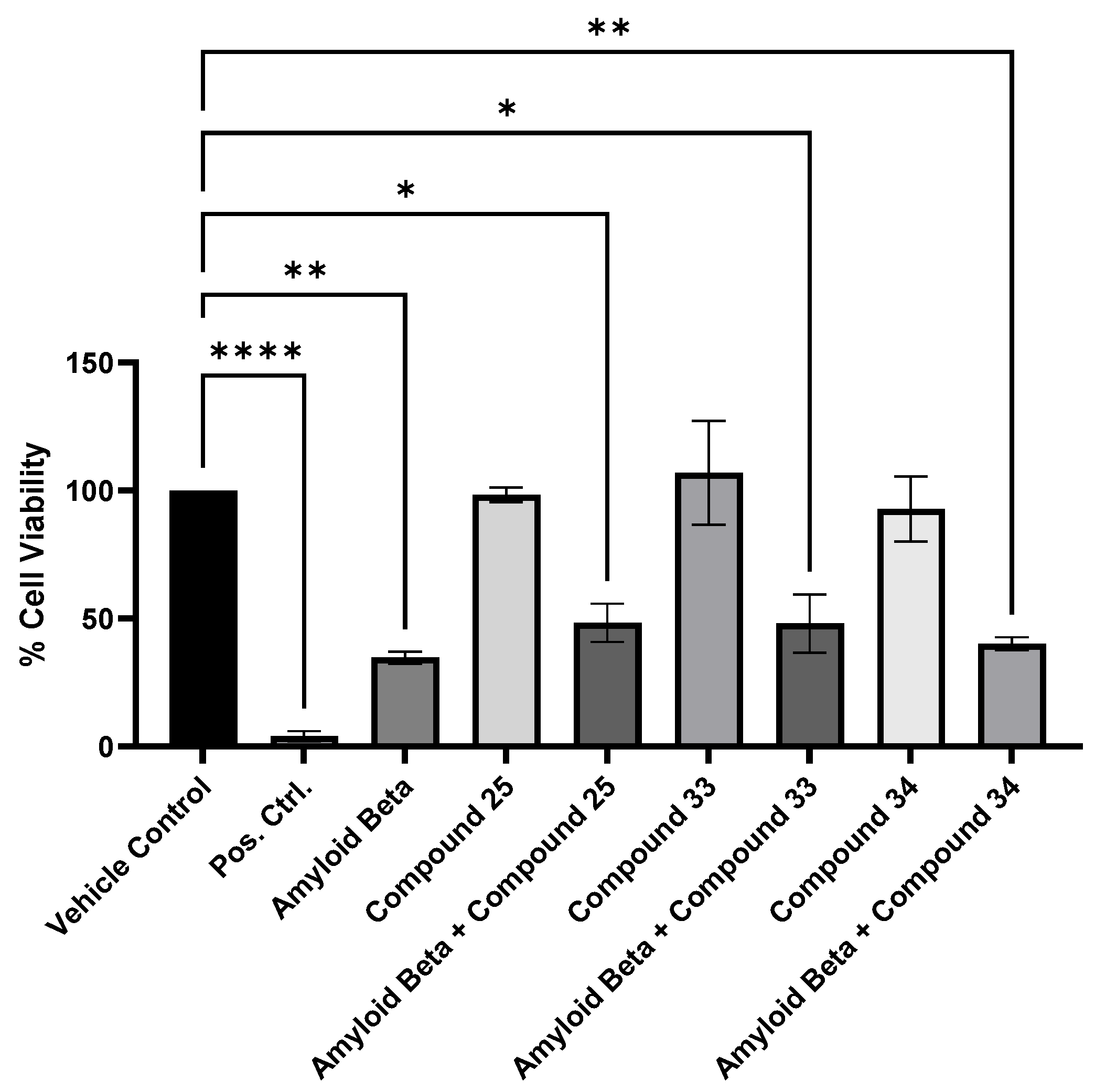
As with our earlier work, we also looked into the cell viability/cell cytotoxicity of our compounds that displayed the greatest anti-amyloidogenic activity [5]. Thus, we employed the 3-(4,5-dimethylthioxzol-2-yl)-2,5-diphenyltretazolium bromide (MTT) assay to assess the cell viability of SH-SY5Y cells when exposed to Compounds 25, 33, and 34 at their IC50 values (Figure 4). Aβ42 alone led to an ~65% decrease in cell viability, corresponding with its well-known neuro-cytotoxicity. Fortunately, all three compounds alone showed no cytotoxicity when administered at their corresponding IC50 value concentrations. When our extended chalcones (again at their IC50 value concentrations) were administered concurrently with Aβ42, a trend toward improvements in cell viability was observed. Notably, the Aβ42-induced cytotoxicity in SH-SY5Y cells after 48 h went from the aforementioned ~65% decrease in cell viability to only a 52, 52, and 60% decrease, when co-administered with Compounds 25, 33, and 34, respectively; although all were just outside of statistical significance.
As the compounds tested displayed notable anti-amyloidogenic activity, no cell cytotoxicity, but no statistically significant neuroprotection when co-administered with Aβ42, we next explored their in vivo activity. We initially tested these compounds on AD-modeled, genetically modified Caenorhabditis elegans (C. elegans, CL2006) [20]. This model of C. elegans are predisposed to developing the aforementioned plaques. We tested compounds at multiple concentrations and observed their mobility, after different time points, when exposed to extended chalcones. The C. elegans displayed notable, partial or complete, reversal of the physiological AD marker, specifically, a lack of mobility. Due to these encouraging results, we focused the remainder of our in vivo work on Drosophila melanogaster (D. melanogaster) that overexpressed Aβ.
To produce our transgenic line of interest, we crossed UAS-Aβ 1-42 female virgins with nSyb-Gal4 males. UAS-Aβ 1-42 has a TM3, Sb balancer on the third chromosome, resulting in the mutant Sb gene, indicative of stubble bristles as a dominant marker [21]. When crossed with the driver line, such as nSyb-Gal4, the transgenic line will either be UAS-Aβ 1-42/nSyb-Gal4 or nSyb-Gal4/TM3, Sb. The transgenic line of interest is the F1 progeny that expressed Aβ 1-42 peptide (UAS-Aβ 1-42/nSyb-Gal4), which have the normal bristle phenotype. All offspring with the inherited balanced nSyb-Gal4TM3, Sb was negatively selected. Once we had generated enough F1 progeny of the transgenic line of interest, we began to test the compounds (Figure 5).
The extended chalcones of interest were dissolved in DMSO to a concentration of 5 mg/mL. This solution was then further diluted into fly food with final concentrations of 1, 10, and 100 mcg/mL of fly food, corresponding to 0.0001%, 0.001%, and 0.01% (w/v), respectively. These three concentrations were then introduced to each respective group. The F1 progeny that express Aβ42 peptide (UAS-Aβ 1-42/nSyb-Gal4), were placed onto the food fortified with the different concentrations of Compound 25, 33, or 34 for 15 days.
Initially, a lifespan assay was performed. Wild Type1118 Bonini flies were placed into corresponding vials of food containing the compounds at the various concentrations. Over the course of the 15 days, both wild type and the transgenic line flies were counted in each vial to determine how many living flies remained. The flies were tipped onto new food with the same compound and concentration three times during the course of the study to avoid potential deaths due to the food becoming too moist from larvae being laid. After the 15 days, the total number of living flies were counted, and a Kaplan–Meier survival was generated. As anticipated, no statistically significant changes in survival between the WT and transgenic line were observed, due to the lack of neurotoxicity previously observed for these compounds (Figure 4). Vials with F2 larvae were kept, after removing the F1 flies, to observe the F2 generation after being exposed to the compounds at various concentrations for the entirety of their life. Interestingly, all vials containing the compounds at the highest concentration had no larvae present. In contrast, at 0.001 and 0.0001%, larvae were present, suggesting that this is a concentration dependent phenomenon.
To investigate the potential physiological benefits of our extended chalcones, a forced climbing assay was conducted on transgenic flies when exposed to our compounds. Flies that overexpress Aβ should exhibit age dependent defects in climbing [22]; conversely, flies that do not overexpress Aβ should exhibit negative geotaxis, allowing them to climb to the top of the vial in 10 s or less. Thus, we conducted this assay five times a day, beginning on the seventh day after the flies were exposed to the compounds. Vials were marked with a top, middle, and bottom sections, separated by one inch. Flies had 10 s to climb after being tapped to the bottom. The percent total in each section, for each trial was calculated and a one-way ANOVA was preformed to compare the percentage of flies in the top, middle, and bottom of the control vs. the transgenic flies when exposed to Compounds 25, 33, and 34 at various concentrations (Figure 6).
Compound 25 at 1 mcg/mL exhibited a statistically significant change in the percentage of flies in the bottom and middle when compared to the control. For example, in the control group, the bottom section contained ~95% of the flies, while flies fed Compound 25 saw a drop to 82.15%. This change corresponded to an increase in flies in the middle section, from 4.33% to 15.56%, for the control and Compound 25, respectively. Transgenic flies fed Compound 33 at 1 mcg/mL also showed a statistically significant change in the percentage in each section of the vial, with the bottom containing 66.00%, middle 26.00%, and the top an impressive 8.00% (compared to the control with 0.33%). Compound 33 also showed statistical significance improvements at 10 mcg/mL (bottom: 74.00% and middle: 22.33%). Finally, Compound 34 showed statistically significant improvements at both 10 and 100 mcg/mL, with 82.50% and 81.26% of the flies in the bottom section and 15.00% and 14.90% in the middle, respectively.
The forced climbing assay results indicate that our extended chalcones exert a physiological effect on the flies with Compound 33 at 1 mcg/mL displaying the greatest effect. Although a larger portion of the flies, for all compounds and concentrations, still remained in the bottom section of the vials, this climbing improvement showcases a clear physiological benefit on the transgenic flies that overexpress Aβ.
To ensure that the compounds only inhibited the formation of the plaques and not the production of Aβ, an enzyme-linked immunosorbent assay (ELISA), using the Human Aβ42 ELISA Kit (Invitrogen: #KHB3441) was performed to quantify the levels of Aβ. The treated and control transgenic flies previously used for the climbing assay were decapitated, and the heads were split among four tubes, to which 100 mcL of lysis buffer was added. Heads were then homogenized using sonication for 30 min, and frozen (−18 °C) until the ELISA was performed.
To perform the ELISA, the samples were first thawed on ice. A protease inhibitor cocktail with AEBSF (a serine protease inhibitor) was added to each sample, with a final concentration of 1 mM, and were then centrifuged to remove the large particulate matter. After centrifugation, samples were diluted with Standard Diluent Buffer at a 1:50 ratio to ensure sample concentrations would be within the range of the standard curve. A serial dilution with native human Aβ42 (Hu Aβ42) was performed in the Standard Diluent Buffer to generate a standard curve. To bind the antigen, 50 mcL of the standard, sample, and control was added to the appropriate wells, with duplicates of each, plus 50 mcL of the detector (Hu Aβ42 Detection Antibody solution). The plate was covered and incubated at room temperature with shaking. After 3 h, the solution was thoroughly aspirated, and the wells were washed four times with 1X Wash Buffer. Anti-Rabbit IgG HRP (100 mcL) was added to every well, and the plate was covered and incubated at room temperature. After 30 min, the solution was thoroughly aspirated, and the wells were washed four times with 1X Wash Buffer. Stabilizing Chromogen (100 mcL) was added to each well and the plate was incubated in the dark. After 30 min, 100 mcL of the Stop Solution was added to every well and the plate was read immediately at an absorbance of 450 nm.
Absorbance measures for each sample and control were averaged, and from the calibration curve, the corresponding level of Aβ was determined. Data were analyzed using a one-way ANOVA. Regardless of what compound was fed to the flies, and at what concentration, no statistically significant change in the levels of Aβ was observed. This further supports our hypothesis that these compounds work by inhibiting plaque formation, opposed to Aβ42 generation. Thus, the physiological benefit observed when the flies were fed either Compound 25, 33, or 34 at varying concentrations, is most likely a result of a decrease in brain plaque burden.

3. Conclusions

Based on our previously published work [8], a more thorough library of extended chalcones was synthesized and tested as potential anti-amyloidogenic agents. Twenty-two modifications were made to Ring A and four to Ring B of the molecular scaffold (Figure 1B), and the effect of substituents was studied. Generally, but not consistently, electron donating groups were found to be superior to electron withdrawing groups on Ring A. Upon increasing the lipophilicity, the anti-aggregation activity also increased, up to a certain point. The meta-position was found to be ideal for mono-substitution on Ring A. Compounds that were di-substituted on Ring A at the meta-positions displayed superior activity. Increasing the lipophilicity of the electron donating group on the para position on Ring B led to an improvement in the anti-aggregation activity. We further assessed the three compounds that displayed the greatest anti-amylogenic activity, namely Compounds 25, 33, and 34, for their toxicity and neuroprotection. None were deemed toxic, and all trended toward displaying neuroprotection. Subsequent testing on an animal model revealed that there were statical improvements in the forced climbing assay of the compounds of up to ~30%, with no alteration in the brain plaque burden. This suggests that, as hypothesized, these compounds do not affect the rate or quantity of AB42 generation, but rather prevent their subsequent aggregation. Thus, there is a potential application of these compounds as an earlier prevention of plaque formation or as an add-on treatment once the brain plague has been removed.

4. Experimental

4.1. Chemistry

4.1.1. General Information

All reagents were purchased from Millipore Sigma (Burlington, VT, USA) and used without further purification. All synthesized compounds were purified using flash column chromatography. 1H-NMR and 13C-NMR were recorded at 400 MHz on a Bruker Topspin 4.2.0 instrument (Bruker Corp., Billerica, MA, USA). EA was performed on a Carlo Erba 1108 elemental analyzer (Thermo Fisher Scientific, Waltham, MA, USA), performed by Atlantic Microlab, Inc. (Norcross, GA, USA). Compounds 6 [23], 9 [24], 10 [24], 15 [25], 16 [26], 17 [27], 23 [28], 24 [23], and 28 [28] were previously reported and compared to for authenticity. For in vitro studies, a fluorometric assay was performed in 96 non-binding microplates from Greiner Bio-One with a clear bottom on a Synergy Bio-tek HTS plate reader (Agilent Technologies, Lexington, KY, USA).

4.1.2. Example Aldol Condensation

  • (2E,4E)-5-(4-(dimethylamino)phenyl)-1-(4-ethylphenyl)penta-2,4-dien-1-one (4)
To a solution of 4′-ethylacetophenone (0.2719 g, 1.83 mmol) in absolute ethanol (10 mL) was added to an aqueous solution of 10% NaOH (5 mL) at 0 °C. The mixture was stirred for 15 min, after which 4-(dimethylamino)cinnamaldehyde (0.2963 g, 1.69 mmol) was added. The reaction mixture was then stirred at room temperature for 24 h. The precipitated product was vacuum filtered and washed with small portions of water/ethanol to yield the desired extended chalcone as a red solid (0.4784 g, 92%). Melting point: 126.9–128.4 °C. 1H NMR (400 MHz, CDCl3) δ 7.91 (d, J = 7.8 Hz, 2H), 7.62 (dd, J = 14.7, 10.9 Hz, 1H), 7.40 (d, J = 8.4 Hz, 2H), 7.30 (d, J = 7.9 Hz, 2H), 7.04–6.91 (m, 2H), 6.85 (dd, J = 15.3, 10.8 Hz, 1H), 6.68 (d, J = 8.4 Hz, 2H), 3.02 (s, 6H), 2.72 (q, J = 7.6 Hz, 2H), 1.27 (t, J = 7.6 Hz, 3H). 13C NMR (101 MHz, CDCl3) δ 190.21, 151.18, 149.31, 146.05, 142.90, 136.48, 128.98, 128.64, 128.11, 124.41, 122.86, 122.68, 112.16, 40.33, 29.06, 15.38. Elemental anal.: calcd. C21H23NO: C, 82.58; H, 7.59; N, 4.59; O, 5.24; found C, 82.51; H, 7.71: N, 4.62.
  • (2E,4E)-5-(4-(dimethylamino)phenyl)-1-(4-propylphenyl)penta-2,4-dien-1-one (5)
Red solid (0.3943 g, 83%). Melting point: 111.9–113.0 °C. 1H NMR (400 MHz, CDCl3) δ 7.90 (d, J = 7.1 Hz, 2H), 7.62 (dd, J = 14.8, 10.8 Hz, 1H), 7.40 (d, J = 8.3 Hz, 2H), 7.28 (s, 2H), 7.04–6.91 (m, 2H), 6.85 (dd, J = 15.3, 10.9 Hz, 1H), 6.68 (d, J = 8.3 Hz, 2H), 3.02 (d, J = 1.5 Hz, 6H), 2.65 (t, J = 7.7 Hz, 2H), 1.68 (h, J = 7.4 Hz, 2H), 0.96 (t, J = 7.3 Hz, 4H). 13C NMR (101 MHz, CDCl3) δ 190.20, 151.17, 147.79, 146.02, 142.89, 136.48, 130.61, 128.96, 128.70, 124.38, 123.90, 122.84, 112.14, 111.85, 40.30, 40.17, 38.15, 24.38, 13.91. Elemental anal.: calcd. C22H25NO: C, 82.72; H, 7.89; N, 4.38; O, 5.01; found C, 82.10; H, 7.82: N, 4.51.
  • (2E,4E)-5-(4-(dimethylamino)phenyl)-1-(4-isopropylphenyl)penta-2,4-dien-1-one (6)
Orange solid (0.3968 g, 43%). Melting Point: 122.5–125.5 °C. 1H NMR (400 MHz, CDCl3) δ 7.62 (dd, J = 14.7, 10.8 Hz, 1H), 7.40 (d, J = 8.8 Hz, 2H), 7.33 (d, J = 8.2 Hz, 2H), 7.04–6.91 (m, 2H), 6.85 (dd, J = 15.2, 10.9 Hz, 1H), 6.68 (d, J = 8.8 Hz, 2H), 3.02 (s, 6H), 2.97 (q, J = 6.9 Hz, 1H), 1.28 (d, J = 6.9 Hz, 6H). 13C NMR (101 MHz, CDCl3) δ 190.12, 153.77, 145.91, 142.74, 136.52, 128.87, 128.56, 126.60, 122.75, 112.13, 40.29, 34.26, 23.76. Elemental anal.: calcd. C22H25NO: C, 82.72; H, 7.89; N, 4.38; O, 5.01; found C, 82.46; H, 7.94: N, 4.51.
  • (2E,4E)-5-(4-(dimethylamino)phenyl)-1-(4-isobutylphenyl)penta-2,4-dien-1-one (7)
Orange solid (0.7069 g, 72%). Melting Point: 121.3–124.3 °C. 1H NMR (400 MHz, CDCl3) δ 7.90 (d, J = 8.3 Hz, 1H), 7.62 (dd, J = 14.7, 10.9 Hz, 1H), 7.40 (d, J = 8.9 Hz, 2H), 7.24 (d, J = 8.2 Hz, 2H), 7.04–6.91 (m, 2H), 6.85 (dd, J = 15.2, 10.9 Hz, 1H), 6.68 (d, J = 8.9 Hz, 1H), 3.02 (s, 5H), 2.54 (d, J = 7.2 Hz, 2H), 1.92 (dh, J = 13.5, 6.8 Hz, 1H), 0.92 (d, J = 6.6 Hz, 6H). 13C NMR (101 MHz, CDCl3) δ 190.16, 151.06, 146.77, 145.95, 142.79, 136.43, 129.26, 128.89, 128.31, 122.82, 122.64, 112.13, 45.45, 40.27, 30.18, 22.40. Elemental anal.: calcd. C22H25NO: C, 82.84; H, 8.16; N, 4.20; O, 4.80; found C, 82.56; H, 8.10; N, 4.30.
  • (2E,4E)-1-(4-cyclohexylphenyl)-5-(4-(dimethylamino)phenyl)penta-2,4-dien-1-one (8)
Orange solid (0.6055 g, 56%). Melting Point: 168.0–171.0 °C. 1H NMR (400 MHz, CDCl3) δ 7.91 (d, J = 8.4 Hz, 2H), 7.62 (dd, J = 14.8, 10.8 Hz, 1H), 7.40 (d, J = 8.8 Hz, 2H), 7.31 (d, J = 8.3 Hz, 2H), 7.04–6.91 (m, 2H), 6.85 (dd, J = 15.3, 10.8 Hz, 1H), 6.70 (d, J = 8.4 Hz, 2H), 3.02 (s, 6H), 2.57 (td, J = 8.5, 4.8 Hz, 1H), 1.95–1.82 (m, 4H), 1.81–1.73 (m, 1H), 1.53–1.37 (m, 4H), 1.38–1.21 (m, 1H). 13C NMR (101 MHz, CDCl3) δ 190.12, 152.95, 145.86, 142.70, 136.50, 128.86, 128.52, 126.99, 122.86, 122.67, 112.13, 77.24, 44.71, 40.29, 34.18, 26.79, 26.09. Elemental anal.: calcd. C25H29NO: C, 83.52; H, 8.13; N, 3.90; O, 4.45; found C, 83.51; H, 8.24: N, 3.99.
  • (2E,4E)-1-(4-bromophenyl)-5-(4-(dimethylamino)phenyl)penta-2,4-dien-1-one (9)
Orange solid (0.7086 g, 70%). Melting Point: 175.3–178.3 °C. 1H NMR (400 MHz, CDCl3) δ 7.83 (d, J = 8.5 Hz, 2H), 7.68–7.57 (m, 3H), 7.40 (d, J = 8.9 Hz, 2H), 6.95 (dd, J = 20.9, 15.0 Hz, 2H), 6.84 (dd, J = 15.3, 11.1 Hz, 1H), 6.68 (d, J = 9.0 Hz, 1H), 3.03 (s, 6H). 13C NMR (101 MHz, CDCl3) δ 189.32, 151.17, 146.97, 143.69, 137.51, 131.76, 129.85, 129.05, 127.25, 122.31, 121.97, 112.09, 77.25, 40.26. Elemental anal.: calcd. C19H18BrNO: C, 64.06; H, 5.09; Br, 22.43; N, 3.93; O, 4.49; found C, 64.10; H, 5.13: N, 3.96.
  • (2E,4E)-5-(4-(dimethylamino)phenyl)-1-(4-iodophenyl)penta-2,4-dien-1-one (10)
Orange solid (1.5643 g, 65%). Melting Point: 184.3–187.6 °C. 1H NMR (400 MHz, CDCl3) δ 7.83 (d, J = 8.5 Hz, 1H), 7.72–7.57 (m, 2H), 7.40 (d, J = 8.9 Hz, 2H), 7.02–6.79 (m, 3H), 6.68 (d, J = 8.9 Hz, 2H), 3.02 (s, 6H). 13C NMR (101 MHz, CDCl3) δ 189.60, 151.23, 147.00, 143.73, 138.07, 137.75, 129.78, 129.05, 124.09, 122.27, 121.90, 112.03, 99.88, 77.25, 40.21. Elemental anal.: calcd. C19H18INO: C, 56.59; H, 4.50; I, 31.47; N, 3.47; O, 3.97; found C, 53.57; H, 4.22: N, 3.11.
  • (2E,4E)-5-(4-(dimethylamino)phenyl)-1-(4-(trifluoromethyl)phenyl)penta-2,4-dien-1-one (11)
Orange solid (0.7117 g, 72%). Melting point: 178.1–181.1 °C. 1H NMR (400 MHz, CDCl3) δ 8.04 (d, J = 8.1 Hz, 2H), 7.73 (d, J = 8.1 Hz, 2H), 7.64 (dd, J = 14.7, 11.1 Hz, 1H), 7.41 (d, J = 8.9 Hz, 2H), 7.04–6.80 (m, 3H), 6.68 (d, J = 8.9 Hz, 2H), 3.03 (s, 6H). 13C NMR (101 MHz, CDCl3) δ 189.54, 151.28, 147.69, 144.25, 141.71, 133.65, 129.16, 128.54, 125.53, 124.03, 122.45, 122.07, 112.06, 40.21. Elemental anal.: calcd. C20H18F3NO: C, 69.56; H, 5.25; F, 16.50; N, 4.06; O, 4.63; found C, 69.47; H, 5.30: N, 4.06.
  • (2E,4E)-5-(4-(dimethylamino)phenyl)-1-(4-nitrophenyl)penta-2,4-dien-1-one (12)
Brown solid (0.5024 g, 54%). Melting Point: 170.9–173.9 °C. 1H NMR (400 MHz, CDCl3) δ 8.32 (d, J = 8.8 Hz, 2H), 8.08 (d, J = 8.8 Hz, 1H), 7.66 (dd, J = 14.7, 11.2 Hz, 1H), 7.42 (d, J = 8.9 Hz, 2H), 7.06–6.79 (m, 3H), 6.68 (d, J = 8.9 Hz, 2H), 3.04 (s, 6H). 13C NMR (101 MHz, CDCl3) δ 188.83, 151.41, 149.75, 148.43, 144.99, 143.85, 130.52, 129.31, 129.16, 123.74, 121.95, 121.62, 112.03, 40.21. Elemental anal.: calcd. C19H18N2O3: C, 70.79; H, 5.63; N, 8.69; O, 14.89; found C, 70.79; H, 5.70: N, 8.67.
  • 4-((2E,4E)-5-(4-(dimethylamino)phenyl)penta-2,4-dienoyl)benzonitrile (13)
Maroon solid (0.7700 g, 89%). Melting point: 165.1–168.2 °C. 1H NMR (400 MHz, CDCl3) δ 8.02 (d, J = 8.5 Hz, 1H), 7.77 (d, J = 8.4 Hz, 1H), 7.65 (dd, J = 14.7, 11.2 Hz, 1H), 7.41 (d, J = 8.9 Hz, 2H), 7.01 (d, J = 15.3 Hz, 1H), 6.92 (s, 1H), 6.91–6.80 (m, 2H), 6.68 (d, J = 8.9 Hz, 2H), 3.03 (s, 6H). 13C NMR (101 MHz, CDCl3) δ 188.96, 151.40, 148.20, 144.80, 142.22, 132.36, 129.27, 128.65, 123.84, 121.95, 121.48, 118.26, 115.33, 111.99, 40.18. Elemental anal.: calcd. C20H18N2O: C, 79.44; H, 6.00; N, 9.26; O, 5.29; found C, 77.70; H, 6.04: N, 9.00.
  • (2E,4E)-1-(4-acetylphenyl)-5-(4-(dimethylamino)phenyl)penta-2,4-dien-1-one (14)
Orange solid (0.9028 g, 92%). Melting point: 134.2–137.2 °C. 1H NMR (400 MHz, CDCl3) δ 8.08–7.98 (m, 4H), 7.64 (dd, J = 14.7, 11.1 Hz, 1H), 7.41 (d, J = 8.8 Hz, 2H), 7.03–6.81 (m, 3H), 6.68 (d, J = 8.9 Hz, 2H), 3.03 (s, 6H), 2.66 (s, 3H). 13C NMR (101 MHz, CDCl3) δ 197.65, 189.89, 151.29, 147.46, 144.11, 142.34, 139.44, 129.14, 128.43, 124.00, 122.24, 122.19, 112.01, 40.19, 26.90. Elemental anal.: calcd. C21H21BrNO2: C, 78.97; H, 6.63; N, 4.39; O, 10.02; found C, 78.29; H, 6.71: N, 4.45.
  • (2E,4E)-5-(4-(dimethylamino)phenyl)-1-(4-methoxyphenyl)penta-2,4-dien-1-one (15)
Orange solid (0.8930 g, 73%). Melting Point: 137.0–140.0 °C. 1H NMR (400 MHz, CDCl3) δ 7.99 (d, J = 8.9 Hz, 2H), 7.62 (dd, J = 14.7, 10.8 Hz, 1H), 7.40 (d, J = 8.9 Hz, 2H), 7.05–6.91 (m, 3H), 6.91–6.79 (m, 1H), 6.68 (d, J = 8.9 Hz, 2H), 3.88 (s, 3H), 3.02 (s, 6H). 13C NMR (101 MHz, CDCl3) δ 188.82, 163.06, 151.03, 145.52, 142.54, 131.62, 130.54, 128.83, 124.38, 122.64, 122.53, 113.71, 112.09, 55.47, 40.26. Elemental anal.: calcd. C20H21NO2: C, 78.15; H, 6.89; N, 4.56; O, 10.41; found C, 78.00; H, 7.00: N, 4.70.
  • (2E,4E)-5-(4-(dimethylamino)phenyl)-1-(4-ethoxyphenyl)penta-2,4-dien-1-one (16)
Orange solid (0.7713, 83%). Melting Point: 145.8–148.8 °C. 1H NMR (400 MHz, CDCl3) δ 7.98 (d, J = 8.9 Hz, 1H), 7.62 (dd, J = 14.7, 10.8 Hz, 1H), 7.40 (d, J = 8.9 Hz, 2H), 7.05–6.90 (m, 3H), 6.85 (dd, J = 15.3, 10.8 Hz, 1H), 6.68 (d, J = 8.9 Hz, 2H), 4.11 (q, J = 7.0 Hz, 2H), 3.02 (s, 5H), 1.45 (t, J = 7.0 Hz, 3H). 13C NMR (101 MHz, CDCl3) δ 188.76, 162.48, 151.00, 145.40, 142.46, 131.39, 130.52, 128.79, 124.35, 122.62, 122.52, 114.13, 112.05, 63.69, 40.21, 14.71. Elemental anal.: calcd. C21H23NO2: C, 78.47; H, 7.21; N, 4.36; O, 9.96; found C, 78.32; H, 7.13: N, 4.46.
  • (2E,4E)-1-(benzo[d][1,3]dioxol-5-yl)-5-(4-(dimethylamino)phenyl)penta-2,4-dien-1-one (17)
Orange solid (0.7941, 80%). Melting point: 141.2–144.2 °C. 1H NMR (400 MHz, CDCl3) δ 7.67–7.53 (m, 2H), 7.49 (d, J = 1.7 Hz, 1H), 7.40 (d, J = 8.9 Hz, 2H), 6.95 (d, J = 14.3 Hz, 2H), 6.92–6.78 (m, 2H), 6.68 (d, J = 8.9 Hz, 2H), 6.05 (s, 2H). 13C NMR (101 MHz, CDCl3) δ 188.33, 151.27, 148.15, 145.82, 142.77, 133.52, 130.33, 128.87, 124.24, 122.54, 122.31, 112.08, 108.37, 107.84, 101.75, 40.25. Elemental anal.: calcd. C20H19NO3: C, 74.75; H, 5.96; N, 4.36; O, 14.93; found C, 74.75; H, 5.95: N, 4.40.
  • (2E,4E)-5-(4-(dimethylamino)phenyl)-1-(4-(methylthio)phenyl)penta-2,4-dien-1-one (18)
Orange solid (0.7720 g, 81%). Melting point: 127.9–130.2 °C. 1H NMR (400 MHz, CDCl3) δ 7.91 (d, J = 8.5 Hz, 2H), 7.63 (dd, J = 14.7, 10.9 Hz, 1H), 7.40 (d, J = 8.9 Hz, 2H), 7.33–7.24 (m, 2H), 7.02–6.92 (m, 2H), 6.85 (dd, J = 15.3, 10.9 Hz, 1H), 6.68 (d, J = 8.9 Hz, 2H), 3.02 (s, 6H), 2.53 (s, 3H). 13C NMR (101 MHz, CDCl3) δ 189.23, 151.06, 146.07, 144.84, 142.97, 135.05, 128.91, 128.77, 125.11, 122.55, 122.35, 112.10, 40.27, 14.91. Elemental anal.: calcd. C20H21NOS: C, 74.27; H, 6.54; N, 4.33; O, 4.95; S, 9.91; found C, 74.20; H, 6.45: N, 4.44.
  • (2E,4E)-1-(4-(1H-imidazol-1-yl)phenyl)-5-(4-(dimethylamino)phenyl)penta-2,4-dien-1-one (19)
Yellow solid (0.4769 g, 73%). Melting point: 200.7–203.7 °C. 1H NMR (400 MHz, CDCl3) δ 8.11 (d, J = 8.3 Hz, 2H), 7.97 (s, 1H), 7.68 (dd, J = 14.7, 11.1 Hz, 1H), 7.51 (d, J = 8.3 Hz, 2H), 7.42 (d, J = 8.5 Hz, 2H), 7.37 (s, 1H), 7.00 (dd, J = 15.0, 5.4 Hz, 2H), 6.88 (dd, J = 15.2, 11.1 Hz, 1H), 6.70 (d, J = 8.6 Hz, 2H), 3.04 (s, 6H). 13C NMR (101 MHz, CDCl3) δ 188.74, 151.26, 147.12, 143.91, 140.05, 137.54, 135.42, 130.96, 130.23, 129.09, 124.03, 122.20, 121.78, 120.74, 117.83, 112.02, 40.20. Elemental anal.: calcd. C22H21N3O: C, 76.94; H, 6.16; N, 12.24; O, 4.66; found C, 76.79; H, 6.17: N, 12.25.
  • (2E,4E)-5-(4-(dimethylamino)phenyl)-1-(3-(trifluoromethyl)phenyl)penta-2,4-dien-1-one (20)
Red solid (0.5782 g, 87%). Melting point: 112.3–115.3 °C. 1H NMR (400 MHz, CDCl3) δ 8.24 (d, J = 1.8 Hz, 1H), 8.17 (d, J = 7.7 Hz, 1H), 7.85–7.78 (m, 1H), 7.75–7.59 (m, 2H), 7.44 (d, J = 9.0 Hz, 2H), 7.08–6.84 (m, 3H), 6.71 (d, J = 8.9 Hz, 2H), 3.05 (s, 6H). 13C NMR (101 MHz, CDCl3) δ 188.90, 151.29, 147.60, 144.24, 139.36, 131.41, 131.20, 130.87, 129.14, 128.63, 125.11, 124.01, 122.53, 122.14, 121.56, 112.03, 40.19. Elemental anal.: calcd. C20H18F3NO: C, 69.56; H, 5.25; F, 16.50; N, 4.06; O, 4.63; found C, 69.27; H, 5.26: N, 4.08.
  • (2E,4E)-1-(3-chlorophenyl)-5-(4-(dimethylamino)phenyl)penta-2,4-dien-1-one (21)
Red solid (0.5911 g, 65%). Melting point: 123.9–126.5 °C. 1H NMR (400 MHz, CDCl3) δ 7.93 (t, J = 1.9 Hz, 1H), 7.83 (d, J = 7.8 Hz, 1H), 7.64 (dd, J = 14.7, 11.1 Hz, 1H), 7.51 (ddd, J = 7.9, 2.2, 1.1 Hz, 1H), 7.41 (dd, J = 8.3, 5.6 Hz, 3H), 7.03–6.80 (m, 3H), 6.68 (d, J = 8.9 Hz, 2H), 3.03 (s, 6H). 13C NMR (101 MHz, CDCl3) δ 189.17, 151.36, 147.37, 144.03, 140.53, 134.85, 132.25, 129.91, 129.20, 128.51, 126.44, 124.16, 122.32, 122.02, 112.13, 40.31. Elemental anal.: calcd. C19HClNO: C, 73.19; H, 5.82; Cl, 11.37; N, 4.49; O, 5.13; found C, 73.06; H, 5.95: N, 4.54.
  • (2E,4E)-5-(4-(dimethylamino)phenyl)-1-(2-(trifluoromethyl)phenyl)penta-2,4-dien-1-one (22)
Red solid (0.8021 g, 82%). Melting point: 113.9–116.9 °C. 1H NMR (400 MHz, CDCl3) δ 7.74 (d, J = 7.0 Hz, 0H), 7.65–7.51 (m, 2H), 7.45–7.35 (m, 1H), 7.40–7.31 (m, 2H), 7.05 (ddd, J = 15.3, 9.4, 1.0 Hz, 1H), 6.86–6.71 (m, 2H), 6.71–6.61 (m, 2H), 6.47 (d, J = 15.3 Hz, 1H), 3.01 (s, 6H). 13C NMR (101 MHz, CDCl3) δ 195.19, 151.24, 149.51, 143.87, 139.47, 131.49, 129.40, 129.16, 128.15, 127.93, 127.61, 127.36, 126.58, 125.05, 121.86, 112.05, 40.22. Elemental anal.: calcd. calcd. C20H18F3NO: C, 69.56; H, 5.25; F, 16.50; N, 4.06; O, 4.63; found C, 69.84; H, 5.31: N, 4.15.
  • (2E,4E)-5-(4-(dimethylamino)phenyl)-1-(o-tolyl)penta-2,4-dien-1-one (23)
Orange solid (0.3992 g, 60%). Melting Point: 76.7–78.6 °C. 1H NMR (400 MHz, DMSO) δ 7.41 (td, J = 8.7, 5.0 Hz, 4H), 7.30 (d, J = 7.0 Hz, 2H), 7.15 (dd, J = 15.1, 9.7 Hz, 1H), 7.05–6.89 (m, 2H), 6.70 (d, J = 8.5 Hz, 2H), 6.61 (d, J = 15.0 Hz, 1H), 2.96 (s, 6H), 2.31 (s, 3H). 13C NMR (101 MHz, CDCl3) δ 197.25, 151.23, 147.94, 143.08, 139.90, 136.54, 131.16, 130.62, 129.98, 129.07, 127.90, 127.71, 125.46, 122.34, 112.15, 40.33, 20.17. Elemental anal.: calcd. C20H21NO: C, 82.44; H, 7.26; N, 4.81; O, 5.49; found C, 82.32; H, 7.24: N, 4.97.
  • (2E,4E)-1-(2-chlorophenyl)-5-(4-(dimethylamino)phenyl)penta-2,4-dien-1-one (24)
Orange solid (0.5252 g, 91%). Melting Point: 92.0–95.9 °C. 1H NMR (400 MHz, CDCl3) δ 7.38 (tt, J = 12.6, 7.9 Hz, 6H), 7.20 (dd, J = 15.2, 10.3 Hz, 1H), 6.91–6.75 (m, 2H), 6.66 (d, J = 8.5 Hz, 2H), 6.54 (d, J = 15.2 Hz, 1H), 3.02 (s, 6H). 13C NMR (101 MHz, CDCl3) δ 194.10, 151.25, 148.48, 143.76, 139.68, 131.11, 130.84, 130.14, 129.14, 126.97, 126.69, 123.96, 122.09, 112.03, 40.22. Elemental anal.: calcd. C19H18ClNO: C, 73.19; H, 5.82; Cl, 11.37; N, 4.49; O, 5.13; found C, 72.92; H, 5.71: N, 4.47.
  • (2E,4E)-1-(3,5-dichlorophenyl)-5-(4-(dimethylamino)phenyl)penta-2,4-dien-1-one (25)
Orange solid (0.4675 g, 82%). Melting Point: 152.5–155.5 °C. 1H NMR (400 MHz, CDCl3) δ 7.81 (d, J = 1.9 Hz, 2H), 7.65 (dd, J = 14.7, 11.2 Hz, 1H), 7.52 (t, J = 1.9 Hz, 1H), 7.41 (d, J = 8.8 Hz, 2H), 7.01 (d, J = 15.3 Hz, 1H), 6.91–6.79 (m, 2H), 6.69 (d, J = 8.4 Hz, 2H), 3.03 (s, 6H). 13C NMR (101 MHz, CDCl3) δ 187.60, 151.42, 148.12, 144.76, 141.45, 135.48, 131.90, 129.33, 126.77, 124.03, 122.11, 121.21, 112.12, 40.29. Elemental anal.: calcd. C19H17Cl2NO: C, 65.91; H, 4.95; Cl, 20.48; N, 4.05; O, 4.62; found C, 65.71; H, 5.02: N, 3.96.
  • (2E,4E)-1-(2,4-dichlorophenyl)-5-(4-(dimethylamino)phenyl)penta-2,4-dien-1-one (26)
Orange solid (0.5911 g, 93%). Melting Point: 138.4–141.4 °C. 1H NMR (400 MHz, CDCl3) δ 7.45 (d, J = 1.9 Hz, 1H), 7.37 (dd, J = 8.6, 2.4 Hz, 3H), 7.33 (d, J = 2.0 Hz, 1H), 7.21 (dd, J = 15.1, 10.7 Hz, 1H), 6.89 (d, J = 15.3 Hz, 1H), 6.79 (dd, J = 15.3, 10.8 Hz, 1H), 6.69–6.63 (m, 2H), 6.52 (d, J = 15.1 Hz, 1H), 3.02 (s, 6H). 13C NMR (101 MHz, CDCl3) δ 192.76, 151.33, 148.72, 144.24, 138.08, 136.24, 132.18, 130.19, 130.03, 129.23, 127.11, 126.45, 123.87, 121.90, 112.01, 40.20. Elemental anal.: calcd. C19H17Cl2NO: C, 65.91; H, 4.95; Cl, 20.48; N, 4.05; O, 4.62; found C, 66.01; H, 4.98: N, 3.91.
  • (2E,4E)-1-(2-chloro-5-(trifluoromethyl)phenyl)-5-(4-(dimethylamino)phenyl)penta-2,4- dien-1-one (27)
Orange solid (0.5608 g, 80%). Melting Point: 138.4–140.7 °C. 1H NMR (400 MHz, CDCl3) δ 7.70–7.60 (m, 2H), 7.56 (d, J = 8.4 Hz, 1H), 7.38 (d, J = 8.9 Hz, 2H), 7.22 (dd, J = 15.1, 10.8 Hz, 1H), 6.91 (d, J = 15.3 Hz, 1H), 6.81 (dd, J = 15.3, 10.8 Hz, 1H), 6.66 (d, J = 8.9 Hz, 2H), 6.53 (d, J = 15.1 Hz, 1H), 3.03 (s, 6H). 13C NMR (101 MHz, CDCl3) δ 192.28, 151.44, 149.26, 144.77, 140.40, 134.98, 130.77, 129.57, 129.34, 129.23,127.42, 127.38, 126.22, 125.96, 124.81, 123.72, 122.10, 121.73, 111.96, 40.16. Elemental anal.: calcd. C20H17ClF3NO: C, 63.25; H, 4.51; Cl, 9.33; F, 15.01; N, 3.69; O, 4.21; found C, 63.15; H, 4.51: N, 3.56.
  • (2E,4E)-5-(4-(dimethylamino)phenyl)-1-(2,5-dimethylphenyl)penta-2,4-dien-1-one (28)
Orange solid (0.4134 g, 59%). Melting point: 123.4–125.4 °C. 1H NMR (400 MHz, CDCl3) δ 7.62–7.50 (m, 3H), 7.36 (d, J = 8.3 Hz, 2H), 7.14 (s, 1H), 6.98–6.88 (m, 2H), 6.81 (dd, J = 15.4, 10.7 Hz, 1H), 6.64 (d, J = 8.3 Hz, 2H), 3.00–2.96 (m, 6H), 2.35 (s, 6H). 13C NMR (101 MHz, CDCl3) δ 197.33, 151.06, 147.59, 142.80, 139.74, 134.88, 133.19, 130.92, 130.61, 128.92, 128.28, 127.67, 122.35, 112.08, 40.25, 20.92, 19.58. Elemental anal.: calcd. C21H23NO: C, 82.58; H, 7.59; N, 4.59; O, 5.24; found C, 82.35; H, 7.77: N, 4.73.
  • 4-(diethylamino)cinnamaldehyde (29)
Commercially available 4-(diethylamino)benzaldehyde (7.00 g, 40 mmol) was dissolved in 30 mL of concentrated sulfuric acid under vigorous stirring. The solution was then cooled to 0°C and acetaldehyde (5.2964 g, 120 mmol) was added dropwise within 3 h. After another hour, the reaction mixture was poured onto ice and neutralized with 20% NaOH. The precipitate was filtered, dried, and purified using flash column chromatography to yield the desired product as a dirty brown solid (1.3409 g, 16%). Melting Point: 71.5–74.5 °C. 1H NMR (400 MHz, CDCl3) δ 9.58 (d, J = 7.9 Hz, 1H), 7.47–7.39 (m, 2H), 7.36 (d, J = 15.6 Hz, 1H), 6.70–6.61 (m, 2H), 6.52 (dd, J = 15.6, 7.9 Hz, 1H), 3.42 (q, J = 7.1 Hz, 4H), 1.20 (t, J = 7.1 Hz, 6H). 13C NMR (101 MHz, CDCl3) δ 193.70, 153.98, 150.12, 130.85, 123.24, 120.99, 111.24, 44.55, 12.55. Elemental anal.: calcd. C13H17NO: C, 76.81; H, 8.43; N, 6.89; O, 7.87; found C, 77.32; H, 8.58: N, 6.56.
  • 4-(piperdin-1-yl)cinnamaldehyde (30)
Commercially available 4-(piperidin-1-yl)benzaldehyde (3.0519 g, 16.12 mmol) was dissolved in 30 mL of concentrated sulfuric acid under vigorous stirring. The solution was then cooled to 0 °C and acetaldehyde (3.4131 g, 77.48 mmol) was added dropwise within 3 h. After another hour, the reaction mixture was poured onto ice and neutralized with 20% NaOH. The precipitate was filtered, dried, and purified using flash column chromatography to yield the desired product as a dirty brown solid (1.1506 g, 33%). Melting Point: 82.7–85.7 °C. 1H NMR (400 MHz, CDCl3) δ 9.61 (d, J = 7.8 Hz, 1H), 7.47–7.42 (m, 2H), 7.37 (d, J = 15.7 Hz, 1H), 6.90–6.86 (m, 2H), 6.56 (dd, J = 15.7, 7.9 Hz, 1H), 3.34 (t, J = 5.2 Hz, 4H), 1.72–1.63 (m, 6H). 13C NMR (101 MHz, CDCl3) δ 193.75, 153.46, 130.40, 124.56, 123.27, 114.49, 48.81, 25.37, 24.32. Elemental anal.: calcd. C14H17NO: C, 78.10; H, 7.96; N, 6.51; O, 7.43; found C, 78.29; H, 8.07: N, 6.62.
  • (2E,4E)-5-(4-(diethylamino)phenyl)-1-phenylpenta-2,4-dien-1-one (31)
Orange solid (0.8400 g, 49%). Melting Point: 103.9–106.9 °C. 1H NMR (400 MHz, CDCl3) δ 7.94 (d, J = 8.1 Hz, 2H), 7.66 (t, J = 13.2 Hz, 1H), 7.44 (dt, J = 25.2, 4.8 Hz, 4H), 7.04–6.91 (m, 2H), 6.85 (dd, J = 15.0, 11.8 Hz, 1H), 6.67 (d, J = 8.0 Hz, 2H), 3.43 (q, J = 7.4 Hz, 4H), 1.22 (d, J = 7.8 Hz, 6H). 13C NMR (101 MHz, CDCl3) δ 189.15, 148.78, 147.16, 143.85, 138.52, 137.18, 129.70, 129.37, 128.75, 123.23, 121.69, 121.59, 111.42, 44.49, 12.65. Elemental anal.: calcd. C21H22ClNO: C, 74.22; H, 6.52; Cl, 10.43; N, 4.12; O, 4.71; found C, 73.55; H, 6.67: N, 4.03.
  • (2E,4E)-1-(4-chlorophenyl)-5-(4-(piperidin-1-yl)phenyl)penta-2,4-dien-1-one (33)
Orange solid (0.1883 g, 86%). Melting Point: 138.2–141.2 °C. 1H NMR (400 MHz, CDCl3) δ 7.91 (d, J = 8.2 Hz, 2H), 7.62 (dd, J = 14.7, 10.9 Hz, 1H), 7.42 (dd, J = 20.9, 8.2 Hz, 4H), 7.01–6.81 (m, 5H), 3.28 (t, J = 5.4 Hz, 4H), 1.92–1.45 (m, 6H). 13C NMR (101 MHz, CDCl3) δ 189.15, 152.45, 146.63, 143.20, 138.68, 136.99, 129.73, 128.87, 128.80, 125.94, 123.13, 122.52, 115.18, 49.34, 25.50, 24.33. Elemental anal.: calcd. C22H22ClNO: C, 75.10; H, 6.30; Cl, 10.07; N, 3.98; O, 4.55; found C, 75.04; H, 6.37: N, 3.97.
  • (2E,4E)-1-(4-chlorophenyl)-5-(4-(piperidin-1-yl)phenyl)penta-2,4-dien-1-one (34)
Orange solid (0.1318 g, 73%). Melting Point: 113.9–115.8 °C. 1H NMR (400 MHz, CDCl3) δ 7.87–7.78 (m, 2H), 7.64 (dd, J = 14.8, 11.0 Hz, 1H), 7.52 (s, 1H), 7.40 (d, J = 8.5 Hz, 2H), 7.00 (d, J = 15.3 Hz, 1H), 6.94–6.81 (m, 4H), 3.29 (t, J = 5.3 Hz, 4H), 1.67 (dt, J = 21.3, 5.2 Hz, 6H). 13C NMR (101 MHz, CDCl3) δ 187.73, 152.68, 147.90, 144.32, 141.39, 135.56, 132.02, 129.19, 126.82, 125.79, 122.95, 121.80, 115.18, 49.35, 25.61, 24.47. Elemental anal.: calcd. C22H21Cl2NO: C, 68.4; H, 5.48; Cl, 18.35; N, 3.63; O, 4.14; found C, 66.94; H, 5.38: N, 3.35.

4.2. In Vitro Studies

To test the anti-aggregation effects of the different compounds, a SensoLyte, Thioflavin T β-Amyloid (1–42) aggregation kit was used and performed as described in the literature [8]. To test neurotoxicity and neuroprotective effects of the compounds, a 3-(4,5-dimethylthiazol-2-yl)-2,5-diphenyltetrazolium bromide (MTT) assay was performed using compounds 25, 33, and 34 as described in the literature [8].

4.2.1. Thioflavin T (ThT) Fluorescence Assay

Samples of ThT (final concentration of 200 μM) and Aβ1-42 peptide (final concentration of 35 μM) were incubated at 37 °C in a black clear bottom 96-well plate. The ThT fluorescence intensity of each sample was immediately measured every 5 min for 120 min, with 440⁄485 nm excitation/emission filters and with 15 sec shaking between reads to facilitate aggregation. An inhibitor control containing Aβ1-42 and a supplied aggregation inhibitor (Phenol Red), at a final concentration of 100 μM. The positive control contained Aβ1-42 without an inhibitor. The vehicle control contained the assay buffer and DMSO of concentrations that did not exceed 1%. The tested compound wells contained Aβ42 peptide and the extended chalcone derivatives at varying concentrations. All the wells were brought to 100 μL as a final volume.

4.2.2. Cell Viability Assay

Cell Culture and Exposure

SH-SY5Y cells (CRL-2266, ATCC, Manassas, VA, USA) were maintained in Dulbecco’s Modified Eagle’s Medium and Ham’s F-12 Medium (DMEM: F12) supplemented with 50 mL fetal bovine serum (10%) and incubated at 37 °C, 5% CO2, and 90% humidity. For the MTT assay, cells were sub-cultured using trypsin-ethylenediaminetetraacetic acid (EDTA) (0.25%) solution into clear 96-well plates and allowed to adhere for 24 h. Following the removal of growth media, compounds (IC50), Aβ42, at a final concentration of 20 μM, compound + Aβ42, along with positive (500 μM menadione) and vehicle controls were added to designated wells in triplicate. All groups were incubated for 48 h.

MTT Assay and Cell Viability

The MTT assay (Ab211091, Abcam, Waltham, MA, USA) was used to measure cell viability with metabolically active cells reducing the MTT reagent into insoluble formazan. Following the removal of the treatment media, 50 μL of serum-free media and 50 μL of MTT Reagent was added to each well. The plate was then incubated at 37 °C for 3 h. After incubation, 150 μL of MTT Solvent was added into each well. The plate was then wrapped in foil and placed on an orbital plate shaker for 15 min. The absorbance was read at 590 nm.

4.3. Statistical Analysis

All statistical analyses were performed using GraphPad Prism 9 (GraphPad, LaJolla, CA, USA). Data were compared via a one-way ANOVA followed by Tukey’s post hoc test to compare the differences between all treatment groups (p < 0.05 considered significant). The results are expressed as the mean ± SEM of multiple experiments where n represents the number of individual cell passages.

Author Contributions

Conceptualization, R.P. and M.Y.; methodology, A.J., M.C., J.B., J.H.B., D.T.P. and K.G.; formal analysis, G.L. and C.N.E.; writing—original draft preparation, A.J., M.C. and J.B.; writing—review and editing, R.P., M.Y. and G.L.; supervision, R.P.; project administration, R.P. All authors have read and agreed to the published version of the manuscript.

Funding

Financial support for this project was provided by the School of Pharmacy at the Massachusetts College of Pharmacy and Health Sciences as well as Massachusetts Life Sciences Center-Capital Grants.

Data Availability Statement

Data is available upon request.

Acknowledgments

The authors would like to thank the Massachusetts Life Sciences Center-Capital Grants for generous financial support of enumerable laboratory instrumentation utilized within this study.

Conflicts of Interest

The authors declare that they have no known competing financial interests or personal relationships that could influence the work reported in this paper.

References

  1. Scarpini, E.; Schelterns, P.; Feldman, H. Treatment of Alzheimer’s disease; current status and new perspectives. Lancet Neurol. 2003, 2, 539–547. [Google Scholar] [CrossRef] [PubMed]
  2. Alzheimer’s Association. Alzheimer’s Facts and Figures Report; Alzheimer’s Association: Chicago, IL, USA, 2022; Available online: https://www.alz.org/alzheimers-dementia/facts-figures (accessed on 19 February 2024).
  3. Panza, F.; Seripa, D.; D’Onofrio, G.; Frisardi, V.; Solfrizzi, V.; Mecocci, P.; Pilotto, A. Neuropsychiatric Symptoms, Endophenotypes, and Syndromes in Late-Onset Alzheimer′s Disease: Focus on APOE Gene. Int. J. Alzheimer’s Dis. 2011, 2011, 721457. [Google Scholar] [CrossRef] [PubMed]
  4. Murphy, M.P.; LeVine, H., 3rd. Alzheimer’s Disease and the Amyloid-β Peptide. J. Alzheimer’s Dis. 2010, 19, 311–323. [Google Scholar] [CrossRef]
  5. Quiroz, J.P.; Zeng, A.; Young, M.; Gordon, P.; Jaipuria, A.; Reed, K.J.; Landry, G.M.; Yang, S.; Asher, S.; Zhang, S.R.C.; et al. Homotaurine and Curcumin Analogues as Potential Anti-Amyloidogenic Agents. Chemistry 2023, 5, 223–241. [Google Scholar] [CrossRef]
  6. Müller, U.C.; Zheng, H. Physiological Functions of APP Family Proteins. Cold Spring Harb. Perspect. Med. 2012, 2, a006288. [Google Scholar] [CrossRef]
  7. Chen, G.F.; Xu, T.H.; Yan, Y.; Zhou, Y.R.; Jiang, Y.; Melcher, K.; Xu, H.E. Amyloid beta: Structure, biology and structure-based therapeutic development. Acta Pharmacol. Sin. 2017, 38, 1205–1235. [Google Scholar] [CrossRef]
  8. Tiwari, S.; Atluri, V.; Kaushik, A.; Yndart, A.; Nair, M. Alzheimer’s disease: Pathogenesis, diagnostics, and therapeutics. Int. J. Nanomed. 2019, 14, 5541–5554. [Google Scholar] [CrossRef]
  9. Olsson, F.; Schmidt, S.; Althoff, V.; Munter, L.M.; Jin, S.; Rosqvist, S.; Lendahl, U.; Multhaup, G.; Lundkvist, J. Characterization of Intermediate Steps in Amyloid Beta (Aβ) Production under Near-native Conditions. J. Biol. Chem. 2014, 289, 1540–1550. [Google Scholar] [CrossRef] [PubMed]
  10. Madav, Y.; Wairkar, S.; Prabhakar, B. Recent therapeutic strategies targeting beta amyloid and tauopathies in Alzheimer’s disease. Brain Res. Bull. 2019, 146, 171–184. [Google Scholar] [CrossRef]
  11. Takami, M.; Nagashima, Y.; Sano, Y.; Ishihara, S.; Morishima-Kawashima, M.; Funamoto, S.; Ihara, Y. γ-Secretase: Successive Tripeptide and Tetrapeptide Release from the Transmembrane Domain of β-Carboxyl Terminal Fragment. J. Neurosci. 2009, 29, 13042–13052. [Google Scholar] [CrossRef]
  12. Roeters, S.J.; Iyer, A.; Pletikapić, G.; Kogan, V.; Subramaniam, V.; Woutersen, S. Evidence for Intramolecular Antiparallel Beta-Sheet Structure in Alpha-Synuclein Fibrils from a Combination of Two-Dimensional Infrared Spectroscopy and Atomic Force Microscopy. Sci. Rep. 2017, 7, 41051. [Google Scholar] [CrossRef] [PubMed]
  13. Nussbaum, J.M.; Schilling, S.; Cynis, H.; Silva, A.; Swanson, E.; Wangsanut, T.; Tayler, K.; Wiltgen, B.; Hatami, A.; Rönicke, R.; et al. Prion-like behaviour and tau-dependent cytotoxicity of pyroglutamylated amyloid-β. Nature 2012, 485, 651–655. [Google Scholar] [CrossRef]
  14. Shipton, O.A.; Leitz, J.R.; Dworzak, J.; Acton, C.E.J.; Tunbridge, E.M.; Denk, F.; Dawson, H.N.; Vitek, M.P.; Wade-Martins, R.W.; Paulsen, O.; et al. Tau Protein Is Required for Amyloid -Induced Impairment of Hippocampal Long-Term Potentiation. J. Neurosci. 2011, 31, 1688–1692. [Google Scholar] [CrossRef] [PubMed]
  15. Chen, L.-L.; Fan, Y.-G.; Zhao, L.-X.; Zhang, Q.; Wang, Z.-Y. The metal ion hypothesis of Alzheimer’s disease and the anti-neuroinflammatory effect of metal chelators. Bioorg. Chem. 2022, 131, 106301. [Google Scholar] [CrossRef] [PubMed]
  16. Kumar, D.; Ganeshpurkar, A.; Kumar, D.; Modi, G.; Gupta, S.K.; Singh, S.K. Secretase inhibitors for the treatment of Alzheimer’s disease: Long road ahead. Eur. J. Med. Chem. 2018, 148, 436–452. [Google Scholar] [CrossRef]
  17. de la Torre, J.C.; Gonzalez-Lima, F. The FDA Approves Aducanumab for Alzheimer’s Disease, Raising Important Scientific Questions1. J. Alzheimer’s Dis. 2021, 82, 881–882. [Google Scholar] [CrossRef]
  18. Verger, A.; Yakushev, I.; Albert, N.L.; van Berckel, B.; Brendel, M.; Cecchin, D.; Fernandez, P.A.; Fraioli, F.; Guedj, E.; Morbelli, S.; et al. FDA approval of lecanemab: The real start of widespread amyloid PET use?—The EANM Neuroimaging Committee perspective. Eur. J. Nucl. Med. Mol. Imaging 2023, 50, 1553–1555. [Google Scholar] [CrossRef]
  19. SwissADME. Available online: http://www.swissadme.ch/index.php (accessed on 15 March 2024).
  20. Alvarez, J.; Alvarez-Illera, P.; Santo-Domingo, J.; Fonteriz, R.I.; Montero, M. Modeling Alzheimer’s Disease in Caenorhabditis elegans. Biomedicines 2022, 10, 288. [Google Scholar] [CrossRef] [PubMed] [PubMed Central]
  21. Miller, D.E.; Cook, K.R.; Arvanitakis, A.V.; Hawley, R.S. Third Chromosome Balancer Inversions Disrupt Protein-Coding Genes and Influence Distal Recombination Events in Drosophila melanogaster. G3 2017, 6, 1959–1967, Correction in G3 2017, 7, 3837. [Google Scholar] [CrossRef]
  22. Sekiya, M.; Iijima, K.M. Phenotypic analysis of a transgenic Drosophila model of Alzheimer’s amyloid-β toxicity. STAR Protoc. 2022, 2, 100501. [Google Scholar] [CrossRef]
  23. Liu, C.; Zhou, H.; Sheng, X.B.; Liu, X.H.; Chen, F.H. Design, synthesis and SARs of novel telomerase inhibitors based on BIBRA1532. Bioorg Chem. 2020, 102, 104007. [Google Scholar] [CrossRef] [PubMed]
  24. Ono, M.; Doi, Y.; Watanabe, H.; Ihara, M.; Ozaki, A.; Saji, H. Structure-activity relationships of radioiodinated diphenyl derivatives with different con-jugated double bonds as ligands for α-synuclein aggregates. RSC Adv. 2016, 6, 44305–44312. [Google Scholar] [CrossRef]
  25. Niu, C.; Tuerxuntayi, A.; Li, G.; Kabas, M.; Dong, C.-Z.; Aisa, H.A. Design, synthesis and bioactivity of chalcones and its analogues. Chin. Chem. Lett. 2017, 28, 1533–1538. [Google Scholar] [CrossRef]
  26. Le, V.-H.; Lien, V.T.K.; Pham, V.T.; Tran, Q.T.; Thuy, P.T.; Ha, C.V.; Doanh, V.V.; Ha, L.T.; Hanh, C.H.; Thao, P.N.; et al. Effect of extended π-conjugation on photophysical characteristics of chalcone and cinnamyli-deneacetophenone. Mater. Sci. Semicond. Process. 2023, 162, 107507. [Google Scholar] [CrossRef]
  27. Alphonsa, A.T. (E)-1-(benzo [d] [1,3] dioxol-5-yl)-3-phenyl prop 2-en-1-ones: Design, synthesis, anti-cancer activity and molec-ular docking studies. Chem. Data Coll. 2020, 25, 100310. [Google Scholar] [CrossRef]
  28. Zhou, B.; Jiang, P.; Lu, J.; Xing, C. Characterization of the Fluorescence Properties of 4-Dialkyaminochalcones and Investigation of the Cytotoxic Mechanism of Chalcones. Arch. Pharm. 2016, 349, 539–552. [Google Scholar] [CrossRef]
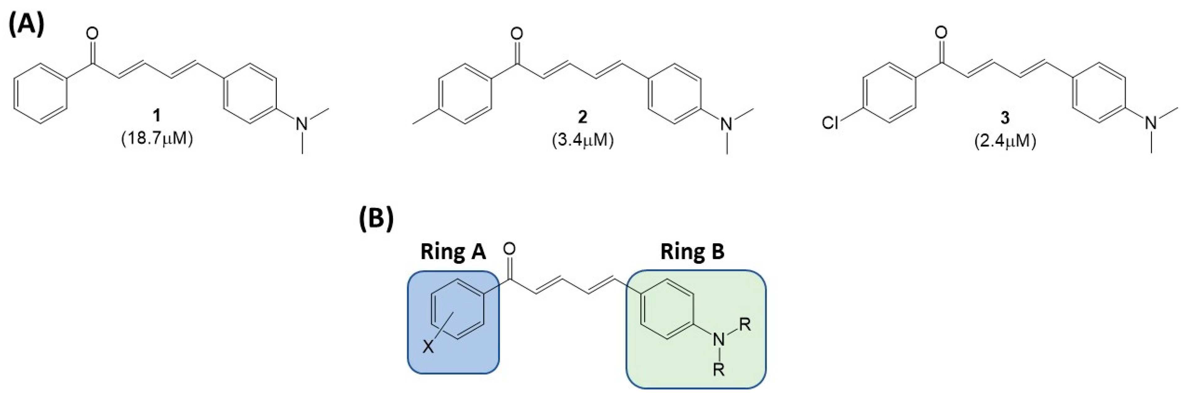
Figure 1. (A) IC50 values of extended chalcone derivatives previously reported [5]. (B) Areas of extended chalcone where modifications will be focused.
Chemistry 06 00089 g001
Scheme 1. Scheme of extended chalcone analogs (428 and 3134).
Chemistry 06 00089 sch001
Figure 2. Thioflavin T fluorescence assay-screening. Compounds 11, 20, 22, 23, and 24 at 10 µM and Phenol Red (100 µM) as positive control.
Chemistry 06 00089 g002
Scheme 2. Acid catalyzed condensation for synthesis of cinnamaldehyde 29 and 30.
Chemistry 06 00089 sch002
Figure 3. IC50 values of extended chalcone library with Ring B modifications.
Chemistry 06 00089 g003
Figure 4.42 significantly decreases cell viability in SH-SY5Y cells alone. In combination with compounds 25, 33, and 34 (at their IC50 value concentrations), no statistically significant neuroprotection after 48 h was observed. Cell viability was measured as an MTT reduction, and data ± SEM (n = 3) were normalized as % vehicle control (black column). Asterisks indicate significant difference from vehicle controls determined by a one-way ANOVA followed by a Tukey’s post hoc test (**** p < 0.0001, ** p < 0.001, * p < 0.05).
Chemistry 06 00089 g004
Figure 5. A representative Punnett square of the cross between UAS-Aβ 1-42 and nSyb-Gal4 with the TM3, Sb balancer on the 3rd chromosome.
Chemistry 06 00089 g005
Figure 6. The percentage climbing for compounds 25, 33, and 34, compared to control, representing the percentage of flies in each section of the vial after 10 s, with standard deviation. Statistical significance is indicated with $ for the top, # for the middle, and * for the bottom. Single symbol indicates a p < 0.05, double indicates a p < 0.001, and triple indicates a p < 0.0001. Compound 25: 1 mcg/mL bottom 95% CI [1.543–24.82] p = 0.0223, middle 95% CI [−18.97–−3.487] p = 0.0024. Compound 33: 1 mcg/mL bottom 95% CI [18.63–40.04] p = <0.0001, middle 95% CI [−29.70–−13.63] p = <0.0001, and top 95% CI [−12.82–−2.516] p = 0.0018, 10 mcg/mL bottom 95% CI [10.63–32.04] p = <0.0001, middle 95% CI [−26.04–−9.964] p = <0.0001. Compound 34: 10 mcg/mL bottom 95% CI [2.157–23.51] p = 0.0142, middle 95% CI [−18.78–−2.558] p = 0.0065. 100 mcg/mL bottom 95% CI [3.395–24.75] p = 0.0064, middle 95% CI [−18.68–−2.463] p = 0.0071.
Chemistry 06 00089 g006
Table 1. IC50 values of extended chalcone library with Ring A modifications.
Chemistry 06 00089 i001
CompoundRIC50 (µM)CompoundRIC50 (µM)
4Chemistry 06 00089 i0021.617Chemistry 06 00089 i0032.7
5Chemistry 06 00089 i0041.518Chemistry 06 00089 i0051.5
6Chemistry 06 00089 i0062.119Chemistry 06 00089 i0072.3
7Chemistry 06 00089 i0081.120Chemistry 06 00089 i0092.0
8Chemistry 06 00089 i010>1021Chemistry 06 00089 i0111.3
9Chemistry 06 00089 i0123.522Chemistry 06 00089 i01310
10Chemistry 06 00089 i0149.323Chemistry 06 00089 i0152.5
11Chemistry 06 00089 i016>1024Chemistry 06 00089 i0171.6
12Chemistry 06 00089 i01820.025Chemistry 06 00089 i0190.91
13Chemistry 06 00089 i0208.826Chemistry 06 00089 i0211.9
14Chemistry 06 00089 i0221.327Chemistry 06 00089 i0231.6
15Chemistry 06 00089 i0243.228Chemistry 06 00089 i0252.2
16Chemistry 06 00089 i0261.9
IC50 values were determined utilizing a Thioflavin T assay. ThT (final concentration of 200 μM) and Aβ1-42 peptide (final concentration of 35 μM) were incubated at 37 °C in a black clear bottom 96-well plate. The ThT fluorescence intensity of each sample was immediately measured every 5 min for 120 min, with 440⁄485 nm excitation/emission filters and with 15 s shaking between reads to facilitate aggregation. An inhibitor control containing Aβ1-42 and a supplied aggregation inhibitor (Phenol Red), at a final concentration of 100 μM. Positive control contained Aβ1-42 without inhibitor. The vehicle control contained the assay buffer and DMSO, of concentrations that did not exceed 1%. The tested compound wells contained Aβ42 peptide and the extended chalcone derivatives at varying concentrations. All the wells were brought to 100 μL as a final volume. All assays were performed in triplicate.
Disclaimer/Publisher’s Note: The statements, opinions and data contained in all publications are solely those of the individual author(s) and contributor(s) and not of MDPI and/or the editor(s). MDPI and/or the editor(s) disclaim responsibility for any injury to people or property resulting from any ideas, methods, instructions or products referred to in the content.