Case Report

Full Digital Workflow for Prosthetic Full-Arch Immediate Loading Rehabilitation Using OT-Bridge System: A Case Report

1
Private Practice, Via Traversa Michelangelo 14, 80026 Casoria, Italy
2
Department of Surgical Sciences, CIR Dental School, University of Turin, 10126 Turin, Italy
3
Department of Prosthodontics, University of Ferrara, Via Luigi Borsari 46, 44121 Ferrara, Italy
*
Author to whom correspondence should be addressed.
Prosthesis 2022, 4(2), 213-223; https://doi.org/10.3390/prosthesis4020021
Submission received: 1 March 2022 / Revised: 17 April 2022 / Accepted: 20 April 2022 / Published: 26 April 2022

Abstract

:
Nowadays, digital technologies have brought very important advancements in clinical prosthetic dentistry. However, a full digital workflow is still considered to be challenging in the management of full-arch implant cases with immediate prosthetic loading. The aim of this case report is to show a full-digital workflow for the fabrication of an implant-prosthetic fixed provisional prosthesis for immediate loading on seven implants in the upper maxilla. The static guided implant surgery and the OT Bridge prosthetic system were used to rehabilitate the patient. In this way, the combination of a well-known surgical technique with a peculiar prosthetic system that allows for a certain degree of tolerance resulted in it being useful for full-arch immediate loading. Future research and studies are necessary to prove the reliability of this full-digital protocol.

1. Introduction

In the last decade, scientific innovations have brought significant advantages to the “conventional loading” protocol originally proposed by Branemark et al. [1]. The immediate loading of dental implants has gained widespread popularity because of its reduced treatment times and improved esthetics and patient acceptance [2]. In addition, considering the earlier factors for success, immediate loading with a fixed prosthesis in edentulous jaws seems to be a predictable treatment in terms of implant survival [3]. Immediate loading seems to induce bone remodeling, generating new bone around implants, with a better healing of hard and soft tissues [4]. Furthermore, the rigid prosthetic framework prevents implants’ micromovements, favoring their stability and an appropriate distribution of occlusal forces [5,6]. However, the passive fitting of the prosthetic framework on implants is always a crucial factor. In the presence of misfit, critical mechanical and/or biological complications may arise [7]. In this view, the mainly applied immediate loading protocols currently described in the literature [8,9] present intrinsic drawbacks due to material properties [10,11,12] and characteristics that may affect the passive fit of the prosthetic framework. The fabrication of gypsum casts or 3D-printed models is prone to errors due to gypsum expansion or to resins shrinkage, which may move implant analogues. Even intraoral impressions, both conventional or digital, are affected by a certain degree of inaccuracies due to the material’s distortions or scanning errors [13,14].
In this way, a full-digital workflow could be useful in order to reduce the number of clinical steps and materials used with their related inaccuracies. Starting from a digital prosthetic and implant planning, clinicians may have the possibility to increase the predictability of their prosthetic rehabilitation, reducing the disadvantages associated with conventional analog or hybrid workflows. Moreover, the use of prosthetic components with a certain degree of tolerance in implant connection is necessary to guarantee a passive fitting of the framework. Today, in order to rigidly connect implants, the most used solution is represented by the multi-unit abutment (M.U.A.), which has the function of changing the implant connection from an internal hexagon to an external one [15]. When titled implants are inserted to correct the implant axis and to solidarize distal implants with the anterior ones, angulated M.U.A.s are necessary [16]. In this way, the connection between the prosthetic framework and implants is provided only by the prosthetic screws [17], which do not permit any discrepancy because of the rigid connection. In the last few years, another fixed system, called OT Bridge® (Rhein 83 srl, Bologna, Italy), has been introduced in the dental market as an alternative to M.U.A. [18]. In this system, the connection of the OT Bridge system is provided both by the screws and by an interlocking system between an acetal ring called Seeger, inserted in a dedicated “extragrade” abutment, and the subequatorial region of the OT equator attachments. In this way, this peculiar type of connection associated with the low-profile attachement may play an important role in the solidarization of the prosthesis on implants.
The aim of this article is to show a clinical case report that allows for the fabrication of an implant-prosthetic fixed provisional prosthesis from the digital implant planning using the OT Bridge prosthetic system.

2. Case Report

A 60-year-old woman presented at our attention with esthetic and functional issues. Her medical history was unsigned and she was classified as ASA1. She complained of a diminished masticatory capacity and loss of retention of the upper removable partial denture. She expressed the desire to change her esthetic, stating that she was dissatisfied with her smile and her removable partial denture. She also expressed the desire to have a more stable masticatory function because of the mobility of the remaining teeth in the upper maxilla. She was very motivated and didn’t want to remain without teeth in case of extractions.
Esthetic analysis highlighted an unbalanced smile line, due to Kelly syndrome, with an anterior rotation of the upper partial removable denture due to maxillary anterior partial resorption (Figure 1).
Clinical examination showed some edentulous areas, especially in the upper maxilla (Figure 2a,b). In particular, the two remaining upper canines and the right lateral incisor showed a hopeless prognosis. The upper partial removable denture showed low retention and stability. The lower maxilla showed a right distal edentulism behind premolars and a single edentulous space in the left premolar region.
The patient’s intraoral records with and without the upper removable partial denture were taken with an intraoral scanner (Trios 3, 3Shape, Copenhagen, Denmark). The upper removable denture was also digitized. The occlusion was unstable with the remaining teeth, so the vertical dimension was recorded with the partial upper removable denture. Then, she underwent an OPT examination that confirmed the hopeless prognosis of the anterior upper teeth, highlighting the necessity of the rehabilitation of the upper maxilla.
Then, in light of the anamnesis, clinical and radiographic examinations, and chief complaint, the treatment of the upper maxilla with a full-arch implant prosthesis in an immediate loading protocol was proposed. For a better evaluation of the upper maxilla bone volumes for implant planning, the patient underwent a CBCT.
Then, the operative sequence was structured as follows:
  • Digital planning for static guided implant surgery planning;
  • A lab workflow for the realization of the provisional prosthesis;
  • A surgical phase of implant insertion and immediate loading of the provisional prosthesis.
The patient accepted the treatment plan and gave written consent to publish photos of her clinical case, including photos of her face.
Digital implant planning:
  • Digital implant planning was initially performed after superimposing the DICOM files of the CBCT with the STL files of the digital intraoral impressions, using a dental implant planning software (RealGuide 3Diemme, Como, Italy). Seven implants (Naturactis tapered implants, LYRA ETK, Paris, France) were virtually inserted in the upper maxilla (Figure 3a,b) based on the final prosthetic project and on the occlusion of the patient. A surgical template for static guided implant surgery was designed (Figure 4) according to the implant positions and patient anatomy. Implant dimensions and positions were specified as follows:
    • Three 4 × 10 mm implants were planned in 1.5, 2.3, and 2.5 positions;
    • Two 3.5 × 12 mm implants were planned in 1.3 and 1.1 positions;
    • One 4 × 12 mm implant was planned in 2.1 position;
    • One 4 × 8 mm implant was planned in 2.6 position.
  • Six low-profile attachments (OT-Equator, Rhein83, Bologna, Italy) were selected and placed in the virtual environment on the corresponding implants. The height of each OT-Equator was selected on the basis of tissue levels previously measured in the implant planning, types, and implant depth (Figure 5a).
  • The digital library files of the dedicated scan abutment were applied to each OT-Equator, performing a “presurgical virtual impression” that obtained the implant’s prosthetic emergence, axis, and positions, (Figure 5b), and the STL file was then exported and sent to the technician for the project and fabrication of the provisional prosthesis.
Lab workflow:
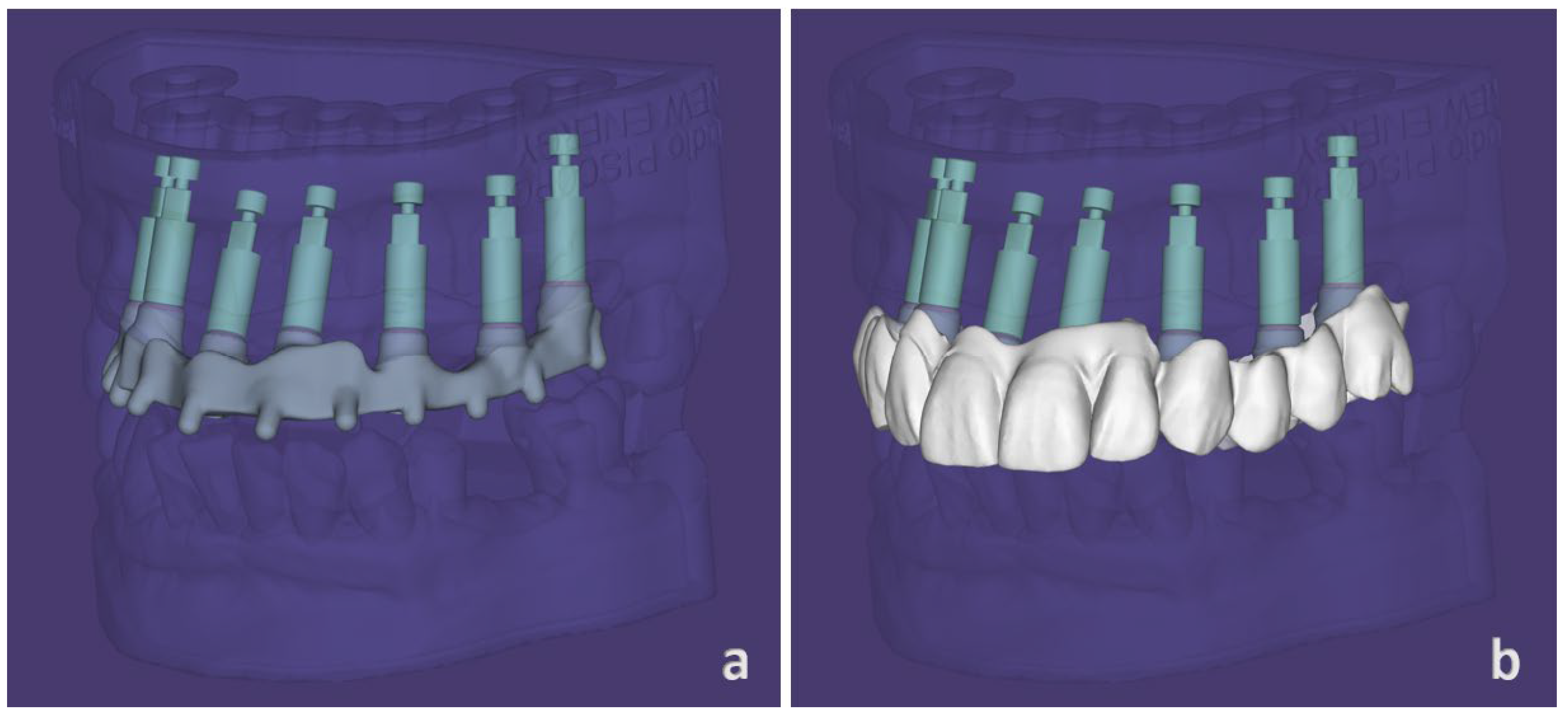
  • The dental technician worked on the STL file that was received as an intraoral digital impression, obtaining the Ti-Base (Extragrade abutment, Rhein83, Bologna, Italy) from the library file of the scan abutment (Figure 6a,b). Then, the appropriate Ti-base height for the OT-Bridge system was selected for each position (Figure 7a,b).
  • A provisional prosthesis composed of a thermosetting resin reinforced with a multidirectional glass fiber framework (Trilor Arch, Bioloren, Saronno, Italy) covered by a PMMA esthetic coverage (BreCam Multicom, Bredent srl, Bolzano, Italy) was digitally designed and produced using a five-axis milling machine (PrograMill PM7, Ivoclar Vivadent, Bolzano, Italy) (Figure 8a,b). The extragrade abutments were cemented manually in the framework using a paste-paste resin-modified glass ionomer luting cement self-mixed with cartridge (FujiCEM 2, GC, Tokio, Japan). The white acetalic Seegers were inserted in the extragrade abutment undercuts and the prosthesis was then sent to the clinician (Figure 9a,b).
  • A 3D-printed surgical mucosa and tooth-supported template was digitally designed and produced (RealGuide 3Diemme, Como, Italy). This was supported by the two contralateral distal molars and was provided with three labial pin holes.
Surgical phase planning:
  • The surgical template was positioned in the upper maxilla using the tooth-supported parts and then stabilized by inserting three vestibular pins in the bone (Figure 10).
  • Seven tapered implants (Naturactis LYRA ETK, Paris, France) were inserted on the upper maxilla in a flapless surgery, as planned (Figure 11a). The implant paths were prepared using a dedicated static guided surgery kit (ETK Implant Guide, LYRA ETK, Paris, France). Implants were manually inserted in an alternate way and by screwing and unscrewing movements before the final positioning. All of the implants showed final peak insertion torque values between 25 and 30 Ncm. OT-Equator attachments were mounted on implants (Figure 11b) and then the prosthesis was placed on them.
  • Clinical and radiographic checks of the passive fit of the prosthesis were performed (Figure 12a–c).

3. Discussion

Static guided surgery with previous digital planning and digital prosthodontics are two of the most explicit examples of the digital revolution that, today, involves dentistry [19,20,21,22,23,24]. With the improvements in the dental softwares, several digital tricks have become possible, resulting in a reduced clinical and lab operating time [19,20,21].
In our technique, the avoidance of the production of the patient models, the elimination of the intraoral impression phase, and the use of the OT Bridge system are the major advantages for the realization of a provisional prosthesis on implants.
In fact, gypsum casts and 3D-printed models may determine some deviations in the position of implant analogues because of the volumetric expansion of gypsum materials during the pouring phase and the shrinkage related to the resin polymerization process, respectively [25]. Convention impression material and intraoral scanners also led to some inaccuracies; shifts of impression transfers may happen during impression tray removal from the patient’s mouth while, in digital impressions, implant positions may be wrongly interpreted by software algorithms, especially in the case of large edentulous areas between scan bodies [13,14].
However, despite all of the surgical attention and the digital implant planning, inaccuracies in implant positions are always reported [26,27]. A systematic review and meta-analysis of the literature reports an accuracy of static computer-assisted implant surgery with a total mean error of 1.12 mm (maximum of 4.5 mm) at the entry point and 1.39 mm at the apex (maximum of 7.1 mm) [28]. Deviations from the digital planning arise from many factors, including the type of surgical template, the surgical procedures, the angulation and depth of the implants and the bone quality. In our protocol, in order to reduce the size of errors during implant positioning, some tricks were applied.
In the digital planning, implant axes were parallelized as much as possible. In this way, a more predictable implant insertion could be reached according to the literature [28]. Consequently, a passive prosthetic fitting could be achieved and possible discrepancies between implant positions and abutment platforms could be simply recognized through clinical and radiographic exams [7].
During the surgical phases, implants were inserted alternately in order to stabilize the surgical template, and movements of screwing and unscrewing implants were performed before the final insertion to obtain the most accurate final implant position. Manual insertion was conducted in order to have a more sensitive positioning of implants without perceivable deviations.
However, from the literature, it seems that immediate loading protocols are incompatible with static guided implant surgery procedures because the implant-prosthetic connection after the surgery lacks a passive fit of the prosthetic framework on implants [9,28]. Then, a prosthetic system with a tolerance in the connection could be a possible solution to avoid misfit.
The OT Bridge is a screw-retained system composed of a low-profile attachment (OT-Equator), a sub-equatorial component represented by an interchangeable acetal ring called Seeger, and a cylindrical titanium “extragrade” abutment. This abutment is cemented in the prosthetic framework and, at its retentive extremity, is provided by a cavity designed for the insertion of the acetal ring, which occupies the undercut created. In this way, the Seeger ring provides a secure and functioning resilient connection that guarantees the stable housing of the prosthesis already on the OT equator attachment, which was initally designed to retain overdentures [17,18]. With this system, discrepancies in implant positioning between digital and real could be compensated for by the peculiar connection of the OT Bridge system, which guarantees a correct fitting of the prosthesis thanks to its low profile. For these reasons, in this clinical case, we use this prosthetic system instead of the MUA system, where the rigid screw connection needs an accuracy in implant positioning that, today, is impossible to achieve. In addition, the low profile of the OT-Equator attachments allows for a more simple tissue management that could also be evaluated in the presurgical phase through digital planning [29,30].
However, this is a case report and the protocol has to be validated with other studies with larger samples considering also different final prosthetic volumes and implant positions. Other factors, such as the type of surgical template, the implant insertion protocol, the use of a technopolymer for the prosthetic framework, and all of the other variables involved in this technique, have to be considered in a broader context with future research and studies.

Author Contributions

Conceptualization, M.P.; methodology, M.P.; software, M.P.; validation, S.C.; formal analysis, F.G.; investigation, F.G.; resources, F.G.; data curation, M.P. and F.G.; writing—original draft preparation, F.G.; writing—review and editing, F.G. and S.C.; visualization, M.P.; supervision, S.C.; project administration, M.P. All authors have read and agreed to the published version of the manuscript.

Funding

This research received no external funding.

Informed Consent Statement

Written informed consent has been obtained from the patient to publish this paper.

Acknowledgments

The authors thank the New Energy LAB of Naples for the fabrication of the prosthetic artifact.

Conflicts of Interest

The authors declare no conflict of interest.

References

  1. Maló, P.; Rangert, B.; Nobre, M. “All-on-Four” Immediate-Function Concept with Brånemark System® Implants for Completely Edentulous Mandibles: A Retrospective Clinical Study. Clin. Implant Dent. Relat. Res. 2003, 5, 2–9. [Google Scholar] [CrossRef] [PubMed]
  2. Chen, J.; Cai, M.; Yang, J.; Aldhohrah, T.; Wang, Y. Immediate versus early or conventional loading dental implants with fixed prostheses: A systematic review and meta-analysis of randomized controlled clinical trials. J. Prosthet. Dent. 2019, 122, 516–536. [Google Scholar] [CrossRef] [PubMed] [Green Version]
  3. De Bruyn, H.; Raes, S.; Östman, P.-O.; Cosyn, J. Immediate loading in partially and completely edentulous jaws: A review of the literature with clinical guidelines. Periodontology 2000 2014, 66, 153–187. [Google Scholar] [CrossRef]
  4. Paleari, A.G.; Presoto, C.D.; Vasconcelos, J.A.; dos Nunes Reis, J.M.S.; Pinelli, L.A.P.; Tavares da Silva, R.H.B.; Quishida, C.C.C. An Alternative Technique for Fabrication of Frameworks in an Immediate Loading Implant Fixed Mandibular Prosthesis. Case Rep. Dent. 2015, 2015, 102189. [Google Scholar] [CrossRef] [Green Version]
  5. Pellegrino, G.; Basile, F.; Relics, D.; Ferri, A.; Grande, F.; Tarsitano, A.; Marchetti, C. Computer-Aided Rehabilitation Supported by Zygomatic Implants: A Cohort Study Comparing Atrophic with Oncologic Patients after Five Years of Follow-Up. J. Clin. Med. 2020, 9, 3254. [Google Scholar] [CrossRef]
  6. Pellegrino, G.; Grande, F.; Ferri, A.; Pisi, P.; Gandolfi, M.G.; Marchetti, C. Three-Dimensional Radiographic Evaluation of the Malar Bone Engagement Available for Ideal Zygomatic Implant Placement. Methods Protoc. 2020, 3, 52. [Google Scholar] [CrossRef]
  7. Sahin, S.; Çehreli, M.C. The Significance of Passive Framework Fit in Implant Prosthodontics: Current Status. Implant Dent. 2001, 10, 85–92. [Google Scholar] [CrossRef]
  8. Bousquet, P.; Carayon, D.; Durand, J.-C. Using the “One Shot” Concept for Immediate Loading Implant Protocol in Edentulous Patient Rehabilitation with a Fixed Prosthesis: A 6-Year Follow-Up. Case Rep. Dent. 2021, 2021, 8872277. [Google Scholar] [CrossRef]
  9. Strub, J.R.; Jurdzik, B.A.; Tuna, T. Prognosis of immediately loaded implants and their restorations: A systematic literature review: Prognosis of immediately loaded implants and restorations. J. Oral Rehabil. 2012, 39, 704–717. [Google Scholar] [CrossRef]
  10. Mobilio, N.; Fasiol, A.; Catapano, S. Survival Rates of Lithium Disilicate Single Restorations: A Retrospective Study. Int. J. Prosthodont. 2018, 31, 283–286. [Google Scholar] [CrossRef]
  11. Mobilio, N.; Catapano, S. The use of monolithic lithium disilicate for posterior screw-retained implant crowns. J. Prosthet. Dent. 2017, 118, 703–705. [Google Scholar] [CrossRef] [PubMed]
  12. Grande, F.; Mochi Zamperoli, E.; Pozzan, M.C.; Tesini, F.; Catapano, S. Qualitative Evaluation of the Effects of Professional Oral Hygiene Instruments on Prosthetic Ceramic Surfaces. Materials 2021, 15, 21. [Google Scholar] [CrossRef] [PubMed]
  13. Baldissara, P.; Koci, B.; Messias, A.M.; Meneghello, R.; Ghelli, F.; Gatto, M.R.; Ciocca, L. Assessment of impression material accuracy in complete-arch restorations on four implants. J. Prosthet. Dent. 2021, 126, 763–771. [Google Scholar] [CrossRef] [PubMed]
  14. Celeghin, G.; Franceschetti, G.; Mobilio, N.; Fasiol, A.; Catapano, S.; Corsalini, M.; Grande, F. Complete-Arch Accuracy of Four Intraoral Scanners: An In Vitro Study. Healthcare 2021, 9, 246. [Google Scholar] [CrossRef] [PubMed]
  15. Kreulen, C.M.; Wolke, J.G.C.; de Baat, C.; Creugers, N.H.J. Attaching single- and multi-unit fixed dental prostheses. Ned. Tijdschr. Voor Tandheelkd. 2013, 120, 633–640. [Google Scholar] [CrossRef]
  16. Hamilton, A.; Jamjoom, F.Z.; Alnasser, M.; Starr, J.R.; Friedland, B.; Gallucci, G.O.; Bdsc, D.A.H.; Bds, M.F.Z.J.; Starr, M.J.R.; BChD, M.B.F.; et al. Tilted versus axial implant distribution in the posterior edentulous maxilla: A CBCT analysis. Clin. Oral Implant. Res. 2021, 32, 1357–1365. [Google Scholar] [CrossRef]
  17. Pozzan, M.C.; Grande, F.; Mochi Zamperoli, E.; Tesini, F.; Carossa, M.; Catapano, S. Assessment of Preload Loss after Cyclic Loading in the OT Bridge System in an “All-on-Four” Rehabilitation Model in the Absence of One and Two Prosthesis Screws. Materials 2022, 15, 1582. [Google Scholar] [CrossRef]
  18. Catapano, S.; Ferrari, M.; Mobilio, N.; Montanari, M.; Corsalini, M.; Grande, F. Comparative Analysis of the Stability of Prosthetic Screws under Cyclic Loading in Implant Prosthodontics: An In Vitro Study. Appl. Sci. 2021, 11, 622. [Google Scholar] [CrossRef]
  19. Ceruti, P.; Mobilio, N.; Bellia, E.; Borracchini, A.; Catapano, S.; Gassino, G. Simplified edentulous treatment: A multicenter randomized controlled trial to evaluate the timing and clinical outcomes of the technique. J. Prosthet. Dent. 2017, 118, 462–467. [Google Scholar] [CrossRef]
  20. Ceraulo, S.; Leonida, A.; Lauritano, D.; Baldoni, A.; Longoni, S.; Baldoni, M.; Caccianiga, G. Proposal for a Clinical Approach to Geriatric Patients with Anchor Need on Implant for Removable Denture: New Technique. Prosthesis 2020, 2, 185–195. [Google Scholar] [CrossRef]
  21. Ortensi, L.; Fisichella, M.L.; Ortensi, M.; Grande, F.; Pellegrino, G.; Ferri, A.; LA Rosa, G.R.; Pedullà, E. A comparison of accuracy between three different facial detection systems for prosthodontic esthetic preview: A single-blinded in vitro study. Minerva Dent. Oral Sci. 2021. [Google Scholar] [CrossRef] [PubMed]
  22. Ortensi, L.; Ortensi, M.; Minghelli, A.; Grande, F. Implant-Supported Prosthetic Therapy of an Edentulous Patient: Clinical and Technical Aspects. Prosthesis 2020, 2, 140–152. [Google Scholar] [CrossRef]
  23. Lorenzetti, M.; Lorenzetti, V.; Carossa, M.; Cavagnetto, D.; Mussano, F. Using a Preoperative Scan Digital Impression and a Digital Index to Build Immediate Interim Full-Arch Implant-Supported Prosthesis. A Case Report and Proof of Concept. Appl. Sci. 2021, 11, 996. [Google Scholar] [CrossRef]
  24. Ortensi, L.; Sigari, G.; La Rosa, G.R.M.; Ferri, A.; Grande, F.; Pedullà, E. Digital planning of composite customized veneers using Digital Smile Design: Evaluation of its accuracy and manufacturing. Clin. Exp. Dent. Res. 2022, 8, 537–543. [Google Scholar] [CrossRef] [PubMed]
  25. Mobilio, N.; Fasiol, A.; Mollica, F.; Catapano, S. Effect of Different Luting Agents on the Retention of Lithium Disilicate Ceramic Crowns. Materials 2015, 8, 1604–1611. [Google Scholar] [CrossRef] [Green Version]
  26. Kero, T.; Pettersson, A.; Fäldt, J.; Andersson, M.; Gillot, L.; Cannas, B.; Nässtrom, K.; Söderberg, R. Virtual variation simulation of CAD/CAM template-guided surgeries performed on human cadavers: Part II. J. Prosthet. Dent. 2010, 104, 48–55. [Google Scholar] [CrossRef]
  27. Landázuri-Del Barrio, R.A.; Cosyn, J.; De Paula, W.N.; De Bruyn, H.; Marcantonio, E. A prospective study on implants installed with flapless-guided surgery using the all-on-four concept in the mandible. Clin. Oral Implant. Res. 2013, 24, 428–433. [Google Scholar] [CrossRef]
  28. Tahmaseb, A.; Wismeijer, D.; Coucke, W.; Derksen, W. Computer Technology Applications in Surgical Implant Dentistry: A Systematic Review. Int. J. Oral Maxillofac. Implant. 2014, 29, 25–42. [Google Scholar] [CrossRef] [Green Version]
  29. Cervino, G.; Cicciù, M.; Fedi, S.; Milone, D.; Fiorillo, L. FEM Analysis Applied to OT Bridge Abutment with Seeger Retention System. Eur. J. Dent. 2021, 15, 047–053. [Google Scholar] [CrossRef]
  30. Montanari, M.; Tallarico, M.; Vaccaro, G.; Ferrari, E.; Scrascia, R.; Ortensi, L.; Cicciù, M.; Meloni, S.M.; Lumbau, A.I.; Avrampou, M.; et al. Two Years after Loading Performance of Implant-Supported Overdenture with Metal Bar and Low-Profile Attachments: A Prospective Case Series Multicenter Clinical Study. Int. J. Dent. 2020, 2020, 8830722. [Google Scholar] [CrossRef]
Figure 1. Extraoral aspect of the patient smile.
Prosthesis 04 00021 g001
Figure 2. Intraoral view with (a) and without (b) upper removable partial denture.
Prosthesis 04 00021 g002
Figure 3. DICOM and STL files matching (a) with surgical and prosthetic project (b).
Prosthesis 04 00021 g003
Figure 4. Virtual design of the surgical template.
Prosthesis 04 00021 g004
Figure 5. Virtual implant planning with OT-Equator attachments (a) and scan abutment library files (b).
Prosthesis 04 00021 g005
Figure 6. Virtual presurgical impression on the scan abutment library file (a) and related extragrade abutments for OT-Bridge system (b).
Prosthesis 04 00021 g006
Figure 7. Computer-aided design framework (a) and final prosthetic volumes (b).
Prosthesis 04 00021 g007
Figure 8. Milling of the framework in technopolymer material (a) and extragade abutments (b).
Prosthesis 04 00021 g008
Figure 9. Provisional pre surgical prosthesis with esthetic characterization: palatal view (a), frontal view (b).
Prosthesis 04 00021 g009
Figure 10. Surgical template correctly positioned in the upper jaw.
Prosthesis 04 00021 g010
Figure 11. Implant insertion (a) and OT-Equator attachments positioning (b).
Prosthesis 04 00021 g011
Figure 12. Provisional prosthesis positioning (a), post-surgical orthopantomography (b) and final extraoral aspect of the patient (c).
Prosthesis 04 00021 g012
Publisher’s Note: MDPI stays neutral with regard to jurisdictional claims in published maps and institutional affiliations.