Review

Fully Digital Workflow in Full-Arch Implant Rehabilitation: A Descriptive Methodological Review

1
Odontology Department, CHU Clermont-Ferrand, University of Clermont Auvergne, F-63000 Clermont-Ferrand, France
2
Clinical Odontology Research Center (CROC), UFR d’Odontologie, University of Clermont Auvergne, F-63000 Clermont-Ferrand, France
*
Author to whom correspondence should be addressed.
Prosthesis 2025, 7(4), 85; https://doi.org/10.3390/prosthesis7040085
Submission received: 17 June 2025 / Revised: 8 July 2025 / Accepted: 11 July 2025 / Published: 16 July 2025

Abstract

Background. Digital dentistry continues to evolve, offering improved accuracy, efficiency, and patient experience across various prosthodontic procedures. Many previous reviews have focused on digital applications in prosthodontics. But the use of a fully digital workflow for full-arch implant-supported prostheses in edentulous patients remains an emerging and underexplored area in the literature. Objective. This article presents a comprehensive methodological review of the digital workflow in full-arch implant-supported rehabilitation. It follows a structured literature exploration and synthesizes relevant technological processes from patient assessment to prosthetic delivery. Methods. The relevant literature was retrieved from the PubMed database on 20 June 2024, to identify the most recent and relevant studies. A total of 22 articles met the eligibility criteria and were included in the review. The majority included case and technical reports. Results. The review illustrates the integration and application of digital tools in implant dentistry, including cone-beam computed tomography (CBCT) exposure, intraoral scanning, digital smile design, virtual patients, guided surgery, and digital scanning. The key findings demonstrate multiple advantages of a fully digital workflow, such as reduced treatment time and cost, increased patient satisfaction, and improved interdisciplinary communication. Conclusions. Despite these benefits, limitations persist due to the low level of evidence, technological challenges, and the lack of standardized protocols. Further randomized controlled trials and long-term clinical evaluations are essential to validate the effectiveness and feasibility of a fully digital workflow for full-arch implant-supported rehabilitation.

1. Introduction

The emergence of advanced digital technologies has considerably influenced modern dentistry, reshaping clinical protocols, enhancing diagnostic accuracy, and expanding the scope of restorative treatment options. The integration of digital tools has streamlined workflows, reduced chair time, and improved communication among clinicians, technicians, and patients [1]. These advancements have been transformative in prosthodontics and implant dentistry, where clinical precision and treatment predictability are principal to achieving successful outcomes [2].
Full-arch edentulism remains a prevalent public health concern, particularly among older populations, affecting oral function, esthetics, and quality of life [3]. While conventional rehabilitation techniques are clinically effective, they require multiple clinical visits and offer limited opportunities for customization. In contrast, digital workflows represent a paradigm shift, enabling faster, more precise, and patient-centered treatment approaches that are personalized to each patient’s anatomical and esthetic needs [4,5].
In implant dentistry, a fully digital workflow involves a sequence of interconnected steps, from initial patient assessment and data acquisition to digital planning, guided surgical intervention, and prosthetic fabrication. Technologies such as intraoral scanners (IOS), cone-beam computed tomography (CBCT), three-dimensional planning software, and computer-aided design and manufacturing (CAD/CAM) systems support this process. Digitalization enhances clinical efficiency and improves treatment accuracy by enabling prosthetically driven planning and real-time visualization of anatomical structures and constraints [1,6].
While numerous reviews have explored digital applications across different disciplines in prosthodontics [5,7,8,9], this review examines the integration of a fully digital workflow for full-arch implant-supported prostheses in fully edentulous patients, a domain that remains relatively underexplored. This paper presents a descriptive methodological review that, unlike a systematic review, does not aim to perform a comprehensive evidence synthesis or formal bias assessment. Instead, it provides a structured overview of current methodologies and digital technologies used in this context. The review evaluates each phase of the digital workflow, including digital diagnostics, virtual treatment planning, guided surgery, and prosthetic fabrication. It also addresses key technological limitations, such as scanning challenges in scanning edentulous arches, variability in scan body systems, and issues with software interoperability, while highlighting innovations aimed at improving accuracy, reliability, and long-term clinical outcomes.

2. Methods

2.1. Research Methodology

A comprehensive literature review was conducted on 20 June 2024 by a calibrated operator using Medical Subject Headings (MeSH) in the PubMed database (www.PubMed.gov) to ensure a standardized and systematic search approach. The review aimed to identify studies addressing the integration of digital workflows in full-arch implant-supported rehabilitation. The focus was on methodological approaches, clinical outcomes, and technological innovations relevant to digital implant dentistry. The process included an initial screening of titles and abstracts, followed by a full-text evaluation of eligible studies. The selection adhered to the guidelines of the Preferred Reporting Items for Systematic Reviews and Meta-Analysis Protocol (PRISMA-P 2020) [10]. Although elements of the PRISMA guidelines were referenced to guide the search strategy and enhance transparency, the review did not follow all PRISMA criteria, such as risk-of-bias assessment or full systematic review procedures. Therefore, this work is presented as a descriptive methodological review rather than a systematic review.

2.2. Article Selection

The search strategy employed a Boolean combination of MeSH terms: (“full digital workflow” [MeSH Terms]) AND (“dental implant”) AND (“full arch”). This formulation was designed to narrow the scope to studies addressing comprehensive digital protocols in the context of full-arch implant rehabilitation. The search was completed on 20 June 2024, ensuring the inclusion of the most recent literature available up to that date.

2.3. Inclusion and Exclusion Criteria

To ensure relevance and scientific rigor, inclusion criteria were defined as follows: peer-reviewed articles published in English or French, involving patients, and addressing digital workflows in implant dentistry. Exclusion criteria included studies older than six years, publications focusing on unrelated topics such as single-unit implants or removable prostheses, and articles lacking sufficient methodological detail.

2.4. Selection Process

A total of 33 articles were initially identified. After applying predefined inclusion and exclusion criteria through a structured screening of titles and abstracts, 25 articles were selected for full-text assessment. Following a comprehensive evaluation, 22 articles met the eligibility criteria and were included in the review (Figure 1). The selected studies include a diverse range of methodological approaches that assessed and described digital workflows in full-arch prosthodontics. The majority of the included studies consisted of case reports and technical reports [2,11,12,13,14,15,16,17,18,19,20,21,22,23,24,25,26,27], which primarily focused on clinical applications and procedural innovations. In contrast, a smaller proportion of studies offered higher levels of evidence, including one randomized clinical trial [28], one case series [29], one retrospective study [30], and one prospective study [31]. Table 1 summarizes the included studies, detailing the year of publication, study design, level of evidence, and principal findings.

3. Results

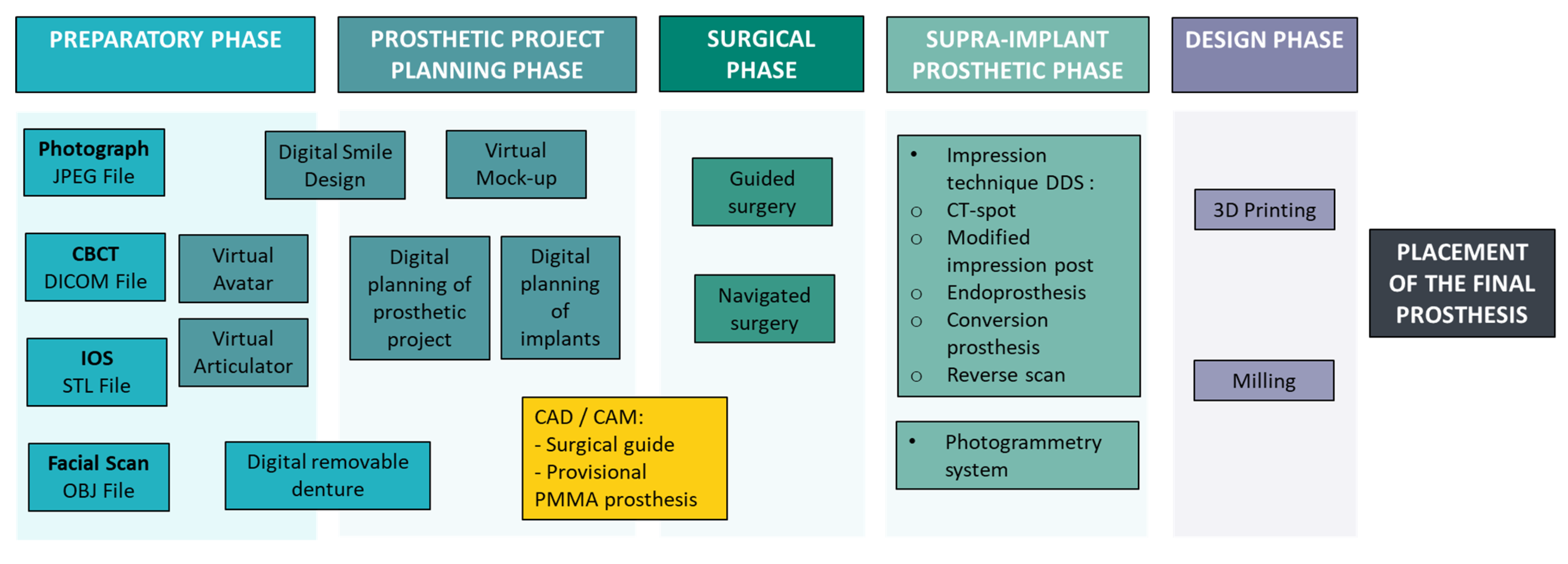
3.1. Preparatory Phase

The main challenge of this phase lies in the superimposition and integration of various digital files to create a coherent and accurate model. This integration is crucial for successful implant planning [21].

3.1.1. Cone-Beam Computed Tomography (CBCT)

CBCT exposure provides 3D visualization of oral and maxillofacial anatomy, which is essential for evaluating bone morphology and accurately planning implant placement [11]. In edentulous patients, the superimposition of CBCT data with intraoral optical scan files is technically challenging due to the absence of anatomical landmarks. To overcome this limitation, radiopaque fiducial markers are often employed. These markers facilitate precise registration between digital surface scan (Standard Tessellation Language or Stereolithography: STL files) and volumetric imaging data (Digital Imaging and Communications in Medicine: DICOM files) [12]. Fiducial markers (CT spots 120; BEEKLEY Medical, Bristol, CT, USA) can be attached to existing prostheses using non-metallic adhesive pads [13,14], or integrated within a radiopaque resin duplicate of the prosthesis [11] (Figure 2). In the maxillary arch, three-dimensional radiopaque markers can be aligned using palatal rugae as reference points, which requires two CBCT scans, one with and one without the prosthesis containing the markers, to achieve accurate registration [14] (Figure 3).

3.1.2. Intraoral Optical Scanning (IOS)

IOS improves patient comfort and reduces many of the inaccuracies associated with conventional impression [15]. However, its effectiveness in full-arch digital rehabilitation is limited by the absence of anatomical reference points in edentulous arches. To address this challenge, radiopaque markers are used with CBCT exposure, to provide stable reference points for digital file alignment. This approach facilitates accurate superimposition of IOS data and volumetric imaging [13].

3.1.3. Digital Smile Design and Virtual Avatar Creation

-
Digital smile design (DSD). The digital smile design (DSD) protocol allows clinicians to create a virtual simulation of the patient’s future smile, allowing for non-invasive evaluation and customization of prosthetic outcomes before treatment. This process requires two facial photographs (at rest and in full smile) with an IOS [16,28]. The resulting STL files and images are imported into specialized software, where reference points guide the alignment of 3D dental models with the 2D facial framework [17]. This technology provides a realistic simulation of treatment results, enhancing patient satisfaction and engagement [22]. With CAD software (NemoStudioR, Nemotec SL, Madrid, Spain), a virtual diagnostic wax-up is generated and positioned in accordance with facial midlines and esthetic landmarks, improving communication between the clinician and dental technician and enabling implant planning in harmony with the patient’s facial anatomy [23].
-
Three-dimensional facial scanning. Three-dimensional facial scanning systems (Cloner II, Done3D; 3DMD face system) provide a detailed and accurate representation of the patient’s facial anatomy, facilitating advanced treatment planning in both dental and implant therapy [23]. The scans generate OBJ files, which are integrated into planning software (3dMDface System, 3dMD, Atlanta, GA, USA) to create virtual avatars that incorporate facial features, bone structure, dentition, and soft tissues. These virtual representations enable clinicians to conduct comprehensive treatment planning, even in the patient’s absence, and help in determining the need for alveolar ridge reduction to achieve optimal esthetic outcomes [24].
-
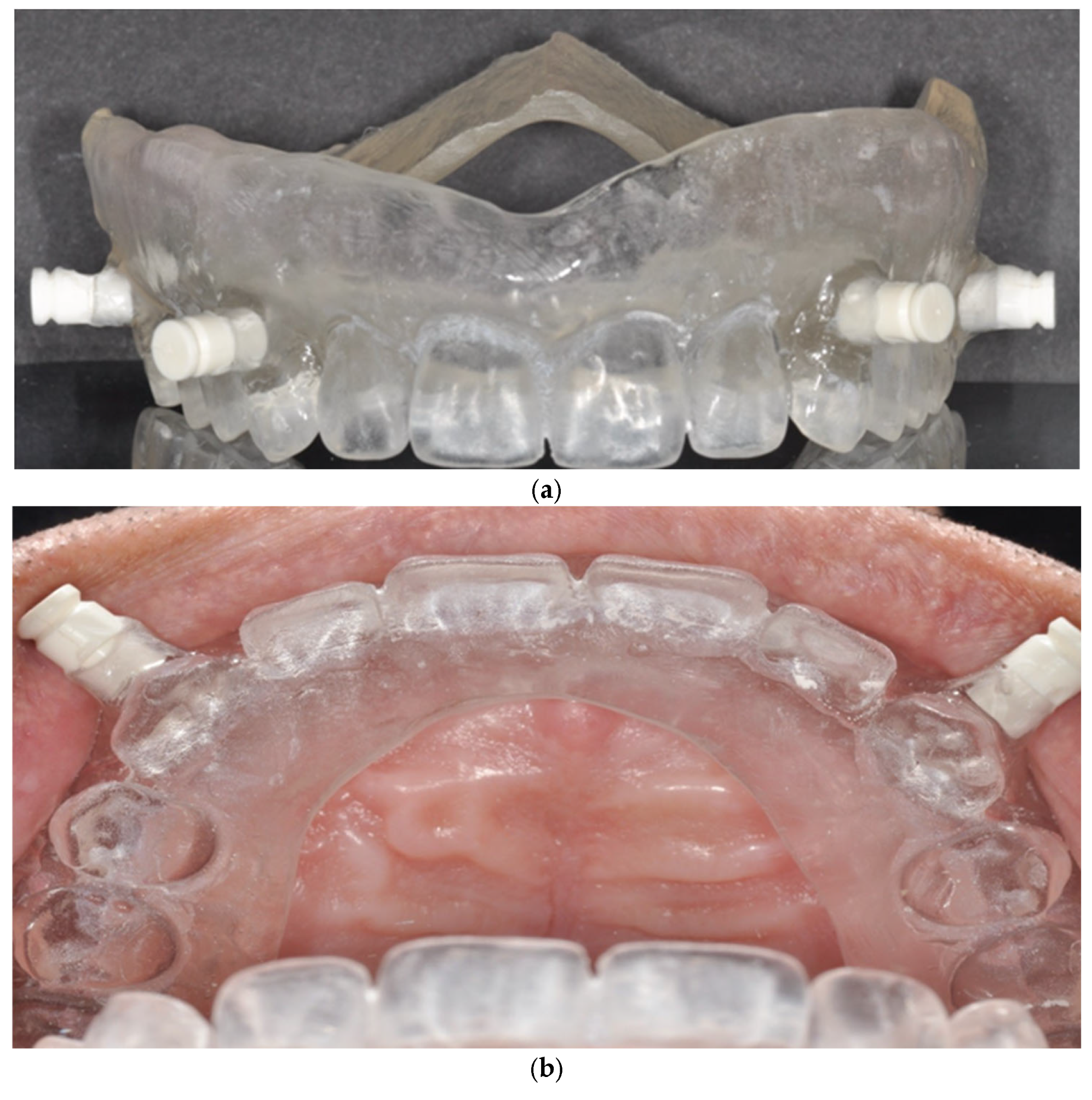
Virtual articulator. Virtual articulators (Zebris Amann Girrbach Artex Articulator, Zebris, Isny, Germany) embedded in CAD/CAM systems (such as CEREC, 3Shape, or Exocad) simulate mandibular movements similar to mechanical articulators, using average condylar settings to articulate digital models [25]. For full-arch implant-supported prosthetic rehabilitation, key parameters (vertical dimension, centric relation, guidance, and esthetics) are digitally transferred from removable prosthesis to the definitive implant restoration [12]. If no existing prosthesis is available, an interim removable denture is required to establish these parameters [13,14,26].
-
Dynamic virtual avatar. Recent advancements have enabled the development of dynamic virtual patients by integrating occlusion data into the digital avatar framework [24] (Figure 4). The static and dynamic relationships between the jaws are recorded using a provisional prosthesis and a mandibular movement tracking system (Jaw Motion analyzer, Zebris, Isny, Germany). A virtual facebow and a reference plane, established via CBCT, are used to mount the digital models on a virtual articulator (Zebris Amann Girrbach Artex Articulator, Zebris, Isny, Germany). The resulting data are then imported into CAD software (Exocad Software; Exocad GmbH, Darm-stadt, Germany) to construct a fully dynamic virtual patient, for precise rehabilitation planning.

3.2. Surgical Phase

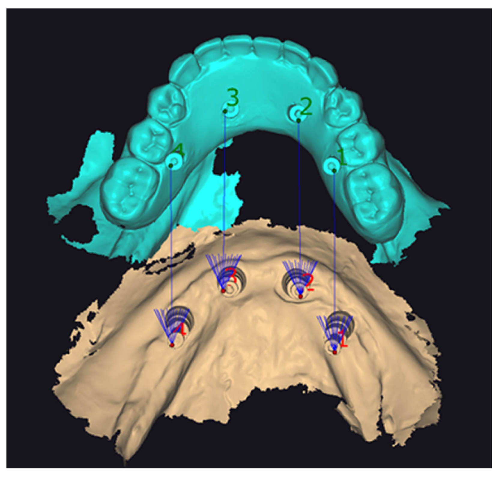
3.2.1. Implant Planning

This phase focuses on determining the optimal implant positions based on the prosthetic design. Implant placement is guided by the planned final prosthesis, the available bone volume, and the location of critical anatomical structures [23]. Using digital libraries, clinicians can select the appropriate implant type, dimensions, and angulation. The finalized digital treatment plan serves multiple functions: ordering implant components, fabricating surgical guides, and integrating with computer-assisted surgery systems. This ensures prosthetically guided implant positioning that meets functional and esthetic objectives.

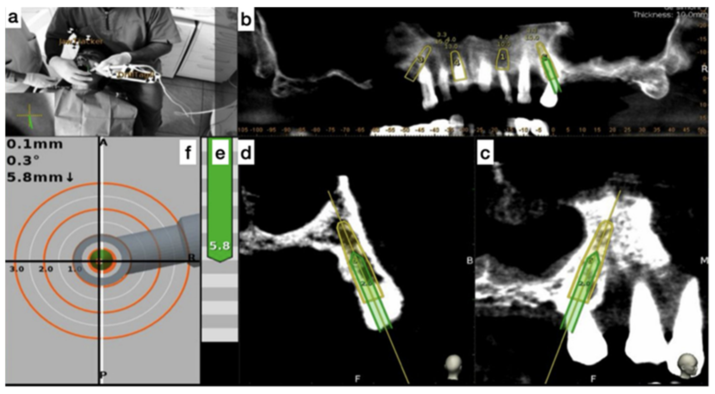
3.2.2. Surgical Phase: Static Guided Surgery

In static guided surgery, digital patient data are used to design custom surgical guides that translate preoperative planning into precise intraoperative execution, without the need for real-time adjustments [12,18].
For full-arch rehabilitations, surgical guides are typically bone-supported and stabilized with fixation pins. Several types of guide systems are available, ranging from simple guides (Figure 5) to sequential and stackable systems. The sequential system facilitates the stepwise placement of different guides (for bone reduction, implant placement, and prosthetic conversion) using the same pin sites.
In stackable systems, a primary guide is first used to place fixation pins. A bone reduction guide is then inserted and stabilized in the same pin sites. Subsequently, the implant placement guide is either magnetically connected [19,23] or mechanically nested [13,16] onto the bone reduction guide for precise execution of the surgical steps. A prefabricated provisional prosthesis may also be stacked onto the bone reduction guide to facilitate immediate loading and conversion (Figure 6).

3.2.3. Surgical Phase: Dynamic Navigated Surgery

Dynamic navigation in implantology allows for real-time adjustment of implant positioning through continuous CT-based tracking during surgery (Figure 7). To track jaw movements, a JawTracker is affixed to one or two teeth using light-cured composite resin, or alternatively, a HeadTracker is positioned on the patient’s head for maxillary procedures. A tracking tag is also mounted on the handpiece to monitor its spatial position.
During implant placement, a target view provides live feedback on the entry point, drilling depth, and angulation of the osteotomy. Additional sagittal and coronal views enable real-time visualization of drill’s trajectory throughout the surgical procedure [29].

3.3. Prosthetic Phase

3.3.1. Supra-Implant Digital Impressions

The accuracy of digital scans in fully edentulous arches is challenged due to the absence of anatomical landmarks in homogeneous soft tissue regions [12,15].
-
Double digital scan (DDS) technique. It requires two digital scans of the same arch: the first is a standard supra-implant impression using scan bodies, and the second is performed with an auxiliary device, such as a provisional prosthesis, an existing denture, an endoprosthesis, or a surgical guide. These auxiliary devices are intended to create essential irregularities within the scanning area.
The endoprosthesis, derived from the digital design of the surgical guide, creates reference zones and captures maxillomandibular relations (Figure 8). It incorporates occlusal stops, sleeves for anchorage pins, and enlarged areas around the implants to accommodate scan bodies [12]. Meanwhile, a conversion prosthesis may also serve as an auxiliary device; however, unlike the endoprosthesis, it does not allow for the placement of scan bodies [13].
In both scenarios, bone fixation pins are retained during the post-surgical phase. These provide stable reference landmarks for the accurate superimposition of STL files. However, when transitioning from a provisional to a definitive prosthesis, particularly when fixation pins have been removed, additional techniques are required to ensure precise alignment of digital files [14,30,31]. One method involves the use of CT-Spot fiducial markers, previously mentioned in the data acquisition phase, although their application is limited to the maxilla (Figure 9).
According to Papaspyridakos et al., 2023 [30], 39 out of 45 full-arch prosthesis prototypes produced using a complete digital workflow (DDS technique) achieved a clinically acceptable fit. The design of the scan bodies and the number of supporting implants significantly influenced the outcomes, with cylindrical scan bodies and prostheses supported by four implants showing the most favorable outcomes [30].
Another study evaluated a novel scan body system (Figure 10) designed to overcome the limitations of placing fiducial markers on soft tissues. It uses long impression posts in combination with new scan bodies. The head of the pin serves as a stable, reproducible landmark for STL file superimposition, regardless of the presence of keratinized mucosa or additional fiducial markers [2].
An extraoral scanning technique has also been implemented to capture implant positions, design a prototype, and fabricate the final monolithic zirconia prosthesis, aimed at restoring both function and esthetics. This method is based on a double digital scan. An extraoral scan is performed after attaching inverted scan bodies to the provisional prosthesis. This scan determines the implant positions, enabling the creation of a precise master digital model (Figure 11) [20].
-
Photogrammetry system. In this review, only one study employed a photogrammetry system, where scan bodies (ICAM bodies, Imetric4D, Imaging, Switzerland) were attached to screw-retained abutments (SRA abutments, Straumann, Switzerland). Implant position data were recorded using a photogrammetry camera (ICAM 4d camera, Imetric4D Imaging Sarl, Courgenay, Switzerland). This study integrated with other digital data, including IOS, CBCT, facial scans, and a virtual facebow, enabling the fabrication of the final prosthesis [24].

3.3.2. Laboratory Phase

Advances in CAD/CAM technology and optimized materials have facilitated a complete digital workflow, offering advantages through milling and 3D printing [12].
-
Three-dimensional Printing. Three-dimensional printing allows the manufacturing of models, surgical guides, endoprostheses, and provisional and definitive prostheses. Mainly used for guides and provisional prostheses, it also offers solutions for permanent prostheses. The materials include PMMA (polymethyl methacrylate) for provisional prostheses, endoprostheses, surgical guides, and resin for surgical guides [12,13,23].
-
Milling. Computer-assisted milling is a subtractive technique used in the digital fabrication of dental prostheses. Unlike 3D printing, which is an additive manufacturing process, milling is subtractive: a machine sculpts and shapes a solid block of raw material (such as zirconia, ceramics, titanium, metal alloys, or PMMA disks) to achieve the desired prosthetic form [2,12,18,26]. One study evaluating 16 edentulous maxillae found that PMMA prototypes fabricated via milling using the DDS technique achieved a perfect fit with no difference based on implant number [31]. Furthermore, monolithic zirconia shows promise as a durable prosthetic material for full-arch implant-supported restorations, potentially reducing clinical visits and minimizing common prosthetic complications such as the chipping or fracture of veneering porcelain [23].
Figure 12 illustrates the complete digital workflow, integrating clinical and technical stages into an integrated protocol. This overview provides a clear framework for understanding the practical application of digital technologies in the rehabilitation of edentulous patients with full-arch implant-supported prostheses.

4. Discussion

The methodological review shows that fully digital workflows in full-arch implant rehabilitation offer several clinical advantages over conventional techniques. These benefits include improved accuracy, shorter chair time, and enhanced patient comfort by avoiding conventional impressions and prolonged intraoral and laboratory procedures. These factors can lead to financial savings for patients, clinicians, and laboratories [2,23,26].
Few studies have addressed digital smile design, likely due to the novelty of tools like facial scanners. However, one study indicates their clinical relevance, particularly in guiding tooth shape selection and performing virtual wax-ups during digital implant rehabilitation [23]. A limitation of the included articles is the lack of details on occlusion management, creating uncertainty about the method to be adopted. Occlusion remains challenging in edentulous patients due to the absence of occlusal references and the complexity of recording intermaxillary relationships. While IOSs are reliable in dentate patients, their accuracy is reduced in edentulous arches. Clinicians employ digital facebows, virtual articulators, 3D facial scanning, photogrammetry, and intraoral jigs to simulate and record occlusal parameters. The lack of standardized protocols and insufficient clinical validation suggest that combining digital acquisition with analog verification is recommended until further evidence supports full digital accuracy.
Guided surgery is commonly used for implant placement in full-arch rehabilitation, enabling flapless procedures that reduce postoperative discomfort. However, limitations include potential overheating due to insufficient irrigation, inability to visualize anatomical landmarks, and errors due to maxillary bone resorption or misfit of surgical guides [28]. Moreover, navigation surgery requires the presence of at least three remaining teeth, limiting its use in edentulous patients [29].
The dual scanning impression technique integrates maxillomandibular relationship recording and the impression into the provisional prosthesis, thus reducing treatment time. In case of a poor fit of a PMMA prototype, it can be sectioned and reconnected intraorally, then rescanned in the laboratory, and milled in zirconia. However, impressions involving anchoring pins pose challenges due to metallic reflections during scanning, often necessitating the use of scanning powders to enhance capture quality [13]. Photogrammetry systems offer advantages in patient comfort, speed, and cost-effectiveness, though their accuracy may vary depending on clinical conditions [24].
CAD-CAM technologies have transformed the manufacturing of fully-arch implant rehabilitations, offering flexibility and speed at a lower cost thanks to the rise of 3D printers [4,7]. Hybrid and 3D-printed prostheses are gaining popularity, and the marginal fit of implants with these methods matches or surpasses the precision of conventional techniques. Traditional techniques are progressively replaced by digital processes such as laser fusion, milling, and hybrid manufacturing technologies [12,27].
Despite advancements in digital implantology, several challenges persist. Data acquisition in edentulous patients remains a limitation due to the lack of fixed anatomical landmarks, increasing the risk of registration and errors. Additionally, the high cost of equipment, software, and training poses financial and logistical barriers to adoption. Clinicians must acquire expertise in digital file handling, diagnosis, and treatment planning, which can increase preparation time [4].
Moreover, error propagation across digital steps remains a concern. Inaccuracies originating during intraoral scanning or CBCT can be aggravated during the superimposition of STL and DICOM files, CAD planning, and final prosthesis fabrication via CAM technologies. These errors may compromise prosthetic fit, leading to biological or mechanical complications [2,23,30]. To address this, several strategies can be considered, including the use of auxiliary devices to enhance scan accuracy, high-precision photogrammetry systems, validation of each digital step with analog references, and improved calibration protocols.
Scan body design also plays a critical role in the precision of digital workflows. Variations in geometry, material, and library compatibility can introduce positional inaccuracies, especially in full-arch cases where minimal deviations may affect passive fit. Furthermore, inconsistencies in digital scan body libraries across software platforms compound the risk of error during the design and fabrication phases.
Interoperability between systems remains a key technical challenge, as many CAD platforms and imaging systems are not fully compatible. This is often due to proprietary software restrictions, which limit seamless data exchange and hinder the integration of STL and DICOM datasets, especially in the absence of shared anatomical reference points. These incompatibilities can compromise workflow efficiency, data accuracy, and ultimately, clinical outcomes. Exploring open-source platforms or adopting standardized, universal file formats may offer more flexible and scalable alternatives. To improve predictability and long-term success in fully digital full-arch implant rehabilitation, future research should focus on developing standardized clinical protocols, optimizing scan body designs, and validating each digital step through rigorous studies.
Given the persistent technical and clinical limitations, future research should explore the integration of artificial intelligence to enhance planning accuracy, develop cost-effective open-source software solutions to overcome interoperability issues, and validate fully digital protocols through high-level, long-term trials. Accentuating training and accessibility will also be key to ensuring wider clinical adoption.
This review has several limitations. Firstly, it includes 22 studies, many of which are case or technical reports, leading to lower levels of evidence and limited generalizability. Most studies provide only short-term data, lacking long-term follow-up on prosthetic survival, patient satisfaction, or biological complications. Additionally, the rapid evolution of digital tools and variability in clinical protocols across studies create inconsistencies in reported outcomes. Several reports highlighted issues with digital file integration and registration errors, yet these technological limitations remain largely unaddressed in the literature. A further limitation lies in the search strategy. The review was limited to a single database (PubMed), and no complementary databases, gray literature sources, or hand-searching were included. This may have resulted in the exclusion of relevant studies and introduced publication bias. Moreover, a bottom-up citation approach was not applied, further affecting the comprehensiveness of the evidence base. Another limitation of the current literature is the predominance of case and technical reports, which restrict the generalizability of the findings. However, Cattoni et al. demonstrated positive long-term outcomes in a randomized clinical trial comparing digital and traditional workflows in “All on Four” cases [28]. Chochlidakis et al. showed that the double digital scanning (DDS) technique yields accurate prosthesis prototypes in a prospective study [31]. These findings highlight the potential of digital protocols but emphasize the need for more rigorous clinical trials. Finally, the lack of standardized protocols further underscores the variability in current digital workflows and highlights the need for comparative and longitudinal research to validate a consistent and reliable fully digital treatment pathway for full-arch implant rehabilitation.

5. Conclusions

This descriptive methodological review demonstrates that full-arch implant rehabilitation using a fully digital workflow is a promising approach in prosthodontics. Current evidence highlights numerous advantages over conventional workflows, including the following:
Improved Efficiency: Reduced chair time, elimination of conventional impressions, and faster laboratory procedures.
Enhanced Accuracy: Advanced data integration from CBCT, IOS, and virtual simulation.
Patient-Centered Benefits: Less invasive procedures, shorter treatment duration, and better communication of expected outcomes via virtual design tools.
Cost-Effectiveness: Streamlined steps benefit clinicians, laboratories, and patients by lowering material- and labor-related expenses.
Despite these advantages, several challenges and limitations persist. Occlusal registration remains difficult in edentulous patients due to the lack of natural reference points. Variability in scan body design and cumulative errors across digital steps, ranging from data acquisition to prosthesis fabrication, can compromise the accuracy and fit of the final restoration. Additionally, the high cost of equipment, the complexity of digital file management, and the absence of standardized clinical protocols present further barriers to widespread implementation. Long-term clinical outcomes related to fully digital workflows for full-arch rehabilitation remain underexplored.
In conclusion, while fully digital full-arch implant rehabilitation shows considerable potential to enhance clinical outcomes and patient satisfaction, its widespread adoption will require further validation. High-quality randomized controlled trials, multicenter studies, and long-term follow-up data are essential to develop standardized, evidence-based digital protocols for full-arch implant rehabilitation.

Author Contributions

C.A.: had the idea for the article, performed the literature search, data analysis, and critically revised the work. T.D.: revised the work. E.N.: had the idea for the article, drafted and critically revised the work. N.E.O.: performed the data analysis, performed the literature search drafted, and critically revised the work. All authors have read and agreed to the published version of the manuscript.

Funding

This work did not receive any specific grant from funding agencies in the public, commercial, or not-for-profit sectors.

Conflicts of Interest

The authors declare no conflicts of interest.

Disclaimer

The opinions expressed in the article are ours and do not constitute an official position of the institutions.

References

  1. Bonnet, G.; Batisse, C.; Travers, C.; Bessadet, M. Integration of CAD/CAM Technology in Global Dental Prosthetic Treatment: A Case Report. Int. Multispecialty J. Health 2016, 2, 9–12. [Google Scholar]
  2. Marinis, A.; Papaspyridakos, P.; Sicilia, E.; Bernardes, S.R.; Touloumi, F.; Chochlidakis, K.; Weber, H.-P. Digital Workflow for Double Complete Arch Zirconia Prostheses Utilizing a Novel Scan Body. J. Prosthodont. 2022, 31, 4–8. [Google Scholar] [CrossRef]
  3. El Osta, N.; Haddad, E.; Fakhouri, J.; Saad, R.; El Osta, L. Comparison of Psychometric Properties of GOHAI, OHIP-14, and OHIP-EDENT as Measures of Oral Health in Complete Edentulous Patients Aged 60 Years and More. Qual. Life Res. 2021, 30, 1199–1213. [Google Scholar] [CrossRef] [PubMed]
  4. El Osta, N.; Bessadet, M.; Drancourt, N.; Batisse, C. Time Efficiency and Cost of Fabricating Removable Complete Dentures Using Digital, Hybrid, and Conventional Workflows: A Systematic Review. J. Prosthet. Dent. 2025, 133, 1194–1208. [Google Scholar] [CrossRef] [PubMed]
  5. Batisse, C.; Nicolas, E. Comparison of CAD/CAM and Conventional Denture Base Resins: A Systematic Review. Appl. Sci. 2021, 11, 5990. [Google Scholar] [CrossRef]
  6. Drancourt, N.; Auduc, C.; Mouget, A.; Mouminoux, J.; Auroy, P.; Veyrune, J.-L.; El Osta, N.; Nicolas, E. Accuracy of Conventional and Digital Impressions for Full-Arch Implant-Supported Prostheses: An In Vitro Study. J. Pers. Med. 2023, 13, 832. [Google Scholar] [CrossRef]
  7. El Osta, N.; Bessadet, M.; Drancourt, N.; Nicolas, E.; Batisse, C. Response to Letter to the Editor Regarding, “Time Efficiency and Cost of Fabricating Removable Complete Dentures Using Digital, Hybrid, and Conventional Workflows: A Systematic Review” by El Osta et al. J. Prosthet. Dent. 2025, 134, 285–286. [Google Scholar] [CrossRef]
  8. Tohme, H.; Lawand, G.; Chmielewska, M.; Makhzoume, J. Comparison between Stereophotogrammetric, Digital, and Conventional Impression Techniques in Implant-Supported Fixed Complete Arch Prostheses: An in Vitro Study. J. Prosthet. Dent. 2023, 129, 354–362. [Google Scholar] [CrossRef]
  9. Lawand, G.; Ismail, Y.; Revilla-León, M.; Tohme, H. Effect of Implant Scan Body Geometric Modifications on the Trueness and Scanning Time of Complete Arch Intraoral Implant Digital Scans: An in Vitro Study. J. Prosthet. Dent. 2024, 131, 1189–1197. [Google Scholar] [CrossRef]
  10. Page, M.J.; McKenzie, J.E.; Bossuyt, P.M.; Boutron, I.; Hoffmann, T.C.; Mulrow, C.D.; Shamseer, L.; Tetzlaff, J.M.; Akl, E.A.; Brennan, S.E.; et al. The PRISMA 2020 statement: An Updated Guideline for Reporting Systematic Reviews. BMJ 2021, 372, n71. [Google Scholar] [CrossRef]
  11. Beretta, M.; Poli, P.P.; Tansella, S.; Aguzzi, M.; Meoli, A.; Maiorana, C. Cast-Free Digital Workflow for Implant-Supported Rehabilitation in a Completely Edentulous Patient: A Clinical Report. J. Prosthet. Dent. 2021, 125, 197–203. [Google Scholar] [CrossRef] [PubMed]
  12. Venezia, P.; Torsello, F.; Santomauro, V.; Dibello, V.; Cavalcanti, R. Full Digital Workflow for the Treatment of an Edentulous Patient with Guided Surgery, Immediate Loading and 3D-Printed Hybrid Prosthesis: The BARI Technique 2.0. A Case Report. Int. J. Environ. Res. Public Health 2019, 16, 5160. [Google Scholar] [CrossRef] [PubMed]
  13. Papaspyridakos, P.; De Souza, A.; Bathija, A.; Kang, K.; Chochlidakis, K. Complete Digital Workflow for Mandibular Full-Arch Implant Rehabilitation in 3 Appointments. J. Prosthodont. 2021, 30, 548–552. [Google Scholar] [CrossRef] [PubMed]
  14. Papaspyridakos, P.; Chochlidakis, K.; Kang, K.; Chen, Y.-W.; Alghfeli, A.; Kudara, Y.; Weber, H.-P. Digital Workflow for Implant Rehabilitation with Double Full-Arch Monolithic Zirconia Prostheses. J. Prosthodont. 2020, 29, 460–465. [Google Scholar] [CrossRef]
  15. Meneghetti, P.; Moura, G.F.; Tavelli, L.; Li, J.; Siqueira, R.; Wang, H.-L.; Mendonça, G. A Fully Digital Approach for Implant Fixed Complete Dentures: A Case Report. J. Esthet. Restor. Dent. 2021, 33, 1070–1076. [Google Scholar] [CrossRef]
  16. Papaspyridakos, P.; Bedrossian, A.; De Souza, A.; Bokhary, A.; Gonzaga, L.; Chochlidakis, K. Digital Workflow in Implant Treatment Planning For Terminal Dentition Patients. J. Prosthodont. 2022, 31, 543–548. [Google Scholar] [CrossRef]
  17. Roberts, M.; Shull, F.; Schiner, B. Maxillary Full-Arch Reconstruction Using a Sequenced Digital Workflow. J. Esthet. Restor. Dent. 2020, 32, 336–356. [Google Scholar] [CrossRef]
  18. Sobczak, B.; Majewski, P. An Integrated Fully Digital Prosthetic Workflow for the Immediate Full-Arch Restoration of Edentulous Patients-A Case Report. Int. J. Environ. Res. Public Health 2022, 19, 4126. [Google Scholar] [CrossRef]
  19. Martins, J.; Rangel, J.; Nobre, M.d.A.; Ferro, A.; Nunes, M.; Almeida, R.; Guedes, C.M. A New Full Digital Workflow for Fixed Prosthetic Rehabilitation of Full-Arch Edentulism Using the All-on-4 Concept. Medicina 2024, 60, 720. [Google Scholar] [CrossRef]
  20. Bedrossian, E.A.; Papaspyridakos, P.; Bedrossian, E.; Gurries, C. The Reverse Scan Body Protocol: Completing the Digital Workflow. Compend. Contin. Educ. Dent. 2023, 44, e1–e4. [Google Scholar]
  21. Carosi, P.; Ferrigno, N.; De Renzi, G.; Laureti, M. Digital Workflow to Merge an Intraoral Scan and CBCT of Edentulous Maxilla: A Technical Report. J. Prosthodont. 2020, 29, 730–732. [Google Scholar] [CrossRef]
  22. Neto, A.D.T.; Costa, A.J.d.M.E.; Gil Choi, I.G.; Santos, A.; Dos Santos, J.F.; Cortes, A.R.G. Digital Workflow for Full-Arch Implant-Supported Prosthesis Based on Intraoral Scans of a Relative of the Patient. J. Oral Implant. 2020, 47, 68–71. [Google Scholar] [CrossRef]
  23. Costa, A.J.d.M.; Neto, A.D.T.; Burgoa, S.; Gutierrez, V.; Cortes, A.R.G. Fully Digital Workflow with Magnetically Connected Guides for Full-Arch Implant Rehabilitation Following Guided Alveolar Ridge Reduction. J. Prosthodont 2020, 29, 272–276. [Google Scholar] [CrossRef]
  24. Wang, J.; Wu, Y.-L.; Ma, J.; Wu, F.; Li, D.-H. A Novel Technique for Implant-Supported Fixed Complete Rehabilitation Based on a Dynamic Virtual Patient. J. Dent. 2023, 137, 104649. [Google Scholar] [CrossRef]
  25. Lepidi, L.; Chen, Z.; Ravida, A.; Lan, T.; Wang, H.-L.; Li, J. A Full-Digital Technique to Mount a Maxillary Arch Scan on a Virtual Articulator. J. Prosthodont. 2019, 28, 335–338. [Google Scholar] [CrossRef]
  26. Parpaiola, A.; Toia, M.; Norton, M.; Rodriguez, Y.; Baena, R.; Todaro, C.; Lupi, S.M. Fully Digital Workflow for Implant-Supported Fixed Restorations Consisting of a Titanium Primary Structure and a Zirconia Secondary Structure. Int. J. Oral Implant. 2023, 16, 351–358. [Google Scholar]
  27. Ragazzini, N.; Dds, P.B.; Monaco, C.; Ciocca, L. Digital Jaw Relation Record of Edentulous Patients in the CAD-CAM Workflow of the Implant-Supported Full-Arch Prosthesis. J Oral Implant. 2020, 47, 57–62. [Google Scholar] [CrossRef] [PubMed]
  28. Cattoni, F.; Chirico, L.; Merlone, A.; Manacorda, M.; Vinci, R.; Gherlone, E.F. Digital Smile Designed Computer-Aided Surgery versus Traditional Workflow in “All on Four” Rehabilitations: A Randomized Clinical Trial with 4-Years Follow-Up. Int. J. Environ. Res. Public Health 2021, 18, 3449. [Google Scholar] [CrossRef] [PubMed]
  29. Stefanelli, L.V.; Mandelaris, G.A.; Franchina, A.; Pranno, N.; Pagliarulo, M.; Cera, F.; Maltese, F.; Angelis, F.D.; Carlo, S.D. Accuracy of Dynamic Navigation System Workflow for Implant Supported Full Arch Prosthesis: A Case Series. Int. J. Environ. Res. Public Health 2020, 17, 5038. [Google Scholar] [CrossRef]
  30. Papaspyridakos, P.; Vazouras, K.; Gotsis, S.; Bokhary, A.; Sicilia, E.; Kudara, Y.; Bedrossian, A.; Chochlidakis, K. Complete Digital Workflow for Prosthesis Prototype Fabrication with Double Digital Scanning: A Retrospective Study with 45 Edentulous Jaws. J. Prosthodont. 2023, 32, 571–578. [Google Scholar] [CrossRef]
  31. Chochlidakis, K.; Romeo, D.; Ercoli, C.; Papaspyridakos, P. Complete Digital Workflow for Prosthesis Prototype Fabrication with the Double Digital Scanning (DDS) Technique: A Prospective Study on 16 Edentulous Maxillae. J. Prosthodont. 2022, 31, 761–765. [Google Scholar] [CrossRef]
Figure 1. Flowchart of the literature search.
Prosthesis 07 00085 g001
Figure 2. (a) Duplicated maxillary and mandibular dentures with radiopaque resin markers; (b) CBCT exposure of the dentures. Reproduced from Venezia et al., 2019, under the Creative Commons Attribution 4.0 International License [12].
Prosthesis 07 00085 g002
Figure 3. (a) Radiographic template; (b) occlusal view of the radiographic template with radiopaque markers. Reproduced with permission from Carosi et al., J Prosthodont 2020; 29(8): 730–732. © Wiley. License number: 6032490782289 [21].
Prosthesis 07 00085 g003
Figure 4. Digital workflow for designing implant-supported complete prostheses using virtual patient integration.
Prosthesis 07 00085 g004
Figure 5. (a) Virtual planning of implant positioning based on the esthetic prosthetic design; (b) stereolithographic surgical guide stabilized in the maxilla using anchor pins; (c) flapless implant placement surgery. Reproduced from Cattoni et al., 2021, under the Creative Commons Attribution 4.0 International License [28].
Prosthesis 07 00085 g005
Figure 6. (a) Surgical guide for alveolar bone reduction (green), soft tissue (red); (b) implant surgical guide (blue) intended to be magnetically connected to the surgical guide (green); (c) implant-supported interim PMMA fixed prosthesis (white), intended to be magnetically connected to the surgical guide (green). Reproduced with permission from Costa et al., J Prosthodont 2020; 29(3): 272–276. © Wiley. License number: 6032461502682 [23].
Prosthesis 07 00085 g006
Figure 7. Following calibration and verification of the drill axis and tip, navigated implant placement is performed using real-time visual guidance. The clinician monitors the entry point, depth, and angulation (yellow) of the osteotomy through various screen views. Illustration of different views on screen during surgery as drill (green) advances into the bone. (a) Tracker video stream; (b) panoramic view; (c) mesiodistal section view; (d) buccolingual section view; (e) depth indicator; (f) target view. Reproduced from Stefanelli et al., 2020, under the Creative Commons Attribution 4.0 International License [29].
Prosthesis 07 00085 g007
Figure 8. (a) Prosthetic stent in position in conjunction with maxillary prosthesis and stabilized using same anchor pins as surgical stent; (b) scan bodies connected to SRA abutments (Straumann). Reproduced from Venezia et al., 2019, under the Creative Commons Attribution 4.0 International License [12].
Prosthesis 07 00085 g008
Figure 9. (a) Use of fiducial markers to facilitate STL file superimposition in CAD software (Exocad DentalCAD, exocad GmbH, Darm-stadt, Germany) during the double-scan (DDS) technique; (b) second digital scan performed with scan bodies and fiducial markers positioned identically, as part of DDS workflow. Reproduced with permission from Papaspyridakos et al., J Prosthodont 2020; 29(6): 460–465. © Wiley. License number: 6032460591924. [14].
Prosthesis 07 00085 g009
Figure 10. Exocad software (Exocad DentalCAD, exocad GmbH, Darm-stadt, Germany) display before the superimposition of the STL files for the scan bodies and mandibular interim prosthesis. Reproduced with permission from Marinis et al., J Prosthodont 2022; 31(1): 4–8. © Wiley. License number: 6032430913487. [2].
Prosthesis 07 00085 g010
Figure 11. Inverted extraoral scanning technique; (a) STL file of the conversion prosthesis showing tooth positioning; (b) scan of the edentulous maxilla illustrating the contours of the residual ridge; (c) conversion prosthesis with inverted scan bodies indicating implant positions. Bedrossian EA, Papaspyridakos P, Bedrossian E, Gurries C. The Reverse Scan Body Protocol: Completing the Digital Workflow. Compend Contin Educ Dent. 2023; 44(7): e1-e4: Figure 6. Copyright © 2025 to Conexiant LLC. All rights reserved. Used with permission of the publisher [20].
Prosthesis 07 00085 g011
Figure 12. Digital chain for a fully digital workflow in full-arch implant rehabilitation.
Prosthesis 07 00085 g012
Table 1. Summary of included studies on digital workflows in full-arch implant rehabilitation.
Author(s)Study DesignLevel of EvidenceKey Findings
Lepidi et al., 2019 [25]Technical reportLevel IVDigital technique for aligning maxillary scans on a virtual articulator.
Venezia et al., 2019 [12]Case reportLevel IVBARI 2.0 technique with 3D-printed hybrid prosthesis, guided surgery, and immediate loading.
Papaspyridakos et al., 2020 [14]Case seriesLevel IVFully digital workflow for double full-arch monolithic zirconia prostheses.
Stefanelli et al., 2020 [29]Case seriesLevel IVEvaluated accuracy of dynamic navigation system for full-arch digital implant workflow.
Roberts et al., 2020 [17]Case reportLevel IVSequenced digital workflow for maxillary full-arch implant reconstruction.
Carosi et al., 2020 [21]Technical reportLevel IVMerging intraoral scan with CBCT for edentulous maxilla rehabilitation.
Costa et al., 2020 [23]Case reportLevel IVApplication of magnetically retained guides in digital full-arch rehabilitation.
Ragazzini et al., 2021 [27]Case reportLevel IVDigital jaw relation record in CAD-CAM workflow for full-arch prosthesis.
Papaspyridakos et al., 2021 [13]Case reportLevel IVComplete digital workflow for mandibular full-arch implant rehabilitation completed in three visits.
Beretta et al., 2021 [11]Case reportLevel IVCast-free digital workflow in a completely edentulous patient.
Meneghetti et al., 2021 [15]Case reportLevel IVFully digital approach for implant-supported complete dentures.
Neto et al., 2021 [22]Case reportLevel IVDigital workflow using intraoral scanning for full-arch prosthesis fabrication.
Cattoni et al., 2021 [28]Randomized clinical trialLevel IIComparison of digital vs. conventional workflows in All-on-4 implant rehabilitation.
Marinis et al., 2022 [2]Case reportLevel IVInnovative scan body use for double full-arch zirconia prostheses via digital workflow.
Papaspyridakos et al., 2022 [16]Case reportLevel IVDigital workflow in implant treatment planning for terminal dentition patients.
Chochlidakis et al., 2022 [31]Prospective studyLevel IIIUse of double digital scanning technique in 16 fully edentulous maxillae.
Sobczak & Majewski, 2022 [18]Case reportLevel IVImmediate full-arch restoration utilizing a fully digital workflow.
Wang et al., 2023 [24]Case reportLevel IVRehabilitation of edentulous patients using a dynamic virtual patient approach.
Bedrossian et al., 2023 [20]Technical reportLevel IVReverse scan body protocol to streamline fully digital workflow in full-arch rehabilitation.
Parpaiola et al., 2023 [26]Case reportLevel IVFully digital protocol with titanium primary and zirconia secondary structures.
Papaspyridakos et al., 2023 [30]Retrospective studyLevel IIIEvaluation of double digital scanning technique in 45 edentulous jaws.
Martins et al., 2024 [19]Case seriesLevel IVFully digital All-on-4 workflow for fixed full-arch supported prosthesis.
Disclaimer/Publisher’s Note: The statements, opinions and data contained in all publications are solely those of the individual author(s) and contributor(s) and not of MDPI and/or the editor(s). MDPI and/or the editor(s) disclaim responsibility for any injury to people or property resulting from any ideas, methods, instructions or products referred to in the content.

