Case Report

Digital Smile Design with AI-Assisted Workflow for Minimally Invasive Veneer Rehabilitation: A Case Report


1
Department of Dental Technology, School of Dentistry, Al-Ahliyya Amman University, Amman 19328, Jordan
2
Department of Esthetic and Prosthetic Dentistry, School of Dentistry, Saint-Joseph University, Beirut 1107 2180, Lebanon
3
Artium Dental Lab., Beirut 1107 2180, Lebanon
4
School of Dentistry, Aldent University, 1001 Tirana, Albania
5
Head of Department of Digital Dentistry, AI, and Evolving Technologies, Faculty of Dental Medicine, Saint-Joseph University of Beirut, Beirut 1107 2180, Lebanon
*
Author to whom correspondence should be addressed.
Prosthesis 2026, 8(5), 45; https://doi.org/10.3390/prosthesis8050045
Submission received: 31 January 2026 / Revised: 3 March 2026 / Accepted: 24 April 2026 / Published: 10 May 2026

Abstract

This case report describes a digital workflow for the aesthetic rehabilitation of a 30-year-old male patient with unaesthetic anterior teeth. The treatment incorporated AI-assisted smile design software (SmileCloud Biometrics) for 2D/3D digital planning and patient communication. Six lithium disilicate veneers (IPS e.max CAD) were fabricated using CAD/CAM technology following mock-up-guided minimally invasive preparation (0.2–0.9 mm reduction). The restorations were adhesively cemented under rubber dam isolation. One-week follow-up confirmed aesthetic integration, occlusal harmony, and patient satisfaction. This case illustrates how digital workflows with AI-assisted tools can support veneer rehabilitation through data-informed planning and conservative preparation while maintaining aesthetic outcomes.

1. Introduction

Rising patient expectations for natural-looking, personalized smile designs have increased the need for precise aesthetic planning tools [1]. While modern ceramics like lithium disilicate (LDS) offer increased durability (5-year survival: 92%) [2], their clinical success depends not only on mechanical properties and marginal adaptation but also substantially on aesthetic outcomes and patient satisfaction [3]. These aesthetic results are highly dependent on accurate smile design and proper preparation protocols.
Traditionally, smile design and veneer fabrication relied heavily on clinician subjectivity, easily leading to inconsistencies in proportions and symmetry. Additionally, the classic procedure involved multiple appointments, physical impressions, wax-ups, and temporary restorations, often leading to extended treatment times and potential inaccuracies. However, the introduction of digital workflows coupled with artificial intelligence (AI) integration enabled precise dental aesthetic analysis, automated tooth proportion calculations, and instant digital wax-ups [4].
The integration of AI into digital dentistry is rapidly expanding, with machine learning and deep learning now helping to automate clinical workflows in specialties such as prosthodontics and orthodontics [5,6]. Within this landscape, AI-based tools have been increasingly integrated into dental software platforms to assist with tasks like image analysis and tooth segmentation [7]. A particularly evolving application of this technology is in digital smile design [8,9]. In this context, machine learning algorithms analyze vast datasets including dental images, facial features, and patient records, to generate smile designs and simulate potential treatment outcomes [10]. More precisely, AI tools perform detailed image analysis and tooth segmentation while assessing facial landmarks, proportions, lip dynamics, and esthetic parameters [11]. By synthesizing facial maps and morphological data, AI can facilitate the virtual restoration of smiles by proposing specific tooth geometries and automatically aligning digital teeth with objective facial reference planes [12].
The integration of digital tools with chairside CAD/CAM systems has facilitated a patient-centered approach to smile design. This digital workflow enables collaborative treatment planning, allowing clinicians and patients to evaluate prosthetic outcomes prior to intervention. The present article describes a fully digital protocol for the rehabilitation of six anterior maxillary teeth using AI-assisted smile design, emphasizing conservative preparation and predictable esthetic outcomes while maintaining clinician oversight.

2. Case Presentation

2.1. Visit I: Diagnosis

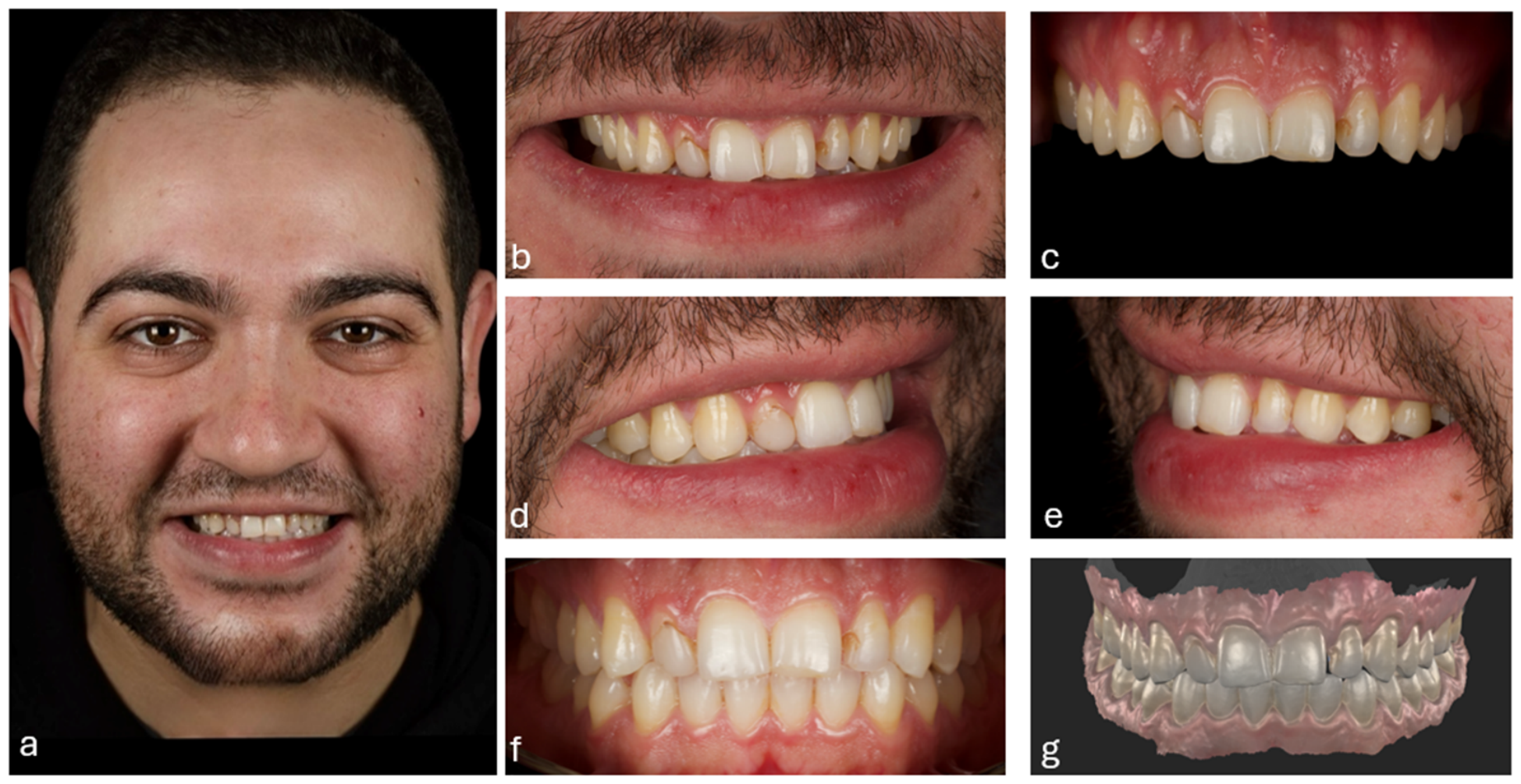
A 30-year-old male patient in good general health presented with concerns of unsatisfactory smile aesthetics regarding his disproportionate tooth morphology and irregular tooth coloration. Intraoral examination revealed peg-shaped lateral incisors with discolored overhanging composite restorations on teeth #12, 21, and 22 and reverse inclination of anterior teeth relative to the occlusal plane.
The initial diagnostic procedure included extraoral and intraoral photographs (Figure 1) and complete-arch digital scans obtained using an intraoral scanner (Primescan, Dentsply Sirona, Bensheim, Germany). The scanner captures high-resolution 3D data with a reported depth resolution of 3.2 μm at 15mm working distance. Calibration was performed according to manufacturer protocols prior to each scanning session.

2.2. Virtual Planning and Digital Workflow

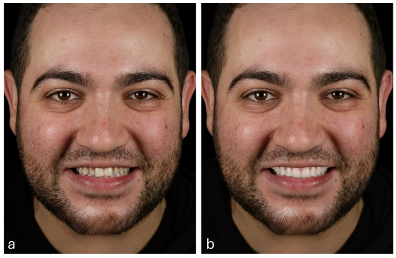
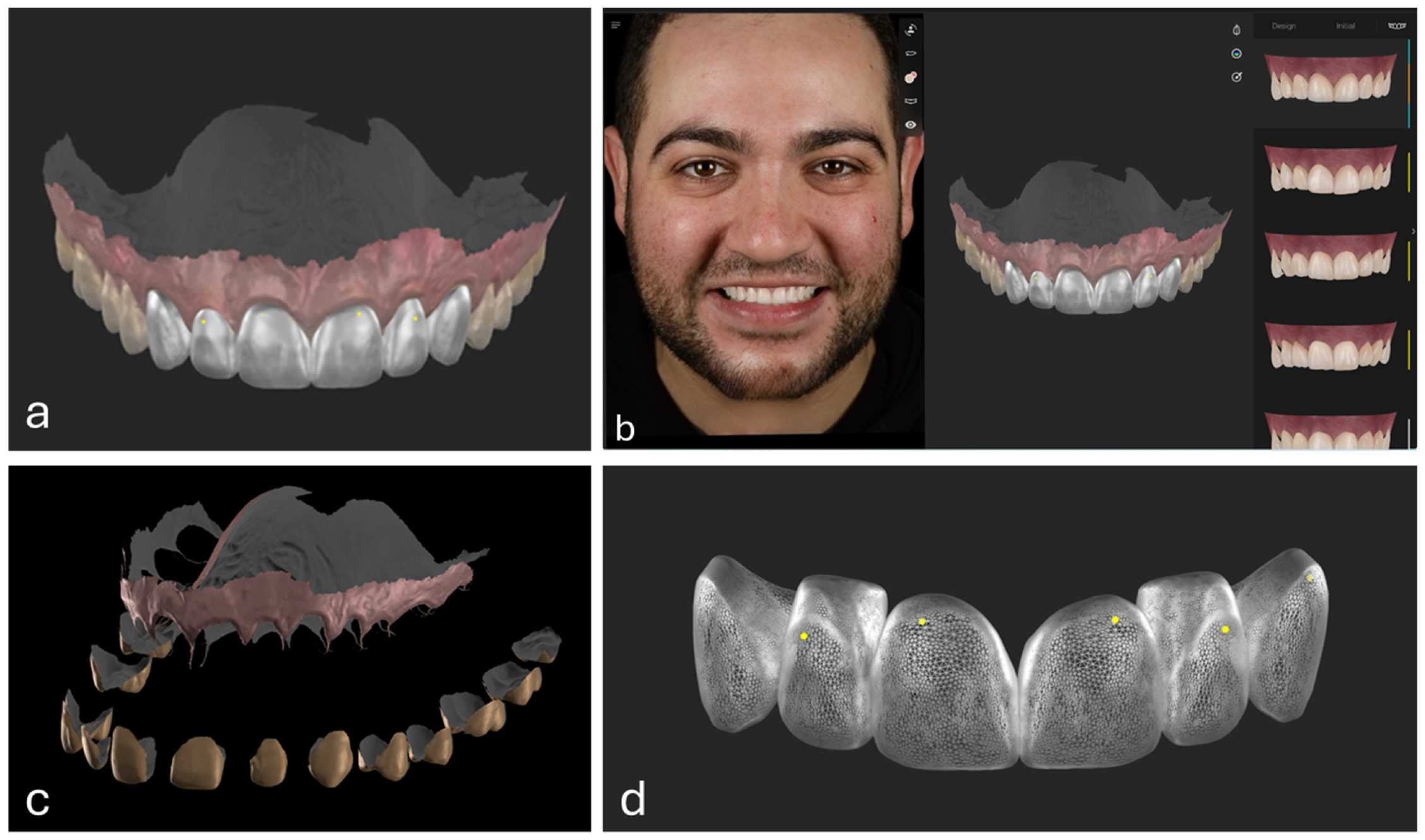
Following data acquisition, the treatment plan progressed to two-dimensional (2D) digital smile design using SmileCloud Biometrics software (SmileCloud SRL, Timișoara, Romania). A 2D digital smile design was developed, outlining proposed treatment for the six anterior teeth (canine to canine) (Figure 2). The workflow proceeded to three-dimensional (3D) smile design using SmileCloud Blueprint empowered with AI-assisted technology for automated teeth segmentation and alignment of intraoral scans with extraoral photographs. Following this process, the 3D tooth templates required manual positioning refinement to optimize both functional occlusion and aesthetic parameters of the proposed smile design (Figure 3). A personalized AI-powered video simulation was generated (SmileCloud YES), creating a dynamic facial representation of the patient to showcase the proposed smile design. Both the 3D digital smile design and accompanying video simulation were presented to the patient for approval prior to initiating treatment.

2.3. Visit II: Mock-Up

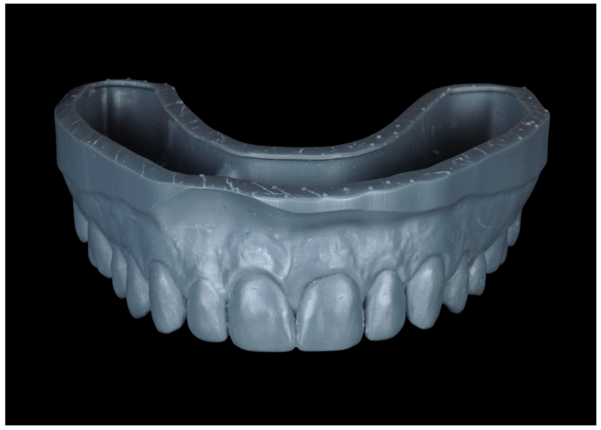
Following patient approval, the digital design files were exported in standard tessellation language (STL) format and imported into a design software (Exocad 3.2 Elefsina prototype, Exocad GmbH, Darmstadt, Germany) for virtual model creation. The finalized model was re-exported as an STL file and processed in RayWare Cloud 2.0/Print Setup on SprintRay Cloud (SprintRay, Los Angeles, CA, USA), where AI-assisted layer optimization technology refined the slicing parameters to maximize printing precision. The model was then fabricated using a SprintRay Pro 2 3D printer (SprintRay, Los Angeles, CA, USA) and Slate dental model resin (SRI-0202095, SprintRay, Los Angeles, CA, USA) (Figure 4).
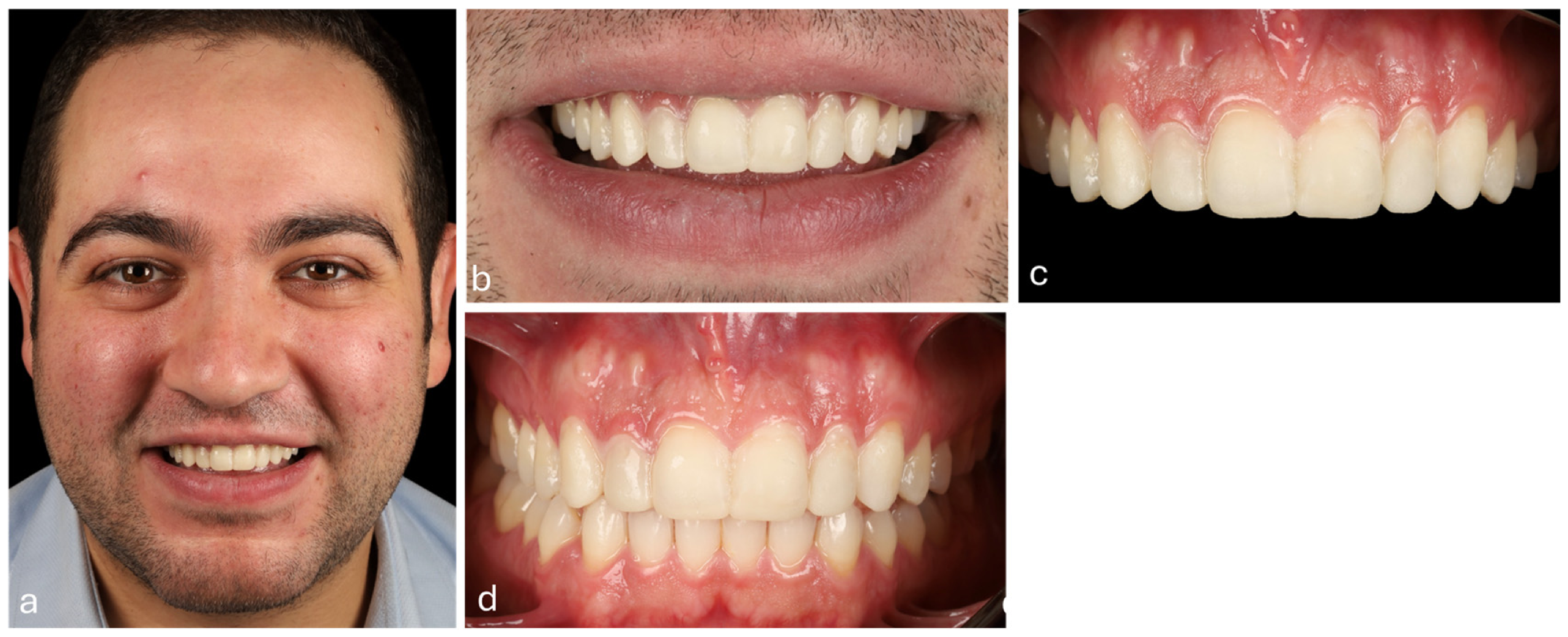
To convert the 3D-printed wax-up into an intraoral mock-up, a clear polyvinyl siloxane (PVS) silicone index using Exafine (GC, Tokyo, Japan) was fabricated. Dual-cure temporary composite resin (TempSmart, GC, Tokyo, Japan) was injected into the index and seated intraorally. After light-curing, excess material was removed using a fine-grit tapered diamond bur (#850314012, Komet, Germany). This functional mock-up served as a trial version of the proposed design, enabling evaluation by both clinician and patient prior to definitive treatment; a fundamental principle of the Aesthetic Pre-evaluative Temporary (APT) concept (Figure 5).

2.4. Veneers Preparation

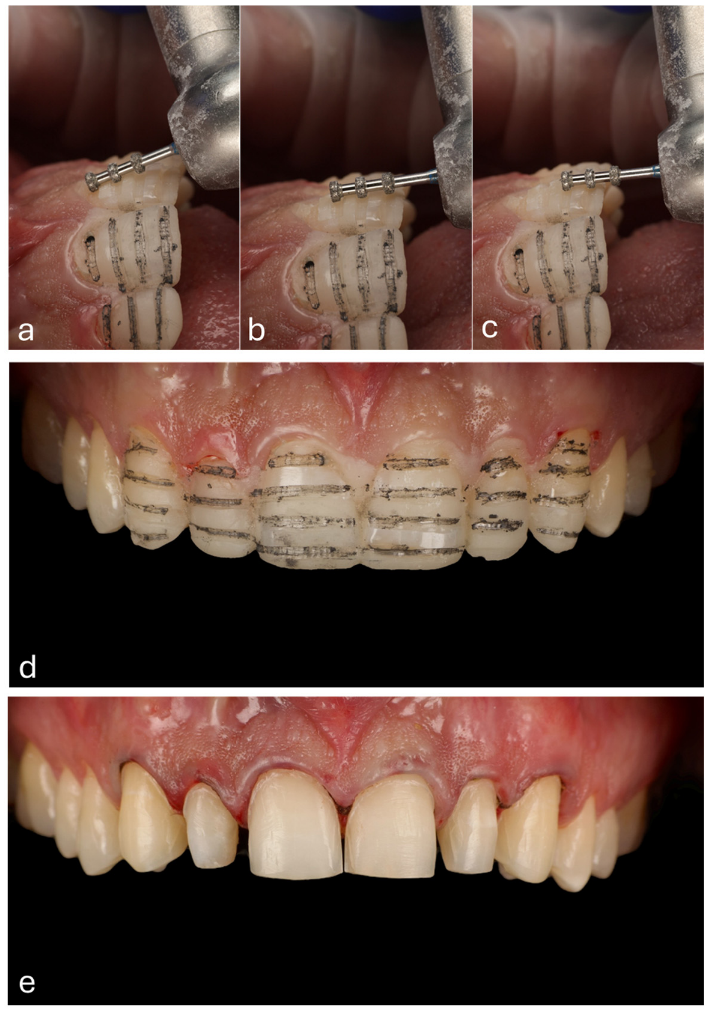
Following patient consent, mock-up-guided tooth preparation was performed. This minimally invasive approach uses the mock-up as a physical guide to preserve maximum healthy tooth structure while achieving the desired aesthetic outcome. Tooth reduction through the mock-up began with a 0.5 mm depth orientation bur (#834, INTESIV SA, Monteggio, Switzerland) at three planes, followed by groove marking with a #2HB graphite pencil. Preparation continued with a chamfer green-grit diamond bur (#FG235AC, INTESIV SA, Monteggio, Switzerland). Final veneer preparation was completed after placing a #00 retraction cord (SureCord, Pascal Company, Bellevue, WA, USA) and refining margins with a red-grit diamond bur (#FG4315S, INTESIV SA, Monteggio, Switzerland) (Figure 6). All preparations were polished using a yellow-grit diamond bur (#FGD18GB, INTESIV SA, Monteggio, Switzerland). Afterwards, shade was recorded, using VITA classical A1–D4 (VITA Zahnfabrik, Essen, Germany) matching the remaining teeth with shade A1.

2.5. Scanning and Designing

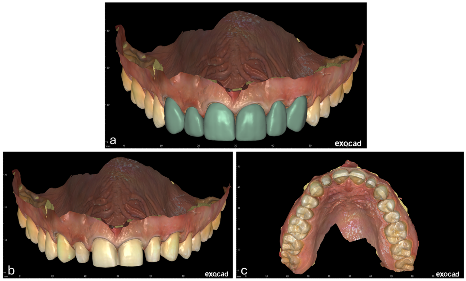
Following tooth preparation, intraoral scans were acquired using the same Primescan scanner (Dentsply Sirona, Bensheim, Germany) with identical calibration and resolution settings. The digital wax-up STL file was then imported into the design project and precisely aligned with best fit alignment with the preparation scan.
Within the Exocad design software (Exocad 3.2 Elefsina prototype, Exocad GmbH, Darmstadt, Germany), the veneers design was adapted to match the pre-operative scan design, shape and contours. Final refinements including occlusal harmony, anatomical detailing, and emergence profile optimization were executed by referencing the original 3D wax-up STL file to ensure aesthetic and functional fidelity (Figure 7). The measured alignment discrepancies directly reflected the preparation depth, which determined the corresponding restorations thicknesses.

2.6. Temporization

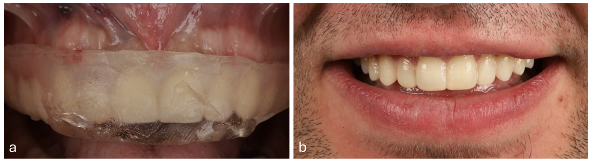
Provisional restorations were fabricated using Filtek™ Supreme XTE (3M ESPE AG, Seefeld, Germany), a highly filled flowable composite. Research indicates that these materials demonstrate favorable mechanical properties, including adequate flexural strength and wear resistance, supporting their use in provisional applications [13]. The clear silicone index, fabricated during the mock-up phase, was used to ensure precise adaptation of the temporaries (Figure 8). For retention, a small enamel area was spot-etched with 35% phosphoric acid (Ultra-Etch, Ultradent) and bonded with adhesive (All-Bond Universal, BISCO, Schaumburg, IL, USA). Final polishing was completed using DIACOMP paste (EVE Ernst Vetter, Pforzheim, Germany) with Jiffy Brushes (Ultradent, South Jordan, UT, USA).

2.7. Laboratory Procedures: Milling and Characterization

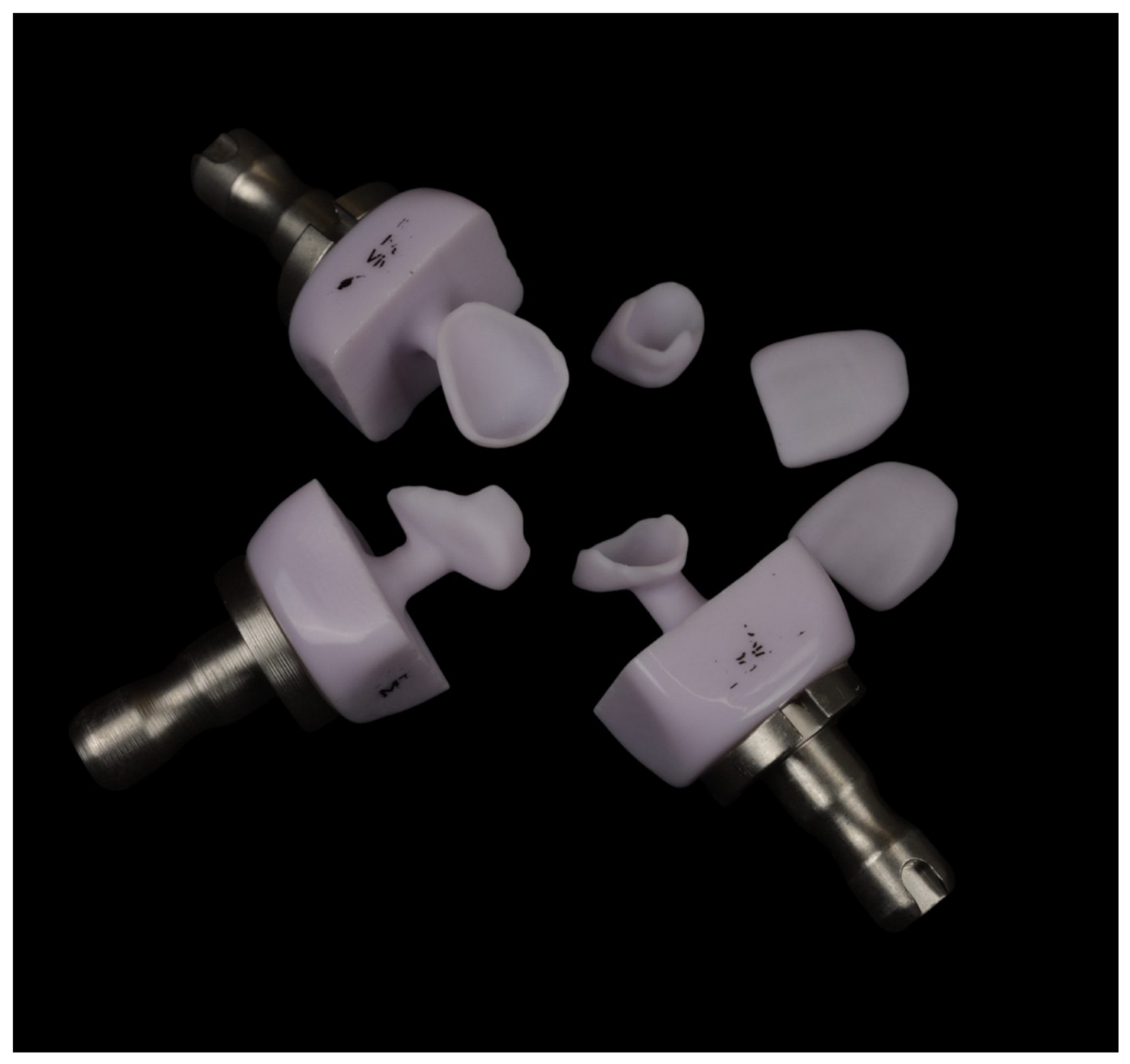
Six LDS veneers were fabricated using MT A1 blocks (IPS e.max CAD, Ivoclar Vivadent AG, Schaan, Liechtenstein) using a 4-axis milling machine (imes-icore GmbH, Eiterfeld, Germany) (Figure 9). The milled restorations underwent crystallization in a VITA VACUMAT® 6000 M furnace (VITA Zahnfabrik, Bad Säckingen, Germany). Following crystallization, veneers were characterized using MiYO ceramic paste material (Jensen Dental GmbH, Metzingen, Germany) received final glaze firing at low temperature in the same furnace. Prior to clinical try-in, the veneers were evaluated on a 3D-printed model to verify marginal adaptation and adjust interproximal contacts as needed (Figure 10).

2.8. Visit III—Try-In and Bonding

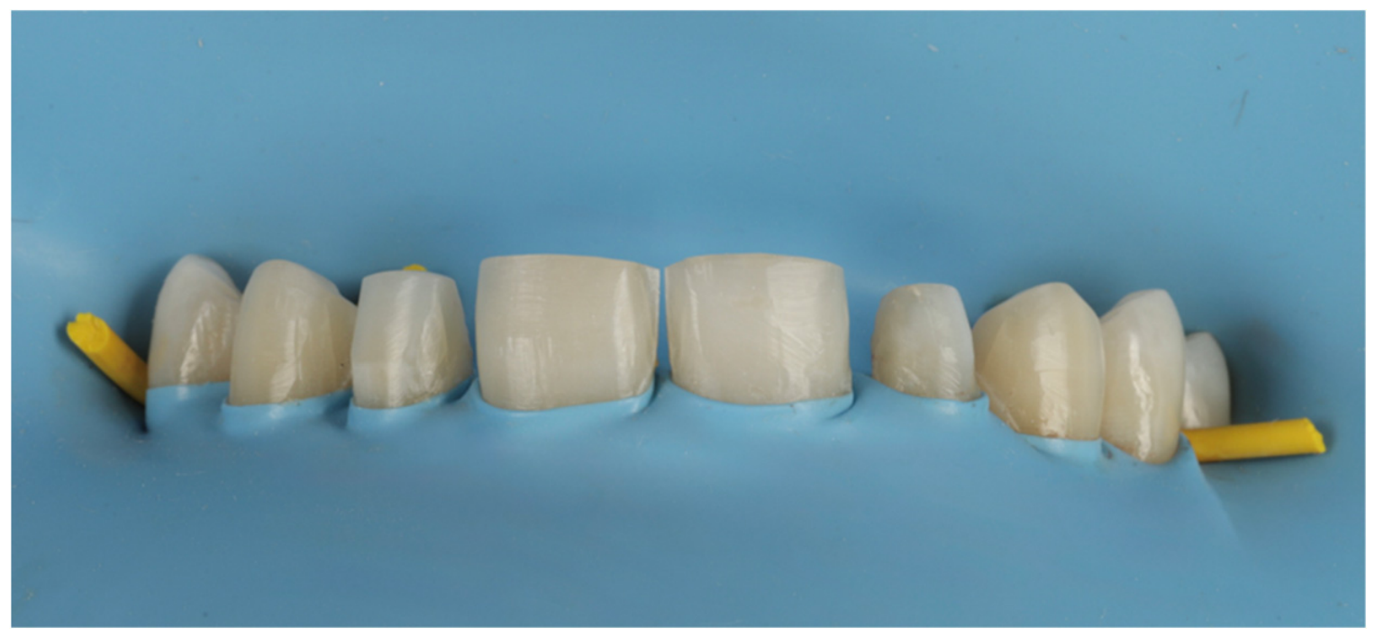
The temporary restorations were removed using an ultrasonic scaler (Sirosonic, Dentsply Sirona, Bensheim, Germany) to ensure complete elimination of provisional material. The ceramic veneers were tried using try-in paste (Variolink Try-In, Ivoclar Vivadent, Schaan, Liechtenstein) following the sequence: centrals, laterals, and then canines. The patient confirmed satisfaction with the shape, color, and overall appearance of the restorations. Following verification, the bonding procedure was initiated. Isolation was achieved with a powder-free latex rubber dam (Sanctuary Healthcare Sdn Bhd, Ipoh, Malaysia) stabilized on the upper first premolars using Wedjets (Coltène/Whaledent, Cuyahoga Falls, OH, USA) (Figure 11).
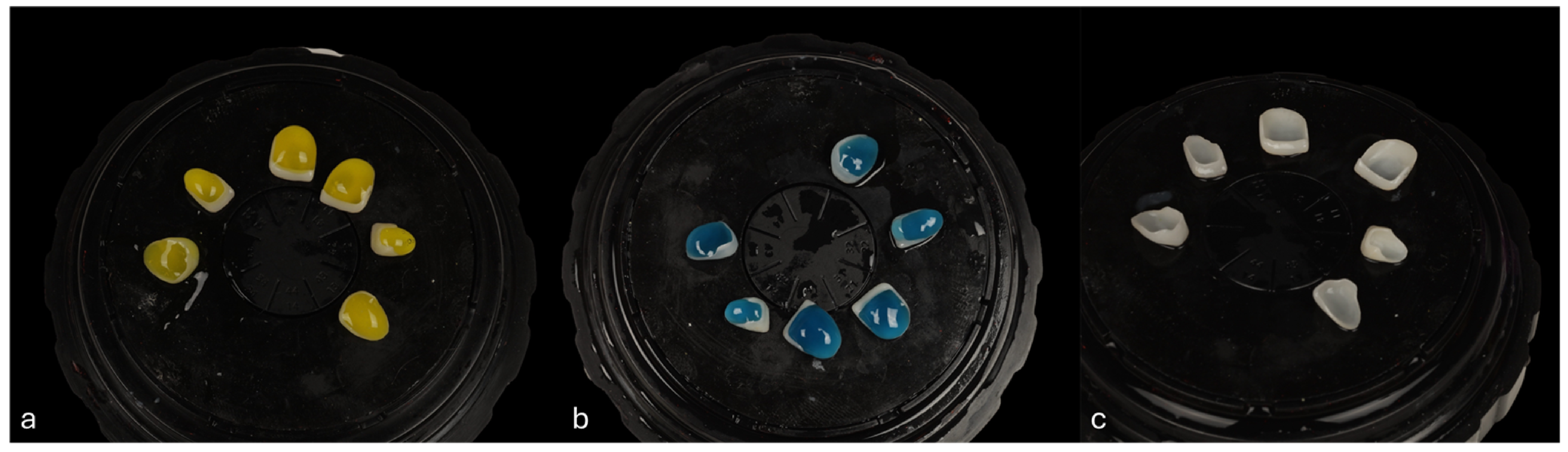
The intaglio surfaces of the veneers were etched with 9% hydrofluoric acid (Porcelain Etch, Ultradent, South Jordan, UT, USA) for 20 s, followed by 37% phosphoric acid (Total Etch, Ivoclar Vivadent, Schaan, Liechtenstein) for 60 s to remove the surface remnants after etching. Each step was followed by copious rinsing with distilled water and drying. A ceramic silane coupling agent (Monobond Plus, Ivoclar Vivadent, Schaan, Liechtenstein) was then applied to the intaglio surfaces for 60 s then air-thinned for 5 s (Figure 12).
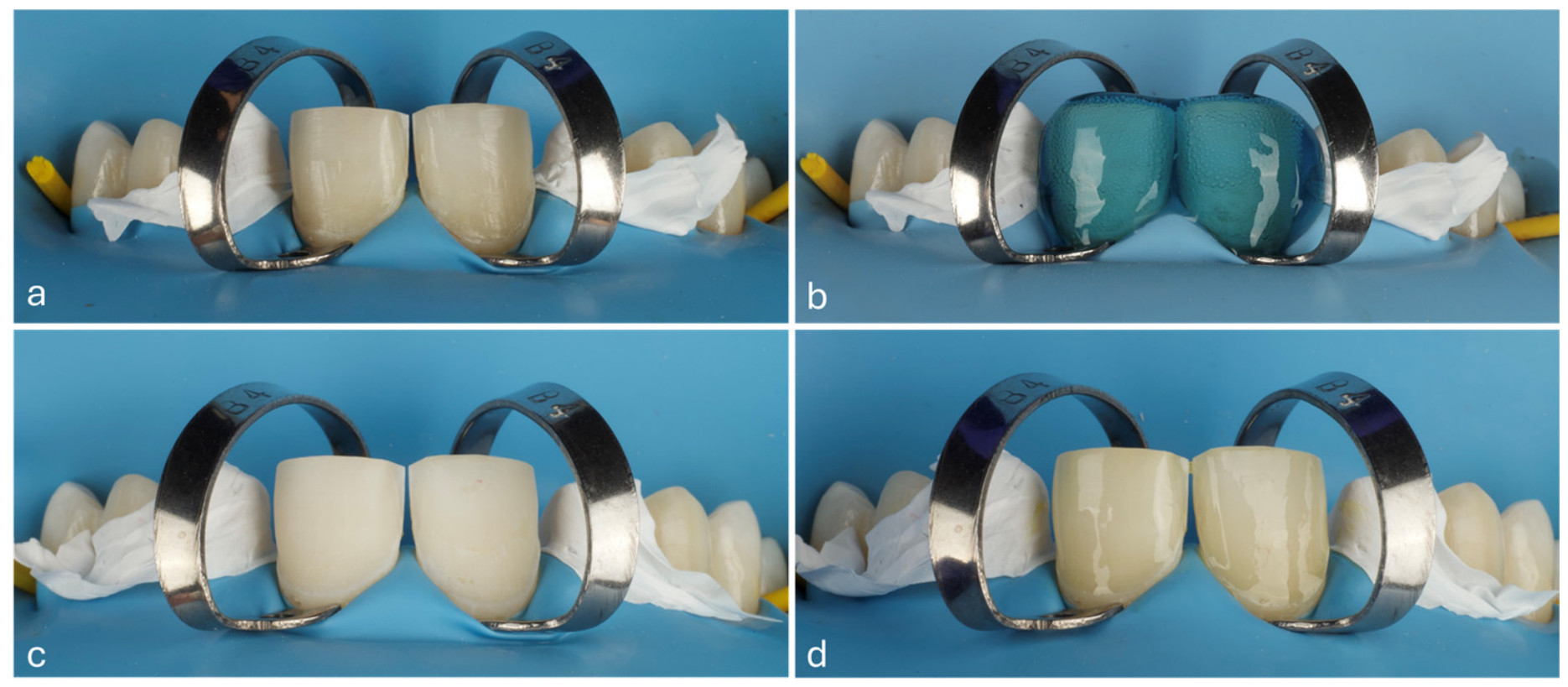
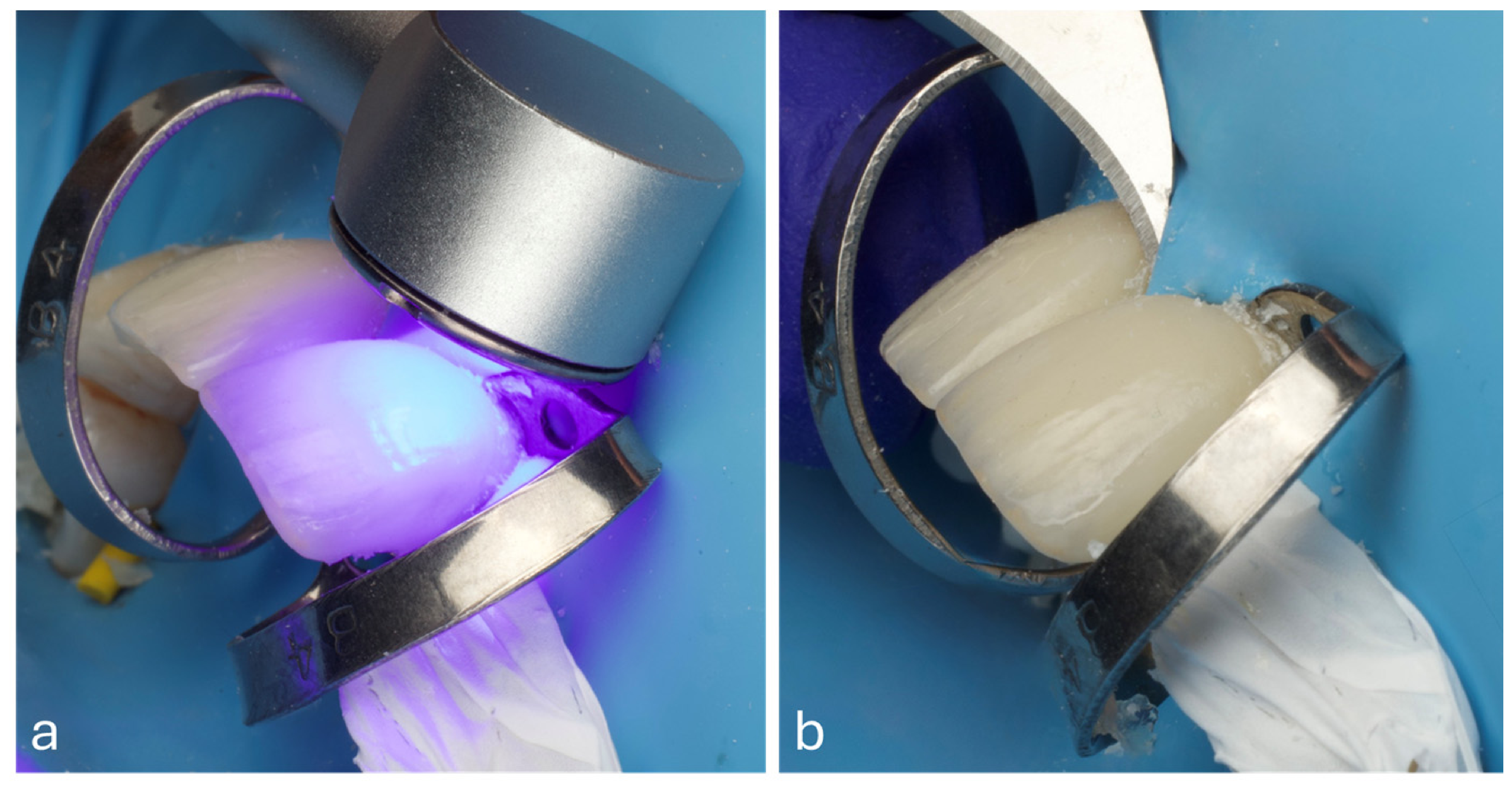
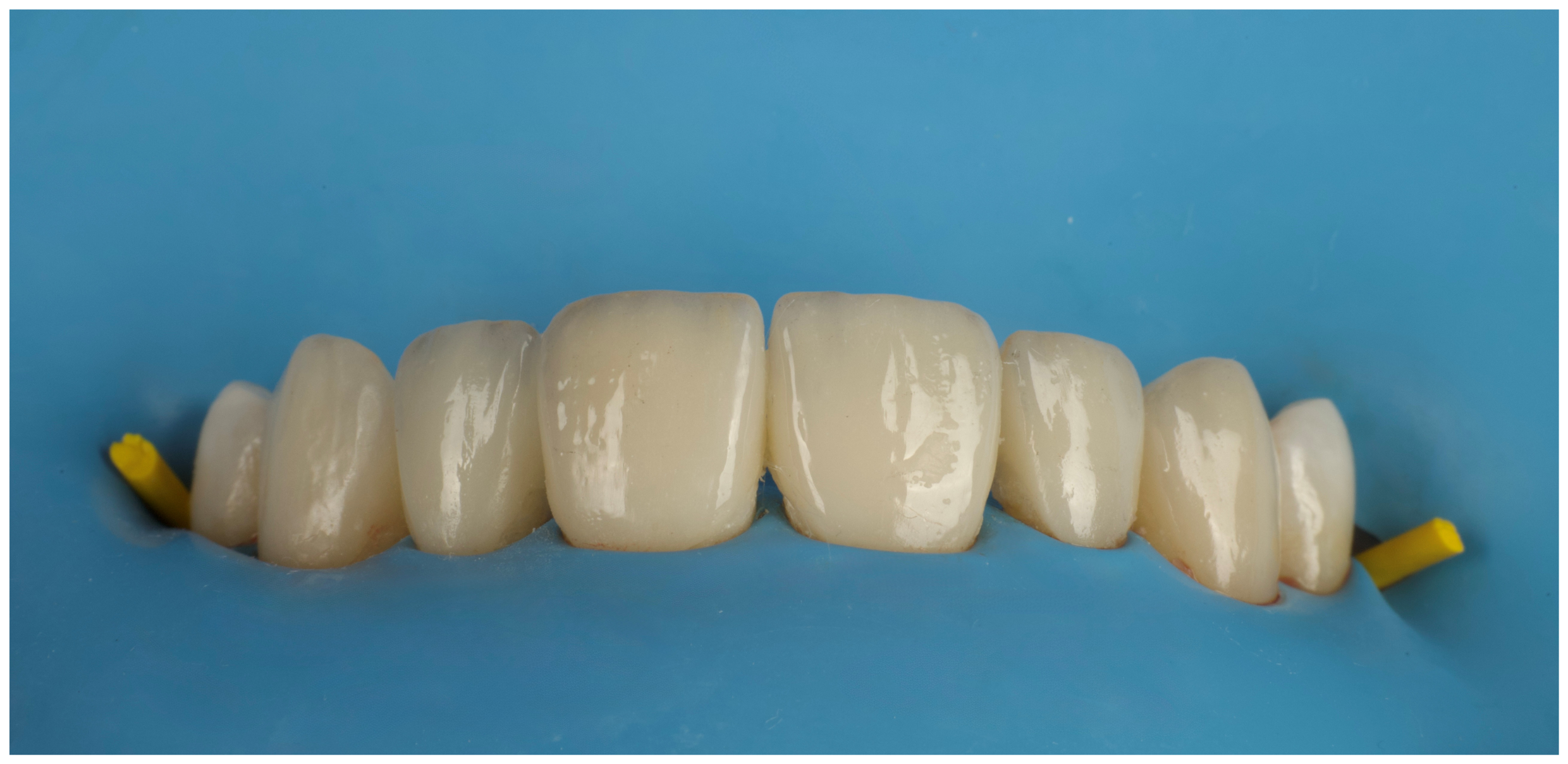
Brinker clamps (Clamp B4, Brinker Hygienic, Coltene, Altstätten, Switzerland) were carefully positioned on the prepared teeth to retract and invert the rubber dam, fully exposing the margins. After thorough cleaning and drying, interproximal surfaces were protected with PTFE tape (PTFE Iso Tape 5 mm Santa Catarina, Brasil). The teeth were etched with 35% phosphoric acid (Ultra-Etch, Ultradent, South Jordan, UT, USA) for 30 s, rinsed, and dried. Universal adhesive (All-Bond Universal, BISCO, Schaumburg, IL, USA) was applied in a double layer, gently air-dried to remove solvents, and each tooth was polymerized for 10 s as per manufacturer instructions (Figure 13). A translucent light-cure resin cement (Variolink Esthetic LC, Ivoclar Vivadent, Schaan, Liechtenstein) was applied to the veneers, which were then seated on the prepared teeth in the predetermined sequence. Excess unpolymerized cement was removed with a brush then each veneer was polymerized for 20 s per surface (facial, palatal, mesial, distal) using light-curing device (1000 mW/cm2, CuringPen-E, Eighteeth, Changzhou, China), finally surgical blade #12 (Swann-Morton, Sheffield, England) was used to remove excess cured resin cement (Figure 14). Veneers were bonded respecting the try-in sequence following the same steps respectively (Figure 15).

2.9. Occlusal Adjustment and Polishing

No occlusal adjustments were required after verifying static and dynamic occlusion with articulating oil of 8 μm (TrollFoil, TrollDental AB, Lincoln, UK). Polishing of the palatal margins was performed using polishing bur (#FG9205, INTESIV SA, Monteggio, Switzerland). The patient was scheduled for follow-up after one week.

2.10. One Week Postoperative Evaluation

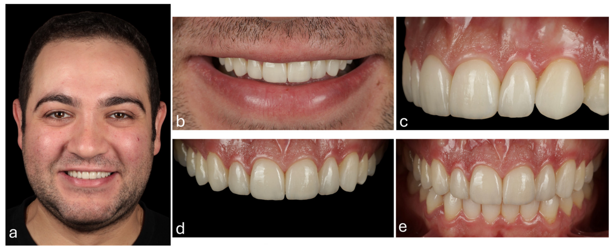
The patient’s smile aesthetics and veneer integration were assessed one-week post-bonding (Figure 16).
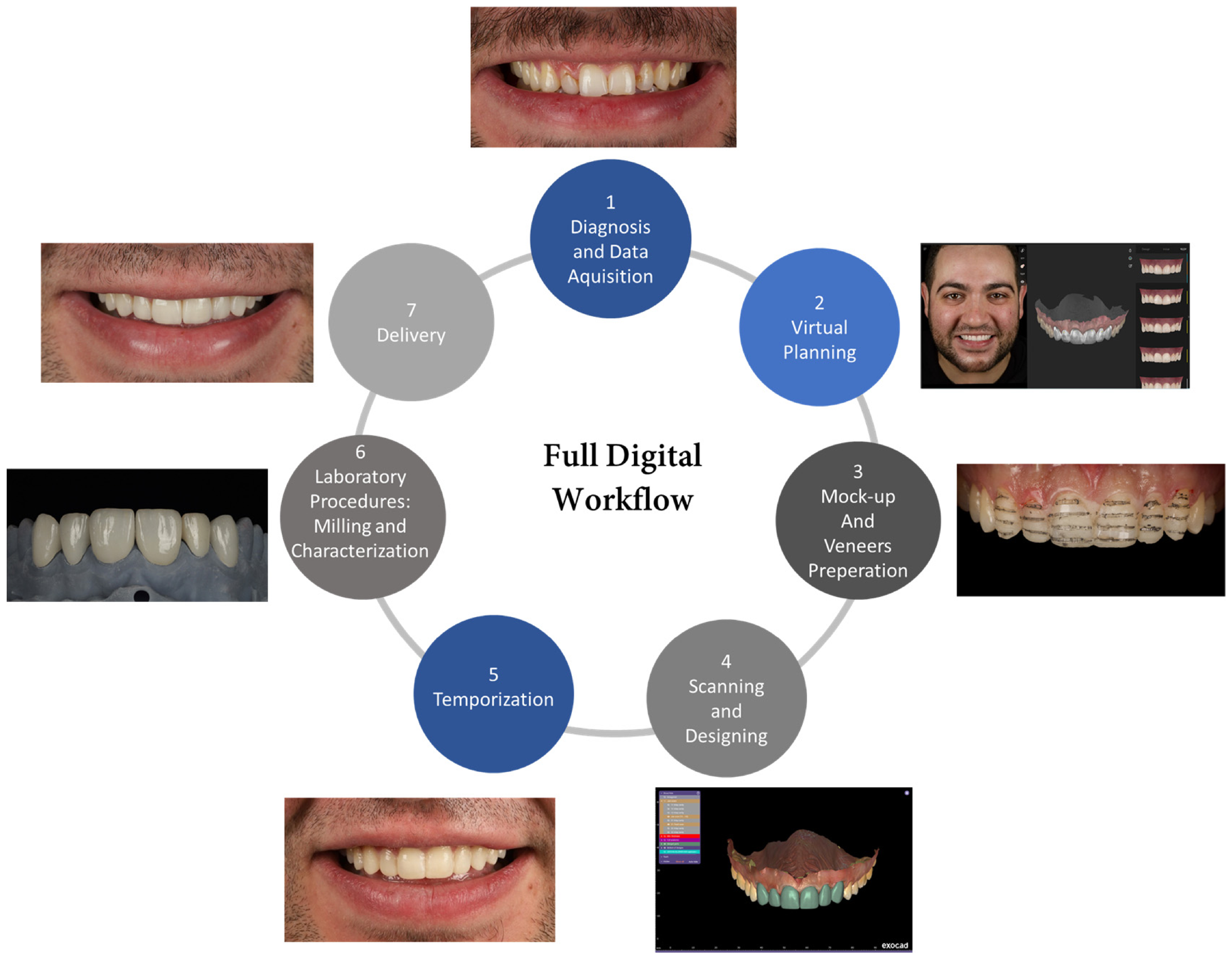
The steps of the implemented full digital workflow are summarized in the flowchart in Figure 17.

3. Discussion

The integration of AI-powered digital workflows in smile design and veneer rehabilitation represents a significant advancement in modern dentistry, as demonstrated in this case report.
The diagnostic workflow incorporated SmileCloud Biometrics software, which uses deep learning algorithms trained on datasets of facial photographs and dental morphologies to analyze patient-specific features. Unlike conventional systems that modify generic tooth shapes to fit a space, this process begins by selecting natural tooth morphology for the defined restorative space. The software applies biometric facial reference points, including interpupillary distance, facial midline, and lip dynamics, to generate proportional tooth proposals. It then integrates user-defined parameters, such as facial references and 2D design outlines, to search a library of natural donor teeth, evaluating tissue architecture, line angles, proportions, and spatial constraints to generate a virtual mock-up [14]. This approach produces a preliminary 2D smile proposal within several minutes, a process that traditionally required more time [7]. Through visualization of potential outcomes and video simulation (SmileCloud YES), the digital workflow supported patient engagement, allowing the patient to participate in decision-making before intervention [14,15].
Following this AI-generated proposal, manual refinements were performed to optimize critical smile parameters including the smile arc, dental midline position, incisal edge placement, and buccal corridor dimensions [16]. Interestingly, the case presented an optimal buccal corridor configuration when referenced against the established biometric parameter of interpupillary distance (specifically, the span between the proximal pupil borders and terminal iris regions [17], ultimately allowing for a conservative treatment approach limited to six anterior veneers while preserving the natural premolar anatomy.
However, 2D AI-driven smile design inherently faces several dimensional and functional limitations that become particularly evident in complex cases [18]. The fundamental constraint of working exclusively with 2D photographic data prevents comprehensive evaluation of crucial 3D occlusal relationships, tooth spatial inclinations, and dynamic arch form characteristics [19]. Furthermore, the proprietary nature and closed architecture of most 2D smile design platforms restrict clinicians’ ability to perform modifications or adaptations to address individual anatomical variations or restorative requirements [20,21]. These limitations highlight the critical importance of transitioning to integrated 3D digital workflows, where advanced dental CAD systems combine intraoral scan data with either 2D facial photographs or 3D facial scans to create a complete virtual patient representation. Modern implementations of this approach often utilize the iterative closest point (ICP) algorithm. However, some systems such as the one employed in this case, feature AI-enhanced ICP optimization, which significantly improves registration accuracy while efficiently compensating for potential scan artifacts or complex anatomical alignments [22].
The fully integrated digital workflow including intraoral scanning, CAD/CAM design, model printing, and veneer milling, contributed to diagnostic accuracy and predictable clinical outcomes. Critically, this approach eliminated error-prone physical impressions and time-consuming manual wax-ups, replacing them with high-precision intraoral scans and AI-optimized smile design combined with rapid 3D-printed model fabrication [23]. Specifically, the 3D-printed model used for mock-up fabrication was produced using SprintRay RayWare with Smart Print AI, a machine learning system trained on thousands of dental print jobs. The software automatically optimizes print orientation, support placement, and slicing parameters based on model type recognition. Intelligent support placement minimizes post-processing work, while Pixel Toning technology uses pixel-by-pixel light adjustment to enhance surface detail, reducing voxelization and improving quality [24,25].
While these AI-assisted tools offer efficiency benefits, combining multiple AI software platforms within a single digital workflow presents potential challenges. Each system operates with its own training dataset, algorithmic decision-making framework. A recent study [26] reported that 6.7% of AI-generated designs required manual intervention due to margin detection failures or die interface errors when encountering suboptimal preparation geometries, highlighting that AI systems may struggle with non-ideal clinical situations. In the present case, these risks were mitigated through consistent clinician oversight throughout the workflow. As previously described, following automated tooth segmentation and template matching in SmileCloud Blueprint, manual positioning refinement was performed to optimize functional occlusion and aesthetic parameters. Similarly, during the 3D printing phase using SprintRay RayWare with Smart Print AI, all automatically generated support structures and print settings were verified before fabrication.
A growing body of evidence supports the use of AI and digital workflows in restorative treatments. Studies by Ceylan et al. and Baaj et al. [8,9] have reported that AI-assisted smile designs achieve aesthetic results comparable to those created by human experts, while also improving standardization and reproducibility. Similarly, Buduru et al. [7] found that AI-assisted smile design applications were well-received by both patients and clinicians but emphasized that AI proposals required refinement for optimal aesthetic outcomes, a finding consistent with the present case, where manual adjustments to AI-generated tooth templates were performed.
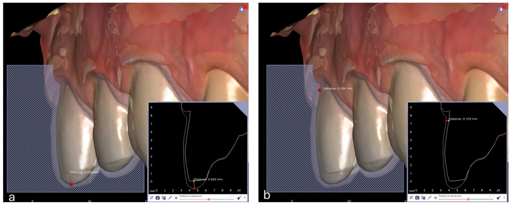
The long-term clinical success of ceramic veneers in restorative dentistry depends on four critical factors: aesthetic integration, mechanical durability, precise bonding protocols, and minimally invasive preparation [27]. In the present case, preparation was optimized through mock-up-guided reduction following the APT concept. Digital diagnostic analysis confirmed an additive restorative approach, enabling ultraconservative tooth reduction of 0.2–0.4 mm (cervical/buccal) to 0.9 mm (incisal edge) (Figure 18), preserving most of the sound enamel. This aligns with clinical guidelines emphasizing minimal dentin exposure to enhance adhesive restoration longevity, as enamel preservation significantly improves bond durability [28]. The final veneers thicknesses were approximately 0.4 mm cervically and 1.0 mm incisally, with an additional 50 μm cement space filled by adhesive resin cement. This protocol successfully balanced biological preservation with clinical outcomes achieving optimal tissue integrity, optimal aesthetic emergence profiles and functional occlusal harmony.
When selecting appropriate restorative materials for veneers, clinicians must evaluate many available options: feldspathic porcelain, resin-matrix hybrid ceramics, high-translucency zirconia, LDS and leucite reinforced ceramics. Among these, LDS has emerged as the gold standard due to its unique combination of aesthetic performance and mechanical resilience. With a flexural strength of 350–400 MPa, significantly higher than feldspathic ceramics (80–120 MPa) and hybrid ceramics (150–200 MPa), LDS demonstrates superior fracture resistance and reduced chipping incidence enabling them to be used in a monolithic form [1,29]. Furthermore, LDS exhibits fewer aesthetic complications (discoloration, surface roughness) compared to both feldspathic porcelain and zirconia-based alternatives [30]. The monolithic design reduces the risk of ceramic shipping, a common failure mode in layered veneers, while maintaining sufficient mechanical properties. The restorations achieved natural characterization through direct staining and glazing with MiYO ceramic paste material (Jensen Dental GmbH, Metzingen, Germany), bypassing the need for traditional layering techniques without compromising optical properties.
Beyond selecting appropriate materials, adhering to proper adhesive protocols and techniques is essential for the clinical success of adhesive restorations. The effectiveness of these dental restorations in clinical settings depends on the strength and durability of the bond, which is crucial for preventing issues such as marginal gaps, bacterial infiltration, postoperative discomfort, secondary caries, and restoration failure [31]. A successful bonding protocol begins with effective tooth isolation to prevent contamination, ensure antisepsis, and control moisture [32]. In the present case, the use of a rubber dam enhanced visibility, facilitated saliva isolation, and improved moisture control, thereby optimizing the quality of the resin adhesive-tooth interface, a critical factor for the long-term success of veneers.

4. Conclusions

This case shows how combining AI with reverse engineering significantly improves dental restorative treatments. Better accuracy, natural aesthetics, and comfortable function are achieved by turning 3D digital smile designs into precise veneers. This workflow, from AI-driven digital design to ceramic veneers manufacturing, reduces guesswork and enhances results, raising the bar for personalized aesthetic rehabilitations.

Author Contributions

Conceptualization, M.Q., M.M., H.T.; methodology, M.Q., M.M., H.T., G.A.; software, M.Q.; validation, M.Q., M.M., H.T.; resources, M.Q., G.A.; writing—original draft preparation, M.Q., M.M., H.T.; writing—review and editing, M.Q., M.M., H.T., E.X.; visualization, M.Q., M.M., H.T., E.X.; supervision, H.T.; project administration, M.Q., M.M., H.T.; funding acquisition, E.X. All authors have read and agreed to the published version of the manuscript.

Funding

This research received no external funding.

Institutional Review Board Statement

As this work is a single case report, Institutional Review Board approval was not required. The Institutional Review Committee at Al-Ahliyya Amman University reviewed and approved the waiver application.

Informed Consent Statement

Written informed consent was obtained from the patient for treatment and for the publication of relevant clinical information and clinical photographs included in this article.

Data Availability Statement

The original contributions presented in this study are included in the article. Further inquiries can be directed to the corresponding author.

Conflicts of Interest

The authors declare no conflicts of interest.

References

  1. Klein, P.; Spitznagel, F.A.; Zembic, A.; Prott, L.S.; Pieralli, S.; Bongaerts, B.; Metzendorf, M.I.; Langner, R.; Gierthmuehlen, P.C. Survival and Complication Rates of Feldspathic, Leucite-Reinforced, Lithium Disilicate and Zirconia Ceramic Laminate Veneers: A Systematic Review and Meta-Analysis. J. Esthet. Restor. Dent. 2025, 37, 601–619. [Google Scholar] [CrossRef]
  2. Peumans, M.; De Munck, J.; Fieuws, S.; Lambrechts, P.; Vanherle, G.; Van Meerbeek, B. A Prospective Ten-Year Clinical Trial of Porcelain Veneers. J. Adhes. Dent. 2004, 6, 65–76. [Google Scholar]
  3. Zarone, F.; Ferrari, M.; Mangano, F.G.; Leone, R.; Sorrentino, R. “Digitally Oriented Materials”: Focus on Lithium Disilicate Ceramics. Int. J. Dent. 2016, 2016, 9840594. [Google Scholar] [CrossRef]
  4. Jafri, Z.; Ahmad, N.; Sawai, M.; Sultan, N.; Bhardwaj, A. Digital Smile Design—An Innovative Tool in Aesthetic Dentistry. J. Oral Biol. Craniofac. Res. 2020, 10, 194–198. [Google Scholar] [CrossRef]
  5. Babu, A.; Andrew Onesimu, J.; Martin Sagayam, K. Artificial Intelligence in Dentistry: Concepts, Applications and Research Challenges. E3S Web Conf. 2021, 297, 01074. [Google Scholar] [CrossRef]
  6. Kazimierczak, N.; Kazimierczak, W.; Serafin, Z.; Nowicki, P.; Nożewski, J.; Janiszewska-Olszowska, J. AI in Orthodontics: Revolutionizing Diagnostics and Treatment Planning—A Comprehensive Review. J. Clin. Med. 2024, 13, 344. [Google Scholar] [CrossRef]
  7. Buduru, S.; Cofar, F.; Mesaroș, A.; Tăut, M.; Negucioiu, M.; Almășan, O. Perceptions in Digital Smile Design: Assessing Laypeople and Dental Professionals’ Preferences Using an Artificial-Intelligence-Based Application. Dent. J. 2024, 12, 104. [Google Scholar] [CrossRef]
  8. Ceylan, G.; Özel, G.S.; Memişoğlu, G.; Emir, F.; Şen, S. Evaluating the facial esthetic outcomes of digital smile designs generated by artificial intelligence and dental professionals. Appl. Sci. 2023, 13, 9001. [Google Scholar] [CrossRef]
  9. Baaj, R.E.; Alangari, T.A. Artificial intelligence applications in smile design dentistry: A scoping review. J. Prosthodont. 2025, 34, 341–349. [Google Scholar] [CrossRef] [PubMed]
  10. Alotaibi, S.; Deligianni, E. Can AI Replace Dentists? Br. Dent. J. 2023, 235, 763. [Google Scholar] [CrossRef] [PubMed]
  11. Lawand, G.; Vera, V.; Nessif, R.; Gonzaga, L.; Martin, W. Artificial intelligence facilitated smile design for the rehabilitation of a fully maxillary edentulous patient with a complete arch implant-supported prosthesis: A dental technique. J. Prosthodont. 2026; in press. [CrossRef] [PubMed]
  12. Saini, R.S.; Kaur, K.; Gurumurthy, V.; Binduhayyim, R.I.; Kaushik, A.; Kuruniyan, M.S.; Alarcón-Sánchez, M.A.; Heboyan, A. Impact of artificial intelligence-based digital smile design on patient and clinician satisfaction and facial esthetic outcomes: A systematic review and meta-analysis. Digit. Health 2025, 11, 20552076251388392. [Google Scholar] [CrossRef]
  13. Francois, P.; Attal, J.P.; Fasham, T.; Troizier-Cheyne, M.; Gouze, H.; Abdel-Gawad, S.; Le Goff, S.; Dursun, E.; Ceinos, R. Flexural properties, wear resistance, and microstructural analysis of highly filled flowable resin composites. Oper. Dent. 2024, 49, 597–607. [Google Scholar] [CrossRef]
  14. Alikhasi, M.; Yousefi, P.; Afrashtehfar, K.I. Smile Design: Mechanical Considerations. Dent. Clin. N. Am. 2022, 66, 477–487. [Google Scholar] [CrossRef]
  15. Alharkan, H.M. Integrating Digital Smile Design into Restorative Dentistry: A Narrative Review of the Applications and Benefits. Saudi Dent. J. 2024, 36, 561–567. [Google Scholar] [CrossRef]
  16. Coachman, C.; Calamita, M. Digital Smile Design: A Tool for Treatment Planning and Communication in Esthetic Dentistry. In Quintessence of Dental Technology (QDT); Quintessence Publishing Co., Inc.: Hanover Park, IL, USA, 2012; Volume 35, pp. 103–111. [Google Scholar]
  17. Ntovas, P.; Karkazi, F.; Özbilen, E.Ö.; Flavio, A.; Ladia, O.; Papazoglou, E.; Yilmaz, H.N.; Coachman, C. Perception of Smile Attractiveness Among Laypeople and Orthodontists Regarding the Buccal Corridor Space, as It Is Defined by the Eyes. An Innovated Technique. J. Esthet. Restor. Dent. 2023, 35, 345–351. [Google Scholar] [CrossRef] [PubMed]
  18. Bourgi, R.; Qaddomi, M.; Hardan, L.; Tohme, H.; Corbani, K.; Abou Isber, S.; Daher, E.A.; Nassar, N.; Kharouf, N.; Haikel, Y. Gingival Contouring and Smile Makeover Through Digital Planning and 3D Guidance. J. Clin. Med. Res. 2025, 6, 6208. [Google Scholar] [CrossRef]
  19. Galibourg, A.; Brenes, C. Virtual Smile Design Tip: From 2D to 3D Design with Free Software. J. Prosthet. Dent. 2019, 121, 863–864. [Google Scholar] [CrossRef] [PubMed]
  20. Feraru, M.; Musella, V.; Bichacho, N. Individualizing a Smile Makeover. J. Cosmet. Dent. 2016, 32, 108–120. [Google Scholar]
  21. Sharma, A.; Luthra, R.; Kaur, P. A Photographic Study on Visagism. Indian J. Oral Sci. 2015, 6, 22. [Google Scholar]
  22. Ferrando-Cascales, Á.; Astudillo-Rubio, D.; Pascual-Moscardó, A.; Delgado-Gaete, A. A Facially Driven Complete-Mouth Rehabilitation with Ultrathin CAD-CAM Composite Resin Veneers for a Patient with Severe Tooth Wear: A Minimally Invasive Approach. J. Prosthet. Dent. 2020, 123, 537–547. [Google Scholar] [CrossRef]
  23. Cacciò, C.; Tallarico, M.; Lumbau, A.I.; Ceruso, F.M.; Pisano, M. The Role of Digital Workflow in Creating a New, Esthetic and Functional Smile in a Periodontally Compromised Patient: A Case Report. Reports 2025, 8, 105. [Google Scholar] [CrossRef] [PubMed]
  24. RayWare on SprintRay Cloud. SprintRay Inc. Available online: https://sprintray.com/en-iq/rayware-on-sprintray-cloud/ (accessed on 27 January 2026).
  25. The Summer of Rayware: SprintRay Pro, Reborn. SprintRay Inc. Available online: https://sprintray.com/the-summer-of-rayware-sprintray-pro-reborn/ (accessed on 27 January 2026).
  26. Xie, B.Y.; He, X.; Hu, L.; Guo, S.L.; Chen, J.L.; Zhang, J.; Shen, X.Q.; Geng, Y.M.; Li, W. Morphological comparison between artificial intelligence-driven and manual CAD design in single tooth restoration: A preliminary study. BMC Oral Health 2025, 25, 1633. [Google Scholar] [CrossRef]
  27. Gonzalez-Martin, O.; Avila-Ortiz, G.; Torres-Muñoz, A.; Del Solar, D.; Veltri, M. Ultrathin Ceramic Veneers in the Aesthetic Zone: A36-month Retrospective Case Series. Int. J. Prosthodont. 2021, 34, 567–577. [Google Scholar] [CrossRef] [PubMed]
  28. Schlichting, L.H.; Resende, T.H.; Reis, K.R.; Dos Santos, A.R.; Correa, I.C.; Magne, P. Ultrathin CAD-CAM Glass-Ceramic and Composite Resin Occlusal Veneers for the Treatment of Severe Dental Erosion: An Up to 3-Year Randomized Clinical Trial. J. Prosthet. Dent. 2022, 128, 158. [Google Scholar] [CrossRef]
  29. De Angelis, F.; D’Arcangelo, C.; Angelozzi, R.; Vadini, M. Retrospective Clinical Evaluation of a No-Prep Porcelain Veneer Protocol. J. Prosthet. Dent. 2023, 129, 40–48. [Google Scholar] [CrossRef]
  30. Villalobos-Tinoco, J.; Floriani, F.; Rojas-Rueda, S.; Mekled, S.; Conner, C.; Colvert, S.; Jurado, C.A. Enhancing Smile Aesthetics and Function with Lithium Disilicate Veneers: A Brief Review and Case Study. Clin. Pract. 2025, 15, 66. [Google Scholar] [CrossRef]
  31. Falacho, R.I.; Melo, E.A.; Marques, J.A.; Ramos, J.C.; Guerra, F.; Blatz, M.B. Clinical In-Situ Evaluation of the Effect of Rubber Dam Isolation on Bond Strength to Enamel. J. Esthet. Restor. Dent. 2023, 35, 48–55. [Google Scholar] [CrossRef]
  32. Jurado, C.A.; Fischer, N.G.; Sayed, M.E.; Villalobos-Tinoco, J.; Tsujimoto, A.; Sayed, M. Rubber Dam Isolation for Bonding Ceramic Veneers: A Five-Year Post-Insertion Clinical Report. Cureus 2021, 13, e20748. [Google Scholar] [CrossRef] [PubMed]
Figure 1. Data acquisition. (a) Full face photo (frontal view). (b) Smile photo. (c) Intraoral photo (frontal view). (d) Right lateral smile. (e) Left lateral smile. (f) Intraoral photo (retracted). (g) Upper and lower scans.
Prosthesis 08 00045 g001
Figure 2. (a) Initial photo. (b) 2D smile design photo.
Prosthesis 08 00045 g002
Figure 3. 3D smile design. (a) 3D digital wax-up (frontal view). (b) 2D, 3D smile designs with AI teeth libraries. (c) Teeth segmentation. (d) Wireframe of 3D digital wax-up.
Prosthesis 08 00045 g003
Figure 4. 3D-printed model.
Prosthesis 08 00045 g004
Figure 5. Mock-up evaluation. (a) Full face photo. (b) Extraoral smile photo. (c) Intraoral upper-arch photo (d) Intraoral retracted photo.
Prosthesis 08 00045 g005
Figure 6. Mock-up driven preparation following three planes. (a) Cervical plane. (b) Mid-plane. (c) Incisal plane. (d) Frontal view (e) Final preparations with retraction cord properly positioned.
Prosthesis 08 00045 g006
Figure 7. (a) Final design. (b) Scan (frontal view). (c) Scan (occlusal view).
Prosthesis 08 00045 g007
Figure 8. Temporization (a) Index and temporaries placed. (b) Smile view.
Prosthesis 08 00045 g008
Figure 9. Milled ceramic veneers (IPS e.max CAD, Ivoclar Vivadent, Liechtenstein).
Prosthesis 08 00045 g009
Figure 10. Veneers evaluation on the 3D-printed model.
Prosthesis 08 00045 g010
Figure 11. Rubber dam isolation and stabilisation with wedjets.
Prosthesis 08 00045 g011
Figure 12. Veneers pre-treatment. (a) Etching with 9% hydrofluoric acid. (b) 35% phosphoric acid application. (c) Silane application.
Prosthesis 08 00045 g012
Figure 13. Teeth treatment. (a) Margin exposure with brinker clamps #B4. (b) 35% phosphoric acid application. (c) Acid rinsing and air drying. (d) Bonding agent application.
Prosthesis 08 00045 g013
Figure 14. (a) Veneers seating and curing. (b) Resin cement excess removal.
Prosthesis 08 00045 g014
Figure 15. Bonded veneers.
Prosthesis 08 00045 g015
Figure 16. One-week post bonding photos. (a) Extraoral full face photo (smile view). (b) Close-up photo (smile view). (c) Intraoral photo (lateral view). (d) Intraoral photo (frontal view). (e) Intraoral photo (retracted view).
Prosthesis 08 00045 g016
Figure 17. Summary of the full digital workflow.
Prosthesis 08 00045 g017
Figure 18. Ultraconservative teeth reduction. (a) Incisal reduction of 0.929 mm. (b). Cervical reduction of 0.329 mm.
Prosthesis 08 00045 g018
Disclaimer/Publisher’s Note: The statements, opinions and data contained in all publications are solely those of the individual author(s) and contributor(s) and not of MDPI and/or the editor(s). MDPI and/or the editor(s) disclaim responsibility for any injury to people or property resulting from any ideas, methods, instructions or products referred to in the content.