
Type I Monteggia Fracture with Associated Ipsilateral Capitellar and Humeral Diaphyseal Fractures in an Adult

 , ,
, ,  and
and 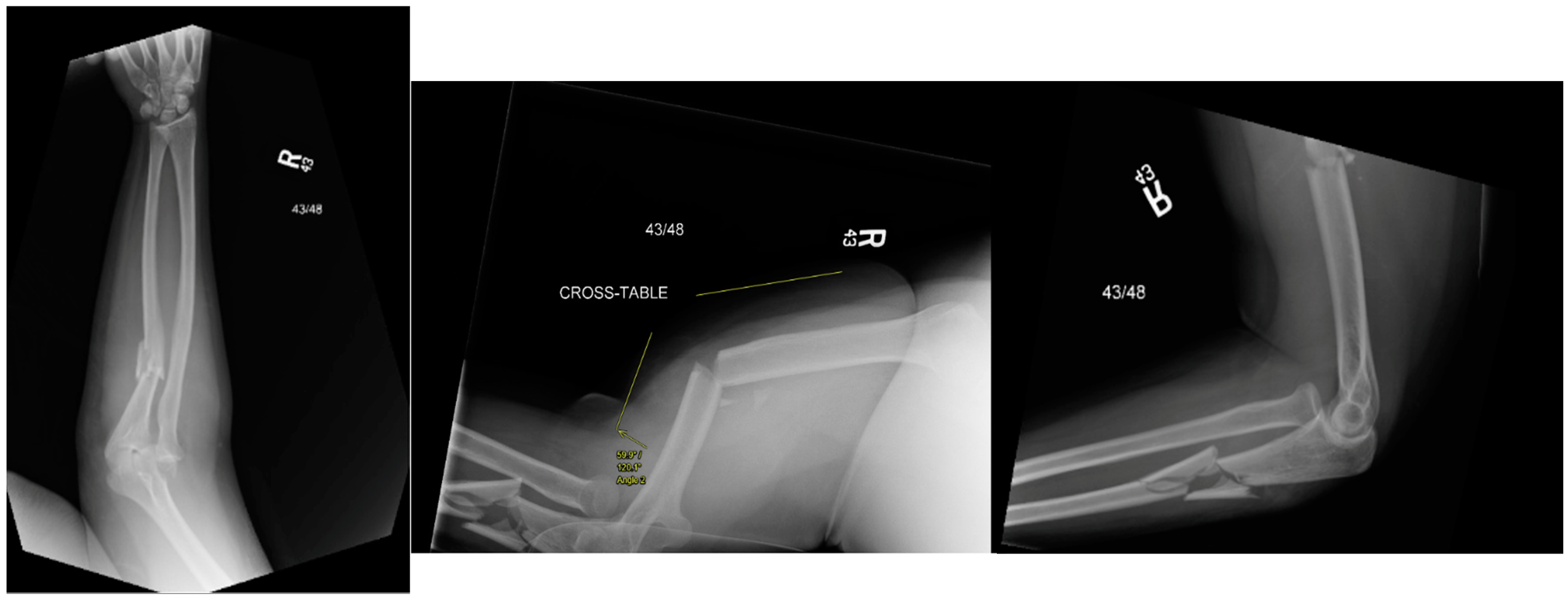
Abstract
1. Introduction
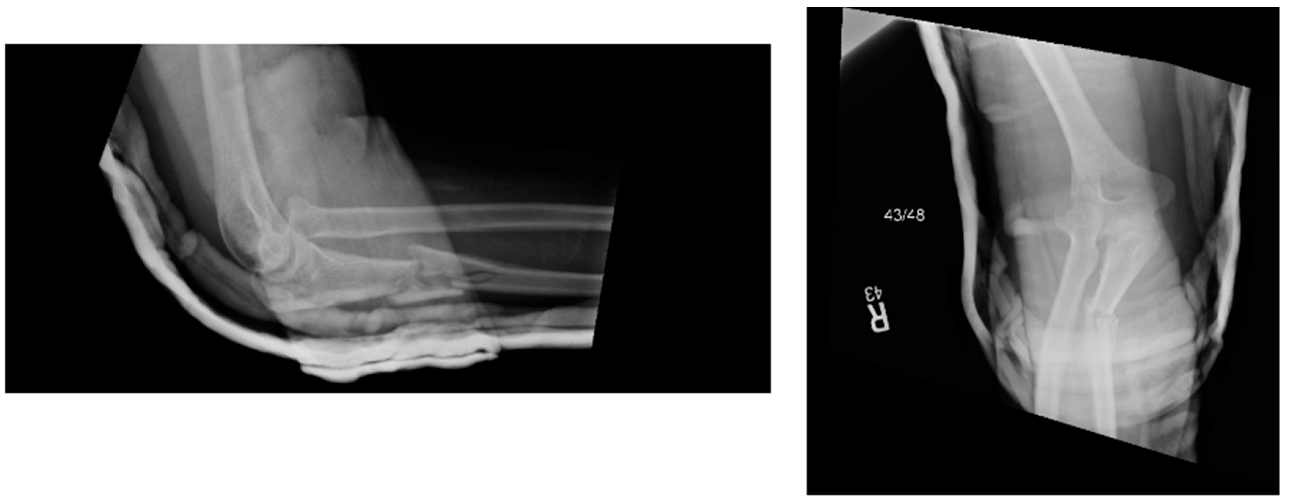
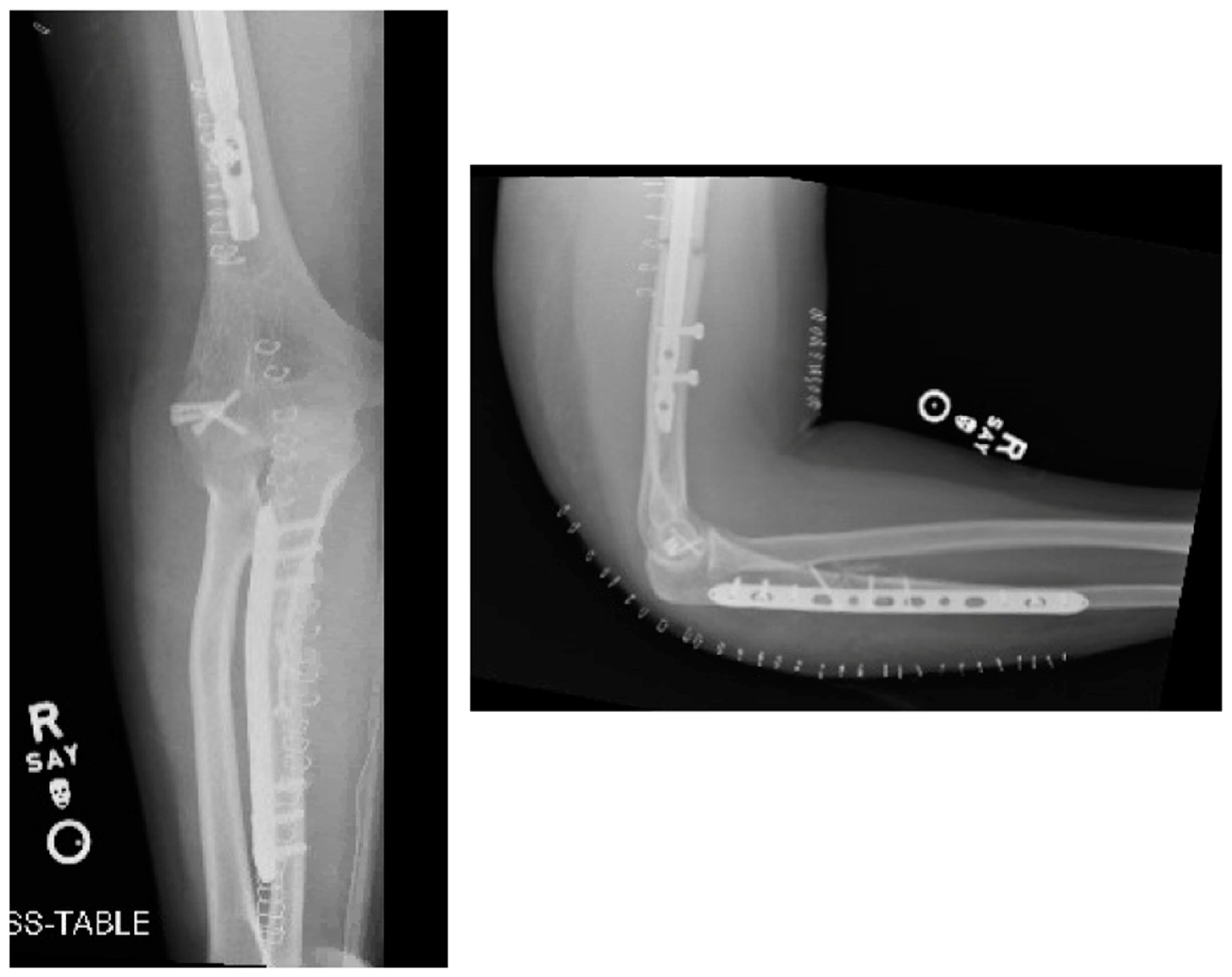
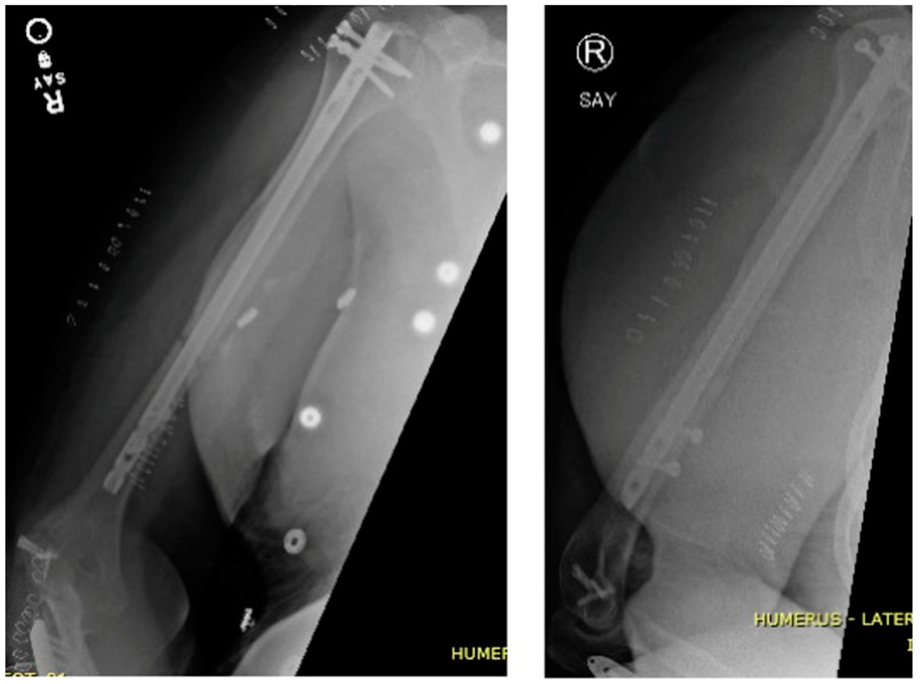
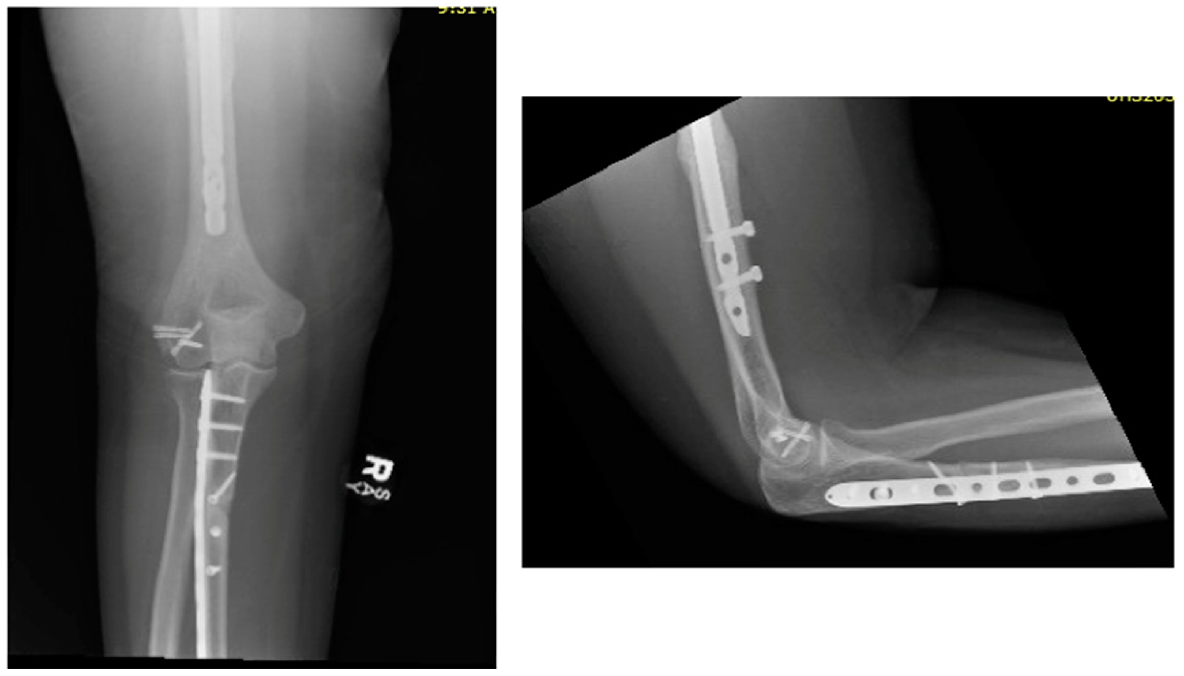
2. Case Presentation
3. Discussion
4. Conclusions
Author Contributions
Funding
Institutional Review Board Statement
Informed Consent Statement
Data Availability Statement
Conflicts of Interest
References
- Suarez, R.; Barquet, A.; Fresco, R. Epidemiology and treatment of monteggia lesion in adults: Series of 44 cases. Acta Ortop. Bras. 2016, 24, 48–51. [Google Scholar] [CrossRef] [PubMed]
- Wong, J.C.; Getz, C.L.; Abboud, J.A. Adult Monteggia and Olecranon Fracture Dislocations of the Elbow. Hand Clin. 2015, 31, 565–580. [Google Scholar] [CrossRef] [PubMed]
- Xiao, R.C.; Chan, J.J.; Cirino, C.M.; Kim, J.M. Surgical Management of Complex Adult Monteggia Fractures. J. Hand Surg. 2021, 46, 1006–1015. [Google Scholar] [CrossRef] [PubMed]
- Rehim, S.A.; Maynard, M.A.; Sebastin, S.J.; Chung, K.C. Monteggia Fracture Dislocations: A Historical Review. J. Hand Surg. 2014, 39, 1384–1394. [Google Scholar] [CrossRef] [PubMed]
- Johnson, N.P.; Silberman, M. Monteggia Fractures. In StatPearls; StatPearls Publishing: Treasure Island, FL, USA, 2024. Available online: http://www.ncbi.nlm.nih.gov/books/NBK470575/ (accessed on 10 March 2024).
- Papaioannou, I.; Repantis, T.; Baikousis, A.; Korovessis, P. Adult Monteggia Lesion with Ipsilateral Distal Radius Fracture: A Case Report and Review of the Literature. J. Orthop. Case Rep. 2018, 8, 77–80. [Google Scholar] [PubMed]
- Sankhla, S.L.; Joshi, P.; Singh, D. An Extremely Rare Combination of Monteggia Equivalent Type 1 Lesion (diaphyseal Ulna and Radial Neck Fractures Without Dislocation) with Ipsilateral Radius Shaft and Distal Radius Fractures in a Child. J. Orthop. Case Rep. 2020, 10, 86–89. [Google Scholar] [PubMed]
- Mundada, G.; Khan, S.M.; Singhania, S.K.; Gupta, V.; Singh, P.K.; Khan, S. Type-I monteggia with ipsilateral fracture of distal radius epiphyseal injury: A rare case report. Ann. Afr. Med. 2017, 16, 30–32. [Google Scholar] [PubMed]
- Guitton, T.G.; Ring, D.; Kloen, P. Long-Term Evaluation of Surgically Treated Anterior Monteggia Fractures in Skeletally Mature Patients. J. Hand Surg. 2009, 34, 1618–1624. [Google Scholar] [CrossRef] [PubMed]
- Orapiriyakul, W.; Apivatthakakul, V.; Theppariyapol, B.; Apivatthakakul, T. Humerus shaft fractures, approaches and management. J. Clin. Orthop. Trauma 2023, 43, 102230. [Google Scholar] [CrossRef] [PubMed]
- Antoon, S.F.; Russo, S.A.; Kozin, S.H.; Zlotolow, D.A. Evaluation of Monteggia Fracture Outcomes: Acute to Chronic. Hand 2023. [Google Scholar] [CrossRef]
- Laun, R.; Wild, M.; Brosius, L.; Hakimi, M. Monteggia-like lesions-treatment strategies and one-year results. GMS Interdiscip. Plast. Reconstr. Surg. DGPW 2015, 4, Doc13. [Google Scholar] [CrossRef]
- Reina, N.; Laffosse, J.-M.; Abbo, O.; Accadbled, F.; Bensafi, H.; Chiron, P. Monteggia equivalent fracture associated with Salter I fracture of the radial head. J. Pediatr. Orthop. Part B 2012, 21, 532–535. [Google Scholar] [CrossRef] [PubMed]
- Bugeja, M.; Avakyan, A.; Bianco, E.Z.; Azzopardi, T. Type III Monteggia Equivalent Lesion with Ipsilateral Fracture Lateral Condyle of Humerus in a Four-year-old Child: A Case Report and Literature Review. J. Orthop. Case Rep. 2018, 8, 19–21. [Google Scholar] [PubMed]
- Peter, N.; Myint, S. Type I Monteggia lesion and associated fracture of the distal radius and ulna metaphysis in a child. CJEM 2007, 9, 383–386. [Google Scholar] [CrossRef] [PubMed]
- Cobanoglu, M.; Şavk, Ş.O.; Cullu, E.; Duygun, F. Ipsilateral supracondylar humerus fracture and Monteggia lesion with a 5-year follow-up: A rare injury in a young girl. BMJ Case Rep. 2015, 2015, bcr2014206313. [Google Scholar] [CrossRef] [PubMed]
- Kembhavi, R.S.; James, B. Type IIA Monteggia Fracture Dislocation with Ipsilateral Distal Radius Fracture in Adult—A Rare Association. J. Clin. Diagn. Res. JCDR 2016, 10, RD01-03. [Google Scholar] [CrossRef] [PubMed]
- Hung, S.-C.; Huang, C.-K.; Chiang, C.-C.; Chen, T.-H.; Chen, W.-M.; Lo, W.-H. Monteggia type I equivalent lesion: Diaphyseal ulna and radius fractures with a posterior elbow dislocation in an adult. Arch. Orthop. Trauma Surg. 2003, 123, 311–313. [Google Scholar] [CrossRef] [PubMed]
- Egol, K.A.; Koval, K.J.; Zuckerman, J.D. Handbook of Fractures, 6th ed.; Wolters Kluwer: Philadelphia, PA, USA, 2020. [Google Scholar]
- Fuad, M.; Elmhiregh, A.; Motazedian, A.; Bakdach, M. Capitellar fracture with bony avulsion of the lateral collateral ligament in a child: Case report. Int. J. Surg. Case Rep. 2017, 36, 103–107. [Google Scholar] [CrossRef] [PubMed]
- Fitzgerald, M.J.; Goodman, H.J.; Kenan, S.; Kenan, S. Did COVID-19 related delays in surgical management lead to patient morbidity in the orthopaedic oncological population? A retrospective observational single centre study. Bone Jt. Open 2021, 2, 236–242. [Google Scholar] [CrossRef] [PubMed]
- Valone, L.C.; Waites, C.; Tartarilla, A.B.; Whited, A.; Sugimoto, D.; Bae, D.S.; Bauer, A.S. Functional Elbow Range of Motion in Children and Adolescents. J. Pediatr. Orthop. 2020, 40, 304–309. [Google Scholar] [CrossRef] [PubMed]


Disclaimer/Publisher’s Note: The statements, opinions and data contained in all publications are solely those of the individual author(s) and contributor(s) and not of MDPI and/or the editor(s). MDPI and/or the editor(s) disclaim responsibility for any injury to people or property resulting from any ideas, methods, instructions or products referred to in the content. |
© 2024 by the authors. Licensee MDPI, Basel, Switzerland. This article is an open access article distributed under the terms and conditions of the Creative Commons Attribution (CC BY) license (https://creativecommons.org/licenses/by/4.0/).
Share and Cite
McDonald, C.; Kannenberg, M.; Goodrum, J.; Eakin, J.; Ryan, P.; Dutta, A. Type I Monteggia Fracture with Associated Ipsilateral Capitellar and Humeral Diaphyseal Fractures in an Adult. Osteology 2024, 4, 45-52. https://doi.org/10.3390/osteology4020004
McDonald C, Kannenberg M, Goodrum J, Eakin J, Ryan P, Dutta A. Type I Monteggia Fracture with Associated Ipsilateral Capitellar and Humeral Diaphyseal Fractures in an Adult. Osteology. 2024; 4(2):45-52. https://doi.org/10.3390/osteology4020004
Chicago/Turabian StyleMcDonald, Casey, Matt Kannenberg, Jason Goodrum, John Eakin, Paul Ryan, and Anil Dutta. 2024. "Type I Monteggia Fracture with Associated Ipsilateral Capitellar and Humeral Diaphyseal Fractures in an Adult" Osteology 4, no. 2: 45-52. https://doi.org/10.3390/osteology4020004
APA StyleMcDonald, C., Kannenberg, M., Goodrum, J., Eakin, J., Ryan, P., & Dutta, A. (2024). Type I Monteggia Fracture with Associated Ipsilateral Capitellar and Humeral Diaphyseal Fractures in an Adult. Osteology, 4(2), 45-52. https://doi.org/10.3390/osteology4020004